3D Imaging Systems A new dimension of confidence better 3D images for a better practice.

Welcome message from author
This document is posted to help you gain knowledge. Please leave a comment to let me know what you think about it! Share it to your friends and learn new things together.
Transcript

3D Im
agin
g Sy
stem
s
A new dimension of confidencebetter 3D images for a better practice.
Would you like to know more?Visit www.carestreamdental.com/3D or call us today at 800.944.6365

Despite your training and years of practice, it’s tough to correctly diagnose what you can’t see…and standard two-dimensional images can be ambiguous at best. Fortunately, with our 3D imaging products, you can finally get the whole picture—all while receiving the best image quality and
highest resolution in the industry. With three systems that each provide a broad spectrum of imaging possibilities for generalists and specialists alike, Carestream Dental gives you everything you need to diagnose with confidence every time.
Your diagnosis is only as good as your information.

Why choose our 3D products? 4 Carestream Dental’s 3D Extraoral Imaging Systems
Our most comprehensive 3D lineup
How good are the images? 6 Orthodontic Imaging
Panoramic, cephalometric and 3D images for a variety of applications
8 Endodontic Imaging 3D images for a variety of endodontic applications
10 Implant Imaging 3D images for a variety of implant applications
12 Oral and Maxillofacial Surgery Imaging 3D images that help make surgery more precise
14 Periodontic Imaging 3D images that let you see the whole picture
Which model is right for you? 17 Comparison Chart
Find the right machine for you
18 CS 9000 3D System Our highest resolution and lowest dose 3D system
20 CS 9300 System Adaptable 3D imaging with a flexible field of view
How easy is it to use? 24 Carestream Dental’s Imaging Software
The software that makes all the difference
What are the details? 26 Specifications/Product Space Requirements
Specifications, doses, dimensions, and more
4
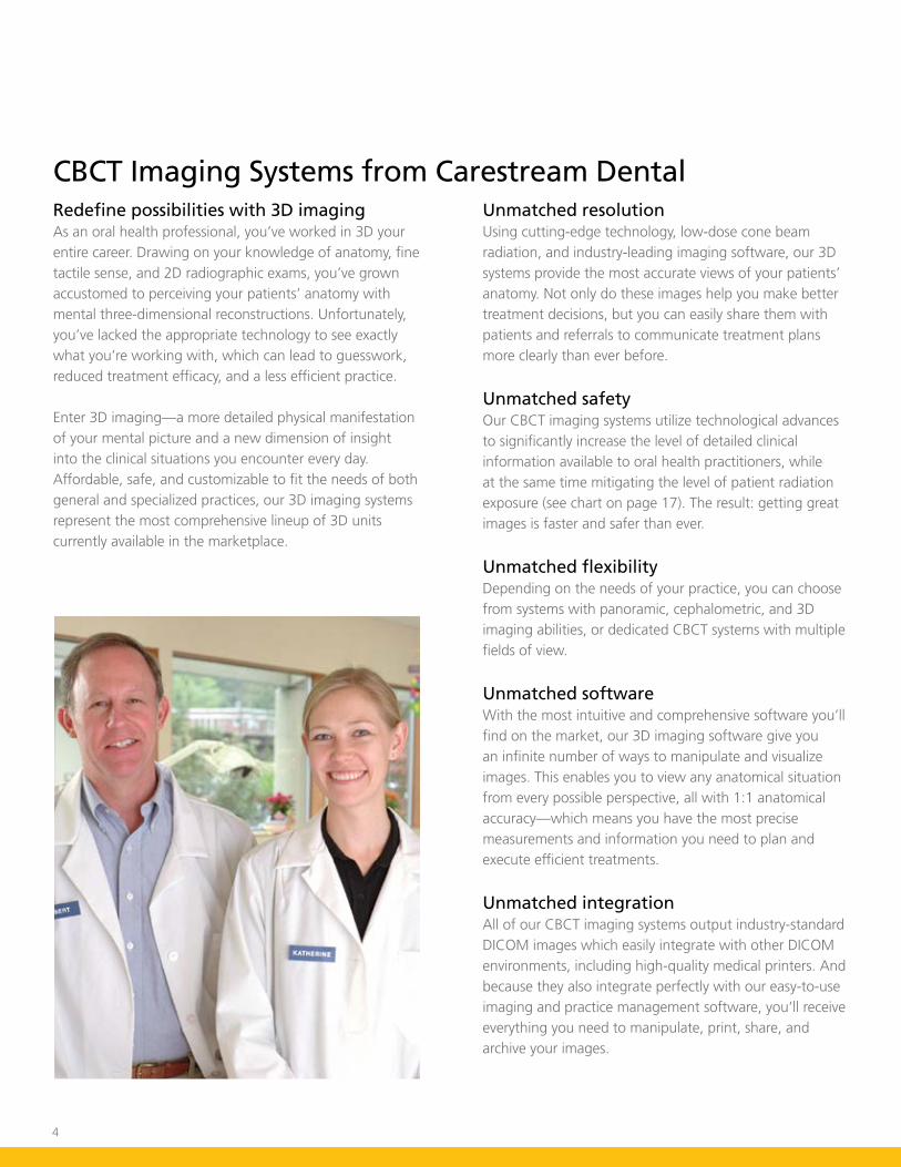
CBCT Imaging Systems from Carestream DentalRedefine possibilities with 3D imagingAs an oral health professional, you’ve worked in 3D your entire career. Drawing on your knowledge of anatomy, fine tactile sense, and 2D radiographic exams, you’ve grown accustomed to perceiving your patients’ anatomy with mental three-dimensional reconstructions. Unfortunately, you’ve lacked the appropriate technology to see exactly what you’re working with, which can lead to guesswork, reduced treatment efficacy, and a less efficient practice.
Enter 3D imaging—a more detailed physical manifestation of your mental picture and a new dimension of insight into the clinical situations you encounter every day. Affordable, safe, and customizable to fit the needs of both general and specialized practices, our 3D imaging systems represent the most comprehensive lineup of 3D units currently available in the marketplace.
Unmatched resolutionUsing cutting-edge technology, low-dose cone beam radiation, and industry-leading imaging software, our 3D systems provide the most accurate views of your patients’ anatomy. Not only do these images help you make better treatment decisions, but you can easily share them with patients and referrals to communicate treatment plans more clearly than ever before.
Unmatched safetyOur CBCT imaging systems utilize technological advances to significantly increase the level of detailed clinical information available to oral health practitioners, while at the same time mitigating the level of patient radiation exposure (see chart on page 17). The result: getting great images is faster and safer than ever.
Unmatched flexibilityDepending on the needs of your practice, you can choose from systems with panoramic, cephalometric, and 3D imaging abilities, or dedicated CBCT systems with multiple fields of view.
Unmatched softwareWith the most intuitive and comprehensive software you’ll find on the market, our 3D imaging software give you an infinite number of ways to manipulate and visualize images. This enables you to view any anatomical situation from every possible perspective, all with 1:1 anatomical accuracy—which means you have the most precise measurements and information you need to plan and execute efficient treatments.
Unmatched integrationAll of our CBCT imaging systems output industry-standard DICOM images which easily integrate with other DICOM environments, including high-quality medical printers. And because they also integrate perfectly with our easy-to-use imaging and practice management software, you’ll receive everything you need to manipulate, print, share, and archive your images.

5
Top Left: The CS 9300 system offers up to seven scalable fields of view and image resolution up to 90µm, giving you greater flexibility and the optimum field of view to suit your patients’ every diagnostic need. With multi-modality panoramic and 3D imaging with exceptional detail and range, the CS 9300 system is the perfect all-in-one machine for your practice.
Middle Left: The CS 9000C 3D system is a hybrid 3-in-1 solution that presents a complete and versatile diagnostic imaging modality. Combining one-shot cephalometric technology, high-quality panoramic imaging, and high-resolution 3D imaging, this unit is the perfect imaging system to cover all your diagnostic imaging needs.
Bottom Left: The CS 9000 3D system also offers a 2-in-1 solution, blending panoramic imaging with 3D imaging technology. This solution can be easily upgraded to include cephalometric capabilities if needed. The system is engineered specifically to extend the benefits of two high-resolution imaging modalities on one machine, including a 3D CBCT modality that features the highest resolution 3D imaging on the market.
Top Right: The CS 9300 system is the perfect solution for applications that require more diagnostic information—such as complex orthodontic cases, implantology, pathologic imaging, orthognathic surgical cases, and generalized radiologic imaging of the maxillofacial area.

6
“ …the CS 9000C 3D panoramic and cephalometric system significantly creates a new benchmark for extraoral radiography and evidence-based dentistry, and unquestionably strengthens our diagnostic confidence level.”
– Eladio DeLeon, JR., DMD, MS – Goldstein Chair of Orthodontics
– John W. Stockstill, DDS, NS and Daniel Levy-Bercowski, DDS, MSD – Medical College of Georgia, Augusta, GA
Complement your cephalometric and panoramic imaging
• Analyze skeletal symmetry
• Assess alveolar ridge shape and volume limitations
• Assess temporomandibular joints and occlusion
• Plan placement of temporary anchorage devices
• Plan orthognathic surgical treatment
• Evaluate ectopic and impacted teeth
• Assess growth
• Design custom appliances and image-guided treatment
Orthodontics: better images for better treatment
Impacted supernumerary teeth can often be difficult to visualize with limited 2D imaging modalities. Fortunately, high-resolution 3D imaging simplifies this process. Information gained from the CS 9000 3D system allows the practitioner to evaluate the presence of supernumerary teeth, their position relative to adjacent teeth, and any pathology associated with the impaction—all with anatomical accuracy. This information promotes a more confident and comprehensive treatment plan, as well as key determinants for surgical approaches to removal, if required.

7
The CS 9300 system gives you the ability to provide comprehensive dental and skeletal assessment of your patients prior to beginning treatment. Create all necessary images from just one 3D scan, including 2D lateral and frontal cephalometric, panoramic, SMV and TMJ images. This scan can even be used to produce a digital model.
The CS 9300 system 8 cm x 8 cm field of view is perfect for multiple impactions cases while limiting the exposure to the area of interest and is very beneficial for performing 3D radiological examinations of the patient before, during, or after orthodontic treatment. In this case, it reveals the exact location of the impacted mandibular canines and their exact position relative to adjacent teeth.
8
• Diagnose endodontic pathosis
• Assess canal morphology
• Assess pathosis of non-endodontic origin
• Evaluate root fractures and trauma
• Analyze external and internal root resorption and invasive cervical resorption
• Plan surgeries and implants
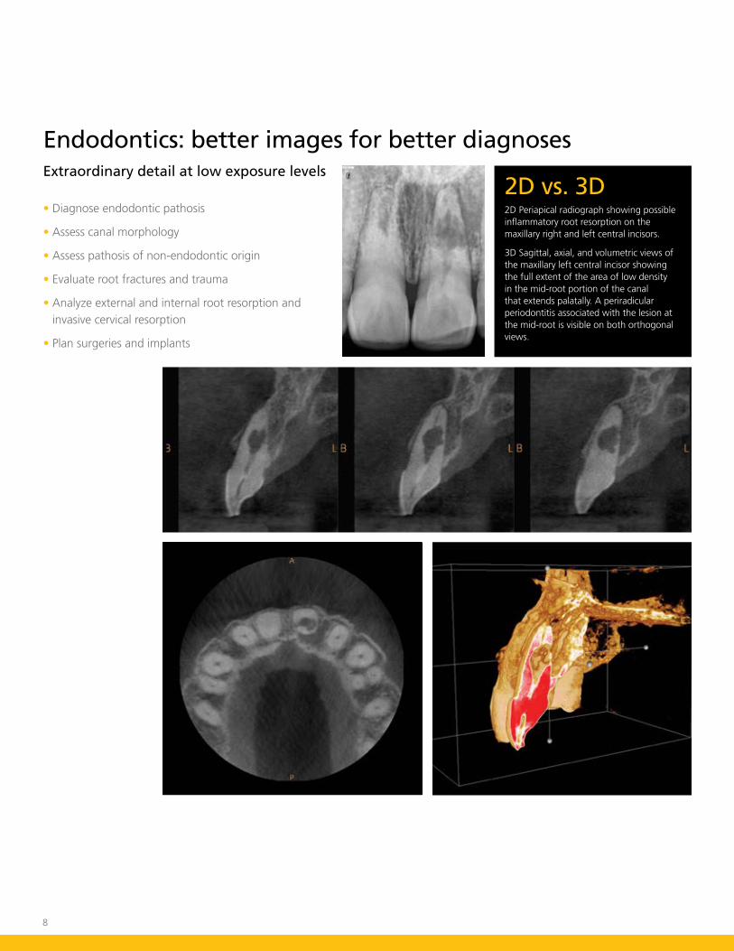
Extraordinary detail at low exposure levels
Endodontics: better images for better diagnoses
2D vs. 3D2D Periapical radiograph showing possible inflammatory root resorption on the maxillary right and left central incisors.
3D Sagittal, axial, and volumetric views of the maxillary left central incisor showing the full extent of the area of low density in the mid-root portion of the canal that extends palatally. A periradicular periodontitis associated with the lesion at the mid-root is visible on both orthogonal views.

9
Top Left: 3D image with a 76 micron high resolution axial slice clearly reveals three canals in the mesial root of the lower left first molar.
Bottom Left: 2D imaging is often limited in its ability to provide the key diagnostic information needed to adequately assess the full apical and lateral extent of the canal morphology prior to treatment.
Bottom Middle: 2D image taken after root canal treatment demonstrating adequate obturation of the three canals in the mesial root. Prior diagnostic knowledge obtained from the 3D image allowed for identification of this canal pre-operatively.
Bottom Right: Intraoperative view confirms the exact canal anatomy as seen on the CS 9000 3D system image obtained pre-operatively. This diagnostic knowledge can lead to expedited and more predictable treatment.
“ Cone Beam technology could be the most powerful addition to an endodontist’s armamentarium in the 21st century. Its ability to bypass the problems of anatomic noise found in 2D radiographs is outstanding. CBCT improves our ability to see apical lesions, predetermine canal morphology, and clearly observe and accurately diagnose resorptions. This helps us provide more accurate diagnoses, prognoses, patient education, and treatment plans. Knowledge of the canal morphology before beginning treatment effectively shortens treatment time and reduces the risk of “missed” canals. This technology has been a great enhancement to my practice of Endodontics.”
– Randolph Todd, DMD, Practice Limited to Endodontics, Diplomate American Board of Endodontics

10
• Evaluate bone quantity and quality
• Identify, localize, and annotate anatomical obstacles (mandibular canal, sinus)
• Take precise measurements using true 1:1 ratio
• Plan implants using specialized 3D software
• Improve case acceptance
• Collaborate with referrals
3D images accurately portray the patient’s anatomy
Implantology: better images for better planning
Implant planning with 3D imaging modalities is rapidly becoming mainstream. The CS 9000 3D system has the ability to scan a wide range of areas. For cases requiring an extended field of view, simply put the CS 9000 3D system into full-arch mode to acquire the maxillary or mandibular arch or acquire a single or dual arch with our versatile CS 9300 system. Our comprehensive CS 3D imaging implant planning software makes treatment planning very simple and helps improve case acceptance.
11
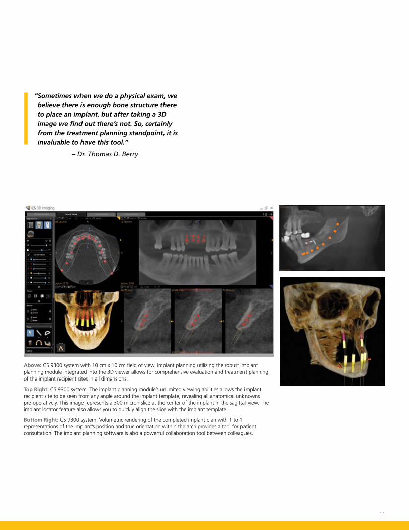
Above: CS 9300 system with 10 cm x 10 cm field of view. Implant planning utilizing the robust implant planning module integrated into the 3D viewer allows for comprehensive evaluation and treatment planning of the implant recipient sites in all dimensions.
Top Right: CS 9300 system. The implant planning module’s unlimited viewing abilities allows the implant recipient site to be seen from any angle around the implant template, revealing all anatomical unknowns pre-operatively. This image represents a 300 micron slice at the center of the implant in the sagittal view. The implant locator feature also allows you to quickly align the slice with the implant template.
Bottom Right: CS 9300 system. Volumetric rendering of the completed implant plan with 1 to 1 representations of the implant’s position and true orientation within the arch provides a tool for patient consultation. The implant planning software is also a powerful collaboration tool between colleagues.
“ Sometimes when we do a physical exam, we believe there is enough bone structure there to place an implant, but after taking a 3D image we find out there’s not. So, certainly from the treatment planning standpoint, it is invaluable to have this tool.”
– Dr. Thomas D. Berry

12
• Identify relationships between impacted teeth and vital anatomical structures
• Visualize cysts and periapical lesions
• Define surgical protocol for impacted tooth extractions, cyst removals, or periapical lesion treatments
• Increase case acceptance rates by allowing patients to visualize and fully understand your treatment plan
• Improve referral relationships by sharing critical 2D and 3D data with referring dentists
• Eliminate office bottlenecks by taking x-ray development out of the equation—2D image acquisition takes a mere 13 seconds
• Easily share images across multiple office locations
Plan treatments more thoroughly than ever
Oral Surgery: better images for better preparation
2D vs. 3D2D Panoramic image shows two supernumerary teeth in maxillary anterior.
3D Volumetric view and sagittal view show precise labial location of supernumerary tooth with no resorption of the central incisor root.
13
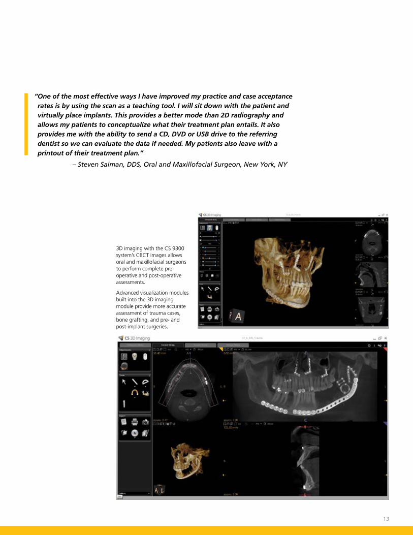
3D imaging with the CS 9300 system’s CBCT images allows oral and maxillofacial surgeons to perform complete pre-operative and post-operative assessments.
Advanced visualization modules built into the 3D imaging module provide more accurate assessment of trauma cases, bone grafting, and pre- and post-implant surgeries.
“ One of the most effective ways I have improved my practice and case acceptance rates is by using the scan as a teaching tool. I will sit down with the patient and virtually place implants. This provides a better mode than 2D radiography and allows my patients to conceptualize what their treatment plan entails. It also provides me with the ability to send a CD, DVD or USB drive to the referring dentist so we can evaluate the data if needed. My patients also leave with a printout of their treatment plan.”
– Steven Salman, DDS, Oral and Maxillofacial Surgeon, New York, NY
14
• Evaluate bone anatomy and proximity to pertinent anatomical areas for implant placement and case planning
• Analyze furcation involvements and intrabony defect patterns
• Diagnose the extent and significance of tooth and alveolar fractures
• Evaluate facial bone plate and associated tooth position for perio-orthodontic considerations in mucogingival or periodontally accelerated osteogenic orthodontic therapy
• Evaluate bone loss patterns in periodontitis and associated regenerative potential of defect morphology
• Evaluate pre- and post- operative bone grafting sites
• Evaluate the patency of the ostium before sinus bone grafting surgery is performed
• Identify exact location of vital structures such as the mandibular nerve and nasopalatine canal
The details you need when you need them
Periodontics: better images for better success
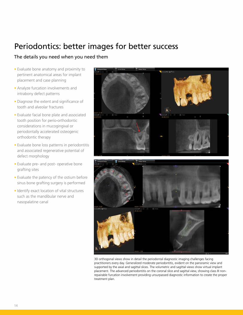
3D orthogonal views show in detail the periodontal diagnostic imaging challenges facing practitioners every day. Generalized moderate periodontitis, evident on the panoramic view and supported by the axial and sagittal slices. The volumetric and sagittal views show virtual implant placement. The advanced periodontitis on the coronal slice and sagittal view, showing class III non-repairable furcation involvement providing unsurpassed diagnostic information to create the proper treatment plan.
15
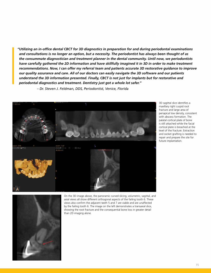
3D sagittal slice identifies a maxillary right cuspid root fracture and large area of periapical low density, consistent with abscess formation. The palatal cortical plate of bone is still attached while the facial cortical plate is breached at the level of the fracture. Extraction and socket grafting is needed to repair and prepare the site for future implantation.
On the 3D image above, the panoramic curved slicing, volumetric, sagittal, and axial views all show different orthogonal aspects of the failing tooth 6. These views also confirm the adjacent teeth 5 and 7 are viable and are unaffected by the failing tooth 6. The image on the left demonstrates a transaxial slice, showing the root fracture and the consequential bone loss in greater detail than 2D imaging alone.
“ Utilizing an in-office dental CBCT for 3D diagnostics in preparation for and during periodontal examinations and consultations is no longer an option, but a necessity. The periodontist has always been thought of as the consummate diagnostician and treatment planner in the dental community. Until now, we periodontists have carefully gathered the 2D information and have skillfully imagined it in 3D in order to make treatment recommendations. Now, I can offer my referral team and patients accurate 3D restorative guidance to improve our quality assurance and care. All of our doctors can easily navigate the 3D software and our patients understand the 3D information presented. Finally, CBCT is not just for implants but for restorative and periodontal diagnostics and treatment. Dentistry just got a whole lot safer.”
– Dr. Steven J. Feldman, DDS, Periodontist, Venice, Florida

16
Use the charts on the opposite page to understand the features and capabilities of our 3D imaging systems so you can determine which is right for your practice.
An easy way to find the model that’s right for you
Features/Applications
The CS 9000 3D system. Cephalometric option also available. For more about the CS 9000 3D System, turn to page 18.
The CS 9300 Select system. Cephalometric option is also available. For more about the CS 9300 Select, turn to page 20.
CS 9000 3D system
• Multi-modality system with 3D imaging and digital panoramic
• Field of view limited to a focused area, single arch, TMJ or Sinus
• Highest resolution CBCT images
• Full arch imaging allows for visualization of impacted teeth, implant planning, simple pathological lesions and surgical guide creation
CS 9300 system
• Multi-modality system with 3D imaging and digital panoramic
• Collimation limits exposure to area of interest and delivers 75-90% less radiation than medical spiral CT units
• Covers widest scope of clinical applications
CS 9300 Select system
• Multi-modality system with 3D imaging and digital panoramic
• Four selectable fields of view ranging from 5 cm x 5 cm to 10 cm x 10 cm
• For general dentists and specialists placing implants, performing endodontic procedures, periodontal treatments or minor oral surgeries
The CS 9300 system. Cephalometric option also available. For more about the CS 9300 system, turn to page 20.

17
Fields of View
CS 9000 3D System
CS 9300 Select System
CS 9300 System
Comparison ChartFeatures & Specifications CS 9000 3D System CS 9300 Select System CS 9300 System
Panoramic 3 3 3
Cephalometric Upgradeable Upgradeable Upgradeable
3D Field of View Size 5 cm x 3.75 cm Extended: 7.5 cm x 3.75 cm
Selectable including 5 cm X 5 cm, 8 cm X 8 cm, 10 cm X 5 cm and 10 cm x 10 cm
Selectable from 5 cm x 5 cm to 17 cm x 13.5 cm
Exposure Time Pan: 14 sec 3D: 10.8 sec Ceph: 1-3.2 sec
Pan: 14 sec 3D: 12-20 sec Ceph: 1-3.2 sec
Pan: 14 sec 3D: 12-20 sec Ceph: 1-3.2 sec
Reconstruction Time 10-55 sec 20-90 sec 20-90 sec
Handicap Accessible 3 3 3
5 cm x 5 cm
5 cm x 5 cm
5 cm x 3.75 cm 7.5 cm x 3.75 cm Extended mode
8 cm x 8 cm
8 cm x 8 cm
10 cm x 5 cm
10 cm x 5 cm
8 cm x 8 cm (TMJ)
8 cm x 8 cm (TMJ) 10 cm x 10 cm
10 cm x 10 cm
17 cm x 11 cm (Sinus)
17 cm x 13.5 cm17 cm x 6 cm

18
Easy for patients, easy for you With a streamlined user interface and computer-controlled system, the CS 9000 3D and CS 9000C 3D system make exams quick and simple. Simply choose your region of interest, and the device automatically positions itself. Next, easily position the patient using a unique bite block and lateral supports. After that, it takes just seconds to make final adjustments, thanks to the laser beam positioning guides. Your patient enjoys a quick, comfortable experience—and you get the best 3D image available today.
The industry’s best quality 3D images are also the most affordableStarting at less than $100,000, you’d think the CS 9000 3D system would be a “no frills” entry system, but the truth is quite the opposite. Featuring award-winning 3D technology and easy-to-use imaging software, the system generates brilliant images quickly at a dosage 10 to 30 times lower than full field of view systems. The unit delivers cutting-edge panoramic images and can be easily upgraded to include cephalometric abilities—making it perfect for initial diagnoses.
Designed to improve treatment outcomes Say goodbye to guesswork and multiple treatment visits—the CS 9000 3D and CS 9000C 3D systems quickly deliver all the information you need to make accurate diagnoses on the spot. And faster treatment, in turn, promotes a more efficient and profitable practice.
Because the CS 9000 3D and CS 9000C 3D systems feature a localized field of view, you can quickly capture high-resolution images at a smaller voxel size, making easy to see minute details other 3D systems miss. Even demanding endodontic and single implant exams are faster and easier than ever.
Lower radiation dose, higher imaging quality The CS 9000 3D system produces a substantially lower radiation dose than what many medium and large FOV CBCT units have reported.* Because exposure is confined only to the area of interest, the system stays true to the principle of ALARA (As Low As Reasonably Achievable), giving you superior quality 3D images with better protection for your patients.
CS 9000 3D System
* References: Ludlow JB: “Dosimetry of CS 9000 3D Small FOV CBCT and Panoramic Unit,” Univ of N Carolina School of Dentistry, Chapel Hill, NC, 2008. Abstract presented to AAOMR Annual session 2009.

19
One-shot cephalometric images: Acquisition time takes about a second, reducing exposure time and the risk of patient movement. It’s all the control of a film unit—with all the advantages of digital.
Superior 3D Images: The CS 9000 3D system offers the highest resolution images and an intuitive interface.
Detailed panoramic images: A powerful low-dose 2D imaging system is at the heart of the CS 9000 3D system.

20
Ultimate practice flexibilityExperience unprecedented levels of imaging capabilities. Which field of view is right for your practice? With the CS 9300 system, you have up to seven selectable fields of view—ranging from 5 cm x 5 cm to 17 cm x 13.5 cm. This gives you greater flexibility and the ability to collimate the field of view to suit your patients’ every diagnostic need.
Multi-modality with 2D panoramic and cephalometric option The all-in-one CS 9300 system is the most versatile multi-modality imaging system available today. In addition to its exceptional 3D imaging capabilities, it also offers 2D digital panoramic imaging with variable focal trough technology that’s crystal clear, every time. It even has a one-shot cephalometric imaging modality upgrade option. For practitioners who have been waiting to integrate cone beam computed tomography into their practice, this is the perfect option with the most capabilities available in one space-saving all-in-one system from Carestream Dental. And, with two configurations available, it’s also perfect for any practice’s needs and budget.
Superb image quality With image resolution up to 90 μm, the superb quality allows you to collect valuable diagnostic information for a range of clinical applications, including focused-field, single jaw, dual jaw, single and double TMJ, dual jaw, sinus and maxillofacial imaging.
CS 9300 System
21
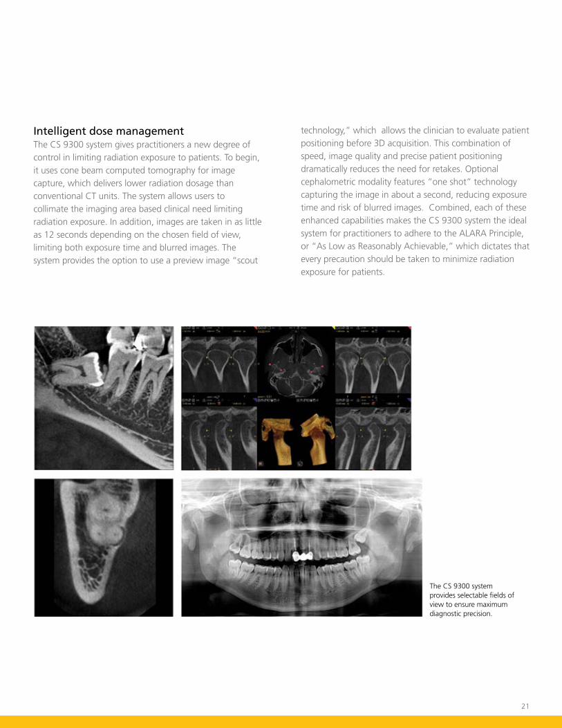
The CS 9300 system provides selectable fields of view to ensure maximum diagnostic precision.
Intelligent dose management The CS 9300 system gives practitioners a new degree of control in limiting radiation exposure to patients. To begin, it uses cone beam computed tomography for image capture, which delivers lower radiation dosage than conventional CT units. The system allows users to collimate the imaging area based clinical need limiting radiation exposure. In addition, images are taken in as little as 12 seconds depending on the chosen field of view, limiting both exposure time and blurred images. The system provides the option to use a preview image “scout
technology,” which allows the clinician to evaluate patient positioning before 3D acquisition. This combination of speed, image quality and precise patient positioning dramatically reduces the need for retakes. Optional cephalometric modality features “one shot” technology capturing the image in about a second, reducing exposure time and risk of blurred images. Combined, each of these enhanced capabilities makes the CS 9300 system the ideal system for practitioners to adhere to the ALARA Principle, or “As Low as Reasonably Achievable,” which dictates that every precaution should be taken to minimize radiation exposure for patients.

22
Simple integration Images from the CS 9300 system are rendered in stunning detail within CS 3D Imaging Software. It’s designed to comprehensively enhance your diagnostic view through its integration with leading imaging programs such as NobelGuide, Simplant and SureSmile. Get the greatest possible clinical value out of your CS 9300 system images and keep your preferred third-party imaging software. Our software is DICOM-compliant, and compatible with PACS* and medical printers.
Enhanced patient communication Show your patients the precise nature of their case that only the third dimension can provide. Focus on specific regions of interest to help patients truly understand their treatment options to make informed decisions. Our software provides you with the details you need to present a more definitive treatment plan, resulting in increased case acceptance. CS 3D Imaging Software lets you view and manipulate the images in a smooth, intuitive fashion, allowing your patients to better visualize their diagnosis and treatment plan. The CS 9300 system’s crystal clear images can boost both your diagnostic confidence and your patients’ confidence in you. The CS 9300 system helps your patients share your vision.
All-inclusive imaging with selectable fields of view
CS 9300 System
Easy case review and planning Our industry leading imaging software has been designed by clinicians for clinicians. The CS 9300 system comes installed with Carestream Dental’s innovative CS 3D imaging software. Our software not only facilitates a number of functions that enhance treatment planning, it delivers fast, accurate results for better patient communication. View images slice-by-slice in axial, coronal, sagittal, cross-sectional and oblique views for enhanced diagnostic interpretation. Our imaging software includes a sophisticated implant planning feature that comes with pre-loaded libraries from implant manufacturers and the flexibility to create custom implant sizes yourself. It also shows visual representation of the long axis, the restorative space and allows planning for customized abutments. There’s also a robust TMJ analysis feature.
Ease of collaboration Intuitive sharable software makes case collaboration easy. Carestream Dental’s imaging software is available free for you to share with your referring doctors. The CS 9300 system enhances your collaborative cases through its CS 3D Imaging Software Viewing Module, which allows users to both view and manipulate volumes of data. Share volumes via USB or CD so you can collaborate with your referring colleagues to facilitate a better patient experience and improved outcomes.
*PACS compatibility requires Trophy DICOM, an optional for-purchase additional software.

23
Examples of field of view
17 c
m x
13.
5 cm
10 c
m x
5 c
m
17 c
m x
11
cm
8 cm
x 8
cm
- TM
J x 1
10 c
m x
10
cm
8 cm
x 8
cm
17 cm
x 6
cm
- TM
J x 2
5 cm
x 5
cm
Options for every practice With two versions available, the CS 9300 family offers a perfect solution for any practice’s needs and budget.
With four selectable fields of view ranging from 5 cm x 5 cm to 10 cm x 10 cm, the CS 9300 Select is perfect for general dentists and specialists during implant placements, endodontic procedures, periodontal treatments, and minor oral surgeries.
Meanwhile, the CS 9300 features seven selectable fields of view ranging from 5 cm x 5 cm to 17 cm x 13.5 cm—making it perfectly suited for practices that focus on oral and maxillofacial surgery, implantology, orthognatics, and orthodontics.
CS 9300 CS 9300 Select
24
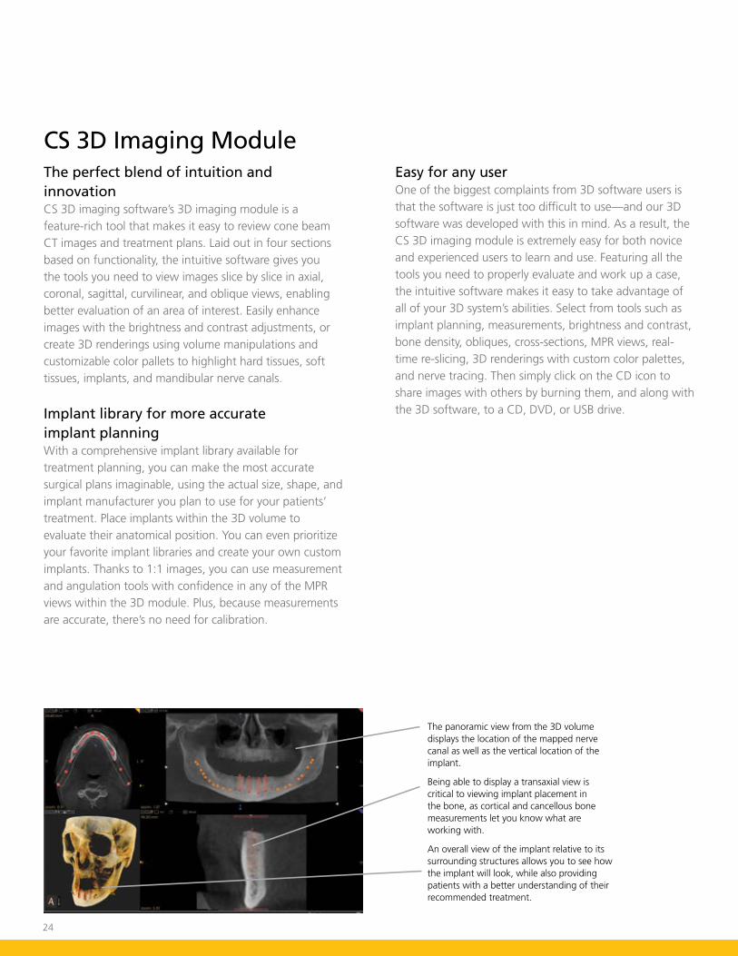
The perfect blend of intuition and innovationCS 3D imaging software’s 3D imaging module is a feature-rich tool that makes it easy to review cone beam CT images and treatment plans. Laid out in four sections based on functionality, the intuitive software gives you the tools you need to view images slice by slice in axial, coronal, sagittal, curvilinear, and oblique views, enabling better evaluation of an area of interest. Easily enhance images with the brightness and contrast adjustments, or create 3D renderings using volume manipulations and customizable color pallets to highlight hard tissues, soft tissues, implants, and mandibular nerve canals.
Implant library for more accurate implant planningWith a comprehensive implant library available for treatment planning, you can make the most accurate surgical plans imaginable, using the actual size, shape, and implant manufacturer you plan to use for your patients’ treatment. Place implants within the 3D volume to evaluate their anatomical position. You can even prioritize your favorite implant libraries and create your own custom implants. Thanks to 1:1 images, you can use measurement and angulation tools with confidence in any of the MPR views within the 3D module. Plus, because measurements are accurate, there’s no need for calibration.
CS 3D Imaging Module
The panoramic view from the 3D volume displays the location of the mapped nerve canal as well as the vertical location of the implant.
Being able to display a transaxial view is critical to viewing implant placement in the bone, as cortical and cancellous bone measurements let you know what are working with.
An overall view of the implant relative to its surrounding structures allows you to see how the implant will look, while also providing patients with a better understanding of their recommended treatment.
Easy for any userOne of the biggest complaints from 3D software users is that the software is just too difficult to use—and our 3D software was developed with this in mind. As a result, the CS 3D imaging module is extremely easy for both novice and experienced users to learn and use. Featuring all the tools you need to properly evaluate and work up a case, the intuitive software makes it easy to take advantage of all of your 3D system’s abilities. Select from tools such as implant planning, measurements, brightness and contrast, bone density, obliques, cross-sections, MPR views, real-time re-slicing, 3D renderings with custom color palettes, and nerve tracing. Then simply click on the CD icon to share images with others by burning them, and along with the 3D software, to a CD, DVD, or USB drive.

25
Easily share data with anyoneData often needs to be shared with colleagues or referring doctors. Fortunately, our 3D imaging module has a CD/DVD burning option that copies data, as well as the entire 3D software, to a disk or USB drive that can be shared.
Printing is easy too!You can easily print views within the 3D imaging module and add them to a patient file for insurance purposes, or to take them into surgery. Templates that show an area of interest can also be displayed in a wide variety of ways.
Integrate images into patient records in secondsCapturing your active workspace into your patient’s file is often a great way to maintain patient records. Panoramic views from the curved slicing tab or cephalometric views from the sagittal view can be easily captured and maintained in the patient’s record. 3D renderings are the most understandable views for patients, and capturing them is quick and easy with the 3D imaging module screen capture tool.
Get instant density readingsUnderstanding the relative density of objects in the 3D volume is critical. Grayscale image values are displayed in the lower right corner of the software whenever you move your cursor over the area of interest.
Top: Plan implants with confidence.
Middle: Clear layout of TMJ slices and 3D rendering.
Bottom: Go beyond right angles to get a complete picture of the anatomy.

Technical Specifications
Panoramic Modality Cephalometric Modality 3D Modality
Sensor Technology
Technology
CCD with Optical Fiber
Panoramic
CCD with Optical Fiber
One-Shot
CMOS with Optical Fiber
Cone beam CT
Gray Scale 14 bit 14 bit 15 bit
3D Field of View n/a n/a Single Volume: 5 cm diameter x 3.75 cm height
Extended Volumes (up to 3 volumes): 7.5 cm diameter x 3.75 cm height
Exam Options Standard Panoramic
Child Mode
Segmented Panoramic
Maxillary Sinus
LA TMJ x 2
LA TMJ x 4
Child Mode
Lateral, Oblique, Frontal (AP/PA), Submento-Vertex, Carpus
Multiple Cephalometric Field Choices: 30 cm x 30 cm, 24 cm x 30 cm, 24 cm x 24 cm, 18 cm x 24 cm, 18 cm x 18 cm
Child Mode
Resolution Options: from 76 to 200 micrometers
Other Specifications Panoramic Modality Cephalometric Modality 3D Modality
Input Voltage 230/240 V - 60 Hz 230/240 V - 60 Hz 230/240 V - 60 Hz
Tube Voltage 60-90 kV (max) 60-90 kV (max) 60-90 kV (max)
Tube Current 2-15 mA (max) 2-15 mA (max) 2-15 mA (max)
Frequency 140 kHZ (max) 140 kHZ (max) 140 kHZ (max)
Total Filtration > 2.5 mm eq. AL > 2.5 mm eq. AL > 2.5 mm eq. AL
Weight 353 lb (160 kg) 438 lb (199 kg) 353 lb (160 kg)
Space Requirements WxDxH (mm)
1500 mm (59") X 2000 mm (78 3/4") X 2400 mm (94 1/2")
2230 mm (88") X 2000 mm (78 3/4") X 2400 mm (94 1/2")
1500 mm (59") X 2000 mm (78 3/4") X 2400 mm (94 1/2")
Warning: Class 2 laser product. Do not stare into the beam.
CS 9000 System
CS 9000 3D system CS 9000C 3D system

Panoramic Modality Cephalometric Modality 3D Modality
Sensor Technology
Technology
TFT
Panoramic
CCD with Optical Fiber
One-Shot
TFT
Cone beam CT
Gray Scale 16 bit 16 bit 16 bit
3D Field of View n/a n/a CS 9300 Select: 5 cm x 5 cm, 10 cm x 5 cm, 8 cm x 8 cm, 10 cm x 10 cm
CS 9300: 5 cm x 5 cm, 10 cm x 5 cm, 8 cm x 8 cm, 10 cm x 10 cm, 17 cm x 6 cm, 17 cm x 11 cm, 17 cm x 13.5 cm
Exam Options Standard Panoramic
Child Mode
Segmented Panoramic
Maxillary Sinus
LA TMJ x 2
LA TMJ x 4
Child Mode
Lateral, Oblique, Frontal (AP/PA), Submento-Vertex, Carpus
Multiple Cephalometric Field Choices: 30 cm x 30 cm, 24 cm x 30 cm, 24 cm x 24 cm, 18 cm x 24 cm, 18 cm x 18 cm
Child Mode
Resolution Options: 90 to 500 micrometers
Other Specifications Panoramic Modality Cephalometric Modality 3D Modality
Input Voltage 230/240 V, 50/60 Hz 230/240 V, 50/60 Hz 230/240 V, 50/60 Hz
Tube Voltage 60-90 kV (max) 60-90 kV (max) 60-90 kV (max)
Tube Current 2-15 mA (max) 2-15 mA (max) 2-15 mA (max)
Frequency 140 kHZ (max) 140 kHZ (max) 140 kHZ (max)
Total Filtration > 2.5 mm eq. AL > 2.5 mm eq. AL > 2.5 mm eq. AL
Weight 353 lb (160 kg) 438 lb (199 kg) 353 lb (160 kg)
Space Requirements WxDxH (mm)
1500 mm (59") X 2000 mm (78 3/4") X 2400 mm (94 1/2")
2230 mm (88") X 2000 mm (78 3/4") X 2400 mm (94 1/2")
1500 mm (59") X 2000 mm (78 3/4") X 2400 mm (94 1/2")
Warning: Class 2 laser product. Do not stare into the beam.
CS 9300 System
CS 9300 System / CS 9300 Select System CS 9300C System / CS 9300C Select System
3D Im
agin
g Sy
stem
sWould you like to know more?Visit www.carestreamdental.com/3D or call us today at 800.944.6365
Learn more about how Carestream Dental’s 3D Imaging products can give you a new dimension of confidence.
© Carestream Health, Inc. 2012. Simplant is a trademark of Materialise Dental. NobelGuide is a trademark of Nobel Biocare. SureSmile is a trademark of OraMetrix. 7701 DE BR
Related Documents