3D and 4D Imaging of the Aortic Root Dominik Fleischmann Department of Radiology Stanford University 20 th Annual Summer Practicum, Masters in Body Imag Jackson Lake Lodge, Moran, Wyom August 8-11, 2

3D and 4D Imaging of the Aortic Root
Feb 24, 2016
20 th Annual Summer Practicum, Masters in Body Imaging Jackson Lake Lodge, Moran, Wyoming August 8-11, 2010. 3D and 4D Imaging of the Aortic Root . Dominik Fleischmann Department of Radiology Stanford University. 20 th Annual Summer Practicum, Masters in Body Imaging - PowerPoint PPT Presentation
Welcome message from author
This document is posted to help you gain knowledge. Please leave a comment to let me know what you think about it! Share it to your friends and learn new things together.
Transcript
3D and 4D Imaging of the Aortic Root
Dominik FleischmannDepartment of Radiology
Stanford University
20th Annual Summer Practicum, Masters in Body ImagingJackson Lake Lodge, Moran, Wyoming
August 8-11, 2010
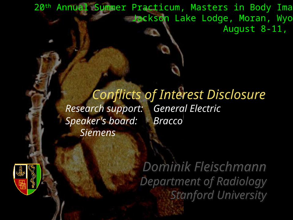
Research support: General ElectricSpeaker's board: Bracco
Siemens
Conflicts of Interest Disclosure
Dominik FleischmannDepartment of Radiology
Stanford University
20th Annual Summer Practicum, Masters in Body ImagingJackson Lake Lodge, Moran, Wyoming
August 8-11, 2010

Progressive root enlargement (Marfan's)
Jan '04
24 mm
45 mm
30 mm
Nov '04
28 mm
47 mm
32 mm
Sep '05
29 mm
49 mm
33 mm
with ECG gating

3D and 4D Imaging of the Aortic Root Learning Objectives / Outline
• Technique: 'gated chest' CT• Surgical anatomy of thoracic aorta• Clinical pre- and postop. imaging in
- Marfan's disease- Bicuspid aortic valve disease / aneurysm
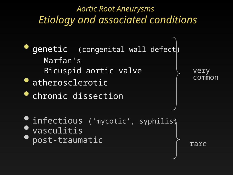
Aortic Root Aneurysms Etiology and associated conditions
• genetic (congenital wall defect)Marfan's Bicuspid aortic valve
• atherosclerotic • chronic dissection
• infectious ('mycotic', syphilis)• vasculitis• post-traumatic rare
verycommon
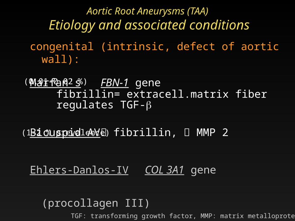
Aortic Root Aneurysms (TAA) Etiology and associated conditionscongenital (intrinsic, defect of aortic wall):
Marfan's FBN-1 gene fibrillin= extracell.matrix fiber regulates TGF-
Bicuspid AV fibrillin, MMP 2
Ehlers-Danlos-IV COL 3A1 gene (procollagen
III)
Loeys-Dietz TGFBR1 or TGFBR2 geneTGF: transforming growth factor, MMP: matrix metalloproteinase;
(0.01-0.02 %)
(1-2 % prevalence)

BAV disease(bicuspid aortic valve disease)
Prevalence 1-2%Complications (>33%)• valve degeneration
and stenosis• endocarditis• aortic root dilatation
(50% of young pts.) ao. root aneurysm
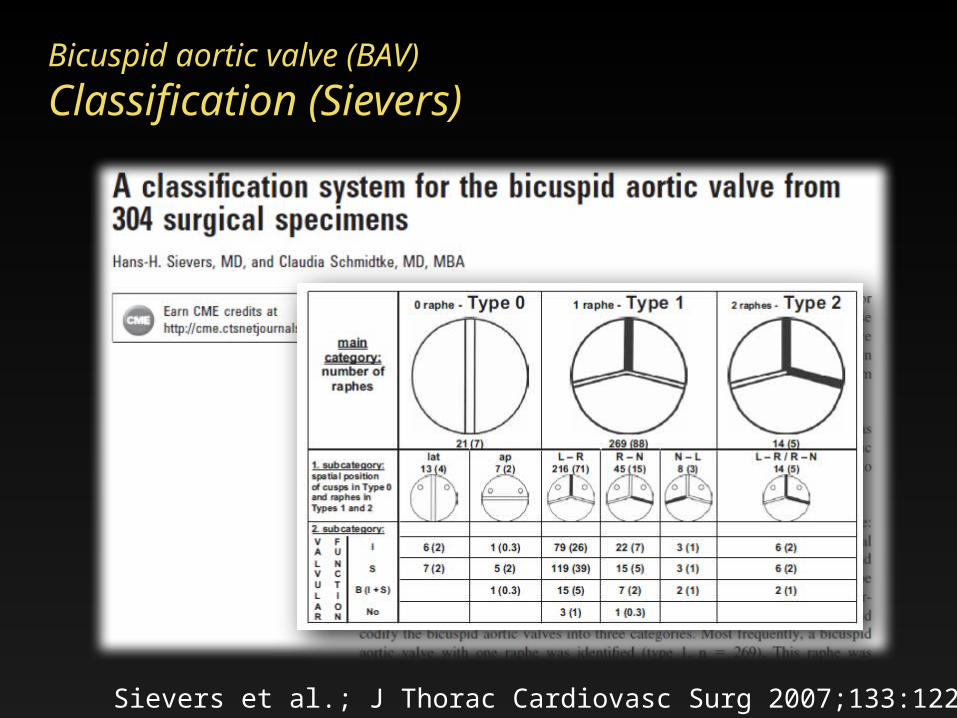
Sievers et al.; J Thorac Cardiovasc Surg 2007;133:1226-33
Bicuspid aortic valve (BAV)Classification (Sievers)

BAV disease(bicuspid aortic valve disease)

3D and 4D Imaging of the Aortic Root Learning Objectives / Outline
• Technique: 'gated chest' CT• Surgical anatomy of thoracic aorta• Clinical focus
pre and postoperative imaging - Marfan's disease- Bicuspid aortic valve disease / aneurysm
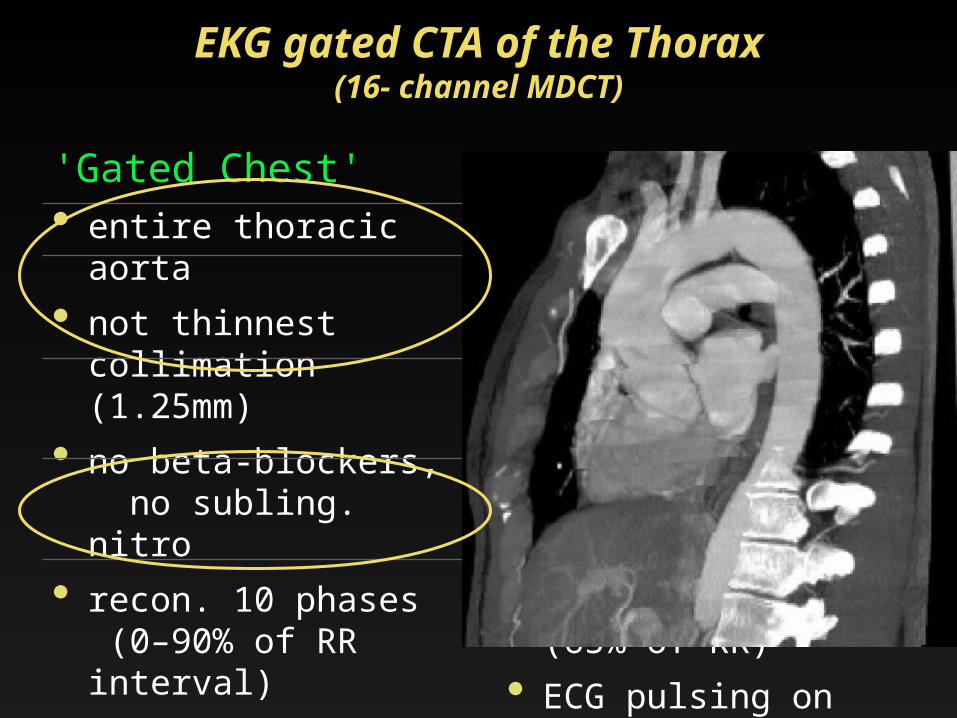
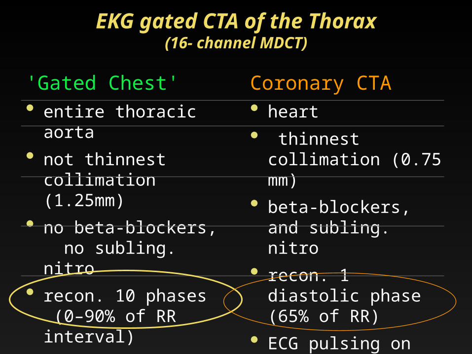
EKG gated CTA of the Thorax(16- channel MDCT)
'Gated Chest'• entire thoracic aorta
• not thinnest
collimation (1.25mm)
• no beta-blockers, no subling. nitro
• recon. 10 phases (0–90% of RR interval)
• no ECG-pulsing (constant mA)
Coronary CTA• heart• thinnest collimation
(0.625mm) • beta-blockers, and
subling. nitro• recon. 1 diastolic
phase (65% of RR) • ECG pulsing on
(dose reduction)
'Gated Chest'• entire thoracic aorta
• not thinnest
collimation (1.25mm)
• no beta-blockers, no subling. nitro
• recon. 10 phases (0–90% of RR interval)
• no ECG-pulsing (constant mA)
Coronary CTA• heart• thinnest collimation
(0.75 mm) • beta-blockers, and
subling. nitro• recon. 1 diastolic
phase (65% of RR) • ECG pulsing on
(dose reduction)
EKG gated CTA of the Thorax(16- channel MDCT)
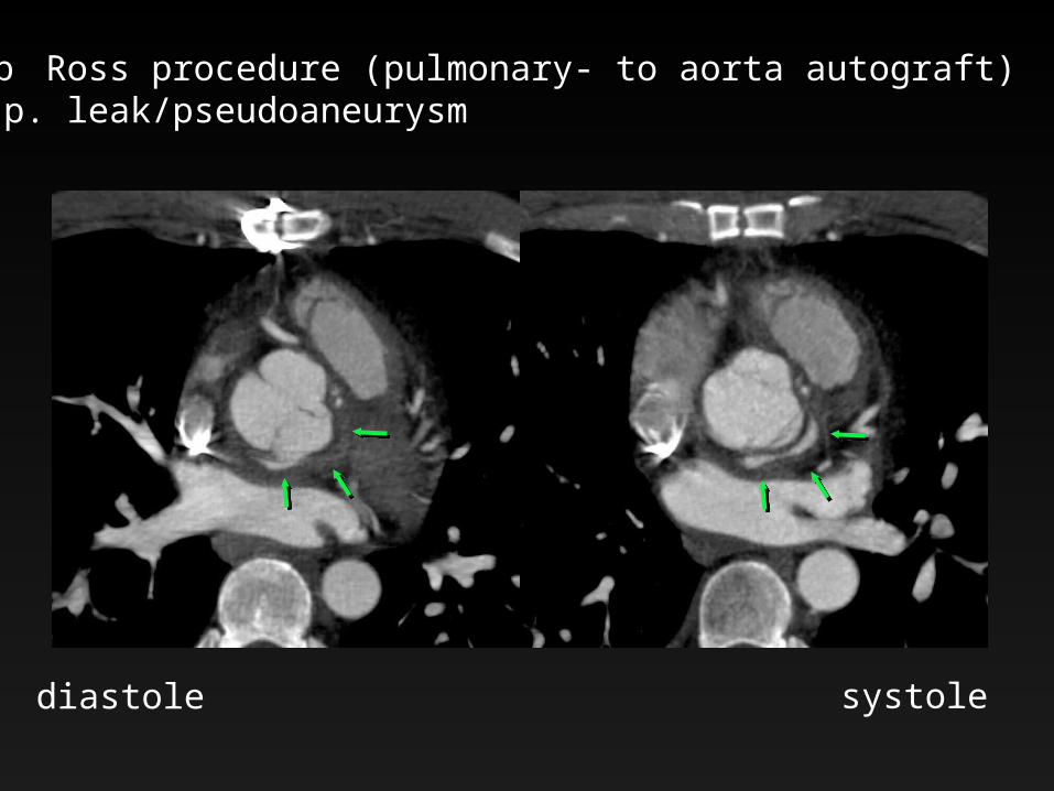
s/p Ross procedure (pulmonary- to aorta autograft) susp. leak/pseudoaneurysm
diastole systole
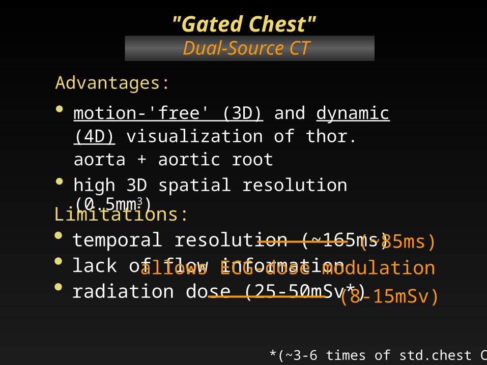
Advantages:• motion-'free' (3D) and dynamic (4D)
visualization of thor. aorta + aortic root
• high 3D spatial resolution (0.5mm3)
"Gated Chest"(16-slice CT, 64-slice CT)
Limitations:• temporal resolution (~165ms)• lack of flow information• radiation dose (25-50mSv*)
*(~3-6 times of std.chest CT)
Dual-Source CT
(~85ms)
(8-15mSv)allows ECG-dose modulation

58 yo woman• abnormal valve (BAV?),• aneurysmal aortic root• LV dilatation, low-normal EF
• 53kg (117 lbs)• 65 bpm heart rate 24
.7 c
m
Gated Chest, Dual-Source CT 27.01 mGy, (667mGy*cm), [~11mSv]
100 kVp ECG pulsing (30-70% of RR Interval)
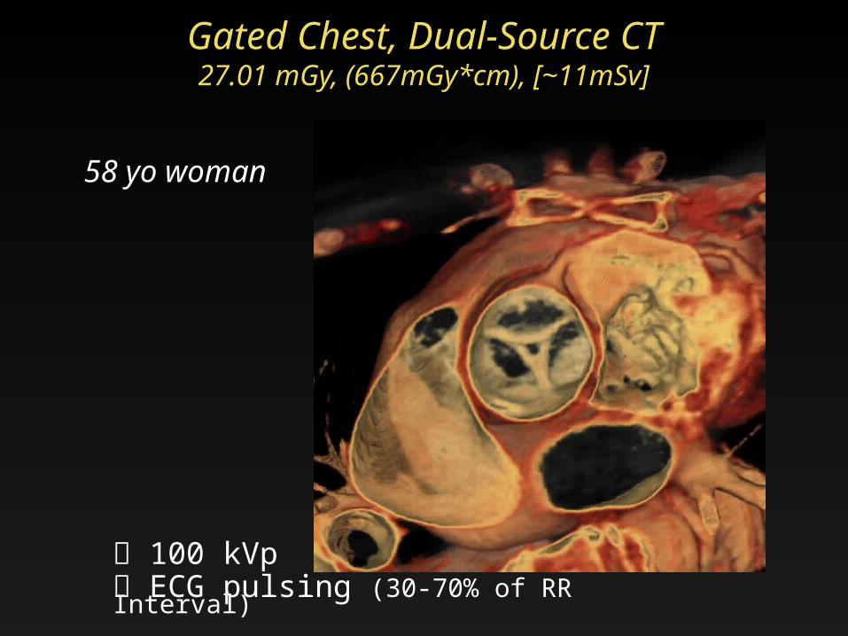
Gated Chest, Dual-Source CT 27.01 mGy, (667mGy*cm), [~11mSv]
58 yo woman
100 kVp ECG pulsing (30-70% of RR Interval)

3D and 4D Imaging of the Aortic Root Learning Objectives / Outline
• Technique: 'gated chest' CT• Surgical anatomy of thoracic aorta• Clinical focus
pre and postoperative imaging - Marfan's disease- Bicuspid aortic valve disease / aneurysm
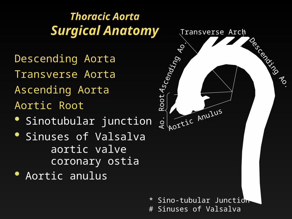
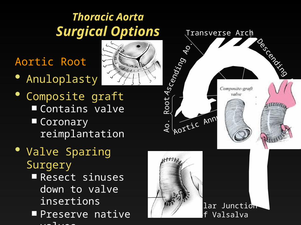
Asce
ndin
g Ao
. Descending Ao.
Transverse Arch
Aortic Anulus
STJ*
* Sino-tubular Junction# Sinuses of Valsalva
Ao.
Roo
t
SOV#
Descending Aorta Transverse AortaAscending Aorta Aortic Root• Sinotubular junction• Sinuses of Valsalva aortic valve coronary ostia• Aortic anulus
Thoracic AortaSurgical Anatomy
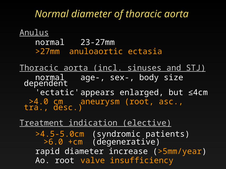
Normal diameter of thoracic aorta
Anulus normal 23-27mm >27mm anuloaortic ectasiaThoracic aorta (incl. sinuses and STJ) normal age-, sex-, body size dependent 'ectatic' appears enlarged, but ≤4cm >4.0 cm aneurysm (root, asc., tra., desc.)
Treatment indication (elective) >4.5-5.0cm (syndromic patients) >6.0 +cm (degenerative)
rapid diameter increase (>5mm/year) Ao. root valve insufficiency

• 73 y/o retired RN ascending aortic aneurysm
MIP (thin-slab) centered at valve
anulu
s
sinus.of Vals.
sin.tub.-junct.
asc.ao.
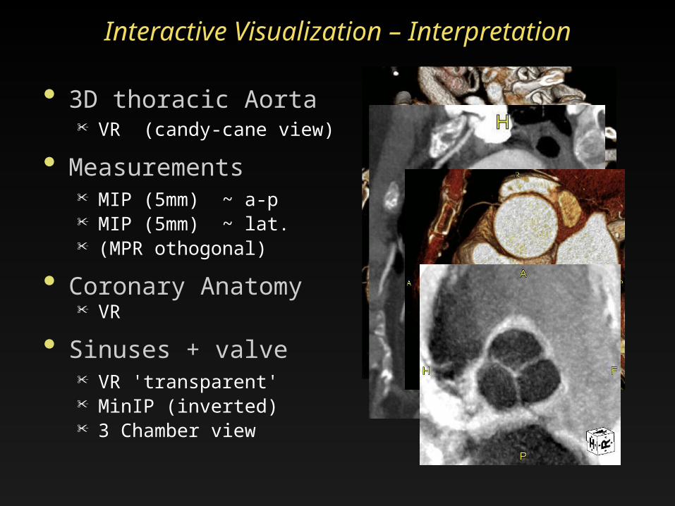
• 3D thoracic Aorta• VR (candy-cane view)
• Measurements • MIP (5mm) ~ a-p• MIP (5mm) ~ lat. • (MPR othogonal)
• Coronary Anatomy• VR
• Sinuses + valve• VR 'transparent' • MinIP (inverted)• 3 Chamber view
Interactive Visualization – Interpretation

3D and 4D Imaging of the Aortic Root Learning Objectives / Outline
• Technique: 'gated chest' CT• Surgical anatomy of thoracic aorta• Clinical focus
pre and postoperative imaging - Marfan's disease- Bicuspid aortic valve disease / aneurysm
Asce
ndin
g Ao
. Descending Ao.
Transverse Arch
Aortic Annulus
STJ*
* Sino-tubular Junction# Sinuses of Valsalva
Ao.
Roo
t
SOV#
Aortic Root• Anuloplasty• Composite graft
Contains valve Coronary
reimplantation• Valve Sparing Surgery
Resect sinuses down to valve insertions
Preserve native valves
Coronary reimpl.
Thoracic AortaSurgical Options
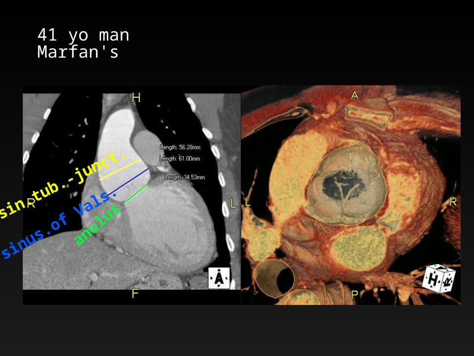
41 yo manMarfan's
anulus
sinus.of Vals.sin.tub.-junct.

Valve Sparing Aortic Root Procedures Tirone David- I “Reimplantation” Technique
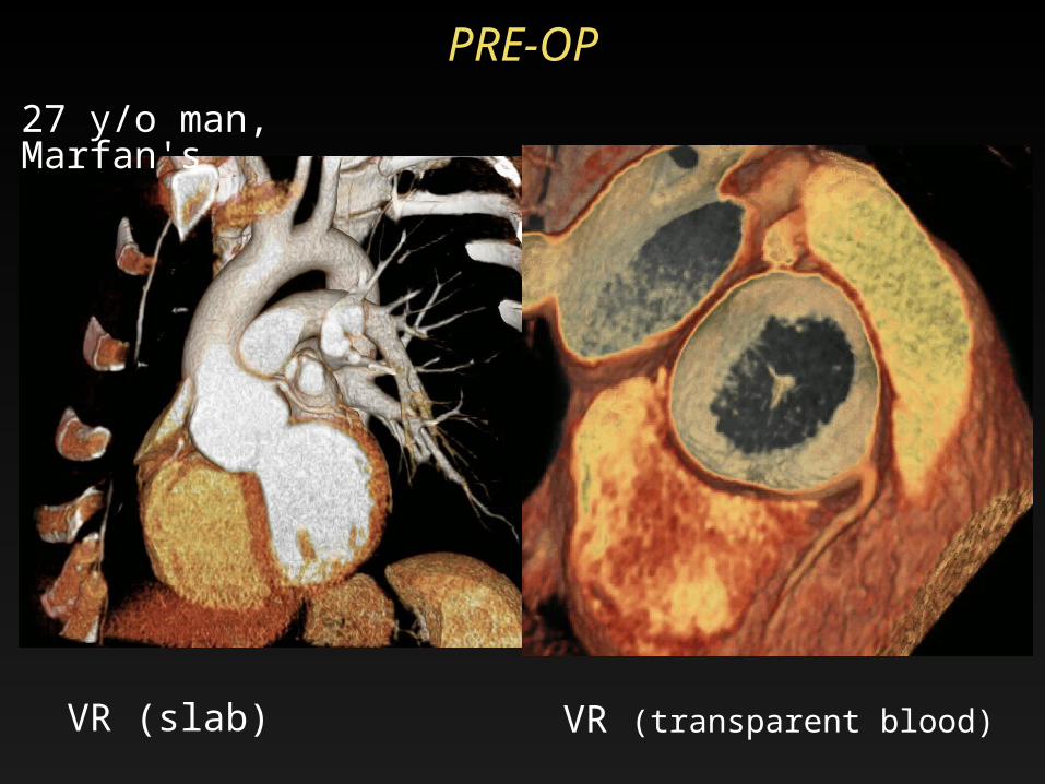
PRE-OP
VR (slab) VR (transparent blood)
27 y/o man, Marfan's

Surgical procedurecoronary
ostium
valveleaflets
coronary reiplanted
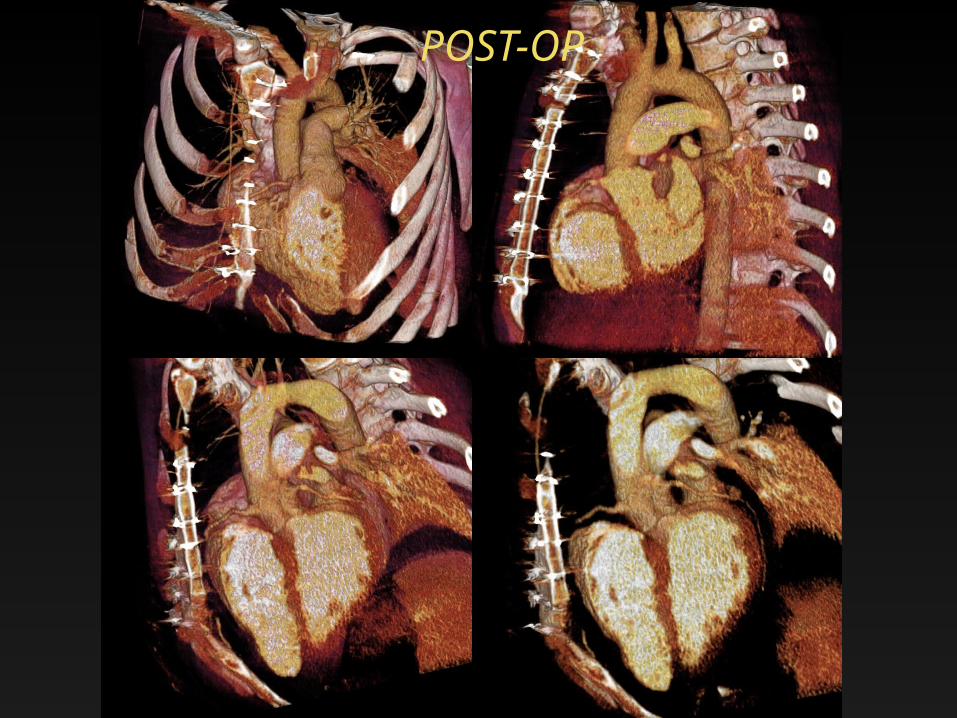
POST-OP
27 year old manbicuspid aortic valve root aneurysmvalve prolaps with severe aortic regurgitation and left ventricular dilatation
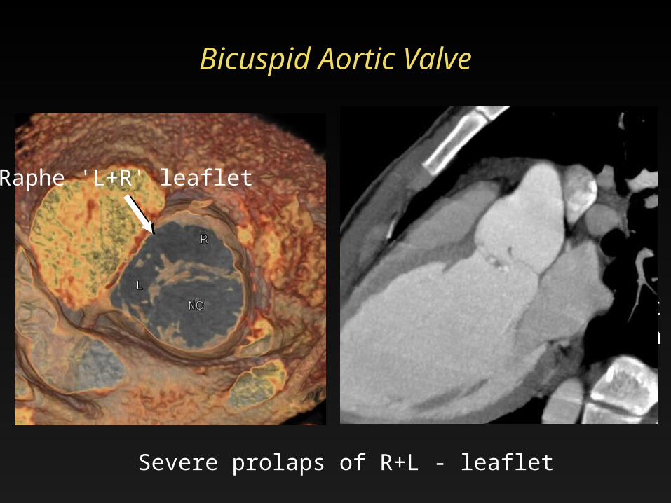
Bicuspid Aortic Valve
Severe prolaps of R+L - leaflet
Raphe 'L+R' leaflet
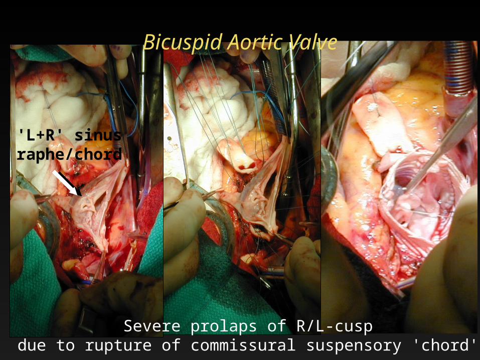
Bicuspid Aortic Valve
Severe prolaps of R/L-cuspdue to rupture of commissural suspensory 'chord'
'L+R' sinusraphe/chord
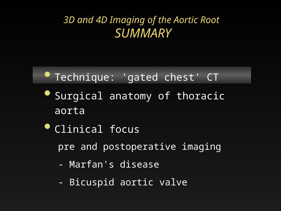
3D and 4D Imaging of the Aortic Root SUMMARY
• Technique: 'gated chest' CT• Surgical anatomy of thoracic aorta• Clinical focus
pre and postoperative imaging - Marfan's disease- Bicuspid aortic valve
Thank you..
DC Miller RS MitchellM Fischbein
20th Annual Summer Practicum, Masters in Body ImagingJackson Lake Lodge, Moran, Wyoming
August 8-11, 2010
Related Documents