Rather than replacing entire hip, as in a total hip replacement with traditional metal-on-plastic implants, (high impact activities are restricted because of increased (plastic) wear and loosening of the prosthesis), hip resurfacing simply reshapes the bone by shaving a few millimeters off the femoral head and socket and caps it with a metal component made of cobalt chrome, which allows the patient to get back to his or her active lifestyle in a matter of months. HISTORY OF HIP RESURFACING Originally developed in the 1970s, many early hip resurfacings resulted in poor outcomes and caused many surgeons to abandon the procedure. ese early attempts at the procedure failed largely because the large, metal femoral head rubbed on the then-poly- ethylene socket, wearing it out. is degenerative wear-and-tear caused the components to loosen, ultimately leading to femoral neck fractures. Surgeons preferred the total hip replacement. In 1997, Derek McMinn, MD, and Ronan Treacy, MD released the original concept of the Birmingham Hip Resurfacing System. Unlike the total hip replacement, in the resurfacing procedure, People who are very physically active — whether in a physical work environ- ment or simply playing sports — are often uneasy when told by a physician that they need hip surgery to fix the severe, arthritic groin pain that can become a chronic problem. This article discusses options to return to pre- surgery activity levels without going through a total hip replacement. by Jill Wehling Birmingham Hip Resurfacing LEARNING OBJECTIVES ▲ Review the relevant anatomy for this procedure ▲ Examine the set-up and surgical positioning for this procedure ▲ Compare and contrast the differences between hip resurfacing and hip replacement ▲ Assess the indications for hip resurfacing, including selecting strong candidates ▲ Evaluate the step-by-step process of the Birmingham Hip Resurfacing System SEPTEMBER 2010 | The Surgical Technologist | 401

Welcome message from author
This document is posted to help you gain knowledge. Please leave a comment to let me know what you think about it! Share it to your friends and learn new things together.
Transcript

Rather than replacing entire hip, as in a total hip replacement with traditional metal-on-plastic implants, (high impact activities are restricted because of increased (plastic) wear and loosening of the prosthesis), hip resurfacing simply reshapes the bone by shaving a few millimeters off the femoral head and socket and caps it with a metal component made of cobalt chrome, which allows the patient to get back to his or her active lifestyle in a matter of months.
H I S T O R Y O F H I P R E S U R F A C I N G
Originally developed in the 1970s, many early hip resurfacings resulted in poor outcomes and caused many surgeons to abandon the procedure. These early attempts at the procedure failed largely because the large, metal femoral head rubbed on the then-poly-ethylene socket, wearing it out. This degenerative wear-and-tear caused the components to loosen, ultimately leading to femoral neck fractures. Surgeons preferred the total hip replacement.
In 1997, Derek McMinn, MD, and Ronan Treacy, MD released the original concept of the Birmingham Hip Resurfacing System. Unlike the total hip replacement, in the resurfacing procedure,
by Nydia Morales, CST
People who are very physically active—whether in a physical work environ-
ment or simply playing sports—are often uneasy when told by a physician
that they need hip surgery to fix the severe, arthritic groin pain that can
become a chronic problem. This article discusses options to return to pre-
surgery activity levels without going through a total hip replacement.
by Jill Wehling
Birmingham Hip Resurfacing
L E A R N I N G O B J E C T I V E S
▲ Review the relevant anatomy for
this procedure
▲ Examine the set-up and surgical
positioning for this procedure
▲ Compare and contrast the differences
between hip resurfacing and
hip replacement
▲ Assess the indications for hip
resurfacing, including selecting
strong candidates
▲ Evaluate the step-by-step process of the
Birmingham Hip Resurfacing System
SEPTEMBER 2010 | The Surgical Technologist | 401
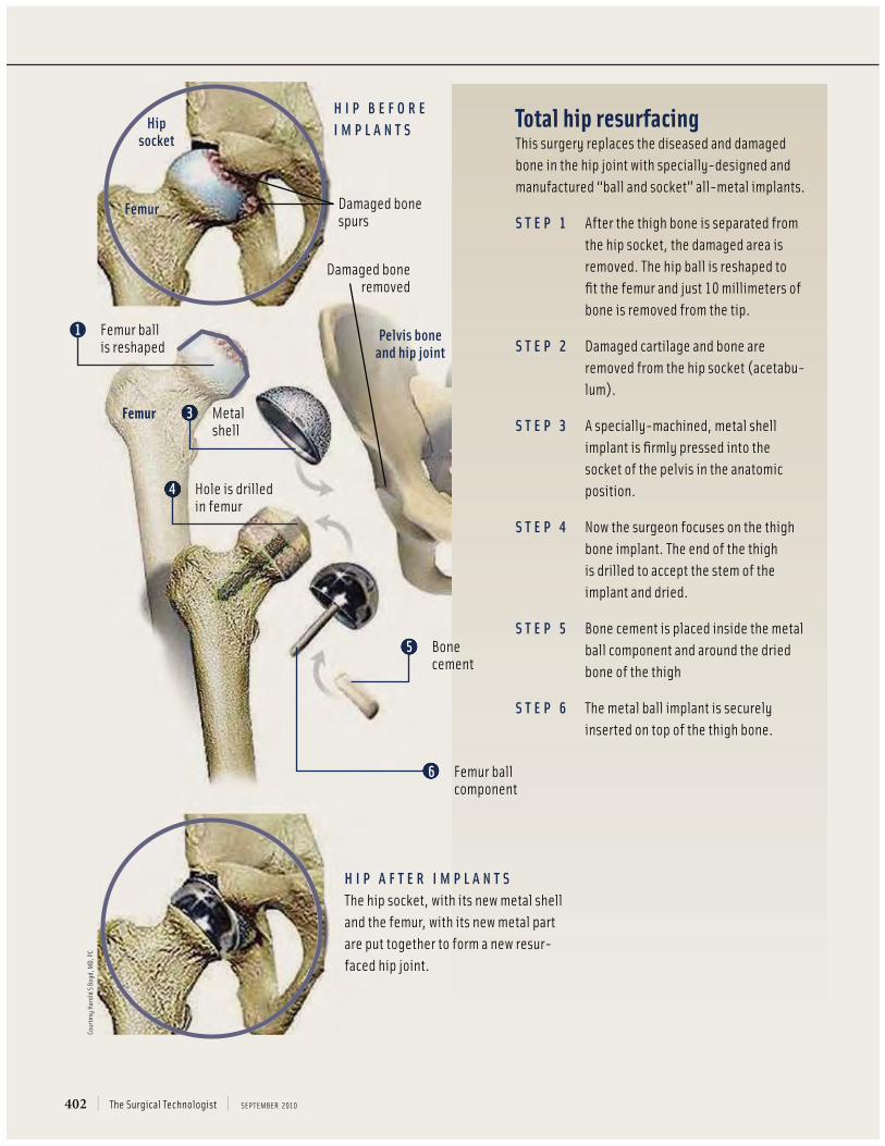
Total hip resurfacingThis surgery replaces the diseased and damaged
bone in the hip joint with specially-designed and
manufactured “ball and socket” all-metal implants.
S T E P 1 After the thigh bone is separated from
the hip socket, the damaged area is
removed. The hip ball is reshaped to
fit the femur and just 10 millimeters of
bone is removed from the tip.
S T E P 2 Damaged cartilage and bone are
removed from the hip socket (acetabu-
lum).
S T E P 3 A specially-machined, metal shell
implant is firmly pressed into the
socket of the pelvis in the anatomic
position.
S T E P 4 Now the surgeon focuses on the thigh
bone implant. The end of the thigh
is drilled to accept the stem of the
implant and dried.
S T E P 5 Bone cement is placed inside the metal
ball component and around the dried
bone of the thigh
S T E P 6 The metal ball implant is securely
inserted on top of the thigh bone.
H I P B E F O R E I M P L A N T S
H I P A F T E R I M P L A N T SThe hip socket, with its new metal shell
and the femur, with its new metal part
are put together to form a new resur-
faced hip joint.
Hip socket
Pelvis bone and hip joint
Femur
Femur
Damaged bone spurs
Damaged bone removed
1 Femur ball is reshaped
3 Metal shell
5 Bone cement
4 Hole is drilled in femur
6 Femur ball component
Cou
rte
sy H
aro
ld S
Bo
yd
, M
D,
PC
| The Surgical Technologist | SEPTEMBER 2010 402
the femoral head and neck is not resected. McMinn’s and Treacy’s model eliminated the polyethylene component of the resurfacing hardware, and with it, prior concerns about it wearing out. This adaptation allows for a much greater level of activity postoperatively. Because of the larger metal-head size, the risk of dislocation is reduced by a factor of 10. Because the femoral shaft is not broached, future revision, if necessary, is much easier and longer lasting.
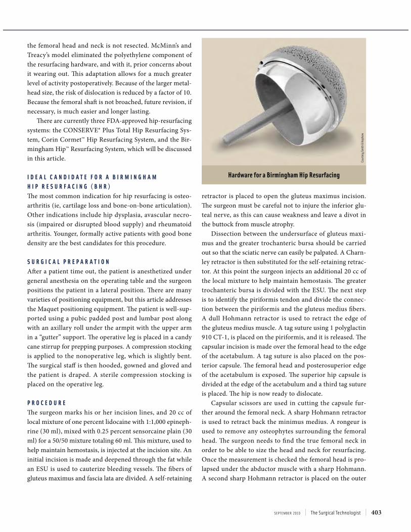
There are currently three FDA-approved hip-resurfacing -
tem, Corin Cormet™ Hip Resurfacing System, and the Bir-mingham Hip™ Resurfacing System, which will be discussed in this article.
I D E A L C A N D I D A T E F O R A B I R M I N G H A M
H I P R E S U R F A C I N G ( B H R )
The most common indication for hip resurfacing is osteo-arthritis (ie, cartilage loss and bone-on-bone articulation). Other indications include hip dysplasia, avascular necro-sis (impaired or disrupted blood supply) and rheumatoid arthritis. Younger, formally active patients with good bone density are the best candidates for this procedure.
S U R G I C A L P R E P A R A T I O N
After a patient time out, the patient is anesthetized under general anesthesia on the operating table and the surgeon positions the patient in a lateral position. There are many varieties of positioning equipment, but this article addresses the Maquet positioning equipment. The patient is well-sup-ported using a pubic padded post and lumbar post along with an axillary roll under the armpit with the upper arm in a “gutter” support. The operative leg is placed in a candy cane stirrup for prepping purposes. A compression stocking is applied to the nonoperative leg, which is slightly bent. The surgical staff is then hooded, gowned and gloved and the patient is draped. A sterile compression stocking is placed on the operative leg.
P R O C E D U R E
The surgeon marks his or her incision lines, and 20 cc of local mixture of one percent lidocaine with 1:1,000 epineph-rine (30 ml), mixed with 0.25 percent sensorcaine plain (30 ml) for a 50/50 mixture totaling 60 ml. This mixture, used to help maintain hemostasis, is injected at the incision site. An initial incision is made and deepened through the fat while an ESU is used to cauterize bleeding vessels. The fibers of gluteus maximus and fascia lata are divided. A self-retaining
retractor is placed to open the gluteus maximus incision. The surgeon must be careful not to injure the inferior glu-teal nerve, as this can cause weakness and leave a divot in the buttock from muscle atrophy.
Dissection between the undersurface of gluteus maxi-mus and the greater trochanteric bursa should be carried out so that the sciatic nerve can easily be palpated. A Charn-ley retractor is then substituted for the self-retaining retrac-tor. At this point the surgeon injects an additional 20 cc of the local mixture to help maintain hemostasis. The greater trochanteric bursa is divided with the ESU. The next step is to identify the piriformis tendon and divide the connec-tion between the piriformis and the gluteus medius fibers. A dull Hohmann retractor is used to retract the edge of the gluteus medius muscle. A tag suture using 1 polyglactin 910 CT-1, is placed on the piriformis, and it is released. The capsular incision is made over the femoral head to the edge of the acetabulum. A tag suture is also placed on the pos-terior capsule. The femoral head and posterosuperior edge of the acetabulum is exposed. The superior hip capsule is divided at the edge of the acetabulum and a third tag suture is placed. The hip is now ready to dislocate.
Capsular scissors are used in cutting the capsule fur-ther around the femoral neck. A sharp Hohmann retractor is used to retract back the minimus medius. A rongeur is used to remove any osteophytes surrounding the femoral head. The surgeon needs to find the true femoral neck in order to be able to size the head and neck for resurfacing. Once the measurement is checked the femoral head is pro-lapsed under the abductor muscle with a sharp Hohmann. A second sharp Hohmann retractor is placed on the outer
Hardware for a Birmingham Hip Resurfacing
Cour
tesy
Sm
ith
& N
ephe
w
SEPTEMBER 2010 | The Surgical Technologist | 403

used to secure the Hohmann retractors. A headed pin is placed below the transverse ligament and below the tear-
the acetabulum is achieved and the surgical technologist is ready for preparation and cup insertion.
The acetabular labrum is fully excised, and the ligamen-tum teres remnant also removed. If an osteophyte is pres-ent on the posterior acetabular wall, it is divided with an osteotome and removed with a rongeur. Sometimes there is soft tissue that needs to be removed. Osteophyte in the acetabular floor is excised with an osteotome and rongeur. The surgeon can now begin reaming the acetabulum. Start-ing with a size 44 or 46 mm reamer, the surgeon reams until he or she is confident that the acetabular floor has been reached—reaming up in 2mm increments until he or she comes close to the final acetabular reaming. At this point, the surgeon will proceed in 1mm increments. It is generally advised to ream one size under the estimated cup size and then verify the measurement by trial. When the surgeon is confident about the fit of the trial acetabular component, the true implant is opened and inserted with an impactor and heavy mallet. The surgeon can often tell by the sound that the acetabular cup is fully seated as it contacts the floor of the acetabulum. The surgeon is aiming for 40 degrees of
inclination and 20 degrees of anteversion. When the surgeon is satisfied with cup positioning, the ace-tabular cables are cut and the impactor cap and the cables are removed. Any protruding osteophyte is removed with a rongeur to prevent impingement. An X-ray-detectable 4x4 is placed in the acetabu-lar cup to protect the cup during the next phase of resurfacing. All retractors are now removed and the headed pin is taken out.
Accuracy is extremely critical in the following steps. The femoral head must be fully exposed to carry out the re-shaping of
the head. In order to obtain the correct varus-valgus align-ment of the femoral component, the template distance from the tip of the lesser trochanter to the desired point on the intertrochanteric crest must now be transferred from the X-ray measurement, into the operative field. A ruler and an 18g spinal needle are used to obtain this measurement, and it is marked with the ESU at its point for pin insertion. The pin is then drilled into the lesser trochanteric space, and a McMinn guide jig is placed to gain proper varus/val-gus alignment. A stylus on the guide jig is used to check perimeters circumferentially on the femoral neck. Once the surgeon is satisfied with the ideal entry point, he or she will then drill a long-guide pin, via a cannulated rod, into the superior femoral head. It is important to check the guide-wire position by using the stylus tip around the femoral neck. The stylus should not touch the femoral neck in any position and should touch the periphery of the femoral head 360 degrees. These are the minimum requirements for guide-wire position.
If the surgeon is not happy with the guide-wire posi-tion, a guide-wire repositioning instrument can be used. The cannulated rod and stylus are removed, and the sur-geon will drill through the lesser trochanter into the canal of the femur to place a drain cannula for suction. A urology drape is then placed around the femoral head to protect
L—Pre-surgical X-ray showing surgeons measurements for the hip resurfacing components.
R—The same hip with the components in place
Cou
rte
sy J
oh
n R
og
ers
on
, M
D
| The Surgical Technologist | SEPTEMBER 2010 404

the soft tissues from being contaminated by bony reaming during the femoral preparation. The guide wire is now over-drilled with two different-sized drilling instruments then replaced with a guide rod. A continuous flow of irrigation is recommended during the next three stages. A head-neck template is used as a protector while reaming the femoral head with the cylindrical reamer. An osteotome is used to remove the peripheral ring of femoral head bone. A rongeur is used to remove any osteophyte left behind on the femo-ral head-neck junction. The cylindrical reamer is then used by hand to remove any fragments left behind. A marking pen will mark the head and neck joint and summit of the head. A plain cutter is used to carefully resect down to the marked point. This is checked with the head-neck template. The chamfer cutter is used until the instrument is fully seated. There is an internal stop in this instrument. This is the final stage of re-shaping the femoral head. Keyholes are drilled with a Wroblewski drill for cement. The guide rod is removed with a slap hammer and the central hole in the femoral head and neck is enlarged with a taper drill. Any cysts seen are removed with a curette. The femoral head is now prepped by pulse lavage and patted dry with lap spong-es and suctioned dry. The surgeon checks the correct size of the femoral head, and the box is opened to surgical staff.
Preparation of cement consists of a mixing bowl, spat-ula, 60 cc catheter tip syringe, cement powder, liquid and stop watch with head impactor with heavy mallet available. The femoral head is draped with three clean lap sponges in alternating directions. At the surgeon’s request, he or she will tell the surgical technologist when to start mixing as he or she has only one minute from the time the liquid con-tacts the powder to implantation. As a surgical technologist, the mixing has to be done quickly, but thoroughly. Ideally, at about 25 seconds, the surgeon should be able to draw up the liquid in the syringe and start filling the femoral head to one-third full. At the one minute mark, the surgeon places the femoral head with the impactor. Any excess cement is wiped away, and a rongeur is used to remove any small osteophyte at the head—neck junction. A pulse lavage is used to irrigate and clean the femoral head and surround-ing tissue. The drain cannula is removed, and a clotting agent is injected into the canal. Forceps are used to retrieve the raytec from the acetabular cup, saline is used to fill the acetabular cup and a bone hook is used to lift femoral head component along with traction on the leg to reduce into the acetabulum. The surgeon now double checks the leg length and range of motion for any impingement.
C L O S U R E
For closure, the surgeon chooses to use a 3/32 drill to place
is used to close the external rotators and capsule along with
CT-1 is used to close the subcutaneous layer, followed by staples for skin. The incision area is cleaned with a wet and dry. Skin glue is applied along with two, sterile 4x4s and an island dressing. An anti-embolism stocking is placed on patient’s leg with one pillow between the legs for the first few hours of recovery. An X-ray is taken in recovery when the patient is awake to ensure that dislocation has not occurred in moving from the operating table.
R E C O V E R Y
Rehabilitation usually starts on the first day after surgery. Patients are on crutches the next day and weight bearing is allowed as tolerated. Physical therapy will start one or two days after surgery, beginning with simple bed exercises that
The femoral head and hip socket after being trimmed
to fit the new prosthesis.
Cou
rte
sy S
cott
Ru
ssin
off
, M
D
SEPTEMBER 2010 | The Surgical Technologist | 405

will strengthen the muscles in the hip and lower extremity. It is the physical therapist’s job to teach the patient proper technique to perform such simple tasks as moving from lying to sitting, sitting to standing and standing to sitting. The patient must learn to perform these movements safely, so that he or she doesn’t dislocate the hip or suffer other injury. Patients will start the use of the pool three days after the operation. The patient should have hip flexion of about 90 degrees four to five weeks postoperatively.
Most patients can return to whatever level of physical activity they previously enjoyed approximately one year postoperatively. In the first three months, patients will be able to golf, and at six months, high-impact activities should be possible. However, during the first year, more conserva-tive, low-impact activities, such as walking, swimming and bicycling are recommended for strengthening the femoral neck and muscles around the resurfaced joint. The bone is strengthened as new bone grows. According to Wolff ’s Law, bone in a healthy person will adapt to the loads it is placed under. If loading on a particular bone increases, the bone will remodel itself over time to become stronger and resist that sort of loading.
Because the femoral neck is maintained in a resurfacing procedure, the weight-bearing forces through the calcar and proximal femur remain normal and calcium deposition is normalized gradually.
C O N C L U S I O N
Hip resurfacing is a technically demanding procedure, but it can be successful, and the results can be satisfying for the patient. Hip resurfacing requires good bone quality. Some acetabular deformities cannot be addressed. However, it is an attractive option for a young patient fearing a potentially difficult future revision.
A B O U T T H E A U T H O R
Wisconsin, and graduated from Mt Hood Community College Surgical Technology Program in Portland, Oregon, in 2003. She currently works at Meriter Hospital in Madi-son, Wisconsin, where she is the lead orthopedic surgical technologist.
ReferencesModern Hip Resurfacing. -
leg; 2009.Birmingham hip resurfacing. Available at: http://orthorogerson.
com. Accessed August 9, 2010.Wolff ’s law. Encyclopædia Britannica. 2010. Encyclopædia Britannica Online. Available at: http://www.britannica.com/EBchecked/topic/646611/Wolffs-law. Accessed July 19, 2010.
CONSERVE is a registered trademark of Wr ight Medical
Tech n olo g y.
Cor in Corm et is a tra d em a rk of th e Cor in G ro u p PLC .
Birm in g h a m HIP Res u r fa cin g System is a tra d em a rk of S m ith
a n d Nep h ew.
O m ni-Tra ct is a registered tra d em a rk of O m ni-Tra ct Su rgical.
Vicryl is a registered tra d em a rk of Eth ico n , a d ivisio n of
Joh n son & Joh n son .
| The Surgical Technologist | SEPTEMBER 2010 406
Related Documents