68 3.1 p Procrss of Seeing and Common Eye Disorders in India Structure: 3.1 Introduction 3.2 Objectives 3.3 Anatomy and Physiology of the Seeing Media 3.4 Different Parts of Eye and Their Function in Seeing 3.5 Process of Seeing 3.6 Common Eye Disorders In India 3.7 Symptoms And Teratment Of Refractive Errors 3.1.1 Introduction: There is a kind of perception that takes place as our brain decides what it is we are actually seeing. You can actually watch this process of settling upon the right image if you look for it. It is especially pronounced if the brain can’t immediately decide what it’s viewing. For example, if you see something in the distance you can’t quite make out the gestalt changes from image to image until the brain is satisfied that it is the correct one. Try to catch it sometime. In any case, we see what we have been taught to see. That is, the process of seeing is learned from the time we are infants. This is basically why all of us see the same things, and why anyone who doesn’t is considered crazy. Artists have long played on the edge of perceptions that are not readily available to the rest of us. Impressionism is a good example. These artists realized that light affected colour and form in unimaginable ways (at that point in the history of art), and painted impressionistic scenes so the rest of us could also see them. Of course, now most of us do, if we allow ourselves to. This really is the essential point—allowing ourselves to. We are much more resilient and stable than we imagine. We can all handle more uncertainty than we imagine. Just because we see or think something out of the ordinary does not mean we’re insane. It’s a normal part of perception.

Welcome message from author
This document is posted to help you gain knowledge. Please leave a comment to let me know what you think about it! Share it to your friends and learn new things together.
Transcript
68
3.1 ppppp Procrss of Seeing and Common Eye Disorders
in India
Structure:
3.1 Introduction
3.2 Objectives
3.3 Anatomy and Physiology of the Seeing Media
3.4 Different Parts of Eye and Their Function in Seeing
3.5 Process of Seeing
3.6 Common Eye Disorders In India
3.7 Symptoms And Teratment Of Refractive Errors
3.1.1 Introduction:
There is a kind of perception that takes place as our brain decides what it is we are
actually seeing. You can actually watch this process of settling upon the right image if
you look for it. It is especially pronounced if the brain can’t immediately decide what
it’s viewing. For example, if you see something in the distance you can’t quite make out
the gestalt changes from image to image until the brain is satisfied that it is the correct
one. Try to catch it sometime. In any case, we see what we have been taught to see. That
is, the process of seeing is learned from the time we are infants. This is basically why
all of us see the same things, and why anyone who doesn’t is considered crazy. Artists
have long played on the edge of perceptions that are not readily available to the rest of
us. Impressionism is a good example. These artists realized that light affected colour
and form in unimaginable ways (at that point in the history of art), and painted
impressionistic scenes so the rest of us could also see them. Of course, now most of us
do, if we allow ourselves to. This really is the essential point—allowing ourselves to.
We are much more resilient and stable than we imagine. We can all handle more
uncertainty than we imagine. Just because we see or think something out of the ordinary
does not mean we’re insane. It’s a normal part of perception.
69
3.1.2 Objectives:
After going through this unit you should be able to:
1. Draw the structure of seeing media
2. Describe the functions of the media
3. Explain the process of seeing
4. Describe the disorders of eye
5. Explain the treatment procedure of refractive errors
3.1.3 Anatomy And Physiologhy Of Seeing Media
The process of perception is done through eye which is the predominant sense
organ of human being. It is a very sensitive organ in our body to be taken care of
properly. Around 85% of the information is received through our eyes. Sight is the
sense through which the brain received approximately 75% of its information. The eye
is essentially formed from both ectoderm and mesoderm. The eye collects information
about size, shape and colour and transmits those to brain where these are interpreted.
So it must be said that eye is the apparatus for seeing. The structure of the orbit, the
ocular adnexa, the ocular muscles, the nerves and the blood supply system are so as to
help the eyeball to see and to protect it from injury. To understand the mechanism of
vision we have to understand the function of the eyeball, the ocular adnexa (the eyelids,
the conjunctiva and the lacrimal system) and the ocular muscles.
3.1.4 Different Parts of Eye And Their Function in Seeing:
The eyeball:
The eyeball rests in a soft cushion of fat protected by the bony orbit of the skull. It is
almost a perfect sphere with clean window in front of cornea. The parts of eyeball are as
follows-
Cornea
The cornea has an important role in image formation; it forms a primary refractive
element in the eye. So it says that cornea is a clear front window of the eye which
transmits and focuses (i.e., sharpness or clarity) light into the eye.
70
Iris:
The coloured part of the eye which helps regulate the amount of light entering the eye.
When there is bright light, the iris closes the pupil to let in less light. And when there is
low light, the iris opens up the pupil to let in more light.
Pupil:
The dark centre opening in the middle of the iris. The pupil changes size to adjust for
the amount of light available (smaller for bright light and larger for low light). This
opening and closing of light into the eye is much like the aperture in most 35 mm
cameras which lets in more or less light depending upon the conditions.
Lens:
Focuses light rays onto the retina. The lens is transparent, and can be replaced if necessary.
The lens is not noticed normally because it is hidden within the dark cavity of the inner
eye. Intraocular lenses are used to replace lenses clouded by cataracts.
Sclera:
The white outer coat of the eye, surrounding the iris. It is similar to the cornea, except
that it is vascular, and has dense, irregular, fibrous connective tissue.
Choroid:
Layer containing blood vessels that lines the back of the eye and is located between the
retina (the inner light-sensitive layer) and the sclera (the outer white eye wall).
Retina:
The nerve layer lining the back of the eye. The retina senses light and creates electrical
impulses that are sent through the optic nerve to the brain.
Macula:
The area in the retina that contains special light-sensitive cells. In the macula these
light-sensitive cells allow us to see fine details clearly in the centre of our visual field.
Fovea:
The centre of the macula which provides the sharp vision.
Ciliary Body
Structure containing muscle and is located behind the iris, which focuses the lens.
Aqueous Humour :
Produced by ciliary processes of ciliary body. It provides nutrients for lens and cornea.
It also maintains intraocular pressure (25mm.Hg), and is replaced several times a day 2
F 1/min).
71
Vitreous Humour:
The, clear, gelatinous substance filling the central cavity of the eye secreted by the
ciliary body up to the time of maturity. It has very loose connective tissue: contains
water, hyalouronic acid and collagen. Pressure from the vitreous humour prevents retinal
detachment. It supports the lens anteriorly and the retina posteriorly. It contains a hyaloid
canal, which is a remnant of blood vessels during development.
Optic Nerve:
A bundle of more than a million nerve fibers carrying visual messages from the retina
to the brain. (In order to see, we must have light and our eyes must be connected to the
brain.) Your brain actually controls what you see, since it combines images. The retina
sees images upside down but the brain turns images right side up. This reversal of the
images that we see is much like a mirror in a camera.
Ocular adnexa:
Accessory structures of the eye, including the eyelids, conjunctiva and the lacrimal
apparatus.
The eyelids:
The chief function of the lids is to protect the eyes from injury and excessive light. The
eyebrow and eyelashes also participate in protective role.
Conjunctiva:
It is continuous with the skin of the eyelids. The palpebral Conjunctiva is the part of the
conjunctiva that covers the inner surface of the Eyelid; the bulbar conjunctiva covers
the surface of the eyeball. It is lined by stratified squamous epithelium, and contains
goblet cells, which secrete the deepest, mucus, layer of tear film, which adheres to the
surface of the globe. It is highly vascular. The conjunctive blends with the skin of the
lid margins as well as with the corneal epithelium. It is also continuous, via the lacrimal
puncta and canaliculi with the mucosa of the nasolacrimal sac and duct and hence nose.
The lacrimal system:
The two main part of lacrimal system are (a) the lacrimal gland which secretes tears and
(b) the lacrimal ducts which carry the tears from the eye into cavity of the nose.
It also contains three layers of the tear film:
1. Deep mucous: from conjunctival goblet cells, adheres tears to the conjunctiva
a. Middle aqueous: from main and third eyelid lacrimal glands; it cleanses,
oxygenates and fills optimal defects.
3. Superficial oily layer: from tarsal glands prevents evaporation
72
Extra ocular muscles:
The muscles which control the movements of eye ball are six in number, all named by
their positions with regard to eyeball. These are as follows-
1. Dorsal rectus muscle
2. Ventral rectus muscle
3. Medial rectus muscle
4. Lateral rectus muscle
5. Dorsal oblique muscle
6. Ventral oblique muscle
Usually carrying out the eye movements two or more muscles work together. In
addition to the co-ordinated action of muscles in one eye, it is essential for proper
vision that there be perfectly co-ordinated muscular action in both eyes.
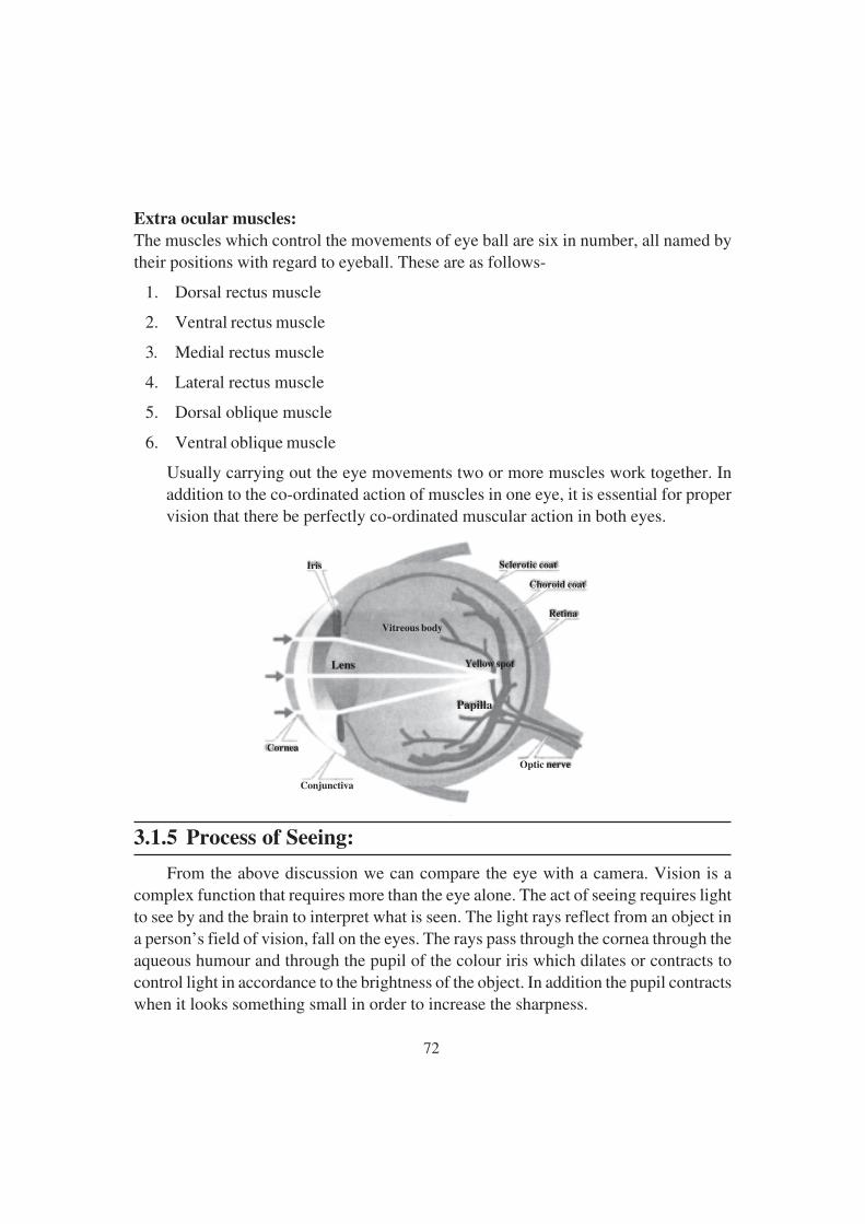
3.1.5 Process of Seeing:
From the above discussion we can compare the eye with a camera. Vision is a
complex function that requires more than the eye alone. The act of seeing requires light
to see by and the brain to interpret what is seen. The light rays reflect from an object in
a person’s field of vision, fall on the eyes. The rays pass through the cornea through the
aqueous humour and through the pupil of the colour iris which dilates or contracts to
control light in accordance to the brightness of the object. In addition the pupil contracts
when it looks something small in order to increase the sharpness.
Vitreous body
Lens
Conjunctiva
Optic nerve
Yellow spot
Papilla
Sclerotic coat
Choroid coat
Retina
Cornea
Iris
73
The rays then pass through the crystalline lens when the eye is relaxed and looking
into the far distance the rays of light are focused on to the retina. When we wish to look
at something nearer say at 6ft the focus of the lens is automatically adjusted by the
surrounding ciliary muscles. The fluid in the aqueous humours in front of the lens and
the vitreous body behind the lens allow it to expand or contract easily. This process of
focusing is called accommodation. The cornea and the lens combine to bend the light
rays as they pass through. The rays pass through the vitreous body and penetrate the
retina, where they set up a photochemical response in the outer most layers, there
stimulating the rods and cones. The impulse is picked by the retinal nerve fibres and
pass along the optic nerve to the brain where upside down image is formed. Based on
experience, the inverted image is psychologically transposed.
The eyes move together and send the brain almost identical images. The brain then
joins these two images into a single mental picture. The slight difference in the images
is needed to produce stereographic vision. By this long process we are able to see.
3.1.6 Common Eye Disoders in India:
Eyesight is one of the most precious gifts that nature has given to mankind. It’s
only because of the eyes; one can enjoy the beauty of this world. It’s impossible to
imagine life without sight. Though a very small part of body, eye is one of the most
complex human organs. It has various parts, all of which are responsible for normal
vision. Smallest structural or functional alteration in the functioning of an eye can cause
tremendous visual disturbances. This type of visual disturbance makes people helpless
and also dependable. The other name of visual disturbance is called visual disorder .on
the other hand it is also known as refractive error. In India maximum cause of the adult
blind is refractive error or injury or accident. If they are identified at first time there is a
chance for curing. But due to lack of knowledge or person’s negligence most of the
time these disorders are not properly treated or identified.
To see external object clearly, it is necessary that sharp images of objects must be
formed upon the retina. The cornea, the aqueous humour, the crystalline lens and the
vitreous body act together as refractive media to bring parallel rays of light reflected
from external object to a focus on the retina. The images become sharp in the macula.
The normal eye is called emmetropic while the abnormal condition is called errors of
refraction or ametropia. Refractive error or need of glasses is one of the most common
eye problems. It can start at any age. This is due to alteration in length, shape & / or
capacity of eyes.
74
What is refraction?
Refraction is the bending of light as it passes through one object to another. Vision
occurs when light rays are bent (refracted) as they pass through the cornea and the lens.
The light is then focused on the retina. The retina converts the light-rays into messages
that are sent through the optic nerve to the brain. The brain interprets these messages
into the images we see.
What are refractive errors?
Refractive errors occur when the shape of the eye prevents light from focusing directly
on the retina. The length of the eyeball (longer or shorter), changes in the shape of the
cornea, or aging of the lens can cause refractive errors.
Not all eyes are optically perfect and consequently light rays may not be brought
accurately to focus on the retina. Faulty optical conditions, or refractive errors may be
classified into four basic categories. These are as follows-
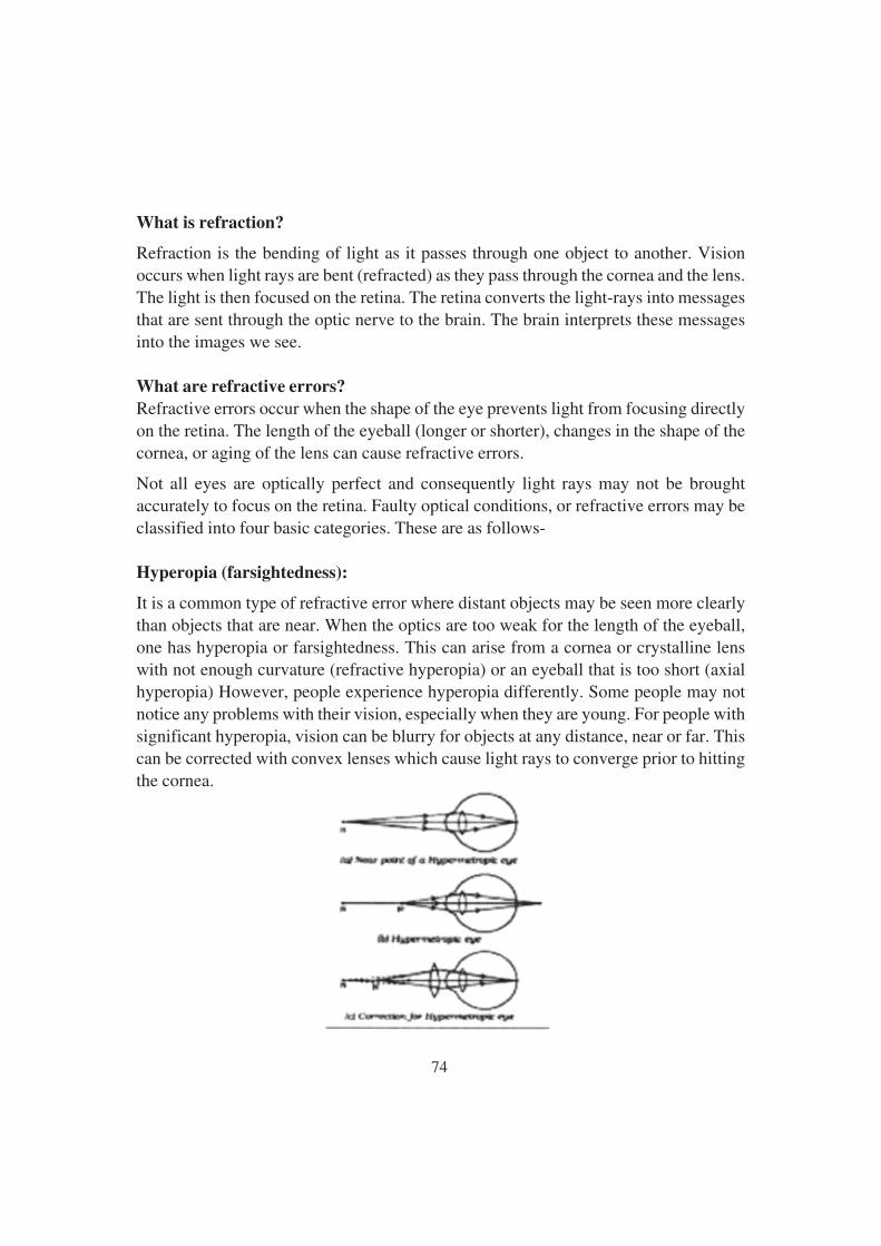
Hyperopia (farsightedness):
It is a common type of refractive error where distant objects may be seen more clearly
than objects that are near. When the optics are too weak for the length of the eyeball,
one has hyperopia or farsightedness. This can arise from a cornea or crystalline lens
with not enough curvature (refractive hyperopia) or an eyeball that is too short (axial
hyperopia) However, people experience hyperopia differently. Some people may not
notice any problems with their vision, especially when they are young. For people with
significant hyperopia, vision can be blurry for objects at any distance, near or far. This
can be corrected with convex lenses which cause light rays to converge prior to hitting
the cornea.
75
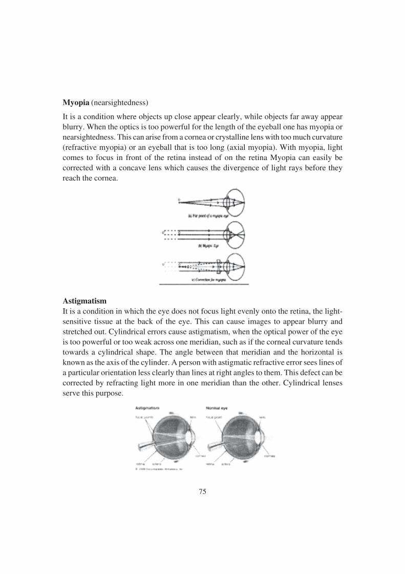
Myopia (nearsightedness)
It is a condition where objects up close appear clearly, while objects far away appear
blurry. When the optics is too powerful for the length of the eyeball one has myopia or
nearsightedness. This can arise from a cornea or crystalline lens with too much curvature
(refractive myopia) or an eyeball that is too long (axial myopia). With myopia, light
comes to focus in front of the retina instead of on the retina Myopia can easily be
corrected with a concave lens which causes the divergence of light rays before they
reach the cornea.
Astigmatism
It is a condition in which the eye does not focus light evenly onto the retina, the light-
sensitive tissue at the back of the eye. This can cause images to appear blurry and
stretched out. Cylindrical errors cause astigmatism, when the optical power of the eye
is too powerful or too weak across one meridian, such as if the corneal curvature tends
towards a cylindrical shape. The angle between that meridian and the horizontal is
known as the axis of the cylinder. A person with astigmatic refractive error sees lines of
a particular orientation less clearly than lines at right angles to them. This defect can be
corrected by refracting light more in one meridian than the other. Cylindrical lenses
serve this purpose.
76
Presbyopia
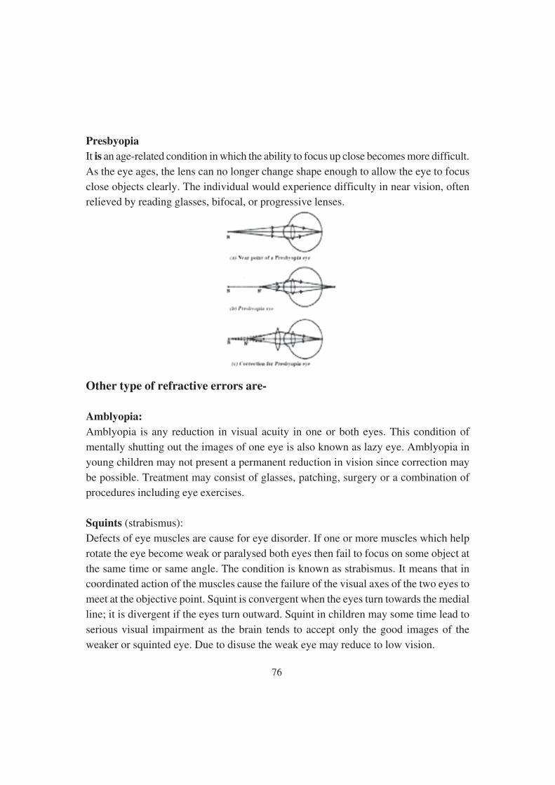
It is an age-related condition in which the ability to focus up close becomes more difficult.
As the eye ages, the lens can no longer change shape enough to allow the eye to focus
close objects clearly. The individual would experience difficulty in near vision, often
relieved by reading glasses, bifocal, or progressive lenses.
Other type of refractive errors are-
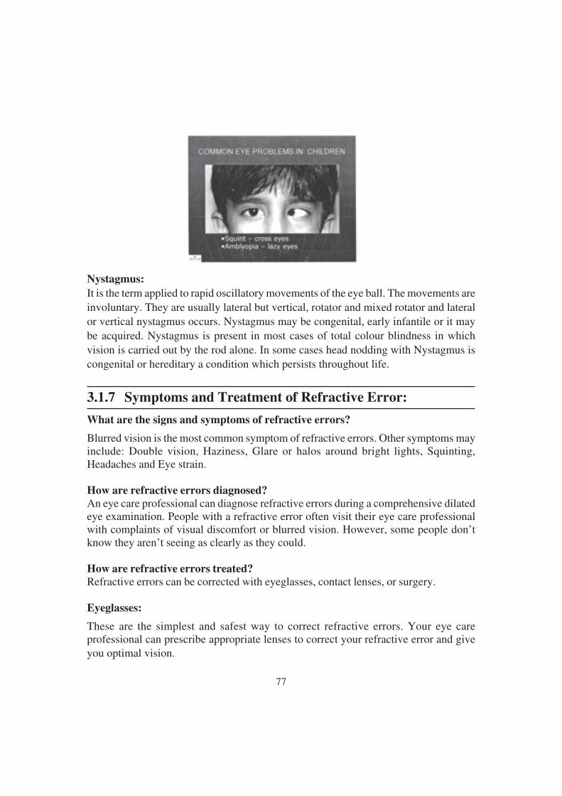
Amblyopia:
Amblyopia is any reduction in visual acuity in one or both eyes. This condition of
mentally shutting out the images of one eye is also known as lazy eye. Amblyopia in
young children may not present a permanent reduction in vision since correction may
be possible. Treatment may consist of glasses, patching, surgery or a combination of
procedures including eye exercises.
Squints (strabismus):
Defects of eye muscles are cause for eye disorder. If one or more muscles which help
rotate the eye become weak or paralysed both eyes then fail to focus on some object at
the same time or same angle. The condition is known as strabismus. It means that in
coordinated action of the muscles cause the failure of the visual axes of the two eyes to
meet at the objective point. Squint is convergent when the eyes turn towards the medial
line; it is divergent if the eyes turn outward. Squint in children may some time lead to
serious visual impairment as the brain tends to accept only the good images of the
weaker or squinted eye. Due to disuse the weak eye may reduce to low vision.
77
Nystagmus:
It is the term applied to rapid oscillatory movements of the eye ball. The movements are
involuntary. They are usually lateral but vertical, rotator and mixed rotator and lateral
or vertical nystagmus occurs. Nystagmus may be congenital, early infantile or it may
be acquired. Nystagmus is present in most cases of total colour blindness in which
vision is carried out by the rod alone. In some cases head nodding with Nystagmus is
congenital or hereditary a condition which persists throughout life.
3.1.7 Symptoms and Treatment of Refractive Error:
What are the signs and symptoms of refractive errors?
Blurred vision is the most common symptom of refractive errors. Other symptoms may
include: Double vision, Haziness, Glare or halos around bright lights, Squinting,
Headaches and Eye strain.
How are refractive errors diagnosed?
An eye care professional can diagnose refractive errors during a comprehensive dilated
eye examination. People with a refractive error often visit their eye care professional
with complaints of visual discomfort or blurred vision. However, some people don’t
know they aren’t seeing as clearly as they could.
How are refractive errors treated?
Refractive errors can be corrected with eyeglasses, contact lenses, or surgery.
Eyeglasses:
These are the simplest and safest way to correct refractive errors. Your eye care
professional can prescribe appropriate lenses to correct your refractive error and give
you optimal vision.
78
Contact Lenses:
It works by becoming the first refractive surface for light rays entering the eye, causing
a more precise refraction or focus. In many cases, contact lenses provide clearer vision,
a wider field of vision, and greater comfort. They are a safe and effective option if fitted
and used properly. It is very important to wash your hands and clean your lenses as
instructed in order to reduce the risk of infection. If you have certain eye conditions you
may not be able to wear contact lenses. Discuss this with your eye care professional.
Refractive Surgery:
It aims to change the shape of the cornea permanently. This change in eye shape restores
the focusing power of the eye by allowing the light rays to focus precisely on the retina
for improved vision. There are many types of refractive surgeries. Your eye care
professional can help you decide if surgery is an option for you.
79
3.2 ppppp Blindess and Low Vision-definition and
Classification
Structure:
3.2.1. Introduction
3.2.2. Objectives
3.2.3. A Brief Historical Review
3.2.4. Definition
3.2.4.1 Blindness
3.2.4.2 Low Vision
3.2.5. Classification
3.2.1 Introduction
It is a true phenomenon that visual impairment tends to evoke more awkwardness from
us than any other disability. For one thing, blindness is visible. The blind person is
usually not one who can easily weave himself into the fabric of a crowd. Unlike many
other exceptional people he stands out. The visually impaired person, however, has a
variety of symbols. Cane, thick or darkened glasses, a guide dog etc.
3.2.2 Objectives
After going through this unit you should be able to:
1. Draw out the position of impairment
2. Know about blind
3. Tell about low vision
4. Also gather knowledge about visual classification
80
3.2.3 A Brief Historical Review:
The history of Special Education in genera] and of visually impaired children in particular
had visualized many ups and downs in its progressive phase of development. Globally
it evolved through the following five stages.
1) Pre-Christian Era-
During this stage, disability was viewed as punishment of past sins and nobody
wanted to interfere in the justice meted out to the disabled persons by God.
2) Christian Era-
In this stage they are protected and pitied to reduce their pains and miseries.
3) Dawn of 19th century-
Institutions were established to provide them separate education.
4) Late 20th century-
The movement started to integrate them in the society.
5) Present age-
The concept of special and integrated system of education has been emerged out
on the basis of needs of disabled persons.
3.2.4 Definitions:
3.2.4.1 Blindness:
The term blindness is used for complete or nearly complete vision loss.
Legal/ medical definitions
The current definition does not make a distinction between those who have “irreversible”
blindness (NO perception of light) and those that have light perception but are still less
than 3/60 in the better eye. The legal definition involves assessment of visual acuity and
field of vision. It is used to determine whether or not an individual qualifies for legal
benefits. The American Medical Association (AMA) proposed this definition in 1934.this
definition is now accepted by American Foundation for the Blind (AFB) and other
Blind Association in different countries.
In India, the broad definition of visual impairment as adopted in the Persons with
Disabilities Act (PWD), 1995 as well as under the National Programme for Control of
Blindness (NPCB) is given as “ Blindness refers to a condition where a person suffers
from any of the following conditions:
81
Total absence of sight or Visual acuity not exceeding 6/60 or 20/200 (Snellen) in the
better eye even with correction lenses or limitation of the field of vision subtending and
angle of 20 degree or worse.”
Educational/functional definition
Many educators are disinterested in the legal or medical definition of blindness. Their
argument that visual acuity is not very accurate prediction of how one will function or
effectively use the remaining sight he has. A common misconception is that legally
blind having absolutely no vision, the vast majority are able to see.
Recognizing the limitations of the legal definition of blindness and partially
sightedness, many have favoured an educational definition.
For educational purpose, “the blind are those who are so severely impaired that
they must be taught to read by Braille, while the partially sighted can read print by
using magnifying glasses or books with large print.”
The educational definition of visual impairment considers the extent to which the
child’s vision affects learning and makes special methods or materials necessary.
Educators often differentiate between blind and low vision students. For deciding the
blindness, the visual acuity as well as field of vision has been considered.
Visual acuity:
It refers to the ability of the eye to see details. The visual acuity for distance is measured
as the maximum distance at which a person can see a certain object, divided by the
maximum distance at which a person with normal eyesight can see the same. Thus a
visual acuity of 6/60 means that the person examined cannot see, at a distance of 6
meters, the object, which a person with normal eyesight would be able to see at 60
meters.
Visual efficiency:
Visual efficiency is the extent to which available vision is used effectively. The term
visual efficiency includes visual acuity at long and at short, control of eye movements,
accommodative ability etc. this also includes the processing ability of the brain. Visual
efficiency is unique to each child. The visual efficiency can be developed by training
but cannot be measured or predicted clinically with any accuracy by medical,
psychological, or educational personnel.
As defined by Barrage, Visual efficiency includes such skills as controlling eye
movements, adapting to the visual impairment, paying attention to visual stimuli and
82
processing visual information rapidly. The fundamental premise in developing visual
efficiency is that children learn to see and must be actively involved in using their own
vision.
Field vision
It refers to the field which both the eyes can easily see in the front. The normal field of
vision is ISO degrees in front of eye.
Visual functioning
The visual functioning refers to the degree to which ability of a person to use vision for
all activities.
3.2.4.2 Low vision
Low vision is a term often used interchangeably with visual impairment and refers to a
loss of vision that may be severe enough to hinder an individual’s ability to complete
daily activities such as reading, cooking, or walking outside safely, while still retaining
some degree of useable vision.
The Person with Disabilities Act, 1995 also recognizes LOW VISION as a category
of disability and defines it as follows:
“Person with low vision means a person with impairment of visual functioning
even after treatment or standard refractive correction but who uses or is potentially
capable of using vision for the planning or execution of a task with appropriate assistive
device.”
This definition is incomplete as it inadvertently omits quantification of the acuity
as well as the field of vision as is done in the case of the WHO definition. It is desirable
to modify this definition and the following quantification should be added:
“Low vision are those who suffer visual acuity between 20/200 to 70/200(Snellen)
or 6/18to 6/60 in the better eye after the best possible correction or a Field of vision
between 20 to 30 degrees.”
In the practice of eye care “LOW VISION” has a specific meaning as defined by
WHO. This is as follows:
“A person with low vision is one who has impairment of visual functioning even
after treatment and/or standard refractive correction, and has a visual acuity of less than
6/18 to light perception, or a visual field of less than 10 degree from the point of fixation,
83
but who uses, or is potentially able to use, vision for planning and/or execution of a
task.” The points emphasize are that there is significantly reduced vision visual
performance is affected but there still is vision that can be used.
For deciding the low vision, the residual vision as well as functional vision has
been considered.
Residual vision
The use of remaining vision by the visually impaired individuals to perform their daily
activities is known as residual vision.
Functional vision
Functional vision is the use of vision for particular activities. Functional visual skills
are required to carry out every day activities.
Central Scotoma
A hazy or dark hole appears in the centre of objects. Causes include macular degeneration
and optic atrophy.
Tunnel vision
Loss of peripheral vision causes a restricted field of vision, Objects in the centre remain
visible. Causes include glaucoma and retinitis pigmentosa.
Accommodation
If while looking at an object situated at infinity, the gaze be transferred to an object near
at hand, some readjustment of the power of the crystalline lens will have to occur,
otherwise the image will fall behind the retina. This adjustment of the power of the lens
is called accommodation.
3.2.5 Classification:
The importance of functional definition lies in the ‘label’ people are given. Someone
with visual acuity of 2/60 can have useful vision, for example, for mobility. However,
he or she will be labelled blind person. The consequence is this person is then treated as
if he or she is blind. This ignores the usable vision. There should be a difference between
legal blindness and functional blindness or low vision. The World Health Organization
uses the following classifications of visual impairment. When the vision in the better
84
eye with best possible glasses correction is: 20/30 to 20/60 : is considered mild vision
loss, or near-normal vision 20/70 to 20/160 : is considered moderate visual impairment,
or moderate low vision 20/200 to 20/400 : is considered severe visual impairment, or
severe low vision 20/500 to 20/1,000 : is considered profound visual impairment, or
profound low vision More than 20/1,000 : is considered near-total visual impairment,
or near total blindness No light perception : is considered total visual impairment, or
total blindness. Blindness is defined by the World Health Organization as vision in a
person’s best eye of less than 20/500 or a visual field of less than 10 degrees
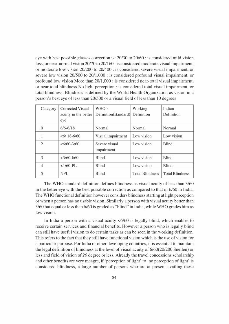
Category Corrected Visual WHO’s Working Indian
acuity in the better Defmition(standard) Definition Definition
eye
0 6/6-6/18 Normal Normal Normal
1 <6/ 18-6/60 Visual impairment Low vision Low vision
2 <6/60-3/60 Severe visual Low vision Blind
impairment
3 <3/60-l/60 Blind Low vision Blind
4 <1/60-PL Blind Low vision Blind
5 NPL Blind Total Blindness Total Blindness
The WHO standard definition defines blindness as visual acuity of less than 3/60
in the better eye with the best possible correction as compared to that of 6/60 in India.
The WHO functional definition however considers blindness starting at light perception
or when a person has no usable vision. Similarly a person with visual acuity better than
3/60 but equal or less than 6/60 is graded as “blind” in India, while WHO grades him as
low vision.
In India a person with a visual acuity <6/60 is legally blind, which enables to
receive certain services and financial benefits. However a person who is legally blind
can still have useful vision to do certain tasks as can be seen in the working definition.
This refers to the fact that they still have functional vision which is the use of vision for
a particular purpose. For India or other developing countries, it is essential to maintain
the legal definition of blindness at the level of visual acuity of 6/60(20/200 Snellen) or
less and field of vision of 20 degree or less. Already the travel concessions scholarship
and other benefits are very meagre, if ‘perception of light’ to ‘no perception of light’ is
considered blindness, a large number of persons who are at present availing these
85
concessions would fall outside the eligibility criteria and thus remain bereft of these
benefits. Alternatively, if these concessions are extended to all the persons with low
vision in the acuity range of 6/18 to ‘perception of light’ as defined by WHO the
appropriate Government may not be able to meet demand due to financial constraints.
For India and other developing countries it is desirable to maintain the definition of
blindness as adopted in the Persons with Disability Act 1995 i.e. visual acuity of 6/
60(20/200) or less and field of vision of 20 degree and less and to consider all the
persons in the range of acuity of 6/18 to 6/60(20/60 to 20/200) as persons with low
vision.
According to above discussion visually impaired are classified as follows-
Partially Sighted
The generally accepted definition for educational purposes now includes:
1. Those students with visual acuity of 20/70 or less in the better eye after the best
possible correction, who can use vision as the main channel of learning.
2. Those students, who in the opinion of eye specialist and educational authorities
will benefit by the use of special facilities provided by the programme for
partially sighted students.
One eyed
The definition of blindness adopted in India excludes people with impairment only in
one eye from the purview of blindness. Generally the impairment of 40% or more is
considered a handicap but in the case of one eyed person it is only 30% according to the
approved definition in medical parlance, a person with one good eye is not a blind person.
Vision loss
It refers to individuals who have trouble seeing, even when wearing glasses or contact
lenses, as well as to individuals who are blind or unable to see at all.
Monocular vision impairment
“Monocular vision impairment” or “Monocular Blindness”; are used both eyes
separately. By using the eyes in this way, as opposed by binocular vision, the field of
view is increased, while depth perception is limited. The fellow eye in these need not
necessarily to be “normal”.
86
Self-reported vision loss
It is determined on an individual basis based on that person’s perceived visual ability
and its effect on daily functioning.
Functional limitation
It refers to the interaction of visual functioning and ability to perform activities of daily
living/instrumental activities of daily living. Common daily activities affected by vision
loss are reading, safe pedestrian travel, self-care, cooking, and recreational activities.
Visual impairment
It is often defined clinically as a visual acuity of 20/70 or worse in the better eye with
best correction, or a total field loss of 140 degrees. Additional factors influencing visual
impairment might be contrast sensitivity, light sensitivity, glare sensitivity, and light/
dark adaptation.
Legal blindness
It is a level of vision loss that has been legally defined to determine eligibility for
benefits. The clinical diagnosis refers to a central visual acuity of 20/200 or less in the
better eye with the best possible correction, and/or a visual field of 20 degrees or less.
Often, people who are diagnosed with legal blindness still have some useable vision.
Total blindness
It refers to an inability to see anything with either eye.
87
3.3 ppppp Demographic Information-NSSO and Census
2011
Structure:
3.3.1 Introduction
3.3.2 Objectives
3.3.3 Demographic Information
3.3.4 Nsso
3.3.5 Census-2011
3.3.1 Introduction:
It is a constitutional obligation of the government to promote the welfare of people by
securing and protecting as possible a social order in which social, economic and political
justice shall inform all the institution of national life. For this reason census is necessary.
There are several estimates about the size of the disabled population in India with
reference to the world situation. 90% of the world’s blind people live in developing
countries. Visually impaired people account for 48.5% of more than 2 core figure in
India. To give them proper prevalence demographic information has great importance.
3.3.2 Objectives:
After studying this unit, you should be able to:
1. Explain the need of census
2. Discuss the role ofcensus 2011
3. Explain the services provided by NSSO
4. Write about demography
3.3.3 Demographic Information
Demography is the statistical study of human population. As a very general science, it
can analyze any kind of dynamic living population, i.e., one that changes over time or
88
space. It encompasses the study of the size, structure, and distribution of these
populations, and spatial and/or temporal changes in them in response to time, birth,
migration, ageing, and death. The word demography taken form Greek word where
demos, means “the people” and -graphy means description or measurement.
Demographics are quantifiable characteristics of a given population. Demographic
analysis can cover whole societies, or groups defined by criteria such as education,
nationality, religion and ethnicity. Educational institutions usually treat demography as
a field of sociology, though there are a number of independent demography departments.
Formal demography limits its object of study to the measurement of population processes,
while the broader field of social demography or population studies also analyzes the
relationships between economic, social, cultural and biological processes influencing a
population. Demographic thoughts can be traced back to antiquity, and are present in
many civilisations and cultures, like Ancient Greece, Ancient Rome, India and China.
There are two types of data collection — direct and indirect — with several different
methods of each type.
Direct methods
Direct data comes from vital statistics registries that track all births and deaths as well
as certain changes in legal status such as marriage, divorce, and migration (registration
of place of residence). In developed countries with good registration systems (such as
the United States and much of Europe), registry statistics are the best method for
estimating the number of births and deaths. A census is the other common direct method
of collecting demographic data. A census is usually conducted by a national government
and attempts to enumerate every person in a country. However, in contrast to vital
statistics data, which are typically collected continuously and summarized on an annual
basis, censuses typically, occur only every 10 years or so and thus are not usually the
best source of data on births and deaths. Analyses are conducted after a census to estimate
how much over or undercounting took place. These compare the sex ratios from the
census data to those estimated from natural values and mortality data. Censuses do
more than just count people. They typically collect information about families or
households in addition to individual characteristics such as age, sex, marital status,
literacy/education, employment status, and occupation, and geographical location. They
may also collect data on migration language, religion, nationality and citizenship. In
countries in which the vital registration system may be incomplete, the censuses are
also used as a direct source of information about fertility and mortality.
Indirect methods
Indirect methods of collecting data are required in countries and periods where full data
are not available, such as is the case in much of the developing world, and most of
89
historical demography. One of these techniques in contemporary demography is the
sister method, where survey researchers ask women how many of their sisters have died
or had children and at what age. With these surveys, researchers can then indirectly
estimate birth or death rates for the entire population. Other indirect methods in
contemporary demography include asking people about siblings, parents, and children.
Other indirect methods are necessary in historical demography. There are a variety of
demographic methods for modelling population processes. They include models of
mortality, fertility, marriage disability, population projections and population momentum.
3.3.4 NSSO
The NSSO (National Sample Survey Organisation), now known as National Sample
Survey Office, is an organization under the Ministry of Statistics of the Government of
India. It is the largest organisation in India conducting regular socio-economic surveys.
It was established in 1950. Employees of NSSO belong to Indian Statistical service and
Subordinate statistical service. NSSO has four divisions: 1. Survey Design and Research
Division (SDRD), 2. Field Operations Division (FOD), 3. Data Processing Division
(DPD) and 4. Co-ordination and Publication Division (CPD)
The Survey Design and Research Division (SDRD)
It is a professional organ of NSSO, mandated to do the job of: Planning of the survey,
Formulation of sample design, Drawing up of schedules of inquiry, Formulation of
concepts and definitions, Preparation of instruction manual for survey field work, Survey
Design and Research Division (SDRD). Training of field and data processing personnel
on survey Methodology Formulation of scrutiny check points Drawing up of tabulation
programme Preparation of survey reports Analysis and presentation of survey results
and Undertaking studies for the improvement of survey methodology SDRD, NSSO is
located at Mahalanobis Bhavan, Kolkata and is headed by an Additional Director General
- a Higher Administrative Grade (HAG) level officer, and has sanctioned strength of
three SAG (Senior Administrative Grade), fifteen JAG (Junior Administrative Grade),
eight STS (Senior Time Scale) and four JTS (Junior Time Scale) level officers of Indian
Statistical Service besides one Deputy Director (Administration) and the supporting
staff members.
The Field Operations Division (FOD)
The one of the four Divisions of the National Sample Survey Office, is responsible for
conducting surveys in the field of Socio- Economic, Industrial Statistics, Agricultural
90
Statistics, Prices, etc. as per the approved programmes. It is also responsible for updating
the frame for conducting Socio-Economic Surveys in urban areas. This Division with
its Headquarters located at New Delhi and Faridabad functions through a network of 6
Zonal Offices, 49 Regional Offices and 116 Sub-Regional Offices spread throughout
the country and have staff strength of about 4000. The Division is headed by Additional
Director General (ADG), an Additional Secretary Level Officer. In Headquarters, four
Deputy Director Generals as well as other officers in the rank of Director/ Joint Director/
Deputy Director/ Assistant Director assist him. All the Zonal Offices are headed by
Deputy Director Generals while the head of Regional Offices are Deputy Director
General/ Director level officers except for Port Blair which is headed by Assistant
Director. Field Operations Division (FOD).
The Data Processing Division (DPD)
This department of NSSO with Headquarters at Kolkata and five Data Processing Centres
outside Kolkata at Ahmadabad, Bangalore, Delhi, Giridih and Nagpur are primarily
mandated to undertake the processing, the tabulation and the dissemination of data
collected through Nation Wide Large Scale Sample Surveys on various Socio-economic
issues conducted by National Sample Survey Office (NSSO) under the Government of
India. This task of transforming large volume of raw data into the final form of Key
Indicators or Estimates in Tabular Format with due process of scrutiny and validation is
carried out by a large number of trained and experienced technical officials in Electronic
Data Processing Cadre under the overall supervision and guidance of the officers of
Indian Statistical Service. The role of DPD starts from the initial stage of formulation
of the Sample Design for NSS Surveys by SDRD wherein apart from providing input
for the formulation it has to undertake the job of sample selection. Later on DPD
undertakes the job of software development for Data Entry, Data Verification, Computer
Edit, Other Data Validations, Howler Checks, Tabulation, etc. DPD also assists the
States by providing complete IT solutions in all their data processing related activities
and also through periodic training/workshop and other interactive methods. With the
advent of Information Technology, DPD is now introducing modern technology to reduce
time and effort in data capturing and transmission besides improving quality of unit
level data. It also helps other countries/organizations in enhancing their capacity building
particularly in data processing/analysis by conducting various need based training
programmes. Main Functions are as follows
1. Selection of samples and preparation of Sample lists. Data Processing Division
(DPD)
91
2. Manual checking of identification particulars and pre data entry scrutiny.
3. In-house development of validation and tabulation software.
4. Data Entry & Verification of filled-in schedules.
5. Validation of data through various stages covering both content check and coverage
check.
6. Preparation of Directory and Multiplier files for estimation of parameters.
7. Tabulation of validated data as per approved tabulation plan.
8. Processing & tabulation of monthly Rural retail price data and release of Quarterly
Rural Price Bulletin.
9. Assistance to state statistical agencies in processing of NSS state sample data.
10. Providing training in application of computer and on data processing.
11. Undertaking special data compilation and tabulation work for: Various analytical
studies, Methodological studies etc. undertaken by NSSO in support of Working
Group/Steering Committee Special users/Committees/Ministries/Depts./Orgs.
12. Organising scrutiny feedback workshop for FOD.
13. Providing technical guidance/assistance to NSS Data Users.
14. Meeting Data requirements (Adhoc tabulation/drawing of Samples etc.) and User’s
queries.
Co-ordination & Publication Division (CPD)
It is located at New Delhi and is responsible for: 1. coordinating the activities of all the
Divisions of NSSO. 2. Dissemination of survey results and analysis through the biannual
technical journal ‘Sarvekshana’ and ‘National Seminars’ to discuss the survey. 3.
Providing technical and secretarial assistance to Steering Committee of National Sample
Surveys. 4. Supplying survey data of various rounds to individuals, researchers, research
institutions and other private and govt. bodies. 5. Liaison with other Departments/
Ministries on various matters concerning NSSO. 6. Providing the technical and secretarial
assistance to DG& CEO of NSSO.
3.3.5 Census 2011
The 15th Indian Census was conducted in two phases, house listing and population
enumeration. House listing phase began on 1 April 2010 and involved collection of
92
information about all buildings. Information for National Population Register was also
collected in the first phase, which will be used to issue a 12-digit unique identification
number to all registered Indians by Unique Identification Authority of India. The second
population enumeration phase was conducted between 9 to 28 February 2011. Census
has been conducted in India since 1872 and 2011 marks the first time biometric
information was collected. According to the provisional reports released on 31 March
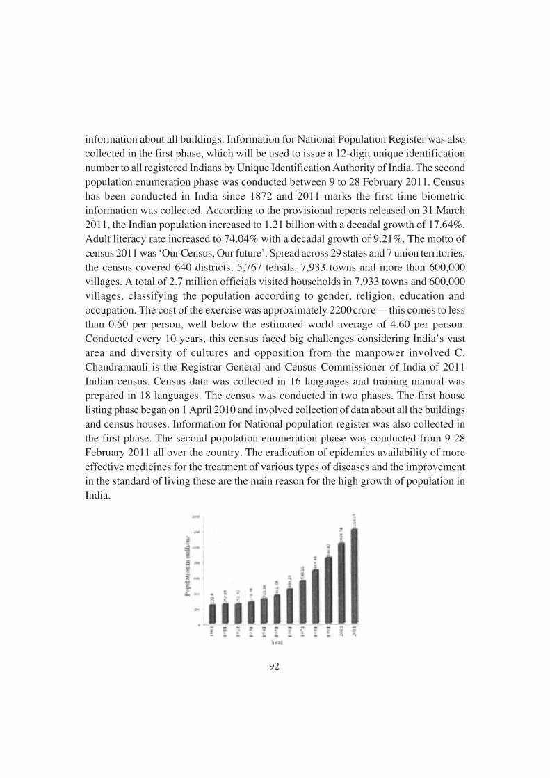
2011, the Indian population increased to 1.21 billion with a decadal growth of 17.64%.
Adult literacy rate increased to 74.04% with a decadal growth of 9.21%. The motto of
census 2011 was ‘Our Census, Our future’. Spread across 29 states and 7 union territories,
the census covered 640 districts, 5,767 tehsils, 7,933 towns and more than 600,000
villages. A total of 2.7 million officials visited households in 7,933 towns and 600,000
villages, classifying the population according to gender, religion, education and
occupation. The cost of the exercise was approximately 2200 crore— this comes to less
than 0.50 per person, well below the estimated world average of 4.60 per person.
Conducted every 10 years, this census faced big challenges considering India’s vast
area and diversity of cultures and opposition from the manpower involved C.
Chandramauli is the Registrar General and Census Commissioner of India of 2011
Indian census. Census data was collected in 16 languages and training manual was
prepared in 18 languages. The census was conducted in two phases. The first house
listing phase began on 1 April 2010 and involved collection of data about all the buildings
and census houses. Information for National population register was also collected in
the first phase. The second population enumeration phase was conducted from 9-28
February 2011 all over the country. The eradication of epidemics availability of more
effective medicines for the treatment of various types of diseases and the improvement
in the standard of living these are the main reason for the high growth of population in
India.
93
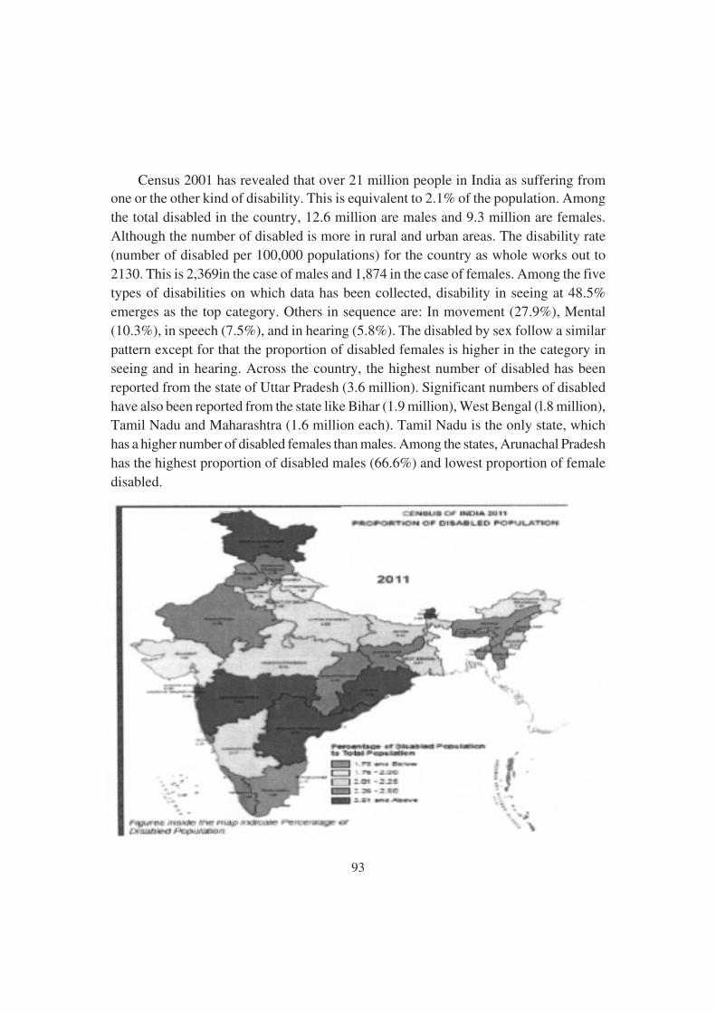
Census 2001 has revealed that over 21 million people in India as suffering from
one or the other kind of disability. This is equivalent to 2.1% of the population. Among
the total disabled in the country, 12.6 million are males and 9.3 million are females.
Although the number of disabled is more in rural and urban areas. The disability rate
(number of disabled per 100,000 populations) for the country as whole works out to
2130. This is 2,369in the case of males and 1,874 in the case of females. Among the five
types of disabilities on which data has been collected, disability in seeing at 48.5%
emerges as the top category. Others in sequence are: In movement (27.9%), Mental
(10.3%), in speech (7.5%), and in hearing (5.8%). The disabled by sex follow a similar
pattern except for that the proportion of disabled females is higher in the category in
seeing and in hearing. Across the country, the highest number of disabled has been
reported from the state of Uttar Pradesh (3.6 million). Significant numbers of disabled
have also been reported from the state like Bihar (1.9 million), West Bengal (l.8 million),
Tamil Nadu and Maharashtra (1.6 million each). Tamil Nadu is the only state, which
has a higher number of disabled females than males. Among the states, Arunachal Pradesh
has the highest proportion of disabled males (66.6%) and lowest proportion of female
disabled.
94
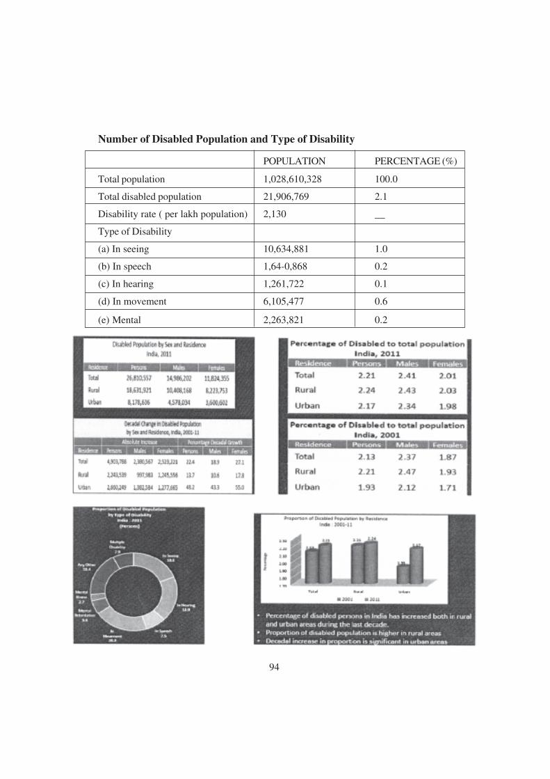
Number of Disabled Population and Type of Disability
POPULATION PERCENTAGE (%)
Total population 1,028,610,328 100.0
Total disabled population 21,906,769 2.1
Disability rate ( per lakh population) 2,130 __
Type of Disability
(a) In seeing 10,634,881 1.0
(b) In speech 1,64-0,868 0.2
(c) In hearing 1,261,722 0.1
(d) In movement 6,105,477 0.6
(e) Mental 2,263,821 0.2
95
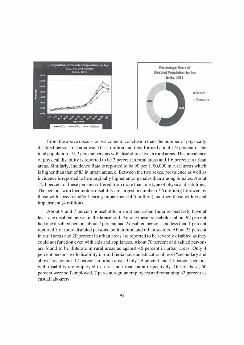
From the above discussion we come to conclusion that- the number of physically
disabled persons in India was 16.15 million and they formed about 1.9 percent of the
total population. 74.3 percent persons with disabilities live in rural areas. The prevalence
of physical disability is reported to be 2 percent in rural areas and 1.6 percent in urban
areas. Similarly, Incidence Rate is reported to be 90 per 1, 00,000 in rural areas which
is higher than that of 83 in urban areas, c. Between the two sexes, prevalence as well as
incidence is reported to be marginally higher among males than among females. About
12.4 percent of these persons suffered from more than one type of physical disabilities.
The persons with locomotors disability are largest in number (7.6 million); followed by
those with speech and/or hearing impairment (4.5 million) and then those with visual
impairment (4 million).
About 9 and 7 percent households in rural and urban India respectively have at
least one disabled person in the household. Among these households, about 92 percent
had one disabled person, about 7 percent had 2 disabled persons and less than 1 percent
reported 3 or more disabled persons, both in rural and urban sectors. About 25 percent
in rural areas and 20 percent in urban areas are reported to be severely disabled as they
could not function even with aids and appliances. About 70 percent of disabled persons
are found to be illiterate in rural areas as against 46 percent in urban areas. Only 4
percent persons with disability in rural India have an educational level “secondary and
above” as against 12 percent in urban areas. Only 29 percent and 25 percent persons
with disability are employed in rural and urban India respectively. Out of these, 60
percent were self employed, 7 percent regular employees and remaining 33 percent as
casual labourers.
96
3.4 ppppp Importance of Early Identification and
Intervention
Structure:
3.4.1 Introduction
3.4.2. Objectives
3.4.3. Early Identification of Vision Problem
3.4.3.1 Importance of Vision And Learning About Vision Loss
3.4.3.2 Symptoms of Vision Problems
3.4.4. Early Intervention Programmes
3.4.4.1 Meaning of Early Intervention
3.4.4.2 Deficit Model
3.4.4.3 Classification of Intervention Programme
3.4.5. Importance
3.4.1 Introduction
‘Catch them young and teach them well’ is the slogan reflected all over the world for
the education of children with special needs. There are a lot of advantages over
identification of children with visual problems at their young ages. Most of the eye
problems are medically treated and cured. After medical correction, most of the children
would see normally. Some medically untreatable conditions of eye defect lead to
blindness. However, a very few children would suffer from total blindness and most of
the children may have residual vision. Therefore early identification of child with visual
problems will help the child to go for medical and educational interventions. In this
unit a detailed discussion is held on early identification, intervention and their importance.
3.4.2 Objectives
After studying this unit, you should be able to:
1. Explain the early identification of child with visual impairment
97
2. List out the factors and behavioural indicators for vision-loss
3. Describe the early intervention programme
4. Able to write about importance of identification and intervention
3.4.3 Early Identification:
Early eye- examination is of utmost importance. All eye surgeons have been exposed to
the frustration of an adult when informed that nothing can be done to improve vision in
the lazy eye. This can be prevented to a great extent if it can be detected around the age
of 3-4 years. It has been observed that 24% have refractive errors and many of these
errors are present at birth and go unnoticed for a long time. Early identification is the
step to set the intervention programmes.
3.4.3.1 Importance of Vision and Learning about Vision Loss
Although every one of our senses plays a role in early development, vision certainly
seems to lead the way. Early bondage of the child with parents is based on the child’s
ability to make eye contact and sustain a gaze with his parents, response to their voices
by gurgling and cooing. An infant tries to move because he sees something. He learns
that things and people exist in the world primarily because he sees and hears them
come and go. He visually tracks an object he pitches to the ground. He can inspire his
parents to play with them by making eye contact, the earliest form of conversation. He
learns about size, shape, and colour, function of objects, social interactions and so much
more just by looking at the world at work. Every child with or without a disability
should have regular and periodic vision checking. If the child is severely disabled, this
can be even more important since their other senses may not be as useful in compensating
for what they miss visually. In fact this is so important that schools should have vision
screening at regular intervals throughout the remainder of the child’s educational career.
Factors And Behavioural Indicators For Vision Loss
A child is at risk for vision loss if the child encounters the following factors:
• Family history of vision loss
• Malformation of the eye
• Prematurity and low birth weight
• Birth trauma
98
• Congenital viral or bacterial
• Meningitis, Encephalitis, Hyperthyroidism, Microcephaly
The following behaviours indicate the child’s vision loss
• The child does not have eyes that look typical
• The child does not recognize caregivers’ faces or smile in response to their smiles
• He does not get excited when he sees other familiar object
• The child’s eyes do not move together when following object
• The child may hold an object very close to his eyes
• The child may over reach or under reach for objects
3.4.3.2 Symptoms of Vision Problem
Young children with vision problems often do not know that the way they see the world
is not the way everyone sees it.
1. Permanent vision loss
2. Learning difficulties
Any changes in the appearance of eyes or vision should be investigated further.
Signs to Watch Out for Early Detection (As Adopted by UNICEF)
General symptoms that may occur from birth
• Squints or blinks when looking at something
• The eyes are crossed
• Favours one eye more than the other when looking at an object
• One or both of the eyes turn in or out
• The pupils are hazy
• Eyes are tearing excessively, they are red or eye-lids are encrusted with matter
• Turns or tilts head abnormally
• Has frequent or persistent sites
May occur from 0-3 Months
• Child does not follow an object in his visual field. Child does not play with his
hands.
99
May occur from 3-6 Months
• Child does not reach for toys in his visual field
• Child does not make eye contact when being fed
• Child does not visually inspect object
May occur from 6-9 Month
• Child’s mother skills such as rolling over, sitting or crawling
• Child does not appear to discriminate between similar objects or people
• Child does not pick up small objects successfully
May occur from 9-12 Months
• Child shuts or covers one eye when focusing
• Child holds playthings very close to eyes
• Child bumps into large objects when crawling
• Child rubs his eyes excessively
• Child does not attempt to grasp spoon
• Child does not appear to notice
May occur from 1-2 Years
• Walking is delayed
• Bumps into large objects
• Child is not interested in playing
• Child not interested in picture book
• Child holds book very close to eye
• Child is afraid to walk
• Child is clumsy and awkward for his age
• Child pays more attention to sound
May occur from 2-5 Years
• Stumbles over small objects
100
• Not interested in task that require Sustained visual concentration
• Complains of headaches, burning, itching of eyes
• Cannot see distant things clearly
• Does not notice colour difference
May occur at School Age
• Short attention span and daydreams
• Uses unusual or fisted pencil grasp, frequently breaking pencil
• Difficulty in remembering what is read
• Loses place while reading
• Cover one eye
• Very hard to read hand writing
• Skips words and re-read
• Difficulty in sequential concepts
• Poor eye hand coordination
• Gets easily frustrated
3.4.4 Early Intervention
The term early intervention refers to services given to very young children with visual
problems, generally from birth until the child turns three. For this reason these
programmes are sometime called “birth to 3” or “zero to 3”. Services included medical
treatment, follow-up service, visual efficiency development, training on daily living
skills and mobility etc.
Deficit Model
Current practice of early intervention is viewed as a deficit model. That is strategies
address deficit of vision. The time to intervene is before the delay occurs. The goal is to
prevent the delay if possible. That is why the identification of a vision problem as early
as possible is essential. As soon as visual problem is identified the sooner intervention
can be provided, the more likely it is that delays can be prevented.

101
Classification Of Intervention Programmes
Early intervention programmes are classified as vision screening, medical intervention
and educational intervention. All these programmes go simultaneously for prevention
of eye deficit, restoration of visionc development of vision effciency.
Vision screening
All children should be screened for possible vision problems, especially those under
age of three with a suspected or identified risk factor, regardless of severity.
The initial screening should be conducted by trained personnel on vision screening
procedures. The trained personnel may be low vision specialist, special teacher,
rehabilitation workers and village nurses. Identified cases of visual problems are referred
to the medical personnel who would attend to thorough eye examination.
Medical intervention
There are many possible defects or diseases of the visual system, but fortunately many
of them appear after the first few years of life. There are still many malformations,
defects, diseases, infection and disorders that can affect the visual system in infants and
toddlers as it is presumed that medical follow up to screening will identify and prescribe
treatment. The medical professionals will take care of treatment aspects for the diseases
and defects of the eyes.
Educational intervention
Educational intervention includes the preschool training such as development of daily
living skills, mobility skill, visual skill etc and placement of the child into formal school
system. The trained teacher or rehabilitation worker who is qualified on visual impairment
takes the child with visual impairment for training on various skills required by the
child. He/she also provides counselling for the parents, family members, relative and
neighbours about the development of the child with visual impairment and their role on
caring the child.
3.4.5 Importance
Early Identiflcation
Early identification is extremely important because early intervention will be most
effective. Sometimes it is unclear whether a child has a vision problem or not. Physical
signs of vision problems include eyelids drooping over one or both eyes, or eyelids that
102
do not completely cover the eyes when the child closes them. If a child has a clear
squint, has jerky eye movements, or has eyes that do not move together, parents should
see a paediatric ophthalmologist. Other signs include: Not looking at others in the eyes,
Reaching in front of or beyond an object, Holding objects very close or very far to see
them, Turning or tilting his head when he uses his eyes, Continuously pushing or poking
his eyes, Looking above, below or off to one side of an object, rather than directly at it
Bumping into objects and having a lot or trouble seeing at night, Feeling for objects on
the ground instead oflooking with her eyes. After the identification of visually impaired
students under these, parents should begin working with an early childhood
interventionist. Young children who are visually impaired are eligible for early
intervention services, which can help a family through the child's first few years oflife.
Early intervention for students with visual impairment is vital in enhancing social,
physical, and intellectual development. When a child who is over three, he will qualify
for special education services if the visual impairment impacts his education. Parents
should contact their district's special education office to locate services for their child.
A child with visual impairment may qualify for services from teachers of students with
visual impairment, an orientation and mobility specialist, a physical therapist, a speech
therapist, or a psychologist, depending on individual needs. Children with visual
impairment should also be provided with modifications and accommodations in an
inclusive classroom.
Early Intervention
Research has shown that the time between birth and age of months is a critical
developmental period in a child's life. These months offer a window of opportunity that
will not be available later. Early intervention programes minimize and in some cases
prevent delays in development of infants and toddlers with disabilities. High quality
early intervention programes for vulnerable infants and toddlers can reduce the incidence
of future problems in their learning, behaviour and health status. They can decrease the
need for special education and related services when a child enters school, and increase
independence. There is an urgent and substantial need to identify as early as possible
those infants and toddlers in need of services to ensure that intervention is provided
when the developing brain is most capable of change. Children whose special needs are
identified and addressed during these crucial early years have a greater chance ofreaching
their full potential. Intervention is likely to be more effective and less costly when it is
provided earlier in life rather than later.
103
3.5 p p p p p Functional Assessment Procedures
Structure:
3.5.1 Introduction
3.5.2 Objectives
3.5.3 What Is Functional Assessment
3.5.4 Functional Assessment Methods
3.5.5 Functional Assessment Procedures For Visually Impaired
3.5.6 Child
3.5.7 Importance
3.5.8 Activities
3.5.1 Introduction
One of the key factors in achieving safety, permanency and well being is the creation of
an effective assessment process. The assessment of needs is, in fact, so critical to the
child and family's well being and dynamic in its focus that no single form, tool or single
event can adequately support it. Needs assessment is a process that continues throughout
the life of each case. Assessment tools are merely instruments that are useful in bringing
attention to issues that need particular focus and in identifying current strengths, needs
and functioning for purposes of decision-making.
3.5.2 Objectives
After going through this unit, you shoul be able to
1. understand and explain functional assessment
2. State the importance of of asessment
3. List out the activities of functional vision assessment
4. Understand about helpers for doing assessment
104
3.5.3 What Is Functional Assessment?
A set of procedures to identify the causes of a maladaptive or socially inappropriate
behaviour and reduce it through teaching replacement behaviours instead of suppressing
it through punishment. The body of empirical and scientific literature which supports
these methods is found in the field of applied behaviour analysis. Within functional
assessment methodology the causes are sought in the immediate environment and the
learning history of the individual. Causes of the maladaptive behaviour based upon
intrapsychic variables or psychodynamic processes are given little attention. The outcome
of the assessment is an analysis of the way the person learned the maladaptive and how
it is presently supported or maintained in the present learning environment. Functional
assessment does not emphasize a search for a diagnosis or classification of symptoms
according to psychodynamic processes. Instead, the purpose of the assessment is to
classify the maladaptive behaviour by its function (cause) and then select treatments or
interventions which are effective in reducing behaviour in that functional category.
Consequently, treatments or interventions are classified by functional categories and
not by form of the maladaptive behaviour. In the field of education many practitioners
choose interventions or treatments based upon topography or form of the behaviour
instead of the function. As a result some recommended interventions actually strengthen
the maladaptive behaviour instead of reducing it. This situation can make school and
their personnel vulnerable to successful legal, administrative and ethical challenges.
3.5.4 Functional Assessment Methods
There are three specific functional assessment methods: (a) Direct Observation, (b)
Informant Methods and (c) Functional Analysis. The terms "functional assessment"
and "functional analysis" are sometimes thought to be the same thing but they are not;
a functional analysis is one specific type of functional assessment.
1. Direct Observation
For direct observational methods, an observer would watch the client engage in activities
within their natural environment. When the challenging behaviour occurs, the observer
would record what happened just before it, what happened just after it and also take
notes on what they perceive to be the potential cause of the behaviour. This method is
used to develop a hypothesis about the function of the behaviour. The terms used for
this method include: Direct Observation. Descriptive Functional Behaviour Assessment.
105
2. Informant Methods
The informant method involves interviews and questionnaires that can be completed by
the client, their parents, staff members, teachers etc. These interviews would be used to
identify what is happening before the behaviour occurs and then what happens after the
behaviour. Just like direct observation, this method is also used to develop a hypothesis
for the function of the behaviour. The terms used for this method include: Indirect
Methods, Indirect Functional Behaviour Assessment, Informant Methods
3. Functional Analysis
This method, functional analysis involves practitioners deliberately changing what
happens before and/or after the behaviour in an effort to test what mIght be causing the
behaviour. Unlike the other two methods that are used to create a hypothesis, this method
is used to actually test the hypothesis and is the only method that can truly predict when
the behaviour will occur.
3.5.5 Functional Assessment Procedures For Visually Imp Aired
Child
Like other disabilities in case of visual impairment, functional assessment is necessary
to improve their remaining functional vision. Functional vision is the ability to use
vision to perform desired tasks. Because of impairment in the eye and other parts of the
system, low- vision children will not learn visually without intervention and help.
Selection of instructional programmes and techniques requires a thorough assessment
and understanding of child's capabilities.
The rocess offunctional assessment should be done-
1) At the age of three months of a baby if the child is not attracted by the light or not
move his/ her neck to see the colourful objects, the parents should report that the
child may be visually impaired.
2) The child has not attracted the colourful toys.
3) If the child complairs about headache, body ache etc
4) The child may complain, to the parent, pain in eye at early stage.
5) At the time of playing the visually impaired child may not hold the ball as easily
as the normal child.
106
6) The normal functions of day to day activities are much more affected — reading,
writing, walking etc.
7) It is always seen searching objects at any time in his/her working expenence.
Who conducts a functional vision assessment?
A functional assessment is typically conducted by a teacher certified in the area of
visual impairment. The specialist is a certified teacher of the visually impaired, trained
to evaluate how a child utilizes vision. The vision specialist will measure and observe
the visual methods a child uses throughout a routine day and will speak with parents,
teachers and other caregivers who know the child well. Information about how the child
uses vision, the conditions and purpose of use, is essential and will be utilized in the
functional vision assessment report. The vision specialist will review records and may
talk to the eye doctor to learn more about the child's visual condition.
3.5.6 Importance
1. It helps to determine the current visual functioning level of the person.
2. It helps to determine the extent of visual stimulation and instruction needed to
help the person make optimum use of remaining vision.
3. It enables the person to use his limited vision in the highest potential
4. It helps to plan the person's mobility training programme
5. It helps in decisions regarding the use of visual stimulation materials
6. It helps to decide upon the nature of the primary reading medium
7. I t enables one to decide on the type of devices needed by the person
3.5.7 Activities
Vision is functional if a child is able to utilize visual information to plan and carry out
a task. A functional vision assessment measures how well a child uses vision to perform
routine tasks in different places and different material throughout a day. Functional
vision assessment has two types of activities like-I. Optical functioning and 2. Perceptual
functioning. Optical functioning may consist of seven activities like- visual awareness,
visual attention, visual fixation, visual focus, visual fusion, visual tracking and visual
scanning. On the other hand perceptual functioning consists of eight activities like-
107
visual discrimination, figure ground, visual memory, visual closure, spatial relation,
mobility, visual motor coordination and form constancy.
VISUAL SKILLS
OPTICAL FUNCTIONING :
Visual Awareness:
To find out the ability of the child to identify an object
Visual Attention:
To find out the ability of the child to attend to the objects.
Visual Scanning:
The ability to search for a particular object among other objects
Visual fixation:
The ability of the child to fix the eyes on the object.
Visual focus:
The ability of the child to see a known object at various distances.
Visual fusion:
The ability of the child to see the object as one.
Visual Tracking:
The ability to follow moving objects.
PERCEPTUAL FUNCTIONING:
Visual Discrimination:
The ability of a person to distinguish different objects on the basis of their colour shape
or size
Visual Figure-Ground Discrimination:
The ability to isolate a particular stimulus from the background, i.e. seeing the distinctive
features of an object
108
Visual Memory:
Ability to store and recall past experiences and integrate them into new ones.
Visual Closure:
Ability to perceive a total picture or object when onlypart of it is visible/available
Form Constancy:
Ability to perceive the same objects at different angles
Visual motor coordination:
This refers to the child's ability to perform a task using eyes and hand /foot in harmony.
It consists of two types these are follows-
Eye-Hand Coordination:
Ability to use hands and eyes in harmony.
Eye-Foot Coordination:
Ability to perform a task using both eyes and foot in co-ordination.
Visual spatial relation:
This refers to the child's ability to identify spatial concepts like direction, distance etc.
Visual mobility:
This refers to the child's ability to indentify right and left concepts through movement.
2.6 Summary
The process of seeing mainly organized through the parts of eye and image sharpness
depends on the functions of the parts of the eye. But sometimes this process is aelayed
or faces problems due to some errors which come under eye disorder. These refractive
errors are curable if they are identified early and also get the treatment properly these.
All these happen if people are aware.
Problem in eye does not come under blindness; blind are those who face problems for
sight disturbance. According to PWD act and WHO’s definition visual impairment has
many classification. And these divisions depend on visual acuity, field vision, visual
effiency, functional vision etc.
109
The NSSO conducted the 15th round of a nation-wide comprehensive survey of dis-
abled persons during 1st April 2010 to 28th February 2011. The survey arrived at an
estimate of 21 million persons having at least one or the other disability, which consti-
tuted 1.9 percent of the total population ofj50 million. The survey revealed that popula-
tion of the visually impaired in India at 850 million level of population.. Among them
minimum of them( come under prevalence facilities.
Without early detection and treatment children's vision problems can lead to permanent
:n vision loss and learning difficulties. So it is very much essential to prevent vision
loss and preserve eyesight. Early intervention programme helps them for guiding properly
and also helps them in their future establishment.
The development of visual ability is not innate or automatic. It requires stimulation and
motivation to use vision in a variety of environments. A visually impaired child owing
to uncertain and frustratingly limited vision may not always be motivated to use his
vision to control and manipulate his environment to achieve specific goals. As a result
he losles the ability to use whatever vision he has efficiently and purposefully. But
modern researchers have proved that systematic techniques can facilitate the emer-
gence of visually guided behaviour in some children and maximize the use of vision in
others. On the other hand functional assessment procedures also measures how well a
child uses vision to perform routine tasks in different places and with different materi-
als throughout the day.
2.7 Unit exercise:
1. Draw a diagram of eye and label
2. Write, about refractive errors
3. How do we see?
4. Who are blind?
5. Write the pauses of low vision.
6. Classify the visual impairment.
7. Write short note on:
(a) Visual acuity (b) visual efficiency (c) field vision
110
8. What is NSSO?
9. How does it function?
10. Write the salien .......... do published in 2011
11. Explain the inportance of census
12. Prepare a list of symptoms of visual problems.
13. What is meant by early intervention?
14. Who should conduct the vision screening to whom and when?
15. What is functional assessment?
16. List out the activities offunctional assessment procedures.
17. How functional assessment help/problem child?
2.8 Suggested Reading:
Smith, M.R. And Neisworth, J.T(1975). THE EXCEPTIONAL CHILD: A
FUNCTIONAL APPROACH. New York: McGraw-Hill Book Coy
Kundu, C.L (2000). STATUS OF DIS ABILITY IN INDIA-2000 New Delhi RCI
Punani, B and Rawal, N. HAND BOOK : VISUAL HANDICAP
M. Dash, 2003, EDUCATION OF EXCEPTIONAL CHILDREN
Ishtiaq Harder, VISUALLY HANDICAPPED CHILDREN
Dr. Jose Murickan, S.J. George Kutty , PERSONS WITH DISABILITIES IN SOCIETY
www.avclinic.com
www.smith.edu
Preston, Samuel, Patrick Heuveline, and Michel Guillot. 2000. DEMOGRAPHY:
MEASURING AND MODELING POPULATION PROCESSES.
Ben J. Wattenberg (2004), HOW THE NEW DEMOGRAPHY OF DEPOPULATION
WILL SHAPE OUR FUTURE.
Punani, Band Rawal, N. HAND BOOK, VISUAL HANDICAP
Manivannan, M. (2005) EDUCATING VISUALLY IMPAIRED CHILDREN
111
Dharial, T.D (2012). STATUS OF DIS ABILITY IN INDIA-2012 New Delhi RCI
www.google.com
http://www.tsbvi.edu/outreach/summer01/early-id.htm
Mani, M.N.G (1986). SEE WITH BLIND
Mason, S. A. & Egel, A. L. (1995). WHAT DOES AMY LIKE? USING A MINI-
REINFORCER ASSESSMENT TO INCREASE STUDENT PARTICIPATION IN
INSTRUCTIONAL ACTIVITIES. TEACHING EXCEPTIONAL CHILDREN,
O'Neill, R. E., Homer, R. H., Albin, R. W., Storey, K., & Sprague, J. R. (1997). FUNC-
TIONAL ASSESSMENT AND PROGRAM DEVELOPMENT: A PRACTICAL
HANDBOOK.
http://www.educateautism.com/functionalassessment/ fbacorrelationcausation.
http://www.educateautism.com/behaviouralprinciples/
functionsofbehaviour. html
http://www.educateautism.com/behaviouralprinciples/
positivereinforcement. html
Related Documents