1 3'-terminal Overhangs Regulate DNA Double-Strand Break Processing in Escherichia coli Edyta Đermić, *,§ Davor Zahradka, †, § Dušica Vujaklija, † Siniša Ivanković ‡ and Damir Đermić † * Department of Plant Pathology, Faculty of Agriculture University of Zagreb, 10 000 Zagreb, Croatia † Division of Molecular Biology, Ruđer Bošković Institute, 10 000 Zagreb, Croatia ‡ Division of Molecular Medicine, Ruđer Bošković Institute, 10 000 Zagreb, Croatia § These authors contributed equally to this work. G3: Genes|Genomes|Genetics Early Online, published on July 14, 2017 as doi:10.1534/g3.117.043521 © The Author(s) 2013. Published by the Genetics Society of America.

Welcome message from author
This document is posted to help you gain knowledge. Please leave a comment to let me know what you think about it! Share it to your friends and learn new things together.
Transcript
1
3'-terminal Overhangs Regulate DNA Double-Strand Break
Processing in Escherichia coli
Edyta Đermić,*,§ Davor Zahradka,†, §
Dušica Vujaklija,†
Siniša Ivanković‡ and Damir Đermić†
* Department of Plant Pathology, Faculty of Agriculture University of Zagreb,
10 000 Zagreb, Croatia
† Division of Molecular Biology, Ruđer Bošković Institute, 10 000 Zagreb,
Croatia
‡ Division of Molecular Medicine, Ruđer Bošković Institute, 10 000 Zagreb,
Croatia
§ These authors contributed equally to this work.
G3: Genes|Genomes|Genetics Early Online, published on July 14, 2017 as doi:10.1534/g3.117.043521
© The Author(s) 2013. Published by the Genetics Society of America.
2
Running title: 3’ tails regulate DSB processing
Keywords: DNA degradation; exonuclease activity; RecA protein; single-
strand specific exonucleases; recB1080 mutant
Correspondence:
Damir Đermić
Division of Molecular Biology
Ruđer Bošković Institute
Bijenička 54, 10 000 Zagreb, Croatia
Fax: +385 1 4561 177
e-mail: [email protected]
3
ABSTRACT
Double-strand breaks (DSBs) are lethal DNA lesions, which are repaired by
homologous recombination in Escherichia coli. To study DSB processing in
vivo, we induced DSBs into the E. coli chromosome by gamma irradiation
and measured chromosomal degradation. We show that the DNA degradation
is regulated by RecA protein concentration and its rate of association with
ssDNA. RecA decreased DNA degradation in wild-type, recB and recD strains,
indicating that it is a general phenomenon in E. coli. On the other hand, DNA
degradation was greatly reduced and unaffected by RecA in the recB1080
mutant (which produces long overhangs) and in a strain devoid of four
exonucleases that degrade a 3’ tail (ssExos). 3’-5’ ssExos deficiency is
epistatic to RecA deficiency concerning DNA degradation, suggesting that
bound RecA is shielding 3’ tail from degradation by 3’-5’ ssExos. Since 3’-tail
preservation is common to all these situations, we infer that RecA
polymerization constitutes a subset of mechanisms for preserving the
integrity of 3’ tails emanating from DSBs, along with 3’ tail’s massive length,
or prevention of their degradation by inactivation of 3’-5’ ssExos. Thus, we
conclude that 3’ overhangs are crucial in controlling the extent of DSB
processing in E. coli. This study suggests a regulatory mechanism for DSB
processing in E. coli, wherein 3’ tails impose a negative feedback loop on
DSB processing reactions, specifically on helicase reloading onto dsDNA
ends.
4
INTRODUCTION
A double-strand break (DSB) is an adverse DNA lesion, which has to be
repaired in order for a cell to survive. DSBs are repaired in all living
organisms by either mutagenic nonhomologous end joining or by much more
universally distributed and precise homologous recombination (HR). During
HR a single 3'-terminated strand is produced from each of two double-strand
DNA (dsDNA) ends of a DSB by a process called DNA end resection, wherein
a combination of helicase and nuclease activities result in degradation of
complementary 5'-terminated strands (Symington 2014). The 3'-end
overhangs emanating from a DSB are bound by a recombinase protein, thus
creating the central recombination intermediate, the nucleoprotein filament.
A recombinase nucleoprotein filament searches for an intact homologous
sequence and invades it, hence restoring continuity of genomic information.
Since evolutionarily conserved recombinase proteins (RecA, RadA, Rad51
(Dmc1) from bacteria, archaea and eukaryotes, respectively) have a lower
affinity of binding to single-strand DNA (ssDNA) than their cognate ssDNA-
binding proteins SSB/RPA, a recombination-mediator class of proteins
(RecBCD and RecFOR proteins in bacteria and BRCA2, PALB2 and Rad52 in
eukaryotes) facilitates recombinase polymerization on ssDNA (Zelensky et al.
2014).
In addition to its role in HR, the RecA nucleoprotein filament in Escherichia
coli serves as a coprotease to promote autocatalytic cleavage of the LexA
5
repressor leading to induction of a SOS response (Little 1991). RecA also
activates a mutagenic DNA polymerase V during SOS induction (Shinagawa
et al. 1988).
In bacteria, both helicase and nuclease activities for DNA end resection are
provided by the functionally related RecBCD, AddAB and AdnAB enzymes
(Wigley 2013). In E. coli the RecBCD enzyme binds to a flush dsDNA end and
unwinds a duplex molecule with its fast and processive helicase activity
(Dillingham and Kowalczykowski 2008). Both of the unwound strands are
degraded by a single nuclease center of the enzyme (residing in its RecB
subunit) (Yu et al. 1998) until the enzyme encounters a regulatory
octanucleotide sequence designated Chi. Interaction with Chi changes
RecBCD’s behavior so that it ceases degradation of the 3’-terminated strand,
while continuing DNA unwinding and degradation of the 5’-terminated strand
(Anderson and Kowalczykowski 1997a). Also, the Chi-modified RecBCD starts
facilitating RecA polymerization onto the post-Chi 3’ strand, hence producing
a RecA-nucleoprotein filament (Anderson and Kowalczykowski 1997b). In
this way Chi switches RecBCD enzyme degradation activity into a repair
activity.
DSB repair in E. coli is active even in the absence of RecBCD due to RecQ
helicase unwinding of duplex DNA, RecJ exonuclease trimming of ssDNA tails
(ssExo) from 5’ end, and RecFOR proteins mediating RecA polymerization
onto the unwound 3’ overhangs. This pathway is operative only when ssExos
6
that degrade 3’-terminated overhangs (e.g. Exonuclease I (ExoI) and SbcCD)
are inactive, which enables stabilization of the recombinogenic substrate
(Persky and Lovett 2008).
There are some mutants that show only partial function of RecBCD, yet are
DSB repair proficient as well. When RecBCD lacks its RecD subunit, the
resulting RecBC enzyme shows reduced helicase rate and processivity
compared to RecBCD, and is completely devoid of nuclease activity and Chi
interaction (Dillingham and Kowalczykowski 2008). In the recD mutant
RecBC enzyme unwinds duplex DNA and constitutively loads RecA protein
onto the unwound 3’ tail (Churchill et al. 1999), while its 5’ complement is
trimmed by RecJ and Exonuclease VII (ExoVII) ssExos (Đermić 2006; Đermić
et al. 2006).
The recB1080 mutation renders the RecBCD enzyme deficient in nuclease
and RecA loading activity, whereas the enzyme’s binding to DNA activity as
well as rate and processivity of its helicase activity is unaffected (Yu et al.
1998; Anderson et al. 1999). In vitro, the RecB1080CD enzyme unwinds linear
DNA duplex, producing full length, RecA-free ss tails in the presence of SSB
(Yu et al. 1998; Anderson et al. 1999). In the recB1080 mutant, the 5’-
ended tail is clipped by RecJ and ExoVII ssExos, while its 3’ complement is
covered with RecA protein with the help of RecFOR proteins (Jockovich and
Myers 2001; Ivančić-Baće et al. 2003; Ivanković et al. 2017). The recB1080
mutant is recombination proficient; however the efficiency of HR depends on
7
trimming of its excessively long 3’ tails, and is lower than in wild-type
bacteria (Ivanković and Đermić 2012; Ivanković et al. 2017). HR is not
regulated by Chi in a recB1080 mutant (Jockovich and Myers 2001).
An E. coli mutant lacking RecA protein is HR deficient and is therefore
unable to repair DSBs, which is reflected in its extreme sensitivity to various
genotoxic agents and reduced viability (~60% of the wild-type) (Kuzminov
1999). This relatively substantial decrease in viability is due to the
exonuclease (ExoV) activity of the RecBCD enzyme (Miranda and Kuzminov
2003), which is actually unregulated, or “reckless”, in a recA mutant
(Miranda and Kuzminov 2003). In this mutant RecBCD degrades DNA
duplexes with free dsDNA ends (either damaged or linear molecules
introduced exogenously) so heavily that a fraction of the recA mutant
population is devoid of a chromosome due to its complete degradation
(Capaldo and Barbour 1975; Skarstad and Boye 1993).
In this study we have characterized processing of DSBs introduced
synchronously into radioactively-labeled E. coli chromosome by gamma
irradiation. Ionizing radiation, such as gamma rays, induce DSBs in DNA by
either directly transferring energy to it or indirectly, by creating reactive
oxygen species in the cell’s cytoplasm which then damage DNA; during
repair of these clustered lesions, DSBs are produced (Bresler et al. 1979;
Wallace 1998). DSB processing was assessed by measuring degradation of
the fragmented chromosome. We show that DNA degradation in gamma-
8
irradiated E. coli is inhibited by RecA protein concentration and its ssDNA
binding affinity. In fact, we show that binding of RecA to ssDNA is sufficient
to protect DNA duplex from degradation. However, DNA degradation as well
as its RecA dependence was diminished in cells with preserved 3’ overhangs.
Our results suggest that 3’ overhangs exert their influence on DSB
processing by inhibiting helicase reloading onto dsDNA ends.
MATERIALS AND METHODS
Bacterial strains, media, growth conditions, phage plating and
microscopy. We used AB1157, a standard HR-, DNA repair- and DNA
degradation-proficient strain (Bachmann 1972). Its derivatives were
constructed by P1 transduction, as described earlier (Miller 1992), and are
listed in Table 1. Bacteria were grown at 37° in LB medium and on LB agar
plates (Miller 1992), supplemented with antibiotics when required.
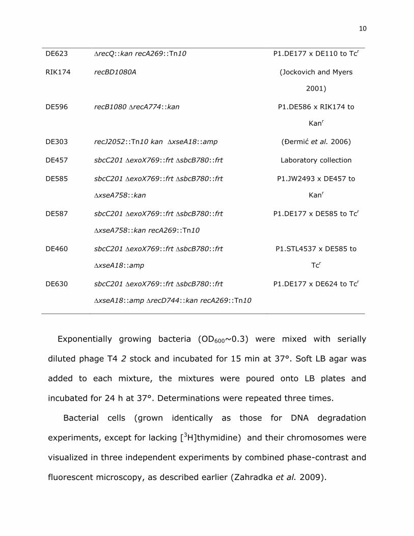
Table 1 Bacterial strains used in this study
Strain Relevant genotype Reference or construction
AB1157 Wild type, rec+, ExoV+ (Bachmann 1972)
DE586 recA774::kan P1.JW2669 x AB1157 to
Kanr
DE127 lexA3 (ind-) malB::Tn9 Laboratory collection

9
DE583 recA730 (E38K) srl::Tn10 sulA::Tn5 Laboratory collection
DE584 recA730 sulA::Tn5 lexA3 malB::Tn9 P1.DE202 x DE583 to
Cmr, UVs
DE656 recB1910::dhfr Laboratory collection
DE657 recA730 srl::Tn10 sulA::Tn5 recB1910::dhfr P1.DE656 x DE583 to Tmr
DE393 recAo281 srlD300::Tn10 lexA3 malB::Tn9 Laboratory collection
DE628 recAo281 srlD300::Tn10 malB732::kan P1.JW3996 x DE393 to
Kanr, UVr
DE189 umuDC::cat Laboratory collection
DE637 umuDC::cat recA774::kan P1.DE586 x DE189 to
Kanr,
DE390 recD744::kan P1.JW2787 x AB1157 to
Kanr
DE595 recD744::kan recA269::Tn10 P1.DE177 x DE390 to Tcr
DE101 recB268::Tn10 (Đermić et al. 2005)
DE589 recB268::Tn10 recA774::kan P1.DE586 x DE101 to
Kanr
DE590 recQ1803::Tn3 P1.JJC405 x AB1157 to
Apr
DE591 recQ1803::Tn3 recB268::Tn10 P1.DE101 x DE590 to Tcr
DE592 recQ1803::Tn3 recB268::Tn10 recA774::kan P1.DE586 x DE591 to
Kanr
DE110 recQ::kan (Ivanković and Đermić
2012)
10
DE623 recQ::kan recA269::Tn10 P1.DE177 x DE110 to Tcr
RIK174 recBD1080A (Jockovich and Myers
2001)
DE596 recB1080 recA774::kan P1.DE586 x RIK174 to
Kanr
DE303 recJ2052::Tn10 kan xseA18::amp (Đermić et al. 2006)
DE457 sbcC201 exoX769::frt sbcB780::frt Laboratory collection
DE585 sbcC201 exoX769::frt sbcB780::frt
xseA758::kan
P1.JW2493 x DE457 to
Kanr
DE587 sbcC201 exoX769::frt sbcB780::frt
xseA758::kan recA269::Tn10
P1.DE177 x DE585 to Tcr
DE460 sbcC201 exoX769::frt sbcB780::frt
xseA18::amp
P1.STL4537 x DE585 to
Tcr
DE630 sbcC201 exoX769::frt sbcB780::frt
xseA18::amp recD744::kan recA269::Tn10
P1.DE177 x DE624 to Tcr
Exponentially growing bacteria (OD600~0.3) were mixed with serially
diluted phage T4 2 stock and incubated for 15 min at 37°. Soft LB agar was
added to each mixture, the mixtures were poured onto LB plates and
incubated for 24 h at 37°. Determinations were repeated three times.
Bacterial cells (grown identically as those for DNA degradation
experiments, except for lacking [3H]thymidine) and their chromosomes were
visualized in three independent experiments by combined phase-contrast and
fluorescent microscopy, as described earlier (Zahradka et al. 2009).
11
Gamma-irradiation. Bacteria were exposed to various doses of gamma-
rays from a 60Co source, with a dose rate of ~2.2 Gy s-1. For a bacterial
survival assay, bacterial cultures were grown to mid-exponential phase
(OD600~0.3), serially diluted in 67 mM phosphate buffer (pH 7.0) and
aliquots spread onto LB plates. The plates were immediately irradiated at
room temperature and then incubated at 37° for 24-48 hours.
Chromosomal DNA degradation. We used the procedure described earlier
(Đermić et al. 2005; Đermić et al. 2006). The cells were grown overnight in
LB medium supplemented with 0.07 MBq ml-1 [3H]thymidine (specific activity
962 GBq mmol-1; Amersham, UK) and 100 µg ml-1 deoxyadenosine.
Unincorporated [3H]thymidine was washed out with phosphate buffer; the
cultures were suspended in a double volume of fresh LB medium and divided
into two counterparts. One served as an unirradiated control, while the other
was irradiated with 400 Gy at 0°. After irradiation the cultures were
incubated at 37° and at intervals duplicate samples were spread onto
Whatman filters pretreated with 0.3 N NaOH. The filters were allowed to dry
at room temperature, and then were suspended for 30 min in 10%
trichloroacetic acid and twice in 5% trichloroacetic acid at 4°. Trichloroacetic
acid precipitates high molecular weight DNA, while the low molecular weight
DNA is washed away. The filters were then washed in 1:1 solution of ether
and ethanol at 4° for 30 min, and then in ether at room temperature. Acid-
precipitable radioactivity of the filters represents the amount of the intact
12
chromosomal DNA, and was measured by scintillation counting (Liquid
Scintillation Analyzer, Tri-Carb 2810 TR, Perkin Elmer, USA). On average,
1500-3000 cpm was measured in unirradiated samples.
The frequency of genomic DSBs inflicted by gamma irradiation ranges from
0.004 to 0.01 DSBs/Gy/Mbp in various organisms (Daly 2009). The dose of
400 Gy is therefore expected to produce at least 8 DSBs per E. coli
chromosome. This is in accord with a measured rate of DSB induction in E.
coli chromosome of 2.7 DSBs per 100 Gy (Ulmer et al. 1979) (which would
amount to about 10 DSBs at 400 Gy), and also with predicted ~ 9 DSBs per
cell at 400 Gy (Shee et al. 2013), based on direct detection of DSBs in
vivo using a fluorescent DSB binding protein.
Preparation of cell-free extracts and Western analysis. Bacterial
cultures (grown identically as those for DNA degradation experiments, except
for lacking [3H]thymidine) were split into two parts, one of which was
irradiated with 400 Gy at 0°. After irradiation, both cultures were incubated
at 37° for additional 35 min. Cell-free extracts were prepared as previously
described (Vujaklija and Maček 2012), with some modifications. Briefly, the
cells were harvested from 20 mL of culture by centrifugation at 5000 g and
washed with 25 mM Tris-HCl pH 8.3 mM NaCl, 1 mM EDTA, 1 mM DTT and
0.5 mM PMSF. The cells were suspended in the same buffer and disrupted by
sonication. Cell debris was removed by centrifugation at 12000 g and the
supernatant was used as a cell-free extract. 15 µg of total protein were
13
loaded into each lane and resolved by SDS-PAGE under reducing conditions
in 10% gels. After electrophoresis, the separated proteins were transferred
to Amersham Hybond-P PVDF membrane (GE Healthcare). The membrane
was stained with Amido Black (Sigma Aldrich, St. Louis, USA) for protein
visualization. This was a loading control and allowed determination of protein
transfer efficiency across the blot. Western analysis performed as described
(Vujaklija and Maček 2012) was used to estimate the relative concentrations
of RecA protein in each sample. RecA was detected on Western blots with the
polyclonal anti-RecA antibody ab63797 (Abcam) diluted 1: 6000. Antibody
binding was visualized with peroxidase-coupled goat anti rabbit antibody
diluted 1: 30000 and Amersham ECLTM Western Blotting Reagent Pack (GE
Healthcare). The specificity of the antibody was examined using E. coli strain
lacking recA gene. Two biological replicates of each strain were analyzed by
immunoblotting. ImageJ, Java-based image processing program (Schneider
et al. 2012) was applied to analyze Western blot signal intensity.
Data availability. All the data necessary for confirming the conclusions
presented in this paper are represented in the paper.
14
RESULTS
Synchronous DSBs were induced into E. coli DNA by gamma irradiation
and their processing was monitored by measuring degradation of the
fragmented chromosome. Chromosomal DNA was [3H]thymidine labeled,
which enabled us to follow its fate.
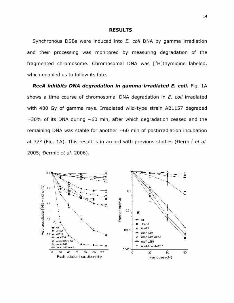
RecA inhibits DNA degradation in gamma-irradiated E. coli. Fig. 1A
shows a time course of chromosomal DNA degradation in E. coli irradiated
with 400 Gy of gamma rays. Irradiated wild-type strain AB1157 degraded
~30% of its DNA during ~60 min, after which degradation ceased and the
remaining DNA was stable for another ~60 min of postirradiation incubation
at 37° (Fig. 1A). This result is in accord with previous studies (Đermić et al.
2005; Đermić et al. 2006).
15
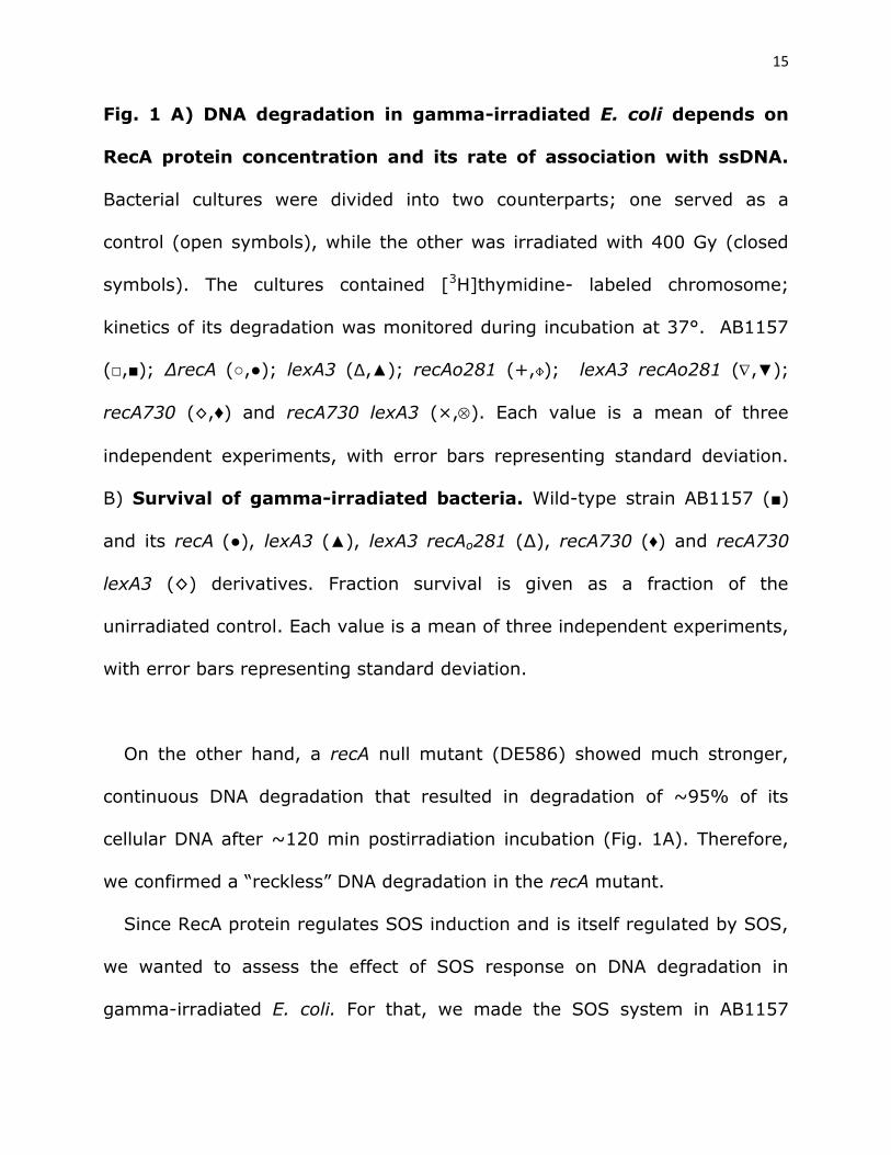
Fig. 1 A) DNA degradation in gamma-irradiated E. coli depends on
RecA protein concentration and its rate of association with ssDNA.
Bacterial cultures were divided into two counterparts; one served as a
control (open symbols), while the other was irradiated with 400 Gy (closed
symbols). The cultures contained [3H]thymidine- labeled chromosome;
kinetics of its degradation was monitored during incubation at 37°. AB1157
(□,■); ∆recA (○,●); lexA3 (∆,▲); recAo281 (+, ); lexA3 recAo281 (,▼);
recA730 (◊,♦) and recA730 lexA3 (×,). Each value is a mean of three
independent experiments, with error bars representing standard deviation.
B) Survival of gamma-irradiated bacteria. Wild-type strain AB1157 (■)
and its recA (●), lexA3 (▲), lexA3 recAo281 (∆), recA730 (♦) and recA730
lexA3 (◊) derivatives. Fraction survival is given as a fraction of the
unirradiated control. Each value is a mean of three independent experiments,
with error bars representing standard deviation.
On the other hand, a recA null mutant (DE586) showed much stronger,
continuous DNA degradation that resulted in degradation of ~95% of its
cellular DNA after ~120 min postirradiation incubation (Fig. 1A). Therefore,
we confirmed a “reckless” DNA degradation in the recA mutant.
Since RecA protein regulates SOS induction and is itself regulated by SOS,
we wanted to assess the effect of SOS response on DNA degradation in
gamma-irradiated E. coli. For that, we made the SOS system in AB1157
16
uninducible by introducing a lexA3 allele, coding for a noncleavable SOS
repressor. By Western analysis we estimated the relative concentrations of
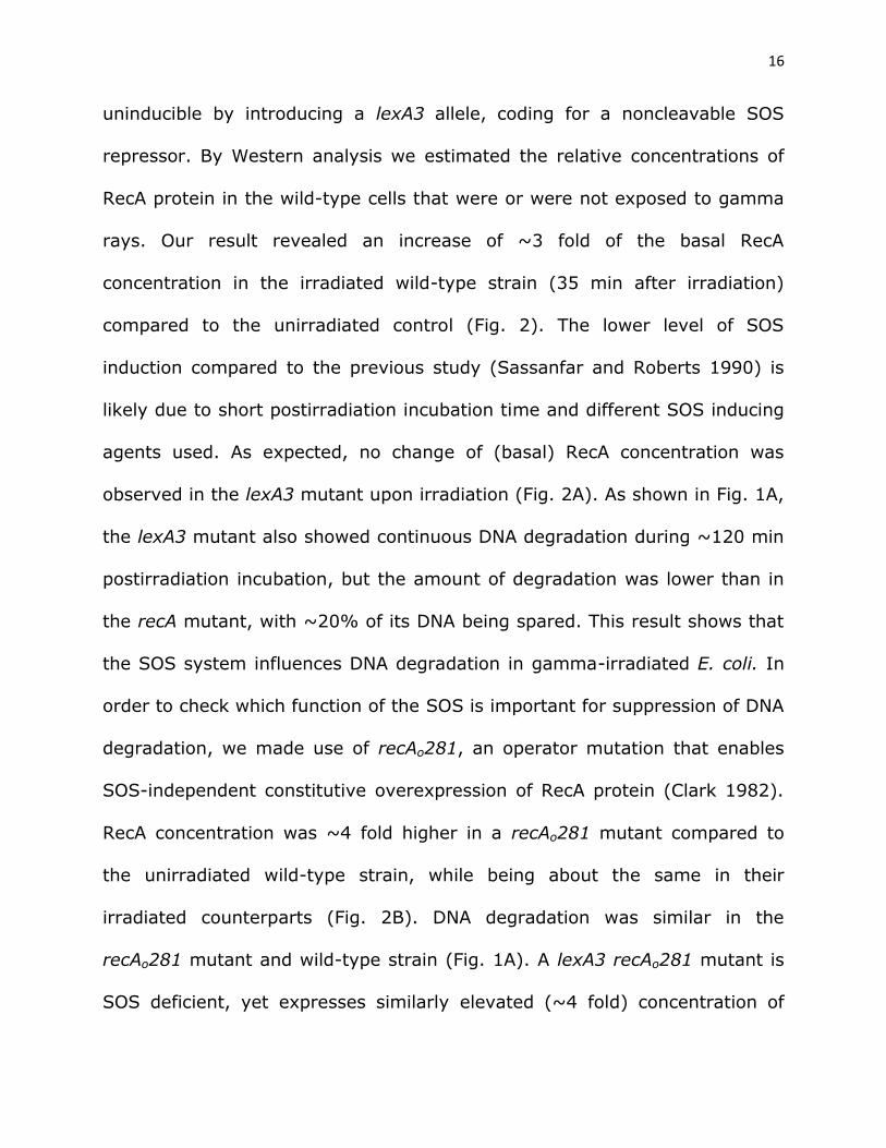
RecA protein in the wild-type cells that were or were not exposed to gamma
rays. Our result revealed an increase of ~3 fold of the basal RecA
concentration in the irradiated wild-type strain (35 min after irradiation)
compared to the unirradiated control (Fig. 2). The lower level of SOS
induction compared to the previous study (Sassanfar and Roberts 1990) is
likely due to short postirradiation incubation time and different SOS inducing
agents used. As expected, no change of (basal) RecA concentration was
observed in the lexA3 mutant upon irradiation (Fig. 2A). As shown in Fig. 1A,
the lexA3 mutant also showed continuous DNA degradation during ~120 min
postirradiation incubation, but the amount of degradation was lower than in
the recA mutant, with ~20% of its DNA being spared. This result shows that
the SOS system influences DNA degradation in gamma-irradiated E. coli. In
order to check which function of the SOS is important for suppression of DNA
degradation, we made use of recAo281, an operator mutation that enables
SOS-independent constitutive overexpression of RecA protein (Clark 1982).
RecA concentration was ~4 fold higher in a recAo281 mutant compared to
the unirradiated wild-type strain, while being about the same in their
irradiated counterparts (Fig. 2B). DNA degradation was similar in the
recAo281 mutant and wild-type strain (Fig. 1A). A lexA3 recAo281 mutant is
SOS deficient, yet expresses similarly elevated (~4 fold) concentration of
17
RecA protein (Fig. 2B). After two hours of postirradiation incubation it
degraded about 40% of its DNA, which is similar to degradation in wild-type
strain, whereas about three-fold more of its DNA was preserved compared to
DNA in the irradiated single lexA3 mutant (Fig. 1A). This suggests that RecA
concentration is important for suppressing DNA degradation, while other
parts of the SOS response have a minor effect. Therefore, our results reveal
an inverse relation between RecA concentration and DNA degradation in
irradiated E. coli, which led us to conclude that RecA protein suppresses DNA
degradation by mass effect.
Fig. 2 Immunoblotting analysis of relative RecA protein
concentrations in E. coli.
Protein samples obtained from the cells that were irradiated with 400 Gy of
gamma-rays and subsequently incubated for 35 min at 37° are marked with
*. PVDF membranes stained with Amido Black were used as a loading control
18
(lower panels). SDS-PAGE gels and PVDF membranes are from one
experiment, while graph depicts mean and SD (error bars) from multiple
replicates (numbered in parentheses) from two independent experiments.
Next we wanted to determine how RecA rate of association with ssDNA
influences DNA degradation in irradiated cells. For that we used a recA730
mutant, which produces the RecAE38K mutant protein that shows an
increased rate of association with ssDNA and hence competes more
efficiently with SSB protein for binding to ssDNA (Lavery and Kowalczykowski
1992). When introduced into AB1157, the recA730 mutation reduced DNA
degradation in the resulting strain about 1.5 fold (Fig.1A). This effect can be
caused by an increased affinity of the RecA E38K for ssDNA, but also by an
increased concentration of the enzyme in that mutant due to constitutive
SOS induction. RecA E38K concentration in unirradiated and irradiated cells
of the recA730 mutant was ~4 and ~8 fold higher, respectively, than the
RecA concentration in an unirradiated wild-type strain (Fig. 2). We measured
the effect of the increased affinity of RecA E38K for ssDNA by using a lexA3
recA730 mutant, which produces a basal level of RecA E38K, which is equal
to that of RecA in the lexA3 mutant (Fig. 2A). The lexA3 recA730 mutant
showed greatly reduced (about two-fold) DNA degradation compared to that
in the lexA3 mutant (Fig. 1A), revealing that RecA dependency of DNA
degradation is proportionate to RecA’s rate of association with ssDNA. In
19
order to check whether the protective effect of RecA E38K protein on DNA
degradation is indeed due to its high affinity to ssDNA, and not to its possible
nonspecific binding to dsDNA, we determined the titer of T4 2 phage in
recA730 and recA730 recB mutants. T4 2 mutant phage genome is a linear
dsDNA with free, blunt ends and hence susceptible to RecBCD binding and
nucleolytic degradation. If RecA E38K would bind to dsDNA, hence blocking
access and activity of RecBCD on it, the titer of T4 2 phage should increase.
In comparison to a recB mutant (which cannot degrade T4 2 phage genome
and therefore enables maximal phage yield), T4 2 phage plating efficiency in
recA730 and wt strain was 0.0019 ± 0.001 and 0.0017 ± 0.0007,
respectively, which is significantly different (P=0.001, two-tailed t-test) from
the recB derivative of recA730 mutant that had 0.94 ± 0.19 of recB’s titer
(n=3), suggesting that RecBCD inhibits phage propagation in recA730
mutant and that phage genome metabolism is otherwise not impaired in that
mutant. Hence, we infer that RecA E38K protein does not interfere with
degradation of purely dsDNA.
The difference in degradation between recA730 and recA730 lexA3
mutants (Fig. 1A) is likely mostly due to different concentrations of the RecA
E38K protein in these strains. The same, moderate, amount of DNA
degradation observed in the lexA3 recA730 and lexA3 recAo281 mutants (Fig.
1A), indicates that for DNA degradation inhibition higher RecA rate of
association with ssDNA can compensate for lower RecA concentration, and
20
that SOS induction is not required per se for protection of cellular DNA from
degradation.
In summary, we have shown that RecA protein protects a fragmented
chromosome from degradation in gamma-irradiated wild type E. coli. The
protective activity depends on RecA concentration and its rate of association
with ssDNA.
DNA degradation is distributed uniformly in wild-type cells. The
DNA degradation experiments used in this study provide bulk population data
that do not give us information on degradation distribution in a population.
To assess the distribution of DNA degradation in a cell population, we
visualized bacteria by microscopy and determined the fraction of cells lacking
DAPI staining material in a population of wild-type (AB1157) and recA
bacteria after 90 min postirradiation incubation. Unirradiated populations of
wild-type and recA cells contained 0.055% ± 0.001% and 8.87% ± 0.93%
anucleate cells, respectively (Fig. 3). On the other hand, wild-type and recA
populations gamma-irradiated with 400 Gy had 0.49% ± 0.10% and 84.1%
± 2.4% of cells lacking DAPI signal, respectively, after 90 min incubation
(Fig. 3). These data suggest that DNA degradation in wild-type cells is
tightly regulated since it is very uniform, so that ~35% of their degraded
cellular DNA (Fig. 1A) is reflected in less than 1% of chromosomeless cells.
Conversely, only ~16% of irradiated recA cells contained DNA, which is
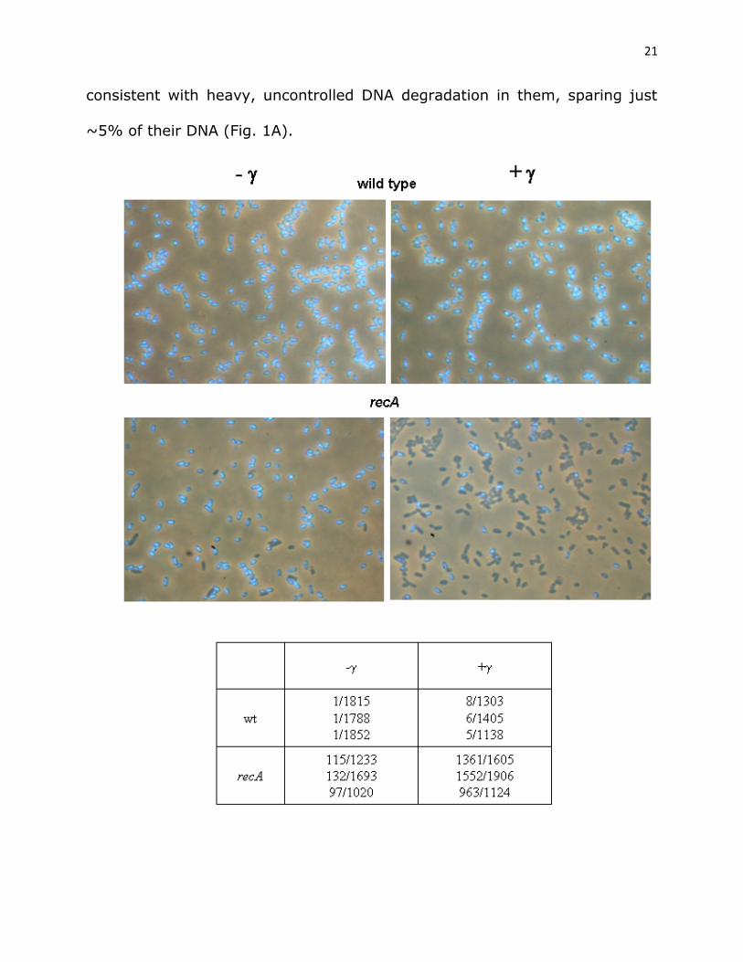
21
consistent with heavy, uncontrolled DNA degradation in them, sparing just
~5% of their DNA (Fig. 1A).
22
Fig. 3 DNA degradation is strictly controlled in wild-type, but not in
recA cells.
A faction of anucleate cells was determined in wild-type (AB1157) and recA
populations, which were either unirradiated (left side of the panel), or
irradiated with 400 Gy of gamma rays and then incubated for 90 min at 37°
(right side of the panel). Cells were fixed with osmium tetroxide, their DNA
stained with DAPI and visualized by fluorescence microscopy. An anucleate
cell count in analyzed population from three experiments is represented in a
table.
Role of RecA protein in survival of gamma-irradiated cells. Since
degradation of a fragmented chromosome reflects DSB processing reactions,
we wanted to relate it to a DSB repair process. Therefore, we determined
survival of gamma-irradiated bacteria as a measure of the efficiency of their
DSB repair. As expected, the recA mutant showed an extreme sensitivity to
gamma rays, at 90 Gy dose its survival was more than three orders of
magnitude lower than that of the wild-type strain AB1157 (Fig. 1B). The
lexA3 mutant had somewhat higher survival than the recA mutant (Fig. 1B),
which is not surprising considering that it retains basal RecA concentration.
Survival of the lexA3 recAo281 mutant was considerably higher than that of
the lexA3 mutant (also due to higher RecA concentration), whereas the
recAo281 mutant had the same survival as the wild-type strain (Fig. 1B).
23
While the recA730 mutant had essentially the same survival as the wild-
type strain, its lexA3 derivative showed extreme sensitivity to gamma rays,
similar to that of the recA and lexA3 mutants (Fig. 1B). Since the recA730
lexA3 mutant showed highly reduced DNA degradation compared to the recA
(and lexA3) mutant, whereas displaying almost identical gamma survival
(compare Figs. 1A and 1B), we conclude that reducing “reckless” degradation
is insufficient to enable DNA repair.
A comparison of gamma survivals shows that the important factors for
higher survival of gamma-irradiated cells are: higher RecA protein
concentration (hence the higher survival of recAo281 lexA3 than lexA3 and
recA730 lexA3 mutants) and SOS induction (causing increased survival of
recAo281 compared to recAo281 lexA3 mutant).
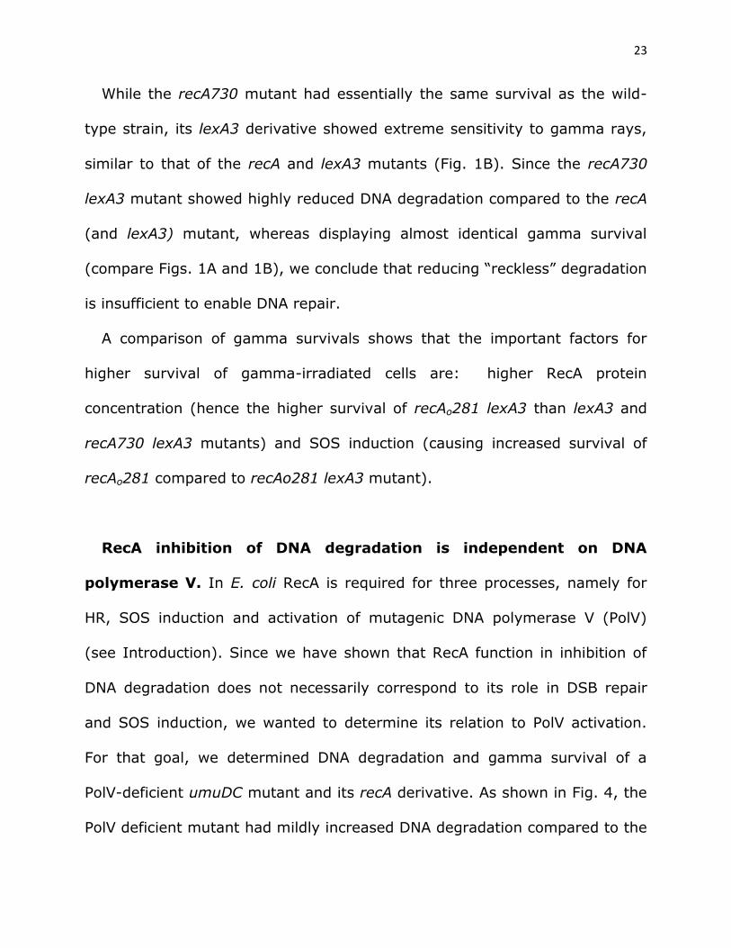
RecA inhibition of DNA degradation is independent on DNA
polymerase V. In E. coli RecA is required for three processes, namely for
HR, SOS induction and activation of mutagenic DNA polymerase V (PolV)
(see Introduction). Since we have shown that RecA function in inhibition of
DNA degradation does not necessarily correspond to its role in DSB repair
and SOS induction, we wanted to determine its relation to PolV activation.
For that goal, we determined DNA degradation and gamma survival of a
PolV-deficient umuDC mutant and its recA derivative. As shown in Fig. 4, the
PolV deficient mutant had mildly increased DNA degradation compared to the
24
wild-type strain, while its gamma survival was the same as that of the wild-
type strain. Furthermore, no difference was observed in DNA degradation
and survival of umuDC recA and recA mutants (Fig. 4). These results
indicate that the RecA protective role during DNA degradation does not
depend on PolV.
Fig. 4 DNA degradation is unrelated to DNA Polymerase V.
Kinetics of [3H]thymidine-labeled DNA degradation (A), and survival (B) of
gamma-irradiated wild-type strain AB1157 (■) and its umuDC (▼), recA (▲),
and umuDC recA (♦) derivatives. For DNA degradation assay, bacterial
cultures were divided into two fractions; one served as a control (open
symbols), while the other was irradiated with 400 Gy (closed symbols).
25
Fraction survival is given as the fraction of the unirradiated control. Each
value is a mean of three independent experiments, with error bars
representing standard deviation.
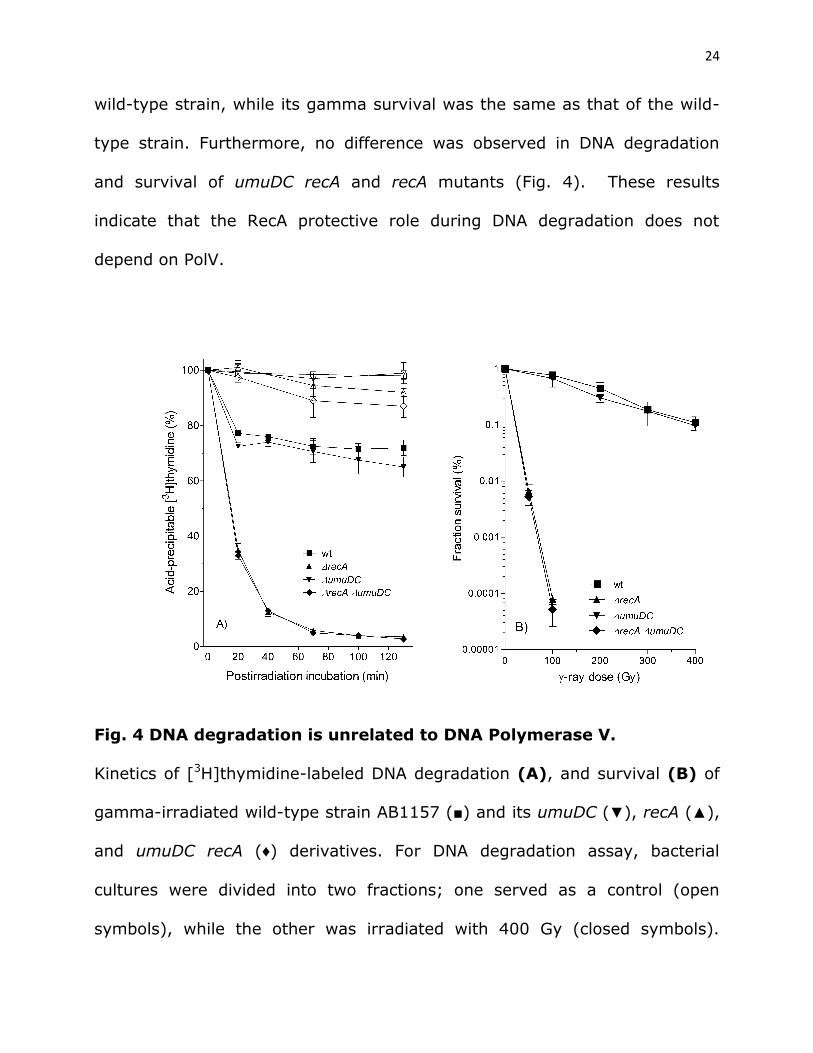
DNA degradation in recB and recD mutants is inhibited by RecA. We
wanted to determine whether the RecA inhibition of DNA degradation is
restricted to RecBCD-catalyzed reactions, or whether it is a more general
phenomenon. For this aim, we determined DNA degradation in gamma-
irradiated recB and recD mutants. No degradation was observed in these
mutants (Figs. 5A, 5B), which is not surprising considering their ExoV-
phenotype. However, their recA derivatives did degrade their damaged DNA.
A recB recA mutant degraded its DNA continuously during postirradiation
incubation, with degradation reaching a level close to that of the wild-type
strain after 130 min (Fig. 5A). This result indicates that RecA protects DNA
from degradation in a recB null mutant. Since in the recB mutant dsDNA
ends are processed by the RecQ helicase, we evaluated the role of RecQ in
DNA degradation in the recB recA mutant. A triple recB recA recQ mutant
degraded ~10% of its DNA after 130 min incubation, which is about two-fold
less compared to its parental RecQ+ strain (Fig. 5A), indicating that a
helicase activity participates in DSB processing reactions that result in
degradation of a shattered E. coli chromosome. At the same time the recQ
mutation did not affect DNA degradation in wild-type, recA and recB null
26
mutants (Fig. 5A), suggesting that the RecQ helicase role is specifically
pronounced in recB recA mutant.
Fig. 5 DNA degradation in recB (A) and recD (B) mutants is inhibited
by RecA protein.
Bacterial cultures were divided into two counterparts; one served as a
control (open symbols), while the other was irradiated with 400 Gy (closed
symbols). The cultures contained [3H]thymidine- labeled chromosome;
kinetics of its degradation was monitored during incubation at 37°. (A)
AB1157 (□,■); recQ (○,●); recB (∆,▲); recB recQ (+, ); recB recA (,▼);
recB recA recQ (◊,♦); recQ recA (×,), recA (---). (B) AB1157 (□,■); recD
(○,●); recD recA (∆,▲); ExoI- ExoVII- ExoX- SbcCD- RecA- RecD- (,▼). Each
27
value is a mean of three independent experiments, with error bars
representing standard deviation.
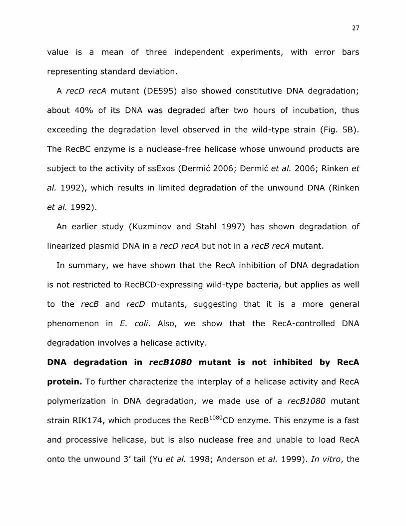
A recD recA mutant (DE595) also showed constitutive DNA degradation;
about 40% of its DNA was degraded after two hours of incubation, thus
exceeding the degradation level observed in the wild-type strain (Fig. 5B).
The RecBC enzyme is a nuclease-free helicase whose unwound products are
subject to the activity of ssExos (Đermić 2006; Đermić et al. 2006; Rinken et
al. 1992), which results in limited degradation of the unwound DNA (Rinken
et al. 1992).
An earlier study (Kuzminov and Stahl 1997) has shown degradation of
linearized plasmid DNA in a recD recA but not in a recB recA mutant.
In summary, we have shown that the RecA inhibition of DNA degradation
is not restricted to RecBCD-expressing wild-type bacteria, but applies as well
to the recB and recD mutants, suggesting that it is a more general
phenomenon in E. coli. Also, we show that the RecA-controlled DNA
degradation involves a helicase activity.
DNA degradation in recB1080 mutant is not inhibited by RecA
protein. To further characterize the interplay of a helicase activity and RecA
polymerization in DNA degradation, we made use of a recB1080 mutant
strain RIK174, which produces the RecB1080CD enzyme. This enzyme is a fast
and processive helicase, but is also nuclease free and unable to load RecA
onto the unwound 3’ tail (Yu et al. 1998; Anderson et al. 1999). In vitro, the
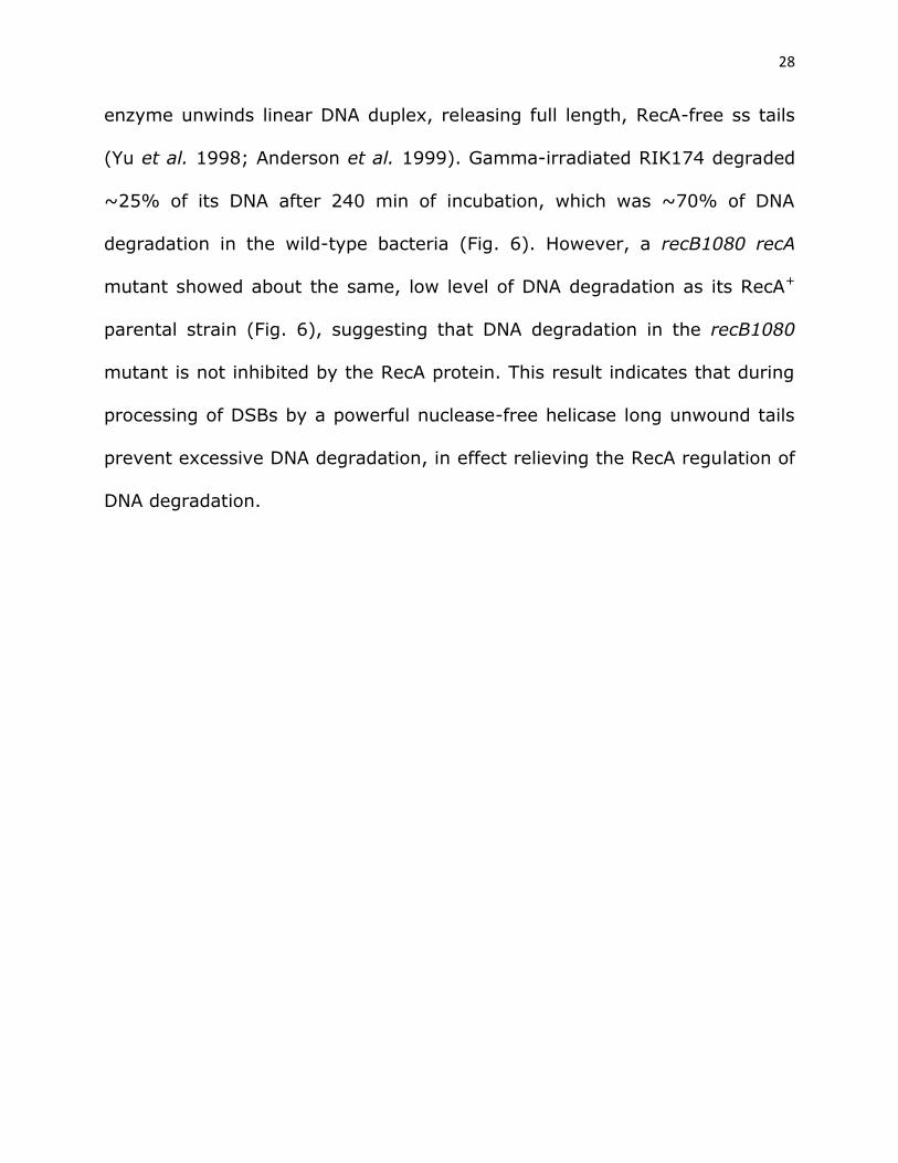
28
enzyme unwinds linear DNA duplex, releasing full length, RecA-free ss tails
(Yu et al. 1998; Anderson et al. 1999). Gamma-irradiated RIK174 degraded
~25% of its DNA after 240 min of incubation, which was ~70% of DNA
degradation in the wild-type bacteria (Fig. 6). However, a recB1080 recA
mutant showed about the same, low level of DNA degradation as its RecA+
parental strain (Fig. 6), suggesting that DNA degradation in the recB1080
mutant is not inhibited by the RecA protein. This result indicates that during
processing of DSBs by a powerful nuclease-free helicase long unwound tails
prevent excessive DNA degradation, in effect relieving the RecA regulation of
DNA degradation.
29
Fig. 6 DNA degradation in recB1080 mutant is weak and uninhibited
by RecA protein.
Bacterial cultures were divided into two counterparts; one served as a
control (open symbols), while the other was irradiated with 400 Gy (closed
symbols). The cultures contained [3H]thymidine- labeled chromosome;
kinetics of its degradation was monitored during incubation at 37°. AB1157
30
(□,■); recB1080 (∆,▲) and recB1080 recA (,▼). Each value is a mean of
three independent experiments, with error bars representing standard
deviation.
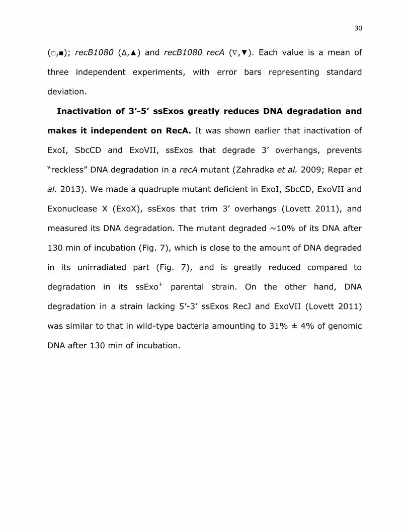
Inactivation of 3’-5’ ssExos greatly reduces DNA degradation and
makes it independent on RecA. It was shown earlier that inactivation of
ExoI, SbcCD and ExoVII, ssExos that degrade 3’ overhangs, prevents
“reckless” DNA degradation in a recA mutant (Zahradka et al. 2009; Repar et
al. 2013). We made a quadruple mutant deficient in ExoI, SbcCD, ExoVII and
Exonuclease X (ExoX), ssExos that trim 3’ overhangs (Lovett 2011), and
measured its DNA degradation. The mutant degraded ~10% of its DNA after
130 min of incubation (Fig. 7), which is close to the amount of DNA degraded
in its unirradiated part (Fig. 7), and is greatly reduced compared to
degradation in its ssExo+ parental strain. On the other hand, DNA
degradation in a strain lacking 5’-3’ ssExos RecJ and ExoVII (Lovett 2011)
was similar to that in wild-type bacteria amounting to 31% ± 4% of genomic
DNA after 130 min of incubation.
31
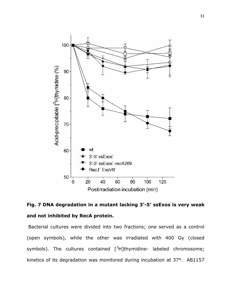
Fig. 7 DNA degradation in a mutant lacking 3’-5’ ssExos is very weak
and not inhibited by RecA protein.
Bacterial cultures were divided into two fractions; one served as a control
(open symbols), while the other was irradiated with 400 Gy (closed
symbols). The cultures contained [3H]thymidine- labeled chromosome;
kinetics of its degradation was monitored during incubation at 37°. AB1157
32
(□,■); ExoI- ExoVII- ExoX- SbcCD- (∆,▲); ExoI- ExoVII- ExoX- SbcCD- RecA-
(,▼), RecJ- ExoVII- (○,●). Each value is a mean of three independent
experiments, with error bars representing standard deviation.
Since the mutant lacking four 3’-5’ ssExos is an ExoV- phenocopy with
respect to DNA degradation, although it expresses an intact RecBCD enzyme,
we checked the enzyme’s nuclease activity by assessing its ability to inhibit
growth of a T4 2 phage. Relative to the titer on the recB mutant, T4 2 phage
plating efficiency on the wild-type strain was 0.0067 ± 0.0043, and that on
the 3’-5’ ssExos deficient mutant was 0.019 ± 0.012 (n=3), which is not
significantly different (P=0.170, two-tailed t-test). This result indicates that
RecBCD is proficient in binding to and degrading phage DNA in the mutant
lacking 3’-5’ ssExos.
A recA derivative of the quadruple ExoI- SbcCD- ExoVII- ExoX- mutant
showed about the same, low-level ~10% degradation as its RecA+
counterpart (Fig. 7), suggesting that DSB processing in bacteria with
preserved 3’ tail is not affected by RecA.
Similarly, DNA degradation in a recD recA mutant was greatly abolished by
inactivation of the four 3’-5’ ssExos (Fig. 5B). Therefore, we conclude that
the constitutive chromosome degradation observed in irradiated recD recA
mutant is dependent on degradation of 3’-ended ss overhangs by the 3’-5’
ssExos.
33
Inactivation of both 5’-3’ ssExos, RecJ and ExoVII, did not inhibit DNA
degradation (Fig. 7).
Hence, our results indicate that 3’ overhangs suppress DSB processing,
especially when spared from 3’-5’ ssExos.
34
DISCUSSION
To gain better insight into the in vivo processing of DSBs we followed
degradation of radioactively-labeled chromosomal DNA of gamma-irradiated
E. coli. DSBs in that bacterium are repaired by RecBCD enzyme, which upon
binding to (nearly blunt) dsDNA end unwinds DNA duplex and degrades both
of the unwound strands. Only after interaction with Chi sequence, the
enzyme starts a resection process, meaning that it continues degradation of
5’ strand, while ceasing trimming of 3’ strand. Therefore, DNA degradation is
an essential and indivisible part of DSB processing in E. coli (there is no
situation in which RecBCD activity on DNA is nuclease free), and by
assessing DNA degradation one can indeed get an insight into DSB
processing. RecBCD enzyme is the strongest DNase in E. coli (Kuzminov
1999), yet its nuclease activity is augmented in bacteria lacking RecA
protein, wherein it becomes unregulated, “reckless”, leading to complete
degradation of a chromosome (Capaldo and Barbour 1975; Skarstad and
Boye 1993). “Reckless” degradation in recA null mutants is attributed to
either impaired Chi regulation of RecBCD enzyme (Kuzminov et al. 1994;
Kuzminov and Stahl 1997), or to a lack of RecA protection of the frayed ends
of a processed DNA molecule (Dabert et al. 1992). We have shown here that
recruitment of RecA onto ssDNA inhibits degradation at DSBs in wild-type,
recB and recD genetic backgrounds, indicating that the RecA protection of
DNA from degradation is a general phenomenon in E. coli. Since there is no
35
Chi activity in the recB and recD mutants and yet degradation of their DNA is
still inhibited by RecA, we infer that the physical protection by RecA binding
on (3’-terminated) ss overhangs is the main mechanism for RecA inhibition
of DNA degradation. A recent study indicated that a RecA-ssDNA complex is
resistant to degradation by nucleases (Kohiyama et al. 2013). Analogously, a
human RecA ortholog, RAD51 recombinase, prevents excessive DNA
nucleolytic degradation in UV-irradiated human cells (Vallerga et al. 2015),
indicating conservation of the inhibitory role of recombinase proteins in DNA
degradation, thus signifying its importance.
However, our results do not exclude that RecBCD’s Chi activity is indeed
impaired in the recA mutant, nor do they determine the possible contribution
of each of the two mechanisms to DNA protection in that mutant.
Interestingly, our results show that the ability of the RecA protein to
protect DNA from degradation differs from its role in DSB repair. The
recA730 lexA3 mutant has greatly suppressed DNA degradation compared
with a lexA3 mutant (both contain about the same concentration of
RecA(E38K) protein), while having similar gamma-survival. This phenotype is
not unique, the recB mutant has it too; it also has RecA inhibited DNA
degradation and extremely poor survival. Thus, our results suggest that the
RecA role in DNA degradation regulation is less challenging than its role in
DSB repair; the former is about preventing 3’-5’ ssExos from degrading 3’tail
(for which, binding of a couple of RecA molecules would likely suffice), while
36
the latter requires production of a functional RecA nucleofilament.
Furthermore, recB and recD null mutants share poor DNA degradation
(suppressed by RecA) while the former is deficient for DSB repair and the
latter is proficient. Also, recA730 lexA3 and recAo281 lexA3 mutants have
about the same amount of DNA degradation, while their gamma-survivals
greatly differ.
However, we revealed two notable exceptions in the RecA-imposed control
of DNA degradation, which enabled further insight into the mechanism of
DSB processing regulation in E. coli. Namely, inactivation of four ssExos
(ExoI, ExoVII, ExoX, SbcCD) that degrade a 3’ tail (Lovett 2011) greatly
reduced DNA degradation in both RecA+ and RecA– bacteria, making them an
ExoV- phenocopy, even though the RecBCD enzyme is active in these cells
and able to degrade DNA (as it prevented proliferation of T4 2 phage, whose
genome is blunt-ended). Since RecBCD poorly degrades fragmented
chromosome in cells lacking 3’-5’ ssExos while retaining ExoV activity, we
infer that the enzyme is affected in binding to DNA, i.e. creation of blunt DNA
ends is suppressed. A previous study showed that a strain lacking three 3’-5’
ssExos (ExoI, ExoVII and SbcCD) retains nearly wt capacity for DNA repair,
whereas its recA derivative had inhibited DNA degradation (ExoV-), thus
indicating that these ssExos are not required for blunting the initial
irradiation-produced dsDNA ends although being critical for reckless DNA
degradation (Repar et al. 2013). Because 3’-5’ ssExos inactivation is
37
epistatic to RecA deficiency for DNA degradation, we conclude that RecA
inhibits DNA degradation by preventing trimming of 3’-terminated overhangs
by 3’-5’ ssExos; hence when these ssExos are absent, RecA becomes
dispensable. Therefore, in cells lacking 3’-5’ ssExos DSB processing is
inhibited by preserved 3’ overhangs, irrespective of RecA protein (Fig. 8A).
Conversely, RecA deficiency is epistatic to 3’-5’ ssExos deficiency for DNA
repair, thus additionally emphasizing difference in roles of RecA protein in
DNA repair and DNA degradation in E. coli.
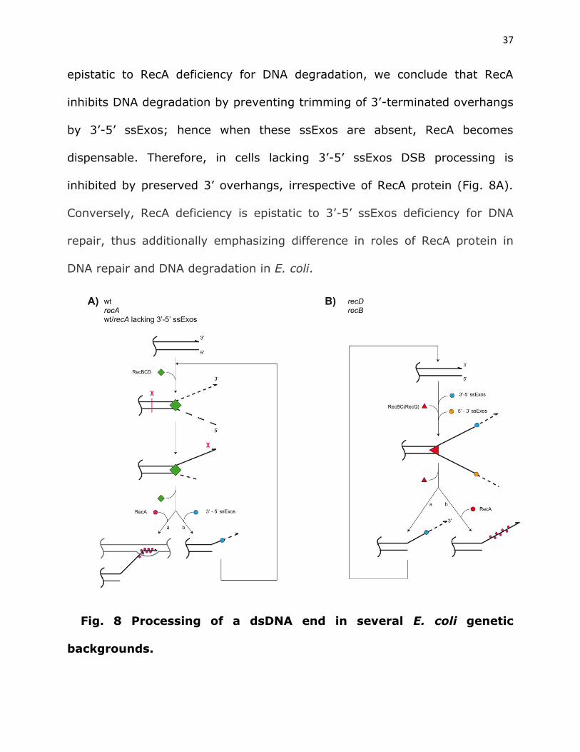
Fig. 8 Processing of a dsDNA end in several E. coli genetic
backgrounds.
38
A) In wt bacteria, dsDNA processing is initiated by binding of RecBCD
enzyme onto it, which unwinds DNA duplex, while degrading both
unwound strands. Upon interaction with Chi site, the enzyme stops the
3’ strand degradation, while continuing DNA unwinding and degradation
of 5’ strand. Chi-modified RecBCD also facilitates RecA loading onto
post-Chi 3’ overhang (a). In recA null mutant RecBCD degrades both
unwound strands before eventually being released from DNA, which
might be independent of Chi, leaving protruding 3’ tail. The 3’ tail
prevents subsequent reloading of RecBCD onto a processed dsDNA end
unless/until 3’-5’ ssExos degrade it (b). Since both RecA+ and RecA-
cells have similar, low DNA degradation when 3’-5’ ssExos are inactive
(blocked (b) path), these exonucleases apparently act on post-Chi-3’-
tail in wt cells (hence path (b) may apply to wt cells too).
B) In recA derivatives of recB and recD mutants, dsDNA end processing
starts with RecQ and RecBC helicase unwinding of DNA duplex,
respectively. The unwound 3’- and 5’-terminated strands are degraded
by 3’-5’ and 5’-3’ ssExos, respectively. The degradation of 5’ strand is
apparently faster than that of its complementary 3’ strand (otherwise
recD mutant would not be HR proficient) and thus the latter remains
after helicase detachment from DNA. The protruding 3’ tail is then
degraded by 3’-5’ ssExos (a), therefore enabling creation of (nearly)
blunt DNA end and hence repeated loading of RecQ or RecBC helicase.
39
In RecA+ variants of recB and recD, RecA binds to 3’ tail (b) and
prevents RecQ reloading onto the DNA end in the recB mutant, while
blocking its trimming and recreation of the blunt DNA end in recD
mutant and hence inhibits RecBC reloading.
Another situation where DNA degradation is unaffected by RecA concerns
the recB1080 mutant whose recA derivative had about the same, low level of
DNA degradation as its parental RecA+ strain. A combination of fast and
processive helicase activity and lack of nuclease and RecA loading activities
of RecB1080CD enzyme likely results in long, RecA-free overhangs in the
recB1080 mutant (as discussed, Ivanković and Đermić 2012; Đermić 2015),
analogously to what was observed in vitro (Yu et al. 1998; Anderson et al.
1999). In this mutant RecA loading is achieved by RecFOR proteins, which
perform it from an ss-ds DNA junction (Morimatsu and Kowalczykowski
2003), i.e. from the 5’ end of that tens of kb long 3’- terminated tail (as
discussed, Ivanković and Đermić 2012). Therefore, ssExos of 3’-5’ polarity
likely need to digest very long ss stretch in order to reach a growing RecA
nucleofilament. This is a heavy task because that ss region is overly reactive;
it may get engaged in inter- and intra-molecular transactions, resulting in
illegitimate recombination intermediates (Ivanković and Đermić 2012) and ds
secondary structures as well as ds products of reannealing with the
complementary 5’ tail, respectively. All of them may inhibit the 3’-5’ ssExos,
40
hence increasing longevity of 3’-terminated tail. Furthermore, ExoI, the most
potent 3’-5’ ssExo (Lovett 2011) degrades 3’ tail with ~275 nt s-1 rate
(Brody et al. 1986), which is ~4-8 fold slower than RecBCD helicase (and
also less processive) (see below). Therefore, excessive length of the
unwound 3’ strands and the mechanism of RecA loading onto them in the
recB1080 mutant seemingly protect them from degradation by ssExos, thus
making RecA protection dispensable. In fact, nucleolytic degradation of 3’
overhangs by 3’-5’ ssExos is essential for survival of recB1080 mutant
(Ivanković et al. 2017).
Since 3’-tail preservation is common to all the aforementioned situations;
we conclude that 3’ overhangs emanating from DSBs are crucial in limiting
the extent of DSB processing in E. coli. How do 3’-tails regulate DSB
processing? Our results reveal a gradient in intensity of DNA degradation in
the recA derivatives of wild-type, recD and recB strains (~95%, ~40% and
~20% degraded DNA, respectively), which unsurprisingly correlates with the
activities of their respective DSB processing helicases RecBCD, RecBC and
RecQ. The RecBCD helicase/nuclease processes at least 30 kb of DNA at
~1000-2000 bp s-1 in vitro (Bianco et al. 2001; Dillingham et al. 2005), the
RecBC helicase is about four fold slower and less processive than RecBCD
(Korangy and Julin 1993; Korangy and Julin 1994), and the RecQ helicase is
even slower (~2 bp s-1, Xu et al. 2003) and less processive than RecBC.
However, this proportionality of DNA degradation with the potency of the
41
DSB processing helicase is disrupted in the recB1080 mutant, whose
RecB1080CD enzyme is equally as powerful a helicase as RecBCD is (Anderson
et al. 1999), and yet, paradoxically, DNA degradation in its recA derivative is
very weak, not stronger than that in the parental RecA+ strain, and certainly
weaker than those observed in the recA recB and recA recD mutants.
Extensive unwound tails produced in recB1080 cells by the powerful
RecB1080CD helicase may certainly prevent its reloading onto dsDNA ends
because RecBCD and its variants (RecBC, RecB1080CD) bind exclusively onto
a (nearly) blunt dsDNA end (Dillingham and Kowalczykowski 2008). On the
other hand, continuous DNA degradation for more than two hours, resulting
in 20%-40% degraded genome (1-2 Mbps) in recA derivatives of recB and
recD mutants (Fig. 8B) reflects perpetual processing of DSBs in those
bacteria, which is possible only if their slow and poorly processive helicases
(RecQ and RecBC) are repeatedly loaded onto the processed DNA ends. As a
consequence, DSB processing catalyzed by repeated reloading of a weak
helicase, such as RecQ or RecBC, turns out to be more vigorous than that
accomplished by a single-round activity of the powerful helicase RecB1080CD.
Consequently, we infer that RecA prevents reloading of RecQ and RecBC
helicases in recB and recD mutants, respectively. Inactivation of 3’-5’ ssExos
in recD mutant alleviates the RecA inhibition of DNA degradation, indicating
that RecA inhibits DNA degradation by protecting 3’ tails from being
degraded by 3’-5’ ssExos. Preserved 3’ tails in turn inhibit RecBC reloading
42
onto the processed DNA end. Lack of DNA degradation in the recB mutant
indicates that an optimal substrate for the RecQ binding, a dsDNA end with a
3’ protrusion (Morimatsu and Kowalczykowski 2014), becomes inaccessible
to RecQ when the tail is bound by RecA. Furthermore, our results show that
3’-5’ ssExos enable DNA degradation in wild-type cells, suggesting that
RecBCD-catalyzed DSB processing is achieved through repeated reloading of
the enzyme, which depends on the removing of the 3’-tails. This indicates
that in wild-type E. coli 3’-5’ ssExos regularly trim RecBCD-produced post-
Chi 3’ tail (Fig. 8A).
However, there is an alternative explanation for the stimulatory effect of
3’-5’ ssExos on DNA degradation. Namely, if RecBCD would release long,
acid-precipitable oligonucleotides while processing DNA duplex (since it has
endonucleolytic rather than exonucleolytic activity), 3’-5’ ssExos may be
actively trimming those oligonucleotides, thus making them acid soluble. In
this way, low acid-soluble DNA content observed in irradiated 3’-5’ ssExos-
deficient cells would mask ongoing DNA degradation in them. However, this
hypothesis may be ruled-out since both un- and UV-irradiated ExoI- ExoVII-
SbcCD- RecA- mutant has preserved genomic DNA, as assessed by DAPI
staining and genome restriction (Repar et al. 2013), suggesting lack of
RecBCD-catalyzed “reckless” DNA degradation.
We show here that DNA degradation in E. coli is inhibited by either DSB-
processing helicase inactivation (e.g. by recB mutation in otherwise wild-type
43
cells and by RecQ inactivation in the recB recA mutant), or by preservation of
3’ tails, again leading to the inability of these helicases to reload on the
processed dsDNA ends. Our results therefore indicate that repeated helicase
loading is the main determinant of the extent of DSB processing, with 3’-
overhang metabolism being the crucial factor in helicase reloading. We
describe three ways by which availability of dsDNA ends to a helicase is
controlled by 3’ tails: i) RecA polymerization onto 3’ tails either directly
inhibits RecQ loading onto them in a recB mutant; or inhibits their
degradation by the 3’-5’ ssExos, thus preventing creation of blunt dsDNA
ends and consequently, reloading of RecBC(D) onto them. ii) Appearance of
blunt dsDNA ends is prevented by inactivation of the 3’-5’ ssExos, hence
making shielding RecA binding onto such a protected 3’ overhangs
dispensable. iii) In the recB1080 mutant lengthy DSB-derived 3’ overhangs
are protected from 3’-5’ ssExos in a RecA-independent manner (instead, this
is likely achieved by their involvement in transactions that produce ds
regions in them), hence preventing recreation of blunt dsDNA ends.
Similarly, excessively long 3’-overhangs produced during resection inhibit
meiotic DSB repair in eukaryotes (Johnson et al. 2007).
Our collective results indicate that resection of a dsDNA end in E. coli
proceeds until a 3’ tail of sufficient length and stability is formed, which then
inhibits further end processing by preventing reloading of a DSB-processing
helicase. Factors that facilitate 3’ tails’ longevity involve: i) 3’ tails protection
44
from degradation by 3’-5’ ssExos by either “insulating” them with the RecA
protein, or by inactivation of these 3’-5’ ssExos; ii) their excessive length.
The 3’tail regulation of DSB processing, that we describe here, is
reminiscent of DNA end-resection in eukaryotes, with a short 3’ overhang
produced during initial DSB resection by the Rad50 and Mre11 nuclease
(orthologues of SbcCD) directing a resected end toward HR and
microhomology-mediated-end-joining pathways and away from
nonhomologous-end-joining repair pathway (Truong et al. 2013). Similarly,
the length of (and RecA binding to) the 3’ overhangs created during DNA end
resection determines the equilibrium of HR and illegitimate recombination
pathways in E. coli (Ivanković and Đermić 2012). Furthermore, SbcCD and
its eukaryotic orthologues are analogously required for enabling (re)loading
of the main DSB processing machines [bacterial RecBCD and eukaryotic EXOI
(ExoI)/BLM DNA2 (Sgs1 Dna2)] that perform long-range dsDNA-end
processing (as discussed recently, Đermić 2015).
Hence, one can note a common regulatory mechanism for DSB processing,
with a 3’ tail produced during end resection acting as the main supervisory
element by imposing a negative feedback loop on subsequent processing
reactions. This mechanism ensures that processing of dsDNA ends continues
until stable and utilizable 3’ tails are produced that enable efficient repair of
DSBs in both bacteria and eukaryotes.
45
ACKNOWLEDGMENTS
We are grateful to Amir Dubravić, Mary Sopta and Nikola Paić for their help
with manuscript preparation. This study was funded by the Croatian Science
Foundation, project HRZZ-IP-11-2013- 2978.
REFERENCES
Anderson, D.G., J.J. Churchill, and S.C. Kowalczykowski, 1999 A single
mutation, RecBD1080A, eliminates RecA protein loading but not Chi recognition
by RecBCD enzyme. J. Biol. Chem. 274: 27139-27144.
Anderson, DG, and S.C. Kowalczykowski, 1997a The recombination hot spot,
Chi, is a regulatory element that switches the polarity of DNA degradation by
the RecBCD enzyme. Genes Dev. 11: 571-581.
Anderson, DG, and S.C. Kowalczykowski, 1997b The translocating RecBCD
enzyme stimulates recombination by directing RecA protein onto ssDNA in a
-regulated manner. Cell 90: 77-86.
Bachmann, B.J., 1972 Pedigrees of some mutant strains of Escherichia coli
K-12. Bacteriol. Rev. 36: 525–557.
Bianco, P.R., L.R. Brewer, M. Corzett, R. Balhorn, Y. Yeh et al., 2001
Processive translocation and DNA unwinding by individual RecBCD enzyme
molecules. Nature 409: 374-378.
46
Bresler, S.E., L.A. Noskin, N.A. Kuzovleva, and I.G. Noskina, 1979 Nature of
the damage to Escherichia coli DNA induced by gamma radiation. Int. J.
Radiat. Biol. 36: 289-300.
Brody, R.S., K.G. Doherty, and P.D. Zimmerman, 1986 Processivity and
kinetics of the reaction of exonuclease I from Escherichia coli with
polydeoxyribonucleotides. J. Biol. Chem. 261: 7136-7143.
Capaldo, F.N., and S.D. Barbour, 1975 DNA content, synthesis and integrity
in dividing and non-dividing cells of rec- strains of Escherichia coli K12. J.
Mol. Biol. 91: 53-66.
Churchill, J.J., D.G. Anderson, and S.C. Kowalczykowski, 1999 The RecBC
enzyme loads RecA protein onto ssDNA asymmetrically and independently of
Chi, resulting in constitutive recombination activation. Genes Dev. 13: 901-
911.
Clark, A.J., 1982 recA operator mutations and their usefulness. Biochimie 64:
669-675.
Dabert, P., S.D. Ehrlich, and A. Gruss, 1992 χ sequence protects against
RecBCD degradation of DNA in vivo. Proc. Natl. Acad. Sci. USA 89: 12073-
12077.
Daly, M.J., 2009 A new perspective on radiation resistance based on
Deinococcus radiodurans. Nat. Rev. Microbiol. 7: 237-245.
47
Đermić, D., 2006 Functions of multiple exonucleases are essential for cell
viability, DNA repair and homologous recombination in recD mutants of
Escherichia coli. Genetics 172: 2057-2069.
Đermić, D., 2015 Double-strand break repair mechanisms in Escherichia coli:
recent insights. Adv. Genomics Genet. 5: 35-42.
Đermić, D., E. Halupecki, D. Zahradka, and M. Petranović, 2005 RecBCD
enzyme overproduction impairs DNA repair and homologous recombination in
Escherichia coli. Res. Microbiol. 156: 304-311.
Đermić, D., D. Zahradka, and M. Petranović, 2006 Exonuclease requirements
for recombination of λ-phage in recD mutants of Escherichia coli. Genetics
173: 2399-2402.
Đermić, D., E. Đermić, D. Zahradka, M. Petranović, and N. Lerš, 2006
Gamma-irradiated RecD overproducers become permanent recB-/recC-
phenocopies for extrachromosomal DNA processing due to prolonged titration
of RecBCD enzyme on damaged Escherichia coli chromosome. Biochimie 88:
379-386.
Dillingham, M.S., and S.C. Kowalczykowski, 2008 RecBCD enzyme and the
repair of double-stranded DNA breaks. Microbiol. Mol. Biol. Rev. 72: 642-
671.
Dillingham, M.S., M.R. Webb, and S.C. Kowalczykowski, 2005 Bipolar DNA
translocation contributes to highly processive DNA unwinding by RecBCD
enzyme. J. Biol. Chem. 280: 37069-37077.
48
Ivančić-Baće, I., P. Peharec, S. Moslavac, N. Škrobot, E. Salaj-Šmic et al.,
2003 RecFOR function is required for DNA repair and recombination in a
RecA loading-deficient recB mutant of Escherichia coli. Genetics 163: 485-
494.
Ivanković, S., and D. Đermić, 2012 DNA end resection controls the balance
between homologous and illegitimate recombination in Escherichia coli. PloS
One 7: e39030. doi: 10.1371/journal.pone.0039030.
Ivanković, S., D., Vujaklija, and D. Đermić, 2017 Nucleolytic degradation of
3’-ending overhangs is essential for DNA-end resection in RecA-loading
deficient recB mutants of Escherichia coli. DNA Repair 57: 56-65.
Jockovich, M.E., and R.S. Myers, 2001 Nuclease activity is essential for
RecBCD recombination in Escherichia coli. Mol. Microbiol. 41: 949-962.
Johnson, R., V. Borde, M.J. Neale, A. Bishop-Bailey, M. North et al., 2007
Excess Single-Stranded DNA Inhibits Meiotic Double-Strand Break Repair.
PLoS Genet. 3: e223. doi:10.1371/journal.pgen.0030223
Kohiyama, M., V. Contremoulins, and X. Baudin, 2013 Trashing of single-
stranded DNA generated during processing of arrested replication fork in E.
coli. J. Mol. Biol. 425: 4837-4844.
Korangy, F., and D.A. Julin, 1993 Kinetics and processivity of ATP hydrolysis
and DNA unwinding by the RecBC enzyme from Escherichia coli.
Biochemistry 32: 4873-4880.
49
Korangy, F., and D.A. Julin, 1994 Efficiency of ATP hydrolysis and DNA
unwinding by the RecBC enzyme from Escherichia coli. Biochemistry 33:
9552-9560.
Kuzminov, A., E. Shabtach, and F.W. Stahl, 1994 χ-sites in combination with
RecA protein increase survival of linear DNA in Escherichia coli by
inactivating exoV activity of RecBCD nuclease. EMBO J. 13: 2764-2776.
Kuzminov, A., and F.W. Stahl, 1997 Stability of linear DNA in recA mutant
Escherichia coli cells reflects ongoing chromosomal DNA degradation. J.
Bacteriol. 179: 880-889.
Kuzminov, A., 1999 Recombinational repair of DNA damage in Escherichia
coli and bacteriophage λ. Microbiol. Mol. Biol. Rev. 63: 751-813.
Lavery, P.E., and S.C. Kowalczykowski, 1992 Biochemical basis of the
constitutive repressor cleveage activity of recA730 protein. A comparison to
recA441 and recA803 proteins. J. Biol. Chem. 267: 20648-20658.
Little, J.W., 1991 Mechanism of specific LexA cleavage-autodigestion and the
role of RecA coprotease. Biochimie 73: 411-422.
Lovett, S.T., 2011 The DNA exonucleases of Escherichia coli. Ecosal. Plus 4.
doi: 10.1128/ecosalplus.4.4.7.
Miller, J.H., 1992 A short course in bacterial genetics. Cold Spring Harbor:
Laboratory Press, Cold Spring Harbor.
50
Miranda, A., and A. Kuzminov, 2003 Chromosomal lesion suppression and
removal in Escherichia coli via linear DNA degradation. Genetics 163: 1255-
1271.
Morimatsu, K., and S.C. Kowalczykowski, 2003 RecFOR proteins load RecA
protein onto gapped DNA to accelerate DNA strand exchange: a universal
step of recombinational repair. Mol. Cell 11: 1337-1347.
Morimatsu, K., and S.C. Kowalczykowski, 2014 RecQ helicase and RecJ
nuclease provide complementary functions to resect DNA for homologous
recombination. Proc. Natl. Acad. Sci. USA 111: E5133-42. doi:
10.1073/pnas.1420009111.
Persky, N.S., and S.T. Lovett, 2008 Mechanisms of recombination: Lessons
from E. coli. Crit. Rev. Biochem. Mol. Bio. 43: 347-370.
Repar, J., N. Briški, M. Buljubašić, K. Zahradka, and D. Zahradka, 2013
Exonuclease VII is involved in "reckless" DNA degradation in UV-irradiated
Escherichia coli. Mutat. Res. 750: 96-104.
Rinken, R., B. Thoms, and W. Wackernagel, 1992 Evidence that recBC-
dependent degradation of duplex DNA in Escherichia coli recD mutants
involves DNA unwinding. J. Bacteriol. 174: 5424-5429.
Sassanfar, M., and J.W. Roberts, 1990 Nature of the SOS-inducing signal in
Escherichia coli. The involvement of DNA replication. J. Mol. Biol. 212: 79-96.
Schneider, C.A., W.S. Rasband, and K.W. Eliceiri, 2012 NIH Image to
ImageJ: 25 years of image analysis. Nat. Methods 9: 671–675.
51
Shee, C., B.D. Cox, F. Gu, E.M. Luengas, M.C. Joshi et al., 2013 Engineered
proteins detect spontaneous DNA breakage in human and bacterial cells.
eLife 2:e01222. doi:10.7554/eLife.01222.
Shinagawa, H., H. Iwasaki, T. Kato, and A. Nakata, 1988 RecA protein-
dependent cleavage of UmuD protein and SOS mutagenesis. Proc. Natl.
Acad. Sci. USA 85: 1806-1810.
Skarstad, K., and E. Boye, 1993 Degradation of individual chromosomes in
recA mutants of Escherichia coli. J. Bacteriol. 175: 5505-5509.
Symington, L.S., 2014 End resection at double-strand breaks: Mechanism
and regulation. Cold Spring Harb. Perspect. Biol. 6: a016436.
Truong, L.N., Y. Li, and L.Z. Shi, 2013 Microhomology-mediated End Joining
and Homologous Recombination share the initial end resection step to repair
DNA double-strand breaks in mammalian cells. Proc. Natl. Acad. Sci. USA
110: 7720-7725.
Ulmer, K.M., R.F. Gomez, and A.J. Sinskey, 1979 Ionizing radiation damage
to the folded chromosome of Escherichia coli K-12: sedimentation properties
of irradiated nucleoids and chromosomal deoxyribonucleic acid. J. Bacteriol.
138: 475-485.
Vallerga, M.B., S.F. Mansilla, M.B. Federico, A.P. Bertolin, and V. Gottifredi,
2015 Rad51 recombinase prevents Mre11 nuclease-dependent degradation
and excessive PrimPol-mediated elongation of nascent DNA after UV
irradiation. Proc. Natl. Acad. Sci. USA 112: E6624-E6633.
52
Vujaklija, D., and B. Maček, 2012 Detecting Posttranslational Modifications of
Bacterial SSB Proteins, pp. 205-218 in Single-Stranded DNA Binding
Proteins. In Methods in molecular biology, edited by J. Keck. Humana Press,
Copyright Holder Springer Science+Business Media, LLC.
Wallace, S.S., 1998 Enzymatic processing of radiation-induced free radical
damage in DNA. Radiat. Res.150: s60-s79.
Wigley, D.B., 2013 Bacterial DNA repair: recent insights into the mechanism
of RecBCD, AddAB and AdnAB. Nature Rev. Microbiol.11: 9-13.
Willets, N.S., and A.J. Clark, 1969 Characteristics of some multiply
recombination-deficient strains of Escherichia coli. J. Bacteriol. 100: 53-66.
Xu, H.Q., E. Deprez, A.H. Zhang, P. Tauc, M.M. Ladjimi, et al., 2003 The
Escherichia coli RecQ helicase functions as a monomer. J. Biol. Chem. 278:
34925-34933.
Yu, M., J. Souaya, and D.A. Julin, 1998 Identification of the nuclease active
site in the multifunctional RecBCD enzyme by creation of a chimeric enzyme.
J. Mol. Biol. 283: 797-808.
Zahradka, K., M. Buljubašić, M. Petranović, and D. Zahradka, 2009 Roles of
ExoI and SbcCD nucleases in "reckless" DNA degradation in recA mutants of
Escherichia coli. J. Bacteriol. 191:1677-1687.
Zelensky, A., R. Kanaar, and C. Wyman, 2014 Mediators of homologous
DNA pairing. Cold Spring Harb Perspect. Biol. 6: a016451.
Related Documents