(3S,4R)-4-(4-Fluorophenyl)-3-(hydroxy- methyl)piperidinium chloride 1 M Nirmala, a B. R. Sreekanth, a Peddy Vishweshwar, a * J. Moses Babu a and Y Anjaneyulu b a Department of Analytical Research, Discovery Research, Dr Reddy’s Laboratories Ltd, Miyapur, Hyderabad 500 049, India, and b Centre for Atmospheric Science, Jawaharlal Nehru Technological University, Hyderabad 500 072, India Correspondence e-mail: [email protected] Received 26 March 2008; accepted 31 March 2008 Key indicators: single-crystal X-ray study; T = 298 K; mean (C–C) = 0.005 A ˚ ; R factor = 0.065; wR factor = 0.203; data-to-parameter ratio = 15.3. The title compound, C 12 H 17 FNO + Cl , is a degradation impurity of paroxetine hydrochloride hemihydrate (PAXIL), an antidepressant belonging to the group of drugs called selective serotonin reuptake inhibitors (SSRIs). Similar to the paroxetine hydrochloride salt with protonation having taken place on the basic piperidine ring, the degradation impurity also exists as the hydrochloride salt. The cyclic six-membered piperidinium ring adopts a chair conformation with the hydroxymethyl and 4-fluorophenyl groups in the equatorial positions. The ions form a tape along the b axis through charge-assisted N + —HCl hydrogen bonds; these tapes are connected by O—HCl hydrogen bonds along the a axis. Related literature For related literature, see: Bower et al. (2007); de Gonzalo et al. (2001); Barnes et al. (1988); Ibers (1999). Experimental Crystal data C 12 H 17 FNO + Cl M r = 245.72 Monoclinic, P2 1 a = 7.697 (4) A ˚ b = 5.958 (3) A ˚ c = 13.393 (8) A ˚ = 95.505 (5) V = 611.4 (6) A ˚ 3 Z =2 Mo K radiation = 0.30 mm 1 T = 298 K 0.50 0.40 0.20 mm Data collection Rigaku Mercury diffractometer Absorption correction: multi-scan (Jacobson, 1998) T min = 0.863, T max = 0.939 6813 measured reflections 2421 independent reflections 2163 reflections with F 2 >2(F 2 ) R int = 0.036 Refinement R[F 2 >2(F 2 )] = 0.065 wR(F 2 ) = 0.203 S = 1.13 2421 reflections 158 parameters H atoms treated by a mixture of independent and constrained refinement Á max = 0.54 e A ˚ 3 Á min = 0.37 e A ˚ 3 Absolute structure: Flack (1983), 938 Friedel Pairs Flack parameter: 0.11 (13) Table 1 Hydrogen-bond geometry (A ˚ , ). D—HA D—H HA DA D—HA O1—H1Cl1 i 0.81 (7) 2.35 (7) 3.114 (4) 160 (6) N1—H11Cl1 0.83 (5) 2.56 (5) 3.234 (5) 140 (4) N1—H12Cl1 ii 0.84 (5) 2.41 (5) 3.144 (5) 147 (6) Symmetry codes: (i) x 1; y; z; (ii) x; y þ 1; z. Data collection: CrystalClear (Pflugrath, 1999); cell refinement: CrystalClear; data reduction: CrystalStructure (Rigaku/MSC, 2006); program(s) used to solve structure: SHELXS97 (Sheldrick, 2008); program(s) used to refine structure: SHELXL97 (Sheldrick, 2008); molecular graphics: X-SEED (Barbour, 2001); software used to prepare material for publication: CrystalStructure. We are grateful to Dr Reddy’s Discovery Research for encouragement. We thank Dr Vijay Vittal Mathad and Mr Naveen Kumar Kolla for providing the sample of paroxetine hydrochloride hemihydrate, and Dr Vyas for valuable suggestions. Supplementary data and figures for this paper are available from the IUCr electronic archives (Reference: TK2259). References Barbour, L. J. (2001). J. Supramol. Chem. 1, 189–191. Barnes, R. D., Wood-Kaczmar, M. W., Curzons, A. D., Lynch, I. R., Richardson, J. E. & Buxton, P. C. (1988). US Patent No. 4 721 723. Bower, J. F., Johannessen, T. R., Szeto, P., Whitehead, A. J. & Gallagher, T. (2007). Chem. Commun. pp. 728–730. Flack, H. D. (1983). Acta Cryst. A39, 876–881. Gonzalo de, G., Brieva,R., Sanchez, V. M., Bayod, M. & Gotor, V. (2001). J. Org. Chem. 66, 8947–8953. Ibers, J. A. (1999). Acta Cryst. C55, 432–434. Jacobson, R. (1998). Private communication to the Rigaku Corporation, Tokyo, Japan. Pflugrath, J. W. (1999). Acta Cryst. D55, 1718–1725. Rigaku/MSC (2006). CrystalStructure. Rigaku/MSC, The Woodlands, Texas, USA. Sheldrick, G. M. (2008). Acta Cryst. A64, 112–122. organic compounds o800 Nirmala et al. doi:10.1107/S1600536808008593 Acta Cryst. (2008). E64, o800 Acta Crystallographica Section E Structure Reports Online ISSN 1600-5368 1 DRL publication number: 693.

Welcome message from author
This document is posted to help you gain knowledge. Please leave a comment to let me know what you think about it! Share it to your friends and learn new things together.
Transcript

(3S,4R)-4-(4-Fluorophenyl)-3-(hydroxy-methyl)piperidinium chloride1
M Nirmala,a B. R. Sreekanth,a Peddy Vishweshwar,a*
J. Moses Babua and Y Anjaneyulub
aDepartment of Analytical Research, Discovery Research, Dr Reddy’s Laboratories
Ltd, Miyapur, Hyderabad 500 049, India, and bCentre for Atmospheric Science,
Jawaharlal Nehru Technological University, Hyderabad 500 072, India
Correspondence e-mail: [email protected]
Received 26 March 2008; accepted 31 March 2008
Key indicators: single-crystal X-ray study; T = 298 K; mean �(C–C) = 0.005 A;
R factor = 0.065; wR factor = 0.203; data-to-parameter ratio = 15.3.
The title compound, C12H17FNO+�Cl�, is a degradation
impurity of paroxetine hydrochloride hemihydrate (PAXIL),
an antidepressant belonging to the group of drugs called
selective serotonin reuptake inhibitors (SSRIs). Similar to the
paroxetine hydrochloride salt with protonation having taken
place on the basic piperidine ring, the degradation impurity
also exists as the hydrochloride salt. The cyclic six-membered
piperidinium ring adopts a chair conformation with the
hydroxymethyl and 4-fluorophenyl groups in the equatorial
positions. The ions form a tape along the b axis through
charge-assisted N+—H� � �Cl� hydrogen bonds; these tapes are
connected by O—H� � �Cl� hydrogen bonds along the a axis.
Related literature
For related literature, see: Bower et al. (2007); de Gonzalo et
al. (2001); Barnes et al. (1988); Ibers (1999).
Experimental
Crystal data
C12H17FNO+�Cl�
Mr = 245.72Monoclinic, P21
a = 7.697 (4) A
b = 5.958 (3) Ac = 13.393 (8) A� = 95.505 (5)�
V = 611.4 (6) A3
Z = 2
Mo K� radiation� = 0.30 mm�1
T = 298 K0.50 � 0.40 � 0.20 mm
Data collection
Rigaku Mercury diffractometerAbsorption correction: multi-scan
(Jacobson, 1998)Tmin = 0.863, Tmax = 0.939
6813 measured reflections2421 independent reflections2163 reflections with F 2 > 2�(F 2)Rint = 0.036
Refinement
R[F 2 > 2�(F 2)] = 0.065wR(F 2) = 0.203S = 1.132421 reflections158 parametersH atoms treated by a mixture of
independent and constrainedrefinement
��max = 0.54 e A�3
��min = �0.37 e A�3
Absolute structure: Flack (1983),938 Friedel Pairs
Flack parameter: �0.11 (13)
Table 1Hydrogen-bond geometry (A, �).
D—H� � �A D—H H� � �A D� � �A D—H� � �A
O1—H1� � �Cl1i 0.81 (7) 2.35 (7) 3.114 (4) 160 (6)N1—H11� � �Cl1 0.83 (5) 2.56 (5) 3.234 (5) 140 (4)N1—H12� � �Cl1ii 0.84 (5) 2.41 (5) 3.144 (5) 147 (6)
Symmetry codes: (i) x � 1; y; z; (ii) x; yþ 1; z.
Data collection: CrystalClear (Pflugrath, 1999); cell refinement:
CrystalClear; data reduction: CrystalStructure (Rigaku/MSC, 2006);
program(s) used to solve structure: SHELXS97 (Sheldrick, 2008);
program(s) used to refine structure: SHELXL97 (Sheldrick, 2008);
molecular graphics: X-SEED (Barbour, 2001); software used to
prepare material for publication: CrystalStructure.
We are grateful to Dr Reddy’s Discovery Research for
encouragement. We thank Dr Vijay Vittal Mathad and Mr
Naveen Kumar Kolla for providing the sample of paroxetine
hydrochloride hemihydrate, and Dr Vyas for valuable
suggestions.
Supplementary data and figures for this paper are available from theIUCr electronic archives (Reference: TK2259).
References
Barbour, L. J. (2001). J. Supramol. Chem. 1, 189–191.Barnes, R. D., Wood-Kaczmar, M. W., Curzons, A. D., Lynch, I. R.,
Richardson, J. E. & Buxton, P. C. (1988). US Patent No. 4 721 723.Bower, J. F., Johannessen, T. R., Szeto, P., Whitehead, A. J. & Gallagher, T.
(2007). Chem. Commun. pp. 728–730.Flack, H. D. (1983). Acta Cryst. A39, 876–881.Gonzalo de, G., Brieva, R., Sanchez, V. M., Bayod, M. & Gotor, V. (2001). J.
Org. Chem. 66, 8947–8953.Ibers, J. A. (1999). Acta Cryst. C55, 432–434.Jacobson, R. (1998). Private communication to the Rigaku Corporation,
Tokyo, Japan.Pflugrath, J. W. (1999). Acta Cryst. D55, 1718–1725.Rigaku/MSC (2006). CrystalStructure. Rigaku/MSC, The Woodlands, Texas,
USA.Sheldrick, G. M. (2008). Acta Cryst. A64, 112–122.
organic compounds
o800 Nirmala et al. doi:10.1107/S1600536808008593 Acta Cryst. (2008). E64, o800
Acta Crystallographica Section E
Structure ReportsOnline
ISSN 1600-5368
1 DRL publication number: 693.

supplementary materials

supplementary materials
sup-1
Acta Cryst. (2008). E64, o800 [ doi:10.1107/S1600536808008593 ]
(3S,4R)-4-(4-Fluorophenyl)-3-(hydroxymethyl)piperidinium chloride
M. Nirmala, B. R. Sreekanth, P. Vishweshwar, J. Moses Babu and Y. Anjaneyulu
Comment
The title compound (I), is a degradation impurity of paroxetine hydrochloride hemihydrate, an orally administered psycho-tropic drug (PAXIL) (Barnes et al., 1988). The crystal structure of paroxetine hydrochloride hemihydrate has been reported(Ibers, 1999). Herein, we report the synthesis and crystal structure of (I).
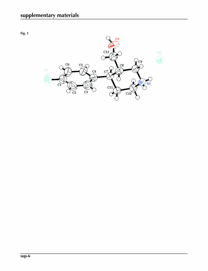
Compound (I) was isolated during degradation studies of paroxetine hydrochloride hemihydrate. The paroxetine drug isavailable in the market as hemihydrate. However, compound (I) is in the anhydrous form, Fig. 1. Similar to the paroxetinehydrochloride salt with protonation having taken place on the basic piperidine ring, the degradation impurity also exists asa hydrochloride salt. The absolute configurations of C7 and C8 atoms were established as R and S, respectively, consistentwith paroxetine hydrochloride hemihydrate. The six-membered piperidinium ring is in the usual chair conformation withthe hydroxylmethyl and 4-fluorophenyl in equatorial positions. The crystal packing shows the formation of a molecular tape
along the b axis through the charge-assisted N+—H···Cl hydrogen bonds (Fig. 2 and Table 1). The tapes thus formed are
connected by O—H···Cl- hydrogen bonds along the a axis.
Experimental
Paroxetine hydrochloride hemihydrate (1.5 gr, 3.5 mmol) was taken in a conical flask and dissolved in acetonitrile andtetrahydrofuran solvent mixture (1:1, 20 ml v/v). About 80 ml of 3% hydrogen peroxide was added to the solution and stirredat 60 °C for 48 h. Chloroform and water was added to the solution and the organic and aqueous layers were separated usingseparating flask. Benzene was added to the aqueous layer and the product (I) was isolated by drying the solution. Singlecrystals were obtained during purification of (I) from chloroform and methanol. The product was characterized by massspectroscopy (M+1 at m/z 210) and NMR.
Refinement
The H atoms bonded to the N and O atoms were located in a difference map and refined isotropically, see Table 1 fordistances. The remaining H atoms were positioned geometrically and refined in the riding model approximation with C—H= 0.93 - 0.97 Å, and with U(H) set to 1.2Ueq(C).
Figures
Fig. 1. Molecular structure of (I) showing the atom numbering scheme. The displacement el-lipsoids are drawn at the 50% probability level. H-atoms are shown by small circles of arbit-rary radii.

supplementary materials
sup-2
Fig. 2. Crystal packing for (I). The molecular tape is sustained through the charge-assistedN+—H···Cl hydrogen bonds, shown as dashed lines.
(3S,4R)-4-(4-Fluorophenyl)-3-(hydroxymethyl)piperidinium chloride
Crystal data
C12H17FNO+·Cl– F000 = 260.00
Mr = 245.72 Dx = 1.335 Mg m−3
Monoclinic, P21Mo Kα radiationλ = 0.71070 Å
Hall symbol: P 2yb Cell parameters from 3399 reflectionsa = 7.697 (4) Å θ = 1.5–27.4ºb = 5.958 (3) Å µ = 0.30 mm−1
c = 13.393 (8) Å T = 298 Kβ = 95.505 (5)º Block, colorless
V = 611.4 (6) Å3 0.50 × 0.40 × 0.20 mmZ = 2
Data collection
Rigaku Mercurydiffractometer 2163 reflections with F2 > 2σ(F2)
Detector resolution: 7.31 pixels mm-1 Rint = 0.036
ω scans θmax = 27.4ºAbsorption correction: multi-scan(Jacobson, 1998) h = −9→9
Tmin = 0.863, Tmax = 0.939 k = −5→76813 measured reflections l = −17→172421 independent reflections
Refinement
Refinement on F2 w = 1/[σ2(Fo2) + (0.1198P)2 + 0.1259P]
where P = (Fo2 + 2Fc
2)/3
R[F2 > 2σ(F2)] = 0.065 (Δ/σ)max = 0.006
wR(F2) = 0.203 Δρmax = 0.54 e Å−3
S = 1.13 Δρmin = −0.37 e Å−3
2421 reflections Extinction correction: none158 parameters Absolute structure: Flack (1983), 938 Friedel PairsH atoms treated by a mixture of Flack parameter: −0.11 (13)

supplementary materials
sup-3
independent and constrained refinement
Special details
Geometry. Bond distances, angles etc. have been calculated using the rounded fractional coordinates. All su's are estimated from thevariances of the (full) variance-covariance matrix. The cell e.s.d.'s are taken into account in the estimation of distances, angles and tor-sion angles
Refinement. Refinement was performed using all reflections. The weighted R-factor (wR) and goodness of fit (S) are based on F2. R-
factor (gt) are based on F. The threshold expression of F2 > 2.0 σ(F2) is used only for calculating R-factor (gt).
Fractional atomic coordinates and isotropic or equivalent isotropic displacement parameters (Å2)
x y z Uiso*/Ueq
F1 −0.4169 (4) 1.0551 (8) 0.5003 (3) 0.1111 (13)O1 −0.1239 (4) 0.4486 (7) 0.1385 (3) 0.0778 (11)N1 0.3679 (4) 0.7472 (8) 0.1251 (2) 0.0576 (10)C1 −0.2900 (5) 0.9849 (10) 0.4441 (4) 0.0738 (16)C2 −0.2390 (6) 1.1230 (9) 0.3728 (4) 0.0764 (14)C3 −0.1118 (5) 1.0490 (8) 0.3135 (3) 0.0651 (12)C4 −0.0374 (4) 0.8397 (6) 0.3271 (2) 0.0505 (10)C5 −0.0914 (5) 0.7080 (9) 0.4028 (2) 0.0584 (11)C6 −0.2175 (5) 0.7774 (10) 0.4632 (3) 0.0715 (16)C7 0.0988 (3) 0.7497 (7) 0.2628 (2) 0.0460 (9)C8 0.0506 (4) 0.7783 (6) 0.1502 (2) 0.0504 (10)C9 0.1889 (4) 0.6685 (7) 0.0934 (2) 0.0545 (11)C10 0.4156 (4) 0.7361 (10) 0.2348 (2) 0.0583 (10)C11 0.2796 (4) 0.8529 (7) 0.2909 (2) 0.0545 (10)C12 −0.1260 (5) 0.6790 (8) 0.1137 (3) 0.0607 (14)Cl1 0.50505 (12) 0.23916 (19) 0.10014 (7) 0.0599 (3)H1 −0.216 (9) 0.388 (14) 0.144 (5) 0.11 (2)*H2 −0.28790 1.26510 0.36350 0.0910*H3 −0.07640 1.14260 0.26370 0.0780*H5 −0.04120 0.56700 0.41370 0.0700*H6 −0.25160 0.68700 0.51450 0.0860*H7 0.10910 0.58820 0.27590 0.0550*H8 0.04830 0.93910 0.13470 0.0600*H11 0.432 (6) 0.658 (9) 0.099 (3) 0.062 (14)*H12 0.378 (10) 0.869 (7) 0.095 (5) 0.12 (2)*H91 0.18390 0.50730 0.10290 0.0650*H92 0.16270 0.69860 0.02230 0.0650*H101 0.52820 0.80710 0.25100 0.0700*H102 0.42530 0.58030 0.25570 0.0700*H111 0.27690 1.01140 0.27430 0.0650*H112 0.31030 0.83830 0.36250 0.0650*H121 −0.21760 0.75500 0.14550 0.0730*H122 −0.14830 0.69780 0.04160 0.0730*

supplementary materials
sup-4
Atomic displacement parameters (Å2)
U11 U22 U33 U12 U13 U23
F1 0.0734 (18) 0.145 (3) 0.120 (2) 0.001 (2) 0.0353 (17) −0.061 (2)O1 0.0611 (19) 0.076 (2) 0.094 (2) −0.0245 (16) −0.0043 (17) 0.0047 (18)N1 0.0510 (15) 0.0604 (19) 0.0638 (17) −0.0101 (18) 0.0179 (13) −0.010 (2)C1 0.050 (2) 0.095 (4) 0.078 (2) −0.005 (2) 0.0143 (19) −0.031 (2)C2 0.065 (2) 0.066 (2) 0.097 (3) 0.009 (2) 0.002 (2) −0.026 (2)C3 0.064 (2) 0.057 (2) 0.074 (2) 0.0021 (19) 0.0046 (19) −0.002 (2)C4 0.0491 (17) 0.0488 (19) 0.0537 (17) −0.0014 (15) 0.0063 (14) −0.0020 (15)C5 0.0510 (17) 0.069 (2) 0.0563 (18) −0.0044 (18) 0.0103 (14) 0.0036 (19)C6 0.058 (2) 0.097 (4) 0.062 (2) −0.010 (2) 0.0187 (16) −0.010 (2)C7 0.0420 (14) 0.0472 (16) 0.0491 (15) −0.0047 (16) 0.0056 (11) −0.0011 (16)C8 0.0494 (16) 0.052 (2) 0.0494 (16) −0.0049 (14) 0.0024 (12) 0.0050 (14)C9 0.055 (2) 0.060 (2) 0.0492 (17) −0.0112 (15) 0.0088 (14) −0.0045 (15)C10 0.0421 (15) 0.070 (2) 0.0624 (19) −0.004 (2) 0.0035 (13) −0.011 (2)C11 0.0438 (17) 0.067 (2) 0.0523 (17) −0.0049 (16) 0.0025 (13) −0.0104 (18)C12 0.052 (2) 0.072 (3) 0.057 (2) −0.0043 (17) −0.0006 (15) 0.0019 (18)Cl1 0.0573 (4) 0.0551 (5) 0.0682 (5) −0.0063 (4) 0.0106 (3) −0.0025 (4)
Geometric parameters (Å, °)
F1—C1 1.355 (6) C8—C12 1.520 (5)O1—C12 1.412 (6) C10—C11 1.515 (5)O1—H1 0.81 (7) C2—H2 0.9300N1—C10 1.481 (4) C3—H3 0.9300N1—C9 1.479 (5) C5—H5 0.9300N1—H12 0.84 (5) C6—H6 0.9300N1—H11 0.83 (5) C7—H7 0.9800C1—C6 1.370 (8) C8—H8 0.9800C1—C2 1.347 (8) C9—H91 0.9700C2—C3 1.390 (6) C9—H92 0.9700C3—C4 1.377 (6) C10—H101 0.9700C4—C5 1.377 (5) C10—H102 0.9700C4—C7 1.517 (4) C11—H111 0.9700C5—C6 1.385 (6) C11—H112 0.9700C7—C11 1.535 (4) C12—H121 0.9700C7—C8 1.528 (4) C12—H122 0.9700C8—C9 1.515 (5)
C12—O1—H1 118 (6) C4—C5—H5 119.00C9—N1—C10 114.0 (3) C6—C5—H5 119.00C9—N1—H12 105 (5) C1—C6—H6 121.00C10—N1—H11 107 (3) C5—C6—H6 121.00H11—N1—H12 106 (6) C4—C7—H7 107.00C10—N1—H12 119 (5) C8—C7—H7 107.00C9—N1—H11 105 (3) C11—C7—H7 107.00F1—C1—C2 118.7 (5) C7—C8—H8 108.00

supplementary materials
sup-5
F1—C1—C6 118.5 (5) C9—C8—H8 108.00C2—C1—C6 122.8 (4) C12—C8—H8 108.00C1—C2—C3 118.7 (5) N1—C9—H91 109.00C2—C3—C4 121.2 (4) N1—C9—H92 109.00C3—C4—C7 123.1 (3) C8—C9—H91 109.00C5—C4—C7 119.4 (3) C8—C9—H92 109.00C3—C4—C5 117.5 (3) H91—C9—H92 108.00C4—C5—C6 122.5 (5) N1—C10—H101 109.00C1—C6—C5 117.1 (4) N1—C10—H102 109.00C8—C7—C11 109.0 (2) C11—C10—H101 109.00C4—C7—C11 112.3 (3) C11—C10—H102 110.00C4—C7—C8 113.9 (2) H101—C10—H102 108.00C9—C8—C12 108.7 (3) C7—C11—H111 110.00C7—C8—C9 109.4 (2) C7—C11—H112 110.00C7—C8—C12 113.5 (3) C10—C11—H111 110.00N1—C9—C8 113.5 (3) C10—C11—H112 110.00N1—C10—C11 110.7 (3) H111—C11—H112 108.00C7—C11—C10 110.4 (3) O1—C12—H121 110.00O1—C12—C8 108.3 (3) O1—C12—H122 110.00C1—C2—H2 121.00 C8—C12—H121 110.00C3—C2—H2 121.00 C8—C12—H122 110.00C2—C3—H3 119.00 H121—C12—H122 108.00C4—C3—H3 119.00
C10—N1—C9—C8 51.5 (5) C5—C4—C7—C11 −103.8 (4)C9—N1—C10—C11 −52.1 (6) C4—C5—C6—C1 0.8 (6)F1—C1—C2—C3 −178.5 (4) C4—C7—C8—C9 −176.0 (3)C6—C1—C2—C3 2.4 (8) C4—C7—C8—C12 −54.5 (4)F1—C1—C6—C5 178.4 (4) C11—C7—C8—C9 57.7 (4)C2—C1—C6—C5 −2.5 (7) C11—C7—C8—C12 179.3 (3)C1—C2—C3—C4 −0.5 (7) C4—C7—C11—C10 172.4 (3)C2—C3—C4—C5 −1.1 (6) C8—C7—C11—C10 −60.4 (4)C2—C3—C4—C7 178.6 (4) C7—C8—C9—N1 −53.7 (4)C3—C4—C5—C6 1.0 (5) C12—C8—C9—N1 −178.2 (3)C7—C4—C5—C6 −178.8 (3) C7—C8—C12—O1 −58.0 (4)C3—C4—C7—C8 −48.1 (5) C9—C8—C12—O1 64.0 (4)C3—C4—C7—C11 76.5 (4) N1—C10—C11—C7 56.7 (5)C5—C4—C7—C8 131.7 (3)
Hydrogen-bond geometry (Å, °)
D—H···A D—H H···A D···A D—H···A
O1—H1···Cl1i 0.81 (7) 2.35 (7) 3.114 (4) 160 (6)N1—H11···Cl1 0.83 (5) 2.56 (5) 3.234 (5) 140 (4)
N1—H12···Cl1ii 0.84 (5) 2.41 (5) 3.144 (5) 147 (6)Symmetry codes: (i) x−1, y, z; (ii) x, y+1, z.
supplementary materials
sup-6
Fig. 1

supplementary materials
sup-7
Fig. 2
Related Documents


![SUPPORTING INFORMATION · 2006-12-18 · 2-Hydroxymethyl-1-[(4-methyl)phenylsulfonyl]pyrrolidine (6): To a solution of (±)-2- (hydroxymethyl)pyrrolidine 3 (0.56 mL, 5.7 mmol) in](https://static.cupdf.com/doc/110x72/5ebcca293aaa475756114192/supporting-2006-12-18-2-hydroxymethyl-1-4-methylphenylsulfonylpyrrolidine.jpg)
