IODINE 33 3. HEALTH EFFECTS 3.1 INTRODUCTION The primary purpose of this chapter is to provide public health officials, physicians, toxicologists, and other interested individuals and groups with an overall perspective on the toxicology of iodine. It contains descriptions and evaluations of toxicological studies and epidemiological investigations and provides conclusions, where possible, on the relevance of toxicity and toxicokinetic data to public health. Section 3.2 contains a discussion of the chemical toxicity of stable iodine; radiation toxicity associated with exposure to radioiodine is discussed in Section 3.3. A glossary and list of acronyms, abbreviations, and symbols can be found at the end of this profile. Health effects of the element iodine are categorized by the chemical nature of stable iodine ( 127 I) and the radioactive nature of unstable isotopes (e.g., 131 I). Four radioactive isotopes ( 123 I, 125 I, 129 I, and 131 I) are of particular interest with respect to human exposures because 125 I and 131 I are used medically and all four isoptopes are sufficiently long-lived to be transported to human receptors after their release into the environment. These isotopes of iodine emit, primarily, beta radiation (that travel short distances in tissues) and gamma radiation (that can penetrate the entire body). The radiation dose from these radionuclides can be classified as either external (if the source is outside the body) or internal (if the source is inside the body). The external dose from iodine radionuclides arises primarily from the penetrating gamma radiation that can travel through air. Beta radiation emitted outside the body is normally of little health concern unless the radioactive material contacts the skin. Skin contact can allow the beta radiation to pass through the epidermis to live dermal tissue where it can contribute to the radiation dose to the skin. At very high external doses, beta (and gamma) radiation (e.g., greater than 3 Gy, 300 rad) can cause such adverse effects as skin erythema, ulceration, or necrosis (Agency for Toxic Substances and Disease Registry [ATSDR] 1999). Once radioactive iodine is internalized, it is absorbed, distributed, and excreted in the same manner as stable iodine. The internal radiation dose from radioactive iodine is actually a measure of the amount of energy that the beta and gamma emissions deposit in tissue. The short-range beta radiation produces a

Welcome message from author
This document is posted to help you gain knowledge. Please leave a comment to let me know what you think about it! Share it to your friends and learn new things together.
Transcript

IODINE 33
3. HEALTH EFFECTS
3.1 INTRODUCTION
The primary purpose of this chapter is to provide public health officials, physicians, toxicologists, and
other interested individuals and groups with an overall perspective on the toxicology of iodine. It
contains descriptions and evaluations of toxicological studies and epidemiological investigations and
provides conclusions, where possible, on the relevance of toxicity and toxicokinetic data to public health.
Section 3.2 contains a discussion of the chemical toxicity of stable iodine; radiation toxicity associated
with exposure to radioiodine is discussed in Section 3.3.
A glossary and list of acronyms, abbreviations, and symbols can be found at the end of this profile.
Health effects of the element iodine are categorized by the chemical nature of stable iodine (127I) and the
radioactive nature of unstable isotopes (e.g., 131I). Four radioactive isotopes (123I, 125I, 129I, and 131I) are of
particular interest with respect to human exposures because 125I and 131I are used medically and all four
isoptopes are sufficiently long-lived to be transported to human receptors after their release into the
environment. These isotopes of iodine emit, primarily, beta radiation (that travel short distances in
tissues) and gamma radiation (that can penetrate the entire body). The radiation dose from these
radionuclides can be classified as either external (if the source is outside the body) or internal (if the
source is inside the body).
The external dose from iodine radionuclides arises primarily from the penetrating gamma radiation that
can travel through air. Beta radiation emitted outside the body is normally of little health concern unless
the radioactive material contacts the skin. Skin contact can allow the beta radiation to pass through the
epidermis to live dermal tissue where it can contribute to the radiation dose to the skin. At very high
external doses, beta (and gamma) radiation (e.g., greater than 3 Gy, 300 rad) can cause such adverse
effects as skin erythema, ulceration, or necrosis (Agency for Toxic Substances and Disease Registry
[ATSDR] 1999).
Once radioactive iodine is internalized, it is absorbed, distributed, and excreted in the same manner as
stable iodine. The internal radiation dose from radioactive iodine is actually a measure of the amount of
energy that the beta and gamma emissions deposit in tissue. The short-range beta radiation produces a

IODINE 34
3. HEALTH EFFECTS
localized dose while the more penetrating gamma radiation contributes to a whole-body dose. Molecular
damage results from the direct ionization of atoms that are encountered by beta and gamma radiation and
by interactions of resulting free radicals with nearby atoms. Tissue damage results when the molecular
damage is extensive and not sufficiently repaired in a timely manner.
In radiation biology, the term absorbed dose is the amount of energy deposited by radiation per unit mass
of tissue, expressed in units of gray (Gy) or rad (see Appendix D for a detailed description of principles of
ionizing radiation). The term dose equivalent refers to the biologically significant dose, which is
determined by multiplying the absorbed dose by a quality factor for the type and energy of the radiations
involved. Dose equivalent is expressed in units of sievert (Sv) or rem. The quality factor is considered to
be unity for the beta and gamma radiation emitted from iodine radionuclides, so for these radionuclides,
the absorbed dose (in Gray or rad) is numerically identical to the dose equivalent (in rem or sievert). The
absorbed dose from internalized iodine radionuclides is estimated by taking into account the quantity of
radionuclides entering the body (via ingestion or inhalation), the biokinetics (retention, distribution, and
excretion) of the radionuclides, the rate at which the radionuclides decay to stable forms, the energies and
intensities of the beta and gamma radiation emitted, and the characteristics of tissues that result in the
energy of the emitted radiation being absorbed by tissues. Each tissue, therefore, can receive a different
absorbed dose for a given amount of radioactivity that enters the body. The total absorbed dose to the
body will reflect the integration of the absorbed doses for the all tissues. In summaries of the radioiodine
toxicology literature provided in this profile, units of activity, absorbed dose, or dose equivalent are cited
as reported in the literature and the corresponding conventional or SI units are provided in parentheses.
The EPA has published a set of internal dose conversion factors for standard persons of various ages
(newborn; 1, 5, 10, or 15 years of age; and adult) in its Federal Guidance Report No. 13 supplemental CD
(EPA 1999). For example, the EPA has estimated that the dose equivalent following ingestion of 1 Bq of 131I is 2.2x10⁻⁸ Sv (assuming an integration time of 50 years for an adult following the initial exposure).
Age-specific dose coefficients for inhalation and ingestion of any of the radioactive isotopes of iodine by
the general public can be found in International Commission on Radiological Protection (ICRP)
publications 71 (ICRP 1995) and 72 (ICRP 1996), respectively. Dose coefficients for inhalation,
ingestion, and submersion in a cloud of iodine radionuclides can be found in U.S. EPA Federal Guidance
Report No. 11 (EPA 1988). Dose coefficients for external exposure to radioisotopes of iodine in air,
surface water, or soil contaminated to various depths can be found in U.S. EPA Federal Guidance Report
No. 12 (EPA 1993).

IODINE 35
3. HEALTH EFFECTS
The ICRP has developed reference values for dose coefficients that relate dose equivalents to a unit of
activity to which a person might be exposed. For example, the ICRP (1996, 2001) has estimated that the
dose coefficient for an acute exposure of an adult to 131I is 2.2x10⁻⁸ Sv/Bq. Age-specific dose coefficients
for inhalation and ingestion of any of the radioactive isotopes of iodine can be found in ICRP publications
71 (ICRP 1995) and 72 (ICRP 1996), respectively.
3.2 DISCUSSION OF HEALTH EFFECTS FOR STABLE IODINE BY ROUTE OF EXPOSURE
Section 3.2 discusses the chemical toxicity of iodine. Radiation toxicity resulting from exposure to
radioiodine is discussed in Section 3.3.
To help public health professionals and others address the needs of persons living or working near
hazardous waste sites, the information in this section is organized first by route of exposure (inhalation,
oral, and dermal) and then by health effect (death, systemic, immunological, neurological, reproductive,
developmental, genotoxic, and carcinogenic effects). These data are discussed in terms of three exposure
periods: acute (14 days or less), intermediate (15–364 days), and chronic (365 days or more).
Levels of significant exposure for each route and duration are presented in tables and illustrated in
figures. The points in the figures showing no-observed-adverse-effect levels (NOAELs) or lowest-
observed-adverse-effect levels (LOAELs) reflect the actual doses (levels of exposure) used in the studies.
LOAELs have been classified into "less serious" or "serious" effects. "Serious" effects are those that
evoke failure in a biological system and can lead to morbidity or mortality (e.g., acute respiratory distress
or death). "Less serious" effects are those that are not expected to cause significant dysfunction or death,
or those whose significance to the organism is not entirely clear. ATSDR acknowledges that a
considerable amount of judgment may be required in establishing whether an end point should be
classified as a NOAEL, "less serious" LOAEL, or "serious" LOAEL, and that in some cases, there will be
insufficient data to decide whether the effect is indicative of significant dysfunction. However, the
Agency has established guidelines and policies that are used to classify these end points. ATSDR
believes that there is sufficient merit in this approach to warrant an attempt at distinguishing between
"less serious" and "serious" effects. The distinction between "less serious" effects and "serious" effects is
considered to be important because it helps the users of the profiles to identify levels of exposure at which
major health effects start to appear. LOAELs or NOAELs should also help in determining whether or not
the effects vary with dose and/or duration, and place into perspective the possible significance of these
effects to human health.

IODINE 36
3. HEALTH EFFECTS
The significance of the exposure levels shown in the Levels of Significant Exposure (LSE) tables and
figures may differ depending on the user's perspective. Public health officials and others concerned with
appropriate actions to take at hazardous waste sites may want information on levels of exposure
associated with more subtle effects in humans or animals (LOAELs) or exposure levels below which no
adverse effects (NOAELs) have been observed. Estimates of levels posing minimal risk to humans
(Minimal Risk Levels or MRLs) may be of interest to health professionals and citizens alike.
Levels of exposure associated with carcinogenic effects (Cancer Effect Levels, CELs) of iodine are
indicated in Tables 3-1 and 3-2 and Figures 3-1 and 3-2.
Estimates of exposure levels posing minimal risk to humans (Minimal Risk Levels or MRLs) have been
made for iodine. An MRL is defined as an estimate of daily human exposure to a substance that is likely
to be without an appreciable risk of adverse effects (noncarcinogenic) over a specified duration of
exposure. MRLs are derived when reliable and sufficient data exist to identify the target organ(s) of
effect or the most sensitive health effect(s) for a specific duration within a given route of exposure.
MRLs are based on noncancerous health effects only and do not consider carcinogenic effects. MRLs can
be derived for acute, intermediate, and chronic duration exposures for inhalation and oral routes.
Appropriate methodology does not exist to develop MRLs for dermal exposure.
Although methods have been established to derive these levels (Barnes and Dourson 1988; EPA 1990),
uncertainties are associated with these techniques. Furthermore, ATSDR acknowledges additional
uncertainties inherent in the application of the procedures to derive less than lifetime MRLs. As an
example, acute inhalation MRLs may not be protective for health effects that are delayed in development
or are acquired following repeated acute insults, such as hypersensitivity reactions, asthma, or chronic
bronchitis. As these kinds of health effects data become available and methods to assess levels of
significant human exposure improve, these MRLs will be revised.
A User's Guide has been provided at the end of this profile (see Appendix B). This guide should aid in
the interpretation of the tables and figures for Levels of Significant Exposure and the MRLs.

IODINE 37
3. HEALTH EFFECTS
3.2.1 Inhalation Exposure
Iodine is absorbed in humans when I2 or methyl iodide vapors are inhaled (Black and Hounam 1968;
Morgan and Morgan 1967; Morgan et al. 1967a, 1967b, 1968). Once absorbed, iodide would be expected
to exert effects that are similar to that of iodide absorbed after ingestion, including effects on the thyroid
gland and thyroid hormone status, sensitivity reactions, and cancer (see Section 3.2.2). Iodine (I2) is a
strong oxidizing agent; therefore, exposure to high air concentrations of I2 vapor could potentially
produce upper respiratory tract irritation and possibly oxidative injury. No studies were located regarding
the following health effects in humans or animals after inhalation exposure to stable iodine:
3.2.1.1 Death
3.2.1.2 Systemic Effects
3.2.1.3 Immunological and Lymphoreticular Effects
3.2.1.4 Neurological Effects
3.2.1.5 Reproductive Effects
3.2.1.6 Developmental Effects
3.2.1.7 Cancer
3.2.2 Oral Exposure
The section that follows provides background information relevant to the various study summaries that
are presented subsequently. A description of the approaches used to calculate doses of stable iodine is
provided. The actual study summaries follow.
A large number of human experimental, clinical, and epidemiological studies on the effects of excess
iodine on human health have been reported. The key studies that provide information on exposures
associated with effects are summarized in this section of the profile. Oral iodine intakes were not directly
assessed in many studies with sufficient accuracy to define dose-response relationships; however,
measurements of urinary iodide provide a basis for estimating intakes in some of the studies (Konno et al.
IODINE 38
3. HEALTH EFFECTS
1993b). Rather than describing the basis for estimating intakes from urinary iodine data in each of the
study descriptions that follow, the general approach used is described here. If a 24-hour urinary iodide
measurement was provided that could be regarded as a steady state value (relatively constant intake for at
least 6 months), the intake was assumed to be equivalent to the 24-hour excretion rate. This assumption
is consistent with information available on the toxicokinetics of iodide that indicates nearly complete
absorption of ingested iodide and that urinary excretion accounts for >97% of the absorbed dose (see
Sections 3.5.1.2 and 3.5.4.2). The assumption is also supported by studies in which 24-hour urinary
iodide was measured before and after supplementation. For example, 31 patients received oral
supplements of 382 µg I/day for 6 months. Prior to the supplementation, the mean 24-hour urinary iodide
excretion rate was 36 µg/day (range, 13–69), whereas after 6 months of iodide supplementation, the mean
24-hour urinary iodide excretion rate was 415 µg/day (Kahaly et al. 1998). The difference between these
two values, 379 µg/day, is nearly identical to the supplemental dose of 382 µg/day.
If a urine iodide concentration was provided for a morning sample that included overnight bladder urine,
the measured concentration was assumed to represent the 24-hour average concentration and iodide intake
was calculated as:
Intake U 1.4I I= ⋅ Equation (1)
where UI is the measured urinary iodine concentration and 1.4 is the average volume of urine excreted per
day (L/day) for a 70-kg adult (ICRP 1981). Equation 1 is in reasonable agreement with observed
relationships between morning bladder urine iodide concentrations and 24-hour iodide excretion rates
(Konno et al. 1993b; Nagata et al. 1998). Urine iodide concentration from untimed (spot) samples, other
than morning samples that included overnight bladder urine, were considered to be potentially too
uncertain to derive intake estimates, unless paired urinary creatinine concentrations or a urinary
iodide:creatinine ratio (µg I:g creatinine) was reported. Urinary iodide:creatinine ratios were converted to
estimated iodide intake as follows, assuming a constant relationship between urinary creatinine excretion
rate and lean body mass. The rate of creatinine excretion (e.g., ECr , g creatinine/day) was calculated from
the relationship between lean body mass (LBM) and ECr:
LBM ECr= ⋅ +0 0272 858. . Equation (2)

IODINE 39
3. HEALTH EFFECTS
where the constants 0.0272 and 8.58 are the weighted arithmetic mean of estimates of these variables
from eight studies reported in Forbes and Bruining (1976). Lean body mass was calculated as follows
(ICRP 1981):
LBM BW males= ⋅0 88. , Equation (3)
LBM BW females= ⋅ 085. ,
where BW is the reported or assumed body weight for males (75 kg) and females (65 kg) (EPA 1997f). A
mean value of 60 kg (females, 55 kg; males, 65 kg) was assumed for body weights of adult populations of
the Asian Pacific countries (e.g., Japan, China, Marshall Islands). Iodide intake was calculated as:
Intake U EI I Cr Cr= ⋅/ Equation (4)
where UI/Cr is the urinary iodide:creatinine ratio (µg I/g creatinine). This approach yields relationships
between 24-hour urinary iodide excretion rates and the urinary iodide:creatinine ratios that are in
reasonable agreement with observations (Konno et al. 1993b).
3.2.2.1 Death
Two recent reviews of the clinical case literature note that deaths have occurred after ingestion of iodine
preparations (FDA 1989b; Pennington 1990b). A review of medical records from the New York City
Medical Examiners Office revealed that, in a period of 6 years, there were 18 deaths from attempted
suicides in which adults ingested iodine tinctures (Finkelstein and Jacobi 1937). Tinctures of iodine
contain a mixture of molecular iodine (I2) and sodium triiodide (NaI3) and have iodine concentrations of
approximately 40 mg/mL. Doses of iodine from ingestion of the tinctures ranged from 1,200 to 9,500 mg
(17–120 mg/kg), and deaths usually occurred within 48 hours of the dose. In one case, an adult male
ingested 15 g of iodine as a potassium iodide solution and survived the episode; 18 hours after the dose,
his serum iodide concentration was 2.95 mg/mL (Tresch et al. 1974). Symptoms of toxicity that have
been observed in lethal or near-lethal poisonings have included abdominal cramps, bloody diarrhea and
gastrointestinal ulceration, edema of the face and neck, pneumonitis, hemolytic anemia, metabolic
acidosis, fatty degeneration of the liver, and renal failure (Clark 1981; Dyck et al. 1979; Finkelstein and
Jacobi 1937; Tresch et al. 1974).

IODINE 40
3. HEALTH EFFECTS
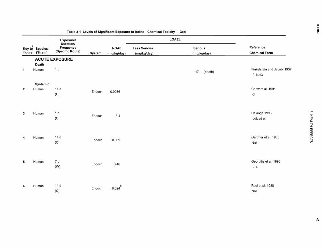
Two cases of infant deaths were reported in which death was from complications related to compression
of the trachea by a goiterous thyroid gland; the mothers had ingested potassium iodide during their
pregnancies at doses of approximately 850 and 1,180 mg I/day (12 and 17 mg/kg/day) (Galina et al.
1962).
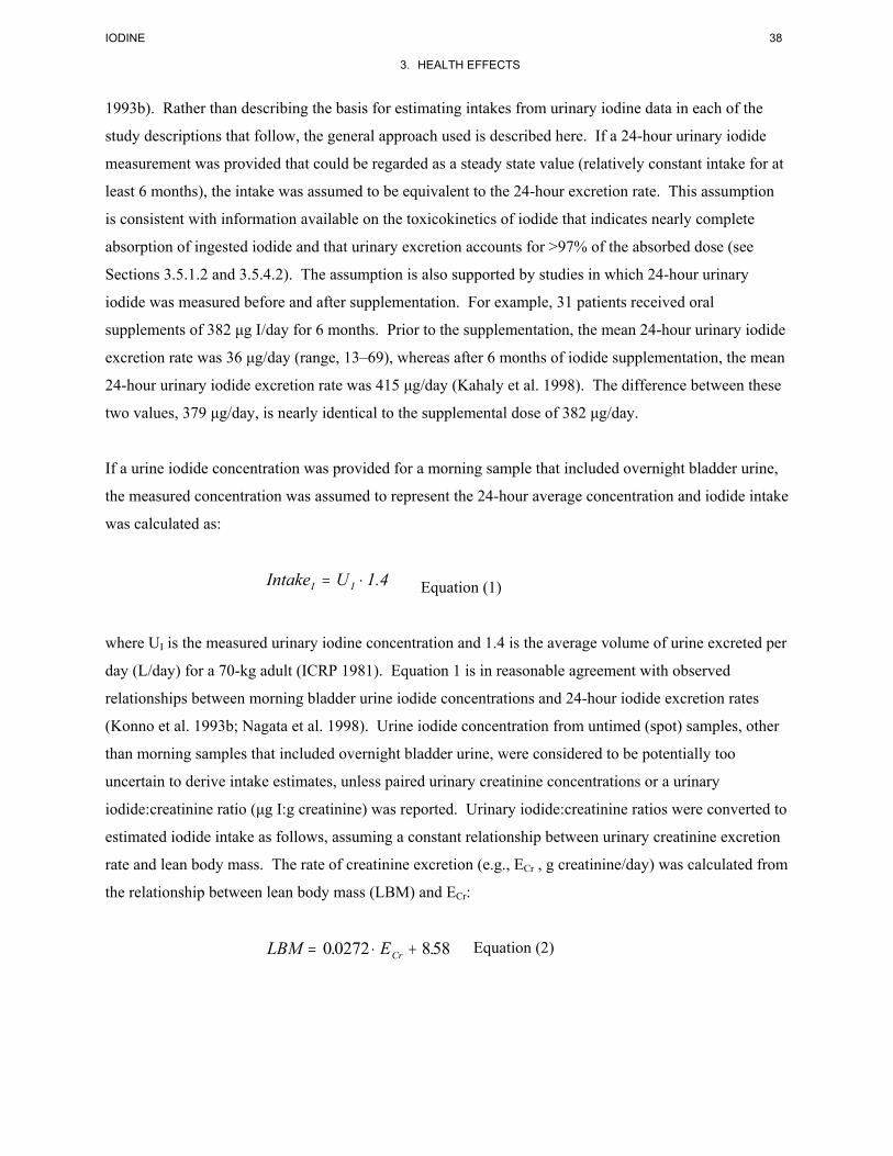
The LOAEL values in humans for exposures by the oral route are presented in Table 3-1 and plotted in
Figure 3-1.
3.2.2.2 Systemic Effects
Systemic effects of oral stable iodine exposure, other than after massive acute iodine overload such as in
cases of attempted suicides (see Section 3.2.2.1), are on the thyroid gland and are discussed in the section
on Endocrine Effects. As noted in the introduction to this chapter of the profile, adverse effects on a wide
variety of other organ systems can result from iodine-induced disorders of the thyroid gland, including
disturbances of the skin, cardiovascular system, pulmonary system, kidneys, gastrointestinal tract, liver,
blood, neuromuscular system, central nervous system, skeleton, male and female reproductive systems,
and numerous endocrine organs, including the pituitary and adrenal glands. The reader is referred to
authoritative references on these subjects for further information (Braverman and Utiger 2000).
Endocrine Effects. The principal direct effects of excessive stable iodine ingestion on the endocrine
system are on the thyroid gland and regulation of thyroid hormone production and secretion. Adverse
effects on the pituitary and adrenal glands derive secondarily from disorders of the thyroid gland. Effects
on the thyroid gland can be classified into three types: hypothyroidism, hyperthyroidism, and thyroiditis.
Hypothyroidism refers to the diminished production of thyroid hormone leading to clinical manifestations
of thyroid insufficiency and can occur with or without goiter, a functional hypertrophy of the gland in
response to suppressed hormone production and elevated serum thyroid stimulating hormone (TSH, also
known as thyrotropin) concentrations. Typical biomarkers of hypothyroidism are a depression in the
circulating levels of thyroxine (T4) and/or triiodothyronine (T3) below their normal ranges. This is always
accompanied by an elevation of the pituitary hormone, TSH, above the normal range. Hyperthyroidism is
an excessive production and/or secretion of thyroid hormones. The clinical manifestation of abnormally
elevated circulating levels of T4 and/or T3 is thyrotoxicosis. Thyroiditis refers to an inflammation of the
gland, which is often secondary to thyroid gland autoimmunity. The above three types of effects can
occur in children and adults, in fetuses exposed in utero, or in infants during lactation.
LOAEL
Less SeriousNOAEL Seriousa
SystemKey tofigure
Reference
Table 3-1 Levels of Significant Exposure to Iodine - Chemical Toxicity - Oral
Chemical Form(mg/kg/day) (mg/kg/day) (mg/kg/day)
Exposure/Duration/
Frequency(Specific Route)
Species(Strain)
ACUTE EXPOSUREDeath
1
60
1717 (death)
1 dHuman Finkelstein and Jacobi 1937
I2, Nal3
Systemic2
0.00860.0086Endocr
77
14 d(C)
Human Chow et al. 1991
KI
3
3.43.4Endocr
78
1 d(C)
Human Delange 1996
Iodized oil
4
0.0690.069Endocr
79
14 d(C)
Human Gardner et al. 1988
NaI
5
0.460.46Endocr
80
7 d(W)
Human Georgitis et al. 1993
I2, I-
6
0.0240.024
bEndocr
81
14 d(C)
Human Paul et al. 1988
NaI
LOAEL
Less SeriousNOAEL Seriousa
SystemKey tofigure
Reference
(continued)Table 3-1 Levels of Significant Exposure to Iodine - Chemical Toxicity - Oral
Chemical Form(mg/kg/day) (mg/kg/day) (mg/kg/day)
Exposure/Duration/
Frequency(Specific Route)
Species(Strain)
7
11Endocr
82
14 d(C)
Human Robison et al. 1998
NaI
8
11Endocr
83
14 d(C)
Human Robison et al. 1998
I2
Immuno/ Lymphoret9
65
2020 (fever)
8 d(C)
Human Horn and Kabins 1972
KI
10
66
4.34.3 (ioderma)
5 d(C)
Human Soria et al. 1990
KI
INTERMEDIATE EXPOSUREDeath
11
130
12
12 (death from trachealcompression by goiter)
9 mo(C)
Human Galina et al. 1962
KI
12
131
17
17 (death from trachealcompression by goiter)
9 mo(C)
Human Galina et al. 1962
KI

LOAEL
Less SeriousNOAEL Seriousa
SystemKey tofigure
Reference
(continued)Table 3-1 Levels of Significant Exposure to Iodine - Chemical Toxicity - Oral
Chemical Form(mg/kg/day) (mg/kg/day) (mg/kg/day)
Exposure/Duration/
Frequency(Specific Route)
Species(Strain)
Systemic13
Endocr
159
23
23 (clinical hyperthyroidism withthyrotoxicosis)
4 mo(C)
Human Ahmed et al. 1974
KI
14Endocr
161
7.37.3 (goiter in neonate)
2 mo(C)
Human Coakley et al. 1989
KI
15Endocr
162
6.4
6.4 (goiter and hypothyroidism inneonate)
9 mo(C)
Human Hassan et al. 1968
KI
16
1515Endocr
163
11 wk(W)
Human Jubiz et al. 1977
KI
17
0.00390.0039Endocr
1650.46
0.46 (subclinical hypothyroidism withgland enlargement)
90 d(C)
Human LeMar et al. 1995
I2 ,I-
18
0.00470.0047Endocr
166
9 mo(C)
Human Liesenkotter et al. 1996
KI
LOAEL
Less SeriousNOAEL Seriousa
SystemKey tofigure
Reference
(continued)Table 3-1 Levels of Significant Exposure to Iodine - Chemical Toxicity - Oral
Chemical Form(mg/kg/day) (mg/kg/day) (mg/kg/day)
Exposure/Duration/
Frequency(Specific Route)
Species(Strain)
19Endocr
167
13
13 (goiter, hypothyroidism inneonate)
9 mo(C)
Human Martin and Rento 1992
KI
20Endocr
1690.39
0.39 (subclinical hypothyroidism withgland enlargement)
28 d(C)
Human Namba et al. 1993
I-
21Endocr
170
5.45.4 (goiter in neonate)
3 mo(C)
Human Penfold et al. 1978
KI
22Endocr
171
6.66.6 (goiter in neonate)
4 mo(C)
Human Penfold et al. 1978
KI
23Endocr
173
0.050.05 (clinical hypothyroidism)
6 mo(C)
Human Shilo and Hirsch 1986
sea-kelp
24Endocr
174
2.6
2.6 (clinical hyperthyroidism withthyrotoxicosis)
7 wk(C)
Human Vagenakis et al. 1972
KI
25Endocr
175
4.64.6 (goiter in fetus)
9 mo(C)
Human Vicens-Colvet et al. 1998
ND

LOAEL
Less SeriousNOAEL Seriousa
SystemKey tofigure
Reference
(continued)Table 3-1 Levels of Significant Exposure to Iodine - Chemical Toxicity - Oral
Chemical Form(mg/kg/day) (mg/kg/day) (mg/kg/day)
Exposure/Duration/
Frequency(Specific Route)
Species(Strain)
Immuno/ Lymphoret26
140
2323 (fever)
NS(C)
Human Horn and Kabins 1972
KI
27
141
1111 (ioderma)
6 mo(C)
Human Kincaid et al. 1981
KI
28
142
8.68.6 (ioderma)
8 mo(C)
Human Soria et al. 1990
KI
CHRONIC EXPOSURESystemic
29
0.010.01
cEndocr
1140.029
0.029 (subclinical hypothyroidism withgland enlargement)
11 yr(W)
Human Boyages et al. 1989
I-
30Endocr
118
2.9
2.9 (clinical hypothyroidism withgoiter in neonate)
2 yr(C)
Human Iancu et al. 1974
NaI
31Endocr
120
1
1 (goiter with elevated serumTSH)
NS(W)
Human Khan et al. 1998
ND

LOAEL
Less SeriousNOAEL Seriousa
SystemKey tofigure
Reference
(continued)Table 3-1 Levels of Significant Exposure to Iodine - Chemical Toxicity - Oral
Chemical Form(mg/kg/day) (mg/kg/day) (mg/kg/day)
Exposure/Duration/
Frequency(Specific Route)
Species(Strain)
32Endocr
121
0.22
0.22 (clinical hypothyroidism withoutautoimmunity)
46 yr(F)
Human Konno et al. 1994
I-
33
0.00460.0046Endocr
122
68 yr(F, W)
Human Laurberg et al. 1998
I-
34
0.00390.0039Endocr
124
16 mo(C)
Human Pedersen et al. 1993
KI
35
0.00230.0023Endocr
125
0.012
0.012 (clinical hypothyroidism withoutautoimmunity; elderly adults)
81 yr(F, W)
Human Szablocs et al. 1997
I-
Immuno/ Lymphoret36
107
1515 (fever)
15 yr(C)
Human Kurtz and Aber 1982
KI
37
108
1414 (ioderma)
1 yr(C)
Human Rosenberg et al. 1972
KI
Cancer38
96
0.0035
0.0035 (thyroid cancer; in endemicgoiter area)
NS(F)
Human Bacher-Stier et al. 1997
I-

LOAEL
Less SeriousNOAEL Seriousa
SystemKey tofigure
Reference
(continued)Table 3-1 Levels of Significant Exposure to Iodine - Chemical Toxicity - Oral
Chemical Form(mg/kg/day) (mg/kg/day) (mg/kg/day)
Exposure/Duration/
Frequency(Specific Route)
Species(Strain)
39
a The number corresponds to entries in Figure 3-1.
b Used to derive an acute oral MRL based on a no-observed-effect-level (NOEL) of 0.01mg/kg/day in healthy adult humans, for changes in serum thyroid hormone levels. Theno-observed-adverse-effect-level (NOAEL) is 0.024 mg/kg/day.
c Used to derive a chronic oral MRL of 0.005 mg/kg/day; dose divided by an uncertainty factor of 2 for human variability.
(C) = capsule; d = day(s); Endocr = endocrine; (F) = feed; kg = kilogram(s); LOAEL = lowest-observed-adverse-effect level; mg = milligram(s); mo = month(s); NOAEL =no-observed-adverse-effect level; NA = not specified; TSH = thyroid-stimulating hormone; (W) = drinking water; wk = week(s); yr = year(s)
98
0.0035
0.0035 (thyroid cancer; in endemicgoiter area)
NS(F)
Human Harach and Williams 1995
I-

0.001
0.01
0.1
1
10
100
Death
1
Endocrine
2
3
4
5
6
7 8
Imm
uno/Lymphor
9
10
mg/kg/day
Figure 3-1. Levels of Significant Exposure to Iodine - Chemical Toxicity - Oral
Acute (≤14 days)
c-Catd-Dogr-Ratp-Pigq-Cow
-Humansk-Monkeym-Mouseh-Rabbita-Sheep
f-Ferretj-Pigeone-Gerbils-Hamsterg-Guinea Pig
n-Minko-Other
Cancer Effect Level-Animals LOAEL, More Serious-Animals LOAEL, Less Serious-Animals NOAEL - Animals
Cancer Effect Level-Humans LOAEL, More Serious-Humans LOAEL, Less Serious-Humans NOAEL - Humans
LD50/LC50 Minimal Risk Level for effects other than Cancer
Systemic
MRL
0.001
0.01
0.1
1
10
100
Death
11
12
Endocrine
13
1415
16
17
17
18
19
20
2122
23
24
25
Imm
uno/Lymphor
26
2728
mg/kg/day
Figure 3-1. Levels of Significant Exposure to Iodine - Chemical Toxicity - Oral (Continued)
Intermediate (15-364 days)
c-Catd-Dogr-Ratp-Pigq-Cow
-Humansk-Monkeym-Mouseh-Rabbita-Sheep
f-Ferretj-Pigeone-Gerbils-Hamsterg-Guinea Pig
n-Minko-Other
Cancer Effect Level-Animals LOAEL, More Serious-Animals LOAEL, Less Serious-Animals NOAEL - Animals
Cancer Effect Level-Humans LOAEL, More Serious-Humans LOAEL, Less Serious-Humans NOAEL - Humans
LD50/LC50 Minimal Risk Level for effects other than Cancer
Systemic
0.001
0.01
0.1
1
10
100
Endocrine
29
29
30
31
32
3334
35
35
Imm
uno/Lymphor
36 37
Cancer *
38 39
mg/kg/day
Figure 3-1. Levels of Significant Exposure to Iodine - Chemical Toxicity - Oral (Continued)
Chronic (≥365 days)
c-Catd-Dogr-Ratp-Pigq-Cow
-Humansk-Monkeym-Mouseh-Rabbita-Sheep
f-Ferretj-Pigeone-Gerbils-Hamsterg-Guinea Pig
n-Minko-Other
Cancer Effect Level-Animals LOAEL, More Serious-Animals LOAEL, Less Serious-Animals NOAEL - Animals
Cancer Effect Level-Humans LOAEL, More Serious-Humans LOAEL, Less Serious-Humans NOAEL - Humans
LD50/LC50 Minimal Risk Level for effects other than Cancer
Systemic
*Doses represent the lowest dose tested per study that produced a tumorigenic
response and do not imply the existence of a threshold for the cancer endpoint.

IODINE 51
3. HEALTH EFFECTS
Measurements of serum levels of thyroid hormones and TSH are often used as biomarkers of
hypothyroidism and thyrotoxicosis in toxicology and epidemiology studies. In interpreting this literature
in terms of human health risks, a distinction must be made between outcomes that have a high potential
for producing clinical manifestations and outcomes that are not clinically significant. In this profile, an
observed increase in serum TSH level and normal T4 and T3 levels is referred to as subclinical
hypothyroidism. Similarly, the term subclinical hyperthyroidism refers to a condition in which the
circulating levels of T4 or T3 are normal and the serum TSH concentration is suppressed. Typical normal
ranges for these hormone levels are discussed in Section 3.9.2
Hypothyroidism
An acute iodide excess (above the preexisting dietary intake) transiently decreases the production of
thyroid hormones in the thyroid gland; this is referred to as the acute Wolff-Chaikoff effect (Wolff et al.
1949). In normal people, this is followed by a return to normal levels of hormone synthesis, referred to as
escape from the acute Wolff-Chaikoff effect, without a significant change in circulating hormone levels.
Escape is thought to be the result of down regulation of the sodium-iodide symport (NIS), the iodide
transporter in the thyroid gland, resulting in a decrease in the intrathyroidal iodine and the resumption of
normoral hormone synthesis (see Section 3.5.3.2 for further details on the Wolff-Chaikoff effect). An
acute or chronic excess of iodide can also decrease circulating T4 and T3 levels and induce a hypothyroid
state in some people who have underlying thyroid disorders. These effects are the result of a failure to
escape from the acute Wolff-Chaikoff effect. Most people who experience iodine-induced
hypothyroidism recover when the excess iodine intake is discontinued. Susceptible individuals include
fetuses and newborn infants, elderly, patients who have underlying thyroid disease, and patients who have
received treatment with antithyroid medications. A complete list of susceptible groups is presented in
Table 2-2, recovery occurs when the excess iodine is discontinued.
Several studies have examined the acute effects of increased intake of iodine on thyroid hormone status in
adults (Chow et al. 1991; Gardner et al. 1988; Georgitis et al. 1993; Namba et al. 1993; Paul et al. 1988;
Robison et al. 1998). These effects, in subjects who have no underlying thyroid disease, result from a
small iodine-induced decrease in thyroid hormone release, which is accompanied by a rise in serum TSH
concentration, to maintain normal thyroid function. The studies included relatively small numbers of
subjects (<30) and, therefore, had low statistical power; this complicates the generalization of findings to
large populations (in particular, findings of no significant effect). However, an important attribute of
these studies is that iodine intakes were controlled and quantified with high certainty. The results of these
studies suggest that acute (14 days) increases in iodine intake of 1,500 µg/day (21 µg/kg/day) above the

IODINE 52
3. HEALTH EFFECTS
preexisting dietary intake can be tolerated without producing a clinically adverse change in thyroid
hormone levels, although such doses may produce a small reversible depression in serum T4
concentrations and a small rise in serum TSH concentrations, both within the normal range of values for
healthy individuals. Changes in thyroid hormone levels within normal ranges are not considered to be
clinically adverse; however, they are indicative of a subtle suppression of thyroid hormone release. The
above conclusions apply to healthy adults who have no prior history of thyroid disease, no detectable
antithyroid antibodies, and no prior history of chronic deficiency or excessive iodine intakes (Gardner et
al. 1988; Paul et al. 1988). One study found that subclinical hypothyroidism was induced by an acute
increase of 500 µg/day (7 µg/kg/day) in elderly adults (Chow et al. 1991), suggesting the possibility that
elderly adults may be less tolerant of an iodide excess than younger adults. Based on estimates of the
background dietary intakes of the subjects in these studies, in most cases estimated from measurements of
urinary iodide excretion, the total iodide intakes (including background dietary intake) that could produce
subclinical hypothyroidism were approximately 1,700 µg/day or approximately 24 µg/kg/day (Gardner et
al. 1988; Paul et al. 1988). Acute intakes of approximately 700 µg/day or approximately 10 µg/kg/day
had no detectable effect on thyroid status in healthy individuals (Gardner et al. 1988; Paul et al. 1988).
One study found no evidence for disturbances in thyroid hormone status in healthy adults who received
doses of 300 µg/kg/day (approximately 20 mg/day) for 14 days (Robison et al. 1998). This suggests that,
at least under certain conditions, exposure levels >10–24 µg/kg/day may be tolerated in some people.
Brief summaries of the relevant studies that provide information on oral exposures to iodine that suppress
the thyroid gland are provided below.
Healthy euthyroid adults (nine males, nine females) who had no history of thyroid disease or detectable
antithyroid antibodies received daily oral doses of 1,500 µg I/day as sodium iodide for 14 days (Paul et al.
1988). Based on 24-hour urinary excretion of iodide prior to the iodide supplement, the background
iodine intake was estimated to be, approximately, 200 µg/day; thus, the total iodide intake was
approximately 1,700 µg I/day (approximately 24 µg/kg/day, assuming a 70-kg body weight). Serum
concentrations of TT4, FT4, and TT3 were significantly depressed (5–10%) compared to pretreatment
levels and serum TSH concentrations were significantly elevated (47%) compared to pretreatment values.
Hormone levels were within the normal range during treatment and, therefore, the subjects were not
hypothyroid. In this same study, nine females received daily doses of 250 or 500 µg I/day for 14 days and
there were no significant changes in serum hormone concentrations. Total intake was approximately
450 or 700 µg/day (6 or 10 µg/kg/day). Some of these women participated in the higher dose study 1 year
earlier.
IODINE 53
3. HEALTH EFFECTS
In a similar type of study, healthy, euthyroid, adult males (n=10) received daily oral doses of 500 µg I/day
(as sodium iodide) for 14 days; there were no effects on serum thyroid hormone or TSH concentrations;
however, dosages of 1,500 or 4,500 µg I/day produced small (10%) but significant, transient decreases in
serum TT4 and FT4 concentrations and an increase (48%) in serum TSH concentration, relative to the
pretreatment values (Gardner et al. 1988). Urinary iodide excretion prior to the dose ranged from 250 to
320 µg/day, suggesting that the background dietary intake was approximately in this same range (see
Sections 3.5.1.2 and 3.5.4.2). The magnitude of the changes at the higher iodide dosages yielded
hormone concentrations that were within the normal range and, thus, would not represent a significant
thyroid suppression. This suggests that an acute oral intake of 500 µg/day above a preexisting dietary
intake, or approximately 800 µg I/day total (11 µg/kg/day), is tolerated without thyroid gland suppression
in healthy adult males, and intakes as high as 4,800 µg I/day (69 µg/kg/day) may be tolerated in some
people without clinically adverse effects.
Another similar experimental study has been reported in which 30 healthy, elderly adult females, without
evidence of thyroid peroxidase antibodies (TPA), received daily doses of 500 µg I/day (as potassium
iodide) for 14 or 28 days (Chow et al. 1991). Serum concentrations of FT4 were significantly decreased
(change from pretreatment level, approximately -1 pmol/L) and serum TSH concentrations were
significantly increased (change from pretreatment level approximately +0.6 mU/L) in the women who
received the iodide supplements, relative to a placebo control group. On average, the magnitude of the
changes did not produce depression in thyroid hormone levels below the normal range; however, five
subjects had serum TSH concentrations that exceeded 5 mU/L, considered mildly elevated. The subjects
had a lower dietary iodine intake than those in the Gardner et al. (1988) study; approximately 72–
100 µg/day, based on urinary iodide measurements. Therefore, the total iodide intake was approximately
600 µg/day (9 µg/kg/day).
Higher acute iodine exposures have been shown to produce reversible thyroid gland hypertrophy, in
addition to hormone suppression. The effects of tetraglycine hydroperiodide, an iodine compound used to
purify drinking water, were examined in an acute experimental study (Georgitis et al. 1993). When
dissolved in water, tetraglycine hydroperiodide releases I2 and iodide (as a reduction product). Seven
healthy adults, who had no history of thyroid disease, ingested 227 mL (8 ounces) of a flavored drink into
which tetraglycine hydroperiodide had been dissolved; the dosage was 32 mg/day of iodine for
7 consecutive days (460 µg/kg/day). Seven age-, weight-, and height-matched controls received water
without added iodine. A statistically significant decrease in serum concentration of T4 and T3 (14–15%)
and a significant increase in TSH concentration (50%) occurred in the treatment group during the

IODINE 54
3. HEALTH EFFECTS
treatment, relative to their pretreatment values, whereas no change occurred in the control subjects. Two
subjects in the treatment group had T4 concentrations below approximately 60 nmol/L, which is slightly
below normal, and two subjects had TSH concentrations that were between 4.5 and 6 mU/L, which were
slightly elevated and suggestive of mild thyroid impairment (it is not clear from the report if these were
the same two subjects). In a more extensive study of similar design, eight healthy euthyroid adults (seven
males, one female), who were negative for thyroid antimicrosomal antibody, ingested approximately
32 mg iodine/day (460 µg/kg/day) as tetraglycine hydroperoxide dissolved in juice or water, for 90 days
(LeMar et al. 1995). The mean pretreatment 24-hour urinary iodide excretion rate was 276 µg/day.
Thyroid gland volumes, as determined from ultrasound measurements, increased significantly during the
treatment, with a peak volume 37% above the pretreatment volume and reverted to pretreatment volumes
7 months after the iodine dosing was discontinued. Serum TSH concentrations increased significantly
during treatment, with only one subject having a 3-fold increase to a value above normal, 6.1 mU/L; this
subject also had the highest thyroid volume during the treatment period. None of the subjects developed
clinical hypothyroidism.
Daily doses of 27 mg I/day (390 µg/kg/day), as licorice lecithin-bound iodide, given for 28 days to
10 healthy, euthyroid adult males who were TPA negative resulted in a statistically significant, 15%
increase in thyroid gland volume, as determined from ultrasound measurements, compared to
pretreatment values (Namba et al. 1993). Serum concentrations of FT4 and T3 were decreased, and serum
TSH and thyroglobulin (Tg) concentrations were significantly elevated, although the values were all
within the normal ranges. All values, including thyroid gland volume returned to normal within 28 days
after the last iodide supplement. In a clinical study of 22 hypothyroid adults from Japan who consumed
an estimated 1–43 mg I/day (17–720 µg/kg/day, from consumption of seaweed), 12 patients reverted to a
euthyroid state after 3 weeks of voluntary dietary iodine restriction (Tajiri et al. 1986). When seven of
these patients who converted to a euthyroid state after dietary restriction received supplements of 25 mg
I/day (420 µg/kg/day) as Lugol’s solution (a mixture of 50 mg/mL I2 and 100 mg/mL potassium iodide
KI) for 2–4 weeks, all reverted to a hypothyroid state (serum TSH concentrations >5 mU/L). In this same
study, 11 healthy euthyroid adults (8 females, 3 males) received 25 mg I/day for 14 days (420 µg/kg/day).
The mean serum TSH concentrations significantly increased (40%) during the treatment compared to
their pretreatment values; however, their TSH concentrations during treatment (3.9 mU/L) did not exceed
the normal range (<5 mM/L).
In contrast to the results of the above studies, no clinical abnormalities in thyroid hormone status occurred
when healthy, euthyroid, adult males (n=6 or 7), who had no history of thyroid-related illness, ingested
IODINE 55
3. HEALTH EFFECTS
daily oral doses of 300 or 1,000 µg I/kg/day as either I2 or sodium iodide for 14 days (Robison et al.
1998). Based on measurements of urinary iodide excretion rates, the pretreatment iodide intakes were
approximately 100 µg/day. The high dosage (1,000 µg I/kg/day) produced a small but statistically
significant increase in serum TSH concentrations compared to a sodium chloride control group; the TSH
concentrations in the control group did not exceed the normal range (<5 mU/L) and reverted to control
levels within 10 days after the iodine supplementation was ended. Serum TT4 and TT3 were not
significantly different in the treatment groups, compared to the control group. As noted previously,
studies of this size have low statistical power, which complicates the interpretation of findings of no
significant effect.
In a more remarkable, intermediate-duration experimental study, four healthy adults (three males, one
female) received a daily oral dose of approximately 1,000 mg I/day as a saturated solution of potassium
iodide (30 drops/day, approximately 36 mg I/drop, 15 mg I/kg/day) for 11 weeks (Jubiz et al. 1977). A
small, statistically significant decrease in the mean serum concentration of T4 occurred (pretreatment,
8.8 µg/dL; treatment minimum 7.6 g/dL) and an increase in TSH concentration (pretreatment, 7.3 mU/L;
treatment maximum, 13.5 mU/L). The above changes were no longer evident within 1 week after the
treatment was discontinued. In a similar study, eight euthyroid adults (seven male, one female), who
were hepatitis patients, received daily oral doses of approximately 360 mg I/day (5 mg/kg/day) as a
saturated solution of potassium iodide (10 drops/day, approximately 36 mg I/drop) for 60 days (Minelli et
al. 1999). A small statistically significant decrease in the mean serum concentration of T4 (pretreatment,
13.8 pmol/L; treatment minimum 13.2 pmol/L) and an increase in TSH concentration (pretreatment,
0.6 mU/L; treatment maximum, 1.7 mU/L) occurred. Two patients were reported to have developed
transient elevated serum TSH concentrations during the iodide treatment, with normal concentrations of
FT4 and FT3. There were no incidences of clinical hypothyroidism or hyperthyroidism. A nearly
identical result was reported for eight euthyroid hepatitis patients who had previously received
recombinant interferon-alpha therapy (but who did not develop thyroid dysfunction during therapy) and
who subsequently received daily doses of approximately 360 mg I/day (5 mg/kg/day) as a saturated
solution of potassium iodide for 60 days (Minelli et al. 1997). As part of the study reported by Jubiz et al.
(1977), 13 patients with obstructive pulmonary disease who were receiving 1,000–2,000 mg I/day (14–
28 mg/kg/day) as a saturated potassium iodide solution for periods of 1 month to 8 years exhibited
unambiguous symptoms of hypothyroidism, including thyroid gland enlargement, depressed serum
concentrations of T4 (mean 2–2.7 µg/dL), and elevated serum TSH concentrations (20–35 mU/L). Serum
T4 and TSH levels returned to normal in all but one of the patients within 1 month after the iodide dosage

IODINE 56
3. HEALTH EFFECTS
was discontinued. However, in the Jubiz et al. (1977) study, the presence of chronic thyroiditis was not
determined.
The results of several epidemiological studies suggest that chronic exposure to excess iodine can result in
or contribute to hypothyroidism. Thyroid status was compared in groups of children, ages 7–15 years,
who resided in two areas of China where drinking water iodide concentrations were either 462 µg/L
(n=120) or 54 µg/L (n=51) (Boyages et al. 1989; Li et al. 1987). Although the subjects were all euthyroid
with normal values for serum thyroid hormones and TSH concentrations, TSH concentrations were
significantly higher in the high iodine group. The prevalence and severity of goiter in the population were
evaluated, the latter based on a goiter severity classification scale (Grade 0, no visible goiter; Grade 1,
palpable goiter that is not visible when the neck is not extended; Grade 2, palpable and visible goiter
when the neck is not extended). The high iodide group had a 65% prevalence of goiter compared to 15%
in the low iodine group. The prevalence of more severe, Grade 2 goiter, was also higher in the high
iodide group (15%) compared to the low iodide group (0%). Urinary iodine was 1,236 µg I/g creatinine
in the high iodine group and 428 µg I/g creatinine in the low iodine group. Assuming a body weight of
40 kg and lean body mass of 85% of body weight, the above urinary iodine/creatinine ratios are
approximately equivalent to iodine excretion rates or steady state ingestion rates of 1,150 µg/day
(29 µg/kg/day) and 400 µg/day (10 µg/kg/day) in the high and low iodide groups, respectively.
Zhao et al. (2000) compared the prevalence of thyroid enlargement among children 5–15 years of age to
drinking water and urinary iodine levels in residents of 65 townships in Jiangsu Province, China. This
area had a high prevalence of childhood goiter, although urinary iodide measurements suggested dietary
iodine sufficiency. Urinary iodine measurements were obtained for adults who resided in the same
townships as the children. The prevalences of goiter and abnormal thyroid volume (not defined in the
report) increased with increasing urine iodine concentration. The prevalences of goiter increased from
15% (802 µg I/L urine) to 38% (1,961 µg I/L urine). The prevalences of abnormal thyroid volume
increased from 5 to 17% over this same range of urinary iodine concentrations. Assuming an adult urine
volume of 1.4 L/day and an adult body weight of 60 kg, the observed range of urinary iodide
concentrations in adults (520–1,961 µg I/L) corresponded to approximate intakes of 730–2,750 µg/day
(12–46 µg/kg/day).
A survey of a group of Peace Corps volunteers revealed a high prevalence of goiter among volunteers
who drank water from iodine filters (Khan et al. 1998). Of 96 volunteers surveyed, 44 (46%) had
enlarged thyroid glands, 33 (34%) had elevated serum TSH concentrations ($4.2 mU/L), and 4 (4%) had

IODINE 57
3. HEALTH EFFECTS
depressed serum TSH concentrations (#0.4 mU/L). The mean iodide concentration in filtered drinking
water was 10 mg I/L, which corresponded to a daily intake of iodide from drinking water of 50–90 mg
I/day (0.7–1.3 mg/kg/day, based on a reported daily water consumption of 5–9 L/day). This estimate was
consistent with measured mean urinary iodide concentration of 11 mg/L, which corresponds to
approximately 55–99 mg I/day excreted or ingested, assuming daily urine volumes similar to water
consumption. When the excess iodine was removed from the drinking water, all measures of thyroid
function returned to normal (Pearce et al. 2002).
In a study of elderly adults, thyroid status was compared in 423 residents (ages 66–70 years) of Jutland,
Denmark who had iodine intakes of 40–60 µg/day (0.7 µg/kg/day) and 100 residents of Iceland who had
intakes of 300–350 µg/day (5 µg/kg/day) (Laurberg et al. 1998). Subjects from the high iodine intake
region had a significantly higher prevalence (18%) of serum TSH levels above the high end of the normal
range (>4 mU/L) compared to subjects from the low iodine region (3.8%). The prevalence of serum TSH
concentrations above 10 mU/L was 4.0% in the high iodine region and 0.9% in the low iodine region.
Females in both regions had a significantly higher prevalence of elevated TSH concentrations than males.
Serum concentrations of T4 were not depressed, even in subjects with TSH concentrations that exceeded
10 mU/L. Thus, although the subjects appeared to be euthyroid, the higher iodine intakes were associated
with a subclinical suppression of the thyroid gland as indicated by a high prevalence of elevated serum
TSH concentrations. A study of elderly nursing home residents in the Carpathian Basin also revealed a
prevalence of hypothyroidism that increased with increasing iodine intake (Szabolcs et al. 1997).
Subjects were from one of three regions where, based on reported urinary iodine levels of 72, 100, or
513 µg I/g creatinine, the iodine intakes were approximately 117, 163, or 834 µg/day (1.7, 2.3, or
12 µg/kg/day for low, n=119; moderate, n=135; or high intake, n=92, respectively). The prevalence of
serum TSH concentrations above the normal range was 4.2, 10.4, and 23.9% in the low, moderate, and
high iodine groups, respectively. The prevalence of elevated serum TSH concentrations together with
serum FT4 concentrations below the normal range was 0.95, 1.5, and 7.6% in the low, moderate, and high
iodine groups, respectively.
Several studies have found increased prevalence of hypothyroidism in residents of areas of Japan where
dietary iodine intake is high as a result of consumption of seaweeds containing a high iodine
concentration. In one study, urinary iodide and serum TSH concentrations were measured in a group of
1,061 adult residents of five coastal areas of Japan and in 4,100 residents of two inland areas (Konno et al.
1993a, 1994). The subjects were classified as having high or normal iodine intakes based on whether
their urinary iodide concentrations were less than or greater than the high end of the normal range,

IODINE 58
3. HEALTH EFFECTS
75 µmol/L (9,500 µg/L). The urine samples were not timed and urinary creatinine concentrations were
not reported; therefore, only rough estimates of the rate of urinary excretion of iodide (µg/day) and iodide
intake can be made. The report indicates that the urine samples were collected in the morning and
included night urine (i.e., urine voided on awakening). If it is assumed that the concentrations of iodide in
the morning urine samples reflect the concentration for a 24-hour sample and that the 24-hour urine
volume is approximately 1.4 L (ICRP 1981), then the 24-hour excretion and intake rates in the high
iodine group may have been approximately 13.3 mg/day (0.22 mg/kg/day, assuming a body weight of
60 kg). Even if the morning urine samples were relatively concentrated compared to the 24-hour average,
the above urine iodide concentrations suggest an iodide intake of several mg/day. This is consistent with
other reported estimates that range from 1 to 5 mg/day in Japan among consumers of seaweed
(Pennington 1990b). Examples of much higher intakes (25–40 mg/day, 0.4–0.7 mg/kg/day) have been
reported in hypothyroid patients who consume seaweed (Tajiri et al. 1986). The prevalences of elevated
serum TSH concentrations (>5 mU/L) and urine iodide concentrations (>9,500 µg/L) were both
significantly higher in the costal regions compared to the inland regions (Konno et al. 1994). Serum TSH
concentrations were positively correlated with the urine iodide concentrations, and the prevalence of
elevated serum TSH concentrations in the seven areas correlated positively with the prevalence of high
urinary iodide concentrations. There were no significant correlations or associations with urine iodide
and suppressed concentrations of serum TSH (<0.15 mU/L) or with the presence of thyroid antibodies.
A study of iodine supplementation for treatment of endemic goiter related to iodine deficiency provides
additional evidence that increases in iodine intake can induce thyroid dysfunction, including thyroid
autoimmunity. Otherwise healthy adults who had goiter but no evidence of clinical hypothyroidism or
hyperthyroidism or antithyroid antibodies received either a placebo (16 females, 15 males) or 200 µg
I/day (3 µg/kg/day total intake) (16 females, 15 males) as potassium iodide for 12 months (Kahaly et al.
1997). A significant decrease in thyroid volume occurred in the treated group relative to the control
group. Three subjects in the treatment group (9.7%, two females and one male) developed elevated levels
of thyroglobulin and thyroid microsomal antibodies compared to none in the control group. Two of these
subjects developed hypothyroidism and one subject developed hyperthyroidism; all three subjects
reverted to normal thyroid hormone status when the iodide supplementation was discontinued. In a
similar study, 31 adult euthyroid patients from an endemic goiter region who had goiter received
500 µg/day potassium iodide (382 µg I/day, 5 mg I/kg/ day based on reported median body weight of
75 kg) for 6 months, and 31 patients received 0.125 µg T4/day (Kahaly et al. 1998). Based on reported
measurements of 24-hour urine iodide excretion, the preexisting iodide intake was approximately
40 µg/day (range, 13–77, 0.6 µg/kg/day); thus, the total intake during treatment was approximately
IODINE 59
3. HEALTH EFFECTS
420 µg I/day (6 µg/kg/day). After 6 months of iodide supplementation, the mean 24-hour urinary iodide
excretion rate was 415 µg/day, which is consistent with the estimate of a total iodide intake of
approximately 420 µg/day. Six of the patients who received iodide (19%) developed high titres of
thyroglobulin and thyroid microsomal antibodies, compared to none in the T4 group. Four of the high
antibody patients became hypothyroid and two patients became hyperthyroid. The thyroid hormone
status reverted to normal and antibody titres decreased during a 6-month period following the treatment in
which the patients received a placebo.
People who have autoimmune thyroid disease may be at increased risk of developing thyroid dysfunction
when exposed to excess iodide. Euthyroid patients (37 females, 3 males) from an iodine-deficient region,
who were diagnosed with Hashimoto’s thyroiditis and who were positive for antithyroid (thyroid
peroxidase) antibodies, received an oral dose of 250 µg potassium iodide (190 µg I/day) for 4 months; a
similar group of thyroiditis patients (41 females, 2 males) served as controls (Reinhardt et al. 1998).
Based on urinary iodide measurements of 72 µg I/g creatinine before the iodide supplementation, the
preexisting iodide intake was approximately 125 µg/day, for a total iodide dosage of 375 µg/day
(5.8. µg/kg/day) in the treatment group. Seven patients in the treatment group developed elevated serum
TSH concentrations (>4 mU/L) and one patient developed overt clinical hypothyroidism with a TSH
concentration of 43.3 mU/L and a serum FT4 concentration of 7 pmol/L. One patient in the treatment
group became clinically hyperthyroid with a serum FT4 concentration of 30 pmol/L and TSH
concentration of <1 mU/L. One patient in the control group developed mild subclinical hypothyroidism.
After the iodine supplementation was discontinued, three of the seven hypothyroid patients in the
treatment group reverted to normal thyroid. An additional patient in the treatment group became
hypothyroid, requiring T4 supplements. The patient who became hyperthyroid while in the treatment
group reverted to normal thyroid status after the iodide supplements were discontinued. In a smaller
clinical study of patients from an iodine-deficient region, four of seven euthyroid patients with
Hashimoto’s thyroiditis who received 180 mg I/day (2.6 mg/kg/day) as a saturated potassium iodide
solution for 6 weeks developed hypothyroidism, which reverted to normal after the iodide
supplementation was discontinued (Braverman et al. 1971a). In addition to autoimmune diseases, other
thyroid disorders predispose people to iodine-induced hypothyroidism (Table 2-2).
Maternal exposures to excess iodine during pregnancy have been shown to produce goiter and
hypothyroidism in neonates. In general, clinical cases have involved maternal exposures to several
hundred mg I/day during pregnancy. For example, in one clinical case, hypothyroidism and life-
threatening goiter occurred in an infant born to a woman who consumed approximately 200 mg I/day

IODINE 60
3. HEALTH EFFECTS
(2.9 mg/kg/day), as sodium iodide, for 2 years, including during pregnancy (Iancu et al. 1974). The infant
was treated with levothyroxine and reverted to a normal gland and hormone status within 3 weeks after
birth, without further hormone therapy. In another case, a woman ingested approximately 260–390 mg
I/day (4.6 mg/kg/day) during pregnancy and her infant developed goiter in utero, which was successfully
treated in utero with levothyroxine; the thyroid gland and hormone status of the infant was normal at birth
(Vicens-Colvet et al. 1998). Coakley et al. (1989) reported, as part of the results of a screening program
for congenital hypothyroidism, two cases in which women ingested iodide during pregnancy and gave
birth to infants who had a transient goiter. In one case, the estimated total dose iodide dose was
approximately 38.3 g I, of which approximately 15.3 g was ingested during the last month of pregnancy.
These doses are equivalent to an average daily total dose of approximately 96 mg I/day during the first
8 months and 510 mg I/day (7.3 mg/kg/day) during the last month of pregnancy. Penfold et al. (1978)
reported two cases, one of goiter without hypothyroidism in an infant born to a mother who ingested
approximately 380 mg I/day (5.4 mg/kg/day) as potassium iodide during the last trimester of pregnancy,
and the other case of goiter with hypothyroidism in an infant born to a mother who had ingested
approximately 460 mg I/day (6.6 mg/kg/day) as potassium iodide during the last 4 months of pregnancy.
In both cases, hypothyroidism and/or goiter were temporary and did not require thyroid hormone therapy.
Hassan et al. (1968) reported three cases of neonatal goiter and hypothyroidism. In each case, the mother
had ingested daily doses of potassium iodide during pregnancy; approximate doses were 450, 688, and
765 mg I/day (6–11 mg/kg/day). The goiter and hypothyroidism reversed with temporary thyroid
hormone therapy. Bostanci et al. (2001) reported a similar outcome in an infant of a mother who ingested
130 mg I/day as potassium iodide during the last 4 months of pregnancy. Martin and Rento (1962)
reported two cases of goiter and severe but reversible hypothyroidism in infants born to mothers who
ingested potassium iodide during pregnancy; the approximate dosages were 920 and 1,530 mg I/day
(13 and 22 mg/kg/day). In two cases, infants died with complications related to a goiterous thyroid gland
compression of the trachea; the mothers had ingested potassium iodide during their pregnancies at doses
of approximately 850 and 1,180 mg I/day (12 and 17 mg/kg/day) (Galina et al. 1962).
The above clinical cases demonstrate that doses of iodide exceeding 200 mg/day (2.8 mg/kg/day) during
pregnancy can result in congenital goiter and hypothyroidism. There is also a large clinical experience
with the lower doses of iodide supplementation given during pregnancy for the purpose of correcting or
preventing potential iodine deficiency and for the management of Graves’ disease during pregnancy. In a
study of 35 women with Graves’ disease who received 6–40 mg iodide (0.1–0.7 mg/kg/day, assuming a
60-kg body weight) as potassium iodide during pregnancy, 2 of 35 infants had serum TSH concentrations
above normal at birth (>20 mU/L) and none had FT4 concentrations below normal at birth (<10 pmol/L;

IODINE 61
3. HEALTH EFFECTS
7 ng/L), suggesting that this level of iodide supplementation did not induce a hypothyroid state in the
newborn, but did produce a subclinical elevation in TSH levels in some infants (Momotani et al. 1992).
In a study of iodide supplementation during pregnancy in an iodide-deficient area of Denmark, 28 women
received daily doses of 200 µg I/day from the 17th–18th week of pregnancy through the first 12 months
after delivery and 26 women received no supplementation (Pedersen et al. 1993). Pretreatment urinary
iodide levels were 51 and 55 µg/L, respectively, in the two groups, suggesting a preexisting dietary iodine
intake of approximately 75 µg/day (assuming that the urine iodide concentration reflected the 24-hour
average and that urine volume was approximately 1.4 L/day) and a total iodide intake of 275 µg/day
(4 µg/kg/day). There were no statistically significant differences in serum TT4, FT4, T3, or TSH
concentrations in the infants in the two groups at birth, and there were no abnormal values for the
hormones in any of the infants. In a similar type of study, 38 pregnant women from a potentially iodine-
deficient region of Germany received daily doses of 230 µg I/day as potassium iodide during pregnancy
and lactation and 70 women received no supplementation. Pretreatment urinary iodide levels were 53 µg
I/g creatinine (median), suggesting a preexisting iodide intake of approximately 90 µg/day (Liesenkötter
et al. 1996) and a total intake of 320 µg/day (5 µg/kg/day). Thyroid gland volumes were significantly
decreased in infants from the supplemented group, compared to the control group (median control,
1.5 mL; median treated, 0.7 mL). One infant (1/38, 2.6%) from the supplemented group was classified as
having an enlarged gland (>1.5 mL) compared to 14 (14/70, 20%) from the control group. The report
indicates that “no hypothyroidism or hyperthyroidism was observed in the mothers or newborns”,
although the end points evaluated, other than serum TSH, were not indicated.
In general, the aforementioned clinical case literature demonstrates that doses of iodide exceeding
200 mg/day (2.8 mg/kg/day) given to a mother during pregnancy can result in congenital goiter and
hypothyroidism in the newborn infant (Coakley et al. 1989; Galina et al. 1962; Hassan et al. 1968; Iancu
et al. 1974; Martin and Rento 1962; Penfold et al. 1978; Vicens-Calvet et al. 1998), although this effect
has not been observed in all studies (Liesenkötter et al. 1996; Pedersen et al. 1993). An iodine-deficient
status of the mother can also lead to goiter in the fetus and neurodevelopmental impairment of the fetus.
Adequate iodine supplementation early in pregnancy can correct the deficiency and prevent maternal and
neonatal goiter formation (Glinoer et al. 2001).
Iodized oil has been used to supplement intakes in populations that are iodine deficient in areas where
supplementation with iodized table salt or drinking water is not practical. Iodized oil (ethiodiol) consists
of a mixture of covalently iodinated fatty acids of poppy seed oil; the iodine content is approximately
38% by weight. Iodine in iodized oil is taken up in adipose tissue and has a much longer retention time in
IODINE 62
3. HEALTH EFFECTS
the body than iodide salts; thus, epidemiological studies of iodized oil cannot be directly compared to
those of iodide. Nevertheless, the studies provide some useful information on oral exposures to iodine
that are tolerated during pregnancy without apparent adverse consequences to the fetal or neonatal
thyroid. Delange (1996) reviewed epidemiological studies in which iodized oil was administered just
prior to and/or during pregnancy to prevent maternal and neonatal hypothyroidism. A study of an iodine-
deficient population in Algeria (with a 53% prevalence of goiter and 1% prevalence of congenital
cretinism) compared thyroid status in infants born to mothers who received a placebo or a single oral dose
of 240 mg I (3.4 mg/kg), as iodized oil, either 1–3 months prior to conception, during the first month of
pregnancy, or during the third month of pregnancy. Neonatal serum concentrations of TSH were
significantly lower in the treated groups compared to controls (treated, 4.6–4.9 mU/L; placebo,
12.4 mU/L) and serum T4 concentrations were significantly higher compared to controls (treated, 10.4–
11 µg/dL; placebo, 6.7 µg/dL). The incidence of infant hypothyroidism was 0 in 554 infants; the
incidence in the placebo control was 2 in 982 (0.2%). A similar outcome occurred in a population from
an iodine-deficient region of Malawi (59% prevalence of goiter, 1% incidence of cretinism), where
pregnant women received either a placebo or an oral dose of 240 mg I as iodized oil (Delange 1996).
Hyperthyroidism
Oral exposure to excess iodide can, under certain circumstances, induce hyperthyroidism and
thyrotoxicosis. The epidemiological and clinical literature suggests that hyperthyroidism occurs most
often in people who have a previous history of iodine deficiency, goiter, or thyroid diseases including
nodular goiter or Graves’ disease (Braverman and Roti 1996; Fradkin and Wolff 1983; Leger et al. 1984;
Paschke et al. 1994). Cases of iodine-induced hyperthyroidism in people who were euthyroid and without
apparent thyroid disease have been reported (Rajatanavin et al. 1984; Savoie et al. 1975; Shilo and Hirsch
1986); however, only a few have provided dose information. In one case, a 72-year-old female without
apparent preexisting thyroid disease developed clinical hyperthyroidism after ingesting approximately
2.8–4.2 mg I/day (0.05 mg/kg/day) in the form of sea-kelp tablets; her thyroid status reverted to normal
within 6 months after she stopped taking the tablets (Shilo and Hirsch 1986). In another case, a 15-year-
old male developed hyperthyroidism and thyrotoxicosis after receiving 1,440 mg I/day (23 mg/kg/day) as
a saturated solution of potassium iodide for 4 months (Ahmed et al. 1974). The thyroid status reverted to
normal within 6 months after the potassium iodide was discontinued.
In a clinical study, eight healthy adult euthyroid females, who had nontoxic goiter, received oral doses of
180 mg I/day (2.6 mg/kg/day) as a saturated potassium iodide solution for 10–18 weeks (Vagenakis et al.
1972). Four of the eight patients developed clinical hyperthyroidism and thyrotoxicosis. Two patients
IODINE 63
3. HEALTH EFFECTS
developed thyrotoxicosis within 7–10 weeks after supplementation began, which became more serious
after supplementation was discontinued. One patient developed clinical hyperthyroidism after 10 weeks
of supplementation and then became overtly thyrotoxic after the iodide supplementation was stopped. A
fourth patient developed subclinical hyperthyroidism during iodide treatment and became clinically
hyperthyroid with thyrotoxicosis after supplementation was stopped.
What has been referred to as an epidemic of hyperthyroidism occurred in the midwestern United States
between the years 1926 and 1928 (Kohn 1975, 1976). Clinical records suggest that the incidence of
mortality from hyperthyroidism increased in Detroit during this period from approximately 2–4 deaths per
100,000 to approximately 11 deaths per 100,000 at the peak of the epidemic. Although there is
considerable debate about the origins of the epidemic, the advent of aggressive supplementation of the
diet with iodide in midwestern endemic goiter areas has been implicated as a contributing factor. More
recent and more rigorous epidemiologic designs have been applied to several populations in which dietary
iodide was supplemented as a prophylaxis for iodine deficiency and goiter (Lind et al. 1998; Stanbury et
al. 1998). These studies confirm that iodide supplementation of iodide-deficient diets does indeed result
in a detectable increase in incidence of hyperthyroidism.
In an epidemiology study conducted in Austria, the annual incidence of hyperthyroidism was evaluated in
patients examined at nuclear medicine centers (where all thyroid examinations are conducted in Austria)
before and after an upward adjustment was made in the use of iodized table salt in 1991 (Mostbeck et al.
1998). The mean urinary iodide concentration before the adjustment was 42–78 µg I/g creatinine and
after the adjustment was 120–140 µg I/g creatinine; these are approximately equivalent to 77–146 µg/day
(1.1–2.1 µg/kg/day) and 225–263 µg/day (3.2–3.8 µg/kg/day), respectively. The analysis included
392,820 patients examined between 1987 and 1995 in 19 nuclear medicine centers. A significant relative
risk of hyperthyroidism, both for Graves’ disease and intrinsic thyroid autonomy, was found when the
annual incidences of each in the postadjustment period (1991–1995) were compared to the preadjustment
period (1987–1989). The highest relative risks were for Graves’ disease, which were 2.19 (2.01–2.38,
95% confidence interval [CI]) for overt clinical disease and 2.47 (2.04–3.00) for subclinical disease. A
regression analysis of the pre- and postadjustment incidences found a significant increasing trend for
hyperthyroidism of both types in the postadjustment period and no trend in the preadjustment period.
When the postadjustment incidence data were stratified by time periods 1990–1992 or 1993–1995, and by
sex and age, higher relative risks were evident for intrinsic thyroid autonomy among males compared to
females and in subjects older than 50 years compared to younger than 50 years. The incidence for
IODINE 64
3. HEALTH EFFECTS
hyperthyroidism (all forms of overt or subclinical) was 70.1 per 100,000 in the preadjustment period and
reached a peak of 108.4 per 100,000 in 1992, after the adjustment.
Data collected on the incidence of hyperthyroidism in Tasmania also show that a 2–4-fold increase in
hyperthyroidism cases occurred within a few months after diets were supplemented with iodide for
preventing endemic goiter from iodide deficiency (Connolly et al. 1970). The approximate supplemental
dose was 80–200 µg/day from the addition to potassium iodide to bread. Mean 24-hour urinary iodide
excretion rates suggested a total postsupplementation iodide intake of approximately 230 µg/day
(3.3 µg/kg/day); range, 94–398 µg/day (1.3 – 5.7 µg/kg/day), some of which may have came from sources
other than supplemented bread (Connolly 1971a, 1971b). The highest incidence of hyperthyroidism after
the iodine supplementation began occurred in people over 50 years of age (Stewart 1975; Stewart and
Vidor 1976).
A large multinational epidemiological study was conducted in Africa to evaluate the effectiveness and
possible adverse consequences of the introduction of iodized salt into diets of populations residing in
iodine-deficient and endemic goiter regions of Africa (Delange et al. 1999). In each study area, urine and
table salt were collected from a group of 100–400 randomly-selected children, ages 6–14 years. Health
care facilities were surveyed for information on thyroid disease in each area. In Zimbabwe, the incidence
of hyperthyroidism increased by a factor of 2.6 within 18 months after the widespread introduction of
iodized salt into the diet (from 2.8 in 100,000 to 7.4 in 100,000). Females accounted for 90% of the
cases, with the highest incidence in the age group 60–69 years. The most common disorders were toxic
nodular goiter (58%) and Graves’ disease (27%) (Todd et al. 1995). Urinary iodide concentration in
children increased by a factor of 5–10 over this time period. Urine samples were reported as “casual
samples” and, thus, there is a large uncertainty in translating the concentrations into intakes. Median
urine iodide concentrations ranged from 290 to 560 µg/L. Reported estimates of iodide intake from salt
and seafood were 500 µg/day (7.1 µg/kg/day) and 15–100 µg/day (0.2-1.4 µg/kg/day), respectively.
Increased numbers of cases of thyrotoxicosis along with an increase in urinary iodide levels (from 16 to
240 µg/L) occurred after iodized salt was introduced into the diet of an iodine-deficient population in the
Kivu region of Zaire (Bourdoux et al. 1996).
An epidemiological study in Switzerland examined the incidence of hyperthyroidism before and after the
iodine content of salt was increased from 7.5 to 15 mg/kg (Baltisberger et al. 1995; Bürgi et al. 1998).
The study population included 109,000 people. The mean urinary iodide concentration was 90 µg I/g
creatinine before the supplementation and 150 µg I/g creatinine after the supplementation. This is

IODINE 65
3. HEALTH EFFECTS
equivalent to an increase in intake from approximately 170 to 280 µg I/day (4 µg/kg/day), assuming a
body weight of 70 kg. During the first year after supplementation began, the combined annual incidence
of hyperthyroidism diagnosed as either Graves’ disease or toxic nodular goiter increased by 27% (from
62.3/100,000 to approximately 80/100,000). Subsequent to this increase, the incidence of
hyperthyroidism steadily declined to 44% of the presupplementation rates, with most of the decrease
resulting from a decline in incidence of toxic nodular goiter.
In an experimental study, adults with goiter who lived in an iodine-deficient region of Sudan received a
single oral dose of 200, 400, or 800 mg iodine (3–11 mg/kg/day) as iodine oil (37–41 subjects per dose
group) and their thyroid status was evaluated for a period of 12 months (Elnagar et al. 1995).
Approximately half of the subjects were clinically hypothyroid with serum T4 concentrations <50 nmol/L
and TSH concentrations >4 mU/L. One week after the iodine oil was administered, there was a dose-
related increase in the incidence of serum TSH concentrations; 1 in 41 (2.5%) in the low-dose group, 3 in
37 (8.1%) in the middle-dose group, and 10 in 39 (25.6%) in the high-dose group, although the number of
subjects exceeding 4 mU/L was not dose-related. One subject in the low-dose group and three subjects in
the high-dose group became hyperthyroid during the observation period. One of the high-dose subjects
remained hyperthyroid 1 year after the dose of iodine oil.
3.2.2.3 Immunological and Lymphoreticular Effects
Information on immunological effects of oral exposure to stable iodine in humans relates to thyroid gland
autoimmunity or immune reactions (e.g., ioderma). The highest NOAEL values and all reliable LOAEL
values in each duration category for immunological and lymphoreticular effects from exposures by the
oral route are presented in Table 3-1 and plotted in Figure 3-1.
Excess iodide intake may be contributing factor in the development of autoimmune thyroiditis in people
who are susceptible (Brown and Bagchi 1992; Foley 1992; Rose et al. 1997, 2002; Safran et al. 1987).
Autoimmune thyroiditis is an inflammation of the thyroid gland that can lead to fibrosis of the gland,
follicular degeneration, follicular hyperplasia, and hypothyroidism (Weetman 2000). IgG autoantibodies
to thyroglobulin and thyroid peroxidase are consistent features of the disorder. Iodine appears to play an
important role in autoimmune response as human lymphocytes recognize and proliferate in response to
iodinated human thyroglobulin, but not iodine-free thyroglobulin (Rose et al. 1997). Poorly iodinated
thyroglobulin is also less antigenic than iodine-rich thyroglobulin (Ebner et al. 1992)

IODINE 66
3. HEALTH EFFECTS
Evidence for iodide inducing autoimmune thyroiditis in humans is incomplete. Autoimmunity, as
indicated by IgG autoantibodies to thyroglobulin and thyroid peroxidase, has been observed in some
studies in individuals whose iodide intakes were <500 µg/day (Hall et al. 1966; Kahaly et al. 1997, 1998;
Koutras et al. 1996), and not in other studies in which intakes were similar or higher (Boyages et al. 1989;
Li et al. 1987). This variable dose-response relationship suggests that factors other than iodide intake
play a role in the development of thyroid autoimmunity. Several studies have been conducted of people
who reside in endemic goiter areas and who received iodide supplementation. In one study, otherwise
healthy adults who had goiter, but no evidence of clinical hypothyroidism or hyperthyroidism or
antithyroid antibodies, received either an oral placebo (16 females, 15 males) or 200 µg I/day
(3 µg/kg/day total intake) (16 females, 15 males) as potassium iodide for 12 months (Kahaly et al. 1997).
Three subjects in the treatment group (9.7%, two females and one male) developed elevated levels of
thyroglobulin and thyroid microsomal antibodies compared to none in the control group. Two of these
subjects developed hypothyroidism and one subject developed hyperthyroidism; all three subjects
reverted to normal thyroid hormone status when the iodide supplementation was discontinued. In a
similar study, 31 adult euthyroid patients from an endemic goiter region who had goiter received either
500 µg/day potassium iodide (382 µg I/day, 5.1 µg I /kg/day based on reported median body weight of
75 kg) for 6 months, and 31 patients received 0.125 µg T4/day (Kahaly et al. 1998). Based on reported
measurements of 24-hour urine iodide excretion, the preexisting iodide intake was approximately
40 µg/day (range, 13–77, 0.6 µg/kg/day); thus, the total intake during treatment was approximately
420 µg I/day (6 µg/kg/day). After 6 months of iodide supplementation, the mean 24-hour urinary iodide
excretion rate was 415 µg/day, which is consistent with the estimate of a total iodide intake of
approximately 420 µg/day. Six of the patients who received iodide (19%) developed high titres of
thyroglobulin and thyroid microsomal antibodies, compared to none in the T4 group. Four of the high
antibody patients became hypothyroid and two patients became hyperthyroid. The thyroid hormone
status reverted to normal and antibody titres decreased during a 6-month period following the treatment in
which the patients received a placebo. A comparison of autoantibody titres of 27 adult patients who were
diagnosed with iodide-induced goiter and/or hypothyroidism with 55 healthy adults revealed a
significantly greater incidence of antibodies to thyroglobulin in the goiter patients (13 of 27, 48%) than in
the healthy controls (9 of 55, 16%) (Hall et al. 1966). Iodide doses in the goiter group varied from 24 to
3,728 mg I/day (0.3–53 mg/kg/day). Koutras (1996) reported that 30% of a group of goiter patients
developed thyroid autoimmunity several weeks after receiving 150 or 300 µg/day potassium iodide
(115 or 130 µg I/day, 1.6–1.9 µg/kg/day); further details of the study were not provided. A small, but
significant, rise in thyroid peroxidase antibodies was observed in Peace Corps workers in West Africa

IODINE 67
3. HEALTH EFFECTS
when they were exposed for months to a greatly increased intake of iodine in their drinking water (Pearce
et al. 2002).
Other studies have not found increases in autoimmunity associated with iodine supplementation. For
example, thyroid status was compared in groups of children, ages 7–15 years, who resided in two areas of
China where drinking water iodide concentrations were either 462 µg/L (n=120) or 54 µg/L (n=51)
(Boyages et al. 1989; Li et al. 1987). Although the subjects were all euthyroid with normal values for
serum thyroid hormones and TSH concentrations, TSH concentrations were significantly higher in the
high iodine group. The high iodide group had a 65% prevalence of goiter and a 15% prevalence of
Grade 2 goiter compared to 15% for goiter and 0% for Grade 2 goiter in the low iodine group. There
were no differences in the serum titres of either thyroglobulin or thyroid peroxidase antibodies between
the high and low iodine groups. Urinary iodine was 1,236 µg I/g creatinine in the high iodine group and
428 µg I/g creatinine in the low iodine group. Assuming a body weight of 40 kg and lean body mass of
85% of body weight, the above urinary iodine/creatinine ratios are approximately equivalent to iodine
excretion rates, or steady state ingestion rates of 1,150 µg/day (29 µg/kg/day) and 400 µg/day
(10 µg/kg/day) in the high and low iodide groups, respectively.
The effects of iodide on the development of autoimmune thyroiditis have been examined in animal
models. In general, iodine does not induce autoimmune thyroiditis in outbred strains of rats; however, a
susceptible inbred strain, the BB/Wor rat, has a high incidence of spontaneous autoimmune thyroiditis
and does respond to iodide with an increased incidence of thyroid autoimmunity (Allen et al. 1986). This
can be detected histologically as a lymphocytic infiltration of the gland (lymphocytic thyroiditis)
accompanied by increased serum titres of antibodies to thyroglobulin, and increased serum TSH
concentrations, indicating thyroid gland suppression (Allen and Braverman 1990). Weanling BB/Wor
rats that were exposed to 0.05% iodide in drinking water for 8 weeks (approximately 85 mg/kg/day) had a
significantly higher incidence of lymphocytic thyroiditis (27 of 35, 77%) compared to a control group
(11 of 36, 30%) that received tap water. Similarly exposed outbred strains did not have an increase in
lymphocytic thyroiditis. The spontaneous incidence of lymphocytic thyroiditis in the Buffalo strain rat (a
Sprague-Dawley strain) is increased after neonatal thymectomy (Noble et al. 1976). In thymectomized
Buffalo rats, 12 weeks of exposure to 0.05% iodide in drinking water (approximately 70 mg/kg/day)
resulted in a significant increase in the incidence of lymphocytic thyroiditis (73%) compared to a control
group that received tap water (31%) (Allen and Braverman 1990). The treatment group also had
significantly higher serum TSH concentrations and significantly higher serum titres of antithyroglobulin
antibody. In both of the above two studies, intake from food (Purina chow) was approximately

IODINE 68
3. HEALTH EFFECTS
0.05 mg/kg/day. Obese strains of chickens are also highly susceptible to lymphocytic thyroiditis when
exposed to excess iodine (Bagchi et al. 1985a).
Oral exposure to markedly excess iodide can produce allergic reactions in sensitive subjects. The
reactions include urticaria, acneiform skin lesions, and fevers (Kubota et al. 2000; Kurtz and Aber 1982;
Rosenberg et al. 1972; Stone 1985). There were also cases of more serious reactions involving
angioedema (localized edema), vasculitis, peritonitis and pneumonitis, and complement activation (Curd
et al. 1979; Eeckhout et al. 1987). Both humoral and cell-mediated immune responses are thought to be
involved (Curd et al. 1979; Rosenberg et al. 1972; Stone 1985). In general, reactions to iodide have
occurred in association with repeated doses exceeding 300 mg I/day.
Oral exposure to markedly excess iodide can produce skin lesions, referred to as ioderma, which are
thought be a form of cell-mediated hypersensitivity and unrelated to thyroid gland function (Rosenberg et
al. 1972; Stone 1985). Characteristic symptoms include acneiform pustules, which can coalesce to form
vegetative (proliferating) nodular lesions on the face, extremities, trunk, and mucous membranes. The
lesions regress and heal when the excess iodide intake is discontinued. The clinical literature includes
cases of ioderma that occurred subsequent to oral doses of iodide at 300–1,000 mg I/day (5–
14 mg/kg/day) (Baumgartner 1976; Khan et al. 1973; Kincaid et al. 1981; Kint and Van Herpe 1977;
PeZa-Penabad et al. 1993; Rosenberg et al. 1972; Shelly 1967; Soria et al. 1990). However, in many of
these cases, preexisting disease and related drug therapy may have contributed to the reaction to the
iodine; thus, the dose-response relationship for ioderma in healthy people remains highly uncertain. A
typical regimen in the case literature was potassium iodide co-administered with theophylline and
phenobarbital for treatment of obstructive lung disease. In at least two cases, transient dermal lesions
typical of ioderma were elicited by a single oral dose of 360 or 500 mg iodide (5.1 or 7.1 mg/kg/day), as
potassium iodide, and similar lesions were induced in these same patients by oral doses of aspirin,
suggesting a possible cross sensitivity (Shelly 1967). In a more typical case, an adult male developed
proliferating (vegetative) dermal lesions of the face, scalp, and trunk 5 days after receiving approximately
300 mg I/day (5.1 mg/kg/day) as potassium iodide (390 mg/day), along with penicillin for an acute
respiratory tract infection (Soria et al. 1990). The lesions healed within 4 weeks after the potassium
iodide was discontinued. Another adult male developed a vegetative dermal lesion of the neck and trunk
after receiving approximately 600 mg I/day (10 mg/kg/day) as potassium iodide (720 mg/day) along with
theophylline for obstructive pulmonary disease for 8 months (Soria et al. 1990). The lesions regressed
within 3 weeks after the potassium iodide was discontinued and returned when an oral provocation dose
of potassium iodide was administered. Another case of ioderma occurred in an adult female who received

IODINE 69
3. HEALTH EFFECTS
oral doses of approximately 740 mg I/day (11 mg/kg/day) as potassium iodide (970 mg/day) for
6 months, as part of a treatment for obstructive lung disease (Kincaid et al. 1981). Other drugs included
in the patient’s treatment were ephedrine, theophylline, and phenobarbital. The lesions occurred on the
face and conjunctiva of the eye, and healed several weeks after the potassium iodide was discontinued. A
similar case occurred in an adult woman, similarly treated for 1 year with 990 mg I/day (14 mg/kg/day) as
potassium iodide (1,300 mg/day) for asthma (along with ephedrine, theophylline, and phenobarbital)
(Rosenberg et al. 1972). The vegetative lesions occurred on her face and arms and healed within 3 weeks
after the potassium iodide was discontinued. In a more complex case, an adult female who was being
treated for a variety of disorders, including polyarteritis nodosa, for which she was receiving
cyclophosphamide and prednisone, and pneumonia, for which she was receiving an expectorant
containing potassium iodide, developed vegetating dermal lesions on her face (Soria et al. 1990). The
lesions healed within 1 month after the iodide expectorant was discontinued. She received vidarabine
during this period, as the dermal lesions were, at that time, suspected of being a herpes simplex infection.
One week after receiving approximately 400 mg I/day (6 mg/kg/day) as potassium iodide (520 mg/day),
similar lesions of the skin and oral mucosa developed. The lesions healed within 3 weeks after the
potassium iodide was discontinued.
Oral exposures to markedly excess iodide can induce fevers that are thought to have an immunological
basis, and appear to be related to thyroid function (Horn and Kabins 1972; Kurtz and Aber 1982).
Reported clinical cases have almost always involved a preexisting disease, usually pneumonia or
obstructive lung disease in which potassium iodide was administered along with other drugs, including
antibiotics, barbiturates, and methylxanthines; thus, the dose-response relationship for healthy people is
highly uncertain. In one case, recurrent fevers occurred in an adult male who was receiving oral doses of
approximately 1,080 mg I/day (15 mg/kg/day) as a potassium iodide solution (assumed, but not specified
in the case report, to be a saturated solution) for approximately 15 years (Kurtz and Aber 1982). The
fevers stopped within 2 weeks after the potassium iodide was discontinued. In another case, an adult
male developed a fever 8 days after the start of a daily regimen of approximately 1,440 mg I/day as a
saturated solution of potassium iodide for treatment of a respiratory illness; the fever stopped within
3 days after the potassium iodide was discontinued (Horn and Kabins 1972). In another case, an adult
female developed a fever after a dosage of approximately 1,620 mg I/day (23 mg/kg/day) as a saturated
potassium iodide solution along with ampicillin to treat pneumonia (Horn and Kabins 1972). The fever
stopped within 36 hours after the potassium iodide was discontinued; at the same time, a regimen of
diazepam, secobarbitol, and glycerol guaiacolate was administered. The fever returned when a challenge
dose of potassium iodide was administered. A fourth case involved an adult female diabetic patient who

IODINE 70
3. HEALTH EFFECTS
received 1,080 mg I/day (15 mg/kg/day) as a saturated potassium iodide solution along with antibiotics,
cortisone, and aminophylline for pneumonia (Horn and Kabins 1972). Four days after the potassium
iodide treatments began, the patient developed a fever, which stopped when the potassium iodide was
discontinued.
3.2.2.4 Neurological Effects
Exposure to excess stable iodine has been shown to produce subclinical hypothyroidism, and in sensitive
individuals who have underlying thyroid disease, may take the form of hypothyroidism. Sensitive
populations include fetuses, newborn infants, and individuals who have thyroiditis or a history of Graves’
disease, many of whom have abnormal autoimmune disorders (see Section 3.2.2.2, Endocrine Effects).
Of these iodine-induced forms of hypothyroidism, that occurring in the fetus or newborn infant has the
greatest potential for producing neurological effects. This is because thyroid hormones are essential to
the development of the neuromuscular system and brain. An iodine-induced hypothyroid state can result
in delayed or deficient brain and neuromuscular development of the newborn (Boyages 2000b). Iodine-
induced hypothyroidism in an older child or adult would be expected to have little or no deleterious
effects on the neuromuscular system.
Exposure to excess stable iodine can also produce hyperthyroidism in sensitive individuals (see
Section 3.2.2.2, Endocrine Effects). These include people who are initially iodine deficient, those who
have thyroid disease, including nodular goiter, Graves’ disease, those who have been previously treated
with antithyroid drugs, , and those who have developed thyrotoxicosis from amiodarone or interferon-
alpha treatments (Roti and Uberti 2001). Patients who develop thyrotoxicosis may experience
neuromuscular disorders, including myopathy, periodic paralysis, myasthenia gravis, peripheral
neuropathy, tremor, and chorea (Boyages 2000a); however, these are not likely to occur in iodine-induced
hyperthyroidism, except in sensitive groups, already at risk for neurological problems.
3.2.2.5 Reproductive Effects
Oral exposure to excess stable iodine may produce hypothyroidism or hyperthyroidism (see
Section 3.2.2.2, Endocrine Effects) and may cause disruption of reproductive function secondary to
thyroid gland dysfunction. Hypothyroidism can produce changes in the menstrual cycle in humans,
including menorrhagia (excessive uterine bleeding) and anovulation (no ovulation). Abortions, stillbirths,
and premature births have also been associated with hypothyroidism (Longcope 2000a). Reproductive

IODINE 71
3. HEALTH EFFECTS
impairments associated with hyperthyroidism include amenorrhea, alterations in gonadotropin release and
sex hormone-binding globulin (SHBG), and changes in the levels and metabolism of steroid hormones in
both females and males (Longcope 2000b).
The highest NOAEL values and all reliable LOAEL values in each duration category for reproductive
effects from exposures by the oral route are presented in Table 3-1 and plotted in Figure 3-1.
3.2.2.6 Developmental Effects
Exposure to excess stable iodine may produce hypothyroidism and hyperthyroidism (see Section 3.2.2.2,
Endocrine Effects), which could give rise to developmental defects secondary to thyroid gland
dysfunction (Boyages 2000a, 2000b). Hypothyroidism may be associated with impairment in
neurological development of the fetus or growth retardation (Boyages 2000a, 2000b; Snyder 2000a).
Martin and Rento (1962) reported two cases of goiter and severe transient hypothyroidism, without
neurological sequellae in infants born to mothers who ingested potassium iodide during pregnancy; the
approximate dosages were 920 and 1,530 mg I/day (13 and 22 mg/kg/day). Growth acceleration may
occur in childhood hyperthyroidism, which is thought to be related to accelerated pituitary growth
hormone turnover or a direct effect of thyroid hormone on bone maturation and growth (Snyder 2000b).
The highest NOAEL values and all reliable LOAEL values in each duration category for developmental
effects from exposures by the oral route are presented in Table 3-1 and plotted in Figure 3-1.
3.2.2.7 Cancer
Cancer effect levels (CELs) for stable iodine exposures by the oral route are presented in Table 3-1 and
plotted in Figure 3-1.
The relationship between stable iodine intake and thyroid cancer has been examined in several
epidemiology studies. The results of these studies suggest that increased iodide intake may be a risk
factor for thyroid cancer in certain populations, in particular, populations residing in iodine-deficient
(Bacher-Stier et al. 1997; Harach and Williams 1995; Franceschi 1998; Franceschi and Dal Maso 1999).
Studies of populations in which iodine intakes are sufficient have not found significant associations
between iodine intake and thyroid cancer (Horn-Ross et al. 2001; Kolonel et al. 1990) however, a
recurrent observation is an apparent shift in the histopathology towards a higher prevalence of papillary

IODINE 72
3. HEALTH EFFECTS
cancers, relative to follicular cancers, after increased iodine intake (e.g., dietary supplementation) in
otherwise iodine-deficient populations (Bakiri et al. 1998; Belfiore et al. 1987; Feldt-Rasmussen 2001;
Kolonel et al. 1990; Pettersson et al. 1991, 1996).
Two case control studies have been conducted on populations whose iodine intakes are sufficient; both
found no significant association between iodine intake and thyroid cancer. A case control study of
women residents of the San Francisco Bay area of the United States examined dietary habits, including
iodine intake and other variables in 608 cases of thyroid cancer and 558 age- and ethnicity-matched
controls, diagnosed during the period 1995–1998 (Horn-Ross et al. 2001). Dietary iodine intakes were
estimated based on the results of a dietary habits questionnaire and published compilations of the iodine
content of various foods. When cases and controls were classified according to dietary iodine intake
(quintile), the risk of papillary thyroid cancer was significantly lower in women who consumed >273 µg
I/day compared to women who consumed <273 µg I/day (<4.2 µg/kg/day); the odds ratio (OR) for the
highest quintile (>537 µg I/day, >8.3 µg/kg/day) was 0.49 (95% confidence interval [CI] 0.29–0.84).
When cases and controls were classified according to seafood consumption rates, ORs for papillary
thyroid cancers were significantly elevated for consumption of >2.0 g/day of fish sauce/dried salted fish
compared to none (limited to Asian women; OR 2.3, 95% CI 1.3–4.0). ORs for other types of seafood
consumed were not significant. Other variables for which ORs were statistically significant included
medical radiation of head or neck (OR 2.7, 95% CI 1.2–6.2), history benign goiter or thyroid nodules
(OR 4.7, 95% CI 3.1–7.2), and family history of thyroid disease (ORs ranged from 1.5 hyper- or
hypothyroidism to 6.1 for thyroid cancer).
Another case control study of residents of Hawaii examined dietary habits, including iodine intake and
other variables in 191 cases of thyroid cancer and 441 age- and sex-matched controls, diagnosed during
the period 1980–1987 (Kolonel et al. 1990). Dietary iodine intakes were estimated based on the results of
a dietary habits questionnaire and published compilations of the iodine content of various foods. Female
cases had significantly higher dietary iodine intakes than controls, although the group mean differences
were not substantial; cases, 394 µg I/day (6.1 µg/kg/day); controls, 326 µg I/day (5.0 µg/kg/day). When
cases and controls were classified according to dietary iodine intake (quartile), the ORs for thyroid cancer
in females increased with increasing iodine intake; however, ORs were not statistically significant and
there were no significant trends in the OR with increasing iodine intake. Other variables for which ORs
were statistically significant included miscarriage (2.4), use of fertility drugs (4.2), and the combination of
either of the former characteristics with an iodine intake exceeding 300 µg I/day or 4.6 µg/kg/day (4.8 or
7.3, respectively), or seafood intake exceeding 27 g/day (3.0 or 6.9, respectively). A limitation of this

IODINE 73
3. HEALTH EFFECTS
study is that iodine intakes were estimated from dietary surveys and were not verified by measurements of
urinary excretion of iodine.
Several cohort studies conducted on populations residing in iodine-deficient regions have found
significant associations between thyroid cancer and iodine intake. A cohort study compared thyroid
cancer rates in iodine-sufficient and iodine-deficient regions of Sweden during the period 1958–1981
(Pettersson et al. 1991, 1996). Iodine-deficient regions were defined as having had a goiter prevalence
that was >33% in females and >15% in males, based on a 1930 survey. In Sweden, dietary iodine intake
has increased over the study period as a result of dietary supplementation, which began in 1936 and was
subsequently increased in 1966 and 1971 (Pettersson et a. 1996). Thus, iodine deficiency, even in the
previously deficient regions has diminished. A multivariate model that included sex, age, dates of
diagnosis, and region (i.e., iodine deficient or sufficient) as variables was applied to a sample of
5,838 thyroid cancer cases to estimate adjusted RR for thyroid cancer, where RR was the ratio of the
adjusted cancer incidence rates for iodine-deficient:iodine-sufficient regions. The RR for papillary
thyroid cancer was 0.8 (95% CI, 0.73–0.88), suggesting lower risk in the iodine-deficient regions, relative
to the iodine-sufficient regions. The RR for follicular thyroid cancer was 1.98 (1.60–2.4) in males and
1.17 (1.04–1.32) in females, suggesting a 1.2- to 2-fold higher risk for follicular cancer in populations
living in the iodine-deficient regions, relative to iodine-sufficient regions. The prevalence of papillary
cancers was significantly higher, and follicular cancers were significantly lower in the iodine sufficient
areas. Analysis of incidence of thyroid cancer as a function of dates of diagnosis revealed a significant
trend for increasing follicular cancers in the iodine-deficient areas, but not in the iodine-sufficient areas.
A significant trend for increasing papillary cancers was evident in both the iodine-sufficient and iodine-
deficient regions.
Another cohort study examined the prevalence of thyroid cancer during the period 1979–1985 in
populations living in iodine-deficient and iodine-sufficient areas of Sicily (Belfiore et al. 1987). Mean
urinary iodine excretion rate in the deficient regions was approximately 19–43 µg I/day (0.3–
0.6 µg/kg/day) and, in the iodine-sufficient regions, was approximately 114 µg I/day (1.6 µg/kg/day); the
intakes in the two regions would be expected to be similar to urinary excretion rates. Randomly selected
subjects from both regions were subjected to radioiodine thyroid scans to determine the presence of cold
thyroid gland nodules, indicative of a possible tumor with suppressed iodine uptake. The prevalence of
cold nodules in the iodine deficient region was significantly greater (72 of 1,683, 4.3%) than in the
iodine-sufficient group (21 of 1,253, 1.7%). In the second phase of this study, all patients who had cold
nodules in the two study areas, 911 patients from the iodine-deficient region, and 2,537 patients from the

IODINE 74
3. HEALTH EFFECTS
iodine-sufficient region, were biopsied. The prevalence of thyroid cancer among patients who had one or
more cold nodules was higher in the iodine-sufficient region (5.48%) than in the iodine-deficient region
(2.96%). The prevalence of papillary tumors, relative to that of follicular tumors, was higher in the
iodine-sufficient region (3.8) than in the iodine-deficient region (1.0). When the thyroid cancer
prevalence among patients with cold nodules was adjusted for the estimated prevalence of cold nodules in
the two regions, the estimated prevalence of thyroid cancer in the iodine-deficient region was
significantly higher (127 in 100,000) than in the iodine-sufficient region (93 in 100,000).
The results of several ecological studies suggest that the incidence of thyroid cancer may increase in
endemic goiter regions after supplementation of the diet with iodine. In Austria, iodized salt was
introduced into the diet in 1963 and then increased further in 1991. The mean urinary iodide
concentration before the adjustment was 42–78 µgI/g creatinine and after the adjustment was 120–
140 µgI/g creatinine; these are approximately equivalent to 77–146 µg/day (1–2 µg/kg/day) and 225–
263 µg/day (3–4 µg/kg/day), respectively (Bacher-Stier et al. 1997; Mostbeck et al. 1998). A
retrospective analysis of medical records in the Tyrol region of Austria (1,063,395 inhabitants) concluded
that the incidence of thyroid cancer increased from 3.1 per 100,000 year for the period 1960–1970 to
7.8 for the period 1990–1994 (Bacher-Stier et al. 1997). The prevalence of papillary tumors appeared to
increase relative to that of follicular tumors after supplementation; the ratio of papillary:follicular tumors
was 0.6 before supplementation and 1.5 after supplementation. Improved diagnosis may have contributed
to the increased incidence. In support of this, a trend was observed towards increased prevalence of less
advanced tumor stages in 439 patients for which complete medical records were available. The authors
reported that “no excessive natural radiation has been found in Tyrol”.
A retrospective analysis of 1,000 consecutive patient records from endocrine wards in Algiers, recorded
during the period 1967–1991, revealed significantly greater prevalence of differentiated follicular thyroid
tumors in patients who resided in an endemic goiter region (53.6%; n=581) than in nonendemic regions
(44.0%; n=236) (Bakiri et al. 1998). The prevalence of follicular tumors was significantly greater than
that of papillary tumors in the endemic areas, whereas follicular tumors were less prevalent than papillary
tumors in the nonendemic region. The ratio of papillary:follicular tumors was 1.2 in the endemic region
and 0.8 in the nonendemic region. The mean urinary iodide concentration in the goiter endemic area was
<50 µg I/g creatinine and was >80 µg I/g creatinine in the nonendemic region; these are approximately
equivalent to <95 µg/day (1.2 µg/kg/day) and >150 µg/day (2.1 µg/kg/day), respectively.
IODINE 75
3. HEALTH EFFECTS
A retrospective analysis of 144 cases of thyroid cancer in the Salta region of Argentina, diagnosed during
the period 1960–1980, found that the prevalence of papillary tumors appeared to increase relative to that
of follicular tumors after dietary iodine supplementation was initiated as prophylaxis for goiter; the ratio
of papillary:follicular tumors was 1.8 before supplementation and 3.0 after supplementation (Harach and
Williams 1995; Harach et al. 1985). The mean urinary iodide concentration before the supplementation
was 9 µg I/g creatinine and after the supplementation was 110–150 µg I/g creatinine; these are
approximately equivalent to 17 µg I/day (0.2 µg/kg/day) and 205–280 µg I/day (3–4 µg/kg/day),
respectively.
3.2.3 Dermal Exposure
3.2.3.1 Death
No information was located on deaths associated with dermal exposure to iodine.
3.2.3.2 Systemic Effects
No information was located regarding respiratory, cardiovascular, gastrointestinal, hematological,
musculoskeletal, hepatic, renal, dermal, ocular, body weight, or other systemic effects of dermal exposure
to stable iodine.
Endocrine Effects. Povidone-iodine is a complex of I2 and polyvinyl pyrrolidone and is widely used
as a topical antiseptic for mouth, skin, and vaginal infections, and surgical procedures. Topical
preparations of povidone-iodine contain approximately 9–12% iodine, of which a small fraction is in free
solution (Lawrence 1998; Rodeheaver et al. 1982). Dermal exposure to povidone-iodine has induced
acute toxicity in humans. In one case, hyperthyroidism and thyrotoxicosis developed in an adult male
who, for 6 months, received povidone-iodine skin washes to treat dermal ulcers but had no other history
of excess iodine intake or treatment with iodine-containing drugs (Shetty and Duthie 1990). The patient
had elevated antithyroglobulin and thyroid peroxidase (thyroid microsomal) antibodies. The disorder
eventually required therapy with propylthiouracil and radioiodine. It is possible that the povidone-iodine
exposure may have aggravated a pre-existing autoimmune disorder in the patient rather than having been
the cause of the thyrotoxicosis.

IODINE 76
3. HEALTH EFFECTS
In a study of 27 neurological ward patients who received topical povidone-iodine treatments for various
procedures and for periods of 3–133 months, serum iodide, T4, and FT4 concentrations were significantly
higher than a group of 13 patients who did not receive povidone-iodine treatments (Nobukini et al. 1997).
Eight of the 27 patients who received povidone-iodine treatments were clinically hyperthyroid (serum FT4
concentration above the normal range) and 3 of 27 patients were suspected of having subclinical
hypothyroidism (serum TSH concentrations above the normal range). None of these patients had elevated
antithyroglobulin or thyroid peroxidase antibodies, suggesting that thyroid autoimmunity was not the
cause of the apparent thyroid hormone disturbances. Serum FT4 concentrations were significantly
positively correlated with the duration of povidone-iodine exposure. In a similar study, the thyroid
hormone status of 16 healthy nurses who regularly used povidone-iodine formulations, mainly for hand-
washing and gargling, was compared to that of 16 hospital workers who had little or no contact with
povidone-iodine (Nobukini and Kawahara 2002). Mean serum FT4 levels were slightly, but significantly,
higher in the groups of nurses compared to the comparison group (1.30±0.15 ng/dL, 1.15"0.14, p<0.01);
however, serum TSH, FT3, and FT4 levels were within the normal range for all study participants.
Several cases of hypothyroidism induced by topical applications of povidone-iodine to wounds have been
described. In one case, an adult female was exposed to approximately 22 mg iodine as povidone-iodine,
3 days/week for 22 months, when an open fistula was swabbed with povidone-iodine and packed with
iodoform impregnated gauze (Prager and Gardner 1979). The patient developed clinical hypothyroidism
with thyroid enlargement and became euthyroid within 6 weeks after the iodine treatment of the wound
was discontinued. Another patient who had a small nodular goiter developed hyperthyroidism following
betadine irrigation of a mediastinal wound, after cardiac bypass surgery (Rajatanavin et al. 1984). In
another study, mouth rinsing with iodine-containing mixtures for gingivitis, for 6 months, induced a small
decrease in serum T4 and a compensatory rise in serum TSH; however, all values were well within the
normal range (Ader et al. 1988).
Povidone-iodine gels are used for vaginal lubrication during labor checks prior to delivery. Use of
povidone-iodine gels has been associated with increased serum iodide concentrations as well as changes
in thyroid hormone status, indicative of subclinical thyroid gland suppression. In a study of 18 women
who received intravaginal treatments with povidone-iodine gel during labor checks, serum iodide
concentrations were significantly higher after the applications than before the applications (Jacobson et al.
1984). Serum TSH concentrations were significantly elevated (5.9 mU/L) in the povidone-iodine group
compared to a group of 13 women who received vaginal lubricants that did not contain iodine
(1.9 mU/L). There were no differences in the levels of T4 or T3 between the iodine and no-iodine groups.
IODINE 77
3. HEALTH EFFECTS
Topical application of povidone-iodine during labor has been found to produce thyroid gland suppression
in newborns. In a study of 30 women who received topical povidone-iodine in preparation for a cesarean
section, newborn serum TSH concentrations (cord blood) were significantly higher than in newborns from
12 mothers who also underwent a cesarean section, but who were not exposed to povidone-iodine
(Novaes et al. 1994); however, the levels were not above the normal range for newborns (>20 mU/L, de
Zegher et al. 1994; Momotani et al 1992). Serum concentrations of T4 and T3 were not different in the
two groups of newborns. In a study of infants delivered by mothers who received intravaginal povidone-
iodine during labor checks, serum TSH concentrations were significantly higher and T4 and T3
concentrations were significantly lower compared to 18 control infants delivered from mothers who were
not exposed to povidone-iodine during labor (l’Allemand et al. 1983). Twenty percent of the infants from
the treated mothers had serum TSH concentrations above the normal range for newborn infants
(>20 mU/L) and serum T4 concentrations below the normal range (<7 µg/dL) and, thus, were
hypothyroid. All infants were euthyroid at 14 days after birth.
Daily douching with betadine for 14 days was associated with an increase in serum iodide levels, small
decreases in T4, small rises in serum TSD, and a decrease in thyroid iodide uptake, All values returned to
baseline within 2 weeks after the exposure (Safran et al. 1982).
Use of povidone-iodine for topical disinfection and surgical wound disinfection in infants has been shown
to induce hypothyroidism and hyperthyroidism. In a prospective study, 17 premature infants (#36 weeks
gestation), who were euthyroid with no indications of thyroid disorders, received topical povidone-iodine
applications for various procedures beginning within 24 hours of birth (Brown et al. 1997). Five of
17 (29%) of the infants had a significant decrease (<50% of pretreatment value) in their serum T4
concentrations compared to none of 14 control infants who received the same clinical procedures, but
with topical application of a noniodine disinfectant (chlorhexidine). These five infants had serum T4
concentrations that were below 40 nmol/L (3.1 µg/dL) 4–6 days after exposure to povidone-iodine,
indicating mild hypothyroidism (60 nmol/L is low end of normal range), although their serum TSH
concentrations were not elevated (<20 mU/L, de Zegher et al. 1994; Momotani et al. 1992). Their T4
status reverted to normal within 10–25 days after treatment. There were no significant differences
between the treatment and control group mean values for serum T4 or TSH. Iodide concentrations in
random untimed urine samples were approximately 24 times higher in the treatment group (1,800–
3,600 µg/L) than in the control group (90–150 µg/L), indicating absorption of some of the topically
applied iodine. In a study of 30 intensive care ward infants who received frequent topical applications of
IODINE 78
3. HEALTH EFFECTS
povidone-iodine for various procedures, five infants (20%) developed clinical hypothyroidism with serum
T4 and T3 concentrations below the normal range, serum TSH concentrations above the normal range, and
thyroid gland enlargement (Chabrolle and Rossier 1978a, 1978b). Urinary iodide excretion at the time of
treatment ranged from 2.9 to 4.8 mg I/day in four of the patients and was 0.14 mg I/day in one of the
patients, suggesting daily absorbed doses of iodine in this same range. The thyroid hormone status
reverted to normal after the povidone-iodine treatments were discontinued. A 30% incidence of
hypothyroidism was reported in 10 intensive care ward newborns who received topical povidone-iodine
applications for various procedures for >2 days in duration (l’Allemand et al. 1987). A newborn infant
who received povidone-iodine irrigations of wound drains became clinically hyperthyroid without
elevated serum titres of antithyroglobulin or thyroid peroxidase (thyroid microsomal) antibodies (Bryant
and Zimmerman 1995). The patient became euthyroid within 1 month after the povidone-iodine
irrigations were discontinued. Thyroid status of four infants with spinal bifida who received daily
povidone-iodine antiseptic dressings were followed; two of the four patients became hypothyroid after
4 weeks of exposure and required treatment with T4 (Barakat et al. 1994). The patients became euthyroid
within 9 months after the povidone-iodine applications were discontinued. In a study of 47 neonatal
intensive care patients who were exposed to topical povidone-iodine for varying lengths of time, no
evidence of hypothyroidism was found (Gordon et al. 1995).
3.2.3.3 Immunological and Lymphoreticular Effects
Dermal exposures to povidone-iodine have produced localized and systemic allergic responses in humans.
In one case, an adult male developed a reaction to application of povidone-iodine to an arm wound. The
reaction consisted of itching of the extremities, urticaria, and angioedema (of the face), which were
ameliorated with antihistamine treatment (López Sáez et al. 1998). A serum specific IgE assay detected
reactivity in the patient’s serum to various povidone-iodine and various other iodine preparations.
Several case reports have been published that describe dermatitis in people who have been exposed to
topical applications of povidone-iodine and subsequently reacted to dermal challenge tests to povidone-
iodine (Nishioka et al. 2000; Okano 1989; Tosti et al. 1990).
Intravaginal applications of povidone-iodine have also induced allergic reactions in humans. In one case,
an adult woman developed a bronchospastic reaction in response to application of povidone-iodine and an
iodine-containing contrast medium (Moneret-Vautrin et al. 1989). The patient reacted in a dermal
challenge test to povidone-iodine, but not the contrast medium, and the patient’s serum tested positive for
histamine release and basophil degranulation in vitro. In another case, anaphylaxis occurred in a patient

IODINE 79
3. HEALTH EFFECTS
after an intravaginal application of povidone-iodine. The patient reacted to povidone-iodine in a dermal
challenge test (Waran and Munsick 1995).
Although the above cases appear to implicate povidone-iodine as the causative agent in the allergic
responses reported, povidone itself, without iodine, has also been shown to produce allergic reactions and
anaphylaxis in humans and may have contributed to the reactions observed in some of these cases (Garijo
et al. 1996).
3.2.3.4 Neurological Effects
No information was located on neurological effects associated with dermal exposure to iodine. Dermal
exposure to excess iodine may produce mild transient hypothyroidism and hyperthyroidism (see
Section 3.2.3.2, Endocrine Effects), which could give rise to neurological manifestations of thyroid gland
dysfunction including impairments in neurological development and myopathies (Boyages 2000a,
2000b). However, based on the mild effects that have been observed in association with dermal
exposures, such severe neurological sequellae are not likely.
3.2.3.5 Reproductive Effects
No information was located on reproductive effects associated with dermal exposure to iodine. Dermal
exposure to excess iodine may produce mild transient hypothyroidism and hyperthyroidism (see
Section 3.2.3.2, Endocrine Effects). Either could give rise to disruption of reproductive systems
secondary to thyroid gland dysfunction; however, based on the mild effects that have been observed in
association with dermal exposures, significant disruptions of reproductive function are not likely.
Hypothyroidism can produce changes in the menstrual cycle in humans, including menorrhagia
(excessive uterine bleeding) and anovulation (no ovulation). Abortions, stillbirths, and premature births
have also been associated with hypothyroidism (Longcope 2000a). Reproductive impairments associated
with hyperthyroidism include amenorrhea, alterations in gonadotropin release, and sex hormone-binding
globulin (SHBG), and changes in the levels and metabolism of steroid hormones in both females and
males (Longcope 2000b).

IODINE 80
3. HEALTH EFFECTS
3.2.3.6 Developmental Effects
No information was located on developmental effects associated with dermal exposure to iodine. Dermal
exposure to excess iodine may produce mild transient hypothyroidism and hyperthyroidism (see
Section 3.2.3.2, Endocrine Effects). Use of povidone-iodine for topical disinfection and surgical wound
disinfection in infants has been shown to induce hypothyroidism and hyperthyroidism, and topical
application of povidone-iodine during labor has been found to produce transient, mild hypothyroidism in
newborns (see Section 3.2.3.2, Endocrine Effects). Hypothyroidism or hyperthyroidism could give rise to
developmental effects secondary to thyroid gland dysfunction (Boyages 2000a, 2000b). Developmental
effects of hypothyroidism include severe impairment in neurological development of the fetus known as
cretinism, or growth retardation (Boyages 2000a, 2000b; Snyder 2000a). Severe impairment of
neurological development or growth retardation are effects only seen with severe, long-standing thyroid
deficiency, not the transient form that has been associated with dermal iodine-induced hypothyroidism.
Growth acceleration may occur in childhood hyperthyroidism, which is thought to be related to
accelerated pituitary growth hormone turnover or a direct effect of thyroid hormone on bone maturation
and growth (Snyder 2000b).
3.2.3.7 Cancer
No information was located on cancer in association with dermal exposure to iodine.
3.2.4 External Exposure
No information was located on health effects associated with external exposure to radioiodine.
3.3 DISCUSSION OF HEALTH EFFECTS FOR RADIOACTIVE IODINE BY ROUTE OF EXPOSURE
Section 3.3 discusses radiation toxicity associated with exposure to radionuclides of iodine and is
organized in the same manner as that of Section 3.2, first by route of exposure (inhalation, oral, and
external) and then by health effect (death, systemic, immunological, neurological, reproductive,
developmental, genotoxic, and carcinogenic effects). These data are discussed in terms of three exposure
periods: acute (14 days or less), intermediate (15–364 days), and chronic (365 days or more).

IODINE 81
3. HEALTH EFFECTS
Levels of significant exposure for each route and duration are presented in tables and illustrated in
figures. The points in the figures showing NOAELs or LOAELs reflect the actual dose (levels of
exposure) used in the studies. Refer to Section 3.2 for detailed discussion of the classification of
endpoints as a NOAEL, less serious LOAEL, or serious LOAEL.
Levels of exposure associated with carcinogenic effects (Cancer Effect Levels, CELs) of radioiodine are
indicated in Tables 3-1, and 3-2 and Figures 3-1 and 3-2. Because cancer effects could occur at lower
exposure levels, Figures 3-1 and 3-2 also show a range for the upper bound of estimated excess risks,
ranging from a risk of 1 in 10,000 to 1 in 10,000,000 (10-4 to 10-7), as developed by EPA.
Refer to Appendix B for a User's Guide, which should aid in the interpretation of the tables and figures
for Levels of Significant Exposure.
3.3.1 Inhalation Exposure
A large amount of epidemiological literature exists on the heath outcomes in populations exposed to
radioiodine as a result of releases from explosions of nuclear bombs (e.g., Marshall Islands, Nevada Test
Site), operational releases from nuclear fuel reprocessing facilities (e.g., Hanford Nuclear Site), and
accidental releases from nuclear power plants (e.g., Chernobyl). Releases of these types resulted in mixed
exposures to a variety of radioisotopes, and radiation doses from both external and internal exposure.
However, doses from radioiodine that are significant to health effects derive largely from internal
exposure to the thyroid gland as a result of absorption and uptake of radioiodine into the thyroid gland
(see Section 3.5.2.2). Inhalation of airborne radioiodine is likely to have occurred after each of these
releases and prior to ground deposition of radioiodine. However, the major contributors to thyroid
radiation dose in each of these incidents are thought to have been from ingestion of milk, grains,
vegetables, and water contaminated from atmospheric deposition of radioiodine. Ingestion of human
breast milk is also considered to have been a contributor to doses received in nursing infants. For
example, it has been estimated that, in seven Ukraine cities following releases of radioiodine from the
Chernobyl nuclear power plant, inhalation of 131I contributed between 2 and 13% of total absorbed
radiation dose, whereas the ingestion pathway contributed from 87 to 98% (IAEA 1991). In the Marshall
Islands, after the BRAVO bomb test, the inhalation pathway is thought to have contributed <1% of the
absorbed radioiodine, with the ingestion pathway contributing 80–99% (Lessard et al. 1985). Because of
the more substantial contribution of the oral pathway to the absorbed thyroid radiation doses, health

IODINE 82
3. HEALTH EFFECTS
effects studies related to the Chernobyl accident, the Marshall Islands, the Hanford Nuclear Site, and the
Nevada Test Site are discussed in the oral section of this profile (Section 3.3.2). However, the effects
observed that have been related to the internal radiation dose to the thyroid gland are also directly relevant
to inhalation exposures since inhaled radioiodine absorbed from either the respiratory tract or
gastrointestinal tract would be expected to distribute to the thyroid gland (see Section 3.5.2.1).
3.3.1.1 Death
Deaths related to thyroid cancers (or to other cancers or causes) following the Chernobyl accident are
being studied with well-controlled epidemiological designs and dose reconstruction efforts, and possible
associations between mortality and radioiodine exposures may become evident once these studies have
been completed. Thus far, very few deaths have been attributed to thyroid cancer. Although radiation-
related deaths were recorded among emergency response personnel on site during the Chernobyl accident,
these deaths were associated with external exposure to gamma radiation from molten fuel areas and not
with exposure to radioiodine.
3.3.1.2 Systemic Effects
All of the information on systemic effects of inhaled radioactive iodine in humans relates to endocrine
effects from exposures to radioiodine following the BRAVO nuclear bomb test in the Marshall Islands,
the Chernobyl accident, and radioiodine releases from the Hanford Nuclear Site. Because oral ingestion
of radioiodine is thought to have been the major contributor to exposure, these studies are discussed in
detail in Section 3.3.2. No information was located regarding respiratory, cardiovascular, gastrointestinal,
hematological, musculoskeletal, hepatic, renal, dermal, ocular, body weight, or other systemic effects of
inhalation exposure to radioiodine. However, one epidemiological study examined health outcomes of
infants of mothers who resided in the Belarus region before or after the Chernobyl accident (Petrova et al.
1997). The health outcomes observed in this study include respiratory, hematological, renal, and dermal
effects; however, their association to radioiodine exposure has not been established. This study is
discussed in greater detail in the sections of reproductive and developmental effects associated with oral
exposures to radioiodine (Sections 3.3.2.5 and 3.3.2.6).
3.3.1.3 Immunological and Lymphoreticular Effects
All of the information on immunological effects of inhaled iodine in humans relates to thyroid gland
autoimmunity and exposures to radioiodine following the BRAVO nuclear bomb test in the Marshall
IODINE 83
3. HEALTH EFFECTS
Islands, the Chernobyl accident, and releases of radioiodine from the Hanford Nuclear Site. Because
exposures in these incidents are thought to have been largely from oral ingestion of radioiodine, these
studies are discussed in detail in Section 3.3.2.
3.3.1.4 Neurological Effects
Although not supported by observations, exposure to radioiodine at sufficient doses to produce
hypothyroidism could potentially give rise to neurological manifestations of thyroid gland dysfunction
including impairments in neurological development and myopathy (Boyages 2000a, 2000b). Congenital
hypothyroidism can be associated with a severe impairment in neurological development of the fetus
termed cretinism, which usually occurs in areas of endemic iodine deficiency. This condition would be
highly unlikely in iodine-induced hypothyroidism secondary to inhalation of iodine.
3.3.1.5 Reproductive Effects
No information was located regarding reproductive effects of inhalation exposure to radioiodine.
However, a large-scale retrospective analysis was conducted to evaluate pregnancy health and
reproductive outcomes of women who were exposed to radiation resulting from releases from the
Chernobyl nuclear power plant, including a major contribution from 131I (Petrova et al. 1997). Although
inhalation of radioiodine certainly occurred in this population, internal radiation doses resulting from this
incident are thought to have been largely from oral ingestion of radioiodine (IAEA 1991). The study is
summarized in greater detail in Section 3.3.2.5, which discusses the reproductive effects of oral exposures
to radioiodine.
3.3.1.6 Developmental Effects
No information was located regarding developmental effects associated with inhalation exposure to
radioiodine other than those related to the thyroid gland (e.g., Marshall Islands, Section 3.3.2.2).
However, one epidemiological study examined health outcomes of infants of mothers who resided in the
Belarus region before or after the Chernobyl accident (Petrova et al. 1997). Exposures resulting from this
incident are thought to have been largely from oral ingestion of radioiodine (IAEA 1991) and, therefore, a
summary of this study can be found in Section 3.3.2.6 on the developmental effects of oral exposures to
radioiodine.

IODINE 84
3. HEALTH EFFECTS
3.3.1.7 Cancer
Thyroid cancers have been associated with exposures to radioiodine following the BRAVO nuclear bomb
test in the Marshall Islands and the Chernobyl accident. The occurrence of thyroid cancers has also been
studied in populations exposed to radioiodine released from nuclear bomb tests at the Nevada Test Site
and from operational releases of radioiodine from the Hanford Nuclear Site. Although the inhalation of
radioiodine occurred in these incidents, oral ingestion of radioiodine is thought to have been the major
contributor to thyroid radiation doses. Summaries of these studies can be found in Section 3.3.2.7 on
cancer effects of oral exposures to radioiodine.
3.3.2 Oral Exposure
The section that follows provides background information on the exposure scenarios from the major
radioiodine-releasing events for which health effects studies have been reported. The actual study
summaries follow. A discussion of the relevant biokinetics of radioiodine is provided in Section 3.5.
Marshall Islands BRAVO Test. Several epidemiologic studies have examined thyroid gland disorders in
residents of the Marshall Islands who were exposed to radioactive isotopes of iodine from atmospheric
fallout after atmospheric nuclear bomb tests, in particular, the 1954 Castle BRAVO test. Residents of
islands near and downwind from the test site on Bikini Atoll (e.g., Ailingnae, Rongelap, Utrik) were
exposed to both internal radionuclides and external gamma radiation from fallout during the 2 days
following the BRAVO test and prior to their evacuation. The estimated cumulative gamma radiation dose
on these islands ranged from 69 to 175 rad (0.7–1.75 Gy) or approximately 10–50% of the estimated
thyroid dose (Conard 1984). Later studies suggest that external radiation contributed approximately 4–
16% of total thyroid dose (Hamilton et al. 1987). Internal exposures to the thyroid, resulting primarily
from radioiodines, were much higher. Although inhalation of airborne radioiodine probably occurred
during the fallout period immediately after the blast, ingestion of deposited radioiodine on locally
prepared foods and drinking water during the subsequent 2 days prior to evacuation is thought to be the
major contributor to the internal exposures (Lessard et al. 1985). Nursing infants would also have
received internal exposures from ingestion of radioiodine in breast milk. Estimated total absorbed doses
to the thyroid gland (external and internal) were 3.3–20 Gy (330–2,000 rad) on Rongelap (highest doses
in children), 1.3–4.5 Gy (130–450 rad) on Ailingnae, and 0.3–0.95 Gy (30–95 rad) on Utrik (Conard
1984). Estimates of the internal radiation dose to the thyroid remain uncertain as they were based

IODINE 85
3. HEALTH EFFECTS
primarily on measurements of radioiodine (principally 131I) in a pooled urine sample, collected 16 days
after exposure, from a subset of exposed people. Although these measurements allowed back
extrapolation of the initial internal 131I exposures, shorter-lived radioiodine species (132I, 133I, 135I) could
not be detected in the urine sample. These isotopes are thought to have contributed 2–3 times the thyroid
radiation dose of 131I (Conard 1984). It is generally agreed that external radiation exposures resulted
nearly entirely from fallout and deposits of radionuclide-containing materials on the skin, rather than from
direct photon irradiation from the blast, as the exposed populations were approximately 100–320 miles
from the detonation site. In this respect, the Marshall Island exposures are very different from the
Hiroshima and Nagasaki exposures, which were the result of an acute (single dose) exposure to mostly
gamma radiation (with neutron contribution in Hiroshima). Sixty-six nuclear bomb tests were conducted
in the Marshall Islands during the period 1946–1958. Comparisons of contemporary measurements 137Cs
in soils in the Marshall Islands with estimates of global fallout in the mid-Pacific region suggest
contamination from local fallout occurred over much of the Marshall Islands (i.e., local 137Cs:global 137Cs ratio>1) with particularly high local:global 137Cs ratios (>10) on the islands of Bikini Atoll (test
site), Enewatak Atoll (test site), Rongelap Atoll, and Utrik Atoll (Simon and Graham, 1997). The most
recent epidemiologic study (Takahashi et al. 1997, 2003) investigated 4,762 inhabitants of the islands
who were alive during the weapons testing years.
Chernobyl Accident. In 1986, a chemical explosion and fire at the nuclear power plant in Chernobyl in
the Ukraine was caused by improper, unstable operation of the reactor, which allowed an uncontrollable
power surge to occur; this resulted in the release of airborne radionuclides to the surrounding regions and
contamination of soil and locally grown foods. The external radiation exposures were contributed largely
by isotopes of cesium (e.g., 137Cs), which accounted for approximately 90–98% of the external radiation
dose accumulated over the subsequent decades of exposure (Mould 2000; UNSCEAR 2000; Vargo 2000).
Radioiodine is estimated to have contributed approximately 50% of the internal radiation dose for
children born in 1986 in the region and approximately 80% of the total radiation dose received during the
first year after the release (Vargo 2000). Estimates of thyroid radiation doses have been derived from
external thyroid gland scans that measure radiation (mostly gamma) from radioiodine in the thyroid.
These measurements suggest that radioiodine doses to the thyroid gland were highest in small children at
the time of the release, and were highest in locations nearest to the nuclear plant where people were not
evacuated rapidly. The highest estimated doses were received within 30 km of the Chernobyl plant;
median doses ranged from 2.3 Gy (230 rad) at age <1 year to 0.4 Gy (40 rad) in adolescents and adults
(UNSCEAR 2000, Annex J, Table 22). Estimated median doses received in populations residing
approximately 200 km from the plant (e.g., Mogilev region) were <0.3 Gy (30 rad) for all age groups

IODINE 86
3. HEALTH EFFECTS
(UNSCEAR 2000). Although inhalation of airborne radioiodine is likely to have occurred after the
accident, the major contributors to the absorbed thyroid radiation dose are thought to have been from
ingestion of milk and leafy vegetables contaminated from atmospheric deposition of radioiodine.
Ingestion of human breast milk is also considered to have been a major contributor to doses received by
nursing infants. For example, it has been estimated that, in seven Ukraine cities, ingestion of 131I
contributed between 87 and 98% of total absorbed radiation dose (IAEA 1991). Endemic goiter in the
Belarus population due to iodine deficiency (Gembicki et al. 1997) secondary to differences in the extent
of use of stable iodine may have also contributed to the differences in the thyroid doses observed in
Belarus compared to similarly contaminated areas of Finland.
Thyroid dose estimates, particularly peak dose rates, are largely based on extrapolations from thyroid
gland 131I measurements made within 1 to several weeks after the major release from the Chernobyl plant
and ground monitoring of atmospheric deposition of radiocesium. One set of measurements of thyroid
gland radioactivity came from postmortem measurements of thyroid glands from 416 people collected
over the period from May 3 (8 days after the initial release) to August 4, 1986 in Bratislava (Beno et al.
1991). Back extrapolation of thyroid gland activities and consideration of temporal trends in both the
thyroid gland data and atmospheric deposition allowed the estimation of transfer coefficients relating
atmospheric deposition of radioiodine (kBq/m2) and thyroid dose (µSv); the coefficients were
641 µSv/kBq-m2 in exposed children and 221 µSv/kBq-m2 in exposed adults (Beno et al. 1992). Based
on this approach, and radiocesium measurements made in Belarus, thyroid radiation doses received in
Belarus may have ranged from 0.12 to 24 µSv (12–2,400 rem) in children and from 0.04 to 8 µSv (4–
800 rem) in adults (Bleuer et al. 1997). In Gomel, where the highest incidence of thyroid cancer in
children has been reported, estimated doses were 1.2–12.3 Sv (120–1,230 rem) in children
(Drobyshevskaya et al. 1996). Various other approaches have been used to estimate thyroid doses
associated with the Chernobyl accident. In Ukraine, most of these rely on exposure estimates based on
measured or assumed relationships between radioiodine and 137Cs air levels, and models simulating
pathways to humans, including milk ingestion (IIyin et al. 1990; Likhtarev et al. 1995). Estimates of
absorbed thyroid doses from 131I based on 137Cs deposition densities in seven Ukraine cities ranged from
80 to 240 cGy (rad) in infants, 64–190 cGy (rad) in children, and 19–57 cGy (rad) in adults (IAEA 1991).
Almost all of the internal radiation exposure of the thyroid gland was received in the first 3 months after
the accident, during which time, the 131I activity decreased to <0.1% of the initial values. The continued 129I exposure can be considered minimal, although it will persist for several decades for some populations
because of environmental contamination and its longer decay half-life.

IODINE 87
3. HEALTH EFFECTS
Nevada Test Site. During the period 1951–1958, 97 atmospheric nuclear bomb tests were conducted at
the Nevada Test Site (NTS) in southern Nevada (NCI 1997). These tests were followed by nine surface
detonations during the period 1962–1968 and approximately 809 below-ground tests, of which 38 were
determined to have resulted in off-site releases of radioactive materials. In response to a mandate from
the U.S. Congress, a dose estimation methodology was developed by the National Cancer Institute (NCI
1997), which has enabled estimates of population radiation doses to the thyroid gland of representative
persons in each of the approximately 3,100 counties of the United States, from direct and indirect (e.g.,
ingestion of cow milk) exposures to 131I resulting from the NTS activities, for the purpose of health
assessments and epidemiologic investigations (Gilbert et al. 1998). The NCI analysis utilized dose
reconstruction methods developed earlier by the off-site Radiation Exposure Review Board Project
(ORERP) (Ng et al. 1990). In addition, an epidemiologic study of thyroid disease in a Utah cohort was
conducted (Kerber et al. 1993) using dosimetric methods described in Simon et al. (1990). Geographic-
specific geometric mean lifetime doses are estimated to have ranged from 0.19 to 43 cGy (rad) for a
hypothetical individual born on January 1, 1952 who consumed milk only from commercial retail
sources, 0.7–55 cGy (rad) for people who consumed milk only from home-reared cows, and 6.4–330 cGy
(rad) for people who consumed milk only from home-reared goats (NCI 1997; NRC 1999). The actual
dose received by any individual depended on age of exposure, location, and milk consumption habits. A
discussion of the uncertainties and limitations of these population dose estimates for use in epidemiology
studies and risk assessment can be found in a review of the NCI (1997) dose estimations conducted by the
Institute of Medicine and the National Research Council (NRC 1999).
Hanford Nuclear Site. The Hanford Nuclear Site in southeastern Washington reprocessed uranium to
produce plutonium. Radioiodine was released to the atmosphere during the early years of operation of the
facility. Approximately 740,000 Ci (27 PBq) of 131I was estimated to have been released to the
atmosphere during the period 1944–1957 (CDC 2002). Thyroid radiation doses have been estimated
using a dosimetry model developed in the Hanford Environmental Dose Reconstruction Project (Shipler
et al. 1996). The estimated mean thyroid radiation dose in a study cohort of 3,191 people who resided
near the facility was 174 mGy (±224, standard deviation [SD]) (17.4±22.4 rad), with a range of 0.0029–
2,823 mGy (0.00029–282 rad). Mean thyroid doses in females and males were similar; 177 mGy
(17.7 rad) and 171 mGy (17.1 rad), respectively. Doses varied geographically, with the highest doses
received by people who lived near and downwind from the site.

IODINE 88
3. HEALTH EFFECTS
3.3.2.1 Death
Although radiation-related deaths were recorded among emergency response personnel on site during the
Chernobyl accident, these deaths were associated with exposure to gamma radiation from molten fuel
areas and not with exposure to radioiodine (see Section 3.2.2 for a more detailed discussion of the
exposures from Chernobyl accident). Deaths related to thyroid cancers (or to other cancers or causes)
following the accident continue to be studied and possible associations between mortality and radioiodine
exposures may eventually become evident. In general, radiation-induced thyroid cancers tend to be
papillary carcinomas; these types of tumors tend to be non-fatal (30-year mortality was estimated to be
approximately 8% in adults (Mazafaferri and Jhiang 1994). However, papillary carcinomas that occur in
young children, the predominant age group for thyroid cancers observed after the Chernobyl accident, are
more fatal then when they occur in adults (Harach and Williams 1995).
The LOAEL values in humans for exposures by the oral route are presented in Table 3-1 and plotted in
Figure 3-1.
3.3.2.2 Systemic Effects
The major systemic effects of exposures to radioiodine are on the thyroid gland; however, other systemic
effects have been observed, including inflammation of the salivary glands (sialadentitis), following
relatively high exposures to radioiodine such as those used for ablative treatment of thyroid cancers.
The highest NOAEL values and all reliable LOAEL values in each duration category for systemic
(endocrine) effects from exposures by the oral route are presented in Table 3-1 and plotted in Figure 3-1.
Gastrointestinal Effects. The major systemic effects of exposures to radioiodine are on the thyroid
gland; however, other systemic effects have been observed, including inflammation of the salivary glands
(sialadentitis), following relatively high exposures to radioiodine such as those used for ablative treatment
of thyroid cancers.
The highest NOAEL values and all reliable LOAEL values in each duration category for systemic
(endocrine) effects from exposures by the oral route are presented in Table 3-1 and plotted in Figure 3-1.
IODINE 89
3. HEALTH EFFECTS
Endocrine Effects.
Effects of Radioiodine on Thyroid Gland Function
Extensive clinical use of radioiodine, principally 123I and 131I, for diagnostic purposes and 131I for
treatment of thyrotoxicosis has provided a wealth of information on the effects of relatively high acute
exposures on thyroid gland function. Radioiodine is cytotoxic to the thyroid gland and produces
hypothyroidism at absorbed effective doses to the thyroid gland exceeding 2,500 rad (25 Gy). Thyroid
gland doses of approximately 10,000-30,000 rad (300 Gy) can completely ablate the thyroid gland
(Maxon and Saenger 2000). Cytotoxic doses of 131I are delivered for treatment of hyperthyroidism or
thyrotoxicosis; administered activities typically range from 10 to 30 mCi (370–1,110 MBq). Higher
activities are administered if complete ablation of the thyroid is the objective; this usually requires 100–
250 mCi (3,700–9,250 MBq). An administered activity of 5–15 mCi (185–555 MBq) yields a radiation
dose to the thyroid gland of approximately 5,000-10,000 rad (50–100 Gy) (Cooper 2000). Current
diagnostic uses of radioiodine involve much smaller exposures, typically 0.1–0.4 mCi (4–15 MBq) of 123I
or 0.005–0.01 mCi 131I (0.2–0.4 MBq). These exposures correspond to approximate thyroid radiation
doses of 1–5 rad (1–5 cGy) and 6–13 rad (6–13 cGy) for 123I and 131I, respectively (McDougall and
Cavalieri 2000). However, historically, higher doses have been used for diagnostic procedures (e.g.,
Dickman et al. 2003; Hall et al. 1996).
Several epidemiological studies have examined the relationship between oral exposure to 131I and thyroid
gland nodularity. Thyroid nodules are irregular growths of the thyroid gland tissue that can be benign or
cancerous. Nodules can be detected by physical palpation of the gland or by various imaging techniques.
Palpation detects only larger (>1 cm) nodules, whereas ultrasound can detect nodules that are not palpable
(e.g., 1 cm or less). The complete description of a study by Rallison (1996) and by Kerber et al. (1993) is
provided in Section 3.3.2.7, as it primarily relates to thyroid neoplasms. The study reported no difference
in prevalence of thyroid nodularity detected by physical examination in a cohort living near the NTS
when compared to a nonexposed cohort living remote from the NTS (Rallison 1996). However, when the
thyroid radiation dose from 131I was calculated for each subject in each location, there was a correlation
between radiation dose and formation of neoplasia of the thyroid, but not to nonneoplastic nodules
(Kerber et al. 1993).
The Hall et al. (1996a) study evaluated 1,005 women for thyroid nodularity who had been exposed to
diagnostic levels of 131I during the period 1952–1977 and whose diagnosis for thyroid abnormalities were
negative. The subjects were evaluated for palpable thyroid nodules during the period 1991–1992. A

IODINE 90
3. HEALTH EFFECTS
comparison group consisted of 248 women who attended a mammography screening clinic with no prior
history of exposure to 131I or thyroid disease. The average total administered 131I activity was 0.95 MBq
(26 µCi). Absorbed radiation doses to the thyroid gland were estimated based on the administered
activity and dosimetry tables from International Commission on Radiological Protection (ICRP 1988).
The average dose was 0.54 Gy (54 rad) (10th–90th percentiles, 0.02–1.45 Gy; 2–145 rad). Thyroid
nodules were detected in 107 of 1,005 (10.6%) exposed women and 29 of 248 (11.7%) nonexposed
women. The relative risk (RR, based on odds ratios [ORs]) for thyroid nodularity for women exposed to 131I was 0.9 (95% CI, 0.6–1.4) and was not statistically significant. A linear quadratic excess relative risk
model revealed a statistically significant dose trend for thyroid nodularity (excess RR, 0.9/Gy). Hall et al.
(1996a) suggest as an explanation for the lack of a significant RR for thyroid nodularity that the
nonexposed control group was self-selected (i.e., the subjects voluntarily sought mammographic
screening) and, therefore, may not have been an appropriate control group for comparison to the group of
women who received radioiodine.
Clinical cases have been reported in which congenital hypothyroidism occurred after maternal exposures
to high doses of 131I during pregnancy for treatment of thyroid gland tumors (Green et al. 1971; Hamill et
al. 1961; Jafek et al. 1974; Russell et al. 1957). However, the complex clinical picture and
pharmacotherapy of the mothers for their thyroid condition during pregnancy makes direct associations
between the radioiodine exposure and the clinical outcomes of the newborns highly uncertain. Exposures
in these cases ranged from 11 to 77 mCi (0.4–2.8 GBq). Effects on the fetal and newborn thyroid would
be expected if mothers received ablative doses of 131I during pregnancy after approximately 12 weeks of
gestation, when the fetal thyroid begins to take up iodide. A study of 73 infants and children born to
70 patients who received 131I for ablative treatment of thyroid cancer 2–10 years (mean, 5.3 years) prior to
pregnancy found no thyroid gland disorders (Casara et al. 1993). The maternal 131I exposures ranged
from 1.85 to 16.55 GBq (50–450 mCi); the mean exposure was 4.40 GBq (120 mCi). A similar finding
was reported in a study of 37 patients (47 infants) who received 131I, 1–60 months prior to conception
(mean, 16.5 months); exposures ranged from 1.1 to 13.1 GBq (30–350 mCi), with a mean exposure of
3.67 GBq (100 mCi) (Lin et al. 1998).
Marshall Islands. Shortly after the BRAVO test, residents on three of the Marshall Islands were
identified as having been exposed to external gamma radiation during the 2 days prior to their evacuation
(Conard 1984): 64 residents of Rongelap (1.90 Gy, 190 rad), 18 residents of Ailingnae (1.10 Gy, 110 rad)
and 150 residents of Utrik (0.11 Gy, 11 rad) (see Section 3.3.2 for a more detailed discussion of exposures
from the Marshall Islands BRAVO test). Estimated total absorbed doses to the thyroid gland (external
IODINE 91
3. HEALTH EFFECTS
and internal) were 3.3–20 Gy (330–2,000 rad) on Rongelap (highest doses in children), 1.3–4.5 Gy (130–
450 rad) on Ailingnae, and 0.3–0.95 Gy (30–95 rad) on Utrik (Conard 1984). As part of a medical
evaluation program, these individuals, the so-called BRAVO cohort, were evaluated periodically for
health consequences of their exposures. Evidence of acute radiation sickness was prevalent early after
exposures, including nausea and vomiting, hematological suppression, and dermal radiation burns. Cases
of thyroid gland disorders began to be detected in the exposed population in 1964, 10 years after the
exposure, particularly in exposed children; these included cases of apparent growth retardation,
myxedema, and thyroid gland neoplasms (Conard et al. 1970). In 1981, when the children from Rongelap
island were screened, it was discovered that 83% of the children who were <1 year of age at the time of
the BRAVO test were found to have evidence of hypothyroidism (i.e., a serum concentration of TSH
>5 mU/L). This group of children had received an estimated thyroid dose exceeding 1,500 rad (15 Gy).
Prevalence of hypothyroidism and thyroid radiation dose decreased with exposure age: 25% for ages 2–
10 years (800–1,500 rad, 8–15 Gy) and 9% for ages $10 years (335–800 rad, 3.35–8.00 Gy). Prevalences
in the exposed groups from Ailignae were 8% for exposure ages >10 years (135–190 rad, 1.35–1.90 Gy)
and 1% on Utrik (30–60 rad, 0.3–0.6 Gy). In an unexposed group (Rongelap residents who were not on
the island at the time of the BRAVO test), the prevalence was 0.3–0.4% (Conard 1984). At about the
same time, in 1964, cases of palpable thyroid gland nodules began to be identified in health screening
programs (Conard 1984). The prevalence of thyroid nodularity had an age/dose profile similar to that of
thyroid hypofunction (i.e., elevated serum TSH). In 1981, thyroid nodules were found in 77% Rongelap
residents exposed before the age of 10 years and in 13% of those exposed after 10 years. Prevalence in
the Ailingnae populations was 29% in the population of children exposed before age 10 years and 33% in
the population exposed after age 10 years. In the Utrik population, the prevalence of thyroid nodules was
8% in the population of children exposed before age 10 years and 12% in the population exposed after
age 10 years. The prevalence of thyroid gland carcinoma, mainly papillary carcinomas, also appeared to
be elevated in the exposed Rongelap population (6%) compared to the unexposed group (1%). In 1994,
thyroid ultrasound examinations were performed on 117 of the original exposure group, 47 from
Rongelap, and 70 for Utrik, and 47 residents of Rongelap who were on Majuro at the time of the BRAVO
test, approximately 480 miles south of the test site on Bikini Atoll (Howard et al. 1997). Over the period
1965–1990, the case rate for thyroid nodules was approximately 3–8% per year in the exposed groups and
approximately 3 times greater in females than in males. However, the 1994 ultrasound evaluations found
relatively high, but not significantly different, prevalences of thyroid nodules in exposed (12–33%) and
nonexposed (25%) groups or between males and females (Howard et al. 1997). The differences in the
outcomes in 1994 and earlier may reflect the age differences at the time of examination, or possibly that
palpation detects only larger (>1 cm) nodules, whereas ultrasound can detect nodules that are not palpable

IODINE 92
3. HEALTH EFFECTS
(e.g., ≤10 mm). Ultrasound is more likely to detect clinically insignificant nodules that are actually
normal variants of thyroid tissue. Another possible contributor to the differences between outcomes is
that earlier studies may have been biased by greater screening/surveillance intensity given to the high-
dose groups, whereas the Howard et al. (1997) study was a more systematic comparison across the dose
range and used a more objective ultrasound criteria for diagnosing nodularity. Thyroid nodule incidence
is highly susceptible to surveillance effects and these studies were not adequately controlled for such
effects. A possibly related observation is an apparent high prevalence of iodine deficiency in the
Marshall Islands, which may have contributed to a high background prevalence of nodular goiter (Hermus
and Huysmans 2000; Takahashi et al. 1999).
A retrospective cohort study reexamined the prevalence of thyroid gland nodularity reported in the 1980s
among residents of the Marshall Islands who were potentially exposed to 131I from atmospheric fallout
from the BRAVO test in 1954 (Hamilton et al. 1987). This study included residents on islands located
112–589 miles from the test site. The cohort consisted of 7,266 people known to have been residents on
the islands (or in utero) in 1954 at the time of the BRAVO test. Each subject was examined for palpable
thyroid nodules during the period 1983–1985. The examiners were blind to the estimated thyroid
radiation dose received by each subject. Radiation doses to the thyroid gland were estimated to have been
21 Gy (2,100 rad) for residents of Rongelap (120 miles from the test site) and 2.80 Gy (280 rad) for
residents of Utrik (321 miles). Residents of 12 other islands, who historically were thought not to have
received exposures to radioiodine based on location (distance and/or position with respect to prevailing
winds), were included in the study. The age-adjusted prevalence of thyroid nodularity was 37% among
residents of Rongelap Island and 10.3% for Utrik Island. Prevalence among residents of the other
12 islands ranged from 0.8 to 10.2% and there were no statistically significant differences in prevalence
among these 12 less-exposed islands. A prevalence of 2.45% was assumed for nonexposed populations,
based on observed prevalence in the two most southern islands (Ebon and Mili), for the purpose of
calculating ORs. A logistic regression model yielded a statistically significant effect of sex on OR for
thyroid nodularity, with an OR 3.7 times higher in females. The model also yielded a significant trend for
decreasing prevalence of thyroid nodularity with both distance and direction from the test site, with
prevalence decreasing 3-fold per 100 miles (OR, 0.3 per 100 miles) from the site and 2-fold for every
10 degrees east or west of the site (OR, 0.59 per 10 degrees). The risk estimate for thyroid nodularity
among the Marshall Islanders was 1,100 excess cases/Gy/year of exposure per 1 million people
(0.0011/person-Gy/year, 0.000011/person-rad/year).

IODINE 93
3. HEALTH EFFECTS
A large-scale screening program for thyroid disease was conducted in the Marshall Islands during the
period 1993–1997 (Fujimori et al. 1996; Takahashi et al. 1997, 1999, 2003). Results of screening of
1,322 residents of Ebeye (in the Kwajalein Atoll, approximately 190 miles from Bikini Atoll) are reported
in Takahashi et al. (1997). Evaluations included neck palpation, thyroid ultrasound, and fine needle
aspiration biopsy if warranted (results on diagnoses relevant to thyroid cancer are discussed in
Section 3.3.1.7). The examiners were blind to the estimated thyroid radiation dose received by each
subject. Among 815 subjects born before 1954, the date of the BRAVO test, 266 (32.6%) were diagnosed
with thyroid nodules, 132 (16.2%) were palpable. The prevalence of thyroid nodules (palpable and
detected by ultrasound) was higher in females than males; however, as was observed in the Hamilton et
al. (1987) study, the difference was significant only for palpable nodules (palpable: females 17.7%, males
9.3%; total nodules: females 35.9%, males 21.0%). In either case, nodule prevalence was 2–3 times
higher among groups born during the bomb testing period (before 1958) than after the testing ended. A
logistic regression model applied to the nodule prevalence data revealed significant effects of sex, age,
and distance from Bikini Atoll on nodule prevalence (Takahashi et al. 1997). A more recent report on the
screening program described the results of thyroid palpation and ultrasound (7,721 subjects), tests of
thyroid hormone (1,050 subjects), and iodine status (urinary iodide, 309 subjects) (Takahashi et al. 1999).
The study group included 5,263 residents of Majuro (approximately 480 miles from Bikini Atoll),
1,610 residents from Ebeye Island (192 miles), and 348 residents from Mejit (398 miles). Of the
7,221 subjects examined in the study (1993–1997), 4,766 (66%) were of an age to have potential
exposures to radioactive fallout from bomb tests. The prevalence of thyroid nodules (palpable and
detected by ultrasound) was approximately 3 times higher in females than males; among females, the
prevalence was highest (13%, 407 of 3,151) among women born before 1959, the date of the last bomb
tests. Thyroid hormone tests (T4, T3, and TSH) revealed no evidence of an unusual prevalence of thyroid
gland dysfunction. Measurements of urinary iodide levels suggested mild to severe iodine deficiency in
the population; approximately 21% of the adult subjects had urinary iodides in the range of 22–45 nmol
I/mmol creatinine (25–50 µg I/g creatinine). This corresponds to a urinary excretion rate and iodine
intake rate of approximately 40–80 µg I/day (based on an assumed body weight of 60 kg). Thyroid
volumes were compared in subjects who had nodules and were iodine deficient with subjects who were
iodine sufficient and who did not have nodules. Although there was no apparent indication of excessive
prevalence of thyroid enlargement in either the iodine-deficient or -sufficient groups, subjects who had
the largest thyroid volumes tended to fall in the deficient-nodular group. Thyroid nodularity occurs in
populations that have experienced prolonged iodine deficiency, although it is usually associated with
goiter (Hermus and Huysmans 2000). The observation of a high prevalence of iodine deficiency in the
Marshall Island population may be an important confounding variable in many of the epidemiology

IODINE 94
3. HEALTH EFFECTS
studies that have attempted to explore relationships between thyroid nodularity and radiation dose in the
Marshall Island populations.
Chernobyl Accident. Subsequent to the release of radioactive materials from the Chernobyl power plant
in 1986, an increased prevalence of thyroid nodules in children of the Belarus region was reported
(Astakhova et al. 1996) (see Section 3.3.2 for a more detailed discussion of exposures from of the
Chernobyl accident). An analysis of the results of ultrasound screening of 20,785 people in Belarus
conducted during the period 1990–1995 revealed a prevalence of thyroid gland nodules that ranged from
4 to 22 per 1,000. Prevalence was highest (16–22 per 1,000) among residents from districts in which
thyroid radiation doses were estimated to have been above 1 Gy (1.3–1.6 Gy, 130–160 rad). Verified
diagnoses from patients who were referred for further examination as a result of ultrasound results
revealed a prevalence of thyroid cancer of 2.5–6.2 per 1,000, or approximately 13–50% of nodule cases,
among cases from districts where thyroid radiation doses were estimated to have been above 1 Gy (1.3–
1.6 Gy, 130–160 rad) (see Section 3.3.1.7 for further discussion of thyroid cancer related to the Chernobyl
release). Adenoma was diagnosed in 7–12% of thyroid nodule cases, nodular goiter was diagnosed in 5–
22% of the thyroid nodule cases, and 7–64% of the nodule cases were diagnosed as benign cysts. In
districts in which thyroid doses were estimated to have been <0.1 Gy, benign cysts predominated the
diagnoses, with no thyroid cancers; approximately 0–25% were diagnosed as adenomas, 0–8% as nodular
goiter, and 75–100% as benign cysts (predominantly cystic-dystropic types of goiter). Dietary iodine
status was assessed from measurements of urinary iodine (Astakhova et al. 1996). Urinary iodide levels
varied across regions in Belarus. Approximately 30–80% (mean 61%) of children and adolescents had
overnight urinary iodine concentrations <100 µg/L, 10–50% (mean 26%) had concentrations <50 µg/L,
and 0–25% (mean 9%) had concentrations <20 µg/L. These results suggest a substantial prevalence (on
average 26 and 50% in some districts) of dietary iodine intakes below 50–70 µg/day (assuming a daily
urine output of 1–1.4 L in children and adolescents). More recent measurements (made in 2000) suggest
that dietary iodide deficiency in Belarus appears to have persisted since the Chernobyl accident (Ishigaki
et al. 2001). The results of other thyroid screening programs (e.g., the Chernobyl Sasakawa Health and
Medical Cooperation Project) also suggest a high prevalence of goiter among people born in Belarus
between the years 1976 and 1986, which would be consistent with a high prevalence of iodine deficiency
in the population (UNSCEAR 2000). Therefore, iodine deficiency may have contributed to the observed
thyroid nodularity and also may be a confounding variable in susceptibility to thyroid cancer (Gembicki
et al. 1997; Robbins et al. 2001).

IODINE 95
3. HEALTH EFFECTS
Hanford Nuclear Site. The CDC (2002) has conducted a follow-up prevalence study of thyroid disease
in populations that resided near the Hanford Nuclear Site in southeastern Washington during the period
1944–1957 (see Section 3.3.2 for a more detailed discussion of releases from the Hanford Nuclear Site).
The study included 3,441 subjects who were born during the period 1940–1946 in counties surrounding
the Hanford Nuclear Site. Thyroid disease was assessed from a clinical evaluation of each subject, which
included assessments of ultrasound or palpable thyroid nodules, thyroid hormone status, thyroid
autoimmunity, and parathyroid hormone status. Historical information on thyroid disease and
information on radiation exposures were obtained by interviews and, when possible, review of medical
records of participants. Thyroid radiation doses were estimated using a dosimetry model developed in the
Hanford Environmental Dose Reconstruction Project. Information on residence history and relevant food
consumption patterns (e.g., milk consumption, breast feeding, consumption of locally harvested produce)
for each study participant was obtained by interview. The estimated mean thyroid radiation dose, based
on 91 participants, was 174 mGy (±224, standard deviation [SD]) (17.4±22.4 rad), and the range was
0.0029–2,823 mGy (0.00029–282 rad). An indication that the statistical power of the study was
appreciably limited by the low distribution of thyroid doses is the fact that only 24 (0.8%) of the study
population had estimated thyroid doses >1 Gy (100 rad) and only 7 (0.2%) had doses >2 Gy (200 rad).
Doses varied geographically, with the highest doses received by people who lived near and downwind
from the site. Health outcomes investigated included thyroid carcinoma, thyroid nodules,
hypothyroidism, and hyperthyroidism (serum TSH levels), including Graves’ disease, thyroid
autoimmunity (serum antimicrosomal antibodies and antithyroid peroxidase), goiter, and hyperpara-
thyroidism. Dose-response relationships were assessed using a linear regression model with adjustments
for the following confounding and effect modifying variables: sex, age of first exposure, age of
evaluation, ethnicity, smoking, and potential exposures from Nevada Test Site releases. Alternatives to
the linear model, including linear quadratic and logistic models, were also explored. Incidence of thyroid
disease was found to be unrelated to thyroid radioiodine dose for all outcomes evaluated (dose
coefficients not significantly different from zero). Estimated dose coefficients, based on the linear model,
were: thyroid cancer, 0.002±0.004 per Gy (CI: <-0.001–0.017, p=0.25, 20 cases, 0.6% prevalence);
thyroid nodules (of any type), -0.007±0.016 per Gy (CI: <-0.023–0.043, p=0.65, 281 cases, 8.2%);
hypothyroidism, -0.006±0.019 per Gy (CI: -0.016–0.047, p=0.61, 267 cases, 7.8%); hyperthyroidism,
0.011±0.015 per Gy (CI: <-0.008–0.052, p=0.22, 161 cases, 4.7%); thyroid autoimmunity,
-0.024±0.027 per Gy (CI:<-0.058–0.048, p=0.8, 659 cases, 19.2%); goiter, -0.001±0.008 (95% upper CL:
0.012, p=0.74, 14 cases, 0.4%); hyperparathyroidism, -0.000±0.018 per Gy (95% upper CL: 0.013,
p=0.61, 14 cases, 0.4%).

IODINE 96
3. HEALTH EFFECTS
The CDC (2002) study was reviewed by the National Academy of Sciences (NAS 2000), which identified
several sources of uncertainty in the study that need to be considered in interpreting the reported results.
In particular, reliance on modeling thyroid radiation doses, based on environmental transfer coefficients,
rather than direct measurements (which was not possible) may have introduced substantial uncertainty in
the risk estimates, that may have been underestimated in the study. In particular, the NAS pointed out
that the study utilized a transfer coefficient for radioiodine from cows to cow milk that was approximately
twice that estimated from other studies. This could have contributed to an overestimate of thyroid doses
in infants and children, and a lower statistical power of the study. Also, the study utilized survey
information on the sources and amounts of milk consumed that was collected 40–50 years after the period
of interest. Large uncertainties in estimates of these model parameters may have also decreased the
statistical power of the study. Loss of power is particularly important in interpreting the generally
negative findings of the study.
Effects of Radioiodine on the Parathyroid Gland
Cases of hypo- and hyperparathyroidism are rare outcomes in patients who receive 131I treatments for
ablative therapy of thyroid cancer or hyperthyroidism. The parathyroid gland is in close proximity to the
thyroid gland. Although in most people, the parathyroid and thyroid glands are separated by more than
1 cm, in approximately 20% of people, the parathyroid gland is located within the thyroid gland capsule
(Glazebrook 1987). The latter configuration would result in irradiation of the parathyroid gland with
β emission from 131I concentrated in the thyroid gland; β emission from 131I has a tissue penetration
distance of approximately 0.5–2 mm (Esselstyn et al. 1982). Cases of parathyroid dysfunction have been
reported after exposures to 131I ranging from 4 to 30 mCi (0.15–1.1 GBq) (Better et al. 1969; Burch and
Posillico 1983; Eipe et al. 1968; Esselstyn et al. 1982; Fjälling et al. 1983; Freeman et al. 1969;
Glazebrook 1987; Jialal et al. 1980; Rosen et al. 1984). A clinical follow-up study evaluated serum
calcium status of 125 patients (106 females, 19 males) who received 131I for treatment of hyperthyroidism
during the period 1951–1960. The follow-up assessments occurred 16–26 years (mean, 21 years) after
exposure to 131I (Fjälling et al. 1983). A group of age- and sex-matched healthy subjects who had no
history of irradiation to the head or neck region served as a control group. Exposures to 131I ranged from
75 to 1,400 MBq (2–38 mCi). These corresponded to radiation doses to the parathyroid of 2–5 Gy in
subjects whose parathyroid gland was 0.2 cm from the surface of the thyroid gland and 3–7.5 Gy in
subjects whose parathyroid gland was at the surface of the thyroid gland. Two patients and two control
subjects were found to have hypercalcemia and verified hyperparathyroidism (the exact basis for the
verification was not reported). The 131I exposures of the two patients were 140 and 450 MBq (3.8 and
12 mCi), respectively.

IODINE 97
3. HEALTH EFFECTS
Hanford Nuclear Site. Hyperparathyroidism was assessed as part of the CDC (2002) study of health
outcomes related to radioiodine releases from the Hanford Nuclear Site (see Section 3.3.2 for a more
detailed discussion of releases from the Hanford Nuclear Site). The study included 3,441 subjects who
were born during the period 1940–1946 in counties surrounding the site. Parathyroid hormone status was
assessed from measurements of serum parathyroid hormone. Historical information on parathyroid
disease was obtained by interviews and, when possible, review of medical records of participants. The
estimated mean thyroid radiation dose, based on 91 participants, was 174 mGy (±224, standard deviation
[SD]) (17.4±22.4 rad), and the range was 0.00029–2,823 mGy (0.002.9–282 rad). Dose-response
relationships were assessed using a linear regression model with adjustments for the following
confounding and effect modifying variables: sex, age of first exposure, age of evaluation, ethnicity,
smoking, and potential exposures from Nevada Test Site releases. Incidence of hyperparathyroidism was
found to be unrelated to thyroid radioiodine dose (dose coefficients were not significantly different from
zero). Estimated dose coefficients based on the liner model were -0.000±0.018 per Gy (95% upper CL:
0.013, p=0.61) based on 14 cases (0.4% prevalence). Incidence of hyperparathyroidism was found to be
unrelated to thyroid radioiodine dose. Uncertainties in the dose estimates for the cases need to be
considered in interpreting these results.
Effects of Radioiodine on Testicular Endocrine Function
High exposures to 131I may affect testicular endocrine function. Studies relevant to these end points
(Wichers et al. 2000) are described in Section 3.3.2.5 (Reproductive Effects).
3.3.2.3 Immunological and Lymphoreticular Effects
Information on immunological effects of oral exposure to radioiodine in humans relates to thyroid gland
autoimmunity. The highest NOAEL values and all reliable LOAEL values in each duration category for
immunological and lymphoreticular effects from exposures by the oral route are presented in Table 3-1
and plotted in Figure 3-1.
Cases of autoimmune hyperthyroidism after exposures to 131I for ablative treatment of hyperthyroidism
have been reported. In three cases, thyrotoxicosis developed with serum antibodies to TSH receptor 3–
6 months after the patients received oral treatments with 40–86 mCi 131I (1.5–3.2 GBq) for reduction of
IODINE 98
3. HEALTH EFFECTS
nontoxic goiter that was compressing the trachea (Huysmans et al. 1997a). Prior to the 131I treatments, the
patients were euthyroid and had no detectable TSH antibodies.
Marshall Islands. Large scale assessments of thyroid autoimmunity have been conducted in the Marshall
Islands, where exposures to 131I occurred as a result of fallout and contamination from test detonations of
nuclear bombs during the period 1946–1958 (see Section 3.3.1.2, Endocrine, for a more complete
description of these studies) (see Section 3.3.2 for a more detailed discussion of exposures from the
Marshall Islands BRAVO test). In a thyroid screening program conducted during the period 1993–1997,
7,721 subjects were evaluated for various end points of thyroid size, nodularity, and function (Fujimori et
al. 1996, Takahashi et al. 1997, 1999). Antithyroglobulin antibodies in serum were detected in 67 of
2,700 (2.5%) subjects examined (Fujimori et al. 1996). Although this prevalence is unremarkable
compared to that found in other populations (10% in healthy adults) (Marcocci and Chiovata 2000;
Takahashi et al. 1999), a statistical comparison with an appropriate referent population was not
conducted. Furthermore, no attempt was made in this study to assess the relationship between antibody
levels and radioiodine exposure.
Chernobyl Accident. A study that compared thyroid cancers in Belarus and Ukraine diagnosed after the
Chernobyl releases with those diagnosed in Italy and France during the same time period found that the
Belarus-Ukraine cases had a higher incidence of thyroid autoimmunity (i.e., elevated antithyroid
peroxidase and thyroglobulin antibodies) than the Italy-France cases (Pacini et al. 1997) (see
Section 3.3.2 for a more detailed discussion of exposures from of the Chernobyl accident). It is unclear to
what extent the autoimmunity may be related to the exposures to radioiodine. Serum antithyroglobulin
antibody titres were measured in 53 children ages 7–14 years (in 1993–1994) who received 0.4–3.2 Gy
(40–320 rad) as a result of the Chernobyl release (Chernyshov et al. 1998). Antibody titres were detected
in 80.6% of exposed children compared to 16.7% of a reference group that had no estimated exposure to 131I, and there was a significant positive correlation between antibody titre and estimated thyroid 131I dose.
These results suggest a possible contribution of thyroid radioiodine exposure to thyroid autoimmunity.
Other screening programs conducted in Belarus have not found relationships between thyroid
autoimmunity and radiation exposure, as assessed by 137Cs soil levels or body 137Cs levels (UNSCEAR
2000). One of the largest programs, the Chernobyl Sasakawa Health and Medical cooperation project
(1991–1996), conducted thyroid examinations, including serum antithyroperoxidase and
antithyroglobulin measurements, on approximately 160,000 children who were <10 years old at the time
of the accident. No association between body or soil 137Cs activity and thyroid antibody levels was
observed in an analysis of this screening program (UNSCEAR 2000, Annex J).
IODINE 99
3. HEALTH EFFECTS
Hanford Nuclear Site. Thyroid autoimmunity was assessed as part of the CDC (2002) study of health
outcomes related to radioiodine releases from the Hanford Nuclear Site (see Section 3.3.2 for a more
detailed discussion of releases from the Hanford Nuclear Site). The study included 3,441 subjects who
were born during the period 1940–1946 in counties surrounding the site. Thyroid autoimmunity was
assessed from measurements of serum antimicrosomal antibody and antithyroid peroxidase. Historical
information on thyroid disease, including autoimmunity and related disorders (e.g., Graves’ disease), was
obtained by interviews and, when possible, review of medical records of participants. The estimated
mean thyroid radiation dose in a population of 3,191 people who resided near the facility was 174 mGy
(±224, standard deviation [SD]) (17.4±22.4 rad), and the range was 0.0029–2,823 mGy (0.00029–
282 rad). Dose-response relationships were assessed using a linear regression model with adjustments for
the following confounding and effect modifying variables: sex, age of first exposure, age of evaluation,
ethnicity, smoking, and potential exposures from Nevada Test Site releases. Incidence of thyroid
autoimmunity was found to be unrelated to thyroid radioiodine dose (dose coefficients were not
significantly difference from zero). Estimated dose coefficients, based on the linear model, were
-0.024±0.027 per Gy (CI: <-0.058–0.048, p=0.8) based on 659 cases (19.2% prevalence). Alternatives to
the linear model including linear quadratic and logistic models were also explored.
Uncertainties in the dose estimation methodology used in this study have been discussed in NAS (2000).
Major sources of uncertainty derived from the reliance on modeling thyroid radiation doses, based on
environmental transfer coefficients, rather than direct measurements. In particular, the NAS pointed out
that the study utilized a transfer coefficient for radioiodine from cows to cow milk that was approximately
twice that estimated from other studies. This could have contributed to an overestimate of thyroid doses
in infants and children, and a lower statistical power of the study. Also, the study utilized survey
information on the sources and amounts of milk consumed that was collected 40–50 years after the period
of interest. Large uncertainties in estimates of these model parameters may have also decreased the
statistical power of the study. Loss of power is particularly important in interpreting the generally
negative findings of the study.
3.3.2.4 Neurological Effects
Exposure of a fetus to large amounts of radioiodine would result in thyroid tissue ablation and in similar
delayed brain and neuromuscular development, if the hypothyroid state was not corrected (e.g., with
hormone replacement therapy) after birth. An example is a case of severe hypothyroidism with
IODINE 100
3. HEALTH EFFECTS
neurological sequellae that developed at age 8 months in an infant whose mother received 99 mCi
(3.7 GBq) of 131I during her 6th week of pregnancy (Goh 1981).
3.3.2.5 Reproductive Effects
A clinical study of the outcomes of 70 pregnancies in patients who received 131I for ablative treatment of
thyroid cancer 2–10 years (mean, 5.3 years) prior to pregnancy revealed only two spontaneous abortions
(Casara et al. 1993). The maternal 131I exposures ranged from 1.85 to 16.55 GBq (50–450 mCi); the
mean exposure was 4.40 GBq (120 mCi). Maternal gonadal radiation doses ranged from 11 to 20 cGy
(11–20 rad). In a similar study, 37 patients received 131I prior to conception (mean, 16.5 months prior to
conception; range 1–60 months); at exposures ranging from 1.1 to 13.1 GBq (30–350 mCi) with a mean
exposure of 3.67 GBq (100 mCi) (Lin et al. 1998); of 58 pregnancies reported, there were 8 spontaneous
abortions and 2 threatened abortions. In a retrospective review of pregnancy outcomes of 154 women
who received ablative 131I therapy for thyroid cancer, two cases of infertility occurred in 35 patients who
attempted to conceive (Smith et al. 1994). The 131I exposure range was 77–250 mCi (2.8–9.2 GBq) with a
mean exposure of 148 mCi (5.5 GBq). The above studies did not have control comparison groups.
The ATSDR (2000a) conducted a retrospective analysis of pregnancy outcomes (pre-term birth rates, fetal
death) and infant deaths among residents who lived near the Hanford Nuclear Site (see Section 3.3.2 for a
more detailed discussion of releases from the Hanford Nuclear Site). The study reviewed records of
outcomes of 72,154 births, 1,957 infant deaths, and 1,045 fetal deaths that occurred in Washington
counties dear the Hanford Nuclear Site during the period 1940–1952. Subjects were assigned to one of
four exposure categories based on the subject’s address (zip code) at the time of the subject birth or infant
death, and estimated 131I exposures in 1945 in those areas were obtained from the Hanford Environmental
Dose Reconstruction (HEDR) project (CDC 2002). The exposure categories were: low, #50th percentile
of the 1945 HEDR estimate for the entire study area; medium low, >50th percentile and <75th percentile;
medium high, ≥75th percentile and # 90th percentile; and high, >90th percentile (radioiodine doses
associated with these percentiles are not reported in CDC 2002). Associations between 131I exposure and
outcomes were evaluated in a multivariate logistic regression model. Co-variates that were explored
included sex of infant, age of mother, race of mother, occupation of father, and history of previous
pregnancies, stillbirths, or infant mortality. Models were evaluated for outcomes recorded for 1945, the
year in which exposures were estimated to be the highest, and also for the period May 1, 1945–April 30,
1946, which could have included exposures to the highest levels during early pregnancy. The adjusted
odds ratios (low-exposure as the reference) for infant death for the high-exposure category were 1.1
IODINE 101
3. HEALTH EFFECTS
(95% CI, 0.7–1.8) for the year 1945 and 1.3 (CI, 0.8–2.1) for the 1945–1946 period. The adjusted ORs
for fetal death for the high-exposure category were 0.6 (CI, 0.2–1.6) for the year 1945 and 0.7 (CI, 0.3–
1.7) for the 1945–1946 period. These results suggest that neither infant nor fetal death were significantly
associated with estimated 131I exposures. The adjusted odds ratios for preterm birth for the high-exposure
category were 1.6 (CI, 1.0–2.6) for the year 1945 and 1.9 (CI, 1.2–3.0) for the 1945–1946 period,
suggesting a possible association between preterm birth and 131I exposures.
An assessment of uncertainties in the CDC (2002) study is provided in NAS (2000). Major sources of
uncertainty derived from the reliance on modeling thyroid radiation doses, based on environmental
transfer coefficients, rather than direct measurements, use of a relatively high value for the transfer
coefficient for radioiodine from cows to cow milk, and reliance on survey information on the sources and
amounts of milk consumed that was collected 40–50 years after the period of interest. Large uncertainties
in estimates of these model parameters may have decreased the statistical power of the study. Loss of
power is particularly important in interpreting the negative findings of the study.
A retrospective analysis was conducted to evaluate pregnancy health and reproductive outcomes of
women who were exposed to radiation resulting from releases from the Chernobyl nuclear power plant,
including a major contribution from 131I (Petrova et al. 1997) (see Section 3.3.2 for a more detailed
discussion of exposures from the Chernobyl accident). Interpretation of the results of this study, in terms
of the contribution of radioiodine to the outcomes, is highly uncertain, as other factors could have affected
the outcomes, including exposure to other forms of radiation, nutrition, or other chemical exposures.
Nevertheless, because it is one of the only large-scale epidemiological studies that has focused on
reproductive and developmental outcomes, and because of the substantial contribution that radioiodine
made to radiation exposures after the Chernobyl releases, a brief description of the study is presented
here. In the retrospective analysis, clinical records on 755,297 pregnancies that occurred in Belarus
during the period 1982–1990 were evaluated. Approximately half of the women resided in Gomel and
Mogilev, two districts that were relatively heavily contaminated with radioiodine and other radionuclides,
and approximately half of the women lived in two relatively lightly contaminated areas, Brest and
Vitebsk. Three categories of outcomes were evaluated: pregnancy outcome, including stillbirths, low
birth weight, and neonatal or postneonatal mortality; maternal morbidity; and infant health, including
intrauterine hypoxia, perinatal infection, respiratory disorders, and congenital anomalies. Annual
incidence of maternal anemia, renal insufficiency (elevated serum BUN and creatinine), and toxemia
appeared to increase more sharply in the heavily contaminated districts after 1986, the year of the
Chernobyl releases (a statistical analysis of trend was not reported). Incidence of congenital

IODINE 102
3. HEALTH EFFECTS
abnormalities and neonatal respiratory disorders also appeared to increase more sharply in the heavily
contaminated districts after 1986 (no statistical analysis of trend was reported). Fetal death rates appeared
to increase or not decline in contaminated districts to the same extent as in less contaminated districts.
A cohort study was conducted as part of this retrospective analysis (Petrova et al. 1997). Health records
on 757 infants and their mothers who resided in radiation-contaminated or relatively uncontaminated
areas of Belarus were analyzed. The prevalence of maternal toxemia was 4–5 times greater among
women who resided in contaminated areas (25–30%) compared to women from the control areas. The
prevalence of atopic dermatitis in infants who resided in contaminated areas was approximately 2 times
higher (approximately 40%) compared to infants from control areas. The prevalence of anemia (low
blood hemoglobin levels) was 6–7 times higher in infants from contaminated areas (18–20%). The
contribution of radioiodine to the observed outcomes is highly uncertain as other factors could have
affected the outcomes, including exposure to other forms of radiation, nutrition, or other chemical
exposures.
Clinical cases of impaired testicular function have been reported following oral exposures to 131I for
ablative treatment of thyroid cancer (Ahmed and Shalet 1985; Handelsman and Turtle 1983; Pacini et al.
1994). Effects observed included low sperm counts, azospermia (absence of spermatozoa), and elevated
serum concentrations of follicle stimulating hormone (FSH), which persisted for more than 2 years of
follow-up. Exposures to radioiodine ranged from 50 to 540 mCi (1.8–20 GBq). A study of 103 patients
who received 131I treatments for thyroid cancer found low sperm counts and elevated serum FSH
concentrations in some patients when examined 10–243 months after treatment (mean, 94 months) (Pacini
et al. 1994). Exposures to radioiodine ranged from 30 to 1,335 mCi (1.1–49.4 GBq) with a mean
exposure of 167 mCi (6.2 GBq).
Wichers et al. (2000) examined testicular endocrine function in 25 patients before and after they received 131I for ablative treatment of thyroid carcinoma. The mean cumulative exposure was 9.8"0.89 GBq
(260 mCi). Serum concentrations of follicle stimulating hormone (FSH), luteinising hormone (LH),
inhibin B, and testosterone were significantly different from pre-exposure levels. Increases in FSH
(300%) and LH (100%), and decrease in inhibin B concentrations (88%) showed similar temporal
patterns, with peak responses 3–6 months after exposure and a return to pre-exposure levels within
18 months following exposure. Peak levels of FSH (21 UU/L) exceeded the upper limit of the normal
range (1.8–9.2 IU/L) and the lowest post-exposure levels of inhibin B (22 pg/mL) were below the lower
limit of the normal range (75-350 pg/mL). Serum concentrations of LH remained within the normal
IODINE 103
3. HEALTH EFFECTS
range (1.6–9.2 IU/L). Serum concentrations of testosterone were significantly higher (50%) than pre-
exposure levels 12 and 18 months after exposure; however, concentrations remained within the normal
range (10.4–34.7 nmol/L). These results suggest that exposures to high levels of 131I may affect testicular
endocrine function. A major limitation of this study is the lack of observations in a set of controls who
underwent thyroidectomy but who were not exposed to 131I.
The highest NOAEL values and all reliable LOAEL values in each duration category for reproductive
effects from exposures by the oral route are presented in Table 3-2 and plotted in Figure 3-2.
3.3.2.6 Developmental Effects
A clinical study of the outcomes of 70 pregnancies in patients who received 131I for ablative treatment of
thyroid cancer 2–10 years (mean, 5.3 years) prior to pregnancy revealed only two spontaneous abortions
(Casara et al. 1993). Of 73 infants born to the patients, one was diagnosed with tetrology of Fallot’s
(pulmonic stenosis, atrial septal defect, and right ventricular hypertrophy) and the two other infants had
low birth weights with subsequent normal growth rates. The maternal 131I exposures ranged from 1.85 to
16.55 GBq (50–450 mCi); the mean exposure was 4.40 GBq (120 mCi). Maternal gonadal radiation
doses ranged from 11 to 20 cGy (11–20 rad). A similar study was reported of 37 patients who received 131I 1–60 months prior to conception (mean, 16.5 months); exposures ranged from 1.1 to 13.1 GBq (30–
350 mCi) with a mean exposure to 3.67 GBq (100 mCi) (Lin et al. 1998); of 58 pregnancies reported,
there were 8 spontaneous abortions and 2 threatened abortions. Birth weights of newborns of women
who received 131I were not different from newborns of maternal age-matched controls who did not receive 131I and who were not thyroid cancer patients. A retrospective review of pregnancy outcomes of women
who received ablative 131I therapy for thyroid cancer found 3 spontaneous abortions and 4 premature
deliveries out of 67 pregnancies in 32 patients (Smith et al. 1994). Two infants were born within 1 year
of the maternal 131I therapy and both died of congenital abnormalities; severe hypoparathyroidism and
hypothyroidism in one case, and Down’s syndrome and cardiac anomalies in the second case. The 131I
exposure range was 77–250 mCi (2.8–9.2 GBq) with a mean exposure of 148 mCi (5.5 GBq). Goh
(1981) reported a case of cretinism that developed at age 8 months in an infant whose mother received
99 mCi (3.7 GBq) of 131I during her 6th week of pregnancy.
ATSDR (2000b) conducted a retrospective analysis of pregnancy outcomes (pre-term birth rates, fetal
death) and infant deaths among residents who lived near the Hanford Nuclear Site (see discussion of
Reproductive Effects of Radioactive Iodine for a more detailed description of this study and Section 3.3.2
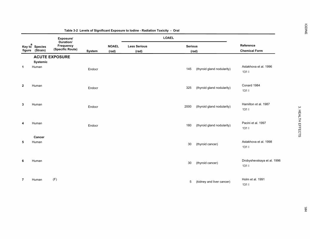
LOAEL
Less SeriousNOAEL Seriousa
SystemKey tofigure
Reference
Table 3-2 Levels of Significant Exposure to Iodine - Radiation Toxicity - Oral
Chemical Form(rad) (rad) (rad)
Exposure/Duration/
Frequency(Specific Route)
Species(Strain)
ACUTE EXPOSURESystemic
1Endocr
54
145145 (thyroid gland nodularity)
Human Astakhova et al. 1996
131 I
2Endocr
55
325325 (thyroid gland nodularity)
Human Conard 1984
131 I
3Endocr
56
20002000 (thyroid gland nodularity)
Human Hamilton et al. 1987
131 I
4Endocr
53
180180 (thyroid gland nodularity)
Human Pacini et al. 1997
131 I
Cancer5
50
3030 (thyroid cancer)
Human Astakhova et al. 1998
131 I
6
51
3030 (thyroid cancer)
Human Drobyshevskaya et al. 1996
131 I
7
57
55 (kidney and liver cancer)
(F)Human Holm et al. 1991
131 I

LOAEL
Less SeriousNOAEL Seriousa
SystemKey tofigure
Reference
(continued)Table 3-2 Levels of Significant Exposure to Iodine - Radiation Toxicity - Oral
Chemical Form(rad) (rad) (rad)
Exposure/Duration/
Frequency(Specific Route)
Species(Strain)
8
58
60006000 (thyroid cancer)
(F)Human Ron et al. 1998
131 I
9
52
2020 (thyroid cancer)
Human Tronko et al. 1996
131 I
CHRONIC EXPOSURESystemic
10
1717Endocr
300
(F)Human CDC 2002
131 I
Cancer11
97
99 (thyroid neoplasm)
(F)Human Gilbert et al. 1998
131 I
12
99
325325 (thyroid neoplasm)
(F)Human Kerber et al. 1993
131 I
13
a the number corresponds to entries in Figure 3-2.
Endocr = endocrine; (f) = feed; LOAEL = lowest-observed-adverse-effect level
100
2525 (thyroid neoplasm)
(F)Human Rallison 1996
131 I

1
10
100
1000
10000
Endocrine
1
2
3
4
Cancer *
5 6
7
8
9
mg/kg/day
Figure 3-2. Levels of Significant Exposure to Iodine - Radiation Toxicity - Oral
Acute (≤14 days)
c-Catd-Dogr-Ratp-Pigq-Cow
-Humansk-Monkeym-Mouseh-Rabbita-Sheep
f-Ferretj-Pigeone-Gerbils-Hamsterg-Guinea Pig
n-Minko-Other
Cancer Effect Level-Animals LOAEL, More Serious-Animals LOAEL, Less Serious-Animals NOAEL - Animals
Cancer Effect Level-Humans LOAEL, More Serious-Humans LOAEL, Less Serious-Humans NOAEL - Humans
LD50/LC50 Minimal Risk Level for effects other than Cancer
*Doses represent the lowest dose tested per study that produced a tumorigenic
response and do not imply the existence of a threshold for the cancer endpoint.
Systemic

1
10
100
1000
Endocrine
10
Cancer *
11
12
13
mg/kg/day
Figure 3-2. Levels of Significant Exposure to Iodine - Radiation Toxicity - Oral (Continued)
Chronic (≥365 days)
c-Catd-Dogr-Ratp-Pigq-Cow
-Humansk-Monkeym-Mouseh-Rabbita-Sheep
f-Ferretj-Pigeone-Gerbils-Hamsterg-Guinea Pig
n-Minko-Other
Cancer Effect Level-Animals LOAEL, More Serious-Animals LOAEL, Less Serious-Animals NOAEL - Animals
Cancer Effect Level-Humans LOAEL, More Serious-Humans LOAEL, Less Serious-Humans NOAEL - Humans
LD50/LC50 Minimal Risk Level for effects other than Cancer
*Doses represent the lowest dose tested per study that produced a tumorigenic
response and do not imply the existence of a threshold for the cancer endpoint.
Systemic

IODINE 108
3. HEALTH EFFECTS
for a more detailed discussion of releases from the Hanford Nuclear Site). The study reviewed records of
outcomes of 72,154 births, 1,957 infant deaths, and 1,045 fetal deaths that occurred in Washington
counties dear the Hanford Nuclear Site during the period 1940–1952. Subjects were assigned to one of
four exposure categories (low, medium low, medium high, high) based on the subject’s address (zip code)
at the time of the subject birth or infant death, and estimated 131I exposures in 1945 in those areas were
obtained from the Hanford Environmental Dose Reconstruction (HEDR) project (CDC 2002).
Associations between 131I exposure and outcomes were evaluated in a multivariate logistic regression
model. Models were evaluated for outcomes recorded for 1945, the year in which exposures were
estimated to be the highest, and also for the period May 1, 1945–April 30, 1946, which could have
included exposures to the highest levels during early pregnancy. The adjusted odds ratios (low-exposure
as the reference) for infant death for the high-exposure category were 1.1 (95% CI, 0.7–1.8) for the year
1945 and 1.3 (CI: 0.8–2.1) for the 1945–1946 period. The adjusted odds ratios for fetal death for the
high-exposure category were 0.6 (CI: 0.2–1.6) for the year 1945 and 0.7 (CI: 0.3–1.7) for the 1945–1946
period. These results suggest that neither infant nor fetal death were significantly associated with
estimated 131I exposures. The adjusted odds ratios for preterm birth for the high-exposure category were
1.6 (1.0–2.6) for the year 1945 and 1.9 (1.2–3.0) for the 1945–1946 period, suggesting a possible
association between preterm birth and 131I exposures.
One epidemiological study has examined health outcomes of infants of mothers who resided in the
Belarus region before or after the Chernobyl accident (Petrova et al. 1997) (see Section 3.3.2 for a more
detailed discussion of exposures from of the Chernobyl accident). Interpretation of the results of this
study, in terms of the contribution of radioiodine to the outcomes, is highly uncertain, as other factors
could have affected the outcomes, including exposure to other forms of radiation, nutrition, or other
chemical exposures. Nevertheless, because it is the only epidemiological study that has focused on
reproductive and developmental outcomes, and because of the substantial contribution that radioiodine
made to radiation exposures after the Chernobyl releases, a brief description of the study is presented
here. As part of a retrospective cohort study, health records were analyzed on 757 infants and their
mothers who resided in heavily radiation-contaminated areas of Belarus resulting from radionuclide
releases from the Chernobyl nuclear power plant or relatively uncontaminated areas (Petrova et al. 1997).
Prevalence of atopic dermatitis in infants who resided in contaminated areas was approximately 2 times
higher (approximately 40%) compared to infants from control areas. The prevalence of anemia (low
blood hemoglobin levels) was 6–7 times higher in infants from contaminated areas (18–20%).
Interpretation of the results of this study, in terms of the contribution of radioiodine, to the outcomes, is
IODINE 109
3. HEALTH EFFECTS
highly uncertain, as other factors could have affected the outcomes, including exposure to other forms of
radiation, nutrition, or other chemical exposures.
The highest NOAEL values and all reliable LOAEL values in each duration category for developmental
effects from exposures by the oral route are presented in Table 3-2 and plotted in Figure 3-2.
3.3.2.7 Cancer
Cancer effect levels (CELs) for iodine exposures by the oral route are presented in Table 3-2 and plotted
in Figure 3-2.
The thyroid gland receives the highest radiation dose of any organ or tissue following an internal
exposure to radioiodine (see Section 3.5, Toxicokinetics) and, therefore, cancer of the thyroid gland is the
major health concern associated with radioiodine exposures. Children, in particular, are highly vulnerable
to radioiodine toxicity. Cancer morbidity and mortality among populations that received exposures to
radioiodine have been examined in several large-scale epidemiology studies. In general, these studies fall
into several categories that can be distinguished by the sources of exposure and estimated radiation doses
to the thyroid gland and include (Table 3-3): (1) exposure to high doses (10–20 mCi, 370–740 MBq;
>10,000 rad, >100 Gy) achieved when 131I is administered to treat hyperthyroidism (even higher doses are
used to treat thyroid cancer); (2) exposures to moderately high doses (40–70 µCi, 1.5–2.6 MBq; 80–
130 rad, cGy) associated with clinical administration of 131I for diagnosis of thyroid gland disorders;
(3) low doses from exposures to fallout from nuclear bomb tests (BRAVO test, 300–2,000 rad, cGy;
Nevada Test Site, 1–40 rad, cGy); (4) low to high doses from exposures to releases from nuclear power
plant accidents (Chernobyl, 10–500 rad, cGy); and (5) low to high environmental exposures from
operational releases from nuclear fuel processing plants (Hanford Nuclear Site, 0.0001–284 rad, cGy). As
a point of reference, the dose-response relationship for thyroid cancer and external radiation appears to
extend down to thyroid doses of 0.1 Gy (10 rad) and predicts an excess relative risk (ERR) of 7/Gy for
ages <15 years at exposure (Ron et al. 1995). Studies of thyroid cancers and external radiation exposure
have found a strong dependence of thyroid cancer risk on age at exposure. Risk is substantially greater

IODINE 110
3. HEALTH EFFECTS
Table 3-3. Estimated Thyroid Radiation Doses in Populations Studied for Radioiodine-related Cancersa
Type of exposure Estimated thyroid radiation dose (cGy)a Referenceb
Radioiodine therapy for hyperthyroidism >5,000c Holm et al. 1991; Ron et al. 1998
Clinical diagnosis of thyroid gland disorders
80–130c Hall et al. 1996b
Marshall Islands BRAVO test 280–2,100c Hamilton et al. 1987; Lessard et al. 1985
Chernobyl power plant accident <1–200d Astrakova et al. 1998
Nevada Test Site nuclear bomb tests 1–30c Gilbert et al. 1998; Kerber et al. 1993; Rallison 1996
Hanford Nuclear Site releases 17±22 (0.003–282)d CDC 2002 a1 cGy=1 rad bSee text for additional references cCohort means darithmetic mean ± SD (range)

IODINE 111
3. HEALTH EFFECTS
for radiation doses received prior to age 15 years when compared to risks for doses received at older ages,
and this increased risk persists, possibly for the lifetime (Ron et al. 1995). This same general trend in
age-dependence would be expected for internal exposures to radioiodine; thus, studies of adult exposures
to radioiodine may not be directly applicable to predicting outcomes from exposures to children. The
relatively high and acutely cytotoxic radiation doses to the thyroid gland that are achieved in the treatment
of thyroid gland disorders, and outcomes on the thyroid, are not relevant for predicting outcomes from the
much lower environmental exposures that occur in most U.S. populations; for example, exposures
received as a result of nuclear bomb testing (Nevada Test Site) or operational releases from nuclear plants
(Hanford Nuclear Site). This is in part because cell killing effects decrease the number of viable cells that
might otherwise be transformed by radiation-associated mutagenesis. Uncertainties in estimating thyroid
doses are also greater in persons who have thyroid abnormalities because of the nonuniform distribution
of radioiodine in the thyroid gland (NCRP 1985). Nevertheless, high-dose studies are summarized
because they provide useful information about the magnitude of radioiodine exposures that would present
an elevated risk for thyroid and extrathyroidal cancers. Although not specified in most of these studies, it
is likely that radioiodine was administered as a single dose by the oral route as either potassium or sodium
iodide, as these are the common clinical practices. However, it is also possible, but highly unlikely, that
some patients received the radioiodine by injection. Since absorption of an oral dose of iodide is nearly
complete, this is unlikely to be a significant issue in interpreting the outcomes of the studies, except in
considering the radiation dose to the gastrointestinal tract.
Breast cancer is also a concern with exposures to high levels of radioiodine after ablative therapy for
hyperthyroidism because breast expresses NIS and can transport and accumulate iodide (see
Sections 3.5.4.2 and 3.6.1, Distribution). However, the epidemiological literature to date has not
implicated such exposures as a significant risk factor for breast cancer (Goldman et al. 1988; Green et al.
1995).
Therapeutic Doses of Radioiodine
Several studies have explored possible associations between radioiodine therapy for thyroid disease and
cancer incidence or mortality. The Ron et al. (1998) study specifically assessed cancer outcomes in
patients who received only 131I, and distinguished these patients from those who received other types of
treatments alone, or in combination with 131I. This is an important design feature, as the study showed
that other forms of treatment appear to be risk factors for cancer mortality. The Ron et al. (1998) study
used a retrospective cohort design to examine cancer mortality in 35,593 patients (79% females; mean
age, 46 years, 3% younger than 20 years) treated for hyperthyroidism (91% Graves’ disease, 8% toxic

IODINE 112
3. HEALTH EFFECTS
nodular goiter) in 25 U.S. hospitals and 1 British hospital (Ron et al. 1998). The mean total activity
administered was 10.4 mCi (385 MBq; 5th–95th percentile, 3–27 mCi, 111–999 MBq). The mean total
administered activity was 10.0 mCi (370 MBq) for treatment of Graves’ disease and 17.0 mCi (629 MBq)
for toxic nodular goiter. Cancers that occurred between the first visit of the patient to the clinic during the
enrollment period (1946–1964) until either the death of the patient or the end of the calendar year 1990
were considered in the analysis. Estimates of expected numbers of cancer deaths were based on U.S.
national mortality rates for the period 1958–1985. Patients were stratified into various categories of
treatment, distinguishing those who received 131I as the only form of treatment from those who received
antithyroid drugs or surgical treatments, alone or in combination with 131I. SMRs (observed/expected
deaths) were calculated for various treatments (131I, surgery, antithyroid drugs, or combinations). This
design allowed an assessment of the effects of possible associations between 131I exposure and cancer
outcomes, independent of the potential effect of other treatments. The study identified 2,960 cancer
deaths, 29 of which were classified as thyroid cancers. Among patients who received 131I as treatment
alone (only 131I), SMRs were significantly elevated only for thyroid cancer (4.91; 95% CI, 2.45–8.79), but
not for other cancers, or all cancers. Among all patients treated with 131I, alone or in combination with
other treatments (any 131I), SMRs were also significantly elevated for thyroid cancer (3.94, 252–5.86),
only. When stratified by latency (1–4 years, 5–9 years, 10 years or longer), in the latter group (any 131I),
SMRs for thyroid cancer were highest 1–4 years after treatment (12.32, 6.38–21.61), but remained
significantly elevated in the 10 years or longer group (2.78, 1.38-4.97). Radiation doses to specific
organs were estimated for each patient based on the administered activity and dosimetry tables developed
by the ICRP (1988). The estimated thyroid dose was 50–70 Gy (5,000–7,000 rad). When stratified by
administered 131I activity (as a surrogate for thyroid dose), the SMRs for thyroid cancer in this group (any 131I) increased with increasing exposure, suggesting a possible dose effect on thyroid cancer mortality.
The highest SMRs occurred in the group that received 15 mCi or more (7.05, 3.05–13.95), and in the
group treated for toxic nodular goiter (18.88, 7.58–38.98), who would have received higher exposures and
doses than Graves’ disease patients (2.84, 1.62–4.61). SMRs for cancers in other tissues were also
significantly elevated in the any 131I group; colorectal cancer, 1–4 years after treatment (1.42, 1.04–1.90);
lung cancer 1–4 years (1.49, 1.01–2.12) and 5-9 years (1.41, 1.02–1.89) after treatment; and for non-
chronic lymphatic leukemia, 5–9 years after treatment ( 2.10, 1.14–3.52). However, interpretation of
these findings, in terms of the potential contribution of 131I to cancer mortality, is complicated by the
finding of elevated SMRs in extra-thyroidal tissues in the groups that received treatments other than 131I,
including bucal cavity, lung, breast, and brain. The results of this study indicate that high exposures to 131I for treatment of hyperthyroidism did not increase overall cancer mortality; however, it did appear to
increase mortality for thyroid cancer. Interpretation of the effect on thyroid cancer mortality is
IODINE 113
3. HEALTH EFFECTS
complicated by the potential impact of thyroid cancers that may have existed in these patients,
undiagnosed, prior to the treatment. The observation that much of the apparent excess risk for thyroid
cancer deaths occurred during the first 1–4 years after 131I treatment, suggests a remarkably short latency
for radiation-induced cancer mortality, or possibly other factors contributed to the outcome. Other
uncertainties in this study include the use of exposure levels (mCi) as a surrogate for absorbed radiation
dose to the thyroid. The relationship between administered activity and thyroid dose in hyperthyroid
patients can be complicated by disease-related variation in thyroid gland size and iodide transport activity.
Also, administered activity can co-vary with the severity of the initial hyperthyroidism; patients who
received the highest activities tend to have the most severe disease, and disease severity could vary,
independently with cancer mortality.
A retrospective cohort study conducted in Sweden examined cancer incidence among 10,552 patients
(85% females; age 13–74 years) who received 131I therapy for treatment of Graves’ disease (51%) or toxic
nodular goiter (42%) (Holm et al. 1991). The mean total activity administered was 506 MBq (13.7 mCi);
however, this varied with the objectives of the therapy; 360 MBq (9.7 mCi) for treatment of Graves’
disease and 700 MBq (18.9 mCi) for toxic nodular goiter. The distribution of the administered activity in
the study population was as follows: 30% <220 MBq (5.9 mCi), mean, 150 MBq (4.1 mCi); 38% 221–
480 MBq (6–13 mCi), mean 315 MBq (8.5 mCi); and 32% >480 MBq (13 mCi), mean 1,063 MBq
(28.7 mCi). Cancers that occurred from 1 year after treatment (on or after 1958) until either the death of
the patient or the end of the calendar year 1985 were considered in the analysis. Expected numbers of
cancers were estimated from data from the Swedish Cancer Register for the period 1958–1985. Standard
incidence ratios (SIR, observed/expected cancers) were significantly elevated for cancers of the lung
(1.32, 95% CI, 1.07–1.59) and kidney (1.39, 95% CI, 1.07–1.76). Among toxic nodular goiter patients,
who received, on average, twice the dose as Graves’ disease patients, the SIR was also significantly
elevated for liver cancer (2.14, 1.20–3.52). Among 10-year survivors, significantly elevated SIRs
included stomach (1.33, 1.01–1.71), kidney (1.51, 1.06–2.08), and brain (1.63, 1.10–2.32). Doses to
specific organs were estimated for each patient based on the administered activity and dosimetry tables
developed by the ICRP (1988). Estimated average radiation doses to these tissues were: thyroid gland,
>10,000 cGy (>10,000 rad); stomach, 25 cGy (25 rad); lung, 7 cGy (7 rad); kidney, 5 cGy (5 rad); liver,
5 cGy (5 rad); and brain (not reported). There were no significant dose trends. Notably, SIRs for thyroid
cancer were not significantly elevated (SIR 1.29, 0.76–2.03). Some of the patients in this study received
treatments other than 131I for thyroid disorders, including antithyroid drugs (14%), surgery (3%), and/or
thyroid hormone supplements (2%). Cancer mortality was examined in the same cohort (Hall et al.
1992a). Standard mortality ratios (SMRs) were calculated based on data from the Swedish Cause-of-

IODINE 114
3. HEALTH EFFECTS
Death Registry. SMRs were significantly elevated for all cancers (1.14, 1.04–1.24), digestive tract
cancers (1.28, 1.16–1.45), and respiratory tract cancers (1.31, 1.01–1.66) among patients who had greater
than a 10-year follow up from the date of their exposure to 131I, and for thyroid gland cancer during the
first year (11.45, 2.8–33.72). There were no significant dose trends, although the SMR for thyroid gland
cancer was approximately 4 times higher in patents who received >480 MBq (13 mCi) than in patients
who received <221 MBq (6 mCi). The results of this study suggest that exposure to high levels of 131I for
treatment of hyperthyroidism increases cancer risk; however, several uncertainties complicate the
interpretation of results, in terms of the contribution of 131I to the elevated cancer risk. These include the
lack of a dose trend for increased cancer incidence or mortality, and the potential contribution of
treatments, other than 131I to cancer incidence and mortality, which were not quantified in this study.
Surgical treatment and antithyroid drug therapy appear to be cancer risk factors in hyperthyroid patients
(Ron et al. 1998).
A retrospective study examined cancer morbidity and mortality in 7,417 patients (83% females; mean
age, 57 years ± 13, SD) treated for hyperthyroidism in the West Midlands region of the United Kingdom
during the period 1950–1991(Franklyn et al. 1999). The mean total activity administered was 308 MBq
(8.3 mCi); 49% received <220 MBq (<6 mCi) and 17% received >481 MBq (>13 mCi). The follow-up
period ranged from 1 year (74%) to $20 years (18%). Estimates of expected numbers of cancer deaths in
England and Wales were based on International Agency for Research on Cancer (IARC) and World
Health Organization (WHO) data. The SIR for all cancer types was 0.83 (95% CI, 0.77–0.90). The SIR
for thyroid cancer was 3.25 (1.69–6.25) and for cancer of the small bowel, 4.81 (2.16–10.72). SIRs for all
other cancers were <1. Similarly, SMRs were 0.90 (0.82–0.98) for all cancer types, 2.78 (1.16–6.67) for
thyroid cancer, and 7.03 (3.16–15.66) for cancer of the small bowel. Significant positive trends for
increasing incidence with increasing cumulative radiation exposure were observed for bladder cancer and
uterine cancer, although SIRs and SMRs for these cancers were not significantly greater than 1. The
results of this study, consistent with those of the Hall et al. (1992a) and Ron et al. (1998), suggest that
exposure to high levels of 131I for treatment of hyperthyroidism increases the risk of cancer mortality;
however, similar to the Hall et al. (1992a) study, the potential contribution of treatments other than 131I
(e.g., surgical treatment and antithyroid drug therapy, Ron et al. 1998), to cancer incidence and mortality,
were not quantified in this study. Surgical treatment and antithyroid drug therapy appear to be cancer risk
factors in hyperthyroid patients (Ron et al. 1998). Other potential uncertainties in interpreting the
Franklyn et al. (1999) study include: (1) thyroid radiation doses were not reported; thus, it is difficult to
compare the doses received to subjects in this studies with those in the Ron et al. (1998), Holm et al.
(1991), and Hall et al. (1992a) studies; (2) the Franklyn et al. (1999) study did not stratify the subjects by
IODINE 115
3. HEALTH EFFECTS
radiation exposure or dose, which may have varied depending on the nature of the original diagnosis of
hyperthyroidism; and (3) the size of the cohort in the Franklyn et al. (1999) study was smaller than the
other three studies (7,417 compared to 35,593 in Ron et al. 1998 study, and 10,552 in Holm et al. 1991
study).
A follow-up cohort study was conducted of cancer morbidity and mortality among 1,762 women who
received ablative 131I therapy for hyperthyroidism during the period 1946–1964 (Goldman et al. 1988).
The follow-up period was 17 years. SMRs and SIRs were estimated based on age-, date-, sex-, and race-
specific mortality incidence and mortality of the United States or Massachusetts population. The cohort
was stratified into treatment categories that included only 131I or 131I in addition to other therapies for
hyperthyroidism. SIRs in the 131I-only group were not significantly elevated for any cancer type or group.
SMRs in the 131I-only group were significantly elevated for cancers of all causes (SMR, 1.2, 1.1–1.4,
95% CI, 10 cases). There were no significant radiation dose trends. Exposures ranged from 0.1 to
>10 mCi (4–370 MBq). Although, like the Ron et al. (1998) study, cancer mortality risk was evaluated in
patients who received only 131I as treatment, the much smaller size of the Goldman et al. (1988) study
makes it difficult to interpret comparisons of results to those from the Ron et al. (1998) study. Like the
Ron et al. (1998) study, Goldman et al (1988) found elevated cancer mortality in patients who received
treatments other than 131I.
A follow-up study was conducted of cancer morbidity among 1,771 patients (21% males) who received
ablative 131I therapy for treatment of thyroid cancer during the period 1950–1990 (de Vathaire et al.
1997). The follow-up period was 10 years. Excess relative risk (ERR) was modeled using linear models
(a quadratic model was also explored), taking into account sex, age at time of treatment, and cumulative
activity of 131I administered as variables. The mean administered activity of 131I was 7.2 GBq (range, 3.8–
57.6; 195 mCi, range, 103–156 mCi) which corresponded to a mean radiation dose to bone marrow of
0.34 Sv (range, 0.13–2.8; 34 rem, range, 13–280 rem). Using the cancer outcomes of patients who
received 1–0.19 GBq of 131I as the reference group, ERRs for colorectal cancer increased with increasing
administered activity. In the patient group that received >3.7–7.5 GBq (>100–203 mCi) the ERR was
4.0 (90% CI, 1.3–12.2) and in the group that received >7.5 GBq>(203 mCi), the ERR was 4.9 (1.2–18.5).
While this is a relatively small study, it supports an outcome of the much larger Ron et al. (1998) study in
which SMRs for colorectal cancer were elevated among patients who received lower administered
activities of 131I for treatment of hyperthyroidism (mean 10.4 mCi, 385 MBq).
IODINE 116
3. HEALTH EFFECTS
Diagnostic Doses of Radioiodine
A retrospective cohort study examined thyroid cancer incidence among 34,104 patients (80% females, 1–
75 years of age) in Sweden who received 131I for diagnosis of thyroid disorders during the period 1950–
1969. The follow-up period was from 1958 to 1990 (Hall et al. 1996b). A total of 2,408 patients (7%)
were exposed before 20 years of age and 316 patients were exposed before 10 years of age (1%). The
diagnostic test was for a suspected thyroid tumor in 10,785 (32%) patients and for hypothyroidism,
hyperthyroidism, or other reasons in 23,319 (68%) patients. The follow-up period ranged from 5 to
39 years after exposure (thyroid cancers detected within 5 years of the diagnostic test were excluded on
the basis that they may have been related to cancer present at the time of the diagnostic test). The mean
total activity administered was 2.4 MBq (65 µCi) for patients suspected of having a thyroid gland tumor
and 1.6 MBq (43 µCi) for other patients. Radiation doses to the thyroid gland were estimated for each
patient based on the administered activity and dosimetry tables developed by the ICRP (1988). The mean
absorbed dose was 1.3 Gy (130 rad) for suspected thyroid tumor patients and 0.8 Gy (80 rad) for other
patients. SIRs were calculated based on sex-, age-, and date-adjusted cancer incidence rates based on the
Swedish Cancer Registry. Sixty-seven thyroid tumors were identified during the period of the study, of
which 42 (63%) were in patients who received 131I for diagnosis of a suspected thyroid gland tumor. SIRs
were significantly elevated only in the latter group (2.86, 95% CI, 2.06–3.86), but not in patients tested
for other suspected thyroid disorders. There were no significant dose trends for thyroid cancer in either
group, and the presence of cancer may have predated the exposures to 131I. A subsequent follow-up of
this same cohort was conducted, which extended the follow-up period an additional 8 years from that
reported in Hall et al. (1996b) and included thyroid cancers diagnosed as early as 2 years after diagnostic
administration of 131I, making the follow-up period 2-47 years (Dickman et al. 2003). Patients (1,767)
who received diagnostic X-rays to the neck, prior to receiving 131I, were also included in the study to
explore the effects of external radiation on thyroid cancer incidence. Among patients who did not receive
X-rays to the neck and who were not referred for diagnostic 131I for suspicion of a possible thyroid tumor,
the SIR for thyroid cancer was 0.91 (95% CI, 0.64–1.26). The estimated dose to the thyroid in this group
was 0.94 Gy (94 rad). However, among patients who did receive X-rays prior to 131I, the SIR was
9.8 (6.3–14.6). The results support the previous findings in this cohort (Hall et al. 1996b) that radiation
doses to the thyroid resulting from diagnostic administration of 131I are not associated with excess risk of
thyroid cancer. The study also identifies X-ray exposures as an important variable that, if not controlled
for, could confound studies of cancer outcomes in patients exposed to 131I.
The incidence of cancer in extrathyroidal organs was examined in this same cohort (Holm et al. 1989). At
that time, the cohort consisted of 35,074 patients, 31% of whom received 131I for diagnosis of a suspected
IODINE 117
3. HEALTH EFFECTS
thyroid gland tumor, 42% for suspected hyperthyroidism, 16% for suspected hypothyroidism, and 8% for
other reasons (the basis for the diagnostic procedure could not be determined for 3% of the patients). The
mean total activity administered was 52 µCi (range 1–960 µCi) (1.9 MBq, 0.04–36 MBq). The mean total
administered activity was 71 µCi (2.6 MBq) for patients suspected of having a thyroid tumor, 48 µCi
(1.8 MBq) for diagnostic tests for hyperthyroidism, and 40 µCi (1.5 MBq) for other diagnostic purposes.
SIRs were significantly elevated for cancers of the endocrine organs other than thyroid gland (1.93, 1.62–
2.29), lymphomas (1.24, 1.03–1.48), and leukemias (1.34, 1.11–1.60). The SIR for nervous system
cancers was 1.19 (1.00–1.41). The SIR for thyroid cancer was significantly elevated only in the 5–9-year
period of follow-up. There were no significant dose trends. In this study, unlike the Hall et al. (1996b)
study, SIRs were calculated for all patients, regardless of the intended purpose of the diagnostic test,
including patients who were administered 131I for the diagnosis of suspected thyroid tumors.
A smaller retrospective cohort study compared thyroid cancer incidence among 789 patients (74%
females) in Germany who received 131I for diagnosis of thyroid disorders before the age of 18 years with
1,118 patients who received a diagnostic procedure on the thyroid that did not involve radioiodine (68%
females) (Hahn et al. 2001). Diagnostic procedures occurred between 1958 and 1978 in the treatment
group, and between 1959 and 1978 in the control group. The diagnosis made at the initial referral in the
treatment groups was nodular goiter in 385 (49%) in patients, no evidence of thyroid disease in 199 (25%)
patients, and hypothyroidism, hyperthyroidism, or other reasons in 205 (26%) patients. In the control
group, the diagnoses included 600 (54%) cases of goiter, 327 (29%) of no evidence of thyroid disease,
and 131 (12%) hypothyroidism, hyperthyroidism, or other reasons. Patients who had a history of external
radiotherapy of the head or neck regions or thyroid cancer were excluded from the study. The follow-up
period (1989–1997) ranged from 13 to 33 years in the treatment group and 9–33 years in the control
group. The median total 131I activity administered in the treatment groups was 0.9 MBq (24 µCi).
Radiation doses to the thyroid gland were estimated for each patient based on the administered activity
and dosimetry tables developed by the ICRP (1988). The mean absorbed dose was 1.0 Gy (100 rad);
however, this varied with age of diagnosis; the range was 0.6–1.2 Gy (60–120 rad). SIRs were calculated
based on sex-, age-, and date-adjusted cancer incidence rates based on the German Democratic Republic
cancer registry for the period 1980–1989. Three cases of thyroid cancer were identified in the treatment
group during the study period and two cases in the control group. SIRs were 5.3 (95% CI, 0.5–15.1) in
the treatment group and 5.3 (1.1–15.3) in the control group. The relative risk (treatment compared to
control) was 0.9 (0.1–5.1). Risk of thyroid cancer was not significantly associated with exposure to
diagnostic levels of 131I. A complication in the interpretation of these findings is that the response rate
was very low: 3 cases in 1,058 patients, 0.28%; 2 in 795 in the treatment group.

IODINE 118
3. HEALTH EFFECTS
A prospective study examined thyroid outcomes of children and adolescents (<20 years old) who received
diagnostic doses of 131I during the period 1946–1967 (Hamilton et al. 1987). Study groups consisted of
3,503 subjects who received diagnostic 131I, 2,495 control subjects who did not receive 131I and who were
matched with the exposed subjects by sex-, age-, and diagnostic-test date, and a group of 1,070 siblings of
the control group. The follow-up period was from entry into the study until 1986. Participants were
surveyed with a questionnaire to identify those who had thyroid or neck surgery during the study period,
and pathology reports and specimens were retrieved and reviewed by a panel of pathologists; neoplasms
were classified and the results were compared with hospital pathology reports. The dose to the thyroid
gland was estimated for each exposed subject based on the reported activity administered, percent thyroid
uptake, and thyroid weight estimated from published thyroid growth tables. The median total absorbed
dose was 20–40 rad (0.2–0.4 Gy) (95th percentile, 200–330 rads 2–3 Gy). The survey response rate was
63%. A total of 34 surgeries were reported, of which 19 were on subjects who did not have any thyroid
disorder diagnosed at the time of entry into the study; 16 of these subjects had confirmed thyroid tumors;
10 benign, 8 of which occurred in the exposed group, and 6 malignant tumors, 5 of which occurred in the
exposed group. Although these results are suggestive of a possible effect of 131I exposure on thyroid
tumor incidence, the differences between the exposed and control groups were not statistically significant.
Shore (1992) reviewed the results of the Hamilton et al. (1987) study and calculated a relative risk for
thyroid cancer of 2.9 (90% CI, 0.6–15) based on the internal comparison of the exposed and unexposed
groups in the Hamilton et al. (1987) study. Based on the Surveillance, Epidemiology and End Results
(SEER) cancer data for 1973–1981 (U.S. DHHS 1985), 3.7 thyroid cancers would have been expected in
the Hamilton et al. (1987) study, compared to the 4 observed during the period of 5 or more years after
the diagnostic test (one of the cancers reported in the Hamilton et al. (1987) study occurred with a latency
of 2 years), which, according to Shore (1992), indicates an SIR of 1.1 (95% CI, 0.3–2.6).
Marshall Islands Nuclear Bomb Test BRAVO. Several epidemiological studies have examined thyroid
gland disorders in residents of the Marshall Islands who were exposed to radioiodine from atmospheric
fallout resulting from nuclear bomb tests (including the so-called BRAVO test; see Section 3.3.2 for a
more detailed discussion of exposures from the Marshall Islands BRAVO test). A more complete
discussion of these studies is presented in Section 3.3.1.2 (Endocrine), as the studies provide dose-
response information on thyroid disorders other than cancer. However, cancer outcomes have been
examined in what has become known as the BRAVO cohort, as well as in larger samples of the Marshall
Island population. Almost all that is known about radioiodine doses to the thyroid from the BRAVO test
exposures derive from a few urinary measurements collected 15 days after the exposures. These have

IODINE 119
3. HEALTH EFFECTS
been estimated to have been (external and internal): 3.3–20 Gy (330–2,000 rad) on Rongelap (highest
doses in children), 1.3–4.5 Gy (130–450 rad) on Ailingnae, and 0.3–0.95 Gy (30–95 rad) on Utrik
(Conard 1984). The BRAVO test was not the only potential source of radioiodine exposure in the
Marshall Island population, as numerous bomb tests were conducted in the Marshall Islands during the
period 1946–1958.
The strengths of the Marshall Island studies include the relatively high range of thyroid radiation doses
and the multiple thyroid screenings, which included, in the more recent studies, relatively objective
assessments of nodularity by ultrasound. Limitations of the studies include: (1) large dose uncertainties
in terms of total thyroid dose; (2) further dose uncertainties in terms of the fraction of the dose that was
from 131I rather than from short-lived isotopes of iodine and gamma radiation; (3) no attempt to estimate
individual thyroid doses; (4) inequities between the exposed and unexposed populations in the intensity of
thyroid screening; (5) the relatively small number of exposed subjects in the BRAVO cohort; (6) the
potential confounding effects of prophylactic iodide administration and thyroid surgery in highly exposed
subjects; and (7) thyroid radiation dose estimates not available for larger scale studies of populations in
the Marshall Islands.
Evidence for a higher prevalence of thyroid cancer among the original 250 people known to have been
heavily exposed as a result of the BRAVO incident has not been established; however, this may reflect
the small size of the cohort (see section 3.3.2 for a more detailed discussion of exposures from the
Marshall Islands BRAVO test). In 1982, a review of the diagnoses for thyroid nodules detected in
250 exposed and 1,303 nonexposed Marshallanese revealed 9 definitive carcinomas (3.6%) and
7 adenomas (2.8%) in the exposed group, and 6 carcinomas (0.5%) and 14 adenomas (1%) in the
nonexposed comparison group (Conard 1984). Subsequent reviews of the thyroid pathology more or less
agree with the conclusions of Conard (1984), although differences in the composition of comparison
group have contributed to slightly different estimates of prevalence in the nonexposed population. For
example, Howard et al. (1997) reported four cancers (1.8%) and one adenoma (0.4%) in a nonexposed
comparison group. Takahashi et al. (1997) reviewed diagnoses of 22 cases of thyroid nodularity
discovered in 1993 in an ultrasound screening program that evaluated 1,275 Marshall Island residents
(mainly from Ebeye). The prevalence of thyroid cancer among patients referred for surgery-based thyroid
gland ultrasound assessments suggested an overall prevalence of thyroid cancer of approximately 1.2%
(15/1,275) in the population evaluated, or a 12% prevalence (15/123) of thyroid cancer among those who
had palpable nodules. A follow-up to this study included the results of thyroid disease screening of
3,709 Marshall Island residents who were born before the BRAVO test and who lived anywhere in the

IODINE 120
3. HEALTH EFFECTS
Marshall Islands during the period of bomb testing. The study group included an estimated 60% of the
still-living population who resided in the Marshall Islands during this period. Combining findings from
the previous study (Takahashi et al. 1997) and the follow-up, a total of 57 thyroid cancers were identified
(1.5%), of which 92% were diagnosed as papillary cancers. Several factors confound attempts to
associate thyroid cancers in the Marshall Islands population with radioiodine exposures, including lack of
definitive dosimetry, outside of the small BRAVO cohort. Changes occurred in diagnostic techniques
used to detect thyroid nodules, which would direct further diagnostic attention; in particular, the use of
ultrasound for detecting small thyroid nodules began only in 1994. More recent studies have also
suggested a relatively high prevalence of iodine deficiency in the Marshall Islands, which may have
affected background thyroid cancer prevalence (Takahashi et al. 1999).
Nevada Test Site Nuclear Bomb Tests. During the period 1951–1958, 119 atmospheric nuclear bomb
tests were conducted at the Nevada Test Site (NTS) in southern Nevada (NCI 1997). These tests were
followed by 9 surface detonations during the period 1962–1968 and approximately 809 below-ground
tests, of which 38 were determined to have resulted in off-site releases of radioactive materials. A dose
estimation methodology was developed by the National Cancer Institute (NCI 1997), which has enabled
estimation of population radiation doses to the thyroid gland from direct and indirect (e.g., in utero,
ingestion of cow milk) exposures to 131I resulting from the NTS activities for the purpose of health
assessments and epidemiologic investigations (Gilbert et al. 1998; Kerber et al. 1993). A discussion of
the uncertainties and limitations of these population dose estimates for use in epidemiology studies and
risk assessment can be found in a review of the NCI (1997) dose estimations conducted by the Institute of
Medicine and the National Research Council (NRC 1999).
The strengths of the NTS studies described above include the attempt to develop a systematic sampling
frame, the careful, multiple thyroid screenings (two or more times), the relatively high follow-up rate, and
the extensive attempt to characterize individual 131I doses. Limitations of the studies include: (1) the
substantial dose uncertainties, since no thyroid exposure measurements were available and individual
milk and vegetable consumption was recalled more than 30 years after the fact; (2) the food-consumption
and behavioral questionnaire was conducted after subjects knew their thyroid outcomes; (3) the modest
sample size and, therefore, small number of thyroid neoplasms found, which limited the statistical power
and precision; (4) the relatively low dose range, which also limited the statistical power and precision;
(5) the restriction of the thyroid examinations to palpation (no ultrasound); and (6) the fact that the
thyroid examinations were only partially blinded (i.e., examiners often knew the subject’s geographic
region).

IODINE 121
3. HEALTH EFFECTS
A cohort study examined thyroid nodularity and performed diagnostic follow up in 2,678 adolescents (age
11–18 years) who resided in Utah or Nevada near the NTS during the early 1950s and in a comparison
population of 2,132 adolescents who lived in Arizona. Examinations were conducted during the period
1965–1970 (Rallison et al. 1974). In a follow-up study conducted in 1985–1987, 1,962 of the original
Utah-Nevada group and 1,160 from the Arizona group were reexamined (Rallison et al. 1990).
Radioiodine doses were estimated for each Utah-Nevada subject based on histories of residence, local
milk and leafy vegetable consumption, records of transport and deposition of radionuclides at their town
and/or county of residence, and age-specific transfer factors relating iodine ingestion with iodine uptake
in the thyroid gland (Kerber et al. 1993; Simon et al. 1990). Mean thyroid dose estimates were 150 mGy
(15 rad) (maximum 4.6 Gy, 460 rad) in the Utah group, 50 mGy (5 rad) (maximum 0.84 Gy, 84 rad) in
the Nevada group, and 13 mGy (1.3 rad) (maximum 0.45 Gy, 45 rad) in the Arizona group (the group
names refer to cohort designations used in the study, which were based on the place of residence during
the potential exposure period, and not necessarily where the entire radiation dose for each individual was
received). In the 1965–1968 examinations, 76 of 4,819 people examined had palpable thyroid gland
nodules, 22 of which were subsequently diagnosed as adenomas (20) or carcinomas (2). The prevalence
of nodules was higher in the Utah-Nevada group (19.7/1,000) than in the Arizona group (10.8/1,000).
Fifteen of the 22 neoplasms were found in the Utah-Nevada group (5.6/1,000) and 7 in the Arizona group
(3.3/1,000) (Rallison et al. 1974). In 1985–1987, 125 new cases of thyroid nodularity were identified,
65 of which were diagnosed as neoplasms and 5 of the latter were carcinomas. Five carcinomas were
reported in the group during the interval between the two examinations. Combining the results of the first
and second evaluations, including the five carcinomas observed during the interval (a total of
12 carcinomas), resulted in similar prevalences in the two groups for nodules (Utah-Nevada 48.6/1,000,
Arizona 36.6/1,000). Prevalence of neoplasms was not disparate: Utah-Nevada, 2.8/1,000 and Arizona,
4.8/1,000 (Rallison et al. 1990). Thyroid nodules were detected in 56 of 2,473 subjects; 38 of these
lesions were diagnosed as nonneoplastic (28 were colloid adenomas, the other 10 were miscellaneous
nonneoplastic lesions), 11 were benign adenomas (of these, 8 were follicular adenomas and there was one
each of papillary, fetal, and Hurthle cell adenomas), and 8 were papillary carcinomas (Rallison 1996).
Stratifying the outcomes by estimated thyroid radiation dose revealed a significant dose trend for
neoplasms, but not for all nodules or for carcinomas alone. The group that received a dose exceeding
0.25 Gy (25 rad) had a thyroid neoplasm prevalence of 21–24/1,000, whereas groups that received
<0.25 Gy had a prevalence of 4–5/1,000. The excess relative risk estimates per Gy were: neoplasms,
7.0 (lower 95% confidence limit [CL], 0.74, p=0.019); nodules, 1.2 (95% CL<0, p=0.16); and
carcinomas, 7.9 (95% CL<0, p=0.096) (Kerber et al. 1993).

IODINE 122
3. HEALTH EFFECTS
In a large scale ecological study, mortality and incidence of thyroid cancer in 3,053 U.S. counties were
compared to estimated exposures to 131I from releases from the NTS (Gilbert et al. 1998). Thyroid cancer
mortality data were obtained from the National Center for Health Statistics for 1957–1994 and thyroid
cancer incidence data from SEER for the period 1973–1994. County-specific or state-specific cumulative
radiation doses were reconstructed based on NCI (1997) and were as follows (cGy, where 1 cGy = 1 rad):
in utero, 4.3 cGy; 0–<1 year, 12.6 cGy; 1–4 years, 10.0 cGy; 5–9 years, 6.7 cGy; 10–14 years, 4.4 cGy;
15–19 years, 3.1 cGy; $20 years, 1.1 cGy. During the study period, there were 12,657 cases of thyroid
cancer and 4,602 thyroid cancer deaths. Age-, calendar-, sex-, and count-specific mortality and incidence
rates in the United States were analyzed in relation to 131I dose estimates, taking into consideration
geographic location, age at exposure, and birth cohort. There were no significant dose-related trends
(linear excess relative risk model) in either thyroid cancer mortality or incidence when all exposure age
groups were composited or when exposure age groups 1–5 years or 1–15 years were considered
separately. However, when the exposure age group <1 year was analyzed, a dose trend was weakly
suggested by highly positive excess relative risks (ERR) for thyroid cancer deaths when doses were
county-specific (ERR 10.6 per Gy, 95% CI, -1.1–29, p=0.085) or state-specific (16.6 per Gy, -0.2–43,
p=0.054), and for thyroid cancer incidence when doses were county-specific (2.4 per Gy, -0.5–5.6).
These outcomes were strongly influenced by two deaths and nine cases of thyroid cancer that occurred in
individuals who received estimated cumulative doses exceeding 9 cGy (9 rad) before they were
12 months of age.
Chernobyl Nuclear Power Plant Accident. Clinical records and cancer registries from the Republics of
Belarus and Ukraine show an increase in the incidence of thyroid cancer in children and adolescents,
which became apparent approximately 4 years after the release of radioactive materials from the
Chernobyl nuclear power plant in April 1986, but which has not been increasing in recent years,
especially among those exposed at older ages (Cherstvoy et al. 1996; Drobyshevskaya et al. 1996;
Prisyazhuik et al. 1991; Tronko et al. 1996) (see Section 3.3.2 for a more detailed discussion of exposures
from the Chernobyl accident). Belarus recorded an annual incidence of 0.09 cases per 100,000 in 1986
among children between the ages of 4 and 17 years and 2.46 per 100,000 in 1991, with the highest
incidence in the Gomel oblast; from 0.24 cases per 100,000 in 1986 to 12.5 per 100,000 in 1991
(Drobyshevskaya et al. 1996). In the Ukraine, annual incidence of thyroid cancer in children and
adolescents (under 15 years of age) increased from approximately 0.05 per 100,000 prior to 1986 to
0.43 per 100,000 in 1992 (Tronko et al. 1996). In 1994, the incidence (per 100,000) was highest in
regions nearest to Chernobyl: Chernihiv, 3.8; Zhytomyr, 1.6; and Kiev, 1 (Tronko et al. 1996). Jacob et al
IODINE 123
3. HEALTH EFFECTS
(1998) estimated excess absolute risk of thyroid cancers in Belarus and Northern Ukraine for the period
1991–1995 using the cancer incidence in southern Ukraine as the control. The relationship between
thyroid cancer risk and the estimated radiation dose to the thyroid was linear, with a slope of 2.3 (95% CI
1.4–3.8) per 10,000 person-year Gy. Although the available data strongly show that radiation exposure
from the accident has led to the excess risk of thyroid cancer, especially in persons exposed as children,
there is also much uncertainty in the radiation dose estimates. The observed trends for increased
prevalence of thyroid cancer, as well as the magnitude of the thyroid cancer risk associated with
radioiodine are highly uncertain because of factors that complicate the epidemiological picture, including
the contribution external exposure, the effect of the intensive screening for thyroid cancer that followed
the accident (Astakhova et al. 1998) on the baseline incidence of thyroid cancer, and the potential effects
of iodine deficiency and endemic goiter in the population (Gembicki et al. 1997; Robbins et al. 2001).
The relationship between childhood thyroid cancer and radiation exposure was examined in a case-control
study of children from Belarus (Astakhova et al. 1998). Cases included all children under age 15 years at
the time of the accident who had confirmed pathology diagnoses of thyroid cancer during the period
1987–1992 and who could participate in the study (107 of 131 applicable cases in Minsk State Medical
Institute records). Cases were matched with two control groups; one control group (Type 1) was
randomly selected from an area of Belarus thought to have relatively low or no exposures from the
Chernobyl accident (Brest, Grodno, and Vitebsk oblasts in north and west Belarus) but was otherwise
matched with cases for age, sex, and urban/rural residence. A second control group (Type 2) was drawn
from each Belarus district, including the more heavily exposed oblasts near Chernobyl (Minsk, Mogilev,
and Gomel), in numbers proportional to the population census and was matched to cases by pathway to
diagnosis, in addition to age, sex, and urban/rural residence. The objective of matching the pathway to
diagnosis was to control for screening intensity as a possible contributor to an increased incidence.
Diagnosis pathways were classified into three elements: (1) systematic endocrine screening; (2) incidental
finding during physical examination not necessarily related to the Chernobyl releases; or (3) examination
prompted by referral because of a swelling of the neck or other symptoms of possible thyroid enlargement
or nodularity.
Average thyroid radiation doses were reconstructed based on thyroid gland 131I measurements made on
200,000 residents of Belarus, after the Chernobyl release, and estimates of cow milk contamination and
consumption for the area of residence of each case or control (vegetable and goat milk consumption was
not included in the exposure estimates). If no cow milk consumption was thought to have occurred,
exposure was assumed to have occurred principally from inhalation. Age-group thyroid doses were

IODINE 124
3. HEALTH EFFECTS
constructed for each area of residence included in the study. Mean (standard deviation) of thyroid doses
in the case group and controls were as follows: cases, 535 mGy (848) mGy; Type I controls, 188 mGy
(386); and Type II controls, 207 mGy (286). For the purpose of estimating odds ratios (ORs), cases and
controls were stratified into three thyroid dose categories. The resulting estimated dose distributions
among thyroid cancer cases were 64/107 (59.8%) in the <0.3 Gy dose category, 26/107 (24.3%) in the
0.3–0.99 Gy dose category, and 17/107 (15.9%) in the $1 Gy dose category. The corresponding
distributions in Type 1 controls were 88/107 (82.2%) for <0.3 Gy, 15/107 (14.0%) for 0.3–0.99 Gy, and
4/107 (3.7%) $1 Gy. The corresponding OR for the $0.3 Gy category compared to <0.3 Gy was
3.11 (95% CI, 1.67–5.81) and for the $1 Gy category compared to <0.3 Gy was 5.84 (1.96–17.3). ORs
were significant when Type 2 controls were the comparison group (controls for pathway to diagnosis).
For routine endocrine screening, ORs were 2.08 (1.0–4.3) for comparison of the dose categories $0.3 Gy
and <0.3 Gy, and 5.04 (1.5–16.7) when the dose category $1 Gy was compared to <0.3 Gy. The OR for
incidental findings was significant, 8.31 (1.1–58) when the dose category $0.3 Gy was compared to
<0.3 Gy. These results suggest that, after controlling for the effects of intensive screening for thyroid
cancer that occurred after the accident, radiation dose to the thyroid gland was a significant contributor to
thyroid cancers diagnosed in children who lived in Belarus during and after the Chernobyl releases and
that this contribution is evident at doses exceeding 0.3 Gy. The OR estimates, however, are highly
uncertain because of the relatively large uncertainties in the dose estimates.
An analysis of 251 thyroid cancer cases in children (14 years or younger) from Belarus who were
diagnosed during the period 1986–1993 revealed a dose trend in incidence when the cases were organized
by districts that reflected their respective mean thyroid doses (Drobyshevskaya et al. 1996). Incidence
ranged from 81 to 201 per 100,000 where estimated average thyroid doses were above 1 Gy (1.2–1.6 Gy,
120–160 rad), and 14–55 per 100,000 where doses were between 0.1 and 0.5 Gy (10–50 rad). The
highest incidence occurred in Bragin where individual thyroid doses were estimated to have ranged from
0.8 to 20 Gy (560, 80–2,000 rad) (mean, 5.6 Gy, 560 rad). Incidence was 9 per 100,000 in Braslav where
the lowest measurable thyroid doses were reported (mean, 0.005 Gy, 0.5 rad). Children who were under
3 years old or in utero at the time of exposure accounted for 53% of thyroid cancer cases. This age-group
was estimated to have received a thyroid radiation dose that was approximately 2–3 times that for older
children (approximately 1.4 Gy average dose). However, 52% of the cancers were diagnosed in children
who received an estimated thyroid dose of <0.3 Gy and 84% in children who received doses <1 Gy.
Children under 3 years old accounted for 38% of the cancer cases among children exposed to <0.3 Gy.
These results suggest that young children were particularly susceptible to lower radiation doses.
IODINE 125
3. HEALTH EFFECTS
An analysis of 531 thyroid cancer cases in children and adolescents (under 18 years of age) from Ukraine
who were diagnosed during the period 1986–1994 revealed that 55% of the cases were under age 6 years
on the date of the Chernobyl release (Tronko et al. 1996). The annual incidence of thyroid cancer in
children and adolescents (under 19 years of age) increased from approximately 0.05 per 100,000 prior to
1986 to 0.43 per 100,000 in 1992. In 1994, the incidence (per 100,000) was highest in regions nearest to
Chernobyl: Chernihiv, 3.8; Zhytomyr, 1.6; and Kiev, 1 (Tronko et al. 1996). Thyroid radiation doses
were estimated to have ranged from 0.01 to >1.5 Gy in the case group analyzed. Approximately 20% of
the cases were estimated to have been exposed to 0.01–0.05 Gy (1–5 rad) and 80% to 0.1–0.3 Gy or less
(10–30 rad).
A comparison of the demographics and pathology of thyroid cancers in Belarus and Ukraine, following
the Chernobyl accident, with those diagnosed in Italy and France during the same time period also is
suggestive of unique causes for the thyroid cancers in Belarus and Ukraine (Pacini et al. 1997). Thyroid
cancers cases in 472 children and adolescents <21 years of age diagnosed in Belarus and Ukraine during
the period 1986–1995 were evaluated. These included approximately 98% of all childhood cases reported
during that period. The comparison group consisted of 369 cases of the same age groups consecutively
diagnosed at two clinics in Italy (n=219) and France (n=150). The study revealed several differences in
the Belarus-Ukraine cases when compared with the Italy-France cases. Most of the Belarus-Ukraine
cases were 5 years of age or less, whereas most of the Italy-France cases occurred after age 14 years. The
female:male ratio of the Italy-France cases was significantly higher (2.5) than the ratio in the Belarus-
Ukraine cases (1.6). Most (94%) of the Belarus-Ukraine cases were papillary carcinomas with follicular
carcinomas accounting for only 5% of cases, whereas 82% of the Italy-France cases were papillary and
15% were follicular carcinomas. Cancers diagnosed in the Belarus-Ukraine group, typical of thyroid
cancer in early childhood, tended to be more invasive with extrathyroidal involvement more frequently
than in the Italy-France cases. The Belarus-Ukraine cases also had a higher incidence of thyroid
autoimmunity (i.e., elevated antithyroid peroxidase and thyroglobulin antibodies) than the Italy-France
cases. These results suggest different factors contributed to the Belarus-Ukraine and Italy-France cases,
radiation dose possibly being at least one factor.
In both Belarus and the Ukraine, the highest rates of childhood thyroid cancer have occurred in areas
where exposure to other industrial contaminants are likely to have occurred and where there is evidence
for widespread iodine deficiency. These factors may have affected the early appearance of thyroid cancer
after the accident, when vigorous public health screening programs for thyroid abnormalities were

IODINE 126
3. HEALTH EFFECTS
initiated. The incidence of thyroid cancer prior to the accident in these areas was poorly documented
(Nikiforov and Fagin 1998).
The strengths of the Chernobyl thyroid studies described above include: (1) the large number of children
who received substantial thyroid doses; (2) the studies included thyroid exposure measurements on more
than 100,000 children; (3) the generally high level of thyroid surveillance in the population after the
accident; and (4) that many children were screened with ultrasound, which provides relatively objective
evidence of thyroid nodularity; one study (Astakhova et al. 1996) attempted to control for the intensity of
thyroid surveillance. Limitations of these studies include: (1) substantial dose uncertainties and use of
average doses in many of the studies rather than estimates of individual doses; (2) no thyroid dose
estimates for many of the thyroid cancer cases; (3) the presence of iodine deficiency in the study
populations may have affected both the thyroid radiation dose received from 131I as well as the likelihood
of a thyroid neoplasm; (4) greater intensity of thyroid screening and surveillance in the areas of highest
exposure than in areas of lower exposure; and (5) lack of rigorous epidemiologic study designs in many
of the studies (i.e., no systematic sampling design, no blinding of examiners with respect to likely thyroid
dose, and irregular variations in thyroid screening). Several international efforts are underway to address
these issues and to provide better information on health risk associated with the exposures that occurred
following the Chernobyl accident (UNSEAR 2000).
Hanford Nuclear Site Releases. The CDC (2002) has conducted a follow-up prevalence study of thyroid
cancer in populations that resided near the Hanford Nuclear Site in southeastern Washington during the
period 1944–1957. The study included 3,441 subjects who were born during the period 1940–1946 in
counties surrounding the Hanford Nuclear Site. Thyroid disease was assessed from a clinical evaluation
of each subject, which included assessments of ultrasound or palpable thyroid nodules. Historical
information on thyroid disease and information on radiation exposures were obtained by interviews and,
when possible, review of medical records of participants, including pathology slides to confirm cancer
diagnosis. Thyroid radiation doses were estimated using a dosimetry model developed in the Hanford
Environmental Dose Reconstruction Project. Information on residence history and relevant food
consumption patterns (e.g., milk consumption, breast feeding, consumption of locally harvested produce)
for each study participant was obtained by interview. The estimated mean thyroid radiation dose, based
on 91 participants, was 174 mGy (±224, standard deviation [SD]) (17.4±22.4 rad), and the range was
0.0029–2,823 mGy (0.00029–282 rad). Doses varied geographically, with the highest doses received by
people who lived near and downwind from the site. Dose-response relationships were assessed using a
linear regression model with adjustments for the following confounding and effect modifying variables:

IODINE 127
3. HEALTH EFFECTS
sex, age of first exposure, age of evaluation, ethnicity, smoking, and potential exposures from Nevada
Test Site releases. Alternatives to the linear model were also explored including linear quadratic and
logistic models. Incidences of thyroid carcinoma or nodules were found to be unrelated to thyroid
radioiodine dose. As noted above, a final report of conclusions has not been published and the study is
currently under review by the National Research Council. Strengths of the Hanford study include: (1) the
extremely careful study design and methods; (2) the systematic sampling and high rates of subject
location and participation; (3) blinded thyroid assessments by multiple examiners, along with ultrasound,
which is a more objective assessment of thyroid nodularity; and (4) extensive attempts to model thyroid
radiation doses in various locales, combined with self-reported or parent-reported estimates of milk and
vegetable consumption to estimate individual thyroid doses. Limitations of the Hanford study include
substantial individual dose uncertainties, since no thyroid exposure measurements were available and
individual milk and vegetable consumption estimates were recalled 30–40 years after the exposure period
studied; and statistical power and precision were limited by the model’s sample size and relatively low
dose range (0.8% of the study population had estimated thyroid doses >1 Gy [100 rad] and 0.2% had
doses >2 Gy [200 rad]).
3.3.3 External Exposure
No studies were located on the toxicity of external exposures to radioiodine. The four radioactive
isotopes of iodine that are of particular interest with respect to human exposures (123I, 125I, 129I, and 131I)
emit, primarily, beta radiation, which would not be expected to produce adverse effects from external
exposures, other than possibly to the upper layers of the skin.
3.4 GENOTOXICITY
Potassium iodide, I2, and povidone iodine (0.1–10 mg/mL) did not show mutagenic effects in L5178Y
mouse lymphoma cells or in transforming activity in Balb/c 3T3 cells grown in culture (Kessler et al.
1980; Merkle and Zeller 1979). Potassium iodide and I2 did not produce lethal mutations in Drosophila
melanogaster when eggs were incubated in 0.38 mg/mL I2 or 0.75 mg/mL potassium iodide (Law 1938).
I2 did not show mutagenic activity in His+ revertant assay in Saccharomyces cerevisiae (Mehta and von
Borstel 1982a) Iodide is a free-radical scavenger and has been shown to decrease hydrogen peroxide-
induced reversion in strain TA104 of Salmonella typhimurium (Han 1992).
IODINE 128
3. HEALTH EFFECTS
Sodium iodate (NaIO3) was not mutagenic when tested in the bacterial Ames assay, mouse bone marrow
micronucleus test, or recessive lethal test in D. melanogaster (Eckhardt et al. 1982). Sodium iodate has
radiosensitizing activity and has been shown to increase the number of gamma radiation-induced single-
strand DNA breaks in bacteria (Myers and Chetty 1973). Iodate is a more active radiosensitizing agent
than is iodide (Kada 1970; Kada et al. 1970; Noguti et al. 1971)
Chromosome aberrations (breakages, dicentrics, micronuclei) have been found in peripheral blood cells of
patients who received 131I ablative therapy for hyperthyroidism, in infants born to mothers who received
such therapy during pregnancy, and in children exposed to radioiodine released from the Chernobyl
nuclear power plant (Ardito et al. 1987; Ballardin et al. 2002; Baugnet-Mähieu et al. 1994; Boyd et al.
1974; Catena et al. 1994; Goh 1981; Gutierrez et al. 1999a; Lehmann et al. 1996; Monteiro et al. 2000;
Ramírez et al. 1997, 2000) (see Section 3.3.2 for a more detailed discussion of exposures from the
Chernobyl accident). The range of 131I exposures in these cases was 15–200 mCi (0.6–7.4 GBq).
A significantly higher frequency of chromosome translocations (number of translocations per cell) was
observed in blood lymphocytes from nine patients who received 131I for ablative treatment of
multinodular or autonomous goiter (0.55–0.85 GBq, 15–23 mCi) compared to lymphocytes obtained from
six healthy adults (Lambert et al. 2001). A study of 21 patients who received various exposures to 131I for
ablative treatment of thyroid carcinoma found a significantly higher frequency of micronuclei in
peripheral blood cells of patients compared to a group of 93 healthy controls (Catena et al. 1994). A
significant exposure response relationship was observed at exposures that ranged from 35 to 202 mCi
(1.3–7.5 GBq). A study of 10 patients who received 131I for ablative treatment of thyroid carcinoma
compared the outcomes of cytogenetic assessment of peripheral blood lymphocytes before or 1 and
10 days after their 131I exposures (Baugnet-Mahieu et al. 1994). The patients received two oral doses of
840 MBq (13.7 mCi) given on 2 consecutive days. A small but statistically significant increase in
“abnormal cells” (2.69%) and dicentrics (1.91%) occurred after exposure to 131I. The presence of
micronuclei and binucleated lymphocytes with micronuclei (BNMN) in blood lymphocytes was assessed
in six patients, before and after they received who received 131I (2.96–5.50 GBq, 80–149 mCi) for
treatment of thyroid carcinoma (Ballardin et al. 2002). The estimated radiation dose to bone marrow was
25.5–52.5 cGy (25.5–52.5 rad). BNMN frequency increased after exposure to 131I, reaching a peak
response (3.6-fold increase above pre-exposure values) 7 days after exposure. Cytogenetic assessments
of peripheral blood lymphocytes of five patients who received 15–40 mCi (0.6–1.5 GBq) for treatment of
hyperthyroidism and four control subjects revealed dicentrics and rings in the treated patients, but no such
abnormalities in the control subjects (Boyd et al. 1974). An increase in the frequency of micronuclei in

IODINE 129
3. HEALTH EFFECTS
peripheral blood lymphocytes was observed of 12 adult women 1 week after they received 100–150 mCi 131I (3.7–5.6 GBq) for treatment of thyroid cancer (Ramírez et al. 1997). The frequency of chromosome
translocations in thyroid tumor tissue was compared among groups of patients who had tumors but no
radiation history (n=24), patients who received 131I or external radiation therapy (n=7), and children
(n=40) who were residents of the Gomel, Brest, or Minsk regions of Belarus at the time of the Chernobyl
accident (Lehmann et al. 1996). The frequency of translocations was highest in the patients who received
radiation therapy and lowest in the patients that had no history of exposure to radiation. Translocation
frequencies among Belarussian children were lower than in the radiation therapy patients and higher than
in the patients who had no radiation history. The highest translocation frequencies among Belarussian
children were observed in children from the Gomel region where 131I exposures and thyroid radiation
doses are considered to have been the highest of the three regions studied.
Goh (1981) reported a case of cretinism that developed at age 8 months in an infant whose mother
received 99 mCi (3.7 GBq) of 131I during her 6th week of pregnancy. The infant was hypothyroid and had
no detectable thyroid gland function. Cytogenetic studies conducted on peripheral blood lymphocytes
revealed chromosomal breakages in both the infant and mother.
3.5 TOXICOKINETICS
3.5.1 Absorption
3.5.1.1 Inhalation Exposure
Molecular iodine (I2) is absorbed when humans are exposed to I2 vapor. In volunteers who inhaled
radioiodine I2 vapor, essentially all of the inhaled vapor was retained and cleared from the respiratory
tract with a half-time of approximately 10 minutes (Black and Hounam 1968; Morgan et al. 1968). Much
of the clearance of the iodine from the respiratory tract was transferred to the gastrointestinal tract,
suggesting that the initial deposition was primarily in the conducting airways and subject to mucocilliary
clearance mechanisms. Observations in humans of relatively rapid absorption of inhaled I2 are supported
by studies in mice, rats, dogs, and sheep (Bair et al. 1963; Willard and Bair 1961).
Methyl iodide is also inhaled when humans are exposed to methyl iodide vapor. In volunteers who
inhaled tracer concentrations of [132I]methyl iodide, approximately 70% of the inhaled iodine was retained
with a half-time in the respiratory tract of approximately 5 seconds, suggesting extremely rapid
absorption at the alveolar-blood interface (Morgan and Morgan 1967; Morgan et al. 1967a, 1967b).

IODINE 130
3. HEALTH EFFECTS
Studies of the absorption of inhaled inorganic iodide in humans are not available. However, in monkeys
that inhaled particulate aerosols of radioiodine as sodium iodide (mass median diameter,
2.32 µm±1.15 SD), inhaled iodide was retained in the respiratory tract with a half-time of approximately
10 minutes (Perrault et al. 1967; Thieblemont et al. 1965). In dogs and rats that were exposed to cesium
chloride aerosols containing 131I (mass median aerodynamic diameter, 1.4 µm±1.7 SD), iodine was
retained and rapidly cleared from the respiratory tract (McClellan and Rupprecht 1968; Thomas et al.
1970). Retention and relatively rapid absorption of iodine has also been observed in mice and sheep that
inhaled radioiodine as either sodium iodide or silver iodide particulate aerosols (mean count diameter,
0.25 µm) (Bair et al. 1963; Willard and Bair 1961).
3.5.1.2 Oral Exposure
Gastrointestinal absorption of iodine is generally considered to be approximately 100% after an ingested
dose of water soluble iodide salts, such as potassium or sodium iodide. This conclusion is based on
several types of observations made in human subjects who received oral doses of radioiodine compounds
(the reader should note that where the chemical form of the radioiodine compound was not reported,
which is the case for most of the radioiodine tracer studies described here, it is likely that it was sodium
iodide, as this is a common form supplied commercially for pharmaceutical use). Fecal excretion of 131I
was <1% of the dose in seven euthyroid adult subjects who ingested a single tracer dose of 131I,
suggesting near complete absorption of the ingested radioiodine (Fisher et al. 1965). In the same study,
20 euthyroid adults received daily oral doses of potassium iodide for 13 weeks (0.25 or 1.0 mg I/day).
Daily urinary iodine excretion was approximately 80–90% of the estimated daily intake, also suggesting
near complete absorption. Similarly, in an acute ingestion study of nine healthy subjects, urinary and
thyroid radioiodine accounted for 97% (±5, SD) of a single ingested tracer dose of radioiodine (131I or 132I), suggesting near complete absorption of the tracer dose (Ramsden et al. 1967). In this same study,
two subjects ingested the tracer dose together with a dose of 5 or 15 mg stable iodide (the chemical form
of the stable iodide was not specified, but presumably, it was either potassium or sodium iodide) and the
recoveries of radioiodine in thyroid and urine were 96 and 98%, respectively. In one subject who
ingested the tracer dose either after a fast (duration not specified) or with a “full stomach”, the recoveries
of radioiodine in thyroid and urine were 97 and 98%, respectively (Ramsden et al. 1967).
Measurement of radioiodine uptake in the thyroid gland is also an indicator of absorption, although such
measurements alone do not allow an accurate quantitative estimate of absorption without other
IODINE 131
3. HEALTH EFFECTS
assumptions about the pharmacokinetics of iodine. Studies of iodine kinetics in subjects who received
intravenous injections of tracer doses of radioiodine have shown that the fraction of an injected dose that
accumulates in the thyroid is affected by many variables; however, it does not vary greatly among
individuals who have the same iodine intake and whose thyroid glands are "normal" (see Section 3.5.2.2).
This fraction has been shown to be similar (20–35%) when radioiodine (123I, 125I, or 131I) is administered
to adults by the intravenous or oral routes, suggesting extensive, if not complete, absorption of ingested
radioiodine (Bernard et al. 1970; Gaffney et al. 1962; Ghahremani et al. 1971; Oddie and Fisher 1967;
Pittman et al. 1969; Robertson et al. 1975; Sternthal et al. 1980; Van Dilla and Fulwyler 1963). Although
the fraction of the oral dose of radioiodine taken up by the thyroid 1–2 days after an oral dose may be
slightly higher in females than males, there is no evidence that this difference results from differences in
absorption (Ghahremani et al. 1971; Quimby et al. 1950; Robertson et al. 1975).
Gastrointestinal absorption of iodine appears to be similar in children, adolescents, and adults, as assessed
from measurements of 24-hour thyroid uptakes of radioiodine administered orally (Cuddihy 1966; Oliner
et al. 1957; Van Dilla and Fulwyler 1963). Absorption in infants, however, may be lower than in children
and adults. Evidence for this comes from studies in which thyroid uptake of radioiodine was measured in
newborns who received tracer doses of radioiodine orally or by injection. In general, injection of the
radioiodine intramuscularly or intravenously resulted in higher thyroid uptakes than when the radioiodine
was administered by gastric tube, suggesting incomplete absorption of the oral dose. For example, in
8 healthy newborn infants (<36 hours postnatal) who each received a tracer dose of 131I by gastric tube,
the average peak thyroid uptake (30 hours after the dose) was approximately 50% of the dose compared
to an average of 70% (25 hours after the dose) in 17 infants who received the tracer dose as an
intramuscular injection (Morrison et al. 1963). The ratio of the thyroid uptakes after the oral and injected
iodine doses suggests a fractional oral absorption of approximately 70%. In a study involving slightly
older newborns (72–96 hours old), 15 newborns each received a tracer dose of 131I by gastric tube and the
average 24-hour uptake of radioiodine in the thyroid was 20% (range, 6–35%) (Ogborn et al. 1960). By
contrast, in a study of seven healthy infants (<3 days old), the mean thyroid uptake 24 hours after an
intramuscular tracer dose of 131I was 70% (range, 46–97) (van Middlesworth 1954). In a study of
26 healthy newborns (<48 hours old) who each received an intravenous tracer dose of 131I, the mean 24-
hour thyroid uptake was 62% (range, 35–88) (Fisher et al. 1962). The rapid changes in iodine status and
biokinetics in the early weeks of postnatal life make interpretations of comparisons between injection data
for a few groups of infants with ingestion data for other groups highly uncertainty. Most or all of the
differences in the thyroid uptakes observed in the above three studies may reflect differences in age and
iodine status

IODINE 132
3. HEALTH EFFECTS
Iodide incorporated into food appears to be nearly completely absorbed. In a dietary balance study in
which dietary iodide intakes (170–180 µg/day) and excretion were measured in 12 healthy adult women
over two 7-day periods, urinary iodide excretion was 96–98% of the daily intake (Jahreis et al. 2001).
Iodine incorporated into bovine milk appears to be nearly completely absorbed when ingested. Cuddihy
(1966) measured thyroid uptakes of radioiodine in euthyroid subjects who ingested radioiodine-
contaminated cow milk for 14 days. The milk was collected from a cow that was fed 131I in feed
(endogenously incorporated). Thyroid uptake 24 hours after the last milk dose was approximately 23% of
the dose. Since this value is within the range of 20–35% observed when a tracer dose of 131I was
administered orally or intravenously, it suggests that iodine that is endogenously incorporated into cow
milk is extensively, if not completely, absorbed. A slightly different observation leads to a similar
conclusion. Comar et al. (1963) compared radioiodine uptakes in each of 11 healthy adults who ingested 131I in a capsule (containing an aqueous solution of radioiodine) or 131I endogenously incorporated into
cow milk. The 24-hour thyroid uptakes were nearly identical under each dosing condition (means, 19 and
20% of the dose) suggesting a similar absorbed fraction of the dose. Pendleton et al. (1963) measured 131I
in dairy cow milk from farms near the NTS, and in the thyroids or total bodies of families who lived on
these farms (measured from external thyroid or total body counting). The average uptake of 131I in
24 individuals was 17% (range, 5–47%) which is similar to that observed after ingestion or injection of
radioiodine. Assessments of gastrointestinal absorption of iodine in other foods are not available,
although Wayne et al. (1964) reported that radioiodine incorporated into watercress was completely
absorbed when ingested by an adult (no details provided).
Little information is available on the gastrointestinal absorption of forms of iodine other than iodide.
Iodine compounds, such as I2 and iodates (e.g., NaIO3), may undergo reduction to iodide before being
absorbed in the small intestine, and absorption may not be complete (Cohn 1932). Iodine from the
sodium salt of the thyroid hormone thyroxine (T4) is absorbed when T4 is ingested. In two adults who
each received a single oral dose of 80 µg [131I]-T4, the rate of fecal excretion of radioiodine was similar to
that observed in three subjects who received the same dose intravenously (10–15% of the dose),
suggesting substantial absorption from the gastrointestinal tract (Myant and Pochin 1950). In this same
study, the sum of urinary excretion of radioiodine and thyroid uptake of radioiodine, 24 hours after the
oral dose of [131I]-T4, was approximately 25% of the dose, compared to an average of 33% (±7) in six
subjects who received the [131I]-T4 dose intravenously. This observation is also consistent with
substantial, if not complete, absorption of T4 from the gastrointestinal tract (at least 75% of the dose).
IODINE 133
3. HEALTH EFFECTS
Observations in humans that indicate extensive absorption of ingested inorganic iodine are supported by
experiments in animals. Iodine is extensively absorbed in rats when it is ingested as either I2 or NaI.
When fasted rats were administered oral gavage tracer doses of 131I as either I2 or NaI, 8–9% of the dose
was excreted in feces in 72 hours and 34–35% of the dose was excreted in the urine (Thrall and Bull
1990). In the same study, similar results were obtained in rats that were allowed free access to food
before the oral radioiodine dose; 6–7% of the dose was excreted in feces in 78 hours and 22–29% was
excreted in urine (22% of the I2 dose and 29% of the NaI dose). These results suggest that tracer doses of
ingested iodine from NaI and I2 are both nearly completely absorbed from the gastrointestinal tract in rats.
In cows, tracer doses of 131I ingested in the diet is nearly completely absorbed (Vandecasteele et al. 2000).
When tracer levels of radioiodine (131I) were administered orally, intravenously, or subcutaneously to four
sheep, the peak thyroid uptake of radioiodine was similar, 17–19% of the dose (these values are not
corrected for radioactive decay of the 131I), suggesting extensive absorption from the oral route (Wood et
al. 1963).
Povidone-iodine is a complex of I2 and polyvinyl pyrrolidone that is widely used as topical antiseptic.
Povidone-iodine preparations contain approximately 9–12% iodine, of which only a small fraction is free
in solution (Lawrence 1998; Rodeheaver et al. 1982). Absorption of iodine ingested as povidone-iodine
has been studied in rats. Rats that received single gavage doses of 125[I]I-povidone (dose not specified)
absorbed approximately 3% of the dose, as assessed by measurements of the radioiodine that was retained
in the gastrointestinal tract 24 hours after the dose (Abdullah and Said 1981). In the same study,
absorption was approximately 10 or 5% when the povidone-iodine was administered in 10% ethanol
solution and 5% when administered as a 0.2% solution of benzalkonium chloride.
3.5.1.3 Dermal Exposure
Systemic iodine toxicity has occurred following dermal exposures to iodine compounds, suggesting that
these compounds of iodine are absorbed across the skin of humans (see Section 3.2.3). Harrison (1963)
attempted to estimate absorption rates for solutions of potassium iodide or iodine (I2), and gaseous I2 in
humans. Subjects received topical applications of 131I as potassium iodide or iodine (I2) and absorption
was estimated from measurements of the cumulative urinary excretion of radioactivity and the 24-hour
activity in the thyroid. Three subjects received a topical application of tracer concentrations of [131I]KI on
a 12.5 cm2 area of the forearm. The site was left uncovered and after 2 hours, all of the applied
radioactivity could be detected on the skin and approximately 90% of the radioactivity could be recovered
from the skin by washing with soap and water. Absorption was estimated to be approximately 0.1% of
IODINE 134
3. HEALTH EFFECTS
the applied dose (range, 0.09–0.13) based on 3-day cumulative urine radioactivity. Thyroid radioactivity
24 hours after the topical dose was below the limits of detection. If it was assumed that the 24-hour
thyroid uptake was 30% of the absorbed dose and that the all of the absorbed activity that was not
recovered in urine was in the thyroid, absorption was approximately 0.16% in the three subjects (range,
0.13–0.19). In two subjects in this same study who received a similar topical application of aqueous
tracer [131I]I2 along with 0.1 mg of [127I]I2 carrier, the absorption was estimated to be 0.06–0.09% of the
applied dose, with the higher estimate assuming thyroid uptake of 30% of the absorbed dose. This study
also estimated iodine absorption after dermal exposure to [131I]I2 vapor. When a 12.5 cm2 area of skin
was isolated and placed in contact with I2 vapor for 30 minutes or 2 hours, approximately 90% of the total
iodine content of the vapor was deposited on the skin. Approximately 50% of the deposited dose could
be washed off with soap and water. Absorption varied depending on the amount of [127I]I2 carrier in the
vapor (the concentration was not reported). At the lowest carrier amount (approximately 0.8 mg applied
to the skin), absorption of 131I was 1.2% of the activity that was on the skin at the end of the 2-hour
exposure. With exposure to 3–5 mg carrier, which produced visible irritation of the skin (reddening or
blistering), absorption was 27–78%. These observations suggest that exposure to I2 vapors can result in
deposition of iodine onto the skin and that dermal irritation produced by I2, and possibly other irritants,
may substantially increase the absorption of iodine after dermal exposure to I2. Dermal absorption of I2
vapor was indicated in an experimental study in which 131I was detected in the thyroid glands of seven
male adult volunteers who were exposed, whole body and without respiratory intake, to 131I2 vapor (the
exposure appears to have been to tracer levels) for up to 4 hours (Gorodinskiy et al. 1979).
Povidone-iodine, a complex with iodine and polyvinyl-pyrrolidone, and alcohol tinctures of iodine are
widely used as a topical antiseptic. Iodine is absorbed to some extent when such preparations are applied
to the skin, although quantitative estimates of the amount absorbed are not available for humans. Urinary
iodine excretion has been shown to increase following the topical application of povidone-iodine to the
hands and arms as part of a surgical scrub routine, indicating systemic absorption (Connolly and Shepard
1972). Increases in iodine concentration in maternal urine and umbilical cord blood have been observed
in pregnant women who received dermal or vaginal applications of povidone-iodine prior to delivery for
disinfection of the skin and fetal scalp electrodes, suggesting that absorption of iodine occurs with these
uses of povidone-iodine as well (l’Allemand et al. 1983; Bachrach et al. 1984). Thyroid enlargement,
hypothyroidism, and elevated urinary iodine excretion also have been observed in hospitalized infants
who received frequent topical antiseptic scrubs with iodine-alcohol preparations as part of preparations
for various clinical procedures (Brown et al. 1997; Chabrolle and Rossier 1978a, 1978b).
IODINE 135
3. HEALTH EFFECTS
Some quantitative information is also available on dermal absorption of iodine in animals. When tracer
levels of radioiodine (131I) were applied to the shaved skin (50–100 cm2) of four sheep, the peak thyroid
uptake of radioiodine was 2–6% of the applied dose compared to 17–19% when the dose was given
orally, subcutaneously, or intravenously (these values are not corrected for radioactive decay of the 131I)
(Wood et al. 1963). In a second study, two sheep received a tracer dose of radioiodine as either an oral
dose or a topical dose; the peak thyroid uptake was 9–14% of the dose at 48–96 hours after the topical
dose, compared to 30% at 48 hours after the oral dose (both values corrected for radioactive decay). The
report of these studies does not specify whether the topical applications were occluded or whether the
animals were restrained in any way from ingesting the topically applied radioiodine (e.g., licking the site
of application). If ingestion of the radioiodine did not occur, then these studies suggest substantial
absorption of topically applied iodine since, during the first 1–4 days after topical dosing, thyroid
radioiodine uptake was approximately 30–50% of that observed after oral dosing, and thyroid uptakes
after oral and parenteral dosing were similar.
Additional evidence for dermal absorption of iodine comes from a study of pigs. A solution (solvent not
specified) containing a mixture of 85% [131I]I2 and 15% [131I]NaI was applied to a 150 cm2 area of
abdominal skin on each of four immature pigs and allowed to dry on the skin; the site of application was
not covered and it is not clear if the site was accessible to licking and ingestion of the applied radioiodine
(Murray 1969). Approximately 95% of the applied dose was removed from the skin by washing the site
of application 2 hours after the dosing. Peak thyroid uptake of radioiodine was approximately 0.2% of
the dose, 1–2 days after dosing (the report does not indicate whether the radioiodine measurements were
corrected for radioactive decay). In the same study, a 150 cm2 area of clipped flank skin on each of four
immature pigs was exposed for 25 minutes to a vapor of 131I containing 85% gaseous 131I, presumably
[131I]I2. The exposed areas were not covered or washed subsequent to exposure. Peak thyroid uptake of
radioiodine was approximately 0.3% of the applied dose 5–7 days after dosing. The lower amount of
absorption of radioiodine in the pigs compared to the results obtained in sheep (Wood et al. 1963) cannot
be interpreted with the available information. It may reflect species differences in skin permeability to
iodine, differences in the chemical form iodine applied to the skin (I2 or I-), or differences in the amounts
of topically applied radioiodine that were ingested from licking the site of application.
Povidone-iodine, an ingredient of some iodine-based topical disinfectants, is absorbed across the skin of
dogs. Topical application of povidone-iodine in dogs resulted in elevated serum iodide concentrations
within 2 hours after application; the amount of iodine absorbed was not determined in this study (Moody
et al. 1988). Evidence for absorption of iodine from topically applied povidone-iodine is also provided by

IODINE 136
3. HEALTH EFFECTS
experiments with rats and mice. Topical application of povidone-iodine to 15–20 mm2 of the shaved skin
of either rats or mice 2 hours prior to an injection of radioiodine decreased radioiodine uptake in the
thyroid by 90%, suggesting competition between the absorbed topically applied iodine and the injected
radioiodine for thyroid uptake (Furudate et al. 1997).
3.5.1.4 Other Routes of Exposure
Iodine is absorbed systemically after intravaginal applications of povidone-iodine. Increases in iodine
concentration in maternal urine, umbilical cord blood, and breast milk, and in infant urine have been
observed following vaginal applications of povidone-iodine to pregnant women prior to delivery for
disinfection of fetal scalp electrodes (l’Allemand et al. 1983). Increases in serum iodine concentrations
have also been observed following irrigation of the lower colon and rectum with povidone-iodine during
surgical procedures, suggesting absorption from the lower bowel (Tsunoda et al. 2000).
3.5.2 Distribution
3.5.2.1 Inhalation Exposure
The distribution of absorbed iodine is expected to be similar regardless of the route of exposure to
inorganic iodine. This is supported by studies in which humans were exposed to tracer levels of
[132I]CH3I and approximately 20–30% of the iodine retained in the respiratory tract was distributed to the
thyroid gland and 30–60% was excreted in urine in approximately 10 hours; essentially identical results
were obtained when a tracer dose of 132[I]NaI was ingested (Morgan et al. 1967a, 1967b). Similar results
were obtained when volunteers inhaled tracer levels of radioiodine as I2 (Black and Hounam 1968;
Morgan et al. 1968). The distribution of inhaled particulate aerosols of sodium iodide in monkeys also
appears to be similar to ingested iodide (Perrault et al. 1967; Thieblemont et al. 1965). A complete
discussion of the distribution of iodine after oral exposures to inorganic iodine is presented in
Section 3.5.2.2, and is applicable to inhalation exposures.
3.5.2.2 Oral Exposure
The human body contains approximately 10–15 mg of iodine, of which approximately 70–90% is in the
thyroid gland, which accumulates iodine in producing thyroid hormones for export to the blood and other
tissues (Cavalieri 1997; Hays 2001; Stather and Greenhalgh 1983). The concentration of iodine in serum
IODINE 137
3. HEALTH EFFECTS
is approximately 50–100 µg/L under normal circumstances (Fisher et al. 1965). Approximately 5% in
serum is in the inorganic form as iodide; the remaining 95% consists of various organic forms of iodine,
principally protein complexes of the thyroid hormones T4 and T3 (Fisher et al. 1965; Nagataki et al. 1967;
Sternthal et al. 1980; Wagner et al. 1961).
The tissue distribution of iodide and organic iodine are very different and are interrelated by metabolic
pathways that lead to the iodination and deiodination of proteins and thyroid hormones in the body (see
Section 3.5.3.2). Iodide is largely confined to the extracellular fluid compartment, with the exception of
tissues that possess specialized transport mechanisms for accumulating iodide; these include the thyroid,
salivary glands, gastric mucosa, choroid plexus, mammary glands, placenta, and sweat glands (Brown-
Grant 1961) (see Section 3.5.1). Serum concentrations of iodide, indicative of extracellular fluid
concentrations, normally range from 5 to 15 µg/L; this would suggest a total extracellular iodide content
of the human body of approximately 85–170 µg, assuming an extracellular fluid volume of approximately
17 L (Cavalieri 1997; Saller et al. 1998).
Iodide concentrations in the thyroid are usually 20–50 times that of serum (0.2–0.4 mg/dL, 15–30 nM);
however, concentrations in excess of 100 times that of blood occur when the gland is stimulated by
thyrotrophin (a TSH) and concentrations in excess of 400 times blood have been observed (Wolff 1964).
Other tissues that can accumulate iodide to a concentration greater than that of blood or serum include the
salivary glands, gastric mucosa, choroid plexus, mammary glands, placenta, and sweat glands (Brown-
Grant 1961). Iodide taken up by the thyroid gland is utilized in the production of thyroid hormones,
which are stored in the gland (see Section 3.5.3.2). This organic fraction of the thyroid iodine content
accounts for approximately 90% of the iodine in the thyroid gland and includes iodinated tyrosine and
tyrosine residues that comprise the thyroid hormones, T4 and T3, and their various synthesis intermediates
and degradation products.
The thyroid hormones, T4 and T3, account for approximately 90–95 and 5% of the organic iodine in
plasma, respectively (Fisher et al. 1965; Sternthal et al. 1980). Nearly all (>99%) of the T4 and T3 in
plasma is bound to protein. The major binding protein for T4 and T3 is thyroxine-binding globulin (TBG),
which has a high affinity for both hormones (Table 3-4) (Larsen et al. 1998; Robbins 1996). Other
proteins that bind thyroid hormones, with lower affinity, include transthyretin (thyroxine-binding
prealbumin), albumin, and various apoproteins of the high density lipoproteins HDL2 and HDL3 (3–6% of
plasma hormones). The distribution of protein-bound thyroid hormones is largely confined to the plasma
space, whereas the free hormones distribute to the intracellular space of a wide variety of tissues where

IODINE 138
3. HEALTH EFFECTS
Table 3-4. Binding Characteristics of Major Human Thyroid Hormone-Binding Proteins
Parameter Thyroxine-binding globulin Transthyretin Albumin
Molecular weight of complex (D) 54,000 54,000 (subunit)a 66,000
Plasma concentration (µmol/L) 0.27 4.6 640
T4 binding capacity (µg T4/dL) 21 350 50,000
Association constants (M-1)
T4 1x10¹⁰ 7x10⁷ 7x10⁵
T3 5x10⁸ 1.4x10⁷ 1x10⁵
Fraction of sites occupied by T4b 0.31 0.02 <0.001
Distribution volume (L) 7 5.7 7.8
Turnover rate (percent/day) 13 59 5
Distribution of thyronines (percent/protein)
T4 68 11 20
T3 80 9 11 aTransthyretin consists of four subunits (54 kD) complexed with retinol binding protein bIn euthyroid state T3 = 3,5,3N-triiodo-L-thyronine; T4 = 3,5,3N,5N-tetraiodo-L-thyronine (thyroxine) Source: Larsen et al. 1998
IODINE 139
3. HEALTH EFFECTS
they exert the metabolic effects attributed to thyroid hormones. TBG and other binding proteins serve as
reservoirs for circulating thyroid hormones and contribute to the maintenance of relatively constant free
hormone concentrations in plasma.
Uptake of T4 and T3 into liver, skeletal muscle, and other tissues occurs by a saturable, energy-dependent
carrier transport system (see Section 3.6.1). Lipoprotein transport mechanisms may also play a role in the
uptake of thyroid hormones into certain tissues (Robbins 1996). Intracellular T4 and T3 exist as free
hormone and are bound to a variety of intracellular proteins.
Maternal exposure to iodine results in exposure to the fetus (ICRP 2002). Radioiodine accumulation in
the fetal thyroid commences in humans at approximately 70–80 days of gestation, and precedes the
development of thyroid follicles and follicle colloid, which are generally detectable at approximately
100–120 days of gestation (Book and Goldman 1975; Evans et al. 1967). Fetal iodide uptake activity
increases with the development of the fetal thyroid and reaches its peak at approximately 6 months of
gestation, at which point, the highest concentrations in thyroid are achieved, approximately 5% of the
maternal dose/g fetal thyroid (approximately 1% of the maternal dose) (Aboul-Khair et al. 1966; Evans et
al. 1967). Fetal radioiodine concentrations 1–2 days following a single maternal dose of radioiodine
generally exceed the concurrent maternal thyroid concentration by a factor of 2–8 with the highest
fetal/maternal ratios occurring at approximately 6 months of gestation (Book and Goldman 1975; Millard
et al. 2001). Following long-term exposure, either from ingestion of administered radioiodine or from
exposure to radioactive fallout, the fetal/maternal ratio for thyroid radioiodine concentration has been
estimated to be approximately 2–3 (Beierwaltes et al. 1963; Book and Goldman 1975; Eisenbud et al.
1963).
Iodine uptake into the thyroid gland is highly sensitive to the iodide intake. At very low intakes,
representing iodine deficiency (e.g., 20 µg/day), uptake of iodide into the thyroid gland is increased
(Delange and Ermans 1996). This response is mediated by TSH, which stimulates iodide transport and
iodothyronine production in the thyroid gland (see Section 3.6.1). At very high intakes of iodine,
representing an intake excess (e.g., >1 mg/day), iodine uptake into the thyroid gland decreases, primarily
as a result of decreased iodothyronine synthesis (Wolff-Chaikoff effect) and iodide transport into the
gland (Nagataki and Yokoyama 1996; Saller et al. 1998). The fraction of an ingested (or injected) tracer
dose of radioiodide that is present in the thyroid gland 24 hours after the dose has been measured in
thousands of patients who received radioiodine for treatment of various thyroid disorders or for the
assessment of thyroid function; these provide a comparative index of effects of various factors on the

IODINE 140
3. HEALTH EFFECTS
distribution of absorbed iodide to the thyroid gland. A single oral dose of 30 mg iodide (as sodium
iodide) decreases the 24-hour thyroid uptake of radioiodine by approximately 90% in healthy adults
(Ramsden et al. 1967; Sternthal et al. 1980). The inhibition of uptake was sustained with repeated oral
doses of sodium iodide for 12 days, with complete recovery to control (presodium iodide) uptake levels
within 6 weeks after the last sodium iodide dose (Sternthal et al. 1980) or within 8 days after a single dose
(Ramsden et al. 1967). Repeated oral doses of 1.5–2.0 mg iodide/m2 of surface area produced an 80%
decrease in thyroid uptake in children (Saxena et al. 1962).
The National Cancer Institute (NCI 1997) has analyzed data on 24-hour thyroid uptakes of radioiodine
reported over the period from 1950 to 1980 and concluded that thyroid uptakes in adults have decreased
in the United States over time from approximately 20–40% of the dose in the 1950–1960 period to
approximately 15–20% currently (Cuddihy 1966; Dunning and Schwartz 1981; Kearns and Phillipsborn
1962; Kereiakes et al. 1972; Oddie and Fisher 1967; Oliner et al. 1957; Pittman et al. 1969; Van Dilla and
Fulwyler 1963). This decrease appears to be related to a concurrent increase in the average dietary intake
of iodide in the population from approximately 200 µg/day to approximately 800 µg/day (NCI 1997).
Twenty-four-hour radioiodine uptakes into the thyroid gland in males and females who experience similar
iodide intakes are similar, although uptakes in females, as a percentage of the dose, appear to be 10–30%
higher than in males (Ghahremani et al. 1971; Oddie et al. 1968a, 1970; Quimby et al. 1950; Robertson et
al. 1975). Thyroid uptakes in newborns are 3–4 times greater during the first 10 days of postnatal life
than in adults, and decline to adult levels after approximately age 10–14 days (Fisher et al. 1962; Kearns
and Phillipsborn 1962; Morrison et al. 1963; Ogborn et al. 1960; Van Middlesworth 1954).
3.5.2.3 Dermal Exposure
The distribution of absorbed iodine is expected to be similar regardless of the route of exposure to
inorganic iodine. A complete discussion of the distribution of iodine after oral exposures to inorganic
iodine is presented in Section 3.5.2.2, and is applicable to inhalation exposures.
3.5.2.4 Other Routes of Exposure
The distribution of absorbed iodine is expected to be similar regardless of the route of exposure to
inorganic iodine. A complete discussion of the distribution of iodine after oral exposures to inorganic
iodine is presented in Section 3.5.2.2, and is applicable to inhalation exposures.
IODINE 141
3. HEALTH EFFECTS
3.5.3 Metabolism
3.5.3.1 Inhalation Exposure
The metabolism of absorbed iodine is expected to be similar regardless of the route of exposure to
inorganic iodine. Inhaled methyl iodide and I2 appear to undergo rapid conversion to iodide based on
nearly identical distribution and excretion kinetics of radioiodine when it is inhaled as either methyl
iodide or I2, or ingested as sodium iodide (Black and Hounam 1968; Morgan and Morgan 1967; Morgan
et al. 1967a, 1967b, 1968). A complete discussion of the metabolism of iodine after oral exposures to
inorganic iodine is presented in Section 3.5.3.2, and is applicable to inhalation exposures.
3.5.3.2 Oral Exposure
Iodide in the thyroid gland is incorporated into a protein, thyroglobulin, as covalent complexes with
tyrosine residues (Figure 3-3). The iodination of thyroglobulin is catalyzed by the enzyme thyroid
peroxidase, which resides predominantly in the apical membrane of thyroid follicle cells, with the active
sites of the enzyme facing the follicular lumen (see Section 3.5.1). The iodination reactions occur at the
follicular cell-lumen interface and consist of the oxidation of iodide to form a reactive intermediate, the
formation of monoiodotyrosine and diiodotyrosine residues in thyroglobulin, and the coupling of the
iodinated tyrosine residues to form T4 (coupling of two diiodotyrosine residues) or T3 (coupling of a
monoiodotyrosine and diiodotyrosine residue) in thyroglobulin (Figure 3-4). The T4/T3 ratio in the
thyroid is approximately 15:1; however, the relative amounts of T4 and T3 produced depend, in part, on
the availability of iodide. Low levels of iodide result in a lower T4/T3 synthesis ratio (Taurog 1996).
Thyroglobulin is stored in the follicular lumen. When the thyroid gland is stimulated to produce and
release thyroid hormones, thyroglobulin is transported into the follicular cells (Taurog 1996). Uptake of
thyroglobulin occurs by endocytosis at the apical membrane, which is followed by fusion of endocytotic
vesicles with lysosomes. Proteolytic enzymes in the lysosomes break down the thyroglobulin into

IODINE 142
3. HEALTH EFFECTS
Figure 3-3. Pathways Uptake and Metabolism of Iodide in the Thyroid Gland
I-Na+
ATP
ADPNa+
K+
-DIT--MIT-
-DIT--MIT---T4----T3--
TPO + H2O2
TPO + H2O2 Tg
I+, HOIor EOI
I-I-
MIT
DIT
T3
T4
(a)
(c)
(d)(e)
(f)
(g)
(h) Apical Membrane
Follicle LumenCellPlasma
Basolateral Membrane
T4
T3
(j)
ITDH
5’-DI
TSH
(i)
I-Na+
I-Na+
ATP
ADPNa+
K+ ATP
ADPNa+
K+ ATP
ADPNa+
K+
-DIT--MIT-
-DIT--MIT---T4----T3--
TPO + H2O2
TPO + H2O2 Tg
I+, HOIor EOI
I-I-
MIT
DIT
T3
T4
(a)
(c)
(d)(e)
(f)
(g)
(h) Apical Membrane
Follicle LumenCellPlasma
Basolateral Membrane
T4
T3
(j)
ITDH
5’-DI
TSH
(i)
METABOLIC STEP INHIBITOR
a. Iodine uptake CIO4-, SCN-, I-
b. Iodine effluxc. Iodination PTU, MMId. Coupling PTU, MMIe. Colloid resorption Colchicine, Li2+
I-, Cytoclasin Bf. Proteolysis I-g. Deiodination of DIT Dinitrotyrosine
and MITh. Deiodination of T4 PTUi. Secretion of T3 and T4j. TSH receptor binding

IODINE 143
3. HEALTH EFFECTS
Figure 3-4. Thyroid Hormones and Metabolic Precursors
OOH CH2CHCOOH
NH2
5 65'
3' 2' 3 2
6'
Thyronine
O
I
OH
I
I
I
CH2CHCOOH
NH2
3,5,3’,5’-Tetraiodothyonine (thyroxine, T4)
OOH
I
I
I
CH2CHCOOH
NH2
Diiodotyrosine
I
I
CH2CHCOOHOHNH2
3,5,3’-Triiodothyonine (T3)
I
CH2CHCOOHOHNH2
Iodotyrosine

IODINE 144
3. HEALTH EFFECTS
constituent amino acid residues, including T4, T3, monoiodotyrosine, and diiodotyrosine. T4 and T3 are
exported to the blood, while monoiodotyrosine and diiodotyrosine residues are retained in the cell and
deiodinated, and the iodide is recycled into the follicular lumen where it is reincorporated into
thyroglobulin. Under circumstances of extreme stimulation of the thyroid gland, monoiodotyrosine,
diiodotyrosine, and iodide can be released into the blood from the gland along with T4 and T3. Although
the T4/T3 ratio in thyroglobulin is approximately 15:1 in the iodide replete state, the hormone secretion
ratio is lower, approximately 10:1; thus, some T4 appears to undergo monodeiodination to T3 in the
thyroid gland.
All of the major steps of thyroid hormone synthesis and release are stimulated by the pituitary hormone,
TSH, including uptake of iodine by the thyroid gland, iodination of thyroglobulin, endocytosis of
thyroglobulin from the follicle lumen, and proteolysis of thyroglobulin to release thyroid hormone for
export to blood (see Section 3.5.1). Hormone synthesis is also responsive to serum iodide concentration.
An acute exposure to high oral doses of iodide (e.g., >1 mg) inhibits the production of iodothyronine in
the thyroid gland; this effect is not dependent on changes in circulating TSH levels, and is referred to as
the Wolff-Chaikoff effect (Wolff and Chaikoff 1948). The effect is temporary, and with repeated
exposure to high doses of iodide, the thyroid gland escapes from the Wolff-Chaikoff effect and hormone
synthesis resumes to normal levels (Wolff et al. 1949). The mechanism for the Wolff-Chaikoff effect
appears to involve inhibition of both iodide transport and iodination reactions, possibly through an
inhibition of the expression of NIS and thyroid peroxidase that is mediated by iodide or an iodinated
metabolic intermediate (Eng et al. 1999; Spitzweg et al. 1999; Uyttersprot et al. 1997). Escape occurs
when transport of iodide into the thyroid gland and the thyroid iodide concentration are sufficiently
depressed to release the gland from inhibition of thyroid peroxidase, or other steps in the production of
iodothyronines (Saller et al. 1998). A variety of chemical inhibitors of iodine thyroid metabolism have
been described (Figure 3-3, see Section 3.10).
The major pathways of metabolism of iodine that occur outside of the thyroid gland involve the
catabolism of T4 and T3, and include deiodination reactions, ether bond cleavage of thyronine, oxidative
deamination and decarboxylation of the side chain of thyronine, and conjugation of the phenolic hydroxyl
group on thyronine with glucuronic acid and sulfate (Figure 3-5). Deiodination products formed in
peripheral tissues are depicted in Figure 3-6. The monodeiodination of T4 to T3 is the major source of
production of peripheral T3, which has a greater hormonal potency than T4, and together with the
production of 3,3',5-triiodo-L-thyronine (reverse T3, rT3), account for approximately 80% of total T4
turnover in humans (Engler and Burger 1984; Visser 1990). The liver and kidney are thought to be major

IODINE 145
3. HEALTH EFFECTS
Figure 3-5. Pathways of Metabolism of Iodothyronines
OH
I
I
CH2CHCOOH
NH2
O
I
OH
I
I
I
CH2CH2NH2
O
I
OH
I
I
I
CH2COOH
O
I
OH
I
I
I
CH2CHCOOH
NH2
O
I
-O3SO
I
I
I
CH2CHCOOH
NH2
O
I
Gluc-O
I
I
I
CH2CHCOOH
NH2
OOH
I
I
I
CH2CHCOOH
NH2
OOH
I
I
CH2CHCOOH
I
NH2
deiodination
deiodination
deiodination
deiodination
deiodination
ether
cleavage
decarboxylation
oxidative deamination
conjugationT4-Glucuronide T4-Sulphate
T4
T3 rT3
5’(3’)-5(3)-
5(3)-5’(3’)-
5’(3’)- 5(3)-
5’(3’)- 5(3)-
5’(3’)- 5(3)-
DIT
Tetram
Tetrac
Source: Köhrle et al. 1987

IODINE 146
3. HEALTH EFFECTS
Figure 3-6. Major Deiodination Pathways of Thyroid Hormones in Peripheral Tissues
40% 40%
O
I*
OH
I
I
R
T3
O
I*
OH
I
R
IrT3
O
I*
OH
I
I
I
CH2CHCOOH
NH2T4
O
I*
OH
I
R
'-T23,3
OOH
I
R
I-T23,5
O
I*
OH R
I -T23',5'
OOH
I
R
-T13
O
I*
OH R
-T13'
OOH R
Thyronine
Source: Engler and Burger 1984

IODINE 147
3. HEALTH EFFECTS
sites of production of T3 in the circulation; however, local tissue production of T3 from T4 is thought to be
the predominant source of T3 in the brain and pituitary. Iodothyronine deiodinases also catalyze the
inactivation of T4 and T3. The activities of deiodinases are under feedback control, mediated by T3,
T4, and reverseT3 (rT3), an inactive deiodination product of T4 (Darras et al. 1999; Peeters et al. 2001).
Deiodination of T4 and T3 also functions to deactivate the thyroid hormones. Iodide released from the
deiodination reactions is either taken up by the thyroid gland or excreted in urine (see Section 3.5.4.2).
Deiodination is catalyzed by selenium-dependent deiodinase enzymes (selenodiodinases) (see
Section 3.6.1).
Oxidative deamination and decarboxylation of the alanine side chain of the iodothyronines represents
approximately 2 and 14% of total of T4 and T3 turnover, respectively (Braverman et al. 1970; Gavin et al.
1980; Pittman et al. 1980; Visser 1990). Enzymes that catalyze these reactions have not been well
characterized. Activity has been demonstrated in homogenates of rat kidney and brain, and the
metabolites have been detected in a variety of tissues, including kidney, liver, and skeletal muscle (Engler
and Burger 1984). The products of side chain deamination and decarboxylation, the acetic acid analogues
of the iodothyronines, undergo deiodination and conjugation with glucuronic acid and sulfate (Engler and
Burger 1984; Green and Ingbar 1961; Pittman et al. 1972).
Sulfate conjugation of the phenolic group of iodothyronines occurs in the liver and probably in other
tissues. In humans, the reaction in liver is catalyzed by phenolic arylsulfotransferase (Young 1990).
Iodothyronines having one iodine moiety on the phenolic ring are preferentially sulfated (Sekura et al.
1981; Visser 1994). The sulfated products undergo deiodination. Although a minor metabolite of the
thyroid hormones under normal conditions, the sulfation pathway becomes more important when Type I
deiodinase is inhibited; for example, by treatment with propylthiourea (Visser 1994).
Glucuronide conjugation of the phenolic hydroxyl group of the iodothyronines occurs in the liver and
probably other tissues. The identity of the glucuronytransferase enzymes that participate in the
conjugation of iodothyronines has not been determined in humans; however, in rats, the activity has been
shown to occur for the microsomal bilirubin, p-nitrophenol, and androsterone uridine diphosphate (UDP)-
glucuronyltransferases (Visser et al. 1993). The activity of the pathway is increased by a variety of
chemicals that induce microsomal enzymes, including benzopyrene, phenobarbital, 3-methylcholanthrene,
polychlorinated biphenyls (PCBs), and 2,3,7,8-tetrachlorodibenzo-p-dioxin (TCDD) (Visser 1990).
IODINE 148
3. HEALTH EFFECTS
Ether bond cleavage is a minor pathway of metabolism of iodotyrosines under normal conditions;
however, it explains the observation of diiodotyrosine in serum of some patients who received high
dosages of T4 or who had severe bacterial infections (Meinhold et al. 1981, 1987, 1991). The reaction has
been observed in phagocytosing leukocytes, which would be abundant during bacterial infections
(Klebanoff and Green 1973).
3.5.3.3 Dermal Exposure
The metabolism of absorbed iodine is expected to be similar regardless of the route of exposure to
inorganic iodine. A complete discussion of the metabolism of iodine after oral exposures to inorganic
iodine is presented in Section 3.5.3.2, and is applicable to inhalation or dermal exposures.
3.5.4 Elimination and Excretion
3.5.4.1 Inhalation Exposure
The excretion of absorbed iodine is expected to be similar regardless of the route of exposure to inorganic
iodine. This is supported by studies in which humans were exposed to tracer levels of radioiodine as
either I2 or methyl iodide, and in studies in which monkeys inhaled particulate aerosols of sodium iodide
(Black and Hounam 1968; Morgan et al. 1967a, 1967b, 1968; Perrault et al. 1967; Thieblemont et al.
1965). A complete discussion of the metabolism of iodine after oral exposures to inorganic iodine is
presented in Section 3.5.3.2, and is applicable to dermal exposures.
3.5.4.2 Oral Exposure
Absorbed iodine is excreted primarily in the urine and feces, but is also excreted in breast milk, exhaled
air, sweat, and tears (Cavalieri 1997). Urinary excretion normally accounts for >97% of the elimination
of absorbed iodine, while fecal excretion accounts for approximately 1–2% (Hays 2001; Larsen et al.
1998). The whole-body elimination half-time of absorbed iodine has been estimated to be approximately
31 days in healthy adult males (Hays 2001); however, there appears to be considerable inter-individual
variability in the half-time (Van Dilla and Fulwyler 1963).
The glucuronide and sulfate conjugates of T4, T3, and metabolites are secreted into bile. Estimates of the
magnitude of the biliary pathway have been obtained from analyses of bile samples collected from
IODINE 149
3. HEALTH EFFECTS
patients who underwent surgical cholecystectomy; the total secretion of T4 and metabolites was
approximately 10–15% of the daily metabolic clearance of T4 (Langer et al. 1988; Myant 1956). More
extensive quantitative information is available on the biliary secretion of iodothyronines conjugates in
experimental animals, although these models may not represent the patterns or amounts of biliary
secretion that occurs in humans. In rats, approximately 30% of T4 clearance is accounted for by the
biliary secretion of the glucuronide conjugate and 5% as the sulfate conjugate; once secreted, the
conjugates undergo extensive hydrolysis with reabsorption of the iodothyronine in the small intestine
(Visser 1990).
Iodide is excreted in human breast milk (Dydek and Blue 1988; Hedrick et al. 1986; Lawes 1992; Morita
et al. 1998; Robinson et al. 1994; Rubow et al. 1994; Spencer et al. 1986). Simon et al. (2002) estimated
a transfer coefficient for 131I from intake to breast milk (ratio of steady-state 131I concentration in breast
milk to 131I intake rate) to be approximately 0.12 day/L milk ("1.5 SD). The fraction of the absorbed
iodide dose excreted in breast milk varies with functional status of the thyroid gland and with iodine
intake. A larger fraction of the absorbed dose is excreted in breast milk in the hypothyroid state
compared to the hyperthyroid state. In the hypothyroid state, uptake of absorbed iodide into the thyroid
and incorporation into iodothyronines is depressed, resulting in greater availability of the absorbed iodide
for distribution to the mammary gland and breast milk. Several examples of this have been reported in
the clinical case literature. A woman who was hyperthyroid and received an oral tracer dose of
radioiodine as [123I]NaI during lactation excreted approximately 2.5% of the dose in breast milk collected
over a 5.5-day period (Morita et al. 1998). The peak excretion (48.5% of the dose) occurred in the first
postdosing collection of breast milk, which occurred 7 hours after the dose. A similar result,
approximately 2.6% of the oral dose excreted in breast milk, was reported by Hedrick et al. (1986) for a
hyperthyroid patient. By contrast, a hypothyroid patient excreted 25% of an oral dose of radioiodine (as
[123I]NaI) in breast milk in 41 hours (Robinson et al. 1994). The fractional transfer of absorbed iodine to
breast milk in goats and cows decreases with increasing intake rates (Crout et al. 2000; Vandecasteel et al.
2000).
Iodide is excreted in human tears. In an adult patient (hypothyroid with thyroid hormone
supplementation) who received an oral tracer dose of 123I radioiodine, approximately 0.01% of the dose
was recovered in tears collected over a 4-hour period. The peak activity in tears was observed 1 hour
after the dose and activity was present in tears 24 hours after the dose (Bakheet et al. 1998).

IODINE 150
3. HEALTH EFFECTS
Iodide is secreted in saliva in humans (Brown-Grant 1961; Mandel and Mandel 2003; Wolff 1983).
Salivary secretion of iodide may be an important pathway for recycling of iodine (Mandel and Mandel
2003). The quantitative contribution of the saliva pathway to excretion of iodine has not been reported,
and is probably minimal, given the relatively small rate of production of saliva under normal
circumstances, most of which is ingested (Brown-Grant 1961; Wolff 1983).
Appreciable amounts of iodide can be excreted in sweat, under conditions of strenuous physical activity
(Mao et al. 2001).
Iodide appears to be excreted into the intestine by a mechanism other than biliary secretion of
iodothyronine (and metabolic conjugates). Evidence in support of this comes from observations of
radioactivity in the colon of patients who have no functioning iodothyronine production and who received
doses of radioiodine. Kinetic analyses of the fecal excretion of radioiodine in euthyroid subjects also
supports a direct blood-to-intestine excretion route for iodide (Hays 1993). Further support for a possible
colonic excretory pathway in humans comes from experimental studies in cats and rats (Hays et al. 1992;
Pastan 1957).
3.5.4.3 Dermal Exposure
The excretion of absorbed iodine is expected to be similar regardless of the route of exposure to inorganic
iodine. A complete discussion of the metabolism of iodine after oral exposures to inorganic iodine is
presented in Section 3.5.3.2, and is applicable to dermal exposures.
3.5.5 Physiologically Based Pharmacokinetic (PBPK)/Pharmacodynamic (PD) Models
Physiologically based pharmacokinetic (PBPK) models use mathematical descriptions of the uptake and
disposition of chemical substances to quantitatively describe the relationships among critical biological
processes (Krishnan et al. 1994). PBPK models are also called biologically based tissue dosimetry
models. PBPK models are increasingly used in risk assessments, primarily to predict the concentration of
potentially toxic moieties of a chemical that will be delivered to any given target tissue following various
combinations of route, dose level, and test species (Clewell and Andersen 1985). Physiologically based
pharmacodynamic (PBPD) models use mathematical descriptions of the dose-response function to
quantitatively describe the relationship between target tissue dose and toxic end points.
IODINE 151
3. HEALTH EFFECTS
PBPK/PD models refine our understanding of complex quantitative dose behaviors by helping to
delineate and characterize the relationships between: (1) the external/exposure concentration and target
tissue dose of the toxic moiety, and (2) the target tissue dose and observed responses (Andersen et al.
1987; Andersen and Krishnan 1994). These models are biologically and mechanistically based and can
be used to extrapolate the pharmacokinetic behavior of chemical substances from high to low dose, from
route to route, between species, and between subpopulations within a species. The biological basis of
PBPK models results in more meaningful extrapolations than those generated with the more conventional
use of uncertainty factors.
The PBPK model for a chemical substance is developed in four interconnected steps: (1) model
representation, (2) model parametrization, (3) model simulation, and (4) model validation (Krishnan and
Andersen 1994). In the early 1990s, validated PBPK models were developed for a number of
toxicologically important chemical substances, both volatile and nonvolatile (Krishnan and Andersen
1994; Leung 1993). PBPK models for a particular substance require estimates of the chemical substance-
specific physicochemical parameters, and species-specific physiological and biological parameters. The
numerical estimates of these model parameters are incorporated within a set of differential and algebraic
equations that describe the pharmacokinetic processes. Solving these differential and algebraic equations
provides the predictions of tissue dose. Computers then provide process simulations based on these
solutions.
The structure and mathematical expressions used in PBPK models significantly simplify the true
complexities of biological systems. If the uptake and disposition of the chemical substance(s) is
adequately described, however, this simplification is desirable because data are often unavailable for
many biological processes. A simplified scheme reduces the magnitude of cumulative uncertainty. The
adequacy of the model is, therefore, of great importance, and model validation is essential to the use of
PBPK models in risk assessment.
PBPK models improve the pharmacokinetic extrapolations used in risk assessments that identify the
maximal (i.e., the safe) levels for human exposure to chemical substances (Andersen and Krishnan 1994).
Similar models have been developed for radionuclides. These models provide a scientifically sound
means to predict the target tissue dose of chemicals and radiation in humans who are exposed to
environmental levels (for example, levels that might occur at hazardous waste sites) based on the results
of studies where doses were higher or were administered in different species. Figure 3-7 shows a
conceptualized representation of a PBPK model. Figures 3-8 through 3-15 show models for radionuclides

IODINE 152
3. HEALTH EFFECTS
Figure 3-7. Conceptual Representation of a Physiologically Based Pharmacokinetic (PBPK) Model for a
Hypothetical Chemical Substance
Skin
Kidney
Richlyperfusedtissues
Slowly perfused tissues
Fat
Liver
Lungs
ARTERIAL
BLOOD
VENOUS
BLOOD
GITract
Vmax Km
Urine
Chemicals in aircontacting skin
Inhaled chemical Exhaled chemical
Ingestion
Feces
Note: This is a conceptual representation of a physiologically based pharmacokinetic (PBPK) model for a hypothetical chemical substance. The chemical substance is shown to be absorbed via the skin, by inhalation, or by ingestion, metabolized in the liver, and excreted in the urine or by exhalation. Source: adapted from Krishnan et al. 1994
IODINE 153
3. HEALTH EFFECTS
in general or specifically for iodine. The ICRP (1994b, 1996) developed a Human Respiratory Tract
Model for Radiological Protection, which contains respiratory tract deposition and clearance
compartmental models for inhalation exposure that may be applied to gases and vapors of iodine
compounds and particulate aerosols of iodine. The ICRP (1979, 1989) also developed a biokinetic model
for human oral exposure that applies to iodine. Several other multicompartmental models of iodine
pharmacokinetics have been described, two of which are also described below because of either their
extensive history of use in clinical applications of radioiodine (Oddie et al. 1955) or their potential value
in environmental risk assessment (Stather and Greenhalgh 1983). The EPA (1998) has adopted the ICRP
(1989, 1994a, 1995) models for assessment of radiologic risks from iodine exposures. The National
Council on Radiation Protection and Measurements (NCRP) has also developed a respiratory tract model
for inhaled radionuclides (NCRP 1997). At this time, the NCRP recommends the use of the ICRP model
for calculating exposures for radiation workers and the general public. Readers interested in this topic are
referred to NCRP Report No. 125; Deposition, Retention and Dosimetry of Inhaled Radioactive
Substances (NCRP 1997). In the appendix to the report, NCRP provides the animal testing clearance data
and equations fitting the data that supported the development of the human model.
Human Respiratory Tract Model for Radiological Protection (ICRP 1994)
Deposition. The ICRP (1994b) has developed a deposition model for behavior of aerosols and vapors in
the respiratory tract. It was developed to estimate the fractions of radioactivity in breathing air that are
deposited in each anatomical region of the respiratory tract. ICRP (1994b) provides inhalation dose
coefficients that can be used to estimate the committed equivalent and effective doses to organs and
tissues throughout the body based on a unit intake of radioactive material. The model applies to three
levels of particle solubility, and a wide range of particle sizes (approximately 0.0005–100 µm in
diameter) and parameter values, and can be adjusted for various segments of the population (e.g., sex,
age, level of physical exertion). This model also allows the evaluation of the bounds of uncertainty in
deposition estimates. Uncertainties arise from natural biological variability among individuals and the
need to interpret some experimental evidence that remains inconclusive. It is applicable to gases and
vapors of volatile iodine compounds (e.g., I2 and methyl iodide) and particulate aerosols containing
iodine, but was developed for a wide variety of radionuclides and their chemical forms.
The ICRP deposition model estimates the amount of inhaled material that initially enters each
compartment (see Figure 3-8). The model was developed with 5 compartments: (1) the anterior nasal
passages (ET1); (2) all other extrathoracic airways (ET2) (posterior nasal passages, the naso- and
oropharynx, and the larynx); (3) the bronchi (BB); (4) the bronchioles (bb); and (5) the alveolar

IODINE 154
3. HEALTH EFFECTS
Figure 3-8. Compartment Model to Represent Particle Deposition and Time-Dependent Particle Transport in the Respiratory Tract*
Anterior Nasal
Naso-oro-pharynx Larynx
0.001
Extrathoracic
ETSEQ12
LNET13
Sequestered in Tissue Surface Transport
Bronchi
bb2
BB2
bbSEQLNTH
0.03 2
0.01
0.01
8
BBSEQ9
Thoracic
56
10
Al3
0.0001
BB1
bb1
7
4
Al1Al2
0.001 0.02
123
GI Tract
Environment16
ET2
ET114
1115
100
1
Bronchioles
Alveolar Interstitium
0.03 10
0.00002
Anterior Nasal
Naso-oro-pharynx Larynx
0.001
Extrathoracic
ETSEQ12
LNET13
Sequestered in Tissue Surface Transport
Bronchi
bb2
BB2
bbSEQLNTH
0.03 2
0.01
0.01
8
BBSEQ9
BBSEQ9
Thoracic
56
10
Al3
0.0001
BB1
bb1
7
4
Al1Al2
0.001 0.02
123
GI Tract
Environment16
ET2
ET114
11ET2
ET114
1115
100
1
Bronchioles
Alveolar Interstitium
0.03 10
0.00002
*Compartment numbers shown in lower right corners are used to define clearance pathways. The clearance rates, half-lives, and fractions by compartment, as well as the compartment abbreviations are presented in Table 3-6. Source: ICRP 1994b

IODINE 155
3. HEALTH EFFECTS
interstitium (AI). Particles deposited in each of the regions may be removed from each region and
redistributed either upward into the respiratory tree or to the lymphatic system and blood by different
particle removal mechanisms.
For extrathoracic deposition of particles, the model uses experimental data (where deposition is related to
particle size and airflow parameters) and scales deposition for women and children from adult male data.
Similar to the extrathoracic region, experimental data served as the basis for lung (bronchi, bronchioles,
and alveoli) aerosol transport and deposition. A theoretical model of gas transport and particle deposition
was used to interpret data and to predict deposition for compartments and subpopulations other than adult
males. Table 3-5 provides reference respiratory values for the general Caucasian population under
several levels of activity.
Deposition of inhaled gases and vapors is modeled as a partitioning process that depends on the
physiological parameters noted above as well as the solubility and reactivity of compound in the
respiratory tract (Figure 3-9). The ICRP (1994b) model defines three categories of solubility and
reactivity: SR-0, SR-1, and SR-2:
• Type SR-0 compounds include insoluble and nonreactive gases (e.g., inert gases such as H2, He). These compounds do not significantly interact with the respiratory tract tissues and essentially all compound inhaled is exhaled. Radiation doses from inhalation of SR-0 compounds are assumed to result from the irradiation of the respiratory tract from the air spaces.
• Type SR-1 compounds include soluble or reactive gases and vapors that are expected to be taken
up by the respiratory tract tissues and may deposit in any or all of the regions of the respiratory tract, depending on the dynamics of the airways and properties of the surface mucous and airway tissues, as well as the solubility and reactivity of the compound. Molecular iodine (I2) and methyl iodide are classified as SR-1 compounds (ICRP 1995). Deposition of molecular iodine vapor is assumed to occur in ET1 (10%), ET2 (40%), and BB (50%) regions of the respiratory tract, whereas 70% of inhaled methyl iodide is assumed to deposit uniformly in ET2 and deeper regions of the respiratory tract (ICRP 1995).
• Type SR-2 compounds include soluble and reactive gases and vapors that are completely retained in the extrathoracic regions of the respiratory tract. SR-2 compounds include sulfur dioxide (SO2) and hydrogen fluoride (HF).
Mechanical Clearance from the Respiratory. This portion of the model identifies the principal clearance
pathways within the respiratory tract. The model was developed to predict the retention of various
radioactive materials. The compartmental model is linked to the deposition model (see Figure 3-8) and to
reference values presented in Table 3-6. Table 3-6 provides clearance rates and deposition fractions for
each compartment for insoluble particles. The table provides rates of insoluble particle transport for each

IODINE 156
3. HEALTH EFFECTS
Table 3-5. Reference Respiratory Values for a General Caucasian Population at Different Levels of Activitya
3 mo 1 yr 5 yr 10 yr 15 yr Adult Male Female Both Male Female Male Female
Resting (sleeping); Maximal workload 8%
Breathing parameters:
VT(L)b 0.04 0.07 0.17 — — 0.3 0.500 0.417 0.625 0.444
B(m3h-1)b 0.09 0.15 0.24 — — 0.31 0.42 0.35 0.45 0.32
fR(min-1)b 38 34 23 — — 17 14 14 12 12
Sitting awake; Maximal workload 12%
Breathing parameters:
VT(L) N/A 0.1 0.21 — — 0.33 0.533 0.417 0.750 0.464
B(m3h-1) N/A 0.22 0.32 — — 0.38 0.48 0.40 0.54 0.39
fR(min-1) N/A 36 25 — — 19 15 16 12 14
Light exercise; Maximal workload 32%
Breathing parameters:
VT(L) 0.07 0.13 0.24 — — 0.58 1.0 0.903 1.25 0.992
B(m3h-1) 0.19 0.35 0.57 — — 1.12 1.38 1.30 1.5 1.25
fR(min-1) 48 46 39 — — 32 23 24 20 21
Heavy exercise; Maximal workload 64%
Breathing parameters:
VT(L) N/A N/A N/A 0.841 0.667 — 1.352 1.127 1.923 1.364
B(m3h-1) N/A N/A N/A 2.22 1.84 — 2.92 2.57 3.0 2.7
fR(min-1) N/A N/A N/A 44 46 — 36 38 26 33
aSee Annex B (ICRP 1994b) for data from which these reference values were derived bVT = Tidal volume, B = ventilation rate, fR = respiration frequency h = hour; L = liter; m = meter; min = minute; mo = months; N/A = not applicable; yr = year(s)
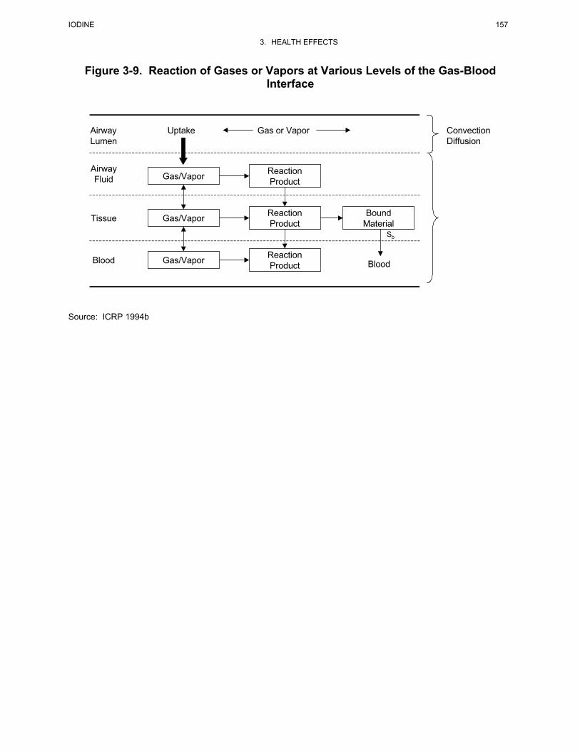
IODINE 157
3. HEALTH EFFECTS
Figure 3-9. Reaction of Gases or Vapors at Various Levels of the Gas-Blood Interface
Uptake Gas or VaporAirwayLumen
AirwayFluid
Tissue
Blood
ConvectionDiffusion
Blood
Sb
ReactionProduct
ReactionProduct
BoundMaterial
Gas/Vapor
Gas/Vapor
ReactionProductGas/Vapor
Source: ICRP 1994b

IODINE 158
3. HEALTH EFFECTS
Table 3-6. Reference Values of Parameters for the Compartment Model to Represent Time-dependent Particle Transport
from the Human Respiratory Tract
Part A
Clearance rates for insoluble particles
Pathway From To Rate (d-1) Half-lifea
m1,4 AI1 bb1 0.02 35 days
m2,4 AI2 bb1 0.001 700 days
m3,4 AI3 bb1 0.0001 7,000 days
m3,10 AI3 LNTH 0.00002 No data
m4,7 bb1 BB1 2 8 hours
m5,7 bb2 BB1 0.03 23 days
m6,10 bbseq LNTH 0.01 70 days
m7,11 BB1 ET2 10 100 minutes
m8,11 BB2 ET2 0.03 23 days
m9,10 BBseq LNTH 0.01 70 days
m11,15 ET2 GI tract 100 10 minutes
m12,13 ETseq LNET 0.001 700 days
m14,16 ET1 Environment 1 17 hours
See next page for Part B
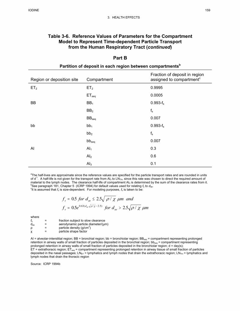
IODINE 159
3. HEALTH EFFECTS
Table 3-6. Reference Values of Parameters for the Compartment Model to Represent Time-dependent Particle Transport
from the Human Respiratory Tract (continued)
Part B
Partition of deposit in each region between compartmentsb
Region or deposition site
Compartment
Fraction of deposit in region assigned to compartmentc
ET2 ET2 0.9995
ETseq 0.0005
BB BB1 0.993-fs
BB2 fs
BBseq 0.007
bb bb1 0.993-fs
bb2 fs
bbseq 0.007
AI AI1 0.3
AI2 0.6
AI3 0.1
aThe half-lives are approximate since the reference values are specified for the particle transport rates and are rounded in units of d-1. A half-life is not given for the transport rate from Al3 to LNTH, since this rate was chosen to direct the required amount of material to the lymph nodes. The clearance half-life of compartment Al3 is determined by the sum of the clearance rates from it. bSee paragraph 181, Chapter 5 (ICRP 1994) for default values used for relating fs to dae. cIt is assumed that fs is size-dependent. For modeling purposes, fs is taken to be:
f for d m and
f e for d ms ae
sd
aeae
= ≤
= >−
05 2 5
05 2 50 63 2 5
. . /
. . /. ( / . )
ρ χ µ
ρ χ µρ χ
where fs = fraction subject to slow clearance dae = aerodynamic particle diameter/(µm) ρ = particle density (g/cm3) χ = particle shape factor AI = alveolar-interstitial region; BB = bronchial region; bb = bronchiolar region; BBseq = compartment representing prolonged retention in airway walls of small fraction of particles deposited in the bronchial region; bbseq = compartment representing prolonged retention in airway walls of small fraction of particles deposited in the bronchiolar region; d = day(s); ET = extrathoracic region; ETseq = compartment representing prolonged retention in airway tissue of small fraction of particles deposited in the nasal passages; LNET = lymphatics and lymph nodes that drain the extrathoracic region; LNTH = lymphatics and lymph nodes that drain the thoracic region Source: ICRP 1994b
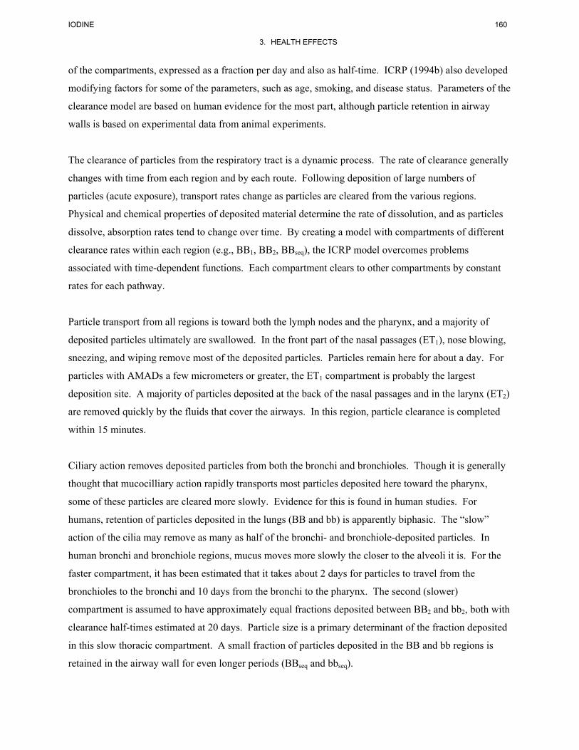
IODINE 160
3. HEALTH EFFECTS
of the compartments, expressed as a fraction per day and also as half-time. ICRP (1994b) also developed
modifying factors for some of the parameters, such as age, smoking, and disease status. Parameters of the
clearance model are based on human evidence for the most part, although particle retention in airway
walls is based on experimental data from animal experiments.
The clearance of particles from the respiratory tract is a dynamic process. The rate of clearance generally
changes with time from each region and by each route. Following deposition of large numbers of
particles (acute exposure), transport rates change as particles are cleared from the various regions.
Physical and chemical properties of deposited material determine the rate of dissolution, and as particles
dissolve, absorption rates tend to change over time. By creating a model with compartments of different
clearance rates within each region (e.g., BB1, BB2, BBseq), the ICRP model overcomes problems
associated with time-dependent functions. Each compartment clears to other compartments by constant
rates for each pathway.
Particle transport from all regions is toward both the lymph nodes and the pharynx, and a majority of
deposited particles ultimately are swallowed. In the front part of the nasal passages (ET1), nose blowing,
sneezing, and wiping remove most of the deposited particles. Particles remain here for about a day. For
particles with AMADs a few micrometers or greater, the ET1 compartment is probably the largest
deposition site. A majority of particles deposited at the back of the nasal passages and in the larynx (ET2)
are removed quickly by the fluids that cover the airways. In this region, particle clearance is completed
within 15 minutes.
Ciliary action removes deposited particles from both the bronchi and bronchioles. Though it is generally
thought that mucocilliary action rapidly transports most particles deposited here toward the pharynx,
some of these particles are cleared more slowly. Evidence for this is found in human studies. For
humans, retention of particles deposited in the lungs (BB and bb) is apparently biphasic. The “slow”
action of the cilia may remove as many as half of the bronchi- and bronchiole-deposited particles. In
human bronchi and bronchiole regions, mucus moves more slowly the closer to the alveoli it is. For the
faster compartment, it has been estimated that it takes about 2 days for particles to travel from the
bronchioles to the bronchi and 10 days from the bronchi to the pharynx. The second (slower)
compartment is assumed to have approximately equal fractions deposited between BB2 and bb2, both with
clearance half-times estimated at 20 days. Particle size is a primary determinant of the fraction deposited
in this slow thoracic compartment. A small fraction of particles deposited in the BB and bb regions is
retained in the airway wall for even longer periods (BBseq and bbseq).

IODINE 161
3. HEALTH EFFECTS
If particles reach and become deposited in the alveoli, they tend to stay imbedded in the fluid on the
alveolar surface or move into the lymph nodes. The one mechanism by which particles are physically
resuspended and removed from the AI region is coughing. For modeling purposes, the AI region is
divided into three subcompartments to represent different clearance rates, all of which are slow.
Particle clearance from the alveolar-interstitial region has been measured in humans. The ICRP model
uses two half-times to represent clearance: about 30% of the particles have a 30-day half-time, and the
remaining 70% are given a half-time of several hundred days. Over time, AI particle transport falls and
some compounds have been found in lungs 10–50 years after exposure.
Absorption into Blood. The ICRP model assumes that absorption into blood occurs at equivalent rates in
all parts of the respiratory tract, except in the anterior nasal passages (ET1), where no absorption occurs.
It is essentially a 2-stage process, as shown in Figure 3-10. First, there is a dissociation (dissolution) of
particles; then the dissolved molecules or ions diffuse across capillary walls and are taken up by the
blood. Immediately following dissolution, rapid absorption is observed. For some elements, rapid
absorption does not occur because of binding to respiratory-tract components. In the absence of specific
data for specific compounds, the model uses the following default absorption rate values for those specific
compounds that are classified as Types F (fast), M (medium), S (slow), and V (instantaneous):
• For Type F, there is rapid 100% absorption within 10 minutes of the material deposited in the BB, bb, and AI regions, and 50% of material deposited in ET2. Thus, for nose breathing, there is rapid absorption of approximately 25% of the deposit in ET and 50% for mouth breathing. Type F iodine compounds include molecular iodine (I2) and particulate aerosols of silver iodide and sodium iodide. For Type M, about 70% of the deposit in AI reaches the blood eventually. There is rapid absorption of about 10% of the deposit in BB and bb, and 5% of material deposited in ET2. Thus, there is rapid absorption of approximately 2.5% of the deposit in ET for nose breathing, and 5% for mouth breathing. ICRP (1995) does not identify any Type M iodine compounds.
• For Type S, 0.1% is absorbed within 10 minutes and 99.9% is absorbed within 7,000 days, so
there is little absorption from ET, BB, or bb, and about 10% of the deposit in AI reaches the blood eventually. ICRP (1995) does not identify any Type S iodine compounds.
• For Type V, complete absorption (100%) is considered to occur instantaneously. Methyl iodide
is classified as a Type V compound (ICRP 1995).

IODINE 162
3. HEALTH EFFECTS
Figure 3-10. The Human Respiratory Tract Model: Absorption into Blood
Particulate Material
Bound Material
Blood
Dissociated Material
Dissolution
Uptake
Particulate Material
Bound Material
Blood
Dissociated Material
Particulate Material
Bound Material
Blood
Dissociated Material
Dissolution
Uptake
Source: ICRP 1994b

IODINE 163
3. HEALTH EFFECTS
EPA (2002) Iodine Biokinetics Models
Description of the models.
EPA (2002a) developed PBPK models of the kinetics of ingested or injected iodide in rats and humans.
The models were developed simultaneously with models of perchlorate biokinetics. When combined, the
iodide and perchlorate models simulate the acute competitive inhibition of iodide transport by perchlorate
in thyroid and other tissues that have NIS activity. The adult rat model has been extended to include
pregnancy and maternal-fetal transfer of iodide, and lactation and maternal-pup iodide transfer through
milk.
The adult rat and human models have the same structure and differ only in values for physiological and
some of iodide parameters (Figure 3-11, Table 3-7). Both models simulate eight tissue compartments:
blood, kidney, liver, skin, stomach, thyroid, fat, other slowly perfused tissues, and other richly perfused
tissues. Uptakes from blood into the vascular compartments of the tissues are simulated as flow-limited
processes. Distributions within blood, skin, stomach, and thyroid are simulated as diffusion-limited
processes with first-order clearance terms. Transport of iodide within tissues that have NIS activity are
simulated with tissue-specific affinity constants and maximum transport velocities. This includes uptake
of iodine into thyroid follicle cells and secretion of iodide into the follicle lumen. Active transport of
iodide into the stomach lumen and in skin is also simulated in the models. Excretion is simulated with a
first-order clearance term for transfer of iodide from the kidney into urine.
Extensions of the adult rat model to simulate iodide kinetics during pregnancy include the addition of two
additional compartments representing the mammary gland and placenta. Uptake of iodide into the
mammary gland tissue from the mammary tissue vascular space is simulated as an affinity- and capacity-
limited transport process, representing the activity of NIS in this tissue. Uptake of iodide into the
placenta from blood is simulated as a flow-limited process. Exchanges of iodide between the placenta
and fetus are simulated with first order clearance terms. The fetal model is identical in structure to the
adult (non-pregnant) model, with adjustments in the physiological and iodide parameters to reflect the
fetus.
The lactating rat model includes a milk compartment in mammary tissue and a first-order clearance term
for describing secretion of iodide form mammary tissue into milk. Transfer of iodide from milk to the
neonate is simulated as a first-order clearance process. The neonate model is identical in structure to the

IODINE 164
3. HEALTH EFFECTS
Table 3-7. Chemical-specific Parameters for the Adult Male Rat and Human PBPK Models for Iodidea
Partition Coefficients (unitless)
Rat
Human
Slowly Perfused/Plasma PS_
0.21
0.21
Richly Perfused/Plasma PR_
0.40
0.40
Fat/Plasma PF_
0.05
0.05
Kidney/Plasma PK_
1.09
0.05
Liver/Plasma PL_
0.44
0.44
Gastric Tissue/Gastric Blood PG_
1.40
0.50
Gastric Juice/Gastric Tissue PGJ_
3.00
3.50
Skin Tissue/Skin Blood PSk_
0.70
0.70
Thyroid Tissue/Thyroid Blood PT_
0.15
0.15
Thyroid Lumen/Thyroid Tissue PDT_
7.00
7.00
Red Blood Cells/Plasma
1.00
1.00
Max Capacity, Vmaxc (ng/hr-kg)
Thyroid Colloid Vmaxc_DT
4.0x10⁻⁷
1.0x10⁸
Thyroid Follicle Vmaxc_T
5.5x10⁴
~1.5x10⁵
Skin Vmaxc_S
5.0x10⁻⁵
7.0x10⁵
Gut Vmaxc_G
1.0x10⁶
9.0105
Plasma Binding Vmaxc_Bp
–
–
Affinity Constants, Km (ng/L)
Thyroid Lumen Km_DT
1.0x10⁹
1.0109
Thyroid km_T
4.0x10⁶
4.0x10⁶
Skin Km_S
4.0x10⁶
4.0106

IODINE 165
3. HEALTH EFFECTS
Table 3-7. Chemical-specific Parameters for the Adult Male Rat and Human PBPK Models for Iodidea
Partition Coefficients (unitless)
Rat
Human
Gut Km_G
4.0x10⁶
4.0106
Plasma BInding km_B
–
–
Permeability Area Cross Products (L/hr-kg)
Gastric Blood to Gastric Tissue PAGc_
0.10
0.20
Gastric Tissue to Gastric Juice PAGJc_
0.10
2.00
Skin Blood to Skin Tissue PASkc_
0.10
0.06
Plasma to Red Blood Cells PARBCc_
1.00
1.00
Follicle to Thyroid Follicle PATc_
1.0x10⁻⁴
1.0x10⁻⁴
Lumen to Thyroid Follicle PADTc_
1.0x10⁻⁴
1.0x10⁻⁴
Clearance Values (L/hr-kg)
Urinary excretion CLUc_
0.05
0.1
Plasma unbinding Clunbc_
–
–
aSource: EPA 2002a
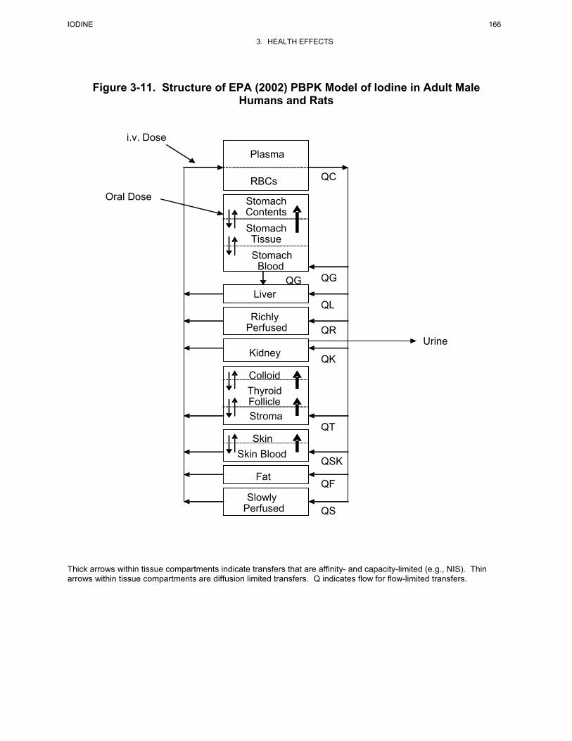
IODINE 166
3. HEALTH EFFECTS
Figure 3-11. Structure of EPA (2002) PBPK Model of Iodine in Adult Male Humans and Rats
Thick arrows within tissue compartments indicate transfers that are affinity- and capacity-limited (e.g., NIS). Thin arrows within tissue compartments are diffusion limited transfers. Q indicates flow for flow-limited transfers.
Kidney
RBCs
Stomach ContentsStomach TissueStomach
Blood
Liver
Richly Perfused
ColloidThyroid Follicle Stroma
SkinSkin Blood
Fat
Slowly Perfused
Oral Dose
QC
QG
QL
QR
QK
Urine
QT
QSK
QF
QS
i.v. Dose Plasma
QG
IODINE 167
3. HEALTH EFFECTS
adult (nonpregnant) model, with adjustments to the physiological and iodide parameter values to reflect
the neonate.
Validation of the model.
The rat iodide model has been evaluated for predicting serum and thyroid iodine concentrations in adult
rats that received acute intravenous injection of radioiodine (EPA 2002a). Model predictions
corresponded reasonably well with observations. The model also predicted reasonably well the inhibition
of radioiodine uptake in the thyroid produced by an acute intravenous dose of perchlorate; however, the
model under-predicted thyroid iodide uptake in rats that received perchlorate in drinking water for
14 days at doses >1 mg/kg/day. Thus, the model simulated a greater inhibition of thyroid uptake of
iodide in animals that received repeated doses of perchlorate than was actually observed. The inability of
the model to accurately predict the effect of repeated exposures to perchlorate on thyroid iodide uptake is
not surprising, since the model does not simulate the hormonal regulation of NIS activity and
organification of iodide in the thyroid. In animals that received repeated exposures to perchlorate,
induction of NIS and thyroid hormone production are likely to have occurred secondary to elevations in
serum TSH (EPA 2002a; Uyttersprot et al. 1997). Such a response could have partially restored thyroid
iodide uptake to higher levels than would be predicted if induction is not taken into account.
The adult human model also predicted reasonably well radioiodine in serum, thyroid, gastric contents, and
urine in subjects who received an intravenous dose of radioiodine (Hays and Solomon 1965), when model
parameters were calibrated to achieve good correspondence to the observations. Similarly, model
predictions of thyroid radioiodine uptake in subjects who received oral doses of perchlorate agreed with
observations when the kinetic parameters for iodide in the thyroid (i.e., maximum transport into the
thyroid follicle) were adjusted to achieve good correspondence to the observations (EPA 2002a). When
the model was calibrated by adjusting the maximum transport rate for iodide into the thyroid follicle, it
accurately predicted the observed time course for radioiodine uptake in a Graves’ disease patient who
received a single tracer dose of radioiodine (Stanbury and Wyngaarden 1952); however, the model
substantially over predicted iodine uptake after the same patient received a dose of perchlorate. Here
again, the error in predictions of the effect of perchlorate on iodine uptake may reflect humoral regulation
of iodide transport and organification mechanisms or the response to perchlorate in Graves’ disease
patients that is not simulated in the model.
The rat maternal/fetal models were evaluated by comparing predictions of radioiodine concentrations in
mammary gland and placenta, and maternal and fetal serum and thyroid following single intravenous
IODINE 168
3. HEALTH EFFECTS
injections of radioiodine, with or without concurrent injection of perchlorate or exposure to perchlorate in
drinking water (EPA 2002a; Versloot et al. 1997). Model predictions were in reasonable agreement with
observations for rats that received single injections of iodide with or without single injections of
perchlorate; however, the model under-predicted maternal thyroid iodide levels in animals that received
repeated oral exposures to perchlorate.
Similar outcomes occurred in evaluations of the lactating dam/neonate model (EPA 2002a). The model
accurately predicted serum and thyroid iodine concentrations in the dam and neonate following single
intravenous injections of radioiodine, with or without concurrent injection of perchlorate. However, the
model under predicted maternal iodide levels in dams that received repeated exposures to perchlorate in
drinking water.
Risk assessment.
The rat and human models have been used to calculate human equivalent exposure levels for perchlorate
that would be expected to produce the same degree of inhibition of iodide uptake into the thyroid gland
(EPA 2002a). These estimates have been used to extrapolate dose-response relationships for perchlorate
observed in rats to humans.
Target tissues.
The models are designed to calculate iodine concentrations in serum and thyroid.
Species extrapolation.
The models are designed for applications to rat or human dosimetry and cannot be applied to other
species without modification.
Interroute extrapolation.
The models are designed to simulate intravenous or oral exposures to radioiodine and cannot be applied to
other routes of exposure without modification.
IODINE 169
3. HEALTH EFFECTS
Berkovski (2002) Iodine Biokinetics Model
Description of the model.
Berkovski (1999a, 1999b) developed compartmental models of the biokinetics of iodine in the pregnant
and lactating human female (Figure 3-12). The most recent description of the models (Berkovski 2002) is
the basis for the ICRP (2002) iodine model. The models simulate the transfer of iodine from the pregnant
woman to the fetus and to breast milk, during lactation. The maternal model simulates the gastrointestinal
cycling of iodine, including absorption to blood from the stomach (slow) and small intestine (fast),
secretion into the stomach, secretion into salivary glands (the latter transfers to the stomach), and
secretion of organic iodine from other tissues into the large intestine (e.g., biliary transfer; the latter
transfers to feces). Iodide in the central blood compartment distributes to the breasts, kidney (to urinary
bladder), ovaries, thyroid gland, and other tissues. The model includes pathways for cycling of iodine
into and out of the organic iodine (e.g., thyroid hormones) pool. This includes the thyroid gland, which
has subcompartments for iodide and organic iodine (e.g., thyroid hormone and precursors). The thyroidal
iodide compartment exchanges with the blood compartment; the organic iodine compartment receives
input from blood and delivers organic iodine to the other tissue compartment. In the other tissue
compartment, organic iodine is deiodinated and the resulting iodide pool exchanges with the iodide in the
blood compartment. The model predicts, for a euthyroid adult who ingests 150 µg iodide/day,
equilibrium contents of approximately 21 µg iodide in blood, 30 µg iodide and 8,000 µg organic iodine in
the thyroid gland, 57 µg iodide and 1,350 µg organic iodine in other tissues, and 21 µg iodide in blood.
The resulting ratio of total iodine in thyroid to that in blood is approximately 400.
The pregnancy model extends the maternal model with additional compartments representing the uterus
and placenta, amniotic fluid compartment, and fetus. Iodide in the maternal blood compartment
exchanges with iodide in the placental/uterine and amniotic fluid compartments. Organic iodine in the
maternal other tissues compartment exchanges with organic iodine in the placental and amniotic fluid
compartments. The fetal compartment includes three subcompartments representing cycling of fetal
iodide into thyroid and extra-thyroidal iodine pools. Iodide can enter the fetal compartment from
exchange with iodide in the placental/uterine compartments or from transfer from the amniotic fluid (i.e.,
fetal ingestion of amniotic fluid). Transfer coefficients vary through gestation to account for changes in
maternal and fetal iodine biokinetics associated with growth of the placenta and fetus, initiation of fetal
thyroid iodine accumulation and hormone production (approximately 12 weeks of gestation), and
increased maternal renal clearance of iodide.

IODINE 170
3. HEALTH EFFECTS
Figure 3-12. Berkovski (2002) Metabolic Model for Iodine Adapted from Berkovski 1999a
Stom
ach
127I 150 µg d-1
I
Salivary glands
Gastric secretory
cells
S
J
λS St
λJ Stv
SIU
LILL
I
λSt SI
λSI ULI
λULI LLI
λLLIF Faeces
(F)
λIS
λSt I
λIJ
Mother
λOI λIO
Ovaries
O
Iodide in blood
I
λSI I
Iodide30 µg
g
Organic I8,000 µg
G
λIg λgI λIG
Thyroid
Iodide
D
Organic I
B
λDI λID
Other
λGBλB ULI
Breasts
Br
Urinary bladder
UB
Kidneys
K
λK Ub
Milk (M)
Urine (U)
λBr M
λUB U
Utero-Placental Unit
Iodide
PI
Organic I
P2
Embryo and Fetus
Fetal iodide
FI
Fetal thyroid
FG
Fetal organic 1
FB
λFG FB
λFI FGv
λFB FI
Iodi
de
A1O
rgan
ic I
A2
Amni
otic
flui
d
λAI FIvv λA2 FI
λFI PI
λPI FI
λBP2 λP2B
λIK
λI PI
λPI I
λI AI
λAi IλBD
λB A2 λA2 B
St
IODINE 171
3. HEALTH EFFECTS
The lactation model includes an additional compartment for breast milk, which receives iodide from the
breast compartment.
Validation of the model.
Berkovski (1999a, 1999b, 2002) presents comparisons of model predictions and observations made in
humans. The model predictions agree well with the observed kinetics of elimination of 131I from amniotic
fluid after intravenous injection of 131I. The model also simulates, with good agreement, observed fetal
thyroid radioiodine uptakes and elimination at various stages of gestation.
Risk assessment.
The Berkovski (2002) model is the basis for the ICRP (2002) model, which is used to establish radiation
dose equivalents (Sv/Bq) of various ingested radioactive isotopes of iodine.
Target tissues.
The model is designed to calculate radioiodine intake limits based on radiation dose to all major organs
that concentrate iodine (relative to blood), including the thyroid gland, salivary glands, ovaries, and fetus.
Species extrapolation.
The model is designed for applications to human dosimetry and cannot be applied to other species without
modification.
Interroute extrapolation.
The model is designed to simulate oral exposures to radioiodine; however, it has been applied to
simulating the biokinetics of intravenous injections of iodide and could be applied other routes of
exposure with modification to include simulations of the absorption from these routes to blood.
ICRP (1989) Iodine Biokinetics Model
Description of the model.
ICRP (1989) developed a compartmental model of the kinetics of ingested iodine in humans with
parameter values that are applicable to infants, children, adolescents, and adults. The model is a

IODINE 172
3. HEALTH EFFECTS
modification and expansion of a similar model described in ICRP (1979; Riggs 1952). Ingested iodine is
assumed to be completely absorbed. Absorbed iodine is assumed to distribute to three compartments:
blood, thyroid gland, and extrathyroid tissues (Figure 3-13). Of the iodine entering the transfer
compartment, 30% is assumed to be transferred to the thyroid gland; the remaining 70% is excreted in
urine. All iodine eliminated from the thyroid gland is assumed to be transferred to the extrathyroidal
tissues compartment as organic iodine (e.g., iodothyronines). Twenty percent of the iodine eliminated
from extrathyroidal tissues is assumed to be excreted in feces; the remaining 80% is transferred to blood.
Elimination half-times of iodine from thyroid, and extrathyroidal tissues are age-dependent, while that
from blood is independent of age (Figure 3-13). The modifications made in this model from ICRP (1979)
include: (1) 20%, rather than 10% of the of iodine eliminated from extrathyroidal tissues is assumed to be
excreted in feces; (2) age-dependent elimination half-times for iodine, which allows the model to be
applied to infants, children, adolescents, and adults; and (3) the extrathyroidal iodine pool is assumed to
be 0.1 of the thyroid pool and the thyroid iodine pool is allowed to be variable, reflecting geographic
variation or other sources of variation in intake.
Validation of the model.
The extent to which the ICRP model has been validated is not described in ICRP (1989).
Risk assessment.
The model has been used to establish radiation dose equivalents (Sv/Bq) of ingested various radioactive
isotopes of iodine (ICRP 1989, 1993).
Target tissues.
The model is designed to calculate radioiodine intake limits based on radiation dose to all major organs,
including the thyroid gland.
Species extrapolation.
The model is designed for applications to human dosimetry and cannot be applied to other species without
modification.

IODINE 173
3. HEALTH EFFECTS
Figure 3-13. International Commission on Radiological Protection (ICRP) (1989)
Metabolic Model for Iodine
ICRP (1989) Metabolic Model for Iodine
Biological half-timea (d) Apparent half-timeb (d)
Age f1
Thyroid uptake (%)
Fecal excretion (%)
Blood T1
Thyroid T2
Extrathyroidal T3 Thyroid
3 months 1 30 20 0.25 11.2 1.12 15
1 year 1 30 20 0.25 15 1.5 20
5 years 1 30 20 0.25 23 2.3 30
10 years 1 30 20 0.25 58 5.8 70
15 years 1 30 20 0.25 67 6.7 80
Adult 1 30 20 0.25 80 12 91
aln2/ki b2–16 days after uptake
Blood
Extra-thyroidal Tissues
Thyroid
Uptake fromIngestion
Feces Urine
f 1 · 1k (1-e) · k 3
(1-f 1 ) · k 3
k2

IODINE 174
3. HEALTH EFFECTS
Interroute extrapolation.
The model is designed to simulate oral exposures to radioiodine and cannot be applied to other routes of
exposure without modification.
Killough and Eckerman (1986) Iodine Biokinetics Model
Description of the model.
Killough and Eckerman (1986) developed a 2-compartment modification of the 3-compartment model as
described by Riggs (1952, the basis for the ICRP 1979 model). In the Killough and Eckerman (1986)
model, the compartment representing the extrathyroidal organic iodine pool in Riggs (1952) has been
eliminated and transfer into the thyroid gland is represented with a first-order rate constant (rather than a
deposition fraction). This change provides for simulation of the short-term kinetics of uptake of iodine
into the thyroid (rather then only maximal uptakes), enabling such observations to be incorporated into
model calibration efforts (Killough and Eckerman 1986). Values for the transfers of iodine into and out
of the thyroid are age-dependent, which is the basis for age-dependence of the biokinetics simulated in the
model.
Validation of the model.
The extent to which the Killough and Eckerman model has been validated is not described in Killough
and Eckerman (1986).
Risk assessment.
The model has been used to establish radiation doses to the thyroid for 4,216 patients administered 131I for
clinical diagnostic purposes (Killough and Eckerman 1986).
Target tissues.
The model is designed to calculate thyroid gland radiation doses associated with administered activities of
radioiodine.
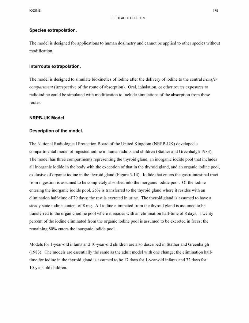
IODINE 175
3. HEALTH EFFECTS
Species extrapolation.
The model is designed for applications to human dosimetry and cannot be applied to other species without
modification.
Interroute extrapolation.
The model is designed to simulate biokinetics of iodine after the delivery of iodine to the central transfer
compartment (irrespective of the route of absorption). Oral, inhalation, or other routes exposures to
radioiodine could be simulated with modification to include simulations of the absorption from these
routes.
NRPB-UK Model
Description of the model.
The National Radiological Protection Board of the United Kingdom (NRPB-UK) developed a
compartmental model of ingested iodine in human adults and children (Stather and Greenhalgh 1983).
The model has three compartments representing the thyroid gland, an inorganic iodide pool that includes
all inorganic iodide in the body with the exception of that in the thyroid gland, and an organic iodine pool,
exclusive of organic iodine in the thyroid gland (Figure 3-14). Iodide that enters the gastrointestinal tract
from ingestion is assumed to be completely absorbed into the inorganic iodide pool. Of the iodine
entering the inorganic iodide pool, 25% is transferred to the thyroid gland where it resides with an
elimination half-time of 79 days; the rest is excreted in urine. The thyroid gland is assumed to have a
steady state iodine content of 8 mg. All iodine eliminated from the thyroid gland is assumed to be
transferred to the organic iodine pool where it resides with an elimination half-time of 8 days. Twenty
percent of the iodine eliminated from the organic iodine pool is assumed to be excreted in feces; the
remaining 80% enters the inorganic iodide pool.
Models for 1-year-old infants and 10-year-old children are also described in Stather and Greenhalgh
(1983). The models are essentially the same as the adult model with one change; the elimination half-
time for iodine in the thyroid gland is assumed to be 17 days for 1-year-old infants and 72 days for
10-year-old children.
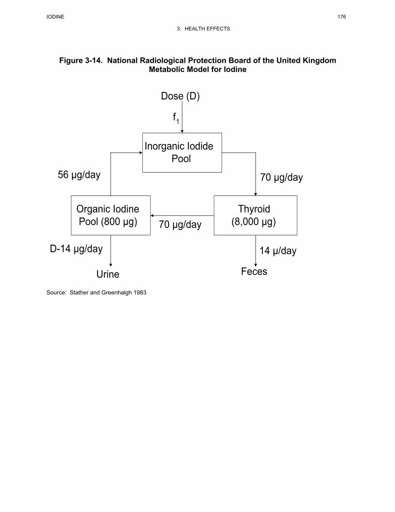
IODINE 176
3. HEALTH EFFECTS
Figure 3-14. National Radiological Protection Board of the United Kingdom Metabolic Model for Iodine
Inorganic IodidePool
Organic IodinePool (800 µg)
Thyroid(8,000 µg)
56 µg/day 70 µg/day
70 µg/day
Urine Feces
14 µ/dayD-14 µg/day
Dose (D)
f1
Source: Stather and Greenhalgh 1983

IODINE 177
3. HEALTH EFFECTS
Validation of the model.
The extent to which the NRPB-UK model has been validated is not described in Stather and Greenhalgh
(1983).
Risk assessment.
The model was developed for calculating radiation doses to populations in the United Kingdom following
release of iodine isotopes into the environment. The extent to which the model has been used for this
purpose is not described in Stather and Greenhalgh (1983).
Target tissues.
The model is designed to calculate intake and exposure limits, based on radiation dose to the NRPB-UK
model thyroid gland.
Species extrapolation.
The model is designed for applications to human dosimetry and cannot be applied to other species without
modification.
Interroute extrapolation.
The model is designed to simulate oral exposures to radioiodine and cannot be applied to other routes of
exposure without modification.
Johnson (1982) Model
Description of the model.
Johnson (1982, 1986) described a compartmental model of iodine biokinetics in humans that included
parameters for simulating pregnancy. The structure of the maternal model is similar to the Stather and
Greenhalgh (1983) model in that it has three compartments representing the thyroid gland, an extra-
thyroidal inorganic iodide pool, and an organic iodine pool, exclusive of organic iodine in the thyroid
gland (Figure 3-15). Iodide that enters the gastrointestinal tract from ingestion or the lungs from
inhalation is assumed to be absorbed into the inorganic iodide pool. From the inorganic iodide pool,
iodide is transferred to the thyroid gland (at a rate equal to loss of thyroidal iodine to the organic iodine

IODINE 178
3. HEALTH EFFECTS
Figure 3-15. Johnson (1982) Metabolic Model for Iodine Adapted from Johnson 1986
1
Mother’s Stomach or Lungs
lr (t)
ls (t)
2
Mother’s and Fetus’ Inorganic Compartment
6
Fetus’ Thyroid
3
Mother’s Thyroid
r8 s8 r2 s2
7
Fetus’ Organic
Compartment
4
Mother’s Organic
Compartment
λ9 λ3
λ1
λ3
λ10 λ4
λ6
5
Bladder Contents
Gastrointestinal Tract
λ1
Urine Feces
1-F1 1-F0 F1 F0

IODINE 179
3. HEALTH EFFECTS
pool), or is excreted in urine and feces. All iodine eliminated from the thyroid gland is assumed to be
transferred to the organic iodine pool, from which it can renter the extra-thyroidal inorganic iodine pool.
Daily thyroid iodide uptakes vary with thyroid gland mass. Thyroid gland growth and mass are age- and
gender-dependent, which are the bases for age- and gender dependence of the biokinetics in the model.
The pregnancy model includes fetal thyroid and fetal organic iodine compartments. Iodide cycles from
the maternal extrathyroidal iodide pool, to the fetal thyroid pool, to the fetal organic iodine pool, from
where it can return to the maternal extrathyroidal iodide pool.
Validation of the model.
The extent to which the Johnson (1982, 1986) model has been validated is not described in either
publication.
Risk assessment.
The model was developed for calculating radiation doses to populations following release of iodine
isotopes into the environment. The extent to which the model has been used for this purpose is not
described in Johnson (1982, 1986).
Target tissues.
The model is designed to calculate intake and exposure limits, based on radiation dose to the thyroid
gland.
Species extrapolation.
The model is designed for applications to human dosimetry and cannot be applied to other species without
modification.
Interroute extrapolation.
The model is designed to simulate oral and inhalation exposures to radioiodine; however, it could be
applied to other routes of exposure with modifications to include simulations of the absorption from these
routes to the extrathyroidal inorganic iodide pool.

IODINE 180
3. HEALTH EFFECTS
Oddie et al. Model
Description of the model.
Oddie et al. (1955) described a compartmental model of absorbed iodine human adults and infants (Fisher
et al. 1962) for predicting 24-hour radioiodine uptake by the thyroid gland in clinical procedures. The
model has two compartments representing the thyroid gland and a central iodide pool that includes all
inorganic iodide in the body with the exception of that in the thyroid gland. An organic iodine pool is not
included in the model. Although this would preclude the model from accurately simulating radioiodide
levels in extrathyroidal tissues, including blood, it was not considered necessary for simulating the initial
uptake of iodide by the thyroid following a single dose of radioiodine, prior to significant release of
organic iodine from the thyroid gland. Iodide that enters the inorganic iodide pool is assumed to be
transferred either to the thyroid gland, represented as a first order rate constant k1, or to the kidney for
urinary excretion, represented by a rate constant k2, usually corrected for loss of iodide in sweat, feces,
and uncollected urine (Oddie and Fisher 1967). In a study of 20 healthy adults, k1 was estimated to be
60x10-5/minute in subjects who ingested a tracer dose of radioiodine (Fisher et al. 1965). In this same
study, the value of k1 was 49x10-5 after 13 weeks of daily ingestion of 252 µg iodide/day and 35x10-5,
after 13 weeks of daily ingestion of 1,000 µg iodide/day. The estimate of k2 from this study was
300x10-5 minute-1. Values for k1 estimated in various populations have ranged from 67 to
134x10-5 minute-1 (Oddie and Fisher 1967). The volume of the iodide space was estimated to be 2.1 L
(Fisher et al. 1965).
The same model has been used to predict thyroid uptakes of iodine in infants. Values for k1 and k2 were
estimated from studies in which 24-hour thyroid uptakes of iodine were measured in 26 euthyroid
newborn infants (Fisher et al. 1962). The values for k1 and k2 were 2.4x10⁻³ minute-1 and
1.1x10⁻³ minute-1, respectively. The iodide space was estimated to be approximately 0.4 L in newborn
infants.
Validation of the model.
The model has been shown to predict 24-hour iodine uptakes in the thyroid in adults who received single
doses of radioiodine. Predicted 24-hour thyroid uptakes of radioiodine were compared to observed
estimates in 1,573 euthyroid subjects reported from various studies; the difference between observed and
predicted estimated for eight studies ranged from 0.7 to 2.1%, with the observed uptakes ranging from
21 to 37% (Oddie and Fisher 1967).
IODINE 181
3. HEALTH EFFECTS
Risk assessment.
The model was developed for predicting the 24-hour uptake of radioiodine in the thyroid after single
doses of radioiodine are given in the clinical setting for assessing thyroid function. It has been evaluated
in terms of its predictive value in detecting abnormal thyroid conditions that affect iodide uptake into the
gland (Oddie et al. 1960). The extent to which the model has been used for risk assessment could not be
ascertained from the available literature.
Target tissues.
The model is designed to predict 24-hour uptakes of radioiodine into the thyroid gland.
Species extrapolation.
The model is designed for applications to humans and cannot be applied to other species without
modification.
Interroute extrapolation.
The model is designed to simulate oral ingestion or parenteral injection (e.g., intramuscular in infants) of
radioiodine and cannot be applied to other routes of exposure without modification.
3.6 MECHANISMS OF ACTION
3.6.1 Pharmacokinetic Mechanisms
Absorption. The mechanism(s) by which iodide is absorbed from the gastrointestinal tract is not
known. Based on the study conducted by Small et al. (1961), absorption appears to occur primarily in the
small intestine in humans. This study measured iodine in the saliva of healthy human subjects who
ingested 0.25 g of potassium iodide (0.19 g iodide) together with a radioopaque suspension of barium
sulfate that allowed the emptying of the stomach to be imaged with a fluoroscope. In five subjects, iodine
was not detected in saliva until 2–3 minutes after the first appearance of the barium sulfate in the
duodenum; the actual time of appearance relative to the oral dose of iodide ranged from 15 to 40 minutes.
An intravenous dose of probanthine, which delays gastric emptying time, given just prior to the oral dose
of potassium iodide, substantially delayed the time of appearance of iodine in saliva to 114–133 minutes;
IODINE 182
3. HEALTH EFFECTS
however, in each of three subjects, iodine was detected in saliva 3–4 minutes after the first appearance of
the radioopaque marker in the duodenum. When iodide was instilled directly into the duodenum together
with the radioopaque marker (two subjects), iodine was detected in saliva 3–4 minutes after the dose was
administered. These observations suggest that the absorption of iodide in humans occurs primarily in the
small intestine and that the stomach may play a minor role in iodide absorption. The mechanisms by
which iodide is transported across the intestinal epithelium are not known. Iodide may be transported by
mechanisms that also transport chloride such as the Cl-/HCO3- antiport (Dalmark 1976; Lambert and
Lowe 1978) or Cl- channels (Katayama and Widdicombe 1991).
While the above studies implicate the small intestine as the major site of absorption of iodide in humans,
studies in rats and dogs indicate that 14–30% of an oral dose of iodide may be absorbed in the stomach in
these species (Small et al. 1961; Cohn 1932)
Distribution.
Iodide Transport. Uptake of iodide into the thyroid is facilitated by a membrane carrier in the basolateral
membrane of the thyroid follicle cell (Carrasco 1993; Levy et al. 1998a; Shen et al. 2001). The carrier, or
NIS, catalyzes the simultaneous transfer Na+ and I- across the basolateral membrane (Chambard et al.
1983; Iff and Wilbrandt 1963; Nilsson et al. 1990). The stoichiometry of transfer reaction is (2)Na+/(1)I-,
which confers to the NIS a net positive charge and, therefore, a sensitivity to transmembrane voltage
(Eskandari et al. 1997; O’Neill et al. 1987). In the presence of an inward-directed electrochemical
gradient for Na+, the NIS can transfer I- into the cell against a pronounced outward-directed
electrochemical gradient for I- (Takasu et al. 1984; Williams 1969; Woodbury and Woodbury 1963).
This enables the follicle cell to achieve intracellular/extracellular concentration ratios of 10–50 for iodide
(Andros and Wollman 1991; Bagchi and Fawcett 1973; Shimura et al. 1997; Vroye et al. 1998; Weiss et
al. 1984b; Wolff 1964).
The NIS has been studied extensively in several in vitro preparations, including isolated plasma
membrane vesicles of mammalian thyroid (O’Neill et al. 1987), FRTL-5 cells, a cell line derived from
normal rat thyroid (Weiss et al. 1984b), Xenopus lavis oocytes transformed by intracellular injection of
FRTL-5 RNA to express NIS (Eskandari et al. 1997), and other mammalian cells cultures transformed to
express NIS (Levy et al. 1997; Nakamura et al. 1990; Smanik et al. 1996; Yoshida et al. 1997). The
apparent Km for I- transport in cell systems is approximately 30–40 µM, which is considerably higher than
the serum iodide concentration of 0.04–0.08 µM (5–10 µg/L) (Eskandari et al. 1997; Weiss et al. 1984b).
The relatively high Km enables the iodide transport rate to be highly sensitive to changes in plasma I-
IODINE 183
3. HEALTH EFFECTS
concentration. Iodide transport by the NIS is inhibited by other anions, most notably, thiocyanate (SCN-)
and perchlorate (ClO4-) (Carrasco 1993; Wolff 1964). Thiocyanate is one of several anions other than I-
that can be transported by the NIS, including SeCN-, NO3-, ClO3
-, Br-, BF4-, IO4
-, and BrO3- (Eskandari et
al. 1997). Perchlorate, on the other hand, does not appear to be transported by NIS (Eskandari et al. 1997;
Yoshida et al. 1997). Thus, thiocyanate and perchlorate, which both inhibit iodide uptake in thyroid in
vivo, do so by different mechanisms; thiocyanate is a competitive substrate for transport, whereas
perchlorate appears to block I- binding to the NIS.
Synthesis of NIS is regulated by the pituitary hormone, TSH, which stimulates iodide uptake into the
thyroid. The mechanism involves both increased transcription of the NIS gene and increased translation
of mRNA for NIS (Kogai et al. 1997; Levy et al. 1997; Ohno et al. 1999; Pekary et al. 1998). Both
responses to TSH follow binding of TSH to a receptor on the basolateral membrane and activation of the
enzyme adenylate cyclase by GTP binding protein Gα (Akamizu et al. 1990; Chazenbalk et al. 1990;
Kogai et al. 1997; Parmentier et al. 1989; Perret et al. 1990; Raspe and Dumont 1995). In FRTL-5 cells
grown in the absence of TSH, NIS activity declines to a minimum level and can be restored by the
addition of TSH to the medium or by treating the cells with dibutryl-cAMP or other agents that increase
the intracellular concentration of cAMP (Pekary et al. 1998; Weiss et al. 1984a, 1984b). Thus, the actions
of TSH appear to involve the activation of adenylate cyclase and subsequent increase in the intracellular
concentration of cAMP. TSH also appears to mediate post-transcriptional regulation of NIS, including
increasing the intracellular elimination half-time of the NIS protein and stimulating the incorporation of
the NIS protein into the thyrocyte cell membrane (Riedel et al. 2001).
Synthesis of NIS also appears to be regulated by plasma iodide concentration through a mechanism that
does not directly involve TSH. In rats exposed to drinking water containing 500 mg/L I as sodium iodide,
expression of mRNA for the NIS in the thyroid decreased by 45% after 1 day of exposure and 60% after
6 days of exposure compared to controls that ingested water without added iodide. Serum iodide
concentrations were 150–200-fold higher in the exposed rats compared to controls, whereas the serum
TSH concentrations were not different between control and treated groups (Eng et al. 1999). A similar
observation was made in dogs made hypothyroid by treatment with propylthiouracil (an inhibitor of
iodination of thyroglobulin) and perchlorate (Uyttersprot et al. 1997). The hypothyroid state elevated
TSH concentrations in serum; nevertheless, a single injection of 0.3 mg potassium iodide (0.23 mg I)
resulted in decreased expression of NIS in the thyroid within 24–48 hours after the dose, without a change
in TSH concentrations in serum. Both iodide and T3 depress the expression of NIS mRNA and iodide
uptakes in rat thyroid follicle cells grown in culture (Sptizweg et al. 1999).

IODINE 184
3. HEALTH EFFECTS
The exact mechanisms by which the NIS gene transcription is regulated have not been determined. The
gene in humans and rats has been sequenced, enabling studies of the mechanisms of gene transcription
regulation (Dai et al. 1996; Smanik et al. 1996). The human gene resides on chromosome 19 (Smanik et
al. 1997). Mutations in the gene sequence have been associated with hypothyroidism, goiter, and
abnormally low thyroid uptake of injected iodide (Fujiwara et al. 1997, 1998, 2000; Kosugi et al. 1998,
2002; Levy et al. 1998c; Pohlenz and Refetoff 1999; Pohlenz et al. 1997). The 5'-flanking region of the
rat NIS gene has been shown to contain one or more promoter regions; however, their role in regulation
of the NIS transcription is not completely understood (Endo et al. 1997; Kogai et al. 2001; Ohno et al.
1999; Schmitt et al. 2000, 2001; Tong et al. 1997). A promoter region in the rat NIS gene appears to
respond to a rise in intracellular cAMP, most likely by binding a cAMP-inducible or cAMP-activated
transcription factor (Chun and Di Lauro 2001; Ohno et al. 1999). NIS expression in FRTL-5 cells is
increased in response to extracellular adenosine, possibly through a mechanism that is independent of
cAMP (Harii et al. 1999). A promoter region in the rat NIS gene responsive to thyroid transcription factor
1 (TTF-1) has also been described (Endo et al. 1997). Tong et al. (1997) found evidence for a promoter
region in the rat NIS gene that could be suppressed in cell cultures that were transformed with the
oncogene PTC1. This may provide a mechanism for the decreased expression of the NIS gene in thyroid
papillary carcinomas and the decreased iodide uptake of some thyroid carcinomas (Smanik et al. 1996,
1997).
Several tissues in humans, other than thyroid, actively express NIS and accumulate iodide; these include
the mammary gland, salivary glands, and gastric mucosa (Brown-Gant 1961; Lacroix et al., 2001; Smanik
et al. 1997; Spitzweg et al. 1998, 1999; Wolff 1983). These tissues can achieve intracellular/extracellular
and/or transepithelial concentration ratios for I- concentrations of 20–40. Transport of iodide in these
tissues is inhibited by thiocyanate and perchlorate; however, transport activity is not responsive to TSH.
Clinical cases of genetic absence or impaired iodide uptake in the thyroid coupled with low uptakes in
saliva and gastric fluid suggest an involvement of an NIS mechanism in these tissues (Fujiwara et al.
1997, 1998; Kosugi et al. 1998; Leger et al. 1987; Pohlenz and Refetoff 1999; Pohlenz et al. 1997; Wolff
1983). Further evidence for extrathyroidal NIS comes from studies of mammary gland. The NIS gene is
expressed in the mammary gland of the human, rat, and many strains of mice (Levy et al. 1997; Perron et
al. 2001; Rillema et al. 2000b; Smanik et al. 1997; Spitzweg et al. 1998; Tazebay et al. 2000). In the rat,
expression of the NIS, or a structurally similar membrane protein, increases during nursing and decreases
after weaning (Cho et al. 2000; Levy et al. 1998a). In the mouse and rat, the induction of NIS appears to

IODINE 185
3. HEALTH EFFECTS
be stimulated by prolactin (Cho et al. 2000; Rillema and Rowady 1997; Rillema et al. 2000b). The NIS
gene is also expressed in human kidney and placenta (Bidart et al. 2000; Spitzweg et al. 2001).
Studies in animals have revealed other tissues that actively secrete or accumulate iodide transport by a
mechanism that is inhibited by perchlorate and thiocyanate, suggestive of an active NIS. These include
choroid plexus, ciliary body of the eye, small intestine (ileum), ovary, placenta, and skin in mammals; and
avian salt gland in marine birds (Brown-Grant 1961). In humans, the NIS gene is expressed in mammary
gland, salivary glands, and gastric mucosa (Lacroix et al. 2001; Smanik et al. 1997; Spitzweg et al. 1998).
An iodide transporter that is distinct from NIS has been characterized in the apical membrane of the
thyroid follicle cell (Royaux et al. 2000; Taylor et al. 2002; Yoshida et al. 2002). This transporter may
function in the facilitated transfer of iodide from the follicle cell in the follicle lumen. Mutations in the
gene coding for the apical transporter occur in Pendred syndrome, an autosomal recessive disorder
characterized by hearing loss, thyroid iodide organification deficits goiter (Scott et al. 2000).
Iodothyronine Transport. Uptake of T4 and T3 into tissues occurs by a saturable, energy-dependent
carrier transport system. Evidence for active transport derives from a variety of observations. The rate of
uptake of T3 into the perfused rat liver is proportional to the concentration of free T3 in the perfusate and
is not related to the total concentration or bound concentration (Mendel et al. 1988). The free cytosolic
concentration of T3 in the in vivo rat liver and heart muscle exceeds that of the simultaneous free
concentration in plasma, suggesting uptake of T3 into these tissues against a chemical gradient for T3
(Oppenheimer and Schwartz 1985). T3 uptake into confluent cultures of human or rat hepatoma cells is
saturable, stereoselective for the active L enantiomer, temperature dependent, and inhibited by metabolic
and membrane transport inhibitors, including phloretin (Movius et al. 1989; Topliss et al. 1989).
Saturable, stereoselective, temperature-dependent, and energy-dependent uptake of T3 and T4 has also
been observed in cultures of human fibroblasts and of T3 in in vitro preparations of rat skeletal muscle
(Centanni and Robbins 1987; Docter et al. 1987).
Metabolism.
Iodination in the Thyroid Gland. Iodination of thyroglobulin is catalyzed by thyroid peroxidase, a
hemoprotein in the apical (luminal) membrane of thyroid follicle cells (Dunn and Dunn 2001). Thyroid
peroxidase catalyzes both the iodination of tyrosine residues in thyroglobulin and the coupling of the
iodinated residues to form the thyroid hormones, T4 and T3, and diiodotyrosine. The iodination reaction
involves the oxidation of iodide (I-) to a reactive species having a sufficiently high oxidation potential to

IODINE 186
3. HEALTH EFFECTS
iodinate the aromatic ring of tyrosine. The oxidizing agent in the reaction is hydrogen peroxide, which is
generated at the apical membrane of follicle cells by an NADPH oxidase (Deme et al. 1994; Dupuy et al.
1991). Although the exact mechanism of the iodination reaction is not completely understood, three
species are suspected as being candidates for the reactive iodinating species: a free radical (I@), iodinium
(I+), or an enzyme-bound hypoiodite ([EOI]-) (Taurog 1996). Human thyroglobulin contains 134 tyrosyl
residues, of which approximately 20 undergo iodination to yield approximately 2–4 molecules of T4 or T3
per molecule of thyroglobulin. The coupling reaction occurs within thyroglobulin, rather than as a
reaction between free iodinated tyrosines. In the formation of T4, two molecules of diiodotyrosine are
coupled, whereas the formation of T3 is a coupling of monoiodotyrosine and diiodotyrosine residues. The
reaction is catalyzed by thyroid peroxidase with hydrogen peroxide serving as the oxidizing agent in the
formation of a reactive intermediate of the contributing diiodotyrosine residue, possibly a free radical
species (Taurog et al. 1994). Specificity of iodination and coupling of tyrosine residues within
thyroglobulin is conferred, in part, by the specificity of thyroid peroxidase and, in part, by the structure of
thyroglobulin (Taurog 1996).
The gene for human thyroid peroxidase has been isolated and sequenced (Kimura et al. 1987; Libert et al.
1987; Magnusson et al. 1987). Transcription of the gene is stimulated by TSH, possibly through a
mechanism involving cAMP (McLachlan and Rapoport 1992).
Deiodination of Iodothyrones in Peripheral Tissues. Deiodination serves both as an important
mechanism for the production of extrathyroidal T3 and for the deactivation of the thyroid hormones, T4
and T3. The deiodination reactions are catalyzed by selenium-dependent deiodinase enzymes
(selenodeiodinases). Three selenodeiodinases have been described that differ in substrate preference,
reaction products, response to inhibitors (propylthiouracil, gold), and response to T3 (Table 3-8). Full
activity of each enzyme requires selenocysteine in the amino acid sequence of the active site, which is the
basis for deiodination activity being responsive to nutritional selenium status (Larsen and Berry 1994; see
Section 3.10).
Excretion.
Urinary Excretion of Iodide. Urinary excretion normally accounts for >97% of the elimination of
absorbed iodine. The renal plasma clearance of iodine has been measured in human subjects during
continuous intravenous infusions of radioiodide (Bricker and Hlad 1955). Under these conditions, only a
negligible amount of radioiodine in the plasma was associated with protein and >98% was ultrafilterable;
thus, the renal clearance of radioiodine can be assumed to reflect that of radioiodide (Bricker and Hlad
IODINE 187
3. HEALTH EFFECTS
Table 3-8. Properties of Human Iodothyronine Selenodeiodinases
Parameter Type 1 Type 2 Type 3
Physiological role Plasma T3 production, deactivate T3 and T4, degrade rT3
Plasma and intracellular T3 production
Deactivate T3 and T4
Tissue location Liver, kidney, thyroid, central nervous system, pituitary
Central nervous system, pituitary, brown fat, placenta, thyroid, skeletal muscle, heart
Central nervous system, placenta, skin
Substrate preference
rT3>>T4>T3 T4$rT3 T3>T4
Molecular weight (D)a
29,000 35,000 31,500
Apparent Km (M) ~10-7 (rT3) ~10-6 (T4)
10-9 (T4) ~10-8 (rT3)
~10-9 (T3) ~10-8 (T4)
Deiodination site Outer and inner ring Outer ring Inner ring
Apparent Ki (M)
Propylthiouracil 2x10⁻⁷ 4x10⁻³ 10-3
Gold ~5x10⁻⁹ ~2x10⁻⁶ 5x10⁻⁶
Response to T3 Increase Decrease Increase aMonomer T3 = 3,5,3N-triiodo-L-thyronine; T4 = 3,5,3N,5N-tetraiodo-L-thyronine (thyroxine); rT3 = reverse T3 Source: Larsen et al. 1998
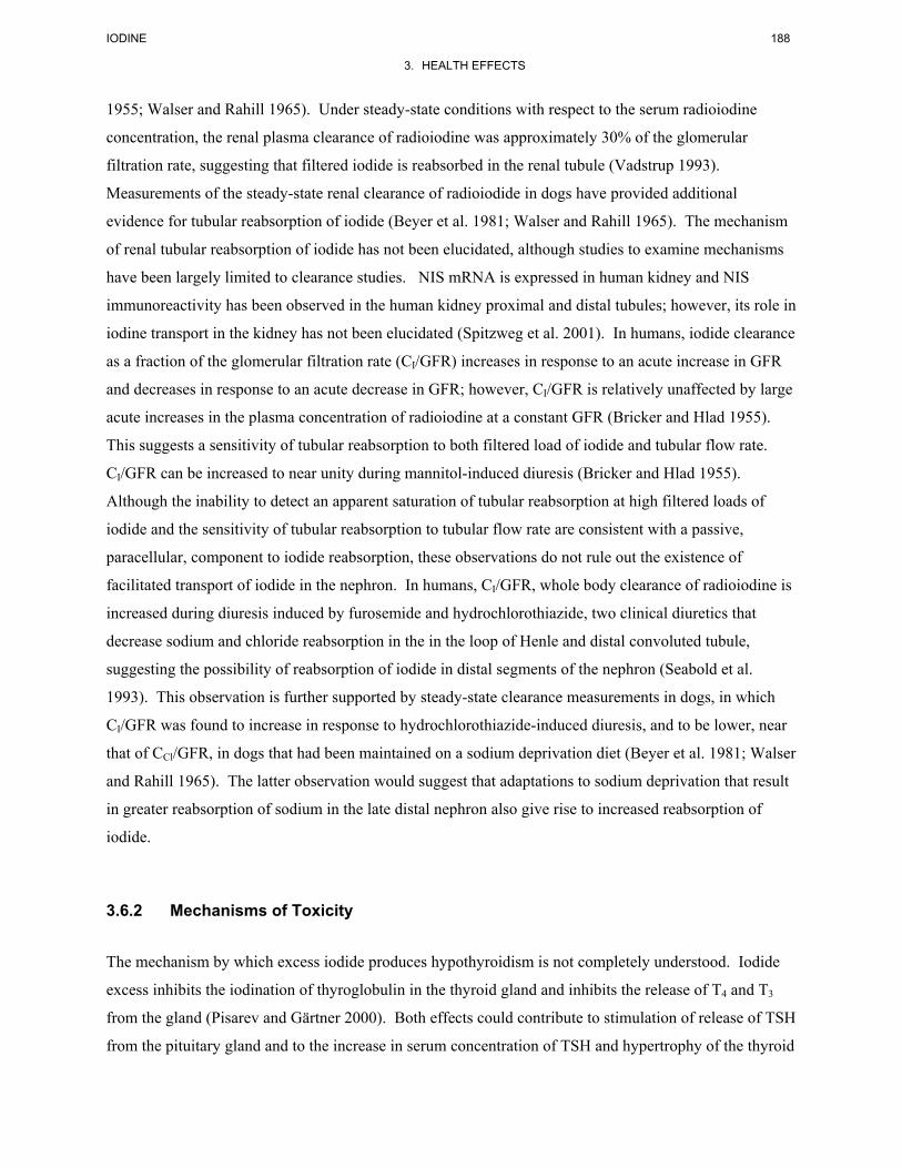
IODINE 188
3. HEALTH EFFECTS
1955; Walser and Rahill 1965). Under steady-state conditions with respect to the serum radioiodine
concentration, the renal plasma clearance of radioiodine was approximately 30% of the glomerular
filtration rate, suggesting that filtered iodide is reabsorbed in the renal tubule (Vadstrup 1993).
Measurements of the steady-state renal clearance of radioiodide in dogs have provided additional
evidence for tubular reabsorption of iodide (Beyer et al. 1981; Walser and Rahill 1965). The mechanism
of renal tubular reabsorption of iodide has not been elucidated, although studies to examine mechanisms
have been largely limited to clearance studies. NIS mRNA is expressed in human kidney and NIS
immunoreactivity has been observed in the human kidney proximal and distal tubules; however, its role in
iodine transport in the kidney has not been elucidated (Spitzweg et al. 2001). In humans, iodide clearance
as a fraction of the glomerular filtration rate (CI/GFR) increases in response to an acute increase in GFR
and decreases in response to an acute decrease in GFR; however, CI/GFR is relatively unaffected by large
acute increases in the plasma concentration of radioiodine at a constant GFR (Bricker and Hlad 1955).
This suggests a sensitivity of tubular reabsorption to both filtered load of iodide and tubular flow rate.
CI/GFR can be increased to near unity during mannitol-induced diuresis (Bricker and Hlad 1955).
Although the inability to detect an apparent saturation of tubular reabsorption at high filtered loads of
iodide and the sensitivity of tubular reabsorption to tubular flow rate are consistent with a passive,
paracellular, component to iodide reabsorption, these observations do not rule out the existence of
facilitated transport of iodide in the nephron. In humans, CI/GFR, whole body clearance of radioiodine is
increased during diuresis induced by furosemide and hydrochlorothiazide, two clinical diuretics that
decrease sodium and chloride reabsorption in the in the loop of Henle and distal convoluted tubule,
suggesting the possibility of reabsorption of iodide in distal segments of the nephron (Seabold et al.
1993). This observation is further supported by steady-state clearance measurements in dogs, in which
CI/GFR was found to increase in response to hydrochlorothiazide-induced diuresis, and to be lower, near
that of CCl/GFR, in dogs that had been maintained on a sodium deprivation diet (Beyer et al. 1981; Walser
and Rahill 1965). The latter observation would suggest that adaptations to sodium deprivation that result
in greater reabsorption of sodium in the late distal nephron also give rise to increased reabsorption of
iodide.
3.6.2 Mechanisms of Toxicity
The mechanism by which excess iodide produces hypothyroidism is not completely understood. Iodide
excess inhibits the iodination of thyroglobulin in the thyroid gland and inhibits the release of T4 and T3
from the gland (Pisarev and Gärtner 2000). Both effects could contribute to stimulation of release of TSH
from the pituitary gland and to the increase in serum concentration of TSH and hypertrophy of the thyroid
IODINE 189
3. HEALTH EFFECTS
gland that has been shown to accompany iodide-induced thyroid gland suppression (see Section 3.2.2.2,
Endocrine). The mechanism by which iodide suppresses iodination and thyroid hormone release appears
to involve inhibition of adenylate cyclase. The stimulatory actions of TSH on the thyroid gland, which
include increased iodide transport and increased iodination of thyroglobulin and production and release of
T4 and T3, occur in response to a rise in intracellular cAMP levels that follow binding of TSH to TSH
receptors on thyroid gland follicle cells. Iodide inhibits adenylate cyclase in thyroid gland follicle cells
and decreases the TSH-induced rise in intracellular cAMP. However, the effect of iodide on adenylate
cyclase can be prevented by inhibitors of iodination, such as propylthiouracil. This has led to the
suggestion that the ultimate active inhibitor is an endogenous iodinated species that is produced in a
reaction requiring thyroid peroxidase. Candidates for the endogenous inhibitor are one or more iodinated
lipids (Filetti and Rapoport 1983; Pereira et al. 1990; Pisarev and Gärtner 2000). The synthesis of NIS
also appears to be regulated by plasma iodide concentration, through a mechanism that does not directly
involve TSH. In rats and dogs, expression of mRNA for the NIS in the thyroid decreased when serum
iodide concentrations were increased by ingestion or injection of iodide, even when serum TSH
concentrations were unchanged (Eng et al. 1999; Uyttersprot et al. 1997).
Excess iodide intake may be a contributing factor in the development of autoimmune thyroiditis in people
who are susceptible (Brown and Bagchi 1992; Foley 1992; Rose et al. 1997; Safran et al. 1987). In
certain inbred strains of rats and mice, exposure to iodide has been shown to increase the incidence of
lymphocytic thyroiditis (Allen and Braverman 1990; Allen et al. 1986; Noble et al. 1976; Rasooly et al.
1996). The mechanism by which iodide stimulates autoimmunity is not completely understood. In the
inbred mouse strain, NODh2h4, both CD4+ and CD8+ T cells are required for iodine-induced acceleration
of autoimmunity (Hutchings et al. 1999). Highly iodinated thyroglobulin may be an antigen in
susceptible animals (or humans) (Dai et al. 2002; Rose et al. 1997; Saboori et al. 1998a, 1998b, 1999;
Sundick et al. 1987). Other proposed mechanisms include effects of iodine on the regulation of major
histocompatibility complex class I and increased expression of thyroid gland TNF-α (Schuppert et al.
2000; Roti and Vagenakis 2000; Ruwhof and Drexhage 2001; Verma et al. 2000). Thyroid autoimmunity
may produce hypothyroidism by stimulating thyroid cell apoptosis (Huang and Kukes 1999; Phelps et al.
2000; Stassi et al. 2000).
Excess iodide can, under certain circumstances, induce hyperthyroidism and thyrotoxicosis; this has been
observed most often after iodine supplementation of iodine-deficient populations (Braverman and Roti
1996; Fradkin and Wolff 1983; Leger et al. 1984; Paschke et al. 1994). The mechanism by which iodide
induces hyperthyroidism is not completely understood. Chronic iodine deficiency results in thyroid gland

IODINE 190
3. HEALTH EFFECTS
proliferation, which may increase the fixation of mutations in the gland and promote the development of
autonomous nodules that are less responsive or unresponsive to regulation in response to serum TSH
concentrations. Iodine excess, under these conditions, could result in increased and unregulated thyroid
hormone production (Corvilain et al. 1998; Dremier et al. 1996; Roti and Uberti 2001).
Extremely high acute doses of iodine in the form of tinctures containing iodine and sodium triiodide have
resulted in deaths (Finkelstein and Jacobi 1937). The mechanism of toxicity is not understood, although
direct chemical injury to the gastrointestinal tract and related secondary consequences, including fluid and
electrolyte loss, massive acute extracellular fluid volume contraction, and cardiovascular shock, may
contribute to the widespread systemic effects that have been observed in lethal or near-lethal poisonings.
3.6.3 Animal-to-Human Extrapolations
The principal health effects of iodine in humans have been characterized in experimental, clinical, and
epidemiological studies of humans. Animal models remain useful for exploring mechanisms, and where
relevant, these studies have been described; for example, the use of inbred rat strains to study iodine-
induced autoimmune thyroiditis (see Section 3.2.2.2, Endocrine). The major features of the toxicokinetics
of iodine in humans, particularly following oral exposures, have been characterized in experimental and
clinical studies of humans. A substantial amount of experience exists in the application of biomarkers for
assessing human exposures to iodine (e.g., urinary iodine excretion and thyroid scintillation scan) and
health effects in humans (e.g., serum thyroid hormone, TSH, and thyroid antibodies). Thus, the
assessment of health effects and health risks associated with exposures to iodine or radioiodine can be
based soundly on human studies rather than on extrapolations from animal studies.
3.7 TOXICITIES MEDIATED THROUGH THE NEUROENDOCRINE AXIS
Recently, attention has focused on the potential hazardous effects of certain chemicals on the endocrine
system because of the ability of these chemicals to mimic or block endogenous hormones. Chemicals
with this type of activity are most commonly referred to as endocrine disruptors. However, appropriate
terminology to describe such effects remains controversial. The terminology endocrine disruptors,
initially used by Colborn and Clement (1992), was also used in 1996 when Congress mandated the
Environmental Protection Agency (EPA) to develop a screening program for “...certain substances
[which] may have an effect produced by a naturally occurring estrogen, or other such endocrine
effect[s]...”. To meet this mandate, EPA convened a panel called the Endocrine Disruptors Screening and
IODINE 191
3. HEALTH EFFECTS
Testing Advisory Committee (EDSTAC), which in 1998 completed its deliberations and made
recommendations to EPA concerning endocrine disruptors. In 1999, the National Academy of Sciences
released a report that referred to these same types of chemicals as hormonally active agents. The
terminology endocrine modulators has also been used to convey the fact that effects caused by such
chemicals may not necessarily be adverse. Many scientists agree that chemicals with the ability to disrupt
or modulate the endocrine system are a potential threat to the health of humans, aquatic animals, and
wildlife. However, others think that endocrine-active chemicals do not pose a significant health risk,
particularly in view of the fact that hormone mimics exist in the natural environment. Examples of
natural hormone mimics are the isoflavinoid phytoestrogens (Adlercreutz 1995; Livingston 1978; Mayr et
al. 1992). These chemicals are derived from plants and are similar in structure and action to endogenous
estrogen. Although the public health significance and descriptive terminology of substances capable of
affecting the endocrine system remains controversial, scientists agree that these chemicals may affect the
synthesis, secretion, transport, binding, action, or elimination of natural hormones in the body responsible
for maintaining homeostasis, reproduction, development, and/or behavior (EPA 1997). Stated differently,
such compounds may cause toxicities that are mediated through the neuroendocrine axis. As a result,
these chemicals may play a role in altering, for example, metabolic, sexual, immune, and neurobehavioral
function. Such chemicals are also thought to be involved in inducing breast, testicular, and prostate
cancers, as well as endometriosis (Berger 1994; Giwercman et al. 1993; Hoel et al. 1992).
Iodine is an endocrine disruptor in that the principal direct effects of excessive iodine ingestion are on the
thyroid gland and on the regulation of thyroid hormone production and secretion. As discussed in
Section 3.2.2.2, Endocrine Effects, the effects of iodine on the thyroid gland include hypothyroidism,
hyperthyroidism, and thyroiditis. The above three types of effects can occur in children and adults, and in
infants exposed in utero or during lactation. Adverse effects on the pituitary and adrenal glands derive
secondarily from disorders of the thyroid gland. A wide variety of effects on other organ systems can
result from disorders of the thyroid gland, including disturbances of the skin, cardiovascular system,
pulmonary system, kidneys, gastrointestinal tract, liver, blood, neuromuscular system, central nervous
system, skeleton, male and female reproductive systems, and numerous endocrine organs, including the
pituitary and adrenal glands (Braverman and Utiger 2000).
3.8 CHILDREN’S SUSCEPTIBILITY
This section discusses potential health effects from exposures during the period from conception to
maturity at 18 years of age in humans, when all biological systems will have fully developed. Potential
IODINE 192
3. HEALTH EFFECTS
effects on offspring resulting from exposures of parental germ cells are considered, as well as any indirect
effects on the fetus and neonate resulting from maternal exposure during gestation and lactation.
Relevant animal and in vitro models are also discussed.
Children are not small adults. They differ from adults in their exposures and may differ in their
susceptibility to hazardous chemicals. Children’s unique physiology and behavior can influence the
extent of their exposure. Exposures of children are discussed in Section 6.6 Exposures of Children.
Children sometimes differ from adults in their susceptibility to hazardous chemicals, but whether there is
a difference depends on the chemical (Guzelian et al. 1992; NRC 1993). Children may be more or less
susceptible than adults to health effects, and the relationship may change with developmental age
(Guzelian et al. 1992; NRC 1993). Vulnerability often depends on developmental stage. There are
critical periods of structural and functional development during both prenatal and postnatal life and a
particular structure or function will be most sensitive to disruption during its critical period(s). Damage
may not be evident until a later stage of development. There are often differences in pharmacokinetics
and metabolism between children and adults. For example, absorption may be different in neonates
because of the immaturity of their gastrointestinal tract and their larger skin surface area in proportion to
body weight (Morselli et al. 1980; NRC 1993); the gastrointestinal absorption of lead is greatest in infants
and young children (Ziegler et al. 1978). Distribution of xenobiotics may be different; for example,
infants have a larger proportion of their bodies as extracellular water and their brains and livers are
proportionately larger (Altman and Dittmer 1974; Fomon 1966; Fomon et al. 1982; Owen and Brozek
1966; Widdowson and Dickerson 1964). The infant also has an immature blood-brain barrier (Adinolfi
1985; Johanson 1980) and probably an immature blood-testis barrier (Setchell and Waites 1975). Many
xenobiotic metabolizing enzymes have distinctive developmental patterns. At various stages of growth
and development, levels of particular enzymes may be higher or lower than those of adults, and
sometimes unique enzymes may exist at particular developmental stages (Komori et al. 1990; Leeder and
Kearns 1997; NRC 1993; Vieira et al. 1996). Whether differences in xenobiotic metabolism make the
child more or less susceptible also depends on whether the relevant enzymes are involved in activation of
the parent compound to its toxic form or in detoxification. There may also be differences in excretion,
particularly in newborns who all have a low glomerular filtration rate and have not developed efficient
tubular secretion and resorption capacities (Altman and Dittmer 1974; NRC 1993; West et al. 1948).
Children and adults may differ in their capacity to repair damage from chemical insults. Children also
have a longer remaining lifetime in which to express damage from chemicals; this potential is particularly
relevant to cancer.

IODINE 193
3. HEALTH EFFECTS
Certain characteristics of the developing human may increase exposure or susceptibility, whereas others
may decrease susceptibility to the same chemical. For example, although infants breathe more air per
kilogram of body weight than adults breathe, this difference might be somewhat counterbalanced by their
alveoli being less developed, which results in a disproportionately smaller surface area for alveolar
absorption (NRC 1993).
Children are highly vulnerable to radioiodine toxicity and related thyroid cancers (NRC 1999).
Radioiodine is secreted into milk in humans, cows, and goats, and infants and children ingest a larger
amount of milk per unit of body mass than adults; they also absorb ingested iodine as avidly as adults. As
a result, children exposed to milk that has been contaminated with radioiodine may receive a larger
internal dose of radioiodine than similarly exposed adults. This larger absorbed iodine dose per unit of
body mass is concentrated in a smaller thyroid mass in infants and children (Aboul-Khair et al. 1966; Kay
et al. 1966; Mochizuki et al. 1963), which can result in a higher radiation dose per unit of thyroid mass.
In addition to a smaller thyroid mass, thyroid iodine uptakes, expressed as a fraction of absorbed dose, are
3–4 times higher during the first 10 days of postnatal life compared to adult uptakes and decline to adult
levels after approximately age 10–14 days (Fisher et al. 1962; Kearns and Phillipsborn 1962; Morrison et
al. 1963; Ogborn et al. 1960; Van Middlesworth 1954). As a result, newborn infants will be particularly
vulnerable to high radiation doses from internal exposure to radioiodine. NCI (1997) estimated that the
radiation dose (rad) to the thyroid gland resulting from ingestion of 1 µCi of 131I activity would increase
with decreasing age in children from approximately 1.5 rad/µCi in adults, to approximately 6.6 rad/µCi at
5 years, 12 rad at 1 year, and 33 rad in newborn infants. Another important factor that contributes to
higher vulnerability of children is that children under 15 years of age appear to be more susceptible to
developing thyroid tumors from thyroid irradiation (Wong et al. 1996). Studies of thyroid cancers and
external radiation exposure have found a strong age-dependence between thyroid radiation dose and
thyroid cancer. Risk is substantially greater for radiation doses received prior to age 15 years when
compared to risks for doses received at older ages (Ron et al. 1995). An age-dependence has been found
for solid tumors of other organs and external radiation dose (Thompson et al. 1994). This same general
trend in age-dependence would be expected for internal exposures to radioiodine; thus, studies of adult
exposures to radioiodine may not be directly applicable to predicting outcomes from exposures to
children.

IODINE 194
3. HEALTH EFFECTS
Evidence for vulnerability of infants and children to radioiodine toxicity derive from studies of
populations that have been exposed to radioiodine fall-out as a result of thermonuclear bomb tests and
nuclear reactor accidents. Several epidemiological studies have examined thyroid gland disorders in
residents of the Marshall Islands who were exposed to radioiodine from atmospheric fallout after an
atmospheric nuclear bomb test (so-called BRAVO test, see Section 3.3.2 for a more detailed discussion of
exposures from the Marshall Islands BRAVO test). The exposures occurred as a result of an unexpected
change in the wind direction after the bomb detonation. Residents of several islands near and downwind
from the test site on Bikini Atoll (e.g., Ailingnae, Rongelap, Utrik) were exposed to both internal
radioiodine and external gamma radiation from fallout during the 2 days prior to their evacuation. The
estimated gamma radiation dose on these islands ranged from 69 to 175 rad (0.7–1.75 Gy) or
approximately 10–50% of the estimated thyroid dose (Conard 1984; Hamilton et al. 1987; Howard et al.
1997; Takahashi et al. 1999). Cases of thyroid gland disorders began to be detected in the exposed
population in approximately 10 years after the exposure, particularly in persons who were exposed as
children; these included cases of apparent growth retardation, myxedema, and thyroid gland nodules and
neoplasms (Conard et al. 1970). In 1981, health screening of children on Rongelap revealed an 83%
prevalence of elevated serum concentrations of TSH (>5 mU/L) among exposed children who were
≤1 year old at the time of the BRAVO test and who received an estimated thyroid radiation dose
exceeding 1,500 rad (15 Gy). Prevalence of elevated serum TSH decreased with exposure age and/or
thyroid dose: 25% for ages 2–10 years (800–1,500 rad, 8–15 Gy) and 9% for ages $10 years (335–
800 rad, 3.3–8 Gy). A similar age-related prevalence of thyroid abnormalities occurred after radioiodine
release from the fire at the Chernobyl nuclear power plant in the Ukraine. Clinical records from the
Republics of Belarus and Ukraine show an increase in the incidence of thyroid nodules and thyroid
cancers in children and adolescents, which became apparent approximately 4 years after the release of
radioactive materials from the Chernobyl nuclear power plant in April 1986 (Astakhova et al. 1998;
Cherstvoy et al. 1996; Drobyshevskaya et al. 1996; Tronko et al. 1996) (see Section 3.3.2 for a more
detailed discussion of exposures from the Chernobyl accident). A comparison of thyroid cancers
diagnosed in children in the Belarus-Ukraine region after the Chernobyl fire with thyroid cancers
diagnosed in children in France and Italy during the same period revealed a striking age difference (Pacini
et al. 1997). Most the Belarus-Ukraine cancers were diagnosed at age ≤5 years, whereas most of the
cases in France and Italy were diagnosed after age 14 years. This observation is consistent with a
radioiodine contribution to the Belarus-Ukraine cancers and a higher vulnerability of infants to
radioiodine toxicity.

IODINE 195
3. HEALTH EFFECTS
Nutritional factors can affect the toxicokinetics of iodine in children and adults. The most important
factor is dietary iodine. Chronic iodine deficiency triggers homeostatic mechanisms to increase iodide
uptake into the thyroid gland in order to sustain adequate thyroid hormone levels to regulate metabolism
(Delange and Ermans 1996). These mechanisms include induction of iodide transport activity and
iodination activity in the thyroid gland, as well as hypertrophy of the gland (i.e., goiter). As a result,
exposures to radioiodine that occur during a state of deficiency can be expected to result in a larger
fraction of the radioiodine dose being deposited in the thyroid gland, which could result in a higher
radiation dose and risk.
Another nutritional factor that could potentially affect iodine biokinetics in infants and children is
selenium deficiency. Selenium is a cofactor in the iodothyronine deiodinases that are important for the
synthesis of the thyroid hormone, T3, in extrathyroidal tissues. Iodine deficiency, in conjunction with
selenium deficiency, has been associated with goiter and cretinism, a developmental impairment related
to prenatal hypothyroidism (Goyens et al. 1987; Vanderpas et al. 1990). In this state, in which the thyroid
gland is responding to a deficiency in T3 production by increasing iodide transport and iodination activity
in the thyroid gland, infants and children (as well as adults) may experience a higher thyroid uptake of
absorbed iodine, and possibly a higher radiation dose to the thyroid when exposed to radioiodine.
As previously discussed in Section 3.5.2.2, exposure to iodine can begin in utero with maternal exposure
and, as a result, the fetus is vulnerable to the potential toxic effects of maternal iodine exposures that
occur during pregnancy. Maternal exposures to excess iodine have been shown to produce thyroid
enlargement and hypothyroidism in neonates (Coakley et al. 1989; Hassan et al. 1968; Iancu et al. 1974;
Martin and Reno 1962; Penfold et al. 1978; Vicens-Colvet et al. 1998). Deaths have occurred in neonates
as a result of tracheal compression from thyroid gland enlargement (Galina et al. 1962). The vulnerability
of the fetal thyroid gland has a toxicokinetic basis. Radioiodine uptake in the fetal thyroid commences in
humans at approximately 70–80 days of gestation and precedes the development of thyroid follicles and
follicle colloid, which are generally detectable at approximately 100–120 days of gestation (Book and
Goldman 1975; Evans et al. 1967). Fetal iodide uptake activity increases with the development of the
fetal thyroid and reaches its peak at approximately 6 months of gestation, at which point, the highest
concentrations in the thyroid are achieved, approximately 5% of the maternal dose/g fetal thyroid
(approximately 1% of the maternal dose) (Aboul-Khair et al. 1966; Evans et al. 1967). Fetal radioiodine
concentrations 1–2 days following a single oral maternal dose of radioiodine generally exceed the
concurrent maternal thyroid concentration by a factor of 2–8, with the highest fetal/maternal ratios
occurring at approximately 6 months of gestation (Book and Goldman 1975). Following exposure to 131I

IODINE 196
3. HEALTH EFFECTS
from maternal ingestion of medically administered radioiodine or from repeated exposure to radioactive
fallout, the fetal/maternal ratio for thyroid radioiodine concentration has been estimated to be
approximately 2–3 (Beierwaltes et al. 1963; Book and Goldman 1975; Eisenbud et al. 1963).
Dermal exposures to iodine, in particular topical antiseptics containing povidone-iodine, can expose the
fetus to iodine. For example, increases in iodine concentration in maternal urine and umbilical cord blood
have been observed in pregnant women who received dermal or vaginal applications of povidone-iodine
prior to delivery for disinfection of the skin and fetal scalp electrodes, suggesting that absorption of iodine
occurs with these uses of povidone-iodine as well (l’Allemand et al. 1983; Bachrach et al. 1984).
Consistent with this are observations that topical application of iodine preparations (i.e., povidone-iodine)
during labor has produced thyroid gland suppression in newborns (l’Allemand et al. 1983; Novaes et al.
1994). Infants can also absorb iodine when such iodine preparations are applied topically. Use of
povidone-iodine for topical and surgical wound disinfection in infants has been shown to induce transient
hypothyroidism or hyperthyroidism (Brown et al. 1997; Chabrolle and Rossier 1978a, 1978b).
Nursing infants can be exposed to iodine in breast milk (Dydek and Blue 1988; Hedrick et al. 1986;
Lawes 1992; Morita et al. 1998; Robinson et al. 1994; Rubow et al. 1994; Spencer et al. 1986). The level
of exposure will depend not only on the maternal exposure, but also on the physiologic status of the
maternal thyroid. A larger fraction of the absorbed dose is excreted in breast milk in the hypothyroid
state compared to the hyperthyroid state; in the hypothyroid state, excretion of radioiodine into breast
milk can be 10 times higher (e.g., 25% of the dose) than in euthyroid or hyperthyroid states (Hedrick et al.
1986; Morita et al. 1998; Robinson et al. 1994).
Iodine is not stored in skeletal tissue or fat to any significant degree and thus, mobilization of these tissues
during pregnancy, for production of the fetal skeleton or breast milk, would not be expected to contribute
to fetal or infant exposure. There is no evidence that iodine metabolism would be appreciably different in
children compared to adults. It is possible that the conjugation of iodothyronines with glucuronic acid
could be limited in newborns as a result of the normal development of glucuronyltransferase activity in
the newborn and infant; however, there is no evidence for an effect on iodine toxicokinetics. In the Gunn
rat, which is a strain of rat that is deficient in glucuronyltransferase activity, glucuronic acid conjugates of
iodothyronines are formed and biliary excretion of iodothyronines is impaired; however, normal
circulating levels iodothyronine appear to be maintained (Curran and DeGroot 1991). This would suggest
that the thyroid gland may not increase uptake of iodine in response to an impairment in glucuronyl-
transferase activity.
IODINE 197
3. HEALTH EFFECTS
Models of the biokinetics of iodine in infants, children, adolescents, and adults have been developed by
ICRP (1989, 1994a, 1995). Models have also been developed that predict, with reasonably high accuracy,
the accumulation of radioiodide in the thyroid gland of infants and children exposed to single doses of
radioiodine for clinical procedures (Fisher et al. 1962).
3.9 BIOMARKERS OF EXPOSURE AND EFFECT
Biomarkers are broadly defined as indicators signaling events in biologic systems or samples. They have
been classified as markers of exposure, markers of effect, and markers of susceptibility (NAS/NRC
1989).
Due to a nascent understanding of the use and interpretation of biomarkers, implementation of biomarkers
as tools of exposure in the general population is very limited. A biomarker of exposure is a xenobiotic
substance or its metabolite(s) or the product of an interaction between a xenobiotic agent and some target
molecule(s) or cell(s) that is measured within a compartment of an organism (NAS/NRC 1989). The
preferred biomarkers of exposure are generally the substance itself or substance-specific metabolites in
readily obtainable body fluid(s), or excreta. However, several factors can confound the use and
interpretation of biomarkers of exposure. The body burden of a substance may be the result of exposures
from more than one source. The substance being measured may be a metabolite of another xenobiotic
substance (e.g., high urinary levels of phenol can result from exposure to several different aromatic
compounds). Depending on the properties of the substance (e.g., biologic half-life) and environmental
conditions (e.g., duration and route of exposure), the substance and all of its metabolites may have left the
body by the time samples can be taken. It may be difficult to identify individuals exposed to hazardous
substances that are commonly found in body tissues and fluids (e.g., essential mineral nutrients such as
copper, zinc, and selenium). Biomarkers of exposure to iodine are discussed in Section 3.9.1.
Biomarkers of effect are defined as any measurable biochemical, physiologic, or other alteration within an
organism that, depending on magnitude, can be recognized as an established or potential health
impairment or disease (NAS/NRC 1989). This definition encompasses biochemical or cellular signals of
tissue dysfunction (e.g., increased liver enzyme activity or pathologic changes in female genital epithelial
cells), as well as physiologic signs of dysfunction such as increased blood pressure or decreased lung
capacity. Note that these markers are not often substance specific. They also may not be directly

IODINE 198
3. HEALTH EFFECTS
adverse, but can indicate potential health impairment (e.g., DNA adducts). Biomarkers of effects caused
by iodine are discussed in Section 3.9.2.
A biomarker of susceptibility is an indicator of an inherent or acquired limitation of an organism's ability
to respond to the challenge of exposure to a specific xenobiotic substance. It can be an intrinsic genetic or
other characteristic or a preexisting disease that results in an increase in absorbed dose, a decrease in the
biologically effective dose, or a target tissue response. If biomarkers of susceptibility exist, they are
discussed in Section 3.11 “Populations That Are Unusually Susceptible”.
3.9.1 Biomarkers Used to Identify or Quantify Exposure to Iodine
Urinary iodine excretion provides a reliable biomarker of steady state iodine intake. Under steady state
conditions, in which exposure to iodine has been reasonably constant for at least 6 months, daily iodine
will approximate the 24-hour urinary iodine excretion. The basis for this relationship is that ingested
iodide is nearly completely absorbed in the gastrointestinal tract and that urine is the principal route of
excretion of the absorbed iodide (see Sections 3.5.1.2 and 3.5.4.2). The use of urinary iodide as a
biomarker of iodide exposure is supported by studies in which 24-hour urinary iodide was measured
before and after supplementation. For example, 31 patients received oral supplements of 382 µg I/day for
6 months. Prior to the supplementation, the mean 24-hour urinary iodide excretion rate was 36 µg/day
(range, 13–69), whereas after 6 months of iodide supplementation, the mean 24-hour urinary iodide
excretion rate was 415 µg/day (Kahaly et al. 1998). The difference between these two values,
379 µg/day, is nearly identical to the supplemental dose of 382 µg/day.
Exposure to 123I, 124I, and 131I can be detected directly from external measurements of gamma radiation
emanating from the thyroid gland. The basis for this is that approximately 90% of the iodine in the body
is in the thyroid gland and absorbed iodine is rapidly taken up into the thyroid gland. The measurement
procedure is known as a thyroid scintillation scan. A scintillation detector device usually consists of a
shielded sodium iodide crystal connected to a collimator and spectrometer. The detector is placed over
the thyroid gland and the spectrometer is tuned to collect gamma emissions having peak energies of the
target isotope (e.g., 0.159, 0.511, or 0.364 MeV for 123I, 124I, or 131I, respectively). Events are corrected
for attenuation by overlying tissue by counting a neck phantom containing a gamma source of known
activity. Because of the relatively short radioactive decay half-times of 123I (13 hours), 124I (4.2 days), and 131I (8 days), thyroid scans must be conducted soon after exposure in order to detect the iodine in the
thyroid gland.

IODINE 199
3. HEALTH EFFECTS
The thyroid scintillation scan is also used in medical practice to identify disease of the thyroid, and can
reflect either idodie excess or deficiency.
3.9.2 Biomarkers Used to Characterize Effects Caused by Iodine
The thyroid gland is the primary and most sensitive target for both chemical and radioiodine toxicity. As
a result, biomarkers of iodine effects are those that allow the detection of preclinical and clinical
suppression or stimulation of the thyroid gland. Effects on the thyroid gland can be classified into three
types: hypothyroidism, hyperthyroidism, and thyroiditis. Hypothyroidism refers to a state of diminished
production of thyroid hormones leading to clinical manifestations of thyroid insufficiency and can occur
with or without goiter, a functional hypertrophy of the gland in response to suppressed hormone
production and elevated serum thyroid stimulating hormone (TSH, also known as thyrotropin)
concentrations. Typical biomarkers of hypothyroidism are depressions in the circulating levels of
thyroxine (T4) and/or triiodothyronine (T3) below their normal ranges. This is always accompanied by an
elevation of the pituitary hormone, TSH, above the normal range. Typical normal ranges are for hormone
levels are shown in Table 3-9. Hyperthyroidism is an excessive production and/or secretion of thyroid
hormones. The clinical manifestation of abnormally elevated circulating levels of T4 and/or T3 is
thyrotoxicosis. Thyroiditis refers to an inflammation of the gland, which is often secondary to thyroid
gland autoimmunity. Thyroid autoimmunity can be detected as a presence of IgG antibodies to
thyroglobulin and thyroid peroxidase in serum antibodies (Table 3-9). In addition to the above
measurements, physical examination, ultrasound and thyroid scintillation scanning can reveal nodules and
other normal or abnormal variations in thyroid gland structure and function. Examples of the use of these
measurements in assessing iodine-induced effects on the thyroid gland are presented in Section 3.2 of the
profile.
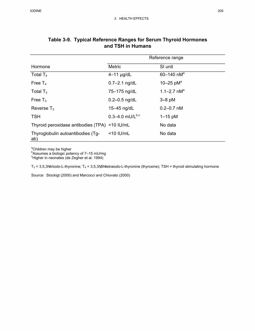
IODINE 200
3. HEALTH EFFECTS
Table 3-9. Typical Reference Ranges for Serum Thyroid Hormones and TSH in Humans
Reference range
Hormone Metric SI unit Total T4 4–11 µg/dL 60–140 nMa
Free T4 0.7–2.1 ng/dL 10–25 pMa
Total T3 75–175 ng/dL 1.1–2.7 nMa
Free T3 0.2–0.5 ng/dL 3–8 pM
Reverse T3 15–45 ng/dL 0.2–0.7 nM
TSH 0.3–4.0 mU/Lb,c 1–15 pM
Thyroid peroxidase antibodies (TPA) <10 IU/mL No data
Thyroglobulin autoantibodies (Tg-ab)
<10 IU/mL No data
aChildren may be higher bAssumes a biologic potency of 7–15 mU/mg cHigher in neonates (de Zegher et al. 1994) T3 = 3,5,3N-triiodo-L-thyronine; T4 = 3,5,3N,5N-tetraiodo-L-thyronine (thyroxine); TSH = thyroid stimulating hormone Source: Stockigt (2000) and Marcocci and Chiovato (2000)

IODINE 201
3. HEALTH EFFECTS
3.10 INTERACTIONS WITH OTHER CHEMICALS
Thioureylenes and Thionamides. Several of thionamide compounds that contain a thioureylene chemical
group have been shown to increase the accumulation of iodide in the thyroid gland and to decrease the
production of iodothyronines (Green 1996):
Propylthiouracil
These include several drugs used in the treatment of thyrotoxicosis and other hyperthyroid states,
(carbimazole, methimazole, and propylthiouracil); as well as the antibiotic, ethionamide; the cancer
chemotherapy agent, 6-mercaptopurine; and goitrin, a natural constituent of the plant genus Brassicae
(rutabaga, turnip, and cabbage). The thionamides exert their effects by inhibiting the iodination of
tyrosine and monoiodotyrosine in the thyroid gland and the coupling of iodotyrosines to form
iodothyronines. The mechanisms for these effects are not completely understood; however, at least two
mechanisms are needed to explain the reversible and irreversible inhibition of iodothyronine production
that is characteristic of these agents. Thionamides agents may act reversibly by reducing I+ or some other
reactive intermediate of iodine required in the iodination reaction, and also through a mechanism that
involves a direct, irreversible reaction with thyroid peroxidase.
Thiouracil and propylthiouracil, and related thioureylenes, are also inhibitors of iodthyronine deoidinase
(Leonard and Koehrle 1996). The mechanism of inhibition involves the formation of a covalent complex
with deiodinase enzymes. Inhibitory potency is highest for Type 1 deiodinase (Table 3-8). The result of
inhibition is a decreased metabolic clearance of iodothyronines.
Analine Derivatives. As a class, para-substituted aminobenzenes have activity similar to that of the
thionamides in that they increase the accumulation of iodide in the thyroid gland and decrease production
of iodothyronines, although possibly not through the same mechanisms (Green 1996). The group
S
N
NCH3CH2CH2
H
O
IODINE 202
3. HEALTH EFFECTS
includes several drugs (and drug classes), amphenone B, carbutamide, amino-glutethimide,
p-aminosalicylic acid, and the sulfonamides.
Substituted Phenols. Various substituted phenols that have hydroxyl groups in the meta positions have
been shown to increase thyroid iodide accumulation and to inhibit iodothyronine production in the
thyroid. These include resorcinol, 2,4-dihydroxybenzoic acid, and 2,4-dihydroxyphenol. These
compounds exert their activity by producing an irreversible inhibition of thyroid peroxidase (Green 1996).
Hydroxypyridines. Hydroxypyridines, including 3-hydroxypyridine and 3,4-dihydroxypyridine, have
been shown to increase thyroid iodide accumulation and to inhibit iodothyronine production in the thyroid
(Green 1996).
Perchlorate and Related Complex Anions. A variety of complex inorganic anions have been shown to
decrease the uptake of iodide in the thyroid gland. When given at high enough dosages, these agents can
induce hypothyroidism and goiter (Green 1996). The complex anions include, in order of potency:
perchlorate (ClO4-), perrhenate (ReO4
-), pertechnetate (TcO4-), and tetrafluorborate (BF4
-). The
mechanism for their activity is competitive inhibition of the NIS (Carrasco 1993; Eskandari et al. 1997;
Wolf 1964). These agents may also be transported by the NIS to varying degrees. Perchlorate does not
appear to be transported by NIS (Eskandari et al. 1997; Yoshida et al. 1997). These anions can also affect
accumulation and/or secretion of iodide in other tissues that have an active iodide transporter, including
the choroid plexus, gastric mucosa, mammary gland, placenta, salivary gland, and sweat gland (Brown-
Gant 1961).
Thiocyanate. Thiocyanate (SCN-) is a potent inhibitor of iodide uptake in the thyroid gland and
iodination of thyroglobulin. The mechanism for the effect on iodide uptake is primarily related to
competitive inhibition of iodide transport by the Na+/I- symport in thyroid gland; however, thiocyanate
may also accelerate iodide efflux from the thyroid by being a substrate with iodide for an anion exchange
mechanism on the basolateral membrane of thyroid follicle cells (Eskandari et al. 1997; Yoshida et al.
1997). Thiocyanate inhibits iodination, apparently by its actions as a competitive oxidation substrate for
thyroid peroxidase (Virion et al. 1980). Unlike other complex anion inhibitors of iodide transport,
thiocyanate is not accumulated in the thyroid gland.
Thiocyanate is a product of the metabolism of cyanide (ATSDR 1997) to which humans are exposed
when they smoke cigarettes, which has prompted interest in the potential effects of smoking on thyroid

IODINE 203
3. HEALTH EFFECTS
iodine metabolism and thyroid disease (Bertelsen and Hegedus 1994). Thiocyanate is a metabolite of
nitroprusside, a drug used in the treatment of acute hypertensive emergencies and cardiac failure.
Impairment of thyroid function in patients on nitroprusside has been reported (Bodigheimer et al. 1979;
Nourok et al. 1964).
Microsomal Enzyme Inducers. Agents that induce hepatic microsomal enzymes increase the activity of
phenolic glucuronyl transferases that catalyze the conjugation of iodothyronines with glucuronic acid
(Curran and DeGroot 1991; Visser 1990). Induction of glucuronyltransferase increases the metabolic
clearance of iodothyronines and, if sufficiently accelerated, can stimulate TSH release and goiter. Such
effects have been observed in rats and other experimental animal models in response to exposures to
2,4-benzopyrene, chlordane, DDT and DDD, 3-methylcholanthrene, PCBs, chlorinated dibenzodioxins
(CDDs), and toxaphene. A variety of drugs have also been shown to exert effects on glucruonide
conjugation of iodothyronines, including the sedative, phenobarbital; the anticonvulsants, phenytoin and
carbamazepine; and the antibiotic, rifampin.
Polychlorinated Biphenyls (PCBs). Depending on dose and duration, PCBs can disrupt the production
and disposition of thyroid hormones at a variety of levels and thereby may potentially interact with iodine
in impairing the thyroid gland. The major findings include (1) histological changes in the thyroid gland
indicative of both stimulation of the gland (e.g., similar to that induced by TSH or a hypothyroid state)
and disruption of the processing of follicular colloid needed for normal production and secretion thyroid
hormone; (2) depression of serum T4 and T3 levels, which may effectively create a hypothyroid state (in
some studies, low doses resulted in elevated serum T4 levels while depressed levels occurred at higher
PCB doses); (3) increased rates of elimination of T4 and T3 from serum; (4) increased activities of T4-
UDP-glucuronyl transferase (UDP-GT) in liver, which is an important metabolic elimination pathway for
T4 and T3; (5) decreased activity of iodothyronine sulfotransferases in the liver, which are also important
in the metabolic elimination of iodothyronines; (6) decreased activity of iodothyronine deiodinases,
including brain Type-2 deiodinase, which provide the major pathways for the production of the active
thyroid hormone, T3; and (7) decreased binding of T4 to transthyretin, an important transport protein for
both T4 and T3 (ATSDR 2000b).
Selenium. Selenium is essential for the activity of the glutathione peroxidases and iodothyronine
deiodinases. In humans, concurrent selenium and iodine deficiency have been associated with goiter and
cretinism, a developmental impairment related to prenatal hypothyroidism (Goyens et al. 1987;
Vanderpas et al. 1990). Supplementation of individuals deficient in both iodine and selenium with

IODINE 204
3. HEALTH EFFECTS
selenium produces a further decrease in thyroid function, but if selenium supplementation is preceded by
normalization of iodine levels, then normal thyroid function is restored (Contempré et al. 1991, 1992).
Selenium intake has been reported to affect thyroid hormone levels in humans; these effects include
decreases in serum T3 and T4 levels and increases in serum TSH levels, suggesting suppression of
thyropid hormone production (Brätter and Negretti De Brätter 1996; Duffield et al. 1999; Hagmar et al.
1998; Hawkes and Keim 1995). In experimental animals, selenium deficiency produces in decreased
metabolic clearance of iodothyronines and decreased extrathyroidal production of T3, as a result of
decreased iodothyronine deiodinase activity, which can be restored to normal by selenium repletion
(Arthur and Beckett 1994; Behne and Kyriakopolous 1993). Selenium deficiency also results in
decreases in thyroid iodine concentrations. The latter effect is thought to involve direct and indirect
effects on thyroid hormone production and secretion. The direct effect is thought to result from decreased
activity of glutathione peroxidase in the thyroid and increased availability of hydrogen peroxide for
utilization in the production of iodothyronines in the thyroid, which can then be exported from the gland.
The indirect effect may involve increased release of TSH from the pituitary gland in response to a
decrease in plasma concentration of T3, resulting from inhibition of deiodination of T4.
Amiodarone. The more serious side effects of the use of the antiarrythmia drug, amiodarone, are effects
on the thyroid, including hypothyroidism, hyperthyroidism, and thyroditis (Bogazzi et al. 2001; Meier and
Burger 1996). Although the exact mechanisms for these effects are not completely understood,
amiodarone contains a large quantity of iodine and has been shown to inhibit the deiodination of
iodothyronines; in particular, the production of T3 from T4, most likely as a result of inhibition of Type 1
deiodinase (Table 3-8). A metabolite of amiodarone, desethyamiodarone, has been shown to inhibit
binding of T3 to thyroid hormone receptors in a variety of tissues (Green 1996). As a thyroid receptor
antagonist, amiodarone (or its metabolite) also stimulates the release of TSH from the pituitary gland.
Lithium. Hypothyroidism and goiter have been associated with chronic therapy with lithium carbonate
for management of bipolar disease (Green 1996; Spaulding et al. 1972). The mechanism for these effects
is not understood, although it has been suggested that lithium may inhibit the coupling reaction in the
synthesis of iodothyronines and may inhibit thyroid hormone secretion.
Propranolol. Propranolol is a drug used in the treatment of hypertension, angina, and other
cardiovascular disorders as well as for the symptomatic treatment of thyrotoxicosis. Although the basis
for its use in treatment of thyrotoxicosis is to counteract the cardiovascular symptoms of the disorder, the
drug is also an inhibitor of iodothyronine deiodination (Meier and Burger 1996). The effect is unrelated

IODINE 205
3. HEALTH EFFECTS
to its activity as a β-adrenergic receptor antagonist, as both the L- and D-isomer (devoid of β-receptor
antagonist activity) inhibit the deiodination of T4. The mechanism for this action is not understood.
Dexamethasone. Although the corticosteroids exert multiple effects on the physiological regulation of
thyroid hormone release (e.g., decreased TSH release from the pituitary), these agents also have
appreciable activity as inhibitors of iodothyronine deiodinase and can decrease the metabolic clearance of
iodothyronines (Meier and Burger 1996).
Iodinated Drugs. A variety of iodine-containing drugs have been shown to inhibit iodothyronine
deiodination and thereby decrease the metabolic clearance of iodothyronines. These include the
antiarrhythmic agent, amiodarone (previously discussed), and several radiographic contrast agents used
for cholecystography such as iopanoic acid, sodium ipodae, and tyropanoate (Meier and Burger 1996).
3.11 POPULATIONS THAT ARE UNUSUALLY SUSCEPTIBLE
A susceptible population will exhibit a different or enhanced response to iodine than will most persons
exposed to the same level of iodine in the environment. Reasons may include genetic makeup, age, health
and nutritional status, and exposure to other toxic substances (e.g., cigarette smoke). These parameters
result in reduced detoxification or excretion of iodine, or compromised function of organs affected by
iodine. Populations who are at greater risk due to their unusually high exposure to iodine are discussed in
Section 6.7, Populations With Potentially High Exposures.
People who consume diets deficient in iodine may be more vulnerable to the toxic effects of exposure to
radioiodine. At very low intakes, representing iodine deficiency (e.g., 20 µg/day), uptake of iodide into
the thyroid gland is increased (Delange and Ermans 1996). This response is mediated by TSH, which
stimulates iodide transport and iodothyronine production in the thyroid gland (see Section 3.6.1). If
exposure to radioiodine was to occur in an individual who is iodine-deficient, a larger fraction of the
absorbed radioiodine may be taken up by the thyroid gland and a larger radiation dose to the thyroid gland
may be received. Iodine deficiency has been suggested to be a possible contributing factor to the increase
in thyroid cancer incidence observed in Belarus after the Chernobyl reactor accident (Gembicki et al.
1997; Robbins et al. 2001).
People who have multinodular goiter or thyroid gland adenomas can have foci of thyroid gland tissues
that produce and secrete thyroid hormone autonomously from control of the gland by TSH. The
IODINE 206
3. HEALTH EFFECTS
mechanisms for thyroid tissue autonomy appear to involve clonal expansion of follicle cells that have a
either a modified TSH receptor or receptor coupling mechanism, or that overexpress growth factors
(Corvilain et al. 2000; Derwahl and Studer, 2001; Krohn et al. 2000). Autonomous nodules can give rise
to hyperthyroidism (e.g., toxic nodular goiter, toxic adenoma). Iodine deficiency and goiter appear to be
risk factors in the development of autonomous nodules (Aghini-Lombardi et al. 1999). People who have
the autonomous nodules appear to be more vulnerable to iodide-induced hyperthyroidism (Braverman and
Roti 1996; Ermans and Camus 1972). This may, in part, explain the increased incidence of
hyperthyroidism that sometimes accompanies the introduction of iodide supplements into the diet of
iodine-deficient populations (Connolly 1971b; Corvilain et al. 1998; Delange et al. 1999). In
experimental studies, supplemental doses of 75–150 µg I/day for 1–2 weeks have induced
hyperthyroidism in euthyroid patients who had autonomous thyroid adenoma (Livadas et al. 1977).
Patients with certain types of thyroid autoimmunity may be more susceptible to developing
hyperthyroidism when exposed to excess iodine (Braverman and Roti 1996; Braverman et al. 1971a; Roti
and Uberti 2001).
Populations with diets that are deficient in selenium may be more susceptible to iodine toxicity. Selenium
is a cofactor in the iodothyronine deiodinases that are important for the synthesis of the thyroid hormone,
T3, in extrathyroidal tissues. Iodine deficiency, in conjunction with selenium deficiency, has been
associated with goiter and cretinism, a developmental impairment related to prenatal hypothyroidism
(Goyens et al. 1987; Vanderpas et al. 1990). In this state, in which the thyroid gland is responding to a
deficiency in T3 production by increasing iodide transport and iodination activity in the thyroid gland,
infants and children (as well as adults) may experience a higher thyroid uptake of absorbed iodine and
possibly a higher radiation dose to the thyroid when exposed to radioiodine.
People who have $-thalassemia, an inherited disorder of hemaglobin production that can lead to anemia,
may be more sensitive to developing hypothyroidism when exposed to excess iodide (Alezandrides et al.
2000).
3.12 METHODS FOR REDUCING TOXIC EFFECTS
This section will describe clinical practice and research concerning methods for reducing toxic effects of
exposure to iodine. However, because some of the treatments discussed may be experimental and
unproven, this section should not be used as a guide for treatment of exposures to iodine. When specific
exposures have occurred, poison control centers and medical toxicologists should be consulted for

IODINE 207
3. HEALTH EFFECTS
medical advice. The following text provides specific information about treatment following exposures to
iodine:
Braverman LE, Utiger RD. 2000. Werner and Ingbar's The thyroid: A Fundamental and Clinical Text.
Philadelphia, PA: Lippincott-Raven.
Treatment of toxicity from exposure to excess iodine is directed at lowering exposure and, if clinical
hypothyroidism or hyperthyroidism persists, correcting the thyroid dysfunction. Treatment of clinical
hypothyroidism includes the administration of thyroid hormone. Treatment of hyperthyroidism involves
administering thyroid hormone synthesis inhibitors.
Treatment of toxicity from exposure to radioiodine is also directed at lowering thyroid gland uptakes of
absorbed iodine, for example, by administration of potassium iodide (see Section 3.12.2). If the exposure
produces persistent hypothyroidism or hyperthyroidism, the treatment strategies for the clinical
abnormalities are the same as those for exposure for nonradioactive iodine.
3.12.1 Reducing Peak Absorption Following Exposure
No information was located on methods to reduce peak absorption following exposure. Mitigation of
toxic effects following exposure to radioiodine is directed at reducing the uptake of absorbed iodine in the
thyroid gland (see Section 3.12.2).
3.12.2 Reducing Body Burden
Approximately 90% of the iodine in the human body is contained in the thyroid gland. The thyroid gland
is also the major toxicity target of radioiodine. Therefore, methods for reducing the uptake and
accumulation of radioiodine in the thyroid gland can reduce the radioiodine body burden, the absorbed
radiation dose to the thyroid gland and body, and the toxic effects of exposure to radioiodine. Iodine
uptake into the thyroid gland is highly sensitive to the iodide intake. At very high intakes of iodine,
representing an intake excess (e.g., >1 mg/day), iodine uptake into the thyroid gland decreases, primarily
as a result of decreased iodothyronine synthesis (Wolff-Chaikoff effect) and iodide transport into the
gland (Nagataki and Yokoyama 1996; Saller et al. 1998). A single oral dose of 30 mg iodide (as sodium
iodide) decreases the 24-hour thyroid uptake of radioiodine by approximately 90% in healthy adults
(Ramsden et al. 1967; Sternthal et al. 1980). The inhibition of uptake was sustained with repeated oral

IODINE 208
3. HEALTH EFFECTS
doses of sodium iodide for 12 days, with complete recovery to control (presodium iodide) uptake levels
within 6 weeks after the last sodium iodide dose (Sternthal et al. 1980), or within 8 days after a single
dose (Ramsden et al. 1967). Repeated oral doses of 1.5–2.0 mg iodide/m3 of surface area produced an
80% decrease in thyroid uptake of radioiodine in children (Saxena et al. 1962). Inhibition of radioiodine
uptake by the thyroid gland that occurs when large doses of iodide are administered results in more rapid
urinary excretion of radioiodine and decreased iodine body burden (Ramsden et al. 1967). The
administration of iodide as prophylaxis for reducing thyroid uptake of radioiodine after accidental
releases of radioiodine has been recommended by the FDA and NCRP (FDA 1978, 2001b; NCRP 1977).
Recommendations regarding the distribution and administration of potassium iodide in the event of a
nulcear accident have been provided by the National Research Council (NRC 2004). Doses of 50 mg
(infants <1 year of age) or 100 mg (adults) I, as potassium iodide, just before or at the time of exposure,
have been found to be effective for blocking (>90%) thyroid uptake of radioiodine (Verger et al. 2001).
The dose of potassium iodide that is effective for achieving this level radioiodine uptake block will
depend on the time of dosing, relative to the exposure, as well as the dietary iodide status; higher doses of
potassium iodide may be needed under conditions of low dietary iodide intake (Zanzonico and Becker
2000). The FDA (2001b) has recommended that potassium iodide be administered at the following doses,
daily doses, until the risk of exposure from inhalation or ingestion no longer exists:
Receptor (years)
Predicted thyroid radiation dose (cGy, rad)
Dose (mg KI/day)
Dose (mg I/day)
Adults >40 years $500 Adults >18–40 years $10 Pregnant or lactating women $5
130 100
Adolescents >12–18 years* Children >3–12 years 65 50
Infants <1 month–3 years 32 24 Infants <1 month
$5
16 12 * Adolescents $70 kg should receive adult dose (130 mg/day)
3.12.3 Interfering with the Mechanism of Action for Toxic Effects
The mechanisms of action of excess iodine in producing goiter, hypothyroidism, hyperthyroidism, or
thyroiditis involve direct interactions between iodide and physiological elements involved in thyroid
hormone synthesis and release, and iodine transport. Therefore, the principal strategy for reducing toxic
effects is to decrease iodine intake or uptake into the thyroid gland (see Section 3.12.2). Numerous cases
of reversal of iodine-induced hypothyroidism or hyperthyroidism after reduction of iodide intake have
been reported and are described in this profile (see Section 3.2.2.2, Endocrine Effects). The principal

IODINE 209
3. HEALTH EFFECTS
clinical strategy for managing permanent hypothyroidism is the administration of T4 (Brent and Larsen
2000). The principal clinical strategies for managing permanent hyperthyroidism is the administration of
agents that inhibit iodination of thyroglobulin, such as propylthiouracil or methimazole, or that inhibit
thyroid uptake of iodine, such as perchlorate, or the destruction of the thyroid gland with radiation. The
latter is usually accomplished by administering a cytotoxic dose of 131I. β-Adrenergic antagonists are also
used to manage some of the symptoms of thyrotoxicosis (Cooper 2000). Cases of massive acute, near-
lethal poisoning from ingestion of tinctures of iodine (mixtures of molecular iodine and sodium triiodide)
have included fluid and electrolyte replacement to manage cardiovascular shock (Finkelstein and Jacobi
1937).
The sulfhydryl compound, amifostine, has been found to reduce the toxic effects of high exposures to 131I
in patients who undergo ablative therapy with 131I for thyroid cancers (Bohuslavizki et al. 1996, 1998a,
1998b, 1999). The mechanism for the protective effect appears to be accumulation of amifostine in the
salivary gland and scavenging of free radicals formed as a result of interactions of ionizing radiation from 131I with tissues.
3.13 ADEQUACY OF THE DATABASE
Section 104(i)(5) of CERCLA, as amended, directs the Administrator of ATSDR (in consultation with the
Administrator of EPA and agencies and programs of the Public Health Service) to assess whether
adequate information on the health effects of iodine is available. Where adequate information is not
available, ATSDR, in conjunction with the National Toxicology Program (NTP), is required to assure the
initiation of a program of research designed to determine the health effects (and techniques for developing
methods to determine such health effects) of iodine.
The following categories of possible data needs have been identified by a joint team of scientists from
ATSDR, NTP, and EPA. They are defined as substance-specific informational needs that if met would
reduce the uncertainties of human health assessment. This definition should not be interpreted to mean
that all data needs discussed in this section must be filled. In the future, the identified data needs will be
evaluated and prioritized, and a substance-specific research agenda will be proposed.

IODINE 210
3. HEALTH EFFECTS
3.13.1 Existing Information on Health Effects of Iodine
The existing data on health effects of inhalation, oral, and dermal exposure of humans and animals to
iodine are summarized in Figures 3-16 and 3-17. The purpose of this figure is to illustrate the existing
information concerning the health effects of iodine. Each dot in the figure indicates that one or more
studies provide information associated with that particular effect. The dot does not necessarily imply
anything about the quality of the study or studies, nor should missing information in this figure be
interpreted as a “data need”. A data need, as defined in ATSDR’s Decision Guide for Identifying
Substance-Specific Data Needs Related to Toxicological Profiles (Agency for Toxic Substances and
Disease Registry 1989), is substance-specific information necessary to conduct comprehensive public
health assessments. Generally, ATSDR defines a data gap more broadly as any substance-specific
information missing from the scientific literature.
3.13.2 Identification of Data Needs
Acute-Duration Exposure. The primary effect of acute exposures to excess iodine in humans is
hypothyroidism. This effect has been studied extensively in experimental studies of humans and is also
well documented in the clinical case literature. Reported NOAELs for iodine-induced hypothyroidism in
humans vary widely for reasons that are not completely understood. Acute exposures to excess iodine
produce allergic reactions in people. The mechanisms for sensitivity and the reactions are not completely
understood.
The effects of acute exposures to radioiodine (primarily 131I) have been extensively studied in humans.
An enormous amount of epidemiological and case literature derives from the clinical use of 131I in
diagnostic procedures and in treatment of thyroid gland enlargement and thyrotoxicosis. Epidemiology
studies have also examined health effects resulting from accidental environmental exposures due to
nuclear bomb detonations (e.g., Marshall Islands) and releases from nuclear power plants (e.g.,
Chernobyl). These studies collectively and convincingly identify the thyroid gland as the primary target
of radioiodine. Other tissues that are either near the thyroid gland, such as the parathyroid gland, or that
accumulate iodine, such as the salivary gland, also are affected by exposures to 131I; however, these
effects occur at absorbed radiation doses that are clearly cytotoxic to the thyroid gland. Breast tissue
expresses NIS and appears capable of accumulating 131I and transferring it to mammary milk; therefore, it
is a potential target of 131I. However, epidemiology studies reported to date have not found a significant
risk of breast cancer even after cytotoxic exposures to 131I.

IODINE 211
3. HEALTH EFFECTS
Figure 3-16. Existing Information on Health Effects of Stable Iodine
Existing Studies
Inhalation
Oral
Dermal
DeathAcute
Interm
ediate
Chronic
Immuno
logic/Lym
phoret
ic
Neurologic
Reproductive
Developmental
Genotoxic
Cancer
Systemic
Animal
Inhalation
Oral
Dermal
DeathAcute
Interm
ediate
Chronic
Immuno
logic/Lymphore
tic
Neurologic
Reproductive
Developmental
Genotoxic
Cancer
Systemic
Human

IODINE 212
3. HEALTH EFFECTS
Figure 3-17. Existing Information on Health Effects of Radioactive Iodine
Existing Studies
Human
Inhalation
Oral
Dermal
DeathAcute
Intermediat
e
Chronic
Immun
ologic/Lym
phoretic
Neurologic
Reprodu
ctive
Developmental
Genotoxic
Cancer
Systemic
External
Inhalation
Oral
Dermal
DeathAcu
teInterm
ediate
Chronic
Immun
ologic/Lym
phoret
ic
Neurologic
Reproductiv
e
Developmental
Genotoxic
Cancer
Systemic
External
Animal
IODINE 213
3. HEALTH EFFECTS
Intermediate-Duration Exposure. The primary effect of intermediate-duration exposures to excess
iodine in humans is hypothyroidism. This effect has been studied extensively in humans and is well
documented in the clinical case literature. Reported LOAELs for iodine-induced hypothyroidism in
euthyroid humans, without goiter, fall within a reasonably narrow range, and are higher than those for
people who are iodine deficient, suggesting a higher sensitivity in these subjects. The mechanisms for
this are not completely understood. Intermediate-duration exposure has also been shown to induce
hyperthyroidism in people who have nontoxic goiter. Here again, the mechanisms are not completely
understood, although clonal expansion of autonomous follicle cells and autoimmunity are suspected
contributors. Intermediate-duration exposures to excess iodine produce allergic reactions in people. The
mechanisms for sensitivity and the reactions are not completely understood.
Chronic-Duration Exposure and Cancer. Epidemiological studies and clinical case literature
identify the thyroid gland as the principal target of chronic exposure to excess iodine. Goiter,
hypothyroidism, hyperthyroidism, and/or thyroid autoimmunity are the main outcomes of chronic
exposure to excess iodine. Which effect occurs appears to be related to the pre-existing iodine intake
(e.g., deficient or replete) and the presence or absence of possibly pre-existing autoimmunity and/or
thyroid gland enlargement (or nodularity).
Genotoxicity. Stable iodine has been tested for genotoxicity in a variety of eukaryotic cell systems
and has been found to be without mutagenic activity. The genotoxicity of radioactive iodine (131I) has
been extensively studied in clinical studies of patients who received 131I for therapy of thyroid cancer and
thyrotoxicosis and in people who were exposed to radioiodine from nuclear power plant accidents (e.g.,
Chernobyl).
Reproductive Toxicity. Several studies of reproductive effects of exposures to 131I have been
reported. These studies indicate that relatively high exposures to radioiodine (i.e., that are cytotoxic to the
thyroid gland) can produce impairment of testicular function. The mechanism for this is not understood,
but the observation of these effects suggests a possible exposure of the testes to 131I. The testis is not
presently known to express NIS; however, studies of the uptake of radioiodine in testes were not located.
Developmental Toxicity. Developmental toxicity of iodine and radioiodine related to effects on the
fetal/neonatal thyroid gland has been well documented in the clinical case literature. The primary effect
is congenital hypothyroidism and associated sequelae.

IODINE 214
3. HEALTH EFFECTS
Immunotoxicity. The epidemiological and clinical case literature has identified thyroid autoimmunity
and allergic reactions as the primary immunologic effects of exposure to excess iodine. Thyroid
autoimmunity is an extremely important mechanism of thyroid gland disease. The mechanisms by which
iodine induces thyroid autoimmunity are not completely understood. The production of antibodies to
highly iodinated thyroglobulin has been proposed as a possible contributor.
Neurotoxicity. The primary target of iodine toxicity is the thyroid gland. A large amount of clinical
literature exists on the neurological sequelae of thyroid gland disorders.
Epidemiological and Human Dosimetry Studies. The epidemiological literature on iodine- and
radioiodine-related health effects is very substantial and provides information on exposures associated
with the primary effect, thyroid gland dysfunction. There remain certain complications in the
interpretation of the major epidemiology studies of environmental exposures to iodine and radioiodine.
These relate to the magnitude of the contribution of iodine deficiency and autoimmunity in the observed
thyroid gland outcomes (e.g., hypothyroidism, hyperthyroidism, thyroid gland nodularity, and cancers).
Studies of human dosimetry of 131I are extensive, in large part, because of the extensive use of 131I in
diagnostic and treatment procedures that require highly certain estimates of the radiation dose delivered to
the thyroid gland. The clinical information has been incorporated into reconstructions of thyroid doses
experienced by the general public.
Biomarkers of Exposure and Effect.
Exposure. The use of urinary iodide for assessing steady state iodine intakes is well substantiated in the
clinical and epidemiological literature and is supported by toxicokinetics studies in humans. Similarly,
the use of external scintillation spectrometry to estimate radioiodine doses to the thyroid gland also has a
substantial clinical history.
Effect. The clinical literature on thyroid gland disorders extensively documents the major biomarkers of
thyroid gland dysfunction that are relevant to iodine toxicity.
Absorption, Distribution, Metabolism, and Excretion. The toxicokinetics of iodine in humans
has been substantially explored and characterized in experimental studies and clinical cases. Radioiodine
toxicity is most likely in tissues that can transport and accumulate iodide. Studies of the expression of

IODINE 215
3. HEALTH EFFECTS
NIS and factors that alter expression of NIS can further advance our understanding of which tissues are at
risk and what factors, including genetic factors, might affect sensitivity to radioiodine in humans.
Comparative Toxicokinetics. The extensive information on the toxicokinetics of iodine in humans
makes extrapolations from animals less important in assessing the health effects of iodine in humans.
Studies of interindividual variability in humans are valuable for identifying sensitive subpopulations.
Methods for Reducing Toxic Effects. The principal method for preventing the toxic effects of
radioiodine is dosing with stable iodine, which decreases the thyroid gland uptake of radioiodine and the
absorbed radiation dose to the gland. The mechanistic basis and effectiveness of this approach is well
established from experimental and clinical studies.
Children’s Susceptibility. Higher susceptibility of the fetus and infants to iodine and radioiodine
toxicity is substantiated by the epidemiological and clinical case studies. The toxicokinetic basis for the
susceptibility of infants and children to iodine exposure is understood. Uncertainties in assessing the
potential health effect of iodine exposures are largely related to estimating exposures, in particular, the
pathways by which environmental releases result in radioiodine uptake into the fetal or infant thyroid
gland (see Section 6.8.1).
Child health data needs relating to exposure are discussed in 6.8.1 Identification of Data Needs:
Exposures of Children.
3.13.3 Ongoing Studies
Ongoing studies pertaining to iodine have been identified and are shown in Table 3-10.

IODINE 216
3. HEALTH EFFECTS
Table 3-10. Ongoing Studies on Health Effects of Radioactive Iodine
Investigator Affiliation Title Sponsor Baker JR University of Michigan
at Ann Arbor Characterization of thyroid autoantibodies and antigens
NCRR
Brent GA University of California, Los Angeles
Regulation of the sodium/iodine symporter in breast
NCI
Burek CL John Hopkins University
Immunotoxic effects of iodine NIH—National Institute of Diabetes and Digestive and Kidney Diseases
Burek CL John Hopkins University
Nod h2h4 mice as a sentinel model for autoimmune thyroid
NIEHS
Carrasco N Yeshiva University Characterization of the thyroid Na +/I-symporter
NIH—National Institute of Diabetes and Digestive and Kidney Diseases
Degroot LJ University of Chicago Pathogenesis and therapy of autoimmune thyroid disease
NIH—National Institute of Diabetes and Digestive and Kidney Diseases
Kong Y-CM Wayne State University
T cell recognition—repertoire in autoimmune thyroiditis
NIH—National Institute of Diabetes and Digestive and Kidney Diseases
Naylor EW Neo Gen Screening, Inc.
Simplified population screening for adult hypothyroidism
NIH—National Institute of Diabetes and Digestive and Kidney Diseases
Refetoff SS University of Chicago Regulation and mechanisms of hormone action
NIH—National Institute of Diabetes and Digestive and Kidney Diseases
Refetoff SS University of Chicago Screening for inherited thyroid defects
NCRR
Sgouros G Sloan-Kettering Institute for Cancer
Modeling and dosimetry for radiolabeled antibody therapy
NCI
St Germain DL Dartmouth College Regulation of thyroid hormone metabolism
NIH—National Institute of Diabetes and Digestive and Kidney Diseases
St Germain DL Dartmouth College The role of the Type 3 deiodinase in development
NIH—National Institute of Diabetes and Digestive and Kidney Diseases

IODINE 217
3. HEALTH EFFECTS
Table 3-10. Ongoing Studies on Health Effects of Radioactive Iodine
Investigator Affiliation Title Sponsor Weintraub BD University of
Maryland, Baltimore Professional School
Structure/function relationships of human thyrotrophin
NIH—National Institute of Diabetes and Digestive and Kidney Diseases
NCI = National Cancer Institute; NCRR = National Center for Research Resources; NIEHS = National Institute of Environmental Health Sciences; NIH = National Institute of Health/National Institute of General Medical Sciences Source: CRISP 2001; National Institutes of Health; Central Repository of Incidents, Solutions, and Problems

Related Documents