2nd International Biophotonics Conference 2nd International Biophotonics Conference SINGAPORE | 1618 NOV17 Exclusive Distributor in Singapore and Brunei Contact: BenchMark Technologies @ +6597778116 Sponsors: microLAMBDA

Welcome message from author
This document is posted to help you gain knowledge. Please leave a comment to let me know what you think about it! Share it to your friends and learn new things together.
Transcript
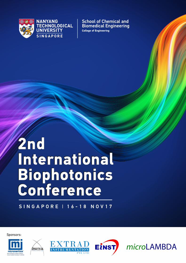
2ndInternationalBiophotonicsConference
2ndInternationalBiophotonicsConferenceS I N G A P O R E | 1 6 � 1 8 N O V 1 7
Exclusive Distributor in Singapore and Brunei
Contact: BenchMark Technologies @ +6597778116
Sponsors:
microLAMBDA Pte Ltd�
2ndInternationalBiophotonicsConference
2ndInternationalBiophotonicsConferenceS I N G A P O R E | 1 6 � 1 8 N O V 1 7
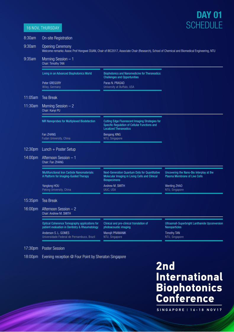
DAY 01SCHEDULE16 NOV, THURSDAY
8:30am
9:30am
9:35am
On-site Registration
Opening CeremonyWelcome remarks: Assoc Prof Hongwei DUAN, Chair of IBC2017, Associate Chair (Research), School of Chemical and Biomedical Engineering, NTU
Morning Session – 1Chair: Timothy TAN
Living in an Advanced Biophotonics World
Peter GREGORYWiley, Germany
11:05am
11:30am
Tea Break
Morning Session – 2Chair: Kanyi PU
NIR Nanoprobes for Multiplexed Biodetection
Fan ZHANGFudan University, China
Cutting Edge Fluorescent Imaging Strategies for Speci�c Regulation of Cellular Functions and Localized Theranostics
Bengang XING NTU, Singapore
12:30pm
14:00pm
Lunch + Poster Setup
Afternoon Session – 1Chair: Fan ZHANG
Multifunctional Iron Carbide Nanomaterials: A Platform for Imaging-Guided Therapy
Yanglong HOU Peking University, China
17:30pm
18:00pm
Poster Session
Evening reception @ Four Point by Sheraton Singapore
Biophotonics and Nanomedicine for Theranostics: Challenges and Opportunities
Paras N. PRASADUniversity at Buffalo, USA
Next-Generation Quantum Dots for Quantitative Molecular Imaging in Living Cells and Clinical Biospecimens
Andrew M. SMITH UIUC, USA
Uncovering the Nano-Bio Interplay at the Plasma Membrane of Live Cells
Wenting ZHAO NTU, Singapore
15:35pm
16:00pm
Tea Break
Afternoon Session – 2Chair: Andrew M. SMITH
Optical Coherence Tomography applications for patient evaluation in Dentistry & Rheumatology
Anderson S. L. GOMES Universidade Federal de Pernambuco, Brazil
Clinical and pre-clinical translation of photoacoustic imaging
Manojit PRAMANIK NTU, Singapore
Ultrasmall-Superbright Lanthanide Upconversion Nanoparticles
Timothy TAN NTU, Singapore
Peter Gregory is Editor-in-Chief Advanced
Materials, Advanced Optical Materials, and
Advanced Materials Interfaces, and Vice
President and Publishing Director at Wiley
in Germany responsible for materials,
physics, and life science journals.
He is a chemist, with an education from
University College London, and research
experience at the University of
Erlangen-Nuremberg in Germany. Over his
career in publishing he led RSC Publishing
for 5 years, and has founded has founded
over 25 journals, both for Wiley and the
RSC, including 15 members of the
“Advanced” journal family. He is a Fellow
of the Royal Society of Chemistry and has
received honorary/guest professorships
from 11 universities and institutes.
Functional optical and photonic materials
play a central role in areas such as
Healthcare, Sustainability, and Technology,
and journals serving the community of
chemists, physicists, life scientists, and
engineers active in this area are among
some of the highest Impact publications in
science. Peter Gregory, Editor in Chief of
the journal Advanced Materials, and
Advanced Optical Materials, will trace the
DAY 01
Peter GREGORY
Editor-in-Chief, Advanced Materials / Vice President & Publishing Director Wiley
Living in an Advanced Biophotonics World
Biography
Journals/services founded:1. Advanced Materials 19892. Advanced Engineering Materials 19993. Advanced Functional Materials 20014. Macromolecular Bioscience 20015. Macromolecular Materials and Engineering 20016. Chemistry World (RSC) 20037. Molecular Biosystems (RSC) 20058. Soft Matter (RSC) 20059. Materials Views 200810. MaterialsViewsChina 200911. Advanced Energy Materials 201112. Advanced Healthcare Materials 201213. Advanced Optical Materials 201314. Particle 201315. Advanced Materials Interfaces 201416. Advanced Science 201417. Advanced Electronic Materials 201518. Advanced Materials Technologies 201619. Advanced Science News 201620. Advanced Biosystems 201721. Advanced Sustainable Systems 201722. Global Challenges 201723. Solar-RRL 201724. Small Methods 201725. Advanced Therapeutics26. Advanced Theory & Simulation27. Advanced Quantum Technology
some of the developments in the world of
journals and publishing, and in topics
related to biophotonics, such as hot topics,
funding priorities, geographical
contributions, and some publishing themes
such as open access, and provide some
food for thought on both how these might
develop in the future and how to maximize
the chances of being published in these top
journals.
PARAS N. PRASAD, Ph.D., is SUNY Distinguished Professor of Chemistry, Physics, Electrical Engineering and Medicine; Samuel P. Capen Chair of Chemistry; and Executive Director of the Institute for Lasers, Photonics and Biophotonics. In 2005, Scienti�c American named him among the top 50 science and technology leaders in the world. He has published more than 790 scienti�c papers, along with four �eld-de�ning monographs in Nonlinear Optics, Biophotonics, Nanophotonics, and Nanomedicine; eight edited books; and numerous patents. He has received the Morley Medal and the Schoellkopf Medal from the American Chemical Society; a Guggenheim Fellowship, a Sloan Fellowship; the Western New York Health Care Industries Technology / Discovery Award; a SUNY Excellence in the
This talk will present our progress in biophotonics dealing with multimodal and multispectral imaging, combined with nanomedicine approach of image guided targeted delivery of therapy to produce an effective paradigm of combined diagnostics and therapy, now popularly referred to as Theranostics. Our recent efforts have focused on using biophotonics to enable in-vitro diagnosis with minimally invasive liquid biopsy. Here we have focused on the development of photon converting nanostructures that can enable imaging and sensing in the spectral regions called NIR window II (1,000 nm to 1,300nm) and NIR window III (1,600nm to 1,870nm) which provide deeper penetration through biological tissues. We introduce Ramanomics which is a new Omics disciplines using Micro Raman Spectrometry with Biomolecular Component Analysis for molecular pro�ling of biological structures . This provides a new biosensing tool to measure concentrations of proteins, DNA, RNA and lipids in the single organelles of live cells, leading to a new set of biomarkers to diagnose progression of diseases such as cancer.
In nanomedicine, our approach has been to develop nanoformulations that provide image guided and
DAY 01
Paras N. PRASAD
SUNY Distinguished Professor Department of Chemistry, Physics, Medicine and Electrical Engineering University at Buffalo, USA
Biophotonics and Nanomedicine for Theranostics: Challenges and Opportunities
Biography
Pursuit of Knowledge award; UB’s �rst Innovation Impact award; and his University’s President’s Medal. Prasad is also the winner of SPIE’s highest honor of President’s Gold medal, Optical Society of America’s Michael Feld Biophotonics award, IEEE‘s Pioneer Award in Nanotechnology, Peter Debye Award from the American Chemical Society in recognition of his numerous pioneering contributions to nonlinear optics, nanophotonics, biophotonics and nanomedicine. He is a fellow of the APS, OSA, and SPIE, and a senior member of IEEE. He is on the Thompson Reuters “Highly Cited Researchers” list for 2014 and 2016. Prasad has received Honorary Doctorates from KTH in Sweden, the Aix-Marseille University in France, MEPhI in Russia and Federal University of Pernambuco in Brazil.
targeted delivery and as well as real time monitoring of therapeutic action. We also make sure that the nanostructures are biocompatible, causing no toxicity and can be bio-eliminated. A major emphasis has been placed on Translational Nanomedicine . A recent direction has been nanodelivery of natural medicine. We have also focused on engaging nuclear physics in radiation-based diagnostics and therapy. An example presented is of our new formulation of boron nanoparticles containing biophotonics probe for Neutron Capture Therapy. A major biomedical application area currently pursued in our lab is brain research in the emerging �eld of Neurophotonics, where we apply �eld responsive materials for functional mapping of the brain using optical and photoacoustic imaging. We have also demonstrated remote and noninvasive actuation of optogenetic stimulation of brain activity using near IR absorbing optical nanotransformers that can provide an effective intervention/augmentation strategy to treat many cognitive disorder and diseases, ranging from Alzheimer, to traumatic brain injury, to retardation.
This talk will conclude with a discussion of existing challenges and new opportunities.

Fan Zhang received his PhD in 2008 from
Fudan University followed by more than 2
years postdoctoral experience
(2008-2010) from the prestigious, world
leading laboratories in University of
California at Santa Barbara (Galen. D.
Stucky’s group) before joining as an
associate professor in the Chemistry
Department of Fudan University in 2010.
The identi�cation of potential diagnostic
markers and target molecules among the
plethora of tumor oncoproteins for cancer
diagnosis requires facile technology that is
capable of quantitatively analyzing multiple
biomarkers in tumor cells and tissues.
Diagnostic and prognostic classi�cations of
human tumors are currently based on
western blotting and single-color
immunohistochemical methods that are
not suitable for multiplexed detection.
Herein, we report a general and novel
method to prepare single-band
upconversion nanoparticles with different
colors. The expression levels of three
biomarkers in breast cancer cells were
DAY 01
Fan ZHANG
Professor Department of Chemistry Fudan University, China
NIR Nanoprobes for Multiplexed Biodetection
Biography
He became a full professor in Fudan
University since 2013. Prof. Zhang has
authored a number of book chapters,
patents and more than 80 peer-reviewed
research papers in international journals
with the high impact factors i.e. Nat.
Commun., J. Am. Chem. Soc., Angew.
Chem. Int. Ed., Nano Lett., Adv. Mater., ACS
Nano.
determined using single-band
upconversion nanoparticles, western
blotting and immunohistochemical
technologies with excellent correlation.
Signi�cantly, the application of
antibody-conjugated single-band
upconversion nanoparticle molecular
pro�ling technology can achieve the
multiplexed simultaneous in situ
biodetection of biomarkers in breast
cancer cells and tissue specimens and
produce more accurate results for the
simultaneous quanti�cation of proteins
present at low levels compared with
classical immunohistochemical
technology.
ReferencesRui Wang, Lei Zhou, Wenxing Wang, Xiaomin Li, Fan Zhang*, In vivo gastrointestinal drug-release monitoring through second near-infrared window �uorescent bioimaging with orally delivered microcarriers, Nat. Commun , 2017, 8: 14702.
Xiaomin Li, Zhenzhen Guo, Tiancong Zhao, Yang Lu, Lei Zhou, Dongyuan Zhao, Fan Zhang *, Filtration Shell Mediated Power Density Independent OrthogonalExcitations–Emissions Upconversion Luminescence, Angew. Chem. Int. Ed. 2016, 55: 2464.
Lei Zhou, Rui Wang, Xiaomin Li, Chi Yao, Chengli Wang, Xiaoyan Zhang, Congjian Xu, Aijun Zeng, Dongyuan Zhao, Fan Zhang*, Single-band upconversion nanoprobes for multiplexed simultaneous in situ molecular mapping of cancer biomarkers, Nat. Comm. 2015, 6: 6938.
Rui Wang, Xiaomin Li, Lei Zhou, Fan Zhang*, Epitaxial Seeded Growth of Rare-Earth Nanocrystals with Ef�cient 800 nm Near-Infrared to 1525 nm Short-Wavelength Infrared Downconversion Photoluminescence for In Vivo Bioimaging, Angew. Chem. Int. Ed. 2014, 53: 12086.
Chi Yao, Lei Zhou, Ahmed Mohamed El-Toni, Yiqing Lu, Xiaomin Li, Fan Zhang *, Facile Peptides Functionalization of Lanthanide-Based Nanocrystals through Phosphorylation Tethering for Ef�cient in Vivo NIR-to-NIR Bioimaging, Analytical Chemistry,2016, 88: 1930.
Chi Yao, Peiyuan Wang, Xiaomin Li, Xiaoyu Hu, Junli Hou, Leyong Wang, Fan Zhang*, Near-Infrared-Triggered Azobenzene-Liposome / Upconversion Nanoparticle Hybrid Vesicles for Remotely Controlled Drug Delivery to Overcome Cancer Multidrug Resistance, Advanced Materials, 2016,28: 9341
1.
2.
3.
4.
5.
6.
Prof. Bengang XING is currently an associate
professor (since 2011) in Division of Chemistry
and Biological Chemistry, School of Physical
and Mathematical Sciences, Nanyang
Technological University, Singapore. His
academic experience: He received his Ph.D in
Department of Chemistry, Nanjing University in
2000. From end of 2000, he moved to the Hong
Kong University of Science and Technology
(HKUST) to work as research associate until
early of 2003. Then from 2003 – 2005, he held
his Post-Doc. Fellow appointment at Crump
In terms of the native 3D cell structures in all
living species, the precise tracking of
complexity and dynamics of cellular functions
with precise resolution in intact cells or
organisms will be important to understand the
biological basis and to conduct related
biomedical applications. Usually, within
complex environment, biological speci�city can
be mediated by the precise regulation of
biomolecule functions in response to intrinsic
development programs and extrinsic signals. In
order to fully decipher the biomolecule
functions and further elucidate their
mechanisms, a simple and reliable assay with
high sensitivity and �delity will be essential to
ef�ciently report the biological process of
interest at the single cell or molecular level.
Generally, �uorescent imaging enables such
rapid, direct and sensitive visualization, mainly
due to their high sensitivity, relative safety, and
DAY 01
Bengang XING
Associate Professor Division of Chemistry and Biological Chemistry School of Physical & Mathematical Sciences Nanyang Technological University Singapore
Cutting Edge Fluorescent Imaging Strategies for Speci�c Regulation of Cellular Functions and Localized Theranostics
Biography
Institute of Molecular Imaging at University of
California, Los Angles (UCLA) and Molecular
Imaging Program at School of Medicine (MIPS),
Stanford University. In December 2005, he was
appointed as Assistant Professor at NTU and
later promoted to Associated Professor in
2011. Currently, Prof. XING is holding
appointment of Deputy Head of Division of
Chemistry and Biological Chemistry, and
Assistant Chair of School of Physical and
Mathematical Sciences.
easily handling, and therefore have become
robust and reliable tools in monitoring
subcellular protein dynamics and analysis of
tumors or pathogen–host interactions. The
systematic imaging investigation of enzymes or
proteins activities in a complicated
environment will offer great possibility for the
in-depth understanding of the biological basis
conferring diseases status, and importantly, for
the facilitating of new discovery of effective
theranostic approaches in vitro and in vivo.
In our group, a series of simple and speci�c
�uorescent/bioluminescent small molecules or
nano-probes have been extensively established
to real-time visualize cellular functions,
importantly, the intrinsic mechanisms to involve
in potent drug activities and relevant pathways
to initiate drug resistance have been well
investigated.
ReferencesAi, X.; Ho, C.-J.; Aw, J.; Attia, A.-B.; Mu, J.; Wang, Y.; Wang, X.; Wang, Y.; Liu, X.; Chen, H.; Gao, M.; Chen, X.; Yeow, K; Liu, G.*; Olivo, M.*; Xing, B.* Nat. Commun. 2016, 7: 10432. Featured in “Biodiscover”, “ScienceNet”, and “X-MoL”.
Min, Y.; Li, J.; Liu, F.; Yeow, K.*; Xing, B.* Angew. Chem. Int. Ed. 2014, 53: 1012.
Ai, X.; Lyu, L.; Zhang, Y.; Tang, Y.; Mu, J.; Liu, F.; Zhou, Y.; Zuo, Z.; Liu, G.*; Xing, B.* Angew. Chem. Int. Ed. 2017, 56: 3031. Featured as “Backcover” and Highlighted in “Advanced Science News, Wiley” and “X-MoL”.
1.
2.
3.
Yanglong Hou is currently a Changjiang Chair Professor of Materials Science and Engineering at Peking University (PKU). He received his M. S. degree (in Applied Chemistry) in 1998 and Ph.D. degree (in Materials Science) in 2000, respectively, from Harbin Institute of Technology. After a short post-doctoral training at Peking University, he worked at the University of Tokyo from 2002-2005 as JSPS foreign special researcher and also at Brown University from 2005-2007 as postdoctoral research associate. In December 2007, Dr. Hou jointed College of Engineering at PKU, where he was appointed as tenure-track Professor. He was promoted to Professor in 2012 and to Chang Jiang Chair Professor in 2014.
Professor Hou’s current research focuses on the design and chemical synthesis of functional nanoparticles and graphene, and their biomedical and energy-related applications. His research makes impacted contribution in monodisperse magnetic nanoparticles, magnetic
DAY 01
Yanglong HOU
Professor Department of Materials Science and Engineering Peking University, China
Multifunctional Iron Carbide Nanomaterials: A Platform for Imaging-Guided Therapy
Biography
nanoparticle-based molecular probes for diagnosis and therapy, and graphene-based composites for energy conversion and storage, in which has produced 11 China patents (pending) and over 110 papers in prestigious journals, including Adv. Mater., J. Am. Chem. Soc., Angew. Chem. Int. Ed. and ACS Nano. Professor Hou’s research excellence was recognized by an elected JSPS fellow in 2003, Outstanding Young Investigator of National Natural Science Foundation of China (NSFC) in 2011, the Green Biomedical Award in 2012, CCS-RSC Young Chemist Award in 2013, Changjiang Chair Professorship of MOE and Young Talent Leadership of Science and Techonology of MOST in 2014, Graphene Award supported by the Internatioal Advanced Materials Association and “Ten-thousand Talent Plan” in 2016. His research has also been highlighted 10 times as the front pages of prestigious journals and widely reported by APS News, Technology Reviews, Materials Views and China Science Newspapers.
Imaging-guided photothermal therapy (PTT) by combination of imaging and PTT has been emerging as a promising therapeutic method for precision therapy. However, the development of multicomponent nanoplatforms with stable structures for both PTT and multi-modal imaging remains a great challenge. Iron carbide nanoparticles (NPs) with magnetic property and near-infrared (NIR) absorption have attracted much attention due to their biomedical applications. Magnetic component offers the high resolution and deep tissue penetration for diagnosis by T2-weighted magnetic resonance imaging (MRI) map. The NIR absorption, on the other hand, converts NIR to heat energy, rendering the NPs for photothermal therapy or photoacoustic tomography.
Bene�ting from these fantastic properties, Fe5C2 NPs exhibited high contrast in MRI, enhanced photoacoustic tomography, and highly ef�cient photothermal therapy. By conjugating ZHER2:342, they targeted to tumor cells with low cytotoxicity, and selectively killed tumor cells through laser radiation in cellular level. They also achieved ef�cient tumor ablation, excellent MRI as well as photoacoustic tomography contrast effect in vivo. No noticeable side effect has been observed at injected doze. These results highlight the great potential of Fe5C2 NPs as multifunctional probe for cancer theranostics. We further optimized iron carbide NPs’ performances by combining Au and iron carbide together to form Au-Fe2C Janus nanoparticles (JNPs), owing to Au excellent optical properties which have rendered it as a classic component of nanocomposites. We demonstrated the use of Au-Fe2C JNPs as agents for the triple-modal MRI/MSOT/CT imaging
and therapeutic PTT. Due to their brand absorption in the NIR range, Au-Fe2C JNPs showed synergetic photothermal effect under 808nm laser irradiation with 30.2% high photothermal transduction ef�ciency. We found that the af�body conjugated Au-Fe2C JNPs (Au-Fe2C-ZHER2:342) have longer tumor retention time and deeper tumor penetration than the non-targeting JNPs (Au-Fe2C-PEG) in vivo. Monodispersed Au-Fe2C JNPs allow the combination of multi-modal imaging techniques and high therapeutic ef�cacy and have great potential for precision theranostics.
Moreover, we developed a nanoplatform based on Fe5C2 NPs by coating bovine serum albumin and loading anticancer drug doxorubicin to achieve controlled drug release under a remote stimulation. The nanoplatform provides a burst drug release when exposed to NIR light or acidic condition. In vitro experiment showed a NIR-regulated cell inhibition, owing to the combination of enhanced drug release and PTT. The Fe5C2 NPs exhibited typical ferromagnetic characteristics, endowing the carrier highly responsive to the external applied magnetic �eld, which enables target drug delivery and monitors theranostic effect by MRI. In vivo experiment demonstrated that the magnetic accumulation of NPs can induce an obvious tumor inhibition due to the photo-chemotherapy and no appreciable side effects to the treated mice were observed. These results highlight that Fe5C2 NPs can be a remote-controlled platform for photo-chemotherapy.
In summary, our work demonstrated that iron carbide nanomaterials have great potential for imaging-guided therapy.
ReferencesJu Y, Zhang H, Hou Y, et al. Monodisperse Au-Fe2C Janus Nanoparticles: an Attractive Multifunctional Material for Triple-Modal Imaging-Guided Tumor Photothermal Therapy[J]. ACS Nano, 2017. DOI: 10.1021/acsnano.7b04461
Yu J, Ju Y, Hou Y, et al. Multistimuli-regulated Photo-Chemothermal Cancer Therapy Remotely Controlled via Fe5C2 Nanoparticles[J]. ACS Nano, 2015, 10(1): 159-169.
Yu J, Yang C, Hou Y, et al. Multifunctional Fe5C2 Nanoparticles: A Targeted Theranostic Platform for Magnetic Resonance Imaging and Photoacoustic Tomography-Guided Photothermal Therapy[J]. Advanced Materials, 2014, 26(24): 4114-4120.
Yang C, Zhao H, Hou Y, et al. Fe5C2 nanoparticles: a Facile Bromide-Induced Synthesis and as an Active Phase for Fischer-Tropsch Synthesis[J]. Journal of the American Chemical Society, 2012, 134(38): 15814-15821.
1.
2.
3.
4.
Andrew Smith is an Assistant Professor of
Bioengineering, Materials Science &
Engineering, Medicine, and Technology
Entrepreneurship at the University of
Illinois at Urbana-Champaign (UIUC).
He joined the UIUC faculty in 2012 and
since has been appointed as
Bioengineering Associate Head and
Faculty Entrepreneurial Fellow. He
received a B.S. in Chemistry in 2002 and
a Ph.D. in Bioengineering in 2008, both
Quantum dots (QDs) are light-emitting
nanocrystals that have been widely applied
for molecular labeling and imaging in cells,
tissues, and animals. These �uorescent
materials have been particularly
transformative for our understanding of
single-molecule cellular processes due to
the unique stability and brightness of their
signals. However, early variants of these
materials suffered from problems related
to uncontrollable emission brightness and
steric hindrance that caused molecular
labeling and process analysis to be
inaccurate. The Smith lab focuses on
engineering the photophysical and colloidal
properties of these materials to overcome
these challenges to enable speci�c
applications in biomedical molecular
imaging. Recently we developed the
capacity to precisely control light emission
DAY 01
Andrew M. SMITH
Assistant Professor Department of Bioengineering University of Illinois at Urbana-Champaign, USA
Next-Generation Quantum Dots for Quantitative Molecular Imaging in Living Cells and Clinical Biospecimens
Biography
from the Georgia Institute of Technology.
He has been awarded numerous grants
from the US National Institutes of Health,
including a K99/R00, has authored 51
publications, and has 7 patents granted
or pending.
Dr. Smith's research interests
include nanomaterials engineering,
single-molecule imaging, molecular
pathology, and new undergraduate and
graduate educational practices.
�ux independently from wavelength,1 to
make these materials both extremely
compact (near 7 nm) and stable, and to
allow rapid and precise attachment to
biomolecular labels such as nucleic acids
and proteins.2 These outcomes have
enabled the new ability to use quantum
dots to observe accurate single molecule
processes in the neuronal synapse, to
count nucleic acids in single cells, to
quantify state populations of intracellular
nanomaterials, and to quantitatively
analyze signal transduction processes at
the single-molecule level. This talk will
describe our ongoing engineering
strategies to improve these materials, our
new applications in molecular imaging in
cells, tissues, and clinical biospecimens, as
well as future directions.
ReferencesLim SJ, et al. Brightness-equalized quantum dots. Nature Communications. 2015, 6: 8210.
Ma L, et al. Multidentate polymer coatings for compact and homogeneous quantum dots with ef�cient bioconjugation. Journal of the American Chemical Society. 2016, 138: 3382-3394.
1.
2.
Dr. Wenting Zhao joined the School of Chemical and Biomedical Engineering at Nanyang Technological University in August 2017. She graduated from Zhejiang University in China with a B. Eng in Bioengineering and later from Hong Kong University of Science and Technology with a Ph.D in Bioengineering. After that, she did her postdoc training at Stanford University, working in both Prof. Yi Cui's group in Materials Science and Engineering and Prof. Bianxiao Cui's
Cellular membranes change conformation strikingly during many cellular processes, such as endocytosis, morphogenesis, migration and vesicle traf�cking. Despite its crucial role in interacting with a variety of bio-imaging probes, the molecular mechanism of how cells generate and control such membrane conformation remains unclear. A key features to characterize membrane conformation is membrane curvature. Extensive studies have been carried out to understand how various proteins participate in modulating the plasma membrane curvature. However, the reciprocal relationship of how the plasma membrane curvature affects the activities of proteins is much less explored, despite recent studies suggesting that the curved membrane itself can act as a signal for biochemical reactions and organizing membrane domains. This is largely due to technical challenges in controlling curvatures of the plasma membrane at nanoscale in live cells. In this talk, I will introduce our efforts in developing a nano�atform to generate pre‐de�ned membrane curvatures in live cells, and demonstrate the curvature
DAY 01
Wenting ZHAO
Assistant Professor School of Chemical & Biomedical Engineering Nanyang Technological University Singapore
Uncovering the Nano-Bio Interplay at the Plasma Membrane of Live Cells
Biography
group in Chemistry. Her research interest is studying the interplay between biology and materials, with speci�c focuses on leveraging cutting-edge nanofabrication technologies to develop new methodologies and platforms for the manipulation of nanoscopic cellular features. Her research works have been published in Nature Nanotechnology, PNAS, Nano Letters, ACS Nano, Chemical Communications, etc.
in�uence on endocytosis and actin polymerization. A wide range of curvature from +100 nm to -500 nm radius can be generated on plasma membranes in live cells using our platform. We �nd that the positively curved membranes are preferred hotspots for clathrin-mediated endocytosis (CME) and that the key CME proteins, clathrin and dynamin, show a strong preference to positive membrane curvatures with a radius < 200 nm. Interestingly, different endocytic proteins besides clathrin and dynamin exhibit distinct curvature sensitivity: only proteins involved in the stages of endocytosis that follow initiation have a strong bias toward pre‐curved membranes. Our results indicate that positive membrane curvatures can facilitate endocytosis by recruiting these endocytic proteins, which opens up a new angle to decipher endocytosis regulation. In addition, actin polymerization could also be guided by membrane curvature generated using our platform. Our work demonstrates the �rst nanofabricated platform to manipulate membrane curvature in live cells, which shed light on a new angle to understand and design bio-imaging probes.

Born December 2, 1956, Anderson Stevens Leonidas Gomes is a native of Recife, Pernambuco. He �nished his undergraduate (1978) and Masters (1982) in Physics, at the Department of Physics, University Federal of Pernambuco. His doctorate in Laser Physics was performed at Imperial College of Science, Technology and Medicine, University of London (1986), and developed a postdoctoral fellowship at Brown University (1992). He is a Full Professor of Physics at the Physics Department, Universidade Federal of Pernambuco, member of the Graduate Program in Physics and Graduate Program in Dentistry of UFPE . His scienti�c activities are in the areas of nanophotonics, biophotonics and nonlinear optics, where he is co-author of over 260 scienti�c papers (H Index: 28), two international patents and supervised more than 30 Master and PhD thesis. He is a Fellow
Optical Coherence Tomography (OCT) is a
well-known imaging diagnostic technique
based on low coherence interferometry,
widely used in Ophtalmology. In this talk, I
shall brie�y review the basics of OCT, and
will describe recent in vivo applications in
clinical environment performed by a
multidisciplinary team involving physicists,
dentists and rheumatologists. In
rheumatology, I will describe how OCT can
be used to evaluate auto- immune
DAY 01
Anderson S. L. GOMES
Professor Laboratory of Photonics and Biophotonics Department of Physics Universidade Federal de Pernambuco Brazil
Optical Coherence Tomography applications for patient evaluation in Dentistry and Rheumatology
Biography
of the Optical Society of America, where he has been the Chair of the International Council (2011-2012). He is also a member of the Brazilian Physical Society, SPIE, Brazilian Society for the Progress of Science (SBPC) and SPIE.
He acted in many higher education Brazilian scienti�c policy committees, at CNPq and CAPES, Brazilian agencies. He served as Associate Editor of Advance in Optics and Photonics 2014-2016 (OSA). He is a member of the National Order of the Scienti�c Merit in Physical Sciences (Presidential Decree, 2010) and is a member of the Brazilian Academy of Sciences. He acted as Pernambuco State Secretary of Science, Technology and Environment (2010), and was the State Secretary of Education of Pernambuco (2011/2012).
diseases, evidenced by skin alterations,
such as systemic sclerosis [1]. In dentistry,
I will report on examples of OCT use to
evaluation of periodontal diseases [2] and
veneers (laminates) placed by aestethical
reasons [3]. I will end the talk with some
future view of OCT challenges for
applications in health care, including an
example of multimodality imaging with
specially desgined nanoparticles [4].
ReferencesPires N. S., et al., Optical coherence tomography as a method for quantitative skin evaluation in systemic sclerosis, Ann Rheum Dis. 2017 doi: 10.1136/annrheumdis-2016-210875. [Epub ahead of print].
Fernandes L.O. et al., In vivo assessment of periodontal structures and measurement of gingival sulcus with Optical Coherence Tomography: a pilot study. J Biophotonics. 2017 Jun;10(6-7):862-869. doi: 10.1002/jbio.201600082. Epub 2016 Aug 9.
Fernandes L. O. et al., Optical coherence tomography investigations of ceramic lumineers, Proceedings Vol. 9692, Lasers in Dentistry XXII; 96920P (2016); doi: 10.1117/12.2213672Event: SPIE BiOS, 2016, San Francisco, California, United States.
Braz A. K. S. et al., TiO2 Coated Fluoride Nanoparticles for Dental Adhesion Multimodal Optical Imaging, J Biophotonics, Accepted for publication, (2017).
1.
2.
3.
4.

Asst/P Manojit Pramanik received his Ph.D. degree (2010) in Biomedical Engineering from Washington University in St. Louis, St. Louis, USA under the tutelage of Dr. Lihong Wang. He joined the School of Chemical and Biomedical Engineering (SCBE) at Nanyang Technological University (NTU), Singapore as Assistant Professor in January 2014. He obtained his masters (M.Tech.) degree from Department of Instrumentation at Indian Institute of Science (IISc), Bangalore, India in 2004. He did his undergraduate (B.Tech) from the Department of Electrical Engineering at Indian Institute of Technology (IIT), Kharagpur, India in 2002. Prior to joining NTU he served as Assistant Professor in the Department of Electrical Engineering at Indian Institute of Science
Photoacoustic imaging is an emerging hybrid biomedical imaging modality combining optical and ultrasound imaging (Wang & Hu, 2012). High optical contrast together with high ultrasound resolution made photoacoustic imaging a new contender for in vivo deep tissue imaging modality for various clinical applications. In photoacoustic imaging a short laser pulse irradiate the tissue. Once the light gets absorbed by the intricsic chromophores in the body (such as blood, melanin or even water), the produces sound wave which is detected using an ultrasound transducers and images are formed. One of the challenge is to translate photoacoustic imaging to clinical use. Several challenges including the cost, size (portability), and applications needs to be overcome before it can become a mainstream diagnostic and treatment tool in hospitals. Here,
DAY 01
Manojit PRAMANIK
Assistant Professor School of Chemical & Biomedical Engineering Nanyang Technological University Singapore
Clinical and pre-clinical translation of photoacoustic imaging
Biography
(IISc), Bangalore, India for one and half years. His industry experiences include two years at General Electric Global Research (GRC), Bangalore, India and one year at Philips Medical System, Bangalore, India. He is the recipient of Department of Atomic Energy (DAE), Government of India Young Scientist Research Award 2013. His research interest include development of photoacoustic and thermoacoustic imaging systems, image reconstruction methods, clinical application areas such as breast cancer imaging, molecular imaging, contrast agent development, monte-carlo simulation for light transport in biological tissue etc. He has more than 120 international journal and conference publications and presentations.
we report two photoacoustic tomography system developed for clinical and pre-clinical applications. First system uses a clinical ultrasound platform modi�ed for handheld photoacoustic imaging (Sivasubramanian, Periyasamy, & Pramanik, 2017 (In Press)). This system is used for non-invasive sentinel lymph node imaging for breast cancer staging. The second system is a low-cost, portable, real-time photoacoustic tomography system for pre-clinical brain imaging (Upputuri & Pramanik, 2017). The use of pulsed laser diode helped in reducing the size of the imaging system into a tabletop form. The application areas we are looking at is breast cancer imaging, small animal brain imaging, circulating tumor cell detection, diabeteics monitoring etc.
ReferencesSivasubramanian, K., Periyasamy, V., & Pramanik, M. (2017 (In Press)). Non-invasive sentinel lymph node mapping and needle guidance using clinical handheld photoacoustic imaging system in small animal. J Biophotonics. doi:http://dx.doi.org/10.1002/jbio.201700061
Upputuri, P. K., & Pramanik, M. (2017). Dynamic in vivo imaging of small animal brain using pulsed laser diode-based photoacoustic tomography system. Journal of Biomedical Optics, 22(9), 090501. doi:http://dx.doi.org/10.1117/1.JBO.22.9.090501
Wang, L. V., & Hu, S. (2012). Photoacoustic Tomography: In Vivo Imaging from Organelles to Organs. Science, 335(6075), 1458-1462. doi:https://doi.org/10.1126/science.1216210
microLAMBDA Pte Ltd�
Timothy Tan obtained his Ph.D in Chemical
Engineering in 2004 from the University of
New South Wales, Australia. He is currently
an Associate Professor in the School of
Chemical and Biomedical Engineering,
Nanyang Technological University,
Singapore. He is interested in the
engineering, manipulation and
interrogation of nanosystems, with an
ultimate goal of enhancing biological and
808 nm-activated upconversion
nanoparticles are amongst the most
promising emerging �uorescent
nanotransducers as they feature merits
such as limited tissue overheating and
deeper penetration depth, hence attractive
for diagnostic and therapeutic applications.
Recent studies indicate that ultrasmall
nanoparticles (<10 nm) are potentially
more suitable for clinical application due to
their favorable biodistribution and safety
pro�les. However, upconversion
nanoparticles in the sub-10 nm range
suffer from poor luminescence due to their
ultrasmall size and greater proportion of
lattice defects. To reconcile these opposing
traits, we have adopted a combinatorial
strategy of energy migration manipulation
and crystal lattice modi�cation, creating
ultrasmall-superbright Nd3+-sensitized
DAY 01
Timothy TAN
Associate Professor School of Chemical & Biomedical Engineering Nanyang Technological University Singapore
Ultrasmall-Superbright Lanthanide Upconversion Nanoparticles
Biography
chemical functions. He has �led 5 patents
with 1 of them granted, edited a book,
published 4 book chapters and more than
90 original papers. His recent awards
include Public Administration Award 2015
by the President of Singapore and Young
Investigator Award in “International
Symposium of Materials on Regenerative
Medicine 2012”.
nanoparticles with two orders of
magnitude enhancement in upconversion
luminescence. Speci�cally, we have
con�gured a sandwich-type nanostructure
with a Yb3+-enriched intermediate layer
[Nd3+]-[Yb3+-Yb3+]-[Yb3+-Tm3+] to
form a positively-reinforced energy
migration system, while introducing Ca2+
into the host lattice to reduce lattice
defects. Furthermore, we have applied the
nanoparticles for 808 nm light mediated
drug release in vitro and in vivo. Our results
indicated a time-dependent cancer cells
killing and better anti-tumor activities.
These ultrasmall-superbright dots have
unraveled new opportunities in
upconversion photomedicine with the
promise of potentially safer and more
effective therapy.[1]
ReferencesY Zhang, Z Yu, J Li, Y Ao, J Xue, Z Zeng, X Yang, TT Tan* “Ultrasmall-Superbright Neodymium-Upconversion Nanoparticles via Energy Migration Manipulation and Lattice Modi�cation: 808 nm-Activated Drug Release” ACS Nano 2017 11 (3), 2846-2857
1.

2ndInternationalBiophotonicsConference
2ndInternationalBiophotonicsConferenceS I N G A P O R E | 1 6 � 1 8 N O V 1 7
DAY 02SCHEDULE17 NOV, FRIDAY
9:30am Morning Session – 1Chair: Hongwei DUAN
Wearable and Bedside Biophotonics: technologies at the intersection between personalized medicine and personal health
Bruce J. TROMBERGUniversity of California, USA
10:20am
10:45am
Tea Break + Group Photo
Morning Session – 1Chair: Mingyuan GAO
Self-assembled Tetrapyrroles for Theranostic Applications
Jonathan F. LOVELLUniversity at Buffalo, USA
Recent developments in high resolution imaging and impact of contrast agents in diagnostic multi-modal optical imaging
Vadakke Matham MURUKESHAN NTU, Singapore
12:10pm
14:00pm
Lunch + Poster Session
Afternoon Session – 1Chair: Jonathan F. LOVELL
Imaging of Multi-Scale Biological Dynamics with Five-Dimensional Optoacoustics
Daniel RAZANSKY Technical University of Munich and Helmholtz Center Munich, Germany
17:15pm
17:20pm
17:30pm
18:30pm
Poster Awards
Closing Remarks
Transportation to Banquet Venue
Appreciation Dinner for speakers
Functional Nanoparticles for Tumor Imaging
Mingyuan GAOSoochow University, China
Advanced In Vivo Fluorescence Imaging: Seeing is Believing
Qiangbin WANG SINANO, China
Development of depth sensitive optical spectroscopy
Quan LIU NTU, Singapore
15:35pm
16:00pm
Tea Break
Afternoon Session – 2Chair: Yanglong HOU
Emerging Photoacoutics Sensing and Imaging
Yuanjing ZHENG NTU, Singapore
Nanosensors for scar classi�cation and monitoring
Chenjie XU NTU, Singapore
Plasmonic Nanostructures Tailored by Reactive Polymers for Biophotonic Applications
Hongwei DUAN NTU, Singapore
Biodegradable Polymer Nanoparticles for Molecular Imaging
Kanyi PU NTU, Singapore

Dr. Tromberg is the Director of the Beckman Laser Institute and Medical Clinic (BLI) at the University of California, Irvine (UCI) and principal investigator of the Laser Microbeam and Medical Program (LAMMP), an NIH National Biomedical Technology Research Center. He is a Professor in the departments of Biomedical Engineering and Surgery, co-leads the Onco-imaging and Biotechnology Program in UCI’s Chao Family Comprehensive Cancer Center, and has been a member of the BLI faculty since 1990. His research interests are in the development of quantitative, broadband
Biophotonics technologies can be designed to provide quantitative, dynamic information about tissue structure and biochemical composition. Their impact spans from medical diagnostic and therapeutic devices to consumer-based wearable sensors. With advances in device miniaturization and high performance photonics components, the line between conventional medical instruments and consumer devices is becoming increasingly blurred. Health care economic pressures are further accelerating this ambiguity by shifting clinical attention from expensive disease treatments to strategies for cost-effective disease management and prevention. This talk introduces
DAY 02
Bruce J. TROMBERG
Professor Department of Biomedical Engineering
Professor Department of Surgery
Director Beckman Laser Institute & Medical Clinic University of California, USA
Wearable and Bedside Biophotonics: technologies at the intersection between personalized medicine and personal health
Biography
Biophotonics technologies for characterizing and imaging tissue structure, function and composition across spatial scales. Dr. Tromberg has more than 440 publications and 18 patents in Biophotonics with applications to cancer, vascular disease, critical care, and neuroscience. He has received the Michael S. Feld Biophotonics Award from The Optical Society (OSA), the Directors Award from the International Society of Optical Engineering (SPIE), and is a fellow of OSA, SPIE, and the American Institute for Medical and Biological Engineers (AIMBE).
emerging Biophotonics technologies that are capable of characterizing tissue structure and biochemical composition spanning from micro- to macroscopic regimes. We will illustrate the power of both wearable and non-contact optical devices for assessing tissue functional parameters including: tissue blood, water and lipid content; tissue oxygenation and oxygen consumption, heart and respiration rate, and tissue blood �ow. Finally, we will consider projected trends in development that are expected to impact how we generate, access, and manage this complex information and improve outcomes for individual patients.
ReferencesO'Sullivan TD, Cerussi AE, Cuccia DJ, Tromberg BJ, Diffuse optical imaging using spatially and temporally modulated light, J Biomed Opt. 2012 Jul;17(7):071311. DOI: 10.1117/1.JBO.17.7.071311. PMCID: PMC3607494.
Tromberg BJ, Anderson RR, Birngruber R, Brinkmann R, Berns MW, Parrish JA, Apiou-Sbirlea G. Biomedical optics centers: forty years of multidisciplinary clinical translation for improving human health. J Biomed Opt. 2016 Dec1;21(12):124001. DOI: 10.1117/1.JBO.21.12.124001.
1.
2.
Dr. Mingyuan Gao is a full Professor from
the Institute of Chemistry, Chinese
Academy of Sciences (CAS). He received
his BSc (1989) and PhD (1995) in Polymer
Chemistry and Physics at Jilin University.
He worked as research assistant and
associate in Germany from 1996 to 2002
and was AvH fellow between 1996 and
1998. He took the professor position upon
a ‘Hundred-talent Program’ of CAS in
DAY 02
Mingyuan GAO
Professor Institute of Chemistry Chinese Academy of Science (CAS) Soochow University, China
Functional Nanoparticles for Tumor Imaging
Biography
2001. He received an award for
Distinguished Young Scholars from NSFC
in 2002. In 2013, he was appointed as a
Chair Professor and Director of the Centre
for Molecular Imaging and Nuclear
Medicine, School of Radiation Medicine
and Protection, Soochow University. He
has published ~140+ peer-reviewed
articles and the total citation number is
9000+.
Through either passive or active targeting,
functional nanoparticles have shown great
potentials in tumor diagnosis and therapy.
Through past years’ efforts, we have been
developing versatile functional
nanoparticles and nanoparticle-based
probes for imaging tiny tumors and
lymphatic micrometastasis, visualizing
tumor microenvironment abnormal
signatures, and tumor photothermal
therapies as well. In this presentation, we
will present our recent results about tumor
theranostic applications of functional
nanoparticles[1-4].
ReferencesGao, Z. Y., et al. Tumor Microenvironment-Triggered Aggregation of Anti-Phagocytosis 99mTc-Labelled Fe3O4 Nanoprobes for Enhanced Tumor Imaging in Vivo. Advanced Materials, 2017, 29(24): 1701095.
Hou, Y., et al. Protease-Activated Ratiometric Fluorescent Probe for pH Mapping of Malignant Tumors, ACS Nano, 2015, 9: 3199-3205.
Qiao, R., et al. Ultrasensitive in Vivo Detection of Primary Gastric Tumor and Lymphatic Metastasis Using Upconversion Nanoparticles, ACS Nano, 2015, 9: 2120-2129.
Zeng, J., et al. Anchoring Group Effects of Surface Ligand on Magnetic Properties of Fe3O4 Nanoparticles: Towards High Performance MRI Contrast Agents, Advanced Materials, 2014, 26: 2694-2698.
1.
2.
3.
4.
Jonathan F. Lovell is an Associate
Professor of Biomedical Engineering at the
State University of New York at Buffalo. Dr.
Lovell’s work has been recognized with
several awards including the NIH Early
Independence Award (2013), the
Biomedical Engineering Society Young
DAY 02
Jonathan F. LOVELL
Associate Professor Department of Biomedical Engineering University at Buffalo, USA
Self-assembled Tetrapyrroles for Theranostic Applications
Biography
Investigator Award (2015), and a NSF
CAREER award (2016). Dr. Lovell’s
research interests include developing
clinically translatable nanoplatforms to
address unmet clinical needs. Dr. Lovell
has co-authored over 80 peer reviewed
manuscripts and 10 patent applications.
For hundreds of millions of years, heme
has served as a bright red contrast agent
for animals to recognize vascular injury.
Other tetrapyrroles have been used in
contrast detection methods dating to 1921
for �uorescence, 1951 for positron
emission tomography (PET) and 1987 for
magnetic resonance. With high extinction
coef�cients in the near infrared,
naphthalocyanines are well-suited for
photoacoustic imaging, when suitably
formulated. Since they can seamlessly
chelate copper-64, they can also serve as
convenient PET contrast agents. We will
discuss recent approaches for formulating
hydrophobic tetrapyrroles using 1) lipid
conjugation and 2) surfactant stripping
approaches. These nanoformulations have
unique properties and have demonstrated
utility in higher order multimodal imaging,
as well as high contrast imaging for optical
photoacoustic imaging in deep tissue (> 10
cm). Furthermore, porphyrin-phospholipid
(PoP) conjugates can be incorporated into
conventional liposomes and behave like a
conventional phospholipids in large part
with two exceptions: (1) Exposure to near
infrared (NIR) light can trigger rapid
permeabilization of the bilayer, depending
on the liposome formulation. (2) PoP
inclusion allows straightforward NIR optical
�uorescence imaging of PoP distribution
and also provides a convenient handle for
seamless copper-64 labeling for positron
emission tomography. Liposomes been
developed that can release anti-cancer
drugs in response to red laser irradiation,
leading to enhanced drug deposition in
irradiated tumors. Inclusion of 2 molar %
PoP imparted optimal near infrared (NIR)
light-triggered release of doxorubicin (Dox)
from conventional sterically stabilized
stealth liposomes. Dox in stealth PoP
liposomes had a circulation half-life in
mice of 21.9 hours and was stable in
storage for months. Following intravenous
injection and NIR tumor irradiation, Dox
deposition increases by about an order of
magnitude in various subcutaneous and
orthotopic tumor models. To our
knowledge, Dox-loaded stealth PoP
liposomes represent the �rst reported
long-circulating nanoparticle capable of
light-triggered drug release. This talk will
discuss recent data and logistics of such a
single agent chemophototherapy
paradigm.
Murukeshan Vadakke Matham is a faculty and Deputy Director (Research and Education) of Centre for Optical & Laser Engineering (COLE), at Nanyang Technological University (NTU), Singapore.
Prof. Murukeshan Vadakke Matham pursued his doctoral degree at the Indian Institute of Technology, Madras and at the University of Oldenburg, Germany with the DAAD Fellowship award and was awarded PhD in 1997 from IIT Madras (INDIA). Since 1997, he has been attached to Nanyang Technological University (NTU), Singapore where he is currently working as an Associate Professor. He has 25+ years of research experience, and close to 19 years of professional experience (full time), which includes 3 and half years of postdoctoral experience and 16 years of teaching experience. He is a Life member of Optical Society of India (OSI), and
DAY 02
Vadakke Matham MURUKESHAN
Deputy Director Center for Optical and Laser Engineering (COLE)
Associate Professor School of Mechanical & Aerospace Engineering Nanyang Technological University Singapore
Recent developments in high resolution imaging and impact of contrast agents in diagnostic multi-modal optical imaging
Biography
regular member of Optical Society of America (OSA) and SPIE. He is also a Fellow of Institute of Physics, UK.
He has Presented more than 35 invited, 12 plenary/Keynote talks and over 15 pedagogical lectures at major Conferences in the area of Biomedical Optics and Nanoscale Optics in the recent past. He was session chairman in many international conferences and also Track Chair or Technical Co- Chair in ICOPEN conferences. He has given numerous research presentations in SPIE/OSA/Other international conferences or workshops held at different countries such as USA, Japan, Europe, Australia, India, and Singapore. He has also taught at conference workshops and given pedagogical lectures in the area of biomedical Optics and Nanoscale Optics. He is a SPIE (USA) Visiting Lecture.
Diagnostic biomedical optics is an interdisciplinary branch of science and technology, which uses optics for improving the basic understanding of biological processes to enhance the diagnostic ef�ciency thereby enabling ef�cient treatment of human diseases. In most of the cases, conventional types of medical imaging may not be able to detect subtle changes occurring in tissues easily. Each imaging modality has its own advantages and limitations and one cannot �t one single modality for all diagnostic applications. Therefore, the need for a multi or hybrid modality imaging arises. In addition, high-resolution probe imaging is also of prime important in today’s imaging world. However, the quest for multi-modality settings for the diagnostic imaging has driven by extracting
certain advantages of the respective individual modalities. From these perspectives, a paradigm shift in medical diagnostics was introduced in the recent past by way of enhancing different parameters of interest using nanoscale contrast agents. A detailed analysis on the proposed schemes based on the recent works carried out by the author’s group for early diagnosis of diseases, and ocular imaging targeting iridocorneal angle and imaging of cornea will be reviewed. Specialty �ber based optical schemes for high resolution imaging will also be introduced.
This research is supported my Ministry of Education (MOE), Singapore through research project (RG 162/15) and ASTAR- MedTech grant.
ReferencesA. Shinde, Perinchery Sandeep Menon and V.M. Murukeshan, "A targeted illumination optical �ber probe for high resolution �uorescence imaging and optical switching," Scienti�c Reports 7, 45654 (2017).
Xun Jie Jeesmond Hong, Vengalathunadakal K. Shinoj, Vadakke Matham Murukeshan, Mani Baskaran, and Tin Aung, “Preclinical imaging of iridocorneal angle and fundus using a modi�ed integrated �exible handheld probe,” Journal of Medical Imaging 4(2), 026001 (2017).
H.-T. Lim and V.M. Murukeshan, “A four-dimensional snapshot hyperspectral video-endoscope for bio-imaging applications,” Scienti�c Reports# 6, 24044 (2016).
1.
2.
3.
Dr. Kanyi Pu has been an Associate
Professor in the School of Chemical and
Biomedical Engineering (SCBE) at Nanyang
Technological University since June 2015.
He did his MS (2007) at Fudan University in
China. He then came to Singapore and did
his PhD (2011) at National University of
Singapore. He moved to Stanford
University School of Medicine for his
postdoctoral study in 2011, and involved in
the molecular imaging program at
DAY 02
Kanyi PU
Associate Professor School of Chemical & Biomedical Engineering Nanyang Technological University Singapore
Biodegradable Polymer Nanoparticles for Molecular Imaging
Biography
Stanford (MIPS) and the Center for Cancer
Nanotechnology Excellence and
Translation (CCNE-T). Dr. Pu has published
more than 90 journal papers, 2 book
chapters and 6 patents. With a h-index of
41, his work has been highlighted by many
world-renown scienti�c journals such as
Nature Biotechnology, Nature Methods,
and Cell Express el al.. He also sits on the
Editorial Board of Advanced Biosystems.
The convergence of medicine and
nanotechnology has been providing new
opportunities to better understand
fundamental biology, monitor health,
perform diagnosis and treat diseases.
Semiconducting polymer nanoparticles
(SPNs) transformed from optically and
electrically active polymers have emerged
as a new class of optical nanomaterials.
As those polymers are completely organic
and biologically inert, SPNs essentially
circumvent the issue of heavy metal
ion-induced toxicity to living organisms,
possessing good biocompatibility.
In this talk, I will present a new kind of
biodegradable SPNs for ultrasensitive
molecular imaging. The potential clinical
applications of these SPNs will be
discussed in imaging-guided surgery
including lymph node mapping and tumor
imaging.
In addition, these nanoparticles can be
developed into useful tools for real-time in
vivo evaluation of drug-induced
hepatotoxicity, a long-standing concern of
modern medicine.

Daniel Razansky is a Professor of Molecular Imaging Engineering at the Technical University of Munich and Helmholtz Center Munich in Germany. He earned his degrees in Electrical and Biomedical Engineering from the Technion - Israel Institute of Technology and completed further training in bio-optics at the Harvard Medical School. His Lab is engaged in development of novel techniques for high performance functional and molecular imaging. The focus is on tools that can broadly impact pre-clinical research and clinical practice by delivering information presently not attainable with the existing state-of-the-art imaging modalities. Dr. Razansky has pioneered multi-spectral optoacoustic tomography (MSOT), near-�eld radiofrequency
DAY 02
Daniel RAZANSKY
Professor Technical University of Munich and Helmholtz Center Munich, Germany
Imaging of Multi-Scale Biological Dynamics with Five-Dimensional Optoacoustics
Biography
thermoacoustic tomography (NRT), and �ve-dimensional optoacoustics and has made other innovations being successfully commercialized worldwide. He has authored over 150 peer-review journal articles and holds 12 inventions in bio-imaging and bio-sensing disciplines. His research has been recognized by the German Innovation Prize, Biovaria Spin-Off Award, Human Frontiers Science Program Award, ERC Starting and ERC Consolidator Awards. Dr. Razansky serves on the editorial boards of leading journals published by Nature Publishing Group, Elsevier, IEEE, and AAPM and has chaired a number of international conferences organized by the OSA, WMIS, EMI, and IFMBE. He is also the co-founding editor of Photoacoustics journal.
In vivo imaging across multiple scales is commonly associated with challenging compromises between the achievable contrast, imaging speed and spatial resolution. Optoacoustic imaging is increasingly attracting the attention of the biomedical research community due to its excellent spatial and temporal resolution, centimeter scale penetration into living tissues, and versatile endogenous and exogenous optical absorption contrast. State-of-the-art implementations of multi-spectral optoacoustic tomography (MSOT) are based on multi-wavelength excitation of tissues to visualize speci�c molecules within opaque tissues [1]. As a result, the MSOT technology can noninvasively deliver structural, functional, metabolic, and molecular information from living tissues. Our recent efforts in the �eld of optoacoustic
functional and molecular imaging have established new technological platforms employing spherical matrix arrays, parallel acquisition hardware, GPU-based data processing and fast-tuning laser systems in order to enable acquisition and visualization of spectroscopic information from entire tissue volumes at video rates. This has set the stage for the so-called �ve dimensional (real-time three-dimensional multi-spectral) optoacoustic imaging that offers unparalleled capabilities among the existing bio-imaging modalities [2]. Applications are explored in the areas of functional neuro-imaging, fast tracking of agent kinetics and biodistribution, cardiovascular research, monitoring of therapies and drug ef�cacy as well as targeted molecular imaging studies [3]. Clinical translation roadmap is further discussed.
ReferencesRazansky, D. (2012) Multi-Spectral Optoacoustic Tomography – Volumetric Color Hearing in Real Time. IEEE J. Sel. Topics Quantum Electron., 18(3), 1234 – 1243.
Deán-Ben, X. L., & Razansky, D. (2014) Adding �fth dimension to optoacoustic imaging: volumetric time-resolved spectrally-en-riched tomography. Light Sci. Appl., 3, e137.
Deán-Ben, X. L., Gottschalk, S., McLarney, B., Shoham, S., & Razansky, D. (2017) Advanced optoacoustic methods for multi-scale imaging of in vivo dynamics. Chem. Soc. Rev. 46, 2158—2198.
1.
2.
3.
Qiangbin Wang earned his Ph.D. in Material
Science from East China University of
Science and Technology in 2002. He was a
postdoctoral researcher at Arizona State
University from 2004 to 2008 after a short
stay in Shanghai Jiaotong University as a
research associate. From 2008, he began
his independent faculty career as a
professor of SINANO, CAS.
DAY 02
Qiangbin WANG
Professor Chinese Academy of Sciences Suzhou Institute of Nano-Tech and Nano-Bionics (SINANO), China Advanced In Vivo Fluorescence Imaging:
Seeing is Believing
Biography
He is the director of Key Laboratory of
Nano-Bio Interface, Chinese Academy of
Sciences. His current research interest is
focused on design and application of the
�uorescence nanoprobes in the second
near-infrared window (900-1700 nm) for
in vivo imaging with deeper penetration
depth and higher spatiotemporal
resolution.
Fluorescent imaging in the second
near-infrared window (NIR-II, 0.9~1.7 μm)
is appealing due to minimal
auto�uorescence and negligible tissue
scattering in this region, affording maximal
penetration depth for deep tissue imaging
with high feature �delity. Herein, for the
�rst time, we reported a new type of NIR-II
QDs-Ag2S QDs and executed a series of in
vivo imaging studies by using Ag2S QDs.
The results show that, by using Ag2S QDs,
the tissue penetration length can reach 1.5
cm, and the spatial and temproal resolution
of the in vivo imaging can down to 25 µm
and several ms, respectively, which are
improved several to dozens of times in
comparison with those using conventional
�uorescence nanoprobes in the visiable
and the �rst near-infrared window
(650-900 nm). With the advanced NIR-II
�uorescence of Ag2S QDs, high signal to
noise ratio imaging of tumor growth and
angiogenesis, imaging-guided targeting
drug-delivery and therapeutics,
imaging-guided precision surgery of
glioma, as well as stem cell tracking and
regeneration in vivo, etc, have been
achieved.
ReferencesDu, Y.; Wang, Q.,* et al. J. Am. Chem. Soc. 2010, 132, 1470- 1471.
Zhang, Y.; Dai, H.*; Wang, Q.,* et al. ACS Nano 2012, 6, 3695-3702.
Hong, G.; Wang, Q.*; Dai, H.,* et al. Angew. Chem. Int. Ed. 2012, 51, 9818-9821.
Chen, G.; Wang, Q.,* et al. Adv. Funct. Mater. 2014, 24, 2481- 2488.
Song, C.; Li*, C.; Wang, Q.,* et al. Adv. Funct. Mater. 2016, 26, 4192-4200.
Li, C.; Wang, Q.,* et al. Adv. Mater. 2017, DOI: 10.1002/adma.201605754.
1.
2.
3.
4.
5.
6.
Dr. Quan Liu received the PhD degree in
Biomedical Engineering from the University
of Wisconsin, Madison. He is currently an
assistant professor in the School of
Chemical and Biomedical Engineering at
Nanyang Technological University in
Singapore. His research interest is focused
on optical imaging and spectroscopy for
medical diagnostics. Dr. Liu has published
more than 50 journal papers and held 16
US patents/applications in the �eld of
biomedical optics. He has also secured a
total amount of external funding more than
DAY 02
Quan LIU
Assistant Professor School of Chemical & Biomedical Engineering Nanyang Technological University Singapore
Development of depth sensitive optical spectroscopy
Biography
3.5 million USD in the past years to
support his group. Dr. Liu has served as a
reviewer for several top journals, such as
Optics Letters, Optics Express and Nature
Communication, and multiple international
funding agencies as well as a
subcommittee member and session chair
for multiple international conferences
such as European Conferences in
Biomedical Optics (ECBO) and Photonics
West. Dr. Liu is a senior member in both
SPIE and OSA.
Many types of tissues such as the skin are
heterogeneous in nature and the optical
properties are depth dependent. This is
also true when a tumor is embeded in a
presumably homogeneous tissue.
Unfortunately, most optical sensors/probes
used in optical spectroscopy average
signals from a large tissue volume. This
limits the accuracy of optical diagnosis in
epithelial cancers and precancers. In
response to this challenge, we published
one of the �rst papers in depth sensitive
�uorescence spectroscopy using an angled
�ber-optic probe design.
To overcome the limitation of the
�ber-optic probe in optical coupling
uncertainty, we have proposed multiple
lens based systems to perform
non-contact depth sensitive optical
spectroscopy. Moreover, relevant
numerical tools based on the Monte Carlo
method were also developed to facilitate
the optimization of such systems. Recently,
we have made some progress in improving
the resolution of depth sensitive optical
spectroscopy so that it can be used in
animal tissues that are much thinner than
humans.
ReferencesWei Liu, Yi Hong Ong, Xiaojun Yu, Jian Ju, Clint Michael Perlaki, Linbo Liu and Quan Liu*, "Snapshot depth-sensitive Raman spectroscopy in layered tissues," Optics Express, 24(25), 28312-28325 (2016).
Fei Gao, Yi Hong Ong, Gaoming Li, Xiaohua Feng, Quan Liu, and Yuanjin Zheng, "Fast photoacoustic-guided depth-resolved Raman spectroscopy: a feasibility study," Optics Letters, 40(15),3568-3571 (2015)
Yi Hong Ong, Caigang Zhu, and Quan Liu*, "Phantom validation of Monte Carlo modeling for non-contact depth sensitive �uorescence measurements in an epithelial tissue model," Journal of Biomedical Optics, 19(8), 85006 (2014)
C. Zhu, Y. H. Ong and Quan Liu*, "Multifocal noncontact color imaging for depth sensitive �uorescence measurements of epithelial cancer," Optics Letters, 39(11), 3250-3253 (2014).
Y. H. Ong, and Quan Liu*, "Fast depth-sensitive �uorescence measurements in turbid media using cone shell con�guration," Journal of Biomedical Optics 18, 110503 (2013).
Y. H. Ong and Quan Liu*, "Axicon lens-based cone shell con�guration for depth-sensitive �uorescence measurements in turbid media," Optics Letters, 38(15), 2647-2649 (2013).
He received the B.Eng. and M.Eng. degrees from Xi’an Jiaotong University, Xi’an, China, in 1993 and 1996, respectively, and the Ph.D. degree from the Nanyang Technological University, Singapore, in 2001. From July 1996 to April 1998, he was with the National Key Laboratory of Optical Communication Technology, University of Electronic Science and Technology of China.
In 2001, he joined the Institute of Microelectronics (IME), Agency for Science, Technology and Research, and had been a Principle Investigator and Group Leader. With the IME, he has led and developed various projects on CMOS RF
DAY 02
Yuanjin ZHENG
Associate Professor School of Electrical & Electronic Engineering Nanyang Technological University Singapore
Emerging Photoacoutics Sensing and Imaging
Biography
transceivers, ultrawideband, and low-power biomedical ICs, etc. In 2009, he joined the Nanyang Technological University, as an Assistant Professor and Program Director for Bioimaging program. He was promoted to Associate Professor in 2016.
His research interests include biomedical sensors and imaging, thermoacoustic and photoacoustic imaging, and SAW/BAW/MEMS sensors. He has authored or coauthored more than 300 international journal and conference papers, 22 patents �led, and several book chapters.
Photoacoustic (PA) technique which refers to generation of acoustic wave by laser illumination, has become an emerging multi-wave modality to measure physiological parameters and reconstruct images of biological tissue in a noninvasive manner. Herein, we will introduce several applications of PA.
Blood core temperature, oxygen saturation (sO2), and glucose concentration are important health indicators which are not easy to measure noninvasively. Thanks to the combination of high contrast of optic wave and low scattering of acoustic wave, PA is capable to penetrate an aggressive depth of 7 cm (Wang & Hu, 2012) while extracting these functional parameters. It is known that PA amplitude is proportional to temperature by Gruneisen coef�cient which enables deep blood vessel temperature monitoring (Yao, Ke, Tai, Zhou, & Wang, 2013). Moreover, due to the speci�c optical absorption properties of blood components (HbR, HbO2 and
glucose) within the near infrared (NIR) range (Maruo, Tsurugi, Tamura, & Ozaki, 2003), PA signals’ amplitude, phase and ratio can be utilized to extract physiological parameters including sO2 and glucose concentration (Zhang et al., 2017). Besides sensing, imaging is the other important application of PA which can acquire both anatomical and functional images simultaneously (Yang et al., 2012) . In PA imaging, nanoparticles are usually adopted to improve image quality as exogenous contrast agent. However, traditional linear method may suffer from poor image contrast due to the strong absorption of endogenous absorbers (hemoglobin, melanin…). Nonlinear PA, on the other hand, is mainly based on the increased Gruneisen coef�cient and thus the heat conversion ef�ciency of an object. It has been reported that nonlinear PA image provides super contrast utilizing specially designed nanoparticles and illuminating methods (Gao et al., 2016; Gao et al., 2017).
ReferencesGao, F., Bai, L., Feng, X., Tham, H. P., Zhang, R., Zhang, Y., . . . Zhao, Y. (2016). Remarkable In Vivo Nonlinear Photoacoustic Imaging Based on Near‐Infrared Organic Dyes. Small, 12(38), 5239-5244.
Gao, F., Bai, L., Liu, S., Zhang, R., Zhang, J., Feng, X., . . . Zhao, Y. (2017). Rationally encapsulated gold nanorods improving both linear and nonlinear photoacoustic imaging contrast in vivo. Nanoscale, 9(1), 79-86.
Maruo, K., Tsurugi, M., Tamura, M., & Ozaki, Y. (2003). In vivo noninvasive measurement of blood glucose by near-infrared diffuse-re�ectance spectroscopy. Applied spectroscopy, 57(10), 1236-1244.
Wang, L. V., & Hu, S. (2012). Photoacoustic tomography: in vivo imaging from organelles to organs. Science, 335(6075), 1458-1462.
Yang, J.-M., Favazza, C., Chen, R., Yao, J., Cai, X., Maslov, K., . . . Wang, L. V. (2012). Simultaneous functional photoacoustic and ultrasonic endoscopy of internal organs in vivo. Nature medicine, 18(8), 1297-1302.
Yao, J., Ke, H., Tai, S., Zhou, Y., & Wang, L. V. (2013). Absolute photoacoustic thermometry in deep tissue. Optics letters, 38(24), 5228-5231.
Zhang, R., Gao, F., Feng, X., Liu, S., Kishor, R., Luo, Y., & Zheng, Y. (2017). Noninvasive photoacoustic measurement of glucose by data fusion. Analyst, 142(16), 2892-2896.
Dr. XU Chenjie received his B.S. from
Nanjing University (2002, China), and
M.Phil. from Hong Kong University of
Science and Technology (2004, Hong
Kong).
After one-year internship at Stanford
University (2005), he continued his Ph.D.
study at Brown University (RI, 2005-2009).
He was awarded Vince Wernig Fellowship
Abnormal scars result from over-exuberant
wound healing and cause signi�cant pain,
impair mobility, and psychological anguish.
Current standard of care is inadequate and
lack of acceptable therapeutics and
diagnostics. Molecular diagnostics are
favourable in identifying high-risk wounds
before maturity. To date, abnormal scars
are diagnosed through symptomatic and
visual appearance. Molecular diagnostics
DAY 02
Chenjie XU
Assistant Professor School of Chemical & Biomedical Engineering Nanyang Technological University Singapore
Nanosensors for scar classi�cation and monitoring
Biography
(2007-2008), Joukowsky Outstanding
Dissertation Prize (2009), and Potter Prize
for Outstanding Doctoral Thesis (2009).
From 2009-2012, Dr. XU was a research
associate in the Harvard-MIT Health
Sciences and Technology (HST) Program
and Harvard medical school. He joined
Nanyang Technological University as an
Assistant Professor in August 2012.
provide clinicians with better information to
make good clinical decision and early
interventions, and to monitor the treatment
progress. Relying on the latest
development of nanoprobes, Dr. Chenjie
Xu’s laboratory is dedicated to address
these challenges. In this talk, he will
provide an update about their effort in
technology development.

Hongwei Duan is an associate professor in
the School of Chemical and Biomedical
Engineering at Nanyang Technological
University (NTU). His current research
focuses on understanding
surface/interface properties of micro- and
The structure-dependant optical, electrical,
magnetic, and catalytic properties of metal,
semiconductor, and metal oxide
nanoparticles have stimulated intense
research and developments in chemistry,
materials sciences, biology and medicine.
The ability to control structural integration
of functional materials at nanometer scale
opens the access to heterogeneous
nanohybrids with functionalities that are
not available in individual building blocks.
Emerging properties of the heterogeneous
nanohybrids hold great promise in
catalysis, theranostics, and combination
therapy, in which the synergistic action of
multiple components is necessitated for
optimal performance. On the other hand,
there is growing fundamental and practical
interest in developing ordered ensembles
DAY 02
Hongwei DUAN
Associate Professor Associate Chair (Research) School of Chemical and Biomedical Engineering Nanyang Technological University Singapore
Plasmonic Nanostructures Tailored by Reactive Polymers for Biophotonic Applications
Biography
nano-structures for tailored optical,
electronic, magnetic, catalytic, and
structural properties, and exploring their
biomedical and environmental
applications.
of metal, semiconductor, and magnetic
nanostructures, in which tailored
interactions of surface plasmons, excitons,
or magnetic moments of the
nanostructures give rise to emerging
collective properties distinctively different
from those of individual building blocks.
This talk summarizes our recent work in
developing tailored plasmonic
nanostructures and well-de�ned
assemblies that were not easily accessible
by traditional colloidal chemistry. We have
shown that our strategies based on the use
of reactive polymers offers new
opportunities in addressing some
fundamental challenges in surface
enhanced spectroscopy, micro�uidics, and
photothermal therapy.
POSTER PRESENTATION 01 ...............
02 ...............
03 ...............
04 ...............
05 ...............
06 ...............
07 ...............
08 ...............
09 ...............
10 ...............
11 ...............
12 ...............
13 ...............
14 ...............
15 ...............
Multifunctional Au‐Fe2C Janus Nanoparticles for MR/MSOT/CT Imaging‐guided Tumor Photothermal TherapyYanmin JUPeking University, College of Life Science
Single nanoparticle orthogonal trichromatic upconvertion for three‐dimension full‐color imagingXuan LIUFudan University, Department of Chemistry
Lanthanide Nanoparticles as an Excellent Fluorescent Probe for Hydrogen Peroxide DetectionLu LIUFudan University, Department of Chemistry
Dissolvable Microneedle Patch for the Transdermal Delivery of Oligonucleotide‐based and mRNA‐ responsive Hypertrophic Scars TheranosticsMengjia ZHENGSchool of Chemical and Biomedical Engineering, Nanyang Technological University, Singapore
A novel quantitative strategy based on orthogonal projection and biochemical component analysis for target species quanti�cation using Raman spectroscopyYanru BAI, Shuo CHEN, Jian JU, Quan LIUSchool of Chemical and Biomedical Engineering, Nanyang Technological University, Singapore
Iron Oxide Nanoparticle‐Powered Micro‐Optical Coherence Tomography for in Situ Imaging the Penetration and Swelling of Polymeric Microneedles in the SkinZayim Razina SEENI SYED, Xiaojun YU, Hao CHANGSchool of Chemical and Biomedical Engineering, Nanyang Technological University, Singapore
A Small‐Molecule FRET Reporter for the Real‐Time Visualization of Cell‐Surface Proteolytic Enzyme FunctionsAlan CHEONG School of Physical and Mathematical Sciences, Nanyang Technological University, Singapore
Remote Regulation of Membrane Channel Activity by Site‐Speci�c Localization of Lanthanide‐Doped Upconversion NanocrystalsXiangzhao AI, Linna LYU, Yang ZHANG, Yanxia TANG, Jing MU, Fang LIU, Yixi ZHOU, Zhenghong ZUO, Gang LIU, Bengang XINGSchool of Physical and Mathematical Sciences, Nanyang Technological University, Singapore
In vivo Covalent Cross‐linking of Photon‐converted Rare‐earth Nanostructures for Tumor Localization and TheranosticsZhimin WANG, Xiangzhao AI, Chris JUN, Junxin AW, Amalina Binte Ebrahim Attia, Jing MU, Yu WANG, Xiaoyong WANG, Yong WANG, Xiaogang LIU, Huabing CHEN, Mingyuan GAO, Xiaoyuan CHEN, Edwin K. L. YEOW, Gang LIU, Malini OLIVO, Bengang XINGSchool of Physical and Mathematical Sciences, Nanyang Technological University, Singapore
Stimulus‐Responsive Short Peptide Nanogels for Controlled Intracellular Drug Release and for Overcoming Tumor ResistanceLinna LYUSchool of Physical and Mathematical Sciences, Nanyang Technological University, Singapore
Near infrared light‐mediated photoactivation of cytotoxic Re(I) complexes by using lanthanidedoped upconversion nanoparticlesMing HU, Jixian ZHAO, Xiangzhao AI, Maja BUDANOVIC, Jing MU, Richard D. WEBSTER, Qian CAO, Zongwan MAO, Bengang XINGSchool of Physical and Mathematical Sciences, Nanyang Technological University, Singapore
Enzyme‐responsive reporter molecules for selective localization and �uorescence imaging of pathogenic bio�lms Hui Ling CHAN, Junxin AW, Frances WIDJAJA, Yichen DING, Jing MU, Liang YANG, Bengang XINGSchool of Physical and Mathematical Sciences, Nanyang Technological University, Singapore
Dye‐Sensitized Upconversion Nanoparticles for Hydrogen Peroxide DetectionZhongzheng YU, Timothy TANSchool of Chemical and Biomedical Engineering, Nanyang Technological University, Singapore
Tailored Plasmonic Nanogapped Nanoparticles: From Nanogap Engineering to MultifunctionalityQirong XIONG, Jiajing ZHOU, Hongwei DUANSchool of Chemical and Biomedical Engineering, Nanyang Technological University, Singapore
Glucose Oxidase‐mimicking Magnetic Nanochains for Label‐Free Colorimetric Biomedical DetectionPeng WANG, Hongwei DUANSchool of Chemical and Biomedical Engineering, Nanyang Technological University, Singapore
* Co-Author(s)
SPONSOR

!"#$%&'()*#$+,-)#%./)'0#-'1#&'2*'3/4+*'#/'5$/62&-'*/7+./)'%)&'-)8%)4-,-)#'2)'#8-'9-7&'/3'-7-4#$/)',24$/*4/5:';2#82)'#8-'<=!<>'$-?2/)@'A-'%$-'4/,,2B-&'#/'*-$6-'4+*#/,-$*'3$/,'#8-'*-,24/)&+4#/$C'723-'*42-)4-'%)&',%#-$2%7'$-*-%$48'
*-?,-)#';2#8'*5-42%7'-,58%*2*'2)'>%)/#-48)/7/?:'
!"#$%&$'()*'+,-.)'/0!'1.223$''4'/.5"#-'0"6#'7%88'+"%9':,;,8'<=>?@A'13#-%B"5,'C@D>A>'0,8*E'FCGCH'4I4'@4>D'J%K*E'FCGCH'4I4'@4@I'L2%38*'3#M"N8$5,)".5&,)O&"2O2P''
!"#$%&"&#'('"&')"%*'+,-.&#'-&/'0"-)"&#'123'+45/,6'
' OneView™ ('+"#$'7,)45*.4&'839:'123'8-;,6-'
' <:='–'>/?-&@,':23'<64A"&#':B)%,;'
' <C<:13'– <6,@")"4&'C4&'<45")$"&#':B)%,;'D46'123':-;E5,'<6,E-6-.4&'
'FCG'H*-&%*;I'JKL'– +"#$'7,)45*.4&'2&,6#B'25,@%64&'!4))':E,@%64)@4EB'M22!:N'
Value through maximized Value through maximized performance:performance:
High-throughput (3 ports) instrument capable of krypton low surface area measurement and micropore analyses
Advanced interactive MicroActive 2.0 software with user defined reports
Small bench top footprint with easy access to connections
Upgradeable mesopore to micropore ports include dedicated 10 and 0.1 torr transducers
Very High Quality (VHQ) manifold construction provides negligible outgas rates for high quality micropore information especially in low relative pressure region
Heated vapor analysis standard. Extended vapor range option is available
SPONSOR
Related Documents