2-Methoxyestradiol (2-ME) reduces the airway inflammation and remodeling in an experimental mouse model S. Huerta-Yepez a,e,f , G.J. Baay-Guzman a,b,d,e , R. Garcia-Zepeda c , R. Hernandez-Pando d , M.I. Vega e,f , C. Gonzalez-Bonilla e , B. Bonavida f, ⁎ a Unidad de Investigación en Enfermedades Oncológicas, Hospital Infantil de México, Federico Gómez, Mexico City, Mexico b Facultad de Medicina, Universidad Nacional Autónoma de México, Mexico City, Mexico c Departamento de Inmunología, Instituto de Investigaciones Biomédicas, Universidad Nacional Autónoma de México, México City, Mexico d Sección de Patología Experimental, Departamento de Patología, Instituto Nacional de Ciencias Médicas y Nutrición Salvador Zubirán, Mexico City, Mexico e Unidad de Investigación Medica en Inmunología e Infectología, CMN “La Raza” IMSS, Mexico City, Mexico f Department of Microbiology, Immunology and Molecular Genetics, Jonsson Comprehensive Cancer Center, David Geffen School of Medicine, University of California, Los Angeles, CA, USA Received 13 June 2008; accepted with revision 12 July 2008 Available online 14 September 2008 KEYWORDS 2-ME; Airway inflammation; Asthma; HIF-1α; VEGF Abstract Patients with asthma experience airway structural changes, termed airway remodeling, in response to persistent inflammation. 2-Methoxyestradiol (2-ME) is an anti- angiogenic agent and downregulates hypoxia-inducible factor 1 (HIF-1) and inhibits HIF-1α- induced transcriptional activation of vascular endothelial growth factor (VEGF) expression. We hypothesized that 2-ME may interfere with the development of the clinical manifesta- tions of asthma. We used a chronic murine model of allergic airway inflammation with subepithelial fibrosis in BALB/c mice. Mice were sensitized with ovalbumin (OVA) that was administered intraperitoneally at days 0–5 and challenged intratracheally (IT) with OVA on days 12–22. The mice received 2-ME IT at days 24, 26 and 28 and sacrificed at day 32. The sensitized/challenged mice developed an extensive cell inflammatory response of the airways. 2-ME administration significantly reduced the cellular infiltrate in the perivascular and peribronchial lung tissues, reduced goblet mucous production, reduced airway fibrosis and thickness of smooth muscle and blood vessels, and reduced eosinophil infiltration. Mice treated with 2-ME had a significant decrease of HIF-1 and VEGF expression in the perivascular, peribronchial, and interstitium of lung tissues. Collagen IV expression was also significantly reduced in 2-ME treated mice compared to untreated mice. The 2-ME treatment was associated with a significant decrease of OVA-specific IgE antibodies. These findings provide the first indication that IT administration of 2-ME is effective in preventing and ⁎ Corresponding author. E-mail address: [email protected] (B. Bonavida). 1521-6616/$ – see front matter © 2008 Elsevier Inc. All rights reserved. doi:10.1016/j.clim.2008.07.023 available at www.sciencedirect.com www.elsevier.com/locate/yclim Clinical Immunology (2008) 129, 313–324

Welcome message from author
This document is posted to help you gain knowledge. Please leave a comment to let me know what you think about it! Share it to your friends and learn new things together.
Transcript
ava i l ab l e a t www.sc i enced i rec t . com
www.e l sev i e r. com/ l oca te /yc l im
Clinical Immunology (2008) 129, 313–324
2-Methoxyestradiol (2-ME) reduces the airwayinflammation and remodeling in an experimentalmouse modelS. Huerta-Yepez a,e,f, G.J. Baay-Guzman a,b,d,e, R. Garcia-Zepeda c,R. Hernandez-Pando d, M.I. Vega e,f, C. Gonzalez-Bonilla e, B. Bonavida f,⁎
a Unidad de Investigación en Enfermedades Oncológicas, Hospital Infantil de México, Federico Gómez, Mexico City, Mexicob Facultad de Medicina, Universidad Nacional Autónoma de México, Mexico City, Mexicoc Departamento de Inmunología, Instituto de Investigaciones Biomédicas, Universidad Nacional Autónoma de México,México City, Mexicod Sección de Patología Experimental, Departamento de Patología, Instituto Nacional de Ciencias Médicas y Nutrición SalvadorZubirán, Mexico City, Mexicoe Unidad de Investigación Medica en Inmunología e Infectología, CMN “La Raza” IMSS, Mexico City, Mexicof Department of Microbiology, Immunology and Molecular Genetics, Jonsson Comprehensive Cancer Center, David GeffenSchool of Medicine, University of California, Los Angeles, CA, USA
Received 13 June 2008; accepted with revision 12 July 2008Available online 14 September 2008
⁎ Corresponding author.E-mail address: bbonavida@medne
1521-6616/$ – see front matter © 200doi:10.1016/j.clim.2008.07.023
KEYWORDS2-ME;Airway inflammation;Asthma;HIF-1α;VEGF
Abstract Patients with asthma experience airway structural changes, termed airwayremodeling, in response to persistent inflammation. 2-Methoxyestradiol (2-ME) is an anti-angiogenic agent and downregulates hypoxia-inducible factor 1 (HIF-1) and inhibits HIF-1α-induced transcriptional activation of vascular endothelial growth factor (VEGF) expression.We hypothesized that 2-ME may interfere with the development of the clinical manifesta-tions of asthma. We used a chronic murine model of allergic airway inflammation withsubepithelial fibrosis in BALB/c mice. Mice were sensitized with ovalbumin (OVA) that was
administered intraperitoneally at days 0–5 and challenged intratracheally (IT) with OVA ondays 12–22. The mice received 2-ME IT at days 24, 26 and 28 and sacrificed at day 32. Thesensitized/challenged mice developed an extensive cell inflammatory response of theairways. 2-ME administration significantly reduced the cellular infiltrate in the perivascularand peribronchial lung tissues, reduced goblet mucous production, reduced airway fibrosisand thickness of smooth muscle and blood vessels, and reduced eosinophil infiltration. Micetreated with 2-ME had a significant decrease of HIF-1 and VEGF expression in theperivascular, peribronchial, and interstitium of lung tissues. Collagen IV expression was alsosignificantly reduced in 2-ME treated mice compared to untreated mice. The 2-ME treatmentwas associated with a significant decrease of OVA-specific IgE antibodies. These findingsprovide the first indication that IT administration of 2-ME is effective in preventing andt.ucla.edu (B. Bonavida).
8 Elsevier Inc. All rights reserved.
314 S. Huerta-Yepez et al.
reversing antigen-induced airway remodeling in the OVA allergen inflammatory murinemodel. The potential role of 2-ME in patients is discussed.© 2008 Elsevier Inc. All rights reserved.
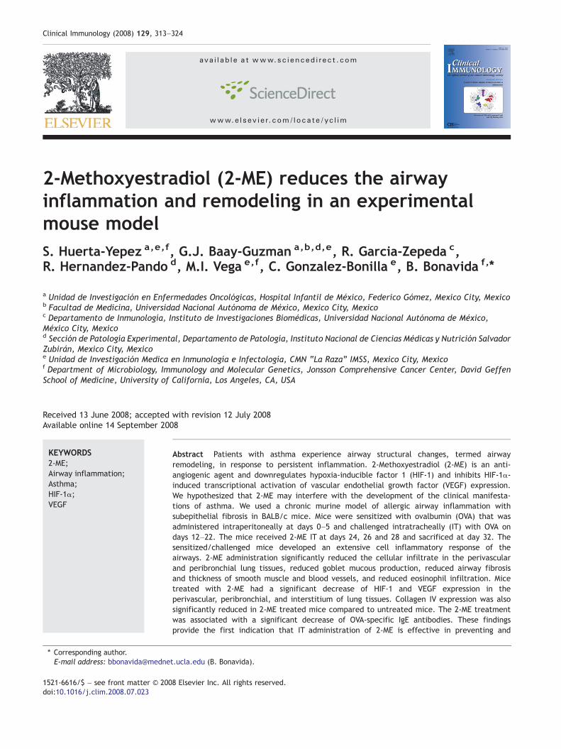
Figure 1 Murine models of airway inflammation and treat-ment with 2-ME. The effects of ITadministered 2-ME and/or OVAwere examined in the context of immunotherapy of establishedatopic lung inflammation.
Introduction
The pathophysiology of asthma involves a number of celltypes and mediators. Airway structural changes that occur inpatients with asthma in response to persistent inflammationare termed airway remodeling and include airway wallthickening, subepithelial fibrosis, and hyperplasia of mucousglands, myofibroblasts, and smooth muscle vasculature [1].Chronic airway inflammation plays a key role in asthma. Therelationship between inflammation and structural changes isspeculative. It is believed that remodeling is a direct resultof a switch from acute to chronic inflammation followed byhealing [2]. For this reason, anti-inflammatory agents willnot necessarily prevent or attenuate the process ofremodeling [3]. All current guidelines focus on the treatmentof inflammation in asthma although there are differencesbetween the processes of airway wall remodeling for whichthere is no defined treatment validated [2].
Long-acting β agonists produce bronchial dilation andimprove asthma symptoms, effects that are maintained withregular use over time. However, immunotherapy with βagonists has been consistently shown to be inferior to the useof inhaled corticosteroids which can reduce the underlyinginflammation associated with asthma. For these reasons,inhaled corticosteroids are recommended as first linemaintenance treatment for asthma [4].
2-ME is a naturally occurring derivative of 17β-estradioland was shown to be a well tolerated small molecule thatpossesses anti-tumor and anti-angiogenic activities [5].Studies by Mabjeesh et al. [6] reported that 2-ME destabilizesmicrotubules and blocks HIF1-α nuclear accumulation andHIF1-α activity by an oxygen- and proteasome-independentpathways. Evidence was provided for a link between themicrotubule cytoskeleton and HIF regulation. Destruction ofthe normal function of the microtubule cytoskeleton isrequired for HIF1-α-induced inhibition by microtubule-targeted drugs such as Taxol and Vincristine.
Several reports have been published suggesting the use ofdifferent strategies for the suppression of antigen-inducedasthma in mice. For instance, the oral administration of CpG-ODNs was effective in preventing and reversing antigen-induced eosinophilia airway inflammation [7]. Also, lowdoses of leukotriene receptor antagonist were an effectivetherapy in the airway remodeling in the ovalbumin mousemodel [8].
Angiogenesis supports the development of many diseasessuch as cancer, rheumatoid arthritis, psoriasis, maculardegeneration, and diabetic retinopathy [9–11]. VEGF is amajor mediator of angiogenesis, whose expression is inducedunder hypoxic conditions [9,12]. The induction of VEGF underhypoxic conditions is a multistage process in which the alphasubunit of HIF (HIF-1α) plays a key role [13]. HIF1-α is rapidlydegraded by the proteasome under normoxic conditions.Following hypoxic stabilization, HIF-1α is translocated intothe nucleus where it heterodimerizes with HIF-1β andactivates the transcription of more than forty genes
important for adaptation and survival under hypoxia [14].Hence, the anti-angiogenic activity of 2-ME may regulate theinflammatory process in asthma and reduce cell infiltration.Therefore, we hypothesized that the local intratracheal (IT)administration of 2-ME may inhibit airway inflammation andremodeling. The objective of this study was to test thishypothesis and determine the effect of IT 2-ME administra-tion in the development of airway remodeling using a chronicmurine model of allergic airway inflammation with subse-quent fibrosis. The findings support our hypothesis anddemonstrate that 2-ME significantly inhibited antigen-induced inflammation and airway remodeling in the OVAallergen inflammatory murine model.
Materials and methods
Animals
Balb/c mice (males) were obtained and maintained in apathogen-free environment in the facilities of the InstitutoNacional de Ciencias Medicas y de la Nutricion (INCMN)(Mexico City). The mice were housed in a temperature-controlled room with 12-h dark/light cycles, and allowedfood and water ad libitum. All the experiments describedbelow were performed in accordance with the INCMNregulations.
Experimental design
The protocol for sensitization and intratracheal challengehas previously been described by us in details [15] andillustrated in Fig.1. Briefly, 6 to 8 week-old Balb/c male micewere sensitized on days 0 and 5 by intraperitoneal injectionof 100 μg chicken egg ovalbumin (OVA, grade V, Sigma, USA)emulsified in 1 mg of aluminum hydroxide (alum) (PierceChemical, USA) in a total volume of 100 μL. Seven days afterthe last sensitization, mice were exposed twice to intra-tracheal (IT) administration of 0.75% OVA. 2-ME waspurchased commercially from Sigma Aldrich (St Louis, MO).The dose of 2-ME chosen for the in vivo experiment wasderived from a report using 100 mg/kg in a cancer murinemodel via the intraperitoneal route [3,16]. We have chosen alesser concentration of 30 mg/kg because 2-ME wasadministered IT. 6 to 8 week-old Balb/c male mice (n=18)
315Inhibition of experimental asthma by 2-ME
were divided into three groups. Group one receivedovalbumin (OVA) (n=6), group two received OVA and thenwas treated with 2-ME (OVA/2-ME) (n=6) and group three(n=6) was control and received saline solution (SS). Groupsone and two were immunized by intraperitoneal (i.p.)injection of 100 μg (100 μL) of OVA complexed with 1 μgalum on days 0 and 5 and then the mice received anintratraqueal (IT) dose of 100 μL of 0.75% of OVA in SS on days12 and 22. The control group three (n=6) received salinesolution (SS) with alum i.p. on days 0 and 5 and SS withoutalum ITon days 12 and 22. A concentrated stock solution of 2-ME was prepared in DMSO and then diluted in SS to prepare asolution of 2-ME (30 mg/kg) that was administrated IT togroup two beginning on day 24 of the protocol and continuinguntil day 28 every other day. The mice received 3 adminis-trations every other day, (D 24, D 26 and D 28) of 100 μL(30 mg/kg) of 2-ME IT. Positive control consisted of micesensitized and OVA-exposed. The non-immunized and OVA-exposed mice were similar to group 1 mice as they did notexhibit detectable airway inflammation. The positive controlOVA mice received SS containing the same concentration ofDMSO as the one used for treatment with 2-ME. All animalswere sacrificed 96 h after the last IT treatment.
Lung histology and morphometric analysis
At 96 h after the last challenge, lungs were removed from themice after sacrifice. Before the lungs were removed, theywere filled intratracheally with a fixative (4% formalin) usinga ligature around the trachea. Lung tissues were fixed with10% (v/v) neutral buffered formalin. The specimens weredehydrated and embedded in paraffin. For histologicalexamination, 4-μm sections of fixed embedded tissueswere cut on a Leica model 2165 rotary microtome (Leica,Nussloch, Germany), placed on glass slides, deparaffinized,and stained sequentially with hematoxylin and eosin (H&E)to assess the inflammatory cell infiltrates. The area (μm2)was calculated from stained slides, selecting venules withdiameter of 100–200 μm and bronchi with 150–300 μm ofdiameter. Four blood vessels or bronchi from four to six lungsections were analyzed for each experimental group, andresults were expressed as average and standard deviations ofinfiltrated area (μm2). Mucus production was assessed fromlung sections stained with periodic acid Schiff (PAS). The PAS-positive material was measured in μm in bronchi with 150–300 μm. Five bronchi from four to six lung sections wereanalyzed for each experimental group and the results wereexpressed as average and standard deviations of PAS-positivearea (μm). Mason's trichrome (MT) was used to determinecollagen deposition in the lung. The histological analyseswere preformed by observers who were not aware of thegroups of mice from which the samples originated.
Bronchoalveolar lavages (BAL)
After the mice were sacrificed by exsanguination, thetracheas were exposed and intubated with a polyethylenecatheter. The BAL fluid was collected by washing with twoseparated aliquots of 1 ml of SS with 2% of FBS through thetrachea. The cells from the first wash were harvested bycentrifugation (500×g for 10 min at 4 °C), counted in a
hemacytometer and adjusted to 1×10⁵ cells/ml. Differentialcounts from four to six H/E stained cytospin preparations foreach experimental group were determined by microscopy,Cells were counted by standard procedures at 40× magnifica-tion. Results are presented as cells/ml averages from all theslides per group (200 cells/slide).
Immunocytochemistry
For immunohistochemistry of VEGF or HIF-1α and type IVcollagen, the deparaffinized 4-μm sections were incubatedsequentially in accordance with the instructions of the LSABkit from DAKO corporation (Carpinteria, CA, USA). In brief,antigen retrieval was performed by immersing the slides in0.01% sodium citrate pH 6.0 incubated for 5 min in boilingwater. The endogenous peroxidase activity was inhibited byimmersing the slides in 3% H2O2-methanol for 25 min, andthe background non-specific binding was reduced byincubating with 1% BSA in PBS for 60 min. The slides wereincubated overnight at room temperature with antibodiesagainst HIF-1 α (1:250) (Santa Cruz Biotechnology, SantaCruz, CA), VEGF (1:750) (Santa Cruz) or type IV collagen(1:500) (Santa Cruz). Finally, the slides were washed fivetimes in PBS 0.1 M pH 7.4 for 8 min. In order to reduce thevariability, all samples from each group were processed atthe same time in a single experiment using a single batch ofantibody diluted in PBS-normal swine serum. After washing,the slides were incubated with a biotinylated secondaryantibody container in the LSAB kit from DAKO corporationfor 30 min at r.t. followed by an incubation with astreptavidin-HRP conjugate container in the LSAB kit for30 min at r.t. and then with 3,3′-diaminobenzidine tetra-hydrochloride (liquid DAB, DAKO corporation) for 1 to 5 minin the dark. The reaction was arrested with distilled waterand the slides were counterstained with hematoxylin.Thereafter, the tissues were washed in tap water for5 min, dehydrated through ethanol baths (70, 90 and100%) in xylene and mounted with E-2 Mount medium(Shandon lab, Pittsburgh, PA, USA). Finally, the slides wereanalyzed under light microscopy (Olympus BX-40). Theanalyses were performed by blinded observers.
Determination of serum OVA-specific IgE
Serum levels of specific anti-OVA IgE were determined byELISA. The ELISA kit was purchased from MD Bioscience, St.Paul, MN (Catalog #OVA IgE 96). The analysis was performedaccording to themanufacturer's instructions. The specificity ofthe ELISA was corroborated by demonstrating that serum fromnaïve non-immunized mice did not reveal any detectablebinding toOVA. Colorwas developedby addingOPD in substratebuffer (Sanofi Diagnostic Pasteur). The reaction was arrestedwith 50 μL H2SO4 and the OD were determined at 495 nm.
Statistical analysis
Data were expressed as mean±S.E.M. Statistical compar-isons were performed using one-way analysis of variancefollowed by the Fisher's test. Significant differences betweengroups were determined using the unpaired Student's test.Statistical significance was set at pb0.05.
316 S. Huerta-Yepez et al.
Results
Intratracheal administration of 2-ME reduces OVA-induced atopic response
Three groups of mice (n=6/group) were examined and weretreated with OVA alone (group one), OVA+ 2-ME (group two)and with saline (SS) (group three) as described in Materialsand methods. The mice were rendered allergic to OVA withtwo i.p. injections of OVA-alum suspension at days 0 and 5and 2 IT challenges at days 12 and 22. Two days aftersensitization group two mice were treated three times with2-ME administered by the intratracheal route every otherday. The intensity of allergic inflammation reaction wasevaluated on day 32 at sacrifice. The schematic diagram ofthe above treatment schedules is also illustrated in Fig. 1.
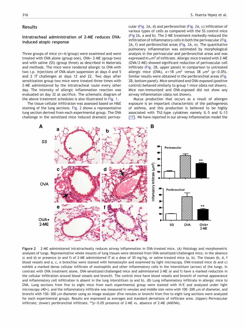
The tissue cellular infiltration was assessed based on H&Estaining of the lung sections. Fig. 2 shows a representativelung section derived from each experimental group. The OVAchallenge in the sensitized mice induced dramatic perivas-
Figure 2 2-ME administered intratracheally reduces airway inflaanalyses of lungs. Representative whole mounts of lung tissues were(c and d) or presence (e and f) of 2-ME administered IT at a doseblood vessels and a, c, e bronchia) were stained with hematoxylinexhibit a marked dense cellular infiltrate of eosinophils and othercontrast with OVA treatment alone, OVA-sensitized/challenged micthe cellular infiltration around blood vessels and bronchi. The contand inflammatory cell infiltration is absent in the lung interstitiumOVA. Lung sections from five to eight mice from each experimemicroscopy (40×), and the inflammatory infiltrate was measured in vbronchi with 150–300 μm diameter using an image analyzer (five vefor each experimental group). Results are expressed as averages ainfiltrate; (lower) peribronchial infiltrate. ⁎⁎pb0.05 presence of 2-
cular (Fig. 2A, d) and peribronchial (Fig. 2A, c) infiltration ofvarious types of cells as compared with the SS control mice(Fig 2A, a and b). The 2-ME treatment markedly reduced theinfiltration of inflammatory cells in both the perivascular (Fig.2A, f) and peribronchial areas (Fig. 2A, e). The quantitativepulmonary inflammation was estimated by morphologicalanalysis in the perivascular and peribronchial areas and wasexpressed in μm2 of infiltrate. Allergicmice treatedwith 2-ME(OVA/2-ME) showed significant reduction of perivascular cellinfiltrate (Fig. 2B, upper panel) in comparison to untreatedallergic mice (OVA), x=18 μm2 versus 38 μm2 (pb0.05).Similar results were obtained in the peribronchial areas (Fig.2B, bottom panel). Mice sensitized and OVA-exposed (positivecontrol) behaved similarly to group 1 mice (data not shown).Mice non-immunized and OVA-exposed did not show anyairway inflammation (data not shown).
Mucus production that occurs as a result of allergenexposure is an important characteristic of the pathogenesisof asthma, and this production is believed to be highlyassociated with Th2-type cytokines namely IL-5 and IL-13[17]. We have reported in our airway inflammation model the
mmation in OVA-treated mice. (A) Histology and morphometricobtained from OVA-sensitized/challenged mice, in the absence
of 30 mg/kg, or saline-treated mice (a, b). The tissues (b, d, fand examined by light microscopy. OVA-treated mice (b and c)inflammatory cells in the interstitium (arrow) of the lungs. Ine and administered 2-ME (e and f) have a marked reduction inrol mice have blood vessels and bronchi of normal appearance(a and b). (B) Lung inflammatory infiltrate in allergic mice tontal group were stained with H/E and analyzed under lightenules and middle size veins with 100–200 μm of diameter, andnules or bronchi from five to eight lung sections were analyzednd standard deviations of infiltrate area. (Upper) PerivascularME vs. absence of 2-ME (ANOVA).
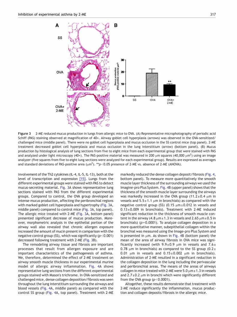
Figure 3 2-ME reduced mucus production in lungs from allergic mice to OVA. (A) Representative microphotography of periodic acidSchiff (PAS) staining observed at magnification of 40×. Airway goblet cell hyperplasia (arrows) was observed in the OVA-sensitized/challenged mice (middle panel). There were no goblet cell hyperplasia and mucus occlusion in the SS control mice (top panel). 2-MEtreatment decreased goblet cell hyperplasia and mucus occlusion in the lung interstitium (arrow) (bottom panel). (B) Mucusproduction by histological analysis of lung sections from five to eight mice from each experimental group that were stained with PASand analyzed under light microscopy (40×). The PAS-positive material was measured in 200 μm squares (40,000 μm2) using an imageanalyzer (five squares from five to eight lung sections were analyzed for each experimental group). Results are expressed as averagesand standard deviations of PAS-positive area (μm2). ⁎⁎pb0.05 presence of 2-ME vs. absence of 2-ME (ANOVA).
317Inhibition of experimental asthma by 2-ME
involvement of the Th2 cytokines (IL-4, IL-5, IL-13), both at thelevel of transcription and expression [15]. Lungs from thedifferent experimental groups were stained with PAS to detectmucus-secreting material. Fig. 3A shows representative lungsections stained with PAS from the different experimentalgroups. Compared to control, the OVA group developed anintense mucus production, affecting the peribronchial regionswith marked goblet cell hyperplasia and hypertrophy (Fig. 3A,middle panel) compared to control mice (Fig. 3A, top panel).The allergic mice treated with 2-ME (Fig. 3A, bottom panel)presented significant decrease of mucus production. More-over, morphometric analysis in the epithelial portion of theairway wall also revealed that chronic allergen exposureincreased the amount ofmucin present in comparison with thenegative control group (SS), which was significantly (pb0.001)decreased following treatment with 2-ME (Fig. 3B).
The remodeling airway tissue and fibrosis are importantprocesses that result from allergen exposure and areimportant characteristics of the pathogenesis of asthma.We, therefore, determined the effect of 2-ME treatment onairway smooth muscle thickness in our experimental murinemodel of allergic airway inflammation. Fig. 4A showsrepresentative lung sections from the different experimentalgroups stained with Mason's trichrome. In OVA-sensitized andchallengedmice, dense collagen deposition/fibrosis was seenthroughout the lung interstitium surrounding the airways andblood vessels (Fig. 4A, middle panel) as compared with thecontrol SS group (Fig. 4A, top panel). Treatment with 2-ME
markedly reduced the dense collagen deposit/fibrosis (Fig. 4,bottom panel). To measure more quantitatively the smoothmuscle layer thickness of the surrounding airways we used theImagine-pro Plus System. Fig. 4B (upper panel) shows that thethickness of the smooth muscle layer surrounding the airwayswas markedly increased in the OVA group (11.2±0.4 μm invessels and 5.5±1.1 μm in bronchials) as compared with thenegative control group (SS) (0.15 μm±0.012 in vessels and0.13±0.009 in bronchials). Treatment with 2-ME inducedsignificant reduction in the thickness of smooth muscle con-tent in the airway (4.8 μm±1.3 in vessels and 2.65 μm±0.5 inbronchials) (p=0.0001). To analyze collagen deposition in amore quantitative manner, subepithelial collagen within thebronchial was measured using the Image-pro Plus System andis presented in μm. As shown in Fig. 4B (bottom panel) themean of the area of airway fibrosis in OVA mice was signi-ficantly increased (with 9.9±0.9 μm in vessels and 7.6±0.78 μm in bronchials) as compared to the SS group (0.2±0.01 μm in vessels and 0.15±0.002 μm in bronchials).Administration of 2-ME resulted in a significant reduction inthe collagen deposition in the lung including the perivascularand peribronchial areas. The means of the areas of airwayscollagen inmice treated with 2-MEwere 5.0 μm±1.3 in vesselsand 2.7±0.2 μm in bronchi which were significantly differentfrom the OVA group (pb0.0001).
Altogether, these results demonstrate that treatment with2-ME reduce significantly the inflammation, mucus produc-tion and collagen deposits/fibrosis in the allergic mice.
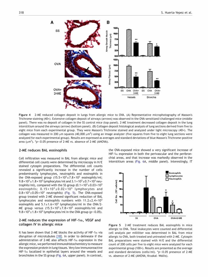
Figure 4 2-ME reduced collagen deposit in lungs from allergic mice to OVA. (A) Representative microphotography of Masson'sTrichrome staining (40×). Extensive collagen deposit of airways (arrows) was observed in the OVA-sensitized/challenged mice (middlepanel). There was no deposit of collagen in the SS control mice (top panel). 2-ME treatment decreased collagen deposit in the lunginterstitium around the airways (arrow) (bottom panel). (B) Collagen deposit histological analysis of lung sections derived from five toeight mice from each experimental group. They were Masson's Trichrome stained and analyzed under light microscopy (40×). Thecollagen was measured in 200 μm squares (40,000 μm2) using an image analyzer (five squares from five to eight lung sections wereanalyzed for each experimental group). Results are expressed as averages and standard deviations of blue Masson's Trichrome-positivearea (μm2). ⁎pb0.05 presence of 2-ME vs. absence of 2-ME (ANOVA).
Figure 5 2-ME treatment reduces BAL eosinophils in miceallergic to OVA. Total leukocytes were counted and differentialcell analysis per milliliter was determined in BAL from miceallergic to OVA, both treated and untreated with 2-ME. CytospinBAL preparations were stained with H/E and the differentialcount of 200 cells per five to eight mice were analyzed for eachexperimental group (100×). Results are presented as the averageand standard deviations (cells/ml). ⁎pb0.05 presence of 2-MEvs. absence of 2-ME (ANOVA; Kruskal–Wallis).
318 S. Huerta-Yepez et al.
2-ME reduces BAL eosinophils
Cell infiltration was measured in BAL from allergic mice anddifferential cell counts were determined by microscopy in H/Estained cytospin preparations. The differential cell countsrevealed a significantly increase in the number of cellspredominantly lymphocytes, neutrophils and eosinophils inthe OVA-exposed group (15.5×10³±7.8×10³ eosinophils/ml;9.8×103±1.8×103 lymphocytes/ml and 3.1×10³±0.7×10³ neu-trophils/ml), compared with the SS group (0.1×10³±0.03×10³
eosinophils; 0.15 × 10³ ± 0.02 × 10³ lymphocytes and0.8×103±0.05×10³ neutrophils) (Fig. 5). The OVA-exposedgroup treated with 2-ME showed significant reduction of BALlymphocytes and eosinophils numbers with 11.2±2.4×10³
eosinophils and 5.1±1.6×10³ lymphocytes/ml in the OVA/2-ME group versus (15.5×10³±7.8×10³ eosinophils/ml and9.8×103±1.8×10³ lymphocytes/ml in the OVA group (pb0.05).
2-ME reduces the expression of HIF-1α, VEGF andcollagen IV in allergic mice
It has been shown that 2-ME blocks the activity of HIF-1α viadisruption of microtubules [18]. In order to delineate if theadministration of 2-ME also affects HIF-1α expression in theallergicmice,weperformed immunohistochemistry tomeasurethe expression protein in lung tissues. Very low immunoreactiveHIF-1α localized in epithelial layers around the vessels andbronchioles in the SS group (Fig. 6A, upper panel). In contrast,
the OVA-exposed mice showed a very significant increase ofHIF-1α expression in both the perivascular and the peribron-chial areas, and that increase was markedly observed in theinterstitium areas (Fig. 6A, middle panel). Interestingly, IT
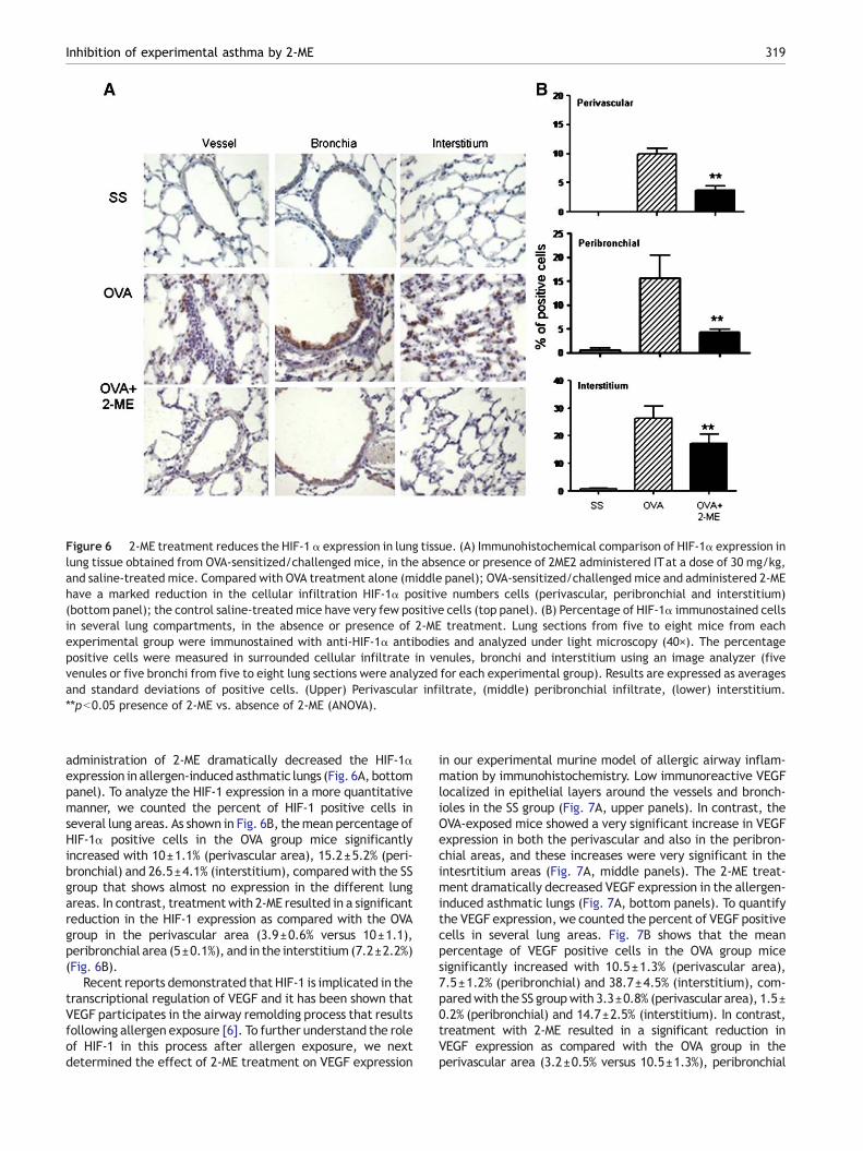
Figure 6 2-ME treatment reduces the HIF-1 α expression in lung tissue. (A) Immunohistochemical comparison of HIF-1α expression inlung tissue obtained from OVA-sensitized/challenged mice, in the absence or presence of 2ME2 administered ITat a dose of 30 mg/kg,and saline-treated mice. Compared with OVA treatment alone (middle panel); OVA-sensitized/challenged mice and administered 2-MEhave a marked reduction in the cellular infiltration HIF-1α positive numbers cells (perivascular, peribronchial and interstitium)(bottom panel); the control saline-treated mice have very few positive cells (top panel). (B) Percentage of HIF-1α immunostained cellsin several lung compartments, in the absence or presence of 2-ME treatment. Lung sections from five to eight mice from eachexperimental group were immunostained with anti-HIF-1α antibodies and analyzed under light microscopy (40×). The percentagepositive cells were measured in surrounded cellular infiltrate in venules, bronchi and interstitium using an image analyzer (fivevenules or five bronchi from five to eight lung sections were analyzed for each experimental group). Results are expressed as averagesand standard deviations of positive cells. (Upper) Perivascular infiltrate, (middle) peribronchial infiltrate, (lower) interstitium.⁎⁎pb0.05 presence of 2-ME vs. absence of 2-ME (ANOVA).
319Inhibition of experimental asthma by 2-ME
administration of 2-ME dramatically decreased the HIF-1αexpression in allergen-inducedasthmatic lungs (Fig. 6A, bottompanel). To analyze the HIF-1 expression in a more quantitativemanner, we counted the percent of HIF-1 positive cells inseveral lung areas. As shown in Fig. 6B, themean percentage ofHIF-1α positive cells in the OVA group mice significantlyincreased with 10±1.1% (perivascular area), 15.2±5.2% (peri-bronchial) and 26.5±4.1% (interstitium), compared with the SSgroup that shows almost no expression in the different lungareas. In contrast, treatmentwith 2-ME resulted in a significantreduction in the HIF-1 expression as compared with the OVAgroup in the perivascular area (3.9±0.6% versus 10±1.1),peribronchial area (5±0.1%), and in the interstitium (7.2±2.2%)(Fig. 6B).
Recent reports demonstrated that HIF-1 is implicated in thetranscriptional regulation of VEGF and it has been shown thatVEGF participates in the airway remolding process that resultsfollowing allergen exposure [6]. To further understand the roleof HIF-1 in this process after allergen exposure, we nextdetermined the effect of 2-ME treatment on VEGF expression
in our experimental murine model of allergic airway inflam-mation by immunohistochemistry. Low immunoreactive VEGFlocalized in epithelial layers around the vessels and bronch-ioles in the SS group (Fig. 7A, upper panels). In contrast, theOVA-exposed mice showed a very significant increase in VEGFexpression in both the perivascular and also in the peribron-chial areas, and these increases were very significant in theintesrtitium areas (Fig. 7A, middle panels). The 2-ME treat-ment dramatically decreased VEGF expression in the allergen-induced asthmatic lungs (Fig. 7A, bottom panels). To quantifythe VEGF expression, we counted the percent of VEGF positivecells in several lung areas. Fig. 7B shows that the meanpercentage of VEGF positive cells in the OVA group micesignificantly increased with 10.5±1.3% (perivascular area),7.5±1.2% (peribronchial) and 38.7±4.5% (interstitium), com-paredwith the SS groupwith 3.3±0.8% (perivascular area), 1.5±0.2% (peribronchial) and 14.7±2.5% (interstitium). In contrast,treatment with 2-ME resulted in a significant reduction inVEGF expression as compared with the OVA group in theperivascular area (3.2±0.5% versus 10.5±1.3%), peribronchial
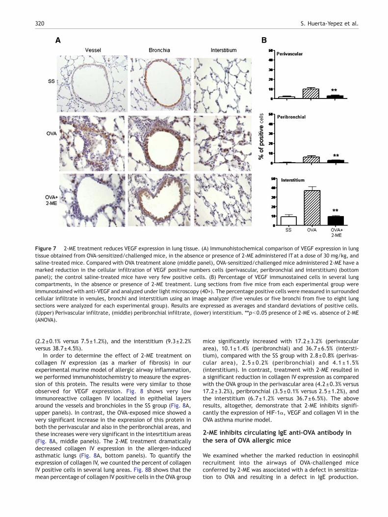
Figure 7 2-ME treatment reduces VEGF expression in lung tissue. (A) Immunohistochemical comparison of VEGF expression in lungtissue obtained from OVA-sensitized/challenged mice, in the absence or presence of 2-ME administered ITat a dose of 30 mg/kg, andsaline-treated mice. Compared with OVA treatment alone (middle panel), OVA-sensitized/challenged mice administered 2-ME have amarked reduction in the cellular infiltration of VEGF positive numbers cells (perivascular, peribronchial and interstitium) (bottompanel); the control saline-treated mice have very few positive cells. (B) Percentage of VEGF immunostained cells in several lungcompartments, in the absence or presence of 2-ME treatment. Lung sections from five mice from each experimental group wereimmunostained with anti-VEGF and analyzed under light microscopy (40×). The percentage positive cells were measured in surroundedcellular infiltrate in venules, bronchi and interstitium using an image analyzer (five venules or five bronchi from five to eight lungsections were analyzed for each experimental group). Results are expressed as averages and standard deviations of positive cells.(Upper) Perivascular infiltrate, (middle) peribronchial infiltrate, (lower) interstitium. ⁎⁎pb0.05 presence of 2-ME vs. absence of 2-ME(ANOVA).
320 S. Huerta-Yepez et al.
(2.2±0.1% versus 7.5±1.2%), and the interstitium (9.3±2.2%versus 38.7±4.5%).
In order to determine the effect of 2-ME treatment oncollagen IV expression (as a marker of fibrosis) in ourexperimental murine model of allergic airway inflammation,we performed immunohistochemistry to measure the expres-sion of this protein. The results were very similar to thoseobserved for VEGF expression. Fig. 8 shows very lowimmunoreactive collagen IV localized in epithelial layersaround the vessels and bronchioles in the SS group (Fig. 8A,upper panels). In contrast, the OVA-exposed mice showed avery significant increase in the expression of this protein inboth the perivascular and also in the peribronchial areas, andthese increases were very significant in the intesrtitium areas(Fig. 8A, middle panels). The 2-ME treatment dramaticallydecreased collagen IV expression in the allergen-inducedasthmatic lungs (Fig. 8A, bottom panels). To quantify theexpression of collagen IV, we counted the percent of collagenIV positive cells in several lung areas. Fig. 8B shows that themean percentage of collagen IV positive cells in the OVA group
mice significantly increased with 17.2±3.2% (perivasculararea), 10.1±1.4% (peribronchial) and 36.7±6.5% (intersti-tium), compared with the SS group with 2.8±0.8% (perivas-cular area), 2.5 ± 0.2% (peribronchial) and 4.1 ± 1.5%(interstitium). In contrast, treatment with 2-ME resulted ina significant reduction in collagen IV expression as comparedwith the OVA group in the perivascular area (4.2±0.3% versus17.2±3.2%), peribronchial (3.5±0.1% versus 2.5±1.2%), andthe interstitium (6.7±1.2% versus 36.7±6.5%). The aboveresults, altogether, demonstrate that 2-ME inhibits signifi-cantly the expression of HIF-1α, VEGF and collagen VI in theOVA asthma murine model.
2-ME inhibits circulating IgE anti-OVA antibody inthe sera of OVA allergic mice
We examined whether the marked reduction in eosinophilrecruitment into the airways of OVA-challenged miceconferred by 2-ME was associated with a defect in sensitiza-tion to OVA and resulting in a defect in IgE production.
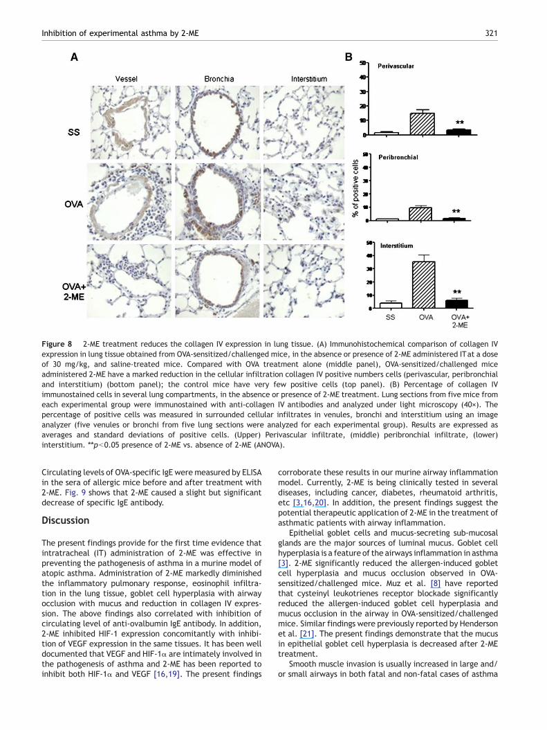
Figure 8 2-ME treatment reduces the collagen IV expression in lung tissue. (A) Immunohistochemical comparison of collagen IVexpression in lung tissue obtained from OVA-sensitized/challenged mice, in the absence or presence of 2-ME administered ITat a doseof 30 mg/kg, and saline-treated mice. Compared with OVA treatment alone (middle panel), OVA-sensitized/challenged miceadministered 2-ME have a marked reduction in the cellular infiltration collagen IV positive numbers cells (perivascular, peribronchialand interstitium) (bottom panel); the control mice have very few positive cells (top panel). (B) Percentage of collagen IVimmunostained cells in several lung compartments, in the absence or presence of 2-ME treatment. Lung sections from five mice fromeach experimental group were immunostained with anti-collagen IV antibodies and analyzed under light microscopy (40×). Thepercentage of positive cells was measured in surrounded cellular infiltrates in venules, bronchi and interstitium using an imageanalyzer (five venules or bronchi from five lung sections were analyzed for each experimental group). Results are expressed asaverages and standard deviations of positive cells. (Upper) Perivascular infiltrate, (middle) peribronchial infiltrate, (lower)interstitium. ⁎⁎pb0.05 presence of 2-ME vs. absence of 2-ME (ANOVA).
321Inhibition of experimental asthma by 2-ME
Circulating levels of OVA-specific IgE were measured by ELISAin the sera of allergic mice before and after treatment with2-ME. Fig. 9 shows that 2-ME caused a slight but significantdecrease of specific IgE antibody.
Discussion
The present findings provide for the first time evidence thatintratracheal (IT) administration of 2-ME was effective inpreventing the pathogenesis of asthma in a murine model ofatopic asthma. Administration of 2-ME markedly diminishedthe inflammatory pulmonary response, eosinophil infiltra-tion in the lung tissue, goblet cell hyperplasia with airwayocclusion with mucus and reduction in collagen IV expres-sion. The above findings also correlated with inhibition ofcirculating level of anti-ovalbumin IgE antibody. In addition,2-ME inhibited HIF-1 expression concomitantly with inhibi-tion of VEGF expression in the same tissues. It has been welldocumented that VEGF and HIF-1α are intimately involved inthe pathogenesis of asthma and 2-ME has been reported toinhibit both HIF-1α and VEGF [16,19]. The present findings
corroborate these results in our murine airway inflammationmodel. Currently, 2-ME is being clinically tested in severaldiseases, including cancer, diabetes, rheumatoid arthritis,etc [3,16,20]. In addition, the present findings suggest thepotential therapeutic application of 2-ME in the treatment ofasthmatic patients with airway inflammation.
Epithelial goblet cells and mucus-secreting sub-mucosalglands are the major sources of luminal mucus. Goblet cellhyperplasia is a feature of the airways inflammation in asthma[3]. 2-ME significantly reduced the allergen-induced gobletcell hyperplasia and mucus occlusion observed in OVA-sensitized/challenged mice. Muz et al. [8] have reportedthat cysteinyl leukotrienes receptor blockade significantlyreduced the allergen-induced goblet cell hyperplasia andmucus occlusion in the airway in OVA-sensitized/challengedmice. Similar findings were previously reported by Hendersonet al. [21]. The present findings demonstrate that the mucusin epithelial goblet cell hyperplasia is decreased after 2-MEtreatment.
Smooth muscle invasion is usually increased in large and/or small airways in both fatal and non-fatal cases of asthma
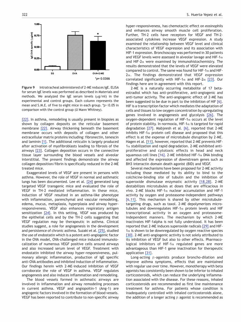
Figure 9 Intratracheal administered of 2-ME reduces IgE. ELISAfor serum IgE levels was performed as described in Materials andmethods. We analyzed the IgE serum levels (μg/ml) in theexperimental and control groups. Each column represents themean and S.M.E. of five to eight mice in each group. ⁎pb0.05 incomparison with the control group (U Mann Whitney).
322 S. Huerta-Yepez et al.
[22]. In asthma, remodeling is usually present in biopsies asshown by collagen deposits on the reticular basementmembrane [22]. Airway thickening beneath the basementmembrane occurs with deposits of collagen and otherextracellular matrix proteins including: fibronectin, tenescinand laminin [1]. The additional reticulin is largely producedafter activation of myofibroblasts leading to fibrosis of theairways [23]. Collagen deposition occurs in the connectivetissue layer surrounding the blood vessels and alveolarinterstitial. The present findings demonstrate the airwaycollagen deposition/fibrin is specifically reduced in the 2-MEtreated mice.
Exaggerated levels of VEGF are present in persons withasthma. However, the role of VEGF in normal and asthmaticlungs has been documented. Lee et al. [24] generated lungtargeted VEGF transgenic mice and evaluated the role ofVEGF in TH-2 mediated inflammation. In these mice,induction of VEGF resulted in an asthma-like phenotypewith inflammation, parenchymal and vascular remodeling,edema, mucus, metaplasia, hyperplasia and airway hyper-responsiveness. VEGF also enhances respiratory antigensensitization [24]. In this setting, VEGF was produced bythe epithelial cells and by the TH-2 cells suggesting thatVEGF regulation may be therapeutic in asthma. Clinicalstudies suggest, a role for angiogenesis in the developmentand persistence of chronic asthma. Suzaki et al. [25], studiedthe use of endostatin which is a potent anti-angiogenic factorin the OVA model, OVA-challenged mice induced immunolo-calization of numerous VEGF positive cells around airwaysand also increased serum level of VEGF. Treatment withendostatin inhibited the airway hyper-responsiveness, pul-monary allergic inflammation, production of IgE specificanti-OVA antibodies and inhibited induction of inflammation.Our findings herein with 2-ME-induced inhibition of VEGFcorroborate the role of VEGF in asthma. VEGF regulatesangiogenesis and also induces inflammation and remodeling.
The blood vessels formed in asthmatic airways areinvolved in inflammation and airway remodeling processesin current asthma. VEGF and angiopoitin-1 (Ang-1) areangiogenic factors involved in the formation of blood vessels.VEGF has been reported to contribute to non-specific airway
hyper-responsiveness, has chemotactic effect on eosinophilsand enhances airway smooth muscle cell proliferation.Further, TH-2 cells have receptors for VEGF and TH-2-associated cytokines increase VEGF expression. A studyexamined the relationship between VEGF level and clinicalcharacteristics of VEGF expression and its association withHIF-1 expression. Bronchoscopy was performed in 30 patientsand VEGF levels were assessed in alveolar lavage and HIF-1αand HIF-2α were examined by immunohistochemistry. Theresults demonstrated that the levels of VEGF were elevatedcompared to control. The same was found for HIF-1α and HIF-2α. The findings demonstrated that VEGF expressioncorrelated significantly with HIF-1α and HIF-2α [23]. Ourfindings here are in agreement with this report.
2-ME is a naturally occurring metabolite of 17 beta-estradiol which has anti-proliferative, anti-angiogenic andanti-tumor activity. The anti-angiogenic effect of 2-ME hasbeen suggested to be due in part to the inhibition of HIF [6].HIF is a transcription factor which mediates the adaptation ofcells and tissues to low oxygen concentration by upregulatinggenes involved in angiogenesis and glycolysis [26]. Theoxygen-dependent regulation of HIF-1α occurs at the levelof protein stability. In normoxia, HIF-1α is targeted for rapiddegradation [27]. Mabjeesh et al. [6], reported that 2-MEinhibits HIF-1α protein cell disease and proposed that thiseffect is at the expense of microtubule disruption by 2-ME.Hagen et al. [11], however, reported that 2-ME prevents HIF-1α stabilization and rapid degradation. 2-ME exhibited anti-proliferative and cytotoxic effects in head and necksquamous cell lines [16]. 2-ME inhibited HIF-1α DNA bindingand affected the expression of downstream genes such asBH3 interactin domain death agonist (BID) and VEGF.
Several mechanisms have been proposed for 2-ME activityincluding those mediated by its ability to bind to thecolchicine-binding site of tubulin and the inhibition ofsuperoxide dismutase enzymatic activity [18,28]. 2-MEdestabilizes microtubules at doses that are efficacious invivo. 2-ME blocks HIF-1α nuclear accumulation and HIF-1activity by oxygen and proteasome-independent pathways[6,11]. This mechanism is shared by other microtubule-targeting drugs, such as taxol. 2-ME depolymerizes micro-tubules and downregulates HIF-1α protein levels and HIFtranscriptional activity in an oxygen and proteasome-independent manners. The mechanism by which 2-MEinactivates HIF-1alpha is not fully understood. It has beenreported that 2-ME induces superoxide radicals [29] and HIF-1α is shown to be downregulated by oxygen reactive species[30]. 2-ME anti-angiogenic activity is not solely attributed toits inhibition of VEGF but also to other effects. Pharmaco-logical inhibitors of HIF-1α regulated genes are moreadvantageous than HIF-1 gene inactivation for therapeuticapplication [31].
Long-acting β-agonists produce broncho-dilation andimprove asthma symptoms, effects that are maintainedwith regular use over time. However, monotherapy with betaagonists has consistently been shown to be inferior to inhaledcorticosteroids, which can reduce the underlying inflamma-tion associated with the disease. For these reasons, inhaledcorticosteroids are recommended as first line maintenancetreatment for asthma. For patients whose condition isinadequately controlled with inhaled corticosteroids alone,the addition of a longer acting β agonist is recommended as
323Inhibition of experimental asthma by 2-ME
symptoms are improved. However, much controversy existsin the use of β agonists in patients with asthma ever since itsapplication over 50 years ago. Regular β agonists usage isassociated with tolerance to the drug and a worsening ofdisease control. Several deaths have been reported with thelong-acting β agonists Salmeterol and there was a four foldincreased risk for asthma related death. The FDA in July of2005 issued a strong warning of increased risk that should beplaced on the labels of all long-acting β agonists with arecommendation that they be used only after other drugshave failed. A meta analysis pooled from 19 treatments with33,826 patients from long-acting β agonists increasedexacerbation and life threatening conditions comparedwith placebo [32]. Similar results were found with Salme-terol and Formoterol and in both children and adults.Concomitant inhaled corticosteroids do not adequatelyprotect against these effects.
Our present findings here demonstrate that 2-ME is aneffective agent in the treatment of airway inflammationassociated with asthma. 2-ME is currently in clinical trials forother diseases and thus toxicity studies are being performed[33, 34]. A phase 2 trial of 2-ME was conducted in 60 patientswith multiple myeloma (MM). 2-ME was administrated orallyat 1000 mg/daily for the first 39 patients and increased to800 mg twice daily for the remaining patients. No partialresponses were observed, although minor responses andprolonged, stable disease were noted. Plasma level of 2-MEwas not adequate [33]. Another study investigated themaximum tolerated dose (MTD) and toxicity of 2-MEadministered orally in 20 patients with solid tumors. TheMTD for 2-ME was not reached and the trial was closed due tolow plasma concentration of 2-ME relative to the dosesadministered. The investigators are currently examining newformulation of 2-ME with improved bio-availability [34]. Aclinical study compared the therapeutic effect of 2-ME aloneor in combination with docetaxel in patients with metastaticbreast cancer. 2-ME alone or in combination with docetaxelwas well tolerated [35]. Although these studies were notpromising for 2-ME administered orally, we propose that thepreferred route of 2-ME administration in asthma is viainhalation using different carriers. Further, this studysuggests that HIF-1α and VEGF are biomarkers in thedevelopment and response to treatment in asthma.
Acknowledgments
This work was supported in part by the Fogarty and by theJonsson Comprehensive Cancer Center at UCLA. The authorsalso acknowledge the assistance of Erica Keng and MaggieYang in the preparation of the manuscript.
References
[1] J.A. Elias, Z. Zhu, G. Chupp, R.J. Homer, Airway remodeling inasthma, J. Clin. Invest. 104 (1999) 1001–1006.
[2] J. Bousquet, P.K. Jeffery, W.W. Busse, M. Johnson, A.M. Vignola,Asthma from bronchoconstriction to airways inflammation andremodeling, Am. J. Respir. Crit. Care Med. 161 (2000)1720–1745.
[3] P.K. Jeffery, Remodeling in asthma and chronic obstructivelung disease, Am. J. Respir. Crit. Care Med. 164 (2001)S28–S38.
[4] M.J. Abramson, A.J. Crockett, P.A. Frith, C.F. McDonald,COPDX: an update of guidelines for the management of chronicobstructive pulmonary disease with a review of recentevidence, Med. J. Aust. 184 (2006) 342–345.
[5] V.S. Pribluda, E.R. Gubish Jr., T.M. Lavallee, A. Treston, G.M.Swartz, S.J. Green, 2-Methoxyestradiol: an endogenous anti-angiogenic and antiproliferative drug candidate, CancerMetastasis Rev. 19 (2000) 173–179.
[6] N.J. Mabjeesh, D. Escuin, T.M. Lavallee, V.S. Pribluda, G.M.Swartz, M.S. Johnson, M.T. Willard, H. Zhong, J.W. Simons,P. Giannakakou, 2-ME inhibits tumor growth and angiogenesisby disrupting microtubules and dysregulating HIF, CancerCell 3 (2003) 363–375.
[7] K. Kitagaki, T.R. Businga, J.N. Kline, Oral administration ofCpG-ODNs suppresses antigen-induced asthma in mice, Clin.Exp. Immunol. 143 (2006) 249.
[8] M.H. Muz, F. Deveci, Y. Bulut, N. Ilhan, H. Yekeler, T. Turgut, Theeffects of low dose leukotriene receptor antagonist therapy onairway remodeling and cysteinyl leukotriene expression in amouse asthma model, Exp. Mol. Med. 38 (2006) 109–118.
[9] J. Folkman, Angiogenesis-dependent diseases, Semin. Oncol.28 (2001) 536–542.
[10] C.G. Lee, H. Link, P. Baluk, R.J. Homer, S. Chapoval, V. Bhandari,M.J. Kang, L. Cohn, Y.K. Kim, D.M. McDonald, J.A. Elias, Vascularendothelial growth factor (VEGF) induces remodeling andenhances TH2-mediated sensitization and inflammation in thelung, Nat. Med. 10 (2004) 1095–1103.
[11] T. Hagen,G. D'Amico,M.Quintero,M.Palacios-Callender, V.Hollis,F. Lam, S. Moncada, Inhibition of mitochondrial respiration by theanticancer agent 2-methoxyestradiol, Biochem. Biophys. Res.Commun. 322 (2004) 923–929.
[12] M.A. Goldberg, T.J. Schneider, Similarities between the oxygen-sensing mechanisms regulating the expression of vascularendothelial growth factor and erythropoietin, J. Biol. Chem.269 (1994) 4355–4359.
[13] J.A. Forsythe, B.H. Jiang, N.V. Iyer, F. Agani, S.W. Leung, R.D.Koos, G.L. Semenza, Activation of vascular endothelial growthfactor gene transcription by hypoxia-inducible factor 1, Mol.Cell Biol. 16 (1996) 4604–4613.
[14] M. Hedtjarn, C. Mallard, S. Eklind, K. Gustafson-Brywe, H.Hagberg, Global gene expression in the immature brain afterhypoxia–ischemia, J. Cereb. Blood Flow Metab. 24 (2004)1317–1332.
[15] S.H. Yepez, R.H. Pando, L.S. Argumedo, M.V. Paredes, A.H.Cueto, A. Isibasi, C.R. Bonilla, Therapeutic efficacy of an E colistrain carrying an ovalbumin allergenic peptide as a fusedprotein to OMPC in a murine model of allergic airwayinflammation, Vaccine 21 (2003) 566–578.
[16] J.L. Ricker, Z. Chen, X.P. Yang, V.S. Pribluda, G.M. Swartz, W.C.Van, 2-methoxyestradiol inhibits hypoxia-inducible factor1alpha, tumor growth, and angiogenesis and augments pacli-taxel efficacy in head and neck squamous cell carcinoma, Clin.Cancer Res. 10 (2004) 8665–8673.
[17] A. Tomkinson, G. Cieslewicz, C. Duez, K.A. Larson, J.J. Lee, E.W.Gelfand, Temporal association between airway hyperrespon-siveness and airway eosinophilia in ovalbumin-sensitized mice,Am. J. Respir. Crit. Care Med. 163 (2001) 721–730.
[18] R.J. D'Amato, C.M. Lin, E. Flynn, J. Folkman, E. Hamel, 2-Methoxyestradiol, an endogenous mammalian metabolite,inhibits tubulin polymerization by interacting at the colchicinesite, Proc. Natl. Acad. Sci. U. S. A. 91 (1994) 3964–3968.
[19] C. Chen, Q. Hu, J. Yan, J. Lei, L. Qin, X. Shi, L. Luan, L. Yang, K.Wang, J. Han, A. Nanda, C. Zhou, Multiple effects of 2-ME andD609 on the cortical expression of HIF-1alpha and apoptoticgenes in a middle cerebral artery occlusion-induced focalischemia rat model, J. Neurochem. 102 (2007) 1831–1841.
[20] A.C. Issekutz, K. Sapru, Modulation of adjuvant arthritis in therat by 2-methoxyestradiol: an effect independent of anti-
324 S. Huerta-Yepez et al.
angiogenic action, Int. Immunopharmnmacology 8 (2008)708–716.
[21] W.R. Henderson Jr., L.O. Tang, S.J. Chu, S.M. Tsao, G.K. Chiang,F. Jones, M. Jonas, C. Pae, H. Wang, E.Y. Chi, A role for cysteinylleukotrienes in airway remodeling in a mouse asthma model,Am. J. Respir. Crit. Care Med. 165 (2002) 108–116.
[22] N. Carroll, J. Elliot, A. Morton, A. James, The structure of largeand small airways in nonfatal and fatal asthma, Am. Rev. Respir.Dis. 147 (1993) 405–410.
[23] C.E. Brewster, P.H. Howarth, R. Djukanovic, J. Wilson, S.T.Holgate, W.R. Roche, Myofibroblasts and subepithelial fibrosisin bronchial asthma, Am. J. Respir. Cell Mol. Biol. 3 (1990)507–511.
[24] S.Y. Lee, S. Kwon, K.H. Kim, H.S. Moon, J.S. Song, S.H. Park, Y.K.Kim, Expression of vascular endothelial growth factor andhypoxia-inducible factor in the airway of asthmatic patients,Ann. Allergy Asthma Immunol. 97 (2006) 794–799.
[25] Y. Suzaki, K. Hamada, M. Sho, T. Ito, K. Miyamoto, S. Akashi,H. Kashizuka, N. Ikeda, Y. Nakajima, M. Iwase, I. Homma, L. Kobzik,H. Kimura, A potent antiangiogenic factor, endostatin prevents thedevelopmentof asthma inamurinemodel, J. AllergyClin. Immunol.116 (2005) 1220–1227.
[26] G.L. Semenza, HIF-1: mediator of physiological and pathophy-siological responses to hypoxia, J. Appl. Physiol. 88 (2000)1474–1480.
[27] A.C. Epstein, J.M. Gleadle, L.A. McNeill, K.S. Hewitson, J.O'Rourke, D.R. Mole, M. Mukherji, E. Metzen, M.I. Wilson, A.Dhanda, Y.M. Tian, N. Masson, D.L. Hamilton, P. Jaakkola, R.Barstead, J. Hodgkin, P.H.Maxwell, C.W. Pugh, C.J. Schofield, P.J.Ratcliffe, C. elegans EGL-9 and mammalian homologs define afamily of dioxygenases that regulate HIF by prolyl hydroxylation,Cell 107 (2001) 43–54.
[28] M. Cushman, H.M. He, J.A. Katzenellenbogen, C.M. Lin, E. Hamel,Synthesis, antitubulin and antimitotic activity, and cytotoxicity ofanalogs of 2-methoxyestradiol, an endogenous mammalian
metabolite of estradiol that inhibits tubulin polymerization bybinding to the colchicine binding site, J. Med. Chem. 38 (1995)2041–2049.
[29] D. Chauhan, G. Li, M. Sattler, K. Podar, C.Mitsiades, N.Mitsiades,N. Munshi, T. Hideshima, K.C. Anderson, Superoxide-dependentand -independent mitochondrial signaling during apoptosis inmultiple myeloma cells, Oncogene 22 (2003) 6296–6300.
[30] Z.Z. Yang, A.Y. Zhang, F.X. Yi, P.L. Li, A.P. Zou, Redox regulationof HIF-1alpha levels and HO-1 expression in renal medullaryinterstitial cells, Am. J. Physiol Renal. Physiol. 284 (2003)F1207–F1215.
[31] D.G. Nagle, Y.D. Zhou, Natural product-based inhibitors ofhypoxia-inducible factor-1 (HIF-1), Curr. Drug Targets 7 (2006)355–369.
[32] S.R. Salpeter, N.S. Buckley, T.M. Ormiston, E.E. Salpeter, Meta-analysis: effect of long-acting beta-agonists on severe asthmaexacerbations and asthma-related deaths, Ann. Intern. Med.144 (2006) 904–912.
[33] S.V. Rajkumar, P.G. Richardson, M.Q. Lacy, A. Dispenzieri, P.R.Greipp, T.E. Witzig, R. Scholssman, C.F. Sidor, K.C. Anderson,M.A. Gertz, Novel therapy with 2-methoxyestradiol for thetreatment of relapsed and plateau phase multiple myeloma,Clin. Cancer Res. 13 (2007) 6162–6167.
[34] W.L. Dahut, N.J. Lakhani, J.L. Gulley, P.M. Arlen, E.C. Kohn,H. Kotz, D. McNally, A. Parr, D. Nguyen, S.X. Yang, S.M.Steinberg, J. Venitz, A. Sparreboom, W.D. Figg, Phase I clinicaltrial of oral 2-methoxyestradiol, an antiangiogenic andapoptotic agent, in patients with solid tumors, Cancer Biol.Ther. 5 (2006) 22–27.
[35] J. James, D.J. Murry, A.M. Treston, A.M. Storniolo, G.W.Sledge, C. Sidor, K.D. Miller, Phase I safety, pharmacokineticand pharmacodynamic studies of 2-methoxyestradiol alone orin combination with docetaxel in patients with locallyrecurrent or metastatic breast cancer, Invest. New Drugs 25(2006) 41–48.
Related Documents