of July 21, 2022. This information is current as Eotaxin-2 Th2 Cytokines, Th17 Cytokines, and of Inflammation Associated with Suppression Alendronate Attenuates Eosinophilic Airway Yamamoto and Makoto Dohi Nakagome, Ryoichi Tanaka, Tetsu Akiyama, Kazuhiko Harada, Taku Matsumoto, Katsuhide Okunishi, Kazuyuki Oh Sasaki, Mitsuru Imamura, Yusuke Yamazumi, Hiroaki http://www.jimmunol.org/content/191/6/2879 doi: 10.4049/jimmunol.1300460 August 2013; 2013; 191:2879-2889; Prepublished online 9 J Immunol Material Supplementary 0.DC1 http://www.jimmunol.org/content/suppl/2013/08/12/jimmunol.130046 References http://www.jimmunol.org/content/191/6/2879.full#ref-list-1 , 26 of which you can access for free at: cites 62 articles This article average * 4 weeks from acceptance to publication Fast Publication! • Every submission reviewed by practicing scientists No Triage! • from submission to initial decision Rapid Reviews! 30 days* • Submit online. ? The JI Why Subscription http://jimmunol.org/subscription is online at: The Journal of Immunology Information about subscribing to Permissions http://www.aai.org/About/Publications/JI/copyright.html Submit copyright permission requests at: Email Alerts http://jimmunol.org/alerts Receive free email-alerts when new articles cite this article. Sign up at: Print ISSN: 0022-1767 Online ISSN: 1550-6606. Immunologists, Inc. All rights reserved. Copyright © 2013 by The American Association of 1451 Rockville Pike, Suite 650, Rockville, MD 20852 The American Association of Immunologists, Inc., is published twice each month by The Journal of Immunology by guest on July 21, 2022 http://www.jimmunol.org/ Downloaded from by guest on July 21, 2022 http://www.jimmunol.org/ Downloaded from

Welcome message from author
This document is posted to help you gain knowledge. Please leave a comment to let me know what you think about it! Share it to your friends and learn new things together.
Transcript
of July 21, 2022.This information is current as
Eotaxin-2Th2 Cytokines, Th17 Cytokines, and
ofInflammation Associated with Suppression Alendronate Attenuates Eosinophilic Airway
Yamamoto and Makoto DohiNakagome, Ryoichi Tanaka, Tetsu Akiyama, KazuhikoHarada, Taku Matsumoto, Katsuhide Okunishi, Kazuyuki Oh Sasaki, Mitsuru Imamura, Yusuke Yamazumi, Hiroaki
http://www.jimmunol.org/content/191/6/2879doi: 10.4049/jimmunol.1300460August 2013;
2013; 191:2879-2889; Prepublished online 9J Immunol
MaterialSupplementary
0.DC1http://www.jimmunol.org/content/suppl/2013/08/12/jimmunol.130046
Referenceshttp://www.jimmunol.org/content/191/6/2879.full#ref-list-1
, 26 of which you can access for free at: cites 62 articlesThis article
average*
4 weeks from acceptance to publicationFast Publication! •
Every submission reviewed by practicing scientistsNo Triage! •
from submission to initial decisionRapid Reviews! 30 days* •
Submit online. ?The JIWhy
Subscriptionhttp://jimmunol.org/subscription
is online at: The Journal of ImmunologyInformation about subscribing to
Permissionshttp://www.aai.org/About/Publications/JI/copyright.htmlSubmit copyright permission requests at:
Email Alertshttp://jimmunol.org/alertsReceive free email-alerts when new articles cite this article. Sign up at:
Print ISSN: 0022-1767 Online ISSN: 1550-6606. Immunologists, Inc. All rights reserved.Copyright © 2013 by The American Association of1451 Rockville Pike, Suite 650, Rockville, MD 20852The American Association of Immunologists, Inc.,
is published twice each month byThe Journal of Immunology
by guest on July 21, 2022http://w
ww
.jimm
unol.org/D
ownloaded from
by guest on July 21, 2022
http://ww
w.jim
munol.org/
Dow
nloaded from
The Journal of Immunology
Alendronate Attenuates Eosinophilic Airway InflammationAssociated with Suppression of Th2 Cytokines, Th17Cytokines, and Eotaxin-2
Oh Sasaki,* Mitsuru Imamura,* Yusuke Yamazumi,† Hiroaki Harada,* Taku Matsumoto,*
Katsuhide Okunishi,* Kazuyuki Nakagome,* Ryoichi Tanaka,* Tetsu Akiyama,†
Kazuhiko Yamamoto,* and Makoto Dohi*
Bisphosphonates (BPs) have been widely used to treat osteoporosis. They act by inhibiting farnesyl diphosphate synthase in the
mevalonate pathway. This resembles the action of statins, whose immune-modulating effect has recently been highlighted. In con-
trast, the effect of BPs on immune responses has not been elucidatedwell. In this study, we examined the effect of alendronate (ALN),
a nitrogen-containing BP, on allergic airway inflammation in a mouse model. BALB/c mice were sensitized twice with OVA and
challenged three times with nebulized OVA to induce eosinophilic airway inflammation. ALN was administered by an intragastric
tube before each inhalation. ALN strongly suppressed airway eosinophilia and Th2, as well as Th17 cytokine production in the lung.
ALN also attenuated eotaxin-2 production in the lung. Immunohistochemistry demonstrated that the major cell source of eotaxin-2
was peribronchial/perivascular macrophages, and flow cytometrical studies confirmed that ALN decreased eotaxin-2 expression in
these macrophages. Furthermore, ALN attenuated eotaxin-2 production from mouse pleural macrophages and human monocyte/
macrophage-like THP-1 cells in vitro. These results suggest that ALN suppressed Ag-induced airway responses in the mouse model.
The suppression of eotaxin-2 production from macrophages appears to be one of ALN’s immunomodulatory effects, whereas
the mechanism by which ALN suppressed Th2 and Th17 responses could not be fully elucidated in this study. Although a clin-
ical study should be conducted, ALN could be a novel therapeutic option for asthma. The Journal of Immunology, 2013, 191:
2879–2889.
Bisphosphonates (BPs) are widely used for the treatment ofosteoporosis in the elderly and for other bone diseases suchas bone cancer. Second-generation “nitrogen-containing
BPs (N-BPs), such as alendronate (ALN) and risedronate, inhibitfarnesyl pyrophosphate synthase in the mevalonate pathway. Themechanism of their action is similar to that of statins, the inhibitionof HMG-CoA reductase.The immune-modulating effect of statins has recently been elu-
cidated (1). The suppressive effects of statins have been reported inmany animal models of immune-mediated disorders (2–6). For ex-ample, several studies have been conducted using simvastatin (7, 8)or pravastatin (9) in models of allergic airway inflammation. Wealso showed that pravastatin suppressed allergic airway inflamma-tion partly by suppressing the Ag-presenting capacity of dendriticcells (10). Based on their similar mechanism of action to statins,
N-BPs may affect allergic airway inflammation; however, this pos-sibility has not yet been examined.N-BPs are mainly distributed in the bones. Few reports have ex-
amined the tissue distribution of BPs in the respiratory system.Hisakaand coworkers (11) reported that ALN was retained in the tracheafor up to 72 h after repeated oral administration. Mochizuki et al. (12,13) described a similar tissue affinity for the trachea after the single orrepeated i.v. administration of ALN. These findings raise the pos-sibility that ALN could suppress allergic airway inflammation. Incontrast, there are no findings on the distribution of other BPs inthe lung.Based on these findings, our study was conducted to examine the
effect of ALN on allergic airway inflammation in mice. Mice weresensitized and challenged three times with the OVA Ag. ALN wasadministered by an intragastric tube 1 h before each challenge. ALNstrongly suppressed airway eosinophilia and Th2 cytokine produc-tion in the lung and thoracic lymph nodes. ALN also decreased theTh17-type response of immunocytes in the lung and thoracic lymphnodes. In addition, ALN strongly suppressed eotaxin-2 production inthe lung. We clarified that macrophages were the main source ofeotaxin-2 in the lung, and that production was suppressed by theALN treatment. These results clearly demonstrate that ALN couldattenuate Ag-induced allergic immune responses in the lung.
Materials and MethodsMice
Male BALB/c mice, 7 wk of age, were obtained from Charles River BreedingLaboratories Japan (Kanagawa, Japan). Mice were housed under conventionalconditions in a specific pathogen-free setting. All of the animal experimentswere approved by the Animal Research Ethics Board of the Department ofAllergy and Rheumatology (University of Tokyo, Tokyo, Japan).
*Department of Allergy and Rheumatology, Graduate School of Medicine, Univer-sity of Tokyo, Tokyo 113-8655, Japan; and †Laboratory of Molecular and GeneticInformation, Institute of Molecular and Cellular Biosciences, University of Tokyo,Tokyo 113-0032, Japan
Received for publication February 15, 2013. Accepted for publication July 8, 2013.
Address correspondence and reprint requests to Dr. Makoto Dohi, Department ofAllergy and Rheumatology, Graduate School of Medicine, University of Tokyo, 7-3-1 Hongo, Bunkyo-ku, Tokyo 113-8655, Japan. E-mail address: [email protected]
The online version of this article contains supplemental material.
Abbreviations used in this article: AHR, airway hyperresponsiveness; ALN, alendro-nate; BALF, bronchoalveolar lavage fluid; BP, bisphosphonate; CMC, carboxyl methylcellulose; FCM, flow cytometry; FOH, farnesol; GGOH, geranylgeraniol; i.g.,intragastric; Mch, methacholine chloride; N-BP, nitrogen-containing bisphosphonate;NC, negative control; PC, positive control; PI, propidium iodide.
Copyright� 2013 by The American Association of Immunologists, Inc. 0022-1767/13/$16.00
www.jimmunol.org/cgi/doi/10.4049/jimmunol.1300460
by guest on July 21, 2022http://w
ww
.jimm
unol.org/D
ownloaded from
Induction of allergic airway inflammation
BALB/c mice were sensitized with 2 mg OVA (Sigma-Aldrich, Poole, U.K.)in 2 mg alum (Serva, Heidelberg, Germany) i.p. on days 0 and 11. Controlmice received i.p. injections of saline on days 0 and 11. Sensitized micewere then challenged with a 3% w/v OVA aerosol in PBS delivered by anebulizer for 10 min every day on days 18–20. Control mice received thePBS aerosol delivered by a nebulizer for 10 min on days 18–20.
In vivo treatment with ALN in the effector phase of Ag-inducedairway inflammation
Mice were sensitized twice and challenged with the OVA solution, as de-scribed earlier. Some of the mice received an intragastric (i.g.) injection ofALN (0, 8, 25 mg/kg, respectively), or the vehicle, 1% w/v carboxyl methylcellulose (CMC) in distilled water, alone 1 h before the OVA challenge ondays 18–20. Control mice received i.g. injections of the vehicle 1 h before thePBS challenges. On day 21 or 22, mice were subjected to analyses. Bron-choalveolar lavage fluid (BALF), thoracic lymph nodes, and lung tissueswere obtained as reported previously (14–17). The cell count and cell dif-ferentials in BALF were calculated.
BALF analyses
BALF analyses were performed as described previously (14, 18). In brief,the lungs were lavaged four times with physiological saline (0.5 ml each).The cell suspension was centrifuged at 300 3 g for 10 min at 4˚C, and thesupernatants were collected and stored at 270˚C to measure the concen-tration of cytokines. Cells were resuspended in 1 ml physiological saline with1% BSA (Wako, Osaka, Japan), and the total cell number was counted witha hemocytometer. Cytospin samples were prepared by centrifuging the sus-pensions at 300 rpm for 10 min. On the basis of the findings obtained withDiff-Quick staining (Kokusai-Shiyaku, Kobe, Japan), cell differentials werecounted with at least 300 leukocytes in each sample.
Histopathological examination
Formalin-fixed, paraffin-embedded lung sections cut at a thickness of 3 mmwere stained with H&E or periodic acid–Schiff. Images were acquired ona BX51 Olympus microscope using its software.
Cytokine production in the lung homogenate, thoracic lymphnode cells, and spleen cells
The left lungs were homogenized in 1.0 ml PBS containing 0.5% Triton X-100 and complete protease inhibitor mixture (Roche, Basel, Switzerland).The lung homogenates were cleared of debris and cells by centrifugation at10,000 3 g for 10 min. Cytokine concentrations in the lung homogenatewere measured by an ELISA. Single-cell suspensions of the thoracic lymphnodes or spleens were stimulated with OVA (1 mg/ml for lymph nodes, and10 or 100 mg/ml for spleens) for indicated periods at 37˚C, 5% CO2, and90% humidity, as described earlier. Cytokine production in the culture su-pernatant was measured by ELISA.
ELISA and cell proliferation assay
Concentrations of mouse IL-4, IL-5, IL-13, IFN-g, and total IgE (BDPharmingen, San Diego, CA), mouse eotaxin-1, mouse eotaxin-2, and humaneotaxin-2 (R&D Systems, Minneapolis, MN) were measured using an ELISAkit, following the manufacturer’s directions. Mouse IL-17 was measuredby ELISA using a purified rat anti-mouse IL-17 mAb for capture andbiotinylated rat anti-mouse IL-17 mAb for detection (BD Pharmingen)(10). The titers of samples for IL-17 were calculated by comparisons withinternal standards. Cell proliferation was measured based on the incorpora-tion of BrdU using an ELISA kit (Roche, Mannheim, Germany). Data wereanalyzed with Microplate Manager III, version 1.45 (Bio-Rad, Tokyo, Japan).
Conditions for cell culture
Throughout the study, complete DMEM was used for the mouse cell incu-bation as reported previously (18). In brief, DMEM (Life Technologies BRL,Grand Island, NY) was supplemented with 10% heat-inactivated FBS, 10 mMHEPES (Life Technologies BRL), 0.1 mM nonessential amino acid (LifeTechnologies BRL), 1 mM sodium pyruvate, 2 mM sodium glutamate (Sigma-Aldrich), 100 U/ml penicillin (Sigma), 100 mg/ml streptomycin (Sigma),and 50 mM 2-ME (Sigma). Human THP-1 cells were grown in RPM 1640medium supplemented with 10% heat-inactivated FBS. Cells were incubatedin a 96-well flat-bottom microtiter assay plate in an incubator (37˚C, 5% CO2,90% humidity) for the specified periods.
Preparation of single-cell suspensions from thoracic lymphnodes and spleen cells
Single-cell suspensions for thoracic lymph nodes and spleens were preparedas reported (14). In brief, thoracic lymph nodes were collected and incu-bated at 37˚C for 20 min after treatment with 0.033% (w/v) collagenase(Sigma-Aldrich) in complete DMEM. Incubation for spleens was at 37˚Cfor 15 min after treatment with a 0.1% (w/v) collagenase solution. The spleenswere then minced, and single-cell suspensions were prepared by passagethrough a cell strainer. RBCs were removed by hypotonic lysis using RBClysing buffer (Roche, Mannheim, Germany). After two washes in HBSS, cellswere resuspended in DMEM at 1.25 3 106 cells/ml for lymph nodes and2.5 3 106 cells/ml for spleen cells, and were cultured for further experiments.
Real-time PCR analysis
Mice were sensitized twice and challenged with the OVA solution, asdescribed earlier. On day 22, mRNAwas extracted from the left lung by theacid-guanidinium phenol chloroform method using Isogen (Nippon Gene,Toyama, Japan). A total of 1.5 mg of each RNA was then reverse tran-scribed at 37˚C for 50 min using Superscript II Reverse Transcriptase andan Oligo (dT) primer. The reverse transcriptase was then inactivated by heatingat 70˚C for 15 min, and RNase was added to remove the template RNA.Eotaxin-1 and eotaxin-2 mRNA levels were determined by real-time quan-titative PCR with a standard protocol on LightCycler 480 Instrument (Roche,Rotkreuz, Switzerland) using a LightCycler 480 SYBR Green I Master(Roche). b-Actin was used as a control for normalization. The sequences ofthe PCR primer pairs (59 to 39) used for each gene were: eotaxin-1, 59-A-GAGCTCCACAGCGCTTCT-39 (forward) and 59-GCAGGAAGTTGGGA-TGGAG-39 (reverse); eotaxin-2, 59-GCAGCATCTGTCCCAAGG-39 (for-ward) and 59-GCAGCTTGGGGTCAGTACA-39 (reverse); b-actin, 59-GG-ATGCAGAAGGAGATTACTGC-39 (forward) and 59-CCACCGATCCA-CACAGAGCA-39 (reverse).
Immunohistochemistry
Sections were cut at a thickness of 3 mm from paraffin-embedded lungblocks, deparaffinized, boiled with unmasking solution (H-3300; Vector Lab-oratories, Burlingame, CA), blocked with Fc-block (553142; BD Pharmin-gen), and stained with anti–eotaxin-1 (1:20, AF-420-NA; R&D Systems) oranti–eotaxin-2 (1:20, AF528; R&D Systems) polyclonal Ab at 4˚C over-night. Secondary biotinylated rabbit anti-goat Ab (Vector Laboratories, Bur-lingame, CA) was applied for 60 min followed by incubation with the ABCcomplex solution and nickel-diaminobenzidine (Biolegend, San Diego, CA).Images were acquired on a BX51 Olympus microscope using its software.
Immunofluorescence
Sections were cut at a thickness of 3 mm from paraffin-embedded lungblocks, deparaffinized, Ag-retrieved, blocked with Fc-block as describedearlier, and stained with goat anti-mouse eotaxin-2 (1:50, AF528; R&DSystems) polyclonal Ab or goat anti-mouse eotaxin-1 (1:50, AF-420-NA;R&D Systems) polyclonal Ab, and rat anti-mouse F4/80 (1:20, MCA497GA[clone CI:A3-1]; AbD Serotec, Raleigh, NC) mAb or rat anti-mouse CD68(1:200, ADMCA1957T; AbD Serotec) mAb at 4˚C overnight. Secondary Abdonkey anti-goat DyLight 488 (1:200, 705-486-147; The Jackson Labora-tory, Bar Harbor, ME) and donkey anti-rat DyLight 594 (1:200, 712-516-153; The Jackson Laboratory) were applied for 30 min. Images were ac-quired on a BX51 Olympus microscope using its software.
Flow cytometry and intracellular cytokine staining
Mice were sensitized twice and challenged with the OVA solution, asdescribed earlier. On day 21, lung cells were obtained according to pre-viously reported methods with a slight modification (18–21). In brief, lungtissues were minced and then treated with 0.033% w/v collagenase/completeDMEM for 30 min. RBCs were removed by hypotonic lysis using RBC lysingbuffer (Roche, Mannheim, Germany). After two washes in PBS, cells werecounted and adjusted to 1.0 3 106/ml aliquots for further experiments.BALF cells were obtained as described earlier. Lung and BALF cells weretreated with Fc-block (553142; BD Pharmingen) for 5 min. To detect F4/80+ macrophages, we stained lung and BALF cells with biotinylated ratanti-mouse F4/80 mAb (123106 [clone BM8]; Biolegend) for 30 min, fol-lowed by washing and incubation with streptavidin-PE/Cy5 (eBioscience,San Diego, CA) for 30 min. Cells were then incubated with intracellularfixation buffer (eBioscience) for 20 min. After being washed with per-meabilization buffer (eBioscience), the cells were stained with goat anti-mouse eotaxin-2 (AF528; R&D Systems) polyclonal Ab or goat anti-mouseeotaxin-1 (AF-420-NA; R&D Systems) polyclonal Ab for 30 min, followedby washing and incubation with donkey anti-goat Alexa 488 (705-546-147;
2880 ALENDRONATE SUPPRESSES IMMUNE RESPONSES IN THE LUNG
by guest on July 21, 2022http://w
ww
.jimm
unol.org/D
ownloaded from
The Jackson Laboratory). Isotype control Abs were biotinylated rat anti-mouse IgG2ak (13-4321-81; eBioscience) and normal goat IgG (SC-3887;Santa Cruz, Santa Cruz, CA). Stained cells were then analyzed by flow cy-tometry (FCM; EPICS XL System II; Coulter). To analyze macrophages,we gated cells according to PE/Cy5-F4/80 parameters. Data analysis wasperformed using WinMDI2.8 software.
In vitro experiments with pleural macrophages
Pleural macrophages were basically collected as previously reported (22)using a conventional method for the isolation of peritoneal macrophages(23). In brief, naive mice were anesthetized and killed by cutting the ab-dominal aorta, and pleural cavities were washed with 1 ml DMEM. Pleuralfluid cells (2.4 3 105 cells/ml) were incubated at 37˚C for 90 min. Duringthis incubation, macrophages stuck to the coverslip. Nonsticking cells werethen removed by washing twice with DMEM. An examination of the ad-hering cells demonstrated that .90% were macrophages based on theirmorphology on cytospins after the Trypsin-EDTA (Life Technologies BRL)treatment. Macrophages were incubated with ALN (0, 1 mmol/l; kindlysupplied by MSD, Tokyo, Japan [previously Banyu Pharmaceutical, Tokyo,Japan]), trans,trans-farnesol (FOH; 0, 3mmol/l; Sigma-Aldrich), all-trans-geranylgeraniol (GGOH; 0, 3mmol/l; Sigma-Aldrich), recombinant mouseIL-4 (0, 20 ng/ml; R&D Systems), and recombinant mouse IL-13 (0, 20 ng/ml; PeproTech, Rocky Hill, U.K.), and were incubated for a further 48 h.
In vitro experiments with THP-1, a human monocyte-like cellline
The human monocytic THP-1 cell line (ECACC) was purchased from DSPharma Biomedical (Osaka, Japan). THP-1 cells were stimulated with PMAand IL-4/13 to produce eotaxin-2 as previously reported with a slight mod-ification (24, 25). In brief, THP-1 cells (5.0 3 105 cells/ml) were culturedwith PMA (50 ng/ml; Sigma-Aldrich), recombinant human IL-4 (0, 20 ng/ml;Wako, Richmond, VA), recombinant human IL-13 (0, 20 ng/ml; Wako,Richmond, VA), and ALN (0, 3mmol/l; kindly supplied by MSD, Tokyo, Japan[previously Banyu Pharmaceutical]), trans,trans-FOH (0, 3 mmol/l; 277541,Sigma-Aldrich), and all-trans-GGOH (0, 3 mmol/l; G3278, Sigma-Aldrich) for48 h.
Detection of apoptosis
Apoptosis was examined by the Annexin/propidium iodide (PI) assay. MouseBALF cells were obtained as described earlier. Mouse pleural macrophagecells or human THP-1 cells were obtained and stimulated as described earlier,cultured with ALN (1, 3, 10 mM) for 48 h, and were then treated with 0.25%trypsin EDTA (Sigma) for 5 min at 37˚C. The obtained cells were resus-pended, washed twice with PBS, and stained with Annexin V-FITC and PIfor 15 min at room temperature using the TACS Annexin V-FITC ApoptosisDetection Kit (Trevigen, Gaithersburg, MD). Stained cells were then ana-lyzed by FCM. Annexin V2 and PI2 staining indicated live and healthycells. Annexin V+ and PI2 staining indicated early apoptotic cells. AnnexinV+ and PI+ staining indicated late apoptotic or necrotic cells. Annexin V2
and PI+ staining indicated damaged, but living cells.
Evaluation of airway hyperresponsiveness
Mice were sensitized twice and challenged with the OVA solution, asdescribed earlier. On day 21, airway hyperresponsiveness (AHR) to meth-acholine chloride (Mch; Sigma-Aldrich) wasmeasured by the enhanced pause(Penh) system (Buxco, Troy, NY) as reported previously (14–16). In brief, atfirst the baseline value of Penh was measured after the inhalation of saline.Increasing concentrations of Mch were then delivered by the nebulizer, thepercent Penh compared with the baseline value was calculated, and AHRwas evaluated by the change in Penh to Mch.
Ag-presenting capacity of lung CD11c+ cells
Mice were sensitized twice and challenged with the OVA solution, as de-scribed earlier. On day 21, CD11c+ APCs in the lungs were positively se-lected, as reported previously (14, 20). In brief, lung tissues were minced andthen treated with 0.033% w/v collagenase/complete DMEM solution for 30min. Single-cell suspensions of the tissues were obtained, and CD11c+ cellswere positively selected using MACS CD11c microbeads (Miltenyi Biotec,Auburn, CA). The population of cells from lung tissues was routinely ∼70%CD11c+, and there was no significant difference in the purity of CD11c+ cellsbetween the different groups of mice. These CD11c+ cells were exposed to3000 Gy gamma radiation. In addition, CD4+ T cells were obtained fromspleen cells of DO11.10 mice using anti-mouse CD4 colloidal super-paramagnetic microbeads (Miltenyi Biotec), as previously reported (14,20). The purity of CD4+ cells, confirmed by FCM, was .95%. To measure
Ag-presenting capacity, we cocultured lung CD11c+ cells (0.253 104, 0.83104, and 2.5 3 104 cells/ml) obtained from each group of mice withCD4+ T cells (2.5 3 105 cells/ml) selected from the spleens of DO11.10mice. After a 5-d coculture, the proliferation of CD4+ T cells was measuredusing a BrdU cell proliferation Kit (Roche, Mannheim, Germany).
In vitro and ex vivo experiments with OVA-stimulated spleencells
In the in vitro experiments, mice were sensitized with OVA/alum on day0 and sacrificed on day 14. Spleen cells were obtained and incubated with 0,0.1, 0.3, 1, 3, or 10 mmol/l ALN solution in DMEM with OVA (10, 100 mg/ml) for 5 d. In the ex vivo experiments, mice were sensitized with OVA/alum on day 0, and ALN (25 mg/kg) was administered on days 0–13. Onday 14, mice were sacrificed. Spleen cells were obtained and restimulatedwith OVA (10, 100 mg/ml) in DMEM for 4 d.
In vivo experiments with BALF cells from mice withoutsensitization or challenge
The BALF cells of naive mice were analyzed as follows. On days 0–2, micewere administered with the vehicle (CMC) or 25 mg/kg ALN. On day 3,BALF was collected. Cytology was evaluated with Diff-Quick staining,and cell viability and apoptosis were analyzed with Annexin V and PIstaining followed by FCM as described earlier.
Statistical analyses
Values are expressed as the mean 6 SD. Dunnett’s test was used tocompare group means in which all test groups were tested against a ref-erence group. The significance of differences between two groups was cal-culated by the unpaired two-tailed Student t test. The p values , 0.05were considered to be significant. We used the R statistical package (http://www.r-project.org) for analyses (26).
ResultsTreatment with ALN significantly reduced eosinophil numbersin BALF
Intragastric treatment with ALN in the effector phase significantlyreduced eosinophil numbers in BALF (Fig. 1A). Eosinophilic air-way inflammation was more prominent with OVA sensitization andOVA challenges (positive control [PC]) than with saline injectionsand PBS challenges (negative control [NC]). Treatment with ALN(8 or 25 mg/kg) attenuated the number of eosinophils in BALF. Incontrast, no significant effect was observed on serum total IgElevels by ALN (Fig. 1B).
Treatment with ALN suppressed the development of allergicairway inflammation
We also examined the effect of ALN on inflammation in the lungby histological examination. Treating mice with 25 mg/kg ALNappeared to reduce inflammatory cell infiltration into lung tissue(Fig. 1C) and the production of mucus in the airway epithelium(Fig. 1D).
ALN attenuated the production of Th2 cytokines in the lung
We then examined the immunosuppressive effect of ALN by an-alyzing cytokine production in the lung homogenate (Fig. 1E–I).ALN significantly attenuated IL-4, IL-5, and IL-13 levels. How-ever, no significant effect was observed on the amount of IFN-glevels. ALN also significantly reduced IL-17 levels.
ALN attenuated cytokine production in regional lymph nodes
We examined cytokine production in the supernatant of OVA-stimulated regional lymph nodes (Fig. 1J–N). ALN significantlyattenuated IL-4, IL-5, and IL-13 levels, but not IFN-g levels. ALNalso significantly reduced IL-17 levels.
ALN attenuated eotaxin-2 production in the lung
We examined the mechanism of ALN that attenuated eosinophilrecruitment into the lungs. We analyzed eotaxin production in lung
The Journal of Immunology 2881
by guest on July 21, 2022http://w
ww
.jimm
unol.org/D
ownloaded from
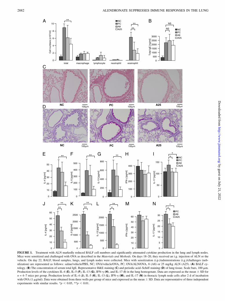
FIGURE 1. Treatment with ALN markedly reduced BALF cell numbers and significantly attenuated cytokine production in the lung and lymph nodes.
Mice were sensitized and challenged with OVA as described in the Materials and Methods. On days 18–20, they received an i.g. injection of ALN or the
vehicle. On day 22, BALF, blood samples, lungs, and lymph nodes were collected. Mice with sensitization (i.p.)/administrations (i.g.)/challenges (neb-
ulization) are represented as follows: saline/vehicle/PBS, NC; OVA/vehicle/OVA, PC; OVA/ALN/OVA, 8 (A8) or 25 mg/kg ALN (A25). (A) BALF cy-
tology. (B) The concentration of serum total IgE. Representative H&E staining (C) and periodic acid–Schiff staining (D) of lung tissue. Scale bars, 100 mm.
Production levels of the cytokines IL-4 (E), IL-5 (F), IL-13 (G), IFN-g (H), and IL-17 (I) in the lung homogenate. Data are expressed as the mean 6 SD for
n = 4–7 mice per group. Production levels of IL-4 (J), IL-5 (K), IL-13 (L), IFN-g (M), and IL-17 (N) in thoracic lymph node cells after 2 d of incubation
with OVA (1 mg/ml). Data were obtained from three wells per group of mice and expressed as the mean6 SD. Data are representative of three independent
experiments with similar results. *p , 0.05, **p , 0.01.
2882 ALENDRONATE SUPPRESSES IMMUNE RESPONSES IN THE LUNG
by guest on July 21, 2022http://w
ww
.jimm
unol.org/D
ownloaded from
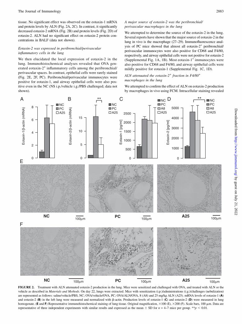
tissue. No significant effect was observed on the eotaxin-1 mRNAand protein levels by ALN (Fig. 2A, 2C). In contrast, it significantlydecreased eotaxin-2 mRNA (Fig. 2B) and protein levels (Fig. 2D) ofeotaxin-2. ALN had no significant effect on eotaxin-2 protein con-centrations in BALF (data not shown).
Eotaxin-2 was expressed in peribronchial/perivascularinflammatory cells in the lung
We then elucidated the local expression of eotaxin-2 in thelung. Immunohistochemical analyses revealed that OVA gen-erated eotaxin-2+ inflammatory cells among the peribronchial/perivascular spaces. In contrast, epithelial cells were rarely stained(Fig. 2E, 2F, PC). Peribronchial/perivascular immunocytes werepositive for eotaxin-1, and airway epithelial cells were also pos-itive even in the NC (NS i.p./vehicle i.g./PBS challenged; data notshown).
A major source of eotaxin-2 was the peribronchial/perivascular macrophages in the lung
We attempted to determine the source of the eotaxin-2 in the lung.Several reports have shown that the major source of eotaxin-2 in thelung in vivo is the macrophage (27–29). Immunofluorescence anal-ysis of PC mice showed that almost all eotaxin-2+ peribronchial/perivascular immunocytes were also positive for CD68 and F4/80,respectively, and airway epithelial cells were not positive for eotaxin-2(Supplemental Fig. 1A, 1B). Most eotaxin-1+ immunocytes werealso positive for CD68 and F4/80, and airway epithelial cells weremildly positive for eotaxin-1 (Supplemental Fig. 1C, 1D).
ALN attenuated the eotaxin-2+ fraction in F4/80+
macrophages in the lung
We attempted to confirm the effect of ALN on eotaxin-2 productionby macrophages in vivo using FCM. Intracellular staining revealed
FIGURE 2. Treatment with ALN attenuated eotaxin-2 production in the lung. Mice were sensitized and challenged with OVA, and treated with ALN or the
vehicle as described in Materials and Methods. On day 22, lungs were extracted. Mice with sensitization (i.p.)/administrations (i.g.)/challenges (nebulization)
are represented as follows: saline/vehicle/PBS, NC; OVA/vehicle/OVA, PC; OVA/ALN/OVA, 8 (A8) and 25 mg/kg ALN (A25). mRNA levels of eotaxin-1 (A)
and eotaxin-2 (B) in the left lung were measured and normalized with b-actin. Production levels of eotaxin-1 (C) and eotaxin-2 (D) were measured in lung
homogenate. (E and F) Representative immunohistochemical staining of lung tissue. Original magnification,3100 (E),3200 (F). Scale bars, 100 mm. Data are
representative of three independent experiments with similar results and expressed as the mean 6 SD for n = 4–7 mice per group. **p , 0.01.
The Journal of Immunology 2883
by guest on July 21, 2022http://w
ww
.jimm
unol.org/D
ownloaded from
that ALN significantly attenuated the eotaxin-2+ fraction in F4/80+
macrophages in the lung (Fig. 3C, 3E, 3F). ALN did not have asignificant effect on the eotaxin-2+ fraction in BALF or on theeotaxin-1+ fraction in the lung (Fig. 3B, 3D–F).
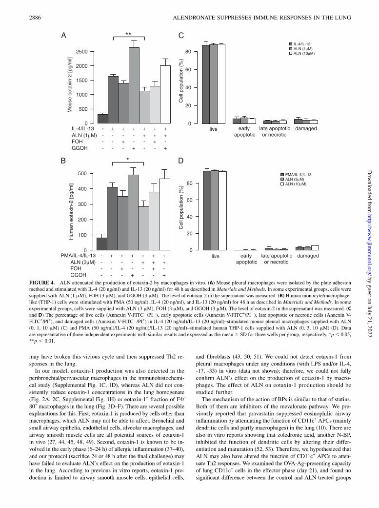
ALN attenuated the production of eotaxin-2 by macrophagesin vitro
We further investigated whether ALN affected eotaxin-2 produc-tion by macrophages in vitro. Pleural macrophages were selectedby the plate adhesion method and stimulated by IL-4 and IL-13for 48 h. Because N-BPs mainly act by inhibiting farnesyl pyro-phosphate synthesis in the mevalonate pathway, the intermediatestrans,trans-FOH or all-trans-GGOH were also added to determinewhether ALN’s effect may be rescued. An ELISA of the super-natants revealed that ALN significantly reduced eotaxin-2 pro-duction and this was reversed by GGOH (Fig. 4A), which indicatedthat ALN acted on farnesyl pyrophosphate synthase. In this in vitroprotocol, we evaluated the percentage of live cells and apoptoticcells by staining with Annexin V and PI; however, ALN did notcause significant cell death or cell damage up to 10 mM (Fig. 4C).We also examined eotaxin-1 production with the same protocol, andeotaxin-1 was not detected even in the PC wells (data not shown).
ALN attenuated the production of eotaxin-2 by THP-1,a human monocyte/macrophage-like cell line, in vitro
The human monocyte/macrophage-like cell line, THP-1, was re-ported to produce eotaxin-2 when stimulated with PMA, IL-4, andIL-13 in vitro (24, 25). Using this cell line, we attempted to confirmwhether ALN could also attenuate human macrophage function.ALN significantly attenuated the production of eotaxin-2 from THP-1 cells, and this effect was reversed by GGOH (Fig. 4B). Cell vi-ability was not significantly affected up to 10 mM of ALN (Fig. 4D).
ALN did not alter AHR in the lung
We evaluated the effect of ALN on AHR in vivo using the enhancedpause (Penh) system. Treatment with ALN on days 18–20 did nothave a significant effect on AHR (Supplemental Fig. 2A), althougha slight suppressive tendency was observed.
ALN did not alter Ag presentation in the lung
We examined the effect of ALN on Ag presentation in vivo. Treat-ment with ALN during the effector phase appeared to have no signi-ficant effect on the Ag-presenting capacity of MACS-isolated CD11c+
cells in the lung (Supplemental Fig. 2B, 2C).
ALN did not exhibit suppressive effects on Th2-mediatedresponses in ex vivo and in vitro experiments with spleen cells
We conducted in vitro and ex vivo experiments with OVA-stim-ulated spleen cells, as described in Materials and Methods. Nosignificant effect was observed by ALN on spleen cells either invitro (Supplemental Fig. 2D–G) or ex vivo (Supplemental Fig.2H–K).
The suppressive effects of ALN were weaker when it wasadministered in the sensitization phase (days 0–17) than in theeffector phase (days 18–20) in vivo
We also evaluated ALN’s effect on the sensitization phase in vivo.In this protocol, ALN was administered throughout the sensiti-zation phase (ALN, days 0–17) or the effector phase (ALN, days18–20), and mice were sacrificed on day 21. No significant effectwas observed on the number of eosinophils in BALF or serumtotal IgE levels in the ALN (days 0–17) group (Supplemental Fig.3A, 3B). The ALN (days 0–17) group showed a moderate but lesssuppressive effect than the ALN (days 18–20) group on the pro-
duction of Th2 cytokines in the lung and OVA-stimulated lymphnode cells (Supplemental Fig. 3C–G, 3J–N). ALN (days 0–17)suppressed eotaxin-1 production, but not eotaxin-2 production, inthe lung homogenate (Supplemental Fig. 3H, 3I).
ALN did not change the cell populations or viability of BALFobtained from mice without sensitization or challenge in vivo
To evaluate ALN’s baseline effect, we analyzed the cytology andviability of BALF cells obtained from mice without sensitizationor challenge. Mice were administered with the vehicle (CMC) or25 mg/kg ALN on days 0–2, and BALF was collected on day 3.The BALF cell population was evaluated with Diff-Quick stain-ing, and the cell viability was evaluated with Annexin V and PIstaining followed by FCM. ALN did not have any significant ef-fects on the cell populations or viability of BALF cells (Supple-mental Fig. 3O, 3P).
DiscussionThe results of this study clearly demonstrated that intragastric de-livery of ALN suppressed Th2-mediated allergic immune responsesin the airway. ALN, administered during the effector phase, sup-pressed eosinophilic airway inflammation and the production of Th2cytokines both in the lung and regional lymph nodes. The productionof IL-17 was also reduced in the lung and regional lymph nodes. Inaddition, ALN significantly suppressed eotaxin-2 production in thelung, the major source of which was the peribronchial/perivascularmacrophages, as confirmed by immunohistochemical examinationsand FCM. ALN also reduced the production of eotaxin-2 frommouse pleural macrophages and human monocytic/macrophagecells (THP-1). Few studies have examined the effect of BP onasthma or asthma-related cells. To the best of our knowledge, oursis the first study to demonstrate the suppressive effect of BP on Ag-induced allergic airway inflammation in vivo.In this study, ALN clearly suppressed Th2 responses in the both
lung and regional lymph nodes (Fig. 1E–N). This effect is consid-ered to be indirect, as BPs mainly distribute to the phagocytes andnot to lymphocytes because of their poor membrane permeability(30–33). ALN did not have systemic suppressive effects in eitherthe in vitro or ex vivo study with spleen cells (Supplemental Fig. 1D–K). Furthermore, we could not confirm a definite effect on CD11c+
APCs in the lung in vivo (Supplemental Fig. 1B, 1C). We cannotcurrently explain the mechanism by which ALN suppressed T cellresponses. Because ALN is selectively incorporated by phagocytes,we assume that the direct effector cell was mainly the macrophage.ALN may have altered some functions of the macrophage, includingthe Ag-presenting capacity and IL-33/ST2 axis, which led to thesuppression of T lymphocytes. However, there remains the possibilityof ALN’s effect on various immunocytes such as lung epithelial cellsand mast cells. Another explanation may be that ALN suppressedthe amplification of Th2 cell-mediated allergic immune responsescaused by eotaxin-2.In this study, ALN strongly suppressed the transcription and
protein production of eotaxin-2 in the lung (Fig. 2B, 2D). Theeotaxin/CCR3 axis is critical in allergic airway inflammation (27,34), although there is a discrepancy in the degree of its contribu-tion (35, 36). In several allergic models, eotaxin-1 was shown to beinvolved in the early phase (6–24 h) (37–40), whereas eotaxin-2was involved in the late phase (24 h or more) (38, 39), and thusplays a dominant role in chronic allergic airway inflammation (27,39). Eotaxin-2 is generally produced by airway epithelial cells andmacrophages upon stimulation with IL-4 or IL-13 in vitro (41–43), orafter an allergen challenge in vivo (44–46). It promotes AHR and IL-13 production in the airway in cooperation with IL-5 (47), resultingin a vicious cycle that amplifies allergic airway inflammation. ALN
2884 ALENDRONATE SUPPRESSES IMMUNE RESPONSES IN THE LUNG
by guest on July 21, 2022http://w
ww
.jimm
unol.org/D
ownloaded from

FIGURE 3. Treatment with ALN reduced the intracellular eotaxin-2+ fraction in F4/80+ macrophages. Mice were sensitized and challenged with OVA
and treated with ALN as described in Materials and Methods. On day 21, BALF and lungs were collected. BALF and digested lungs were stained with F4/
80-biotin-streptavidin-PE/Cy5 and with eotaxin-1–Alexa 488 or eotaxin-2–Alexa 488 after permeabilization. Mice with sensitization (i.p.)/administrations
(i.g.)/challenges (nebulization) are represented as follows: saline/vehicle/PBS, NC; OVA/vehicle/OVA, PC; OVA/ALN/OVA, 25 mg/kg ALN (A25). (A)
Representative flow cytometric isolation of intracellular eotaxin-2+ macrophages determined by F4/80. (B–D) Representative contour plots demonstrating
gating quadrants of intracellular eotaxin-1/eotaxin-2+ F4/80+ macrophages of BALF (B, eotaxin-2) and the lung (C, eotaxin-2; D, eotaxin-1). Percentages in
brackets represent the double-positive ratio to F4/80+ macrophages in the R1 region. (E and F) The cell populations of intracellular eotaxin-1/eotaxin-2+ F4/
80+ macrophages, the ratio to whole cells in the R1 region (E), and the ratio to F4/80+ macrophages in the R1 region (F). Data are representative of three
independent experiments with similar results and are expressed as the mean 6 SD for n = 2–4 mice per group. *p , 0.05.
The Journal of Immunology 2885
by guest on July 21, 2022http://w
ww
.jimm
unol.org/D
ownloaded from
may have broken this vicious cycle and then suppressed Th2 re-sponses in the lung.In our model, eotaxin-1 production was also detected in the
peribronchial/perivascular macrophages in the immunohistochemi-cal study (Supplemental Fig. 1C, 1D), whereas ALN did not con-sistently reduce eotaxin-1 concentrations in the lung homogenate(Fig. 2A, 2C, Supplemental Fig. 1H) or eotaxin-1+ fraction of F4/80+ macrophages in the lung (Fig. 3D–F). There are several possibleexplanations for this. First, eotaxin-1 is produced by cells other thanmacrophages, which ALN may not be able to affect. Bronchial andsmall airway epithelia, endothelial cells, alveolar macrophages, andairway smooth muscle cells are all potential sources of eotaxin-1in vivo (27, 44, 45, 48, 49). Second, eotaxin-1 is known to be in-volved in the early phase (6–24 h) of allergic inflammation (37–40),and our protocol (sacrifice 24 or 48 h after the final challenge) mayhave failed to evaluate ALN’s effect on the production of eotaxin-1in the lung. According to previous in vitro reports, eotaxin-1 pro-duction is limited to airway smooth muscle cells, epithelial cells,
and fibroblasts (43, 50, 51). We could not detect eotaxin-1 frompleural macrophages under any conditions (with LPS and/or IL-4,-17, -33) in vitro (data not shown); therefore, we could not fullyconfirm ALN’s effect on the production of eotaxin-1 by macro-phages. The effect of ALN on eotaxin-1 production should bestudied further.The mechanism of the action of BPs is similar to that of statins.
Both of them are inhibitors of the mevalonate pathway. We pre-viously reported that pravastatin suppressed eosinophilic airwayinflammation by attenuating the function of CD11c+ APCs (mainlydendritic cells and partly macrophages) in the lung (10). There arealso in vitro reports showing that zoledronic acid, another N-BP,inhibited the function of dendritic cells by altering their differ-entiation and maturation (52, 53). Therefore, we hypothesized thatALN may also have altered the function of CD11c+ APCs to atten-uate Th2 responses. We examined the OVA-Ag–presenting capacityof lung CD11c+ cells in the effector phase (day 21), and found nosignificant difference between the control and ALN-treated groups
FIGURE 4. ALN attenuated the production of eotaxin-2 by macrophages in vitro. (A) Mouse pleural macrophages were isolated by the plate adhesion
method and stimulated with IL-4 (20 ng/ml) and IL-13 (20 ng/ml) for 48 h as described in Materials and Methods. In some experimental groups, cells were
supplied with ALN (1 mM), FOH (3 mM), and GGOH (3 mM). The level of eotaxin-2 in the supernatant was measured. (B) Human monocyte/macrophage-
like (THP-1) cells were stimulated with PMA (50 ng/ml), IL-4 (20 ng/ml), and IL-13 (20 ng/ml) for 48 h as described in Materials and Methods. In some
experimental groups, cells were supplied with ALN (3 mM), FOH (3 mM), and GGOH (3 mM). The level of eotaxin-2 in the supernatant was measured. (C
and D) The percentage of live cells (Annexin V-FITC2/PI2), early apoptotic cells (Annexin V-FITC+/PI2), late apoptotic or necrotic cells (Annexin V-
FITC+/PI+), and damaged cells (Annexin V-FITC2/PI+) in IL-4 (20 ng/ml)/IL-13 (20 ng/ml)–stimulated mouse pleural macrophages supplied with ALN
(0, 1, 10 mM) (C) and PMA (50 ng/ml)/IL-4 (20 ng/ml)/IL-13 (20 ng/ml)–stimulated human THP-1 cells supplied with ALN (0, 3, 10 mM) (D). Data
are representative of three independent experiments with similar results and expressed as the mean 6 SD for three wells per group, respectively. *p, 0.05,
**p , 0.01.
2886 ALENDRONATE SUPPRESSES IMMUNE RESPONSES IN THE LUNG
by guest on July 21, 2022http://w
ww
.jimm
unol.org/D
ownloaded from
(Supplemental Fig. 2B, 2C). We also conducted a quantitative PCRanalysis on in vivo lung mRNA for CCL17 (TARC), a potent che-moattractant for Th2 cells produced mainly by dendritic cells (29,54–56); however, ALN did not have a significant effect on its tran-scription (data not shown). Therefore, we consider the contributionof dendritic cell suppression in this study to be weak.There are limited numbers of reports regarding ALN’s effect on
the immune system both in vitro and in animal models in vivo.Sansoni et al. (57) reported that ALN inhibited the Ag-presentingcapabilities and LPS-induced generation of IL-1b, IL-6, and TNF-ain activated human monocytes in vitro. Jiang et al. (58) reportedthat ALN inhibited b2-adrenergic receptor internalization and thedownregulation of human airway smooth muscle cells in vitro.ALN’s effects are controversial in in vivo studies. Deng et al. (59)reported that an i.p. injection of 40 mmol/kg (13 mg/kg) ALNinduced IL-1–dependent inflammatory reactions in the spleen, lung,and liver. In contrast, the oral administration of 25 or 75 mg/kgALN, a similar dosage to that used in this study (8 or 25 mg/kg),decreased colonic damage in a model of inflammatory bowel dis-ease in rats (60). Differences in the dosage as well as the route ofadministration may influence whether ALN reveals a proinflammatoryor anti-inflammatory effect.A critical factor influencing the effect of ALN on immune
responses is tissue distribution. When administered, BPs are gen-erally distributed to the bones and remain there for months to years.Their distribution in the respiratory system has seldom beenstudied. Only two reports have examined this issue (11–13). Inthese reports, ALN was distributed to the trachea even 72 h aftermultiple oral administrations (5 mg/kg, 7 d) or a single i.v. ad-ministration (0.05 mg/kg) in the rat. In contrast, its distribution toother organs such as the liver, spleen, kidney, and lymph nodeswas very low. This high affinity of ALN for bronchial tissue maycontribute to a suppressive effect specifically in the lung and couldexplain why ALN failed to suppress Th2-mediated systemic im-mune responses to OVA in ex vivo and in vitro experiments withspleen cells (Supplemental Fig. 1D–K). ALN moderately sup-pressed Th2 cytokine production in the lung and lymph nodeswhen administered during the sensitization phase (days 0–17)in vivo (Supplemental Fig. 3). This may have been caused by themaintenance of ALN in the trachea 72 h after the last adminis-tration until the effector phase (days 18–20).In this study, we adopted an intragastric route of administration
for the following reasons: 1) ALN is usually administered orally inclinical use, 2) ALN was reported to distribute to trachea after oraladministration (11), and 3) oral administration of ALN was re-ported to attenuate colonic damage in a model of inflammatorybowel disease (60). We mainly used the dosage of 25 mg/kg be-cause 25 or 75 mg/kg of oral administration of ALN was reportedto decrease bowel inflammation in a rat model (60), and because25 mg/kg oral ALN appeared to be more effective than 8 mg/kg inour model (Fig. 1). The mean bioavailability of ALN after oralintake was 0.64% relative to its i.v. equivalent (61). Therefore, therelatively low dose of ALN in the bronchus in this study, deliveredby an intragastric tube, may have demonstrated an anti-inflammatoryeffect in the lung. The intratracheal administration of ALN may bea potential alternative to decrease the dose required for the immuno-suppressive effect on allergic airway inflammation.Regarding its clinical use, ALN has been administered at 0.2
mg/kg (10-mg tablet) daily or 1.4 mg/kg (70-mg tablet) weekly,and a single administration of 70 mg achieved maximal plasmaticconcentrations as 82.37–110.71 ng/ml, relevant to 0.25–0.34 mM(62). After the single or repeated (7 d) oral administration of 5 mg/kg to rats, the concentration of ALN in the trachea was shown tobe ∼4 or 20 times higher than maximal plasmatic concentrations,
respectively (11). Therefore, the in vitro ALN concentrations usedin this study (1–3 mM) do not appear to differ from those achievablein the clinical use. However, the in vivo dose used in this study (25mg/kg 3 d) was 125 times higher than that used clinically, and bloodconcentration data at this dosage were not reported in any species.BPs are known to induce apoptosis in macrophages at high
concentrations. The i.v. administration of liposomal clodronate isa conventional method of depleting macrophages (63). ALN wasshown to induce apoptosis in mouse macrophage-like J774.1 cellsat a very high concentration (100 mM) only in vitro (64), which is.30 times higher than that which inhibited the production ofeotaxin-2 in mouse pleural macrophages (1 mM) or the humanmonocytic cell line (THP-1; 3 mM) in this study (Fig. 4A, 4B).Furthermore, FCM evaluation with Annexin V and PI revealed nosignificant difference in the viability and apoptosis of mouse mac-rophages or human THP-1 cells after incubation with up to 10 mMALN for 48 h from the PC in vitro (Fig. 4C, 4D). BALF cells (mainlymacrophages) of nonsensitized and nonchallenged mice adminis-tered the vehicle (CMC) or 25 mg/kg ALN exhibited no significantdifference in cytology or viability in vivo (Supplemental Fig. 3O,3P). Therefore, we consider that ALN exhibited a suppressive effectwithout inducing cell death or cell damage in macrophages.In summary, the oral administration of ALN during airway
inflammation effectively suppressed Ag-induced airway responsesin a mouse model. ALN attenuated the production of eotaxin-2from lung macrophages, which may contribute to suppress allergicairway inflammation in vivo. The mechanism by which ALNsuppressed Th2 and Th17 responses has not been fully clarified.Our results with human monocyte/macrophage-like (THP-1) cellsappear to support ALN’s anti-inflammatory effect on humans.Therefore, ALN could be a novel therapeutic option for asthma.
AcknowledgmentsWe thank Kanae Kurosaki for technical assistance and Akihiro Hisaka for
helpful discussions.
DisclosuresThe authors have no financial conflicts of interest.
References1. Greenwood, J., L. Steinman, and S. S. Zamvil. 2006. Statin therapy and auto-
immune disease: from protein prenylation to immunomodulation. Nat. Rev.Immunol. 6: 358–370.
2. Youssef, S., O. Stuve, J. C. Patarroyo, P. J. Ruiz, J. L. Radosevich, E. M. Hur,M. Bravo, D. J. Mitchell, R. A. Sobel, L. Steinman, and S. S. Zamvil. 2002. TheHMG-CoA reductase inhibitor, atorvastatin, promotes a Th2 bias and reversesparalysis in central nervous system autoimmune disease. Nature 420: 78–84.
3. Hakamada-Taguchi, R., Y. Uehara, K. Kuribayashi, A. Numabe, K. Saito,H. Negoro, T. Fujita, T. Toyo-oka, and T. Kato. 2003. Inhibition of hydroxy-methylglutaryl-coenzyme a reductase reduces Th1 development and promotes Th2development. Circ. Res. 93: 948–956.
4. Dunn, S. E., S. Youssef, M. J. Goldstein, T. Prod’homme, M. S. Weber,S. S. Zamvil, and L. Steinman. 2006. Isoprenoids determine Th1/Th2 fate inpathogenic T cells, providing a mechanism of modulation of autoimmunity byatorvastatin. J. Exp. Med. 203: 401–412.
5. Leung, B. P., N. Sattar, A. Crilly, M. Prach, D. W. McCarey, H. Payne,R. Madhok, C. Campbell, J. A. Gracie, F. Y. Liew, and I. B. McInnes. 2003. Anovel anti-inflammatory role for simvastatin in inflammatory arthritis. J. Immunol.170: 1524–1530.
6. Lin, H., Y. Xiao, G. Chen, D. Fu, Y. Ye, L. Liang, J. Fan, X. Yang, L. Sun, andH. Xu. 2011. HMG-CoA reductase inhibitor simvastatin suppresses Toll-likereceptor 2 ligand-induced activation of nuclear factor kappa B by preventingRhoA activation in monocytes from rheumatoid arthritis patients. Rheumatol.Int. 31: 1451–1458.
7. McKay, A., B. P. Leung, I. B. McInnes, N. C. Thomson, and F. Y. Liew. 2004. Anovel anti-inflammatory role of simvastatin in a murine model of allergicasthma. J. Immunol. 172: 2903–2908.
8. Kim, D. Y., S. Y. Ryu, J. E. Lim, Y. S. Lee, and J. Y. Ro. 2007. Anti-inflammatory mechanism of simvastatin in mouse allergic asthma model. Eur.J. Pharmacol. 557: 76–86.
The Journal of Immunology 2887
by guest on July 21, 2022http://w
ww
.jimm
unol.org/D
ownloaded from
9. Yeh, Y. F., and S. L. Huang. 2004. Enhancing effect of dietary cholesterol andinhibitory effect of pravastatin on allergic pulmonary inflammation. J. Biomed.Sci. 11: 599–606.
10. Imamura, M., K. Okunishi, H. Ohtsu, K. Nakagome, H. Harada, R. Tanaka,K. Yamamoto, and M. Dohi. 2009. Pravastatin attenuates allergic airway in-flammation by suppressing antigen sensitisation, interleukin 17 production andantigen presentation in the lung. Thorax 64: 44–49.
11. Hisaka, A., N. Takenaga, K. Hara, T. Kamei, Y. Esumi, Y. Karasawa, H. Seki,K. Ichige, and T. Sakuma. 1998. Absorption, distribution, excretion, and sexdifferences in the disposition of the antiosteolytic agent, alendronate after oraladministration in rats. Drug Metab. Pharmacokinet. 13: 252–258.
12. Mochizuki, T., S. Nishimura, K. Okabe, Y. Azuma, S. Kudoh, S. Ishii, T. Ohta,S. Kondo, and M. Kiyoki. 1995. Metabolic fate of 4-amino-1-hydroxybutylidene-1,1-bisphosphonate (alendronate) (I): plasma concentration, distribution and excre-tion after intravenous administration of alendronate to rats. Drug Metab. Phar-macokinet. 10: 161–173.
13. Mochizuki, T., S. Nishimura, K. Okabe, Y. Azuma, A. Umeda, S. Kudoh,N. Harakawa, A. Ishii, T. Ohta, S. Kondo, and M. Kiyoki. 1995. Metabolic fateof 4-amino-1-hydroxybutylidene-1, 1-bisphosphonate (alendronate) (II): plasmaconcentration, distribution and excretion after repeated intravenous administration to7-week old rats and after single intravenous administration to 30-week old rats, andtransfer into the fetus and milk in rats. Drug Metab. Pharmacokinet. 10: 174–189.
14. Okunishi, K., M. Dohi, K. Nakagome, R. Tanaka, and K. Yamamoto. 2004. Anovel role of cysteinyl leukotrienes to promote dendritic cell activation in theantigen-induced immune responses in the lung. J. Immunol. 173: 6393–6402.
15. Dohi, M., S. Tsukamoto, T. Nagahori, K. Shinagawa, K. Saitoh, Y. Tanaka,S. Kobayashi, R. Tanaka, Y. To, and K. Yamamoto. 1999. Noninvasive systemfor evaluating the allergen-specific airway response in a murine model ofasthma. Lab. Invest. 79: 1559–1571.
16. To, Y., M. Dohi, R. Tanaka, A. Sato, K. Nakagome, and K. Yamamoto. 2001.Early interleukin 4-dependent response can induce airway hyperreactivity beforedevelopment of airway inflammation in a mouse model of asthma. Lab. Invest.81: 1385–1396.
17. Vermaelen, K. Y., I. Carro-Muino, B. N. Lambrecht, and R. A. Pauwels. 2001.Specific migratory dendritic cells rapidly transport antigen from the airways tothe thoracic lymph nodes. J. Exp. Med. 193: 51–60.
18. Nakagome, K., M. Dohi, K. Okunishi, Y. Komagata, K. Nagatani, R. Tanaka,J. Miyazaki, and K. Yamamoto. 2005. In vivo IL-10 gene delivery suppressesairway eosinophilia and hyperreactivity by down-regulating APC functions andmigration without impairing the antigen-specific systemic immune response ina mouse model of allergic airway inflammation. J. Immunol. 174: 6955–6966.
19. Nakagome, K., K. Okunishi, M. Imamura, H. Harada, T. Matsumoto, R. Tanaka,J. Miyazaki, K. Yamamoto, and M. Dohi. 2009. IFN-gamma attenuates antigen-induced overall immune response in the airway as a Th1-type immune regulatorycytokine. J. Immunol. 183: 209–220.
20. Okunishi, K., M. Dohi, K. Nakagome, R. Tanaka, S. Mizuno, K. Matsumoto,J. Miyazaki, T. Nakamura, and K. Yamamoto. 2005. A novel role of hepatocytegrowth factor as an immune regulator through suppressing dendritic cell func-tion. J. Immunol. 175: 4745–4753.
21. Nakagome, K., M. Imamura, H. Okada, K. Kawahata, T. Inoue, K. Hashimoto,H. Harada, T. Higashi, R. Takagi, K. Nakano, et al. 2011. Dopamine D1-likereceptor antagonist attenuates Th17-mediated immune response and ovalbuminantigen-induced neutrophilic airway inflammation. J. Immunol. 186: 5975–5982.
22. Cuzzocrea, S., E. Mazzon, G. Calabro, L. Dugo, A. De Sarro, F. A. van De LOO,and A. P. Caputi. 2000. Inducible nitric oxide synthase-knockout mice exhibitresistance to pleurisy and lung injury caused by carrageenan. Am. J. Respir. Crit.Care Med. 162: 1859–1866.
23. Zhang, X., R. Goncalves, and D. M. Mosser. 2008. The isolation and charac-terization of murine macrophages. Curr. Protoc. Immunol. Chapter 14: Unit 14.1.
24. Tjiu, J. W., J. S. Chen, C. T. Shun, S. J. Lin, Y. H. Liao, C. Y. Chu, T. F. Tsai,H. C. Chiu, Y. S. Dai, H. Inoue, et al. 2009. Tumor-associated macrophage-induced invasion and angiogenesis of human basal cell carcinoma cells bycyclooxygenase-2 induction. J. Invest. Dermatol. 129: 1016–1025.
25. Levano, K. S., E. H. Jung, and P. A. Kenny. 2011. Breast cancer subtypes expressdistinct receptor repertoires for tumor-associated macrophage derived cytokines.Biochem. Biophys. Res. Commun. 411: 107–110.
26. Checkley, W., K. P. West, Jr., R. A. Wise, M. R. Baldwin, L. Wu, S. C. LeClerq,P. Christian, J. Katz, J. M. Tielsch, S. Khatry, and A. Sommer. 2010. Maternalvitamin A supplementation and lung function in offspring. N. Engl. J. Med. 362:1784–1794.
27. Pope, S. M., N. Zimmermann, K. F. Stringer, M. L. Karow, and M. E. Rothenberg.2005. The eotaxin chemokines and CCR3 are fundamental regulators of allergen-induced pulmonary eosinophilia. J. Immunol. 175: 5341–5350.
28. Kurowska-Stolarska, M., B. Stolarski, P. Kewin, G. Murphy, C. J. Corrigan,S. Ying, N. Pitman, A. Mirchandani, B. Rana, N. van Rooijen, et al. 2009. IL-33amplifies the polarization of alternatively activated macrophages that contributeto airway inflammation. J. Immunol. 183: 6469–6477.
29. Crapster-Pregont, M., J. Yeo, R. L. Sanchez, and D. A. Kuperman. 2012.Dendritic cells and alveolar macrophages mediate IL-13-induced airway in-flammation and chemokine production. J. Allergy Clin. Immunol. 129: 1621–1627.e3.
30. Fleisch, H. 1998. Bisphosphonates: mechanisms of action. Endocr. Rev. 19: 80–100.
31. Cecchini, M. G., R. Felix, H. Fleisch, and P. H. Cooper. 1987. Effect ofbisphosphonates on proliferation and viability of mouse bone marrow-derivedmacrophages. J. Bone Miner. Res. 2: 135–142.
32. Cecchini, M. G., and H. Fleisch. 1990. Bisphosphonates in vitro specificallyinhibit, among the hematopoietic series, the development of the mouse mono-nuclear phagocyte lineage. J. Bone Miner. Res. 5: 1019–1027.
33. Rogers, M. J., X. Xiong, X. Ji, J. Monkkonen, R. G. Russell, M. P. Williamson,F. H. Ebetino, and D. J. Watts. 1997. Inhibition of growth of Dictyosteliumdiscoideum amoebae by bisphosphonate drugs is dependent on cellular uptake.Pharm. Res. 14: 625–630.
34. Fulkerson, P. C., C. A. Fischetti, M. L. McBride, L. M. Hassman, S. P. Hogan,and M. E. Rothenberg. 2006. A central regulatory role for eosinophils and theeotaxin/CCR3 axis in chronic experimental allergic airway inflammation. Proc.Natl. Acad. Sci. USA 103: 16418–16423.
35. Humbles, A. A., B. Lu, D. S. Friend, S. Okinaga, J. Lora, A. Al-Garawi,T. R. Martin, N. P. Gerard, and C. Gerard. 2002. The murine CCR3 receptorregulates both the role of eosinophils and mast cells in allergen-induced airwayinflammation and hyperresponsiveness. Proc. Natl. Acad. Sci. USA 99: 1479–1484.
36. Voehringer, D., N. van Rooijen, and R. M. Locksley. 2007. Eosinophils developin distinct stages and are recruited to peripheral sites by alternatively activatedmacrophages. J. Leukoc. Biol. 81: 1434–1444.
37. Conroy, D. M., and T. J. Williams. 2001. Eotaxin and the attraction of eosino-phils to the asthmatic lung. Respir. Res. 2: 150–156.
38. Menzies-Gow, A., S. Ying, I. Sabroe, V. L. Stubbs, D. Soler, T. J. Williams, andA. B. Kay. 2002. Eotaxin (CCL11) and eotaxin-2 (CCL24) induce recruitment ofeosinophils, basophils, neutrophils, and macrophages as well as features of early-and late-phase allergic reactions following cutaneous injection in human atopicand nonatopic volunteers. J. Immunol. 169: 2712–2718.
39. Ben-Yehuda, C., R. Bader, I. Puxeddu, F. Levi-Schaffer, R. Breuer, andN. Berkman. 2008. Airway eosinophil accumulation and eotaxin-2/CCL24 ex-pression following allergen challenge in BALB/c mice. Exp. Lung Res. 34: 467–479.
40. Rothenberg, M. E., J. A. MacLean, E. Pearlman, A. D. Luster, and P. Leder.1997. Targeted disruption of the chemokine eotaxin partially reduces antigen-induced tissue eosinophilia. J. Exp. Med. 185: 785–790.
41. van Wetering, S., S. Zuyderduyn, D. K. Ninaber, M. A. van Sterkenburg,K. F. Rabe, and P. S. Hiemstra. 2007. Epithelial differentiation is a determinantin the production of eotaxin-2 and -3 by bronchial epithelial cells in response toIL-4 and IL-13. Mol. Immunol. 44: 803–811.
42. Pope, S. M., P. C. Fulkerson, C. Blanchard, H. S. Akei, N. M. Nikolaidis,N. Zimmermann, J. D. Molkentin, and M. E. Rothenberg. 2005. Identification ofa cooperative mechanism involving interleukin-13 and eotaxin-2 in experimentalallergic lung inflammation. J. Biol. Chem. 280: 13952–13961.
43. Watanabe, K., P. J. Jose, and S. M. Rankin. 2002. Eotaxin-2 generation is dif-ferentially regulated by lipopolysaccharide and IL-4 in monocytes and macro-phages. J. Immunol. 168: 1911–1918.
44. Ying, S., D. S. Robinson, Q. Meng, J. Rottman, R. Kennedy, D. J. Ringler,C. R. Mackay, B. L. Daugherty, M. S. Springer, S. R. Durham, et al. 1997.Enhanced expression of eotaxin and CCR3 mRNA and protein in atopic asthma.Association with airway hyperresponsiveness and predominant co-localizationof eotaxin mRNA to bronchial epithelial and endothelial cells. Eur. J. Immunol.27: 3507–3516.
45. Ying, S., Q. Meng, K. Zeibecoglou, D. S. Robinson, A. Macfarlane, M. Humbert,and A. B. Kay. 1999. Eosinophil chemotactic chemokines (eotaxin, eotaxin-2,RANTES, monocyte chemoattractant protein-3 (MCP-3), and MCP-4), and C-Cchemokine receptor 3 expression in bronchial biopsies from atopic and non-atopic (Intrinsic) asthmatics. J. Immunol. 163: 6321–6329.
46. Ravensberg, A. J., F. L. Ricciardolo, A. van Schadewijk, K. F. Rabe, P. J. Sterk,P. S. Hiemstra, and T. Mauad. 2005. Eotaxin-2 and eotaxin-3 expression is as-sociated with persistent eosinophilic bronchial inflammation in patients withasthma after allergen challenge. J. Allergy Clin. Immunol. 115: 779–785.
47. Yang, M., S. P. Hogan, S. Mahalingam, S. M. Pope, N. Zimmermann,P. Fulkerson, L. A. Dent, I. G. Young, K. I. Matthaei, M. E. Rothenberg, andP. S. Foster. 2003. Eotaxin-2 and IL-5 cooperate in the lung to regulate IL-13production and airway eosinophilia and hyperreactivity. J. Allergy Clin. Immu-nol. 112: 935–943.
48. Humbles, A. A., D. M. Conroy, S. Marleau, S. M. Rankin, R. T. Palframan,A. E. Proudfoot, T. N. Wells, D. Li, P. K. Jeffery, D. A. Griffiths-Johnson, et al.1997. Kinetics of eotaxin generation and its relationship to eosinophil accu-mulation in allergic airways disease: analysis in a guinea pig model in vivo. J.Exp. Med. 186: 601–612.
49. Nagarkar, D. R., E. R. Bowman, D. Schneider, Q. Wang, J. Shim, Y. Zhao,M. J. Linn, C. L. McHenry, B. Gosangi, J. K. Bentley, et al. 2010. Rhinovirusinfection of allergen-sensitized and -challenged mice induces eotaxin releasefrom functionally polarized macrophages. J. Immunol. 185: 2525–2535.
50. Rahman, M. S., A. Yamasaki, J. Yang, L. Shan, A. J. Halayko, and A. S. Gounni.2006. IL-17A induces eotaxin-1/CC chemokine ligand 11 expression in humanairway smooth muscle cells: role of MAPK (Erk1/2, JNK, and p38) pathways. J.Immunol. 177: 4064–4071.
51. Komiya, A., H. Nagase, H. Yamada, T. Sekiya, M. Yamaguchi, Y. Sano,N. Hanai, A. Furuya, K. Ohta, K. Matsushima, et al. 2003. Concerted expressionof eotaxin-1, eotaxin-2, and eotaxin-3 in human bronchial epithelial cells. Cell.Immunol. 225: 91–100.
52. Wolf, A. M., H. Rumpold, H. Tilg, G. Gastl, E. Gunsilius, and D. Wolf. 2006.The effect of zoledronic acid on the function and differentiation of myeloid cells.Haematologica 91: 1165–1171.
53. Bringmann, A., S. M. Schmidt, M. M. Weck, K. M. Brauer, K. vonSchwarzenberg, D. Werth, F. Grunebach, and P. Brossart. 2007. Zoledronic acid
2888 ALENDRONATE SUPPRESSES IMMUNE RESPONSES IN THE LUNG
by guest on July 21, 2022http://w
ww
.jimm
unol.org/D
ownloaded from

inhibits the function of Toll-like receptor 4 ligand activated monocyte-deriveddendritic cells. Leukemia 21: 732–738.
54. Alferink, J., I. Lieberam, W. Reindl, A. Behrens, S. Weiss, N. Huser, K. Gerauer,R. Ross, A. B. Reske-Kunz, P. Ahmad-Nejad, et al. 2003. Compartmentalizedproduction of CCL17 in vivo: strong inducibility in peripheral dendritic cellscontrasts selective absence from the spleen. J. Exp. Med. 197: 585–599.
55. Medoff, B. D., E. Seung, S. Hong, S. Y. Thomas, B. P. Sandall, J. S. Duffield,D. A. Kuperman, D. J. Erle, and A. D. Luster. 2009. CD11b+ myeloid cells arethe key mediators of Th2 cell homing into the airway in allergic inflammation. J.Immunol. 182: 623–635.
56. Perros, F., H. C. Hoogsteden, A. J. Coyle, B. N. Lambrecht, and H. Hammad.2009. Blockade of CCR4 in a humanized model of asthma reveals a critical rolefor DC-derived CCL17 and CCL22 in attracting Th2 cells and inducing airwayinflammation. Allergy 64: 995–1002.
57. Sansoni, P., G. Passeri, F. Fagnoni, N. Mohagheghpour, G. Snelli, V. Brianti, andE. G. Engleman. 1995. Inhibition of antigen-presenting cell function by alendronatein vitro. J. Bone Miner. Res. 10: 1719–1725.
58. Jiang, X., H. Pan, J. F. Nabhan, R. Krishnan, C. Koziol-White, R. A. Panettieri,and Q. Lu. 2012. A novel EST-derived RNAi screen reveals a critical role forfarnesyl diphosphate synthase in b2-adrenergic receptor internalization anddown-regulation. FASEB J. 26: 1995–2007.
59. Deng, X., Z. Yu, H. Funayama, K. Yamaguchi, T. Sasano, S. Sugawara, andY. Endo. 2007. Histidine decarboxylase-stimulating and inflammatory effects ofalendronate in mice: involvement of mevalonate pathway, TNFalpha, macro-phages, and T-cells. Int. Immunopharmacol. 7: 152–161.
60. Ballester, I., A. Daddaoua, R. Lopez-Posadas, A. Nieto, M. D. Suarez,A. Zarzuelo, O. Martınez-Augustin, and F. Sanchez de Medina. 2007. Thebisphosphonate alendronate improves the damage associated with trinitro-benzenesulfonic acid-induced colitis in rats. Br. J. Pharmacol. 151: 206–215.
61. Sarin, J., S. S. DeRossi, and S. O. Akintoye. 2008. Updates on bisphosphonatesand potential pathobiology of bisphosphonate-induced jaw osteonecrosis. OralDis. 14: 277–285.
62. Rhim, S. Y., J. H. Park, Y. S. Park, M. H. Lee, D. S. Kim, L. M. Shaw,S. C. Yang, and J. S. Kang. 2009. Bioavailability and bioequivalence of two oralformulations of alendronate sodium 70 mg: an open-label, randomized, two-period crossover comparison in healthy Korean adult male volunteers. Clin.Ther. 31: 1037–1045.
63. Buiting, A. M., and N. Van Rooijen. 1994. Liposome mediated depletion ofmacrophages: an approach for fundamental studies. J. Drug Target. 2: 357–362.
64. Moreau, M. F., C. Guillet, P. Massin, S. Chevalier, H. Gascan, M. F. Basle, andD. Chappard. 2007. Comparative effects of five bisphosphonates on apoptosis ofmacrophage cells in vitro. Biochem. Pharmacol. 73: 718–723.
The Journal of Immunology 2889
by guest on July 21, 2022http://w
ww
.jimm
unol.org/D
ownloaded from
Related Documents