Welcome message from author
This document is posted to help you gain knowledge. Please leave a comment to let me know what you think about it! Share it to your friends and learn new things together.
Transcript


ArteriesArteries
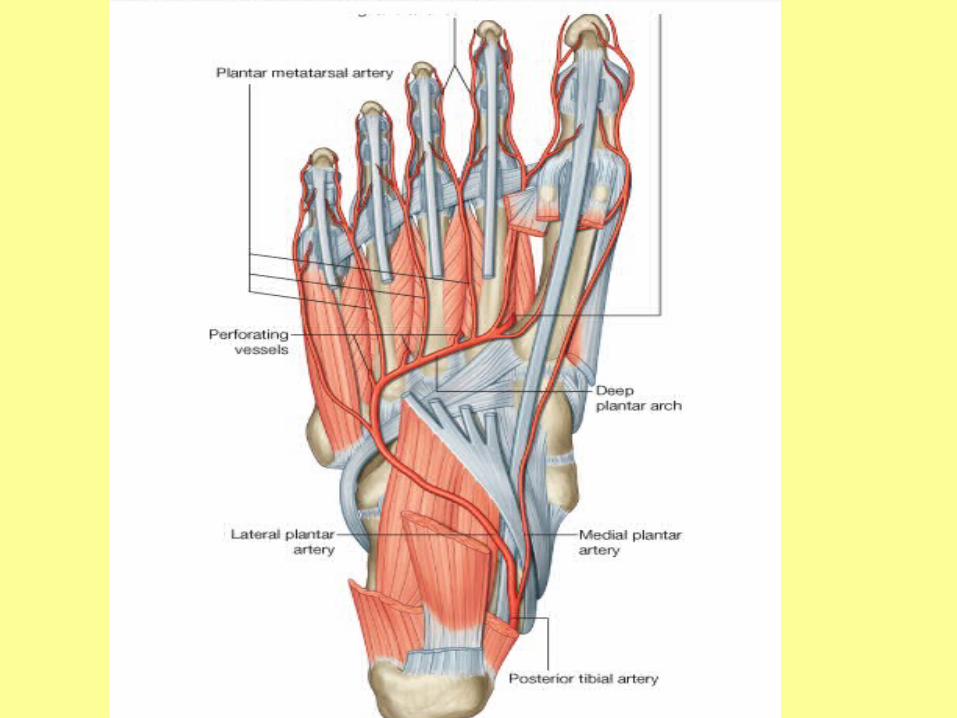
Blood supply to the foot is by branches of the posterior tibial and dorsalis pedis (dorsal artery of the foot) arteries.

• The posterior tibial artery enters the sole and bifurcates into lateral and medial plantar arteries.
• The lateral plantar artery joins with the terminal end of the dorsalis pedis artery (the deep plantar artery) to form the deep plantar arch. Branches from this arch supply the toes.

• The dorsalis pedis artery is the continuation of the anterior tibial artery, passes on the dorsal aspect of the foot and then inferiorly, as the deep plantar artery, between metatarsals I and II to enter the sole of the foot.

Dorsalis pedis artery Dorsalis pedis artery
• The dorsalis pedis artery is the continuation of the anterior tibial artery and begins as the anterior tibial artery crosses the ankle joint .
• It passes anteriorly over the dorsal aspect of the talus, navicular, and intermediate cuneiform bones, and then passes inferiorly, as the deep plantar artery, between the two heads of the first dorsal interosseous muscle to join the deep plantar arch in the sole of the foot.
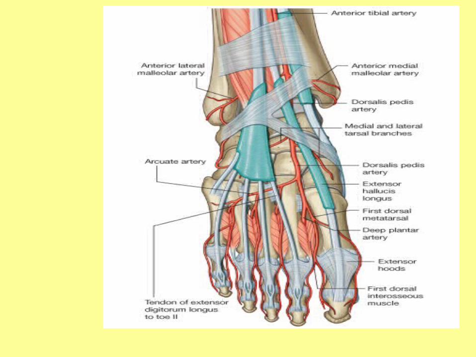

• The pulse of the dorsalis pedis artery on the dorsal surface of the foot can be felt by gently palpating the vessel against the underlying tarsal bones between the tendons of extensor hallucis longus and the tendon of extensor digitorum longus to the second toe.

• Branches of the dorsalis pedis artery include
• lateral and medial tarsal branches,
• an arcuate artery,
• a first dorsal metatarsal artery:

Posterior tibial artery and plantar Posterior tibial artery and plantar arch arch
• The posterior tibial artery enters the foot through the tarsal tunnel on the medial side of the ankle and posterior to the medial malleolus.
• Midway between the medial malleolus and the heel, the pulse of the posterior tibial artery is palpable because here the artery is covered only by a thin layer of retinaculum, by superficial connective tissue, and by skin.

• Near this location, the posterior tibial artery bifurcates into a small medial plantar artery and a much larger lateral plantar artery.
Tibial nerve Tibial nerve
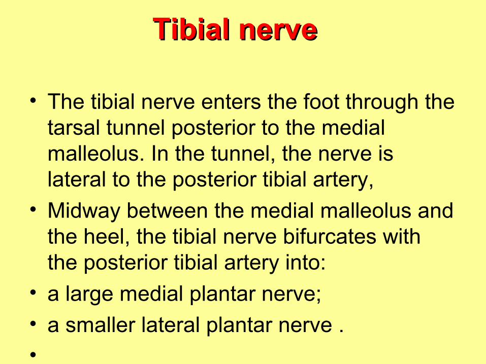
• The tibial nerve enters the foot through the tarsal tunnel posterior to the medial malleolus. In the tunnel, the nerve is lateral to the posterior tibial artery,
• Midway between the medial malleolus and the heel, the tibial nerve bifurcates with the posterior tibial artery into:
• a large medial plantar nerve;
• a smaller lateral plantar nerve .
•
Medial plantar nerve Medial plantar nerve
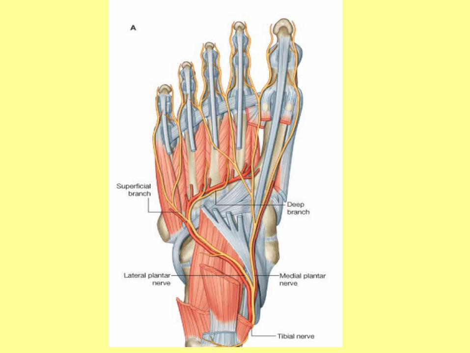
• The medial plantar nerve is the major sensory nerve in the sole of the foot. It innervates skin on most of the anterior two-thirds of the sole and adjacent surfaces of the medial three and one-half toes, which includes the great toe. In addition to this large area of plantar skin, the nerve also innervates four intrinsic muscles-abductor hallucis, flexor digitorum brevis, flexor hallucis brevis, and the first lumbrical.

• The medial plantar nerve supplies a digital branch (proper plantar digital nerve) to the medial side of the great toe and then divides into three nerves (common plantar digital nerves) on the plantar surface of flexor digitorum brevis, which continue forward to supply proper plantar digital branches to adjacent surfaces of toes I to IV. The nerve to the first lumbrical originates from the first common plantar digital nerve.


More than you ever wanted to know about the foot

FUNCTIONS OF FOOTFUNCTIONS OF FOOT
• Support body weightSupport body weight
• Serves as a lever to propel the Serves as a lever to propel the body forward in walking & runningbody forward in walking & running

FUNCTIONS OF FOOTFUNCTIONS OF FOOT
IF THE FOOT IF THE FOOT POSSESSED A POSSESSED A
SINGLE BONESINGLE BONE• It cannot adapt itself It cannot adapt itself
to uneven surfacesto uneven surfaces• Its propulsive action Its propulsive action
depends entirely on depends entirely on gastrocnemius & gastrocnemius & plantarisplantaris
BUTBUT
Gastrocnemius & plantaris

FUNCTIONS OF FOOTFUNCTIONS OF FOOT
IF THE FOOT IS IF THE FOOT IS FORMED OFFORMED OF
SMALL BONES & SMALL BONES & MANY JOINTSMANY JOINTS
• It can adapt itself to It can adapt itself to uneven surfacesuneven surfaces
• Long flexors & small Long flexors & small muscles of foot muscles of foot assist in propulsive assist in propulsive actionaction

WHY THERE ARE ARCHES?WHY THERE ARE ARCHES?
• A segmented structure can hold up A segmented structure can hold up weight only if it is built in the form of weight only if it is built in the form of archesarches
• Weight will be distributed on: Weight will be distributed on: 1) the 1) the heelheel (behind) & (behind) & 2) heads of metatarsal 2) heads of metatarsal bonesbones (in front): pressure will be (in front): pressure will be minimized on nerves & vessels in soleminimized on nerves & vessels in sole
• Forward propulsive action will be Forward propulsive action will be easiereasier
ARCHES OF FOOTARCHES OF FOOT
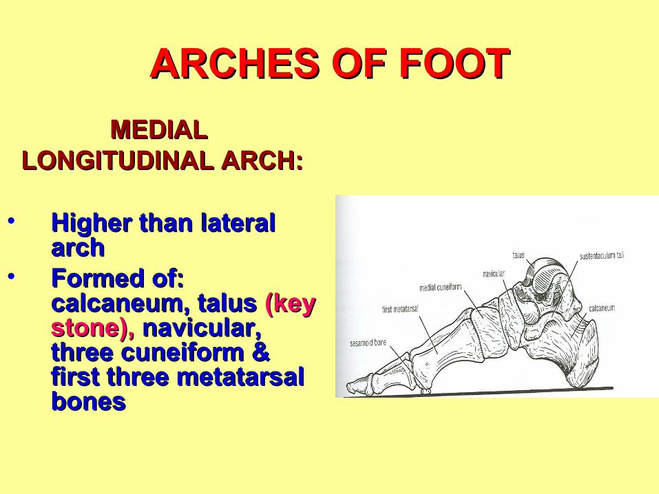
MEDIAL MEDIAL LONGITUDINAL ARCH:LONGITUDINAL ARCH:
• Higher than lateral Higher than lateral archarch
• Formed of: Formed of: calcaneum, talus calcaneum, talus (key (key stone),stone), navicular, navicular, three cuneiform & three cuneiform & first three metatarsal first three metatarsal bonesbones

ARCHES OF FOOTARCHES OF FOOT
LATERAL LATERAL LONGITUDINAL ARCH:LONGITUDINAL ARCH:
• Lower than medial Lower than medial archarch
• Formed of: calcaneum, Formed of: calcaneum, cuboid cuboid (key stone),(key stone), fourth & fifth fourth & fifth metatarsal bonesmetatarsal bones


ARCHES OF FOOTARCHES OF FOOT
TRANSVERSE TRANSVERSE ARCH:ARCH:
• It is only half an It is only half an archarch
• It is formed of: It is formed of: bases of metatarsal bases of metatarsal bones, cuboid & bones, cuboid & three cuneiform three cuneiform bonesbones

FACTORS MAINTAINING FACTORS MAINTAINING ARCHES OF FOOTARCHES OF FOOT
• Shape of bonesShape of bones
• Strength of ligamentsStrength of ligaments
• Tone of musclesTone of muscles
MECHANISM OF ARCH MECHANISM OF ARCH SUPPORTSUPPORT
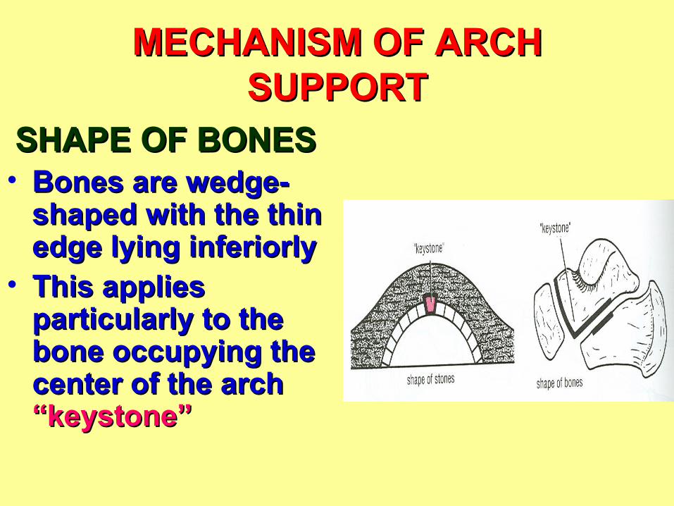
SHAPE OF BONESSHAPE OF BONES• Bones are wedge-Bones are wedge-
shaped with the thin shaped with the thin edge lying inferiorlyedge lying inferiorly
• This applies This applies particularly to the particularly to the bone occupying the bone occupying the center of the arch center of the arch “keystone”“keystone”

MECHANISM OF ARCH MECHANISM OF ARCH SUPPORTSUPPORT
INFERIOR INFERIOR EDGES OF EDGES OF
BONES ARE BONES ARE TIED TIED
TOGETHERTOGETHER

MECHANISM OF ARCH MECHANISM OF ARCH SUPPORTSUPPORT
INFERIOR EDGES OF BONES ARE INFERIOR EDGES OF BONES ARE TIED TOGETHERTIED TOGETHER
• Medial longtitudinal arch: Medial longtitudinal arch: plantar plantar calcaneonavicular ligament, tibialis calcaneonavicular ligament, tibialis posteriorposterior
• Lateral longtitudinal arch: Lateral longtitudinal arch: long & short long & short plantar ligamentsplantar ligaments
• Transverse arch:Transverse arch: deep transverse deep transverse ligaments, transverse head of adductor ligaments, transverse head of adductor hallucis, dorsal interosseihallucis, dorsal interossei


MECHANISM OF ARCH MECHANISM OF ARCH SUPPORTSUPPORT
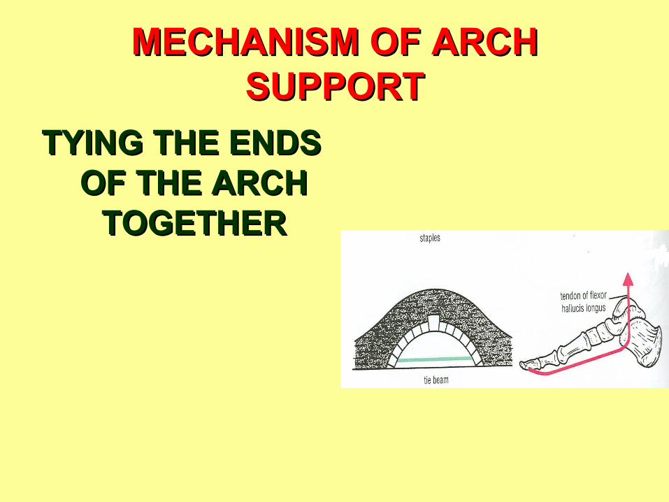
TYINGTYING THE ENDS THE ENDS OF THE ARCH OF THE ARCH
TOGETHERTOGETHER

MECHANISM OF ARCH MECHANISM OF ARCH SUPPORTSUPPORT
TYINGTYING THE ENDS OF THE ARCH THE ENDS OF THE ARCH TOGETHERTOGETHER
• Medial longtitudinal arch: Medial longtitudinal arch: plantar plantar aponeurosis, medial part of flexor digitorum aponeurosis, medial part of flexor digitorum longus & brevis, flexor hallucis longus, flexor longus & brevis, flexor hallucis longus, flexor hallucis brevis, abductor hallucishallucis brevis, abductor hallucis
• Lateral longtitudinal arch: Lateral longtitudinal arch: plantar plantar aponeurosis, lateral part of flexor digitorum aponeurosis, lateral part of flexor digitorum longus & brevis, abductor digiti minimi, longus & brevis, abductor digiti minimi, flexor digiti minimiflexor digiti minimi
• Transverse arch: Transverse arch: peroneus longusperoneus longus

MECHANISM OF ARCH MECHANISM OF ARCH SUPPORTSUPPORT
SUSPENDING SUSPENDING THE ARCH THE ARCH
FROM ABOVEFROM ABOVE
MECHANISM OF ARCH MECHANISM OF ARCH SUPPORTSUPPORT
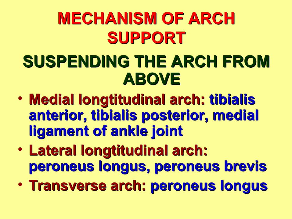
SUSPENDING THE ARCH FROM SUSPENDING THE ARCH FROM ABOVEABOVE
• Medial longtitudinal arch: Medial longtitudinal arch: tibialis tibialis anterior, tibialis posterior, medial anterior, tibialis posterior, medial ligament of ankle jointligament of ankle joint
• Lateral longtitudinal arch: Lateral longtitudinal arch: peroneus longus, peroneus brevisperoneus longus, peroneus brevis
• Transverse arch: Transverse arch: peroneus longusperoneus longus

PES PLANUS (FLAT FOOT)PES PLANUS (FLAT FOOT)
• A condition in which the medial A condition in which the medial longitudinal arch is depressedlongitudinal arch is depressed
• The forefoot is evertedThe forefoot is everted
• The head of talus is forced downward & The head of talus is forced downward & mediallymedially
• The causes are both congenital and The causes are both congenital and acquiredacquired
Related Documents