Open Research Online The Open University’s repository of research publications and other research outputs Investigation of the signal transduction pathways involved in the induction of T-lymphocyte motility Thesis How to cite: Dixon, Richard (1996). Investigation of the signal transduction pathways involved in the induction of T-lymphocyte motility. PhD thesis The Open University. For guidance on citations see FAQs . c 1996 Richard Dixon https://creativecommons.org/licenses/by-nc-nd/4.0/ Version: Version of Record Link(s) to article on publisher’s website: http://dx.doi.org/doi:10.21954/ou.ro.0000f7c4 Copyright and Moral Rights for the articles on this site are retained by the individual authors and/or other copyright owners. For more information on Open Research Online’s data policy on reuse of materials please consult the policies page. oro.open.ac.uk

Welcome message from author
This document is posted to help you gain knowledge. Please leave a comment to let me know what you think about it! Share it to your friends and learn new things together.
Transcript
Open Research OnlineThe Open University’s repository of research publicationsand other research outputs
Investigation of the signal transduction pathwaysinvolved in the induction of T-lymphocyte motilityThesisHow to cite:
Dixon, Richard (1996). Investigation of the signal transduction pathways involved in the induction of T-lymphocytemotility. PhD thesis The Open University.
For guidance on citations see FAQs.
c© 1996 Richard Dixon
https://creativecommons.org/licenses/by-nc-nd/4.0/
Version: Version of Record
Link(s) to article on publisher’s website:http://dx.doi.org/doi:10.21954/ou.ro.0000f7c4
Copyright and Moral Rights for the articles on this site are retained by the individual authors and/or other copyrightowners. For more information on Open Research Online’s data policy on reuse of materials please consult the policiespage.
oro.open.ac.uk
INVESTIGATION OF THE SIGNAL
TRANSDUCTION PATHWAYS INVOLVED IN
THE INDUCTION OF T-LYMPHOCYTE
MOTILITY
Richard Dixon B.Sc. (Hons.)
A thesis submitted for the degree of
Doctor of Philosophy
in the
Open University
Yamanouchi Research Institute
Oxford
September 1996

ProQuest Number: 27701054
All rights reserved
INFORMATION TO ALL USERS The quality of this reproduction is dependent upon the quality of the copy submitted.
In the unlikely event that the author did not send a com p le te manuscript and there are missing pages, these will be noted. Also, if material had to be removed,
a note will indicate the deletion.
uestProQuest 27701054
Published by ProQuest LLO (2019). Copyright of the Dissertation is held by the Author.
All rights reserved.This work is protected against unauthorized copying under Title 17, United States C ode
Microform Edition © ProQuest LLO.
ProQuest LLO.789 East Eisenhower Parkway
P.Q. Box 1346 Ann Arbor, Ml 48106- 1346

is as strange a maze as e*er men trod.
And there is in this business more than nature
Was ever conduct of*
William Shakespeare - The Tempest, Act V. Scene 1
ABSTRACT
Induction of lymphocyte motility is an essential early step in extravasation of lymphocytes
into inflammatory sites and also into lymphoid tissues in the process of lymphocyte
recirculation. Lymphocyte motihty requires a change from a spherical morphology to a
constantly changing irregular shape. In this study, a variety of agents have been
investigated for induction of this shape changing morphology in freshly isolated human
peripheral blood T-lymphocytes (PBTLs) and a non-motile variant of the MOLT-4 human
lymphoid cell line. The MOLT-4 cells proved to be non-responsive to most of the agents
tested, however, 5 agents were found to cause significant polarisation in PBTLs. IL-2, IL-
15, fetal calf serum (PCS) and nocadazole induce shape change in 20-40% of PBTLs.
However, the most potent inducer of shape change found were the PKC inhibitors of the
bisindolylmaleimide (Bis) type, which show effects on over 60% of PBTLs, as reported
recently^^^*\ Do these diverse inducers of shape changing in PBTLs act by a common signal
transduction pathway? With IL-2, IL-15, PCS, nocadazole and Bis., no common changes in
intracellular calcium flux, intracellular pH, inositol triphosphate levels, renaturable kinase
activity and tyrosine phosphorylation have been found. So if a final common signal
transduction pathway exists, it must involve other second messenger systems.
However, a number of pharmacological agents were found to prevent the induction of
shape change in PBTLs, indicating that they could be targeting a common second
messenger element involved in motility signal transduction. Comparisons of their chemical
structures revealed no common structural motifs that would explain their common effects
on lymphocyte motility
ii
Contents
page
Abstract............................................................................ ii
Contents................................................................... ....... iii
List of tables.......................... :......................................... yi
List of figures...................................... ............................ vii
Abbreviations................................................................... xii
Acknowledgements......................................................... xiv
Chapter 1. Introduction.......................................................... 1
1.1 Background............................................................... 4
1.2 Lymphocyte-endothelial recognition......................... 6
1.3 Lymphocyte chemoattractants.................................. 13
1.4 Signal transduction events......................................... 20
1.5 Actin modulation....................................................... 31
1.6 Aims of the project............................... .................... 37
Chapter 2. Materials and Methods.......................................... 38
2.1 Cells and media.............. ..................... ..................... 39
2.2 Solutions and buffers ......................................... 39
2.3 General reagents......................................................... 41
2.4 Isolation of human peripheral blood T-lymphocytes... 41
2.5 Cell polarisation assay................................................. 42
2.6 Time-lapse video microscopy...................................... 43
2.7 Transmigration assay................................................... 43
2.8 Intracellular calcium measurements............................. 44
111
2.9 Intracellular pH measurements............................... 45
2.10 D-/wyo-inositol 1,4,5-triphosphate assay.................... 47
2.11 Preparation of acrylamide gels................................... 47
2.12 Renaturable kinase assay............................................. 48
2.13 Western blotting for tyrosine phosphorylation............ 49
2.14 Immunofluorescence staining for actin and tubulin 50
2.15 Measurement of taurine efflux.............................. 51
RESULTS
Chapter 3. The investigation for inducers of motility.................54
3.1 Introduction.................................................................... 54
3.2 Polarisation assay........................................................... 54
3.3 Transmigration assay...................................................... 57
Chapter 4. Investigations into the roles of intracellular calcium and
phosphoinositides in lymphocyte motility ......................... 66
4.1 Introduction....................................................................66
4.2 Intracellular calcium studies............................................66
4.3 Phosphbinositide studies.................................................70
Chapter 5. Investigations into the roles of intracellular pH and ion
channels in lymphocyte motility................................. .................. 83
5.1 Introduction.....................................................................83
5.2 Intracellular pH measurements........................................84
5.3 Role of ion channels in motility.......................................84
5.4 Chloride channels........................................................... 89
iv
Chapter 6. Investigations into the roles of renaturable kinases in
lymphocyte motility......................................................................... 106
6.1 Introduction..................................................................... 106
6.2 Renaturable kinase assay................................................. 106
Chapter 7. Investigations into the roles of tyrosine phosphorylation
in lymphocyte motility.................................................................... 109
7.1 Introduction..................................................................... 109
7.2 Tyrosine phosphorylation studies..................................... 109
Chapter 8. Investigations into the roles of microtubules in lymphocyte
motility............................................................................................. 115
8.1 Introduction.................................................................... 115
8.2 Microtubule studies......................................................... 115
Chapter 9. Structure-activity relationship of inhibitors of lymphocyte
^ motility............................................................................................. 127
10. Discussion.............................................................................. 138
11. REFERENCES....................... 151
12. List of publications............................................................... 209
o
List of tables
Chapter 3
Table page
3.1: Sunttnary of lymphocyte polarisation assay results.......................................... 59
Chapter 4
4.1: Summary of intracellular calcium studies on PBTLs....................................... 74
VI
List of figures
Chapter 1
figure page
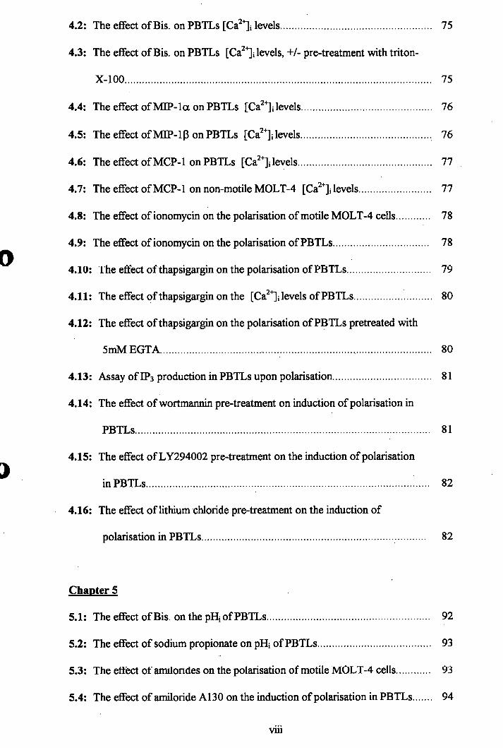
1.1: The four step model of lymphocyte transendothelial migration..................... 3
1.2 : Adhesion molecules involved in lymphocyte-endothelial interactions........... 7
Chapter 2
2.1: Correlation between 490/440nm ratio of BCECF and pH............................. 46
Chapter 3
3.1: Freshly isolated PBTLs................................................................................. 60
3.2: PBTLs treated with lOfiM Bis...................................................................... 60
3.3: PBTLs treated with 50pM nocadazole......................................................... 61
3.4: Dose response of PBTLs polarisation to Bis................................................. 61
3.5: Dose response of PBTLs polarisation to nocadazole.................................... 62
3.6: Dose response of PBTLs polarisation to DL-2 and IL-15.............................. 62
3.7: Dose response of PBTLs polarisation to FCS................................ .......... 63
3.8: The effect of Bis. on the transmigration and polarisation of PBTLs.............. 63
3.9: The effect of IL-2 on the transmigration and polarisation of PBTLs............. 64
3.10; The effect of IL-15 on the transmigration and polarisation of PBTLs 64
3.11: The effect of nocadazole on the transmigration and polarisation of PBTLs.. 65
3.12: The effect of FCS on the transmigration and polarisation of PBTLs 65
Chapter 4
4.1: The effect of lOpM ionomycin on PBTLs [Ca^ ]i levels................................ 74
vu
4.2: The effect of Bis. on PBTLs [Ca^^i levels..................................................... 75
4.3: The effect of Bis. on PBTLs [Ca^ ]i levels, +/- pre-treatment with triton-
X-100............................................................................................................ 75
4.4: The effect of M IP-la on PBTLs [Ca^ ]i levels............................................. 76
4.5: The effect of MIP-ip on PBTLs [Ca^ ]i levels............................................. 76
4.6: The effect of MCP-1 on PBTLs [Ca^ ]i levels............................................... 77
4.7: The effect of MCP-1 on non-motile MOLT-4 [Ca^^i levels......................... 77
4.8: The effect of ionomycin on the polarisation of motile MOLT-4 cells 78
4.9: The effect of ionomycin on the polarisation of PBTLs................................. 78
4.10: The effect of thapsigargin on the polarisation of PBTLs............................. 79
4.11: The effect of thapsigargin on the [Ca ' i levels of PBTLs........................... 80
4.12: The effect of thapsigargin on the polarisation of PBTLs pretreated with
5mMEGTA.................................... 80
4.13: Assay of IP3 production in PBTLs upon polarisation.................................. 81
4,14: The effect of wortmannin pre-treatment on induction of polarisation in
PBTLs........................................................................................................ 81
4.15: The effect of LY294002 pre^treatment on the induction of polarisation
in PBTLs............................................................................................... 82
4.16: The effect of lithium chloride pre-treatment on the induction of
polarisation in PBTLs............................................................................... 82
Chapter 5
5.1 : The effect of Bis. on the pH; of PBTLs.......................................... 92
5.2: The effect of sodium propionate on pH. of PBTLs...................................... 93
5.3: The effect of amilorides on the polarisation of motile MOLT-4 cells 93
5.4: The effect of amiloride A130 on the induction of polarisation in PBTLs 94
viii
5.5: The effect of amiloride A149 on the induction of polarisation in PBTLs 94
5.6: The effect of amiloride A171 on the induction of polarisation in PBTLs 95
5.7: The effect of amiloride A125 on the induction of polarisation in PBTLs 95
5.8: The effect of amiloride A113 on the induction of polarisation in PBTLs 96
5.9: The effect of amiloride A130 on the pHi of motile MOLT-4 cells................ 96
5.10: The effect of A171 on the ability of motile MOLT-4 cells to recover from
an acute acid load...................................................................................... 97
5.11: The effect of sodium propionate on the pHi of motile MOLT-4 cells 97
5.12: The effect of sodium propionate on the polarisation of motile MOLT-4
cells...................................................................................................................... 98
5.13: Comparison between motile and non-motile MOLT-4 cells in their ability
to recover from an acute acid load............................................................. 98
5.14: The effect of Na^/H^ antiport inhibitors on the polarisation of motile
MOLT-4 cells............................................................................................. 99
5.15: The effect of antiport inhibitor-1 on the induction of polarisation in PBTLs 99
5.16: The effect of antiport inhibitor-3 on the induction of polarisation in PBTLs 100
5.17: The effect of antiport inhibitor-4 on the induction of polarisation in PBTLs 100
5.18: The effect of antiport inhibitor-5 on the induction of polarisation in PBTLs 101
5.19: The effect of antiport inhibitor-3 on the ability of motile MOLT-4 cells
to recover from an acute acid load............................................................. 101
5.20: The effect of Cl' channel inhibitors on the polarisation of motile MOLT-4
cells............................................................................................................ 102
5.21: The effect of tamoxifen on the polarisation of motile MOLT-4 cells 102
5.22: The effect of tamoxifen on the induction of polarisation in PBTLs.............. 103
5.23: The effect of NPPB on the induction of polarisation in PBTLs................... 103
5.24: The effect of quinidine on the induction of polarisation in PBTLs............... 104
ix
5.25: The effect of niflumic acid on the induction of polarisation in PBTLs 104
5.26: Comparison of C taurine efflux from motile and non-motile MOLT-4
cells............................................................................................................. 105
5.27: Do the Cl' channel blockers block volume regulated chloride channels in
motile MOLT-4 cells as assayed by ^ C taurine efflux................................ 105
Chapter 6
6.1: The effect of induction of polarisation in PBTLs on renaturable kinases
autophosphorylation................................................................................... 108
6.2: The effect of induction of polarisation in PBTLs on renaturable kinases
autophosphorylation................................................................................... 108
Chapter 7
7.1: The effect of induction of polarisation in PBTLs on tyrosine
phosphorylation in PBTLs............................ 112
7.2:, The effects of IL-2 and IL-15 on tyrosine phosphorylation in PBTLs 112
7.3: The effect of herbimycin A pre-treatment on induction of polarisation
in PBTLs................................................................................ 113
7.4: The effect of herbimycin A pre-treatment on tyrosine phosphorylation
induced by IL-2 in PBTLs.......................................................................... 114
7.5: The effect of herbimycin A pre-treatment on tyrosine phosphorylation
induced by IL-2 and IL-15 in PBTLs......................................................... 114
Chapter 8
8 .1: The effect of taxol on the induction of polarisation in PBTLs....................... 119
8.2: The effect of vinblastine on the polarisation of PBTLs................................... 120
X
8.3: The effect of colchicine on the polarisation of PBTLs......................... 120
8.4: The effect of nocadazole on the polarisation of PBTLs....................... 121
8.5: The effect of vincristine on the polarisation of PBTLs......................... 121
8.6 : The effect of colcemid on the polarisation of PBTLs........................... 122
8.7: Untreated PBTLs stained for polymerised P-tubulin............................ 123
8.8: PBTLs treated with lOpM Bis. stained for polymerised P-tubulin................. 124
8.9: Untreated PBTLs stained for depolymerised p-tubulin................................. 125
8.10: PBTLs treated with lOpM Bis. stained for depolymerised P-tubulin 126
Chapter 9
9.1: Ionomycin.............................. 131
9.2: Thapsigargin................................................................................................... 131
9.3: Amiloride compounds.................................................................. 132
9.4: Chloride channel blockers.............................................................................. 133
9.5: Microtubule targetting drugs.......................................................................... 134
9.6: Phosphoinositide 3-kinase inhibitors............................................................... 135
9.7: Herbimycin A.................................................................................................. 136
9.8: Trifluoperazine................................................................. 136
9.9: The effect of trifluoperazine on induction of polarisation in PBTLs............... 137
XI
Abbreviations
ATP: adenosine tris-phosphate
Bis : bisindolylmaleimide
BSA: bovine serum albumin
BSS: balanced salt solution
cAMP: cyclic adenosine-3%5'-monophosphate
DAG: diacylglycerol
DMSO: dimethyl sulphoxide
DTT: dithiothreitol
EDTA: ethylenediamine tetracetic acid
EGF: epidermal growth factor
EGTA: ethyleneglycol-bis-(b-aminoethylether) N,N,N’,N’ tetra-acetic acid
FCS: fetal calf serum
fMLP : N-formyl-methionyl-leucyl-phenylalanine
GAP: GTPase activating protein
GDI: GDP dissociation inhibitor
GDP: guanosine 5’-diphosphate
GEF: guanine nucleotide exchange factor
HSA: human serum albumin
IP-10: inducible protein-10
MCP-1: monocyte chemotactic protein
MDGP-la: macrophage inflammatory protein
MIP-lp:
MGSA: melanoma growth stimulating activity
Na-EDTA: ethylenediamine tetracetic acid (disodium salt)
XU
NZ: nocadazole
PBS; phosphate buffered saline
PBTLs: peripheral blood T-lymphocytes
PDGF : platelet derived growth factor
PI: phosphatidylinositol
PI3K: phosphatidylinositol 3-kinase
PIP2: phosphatidylinositol bis-phosphate
PKC: protein kinase C
PMSF : phenyl-methyl-sulphonyl-fluoride
RANTES: regulated on activation, normal T cell expressed and secreted
SDS-PAGE: sodium dodecyl sulphate - polyacrylamide gel electrophoresis
TEMED : N,N,N’ ,N-tetra-methylethylenediamine
TNF-a: tumour necrosis factor
xm
Acknowledgements
I would like to thank the Yamanouchi Research Institute (YRI) for funding my PhD project
and all the people who have helped me in the three years I was there. Thank you also to all
the people at YRI who have donated blood, as without them this research project would not
have been possible.
A special thanks goes to my supervisor Dr. Nick Matthews, for all his support and also to
my colleague Kate Thorp, for the invaluable discussions.
To Sven the Beserk and the Milk Tray Man, cheers for the most excellent session evenings
and also to the horse for his timely appearances! Appreciation goes to The Elm Tree for
their excellent Guinness and late acoustic nights.
Most important of all, a special thanks to Suzanne for keeping me sane during my PhD in
Oxford.
XIV
i
1. Introduction
I
The circulatory and migratory properties of white blood cells have evolved to allow
efficient surveillance of tissues for infectious pathogens and rapid accumulation at sites
of injury and infection. Lymphocytes exhibit complex migration pathways in the body.
Resting blood lymphocytes which are predominantly non-motile recirculate selectively
through specific lymphoid tissues; activated lymphocytes migrate selectively into
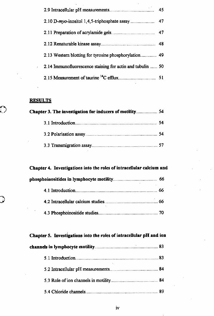
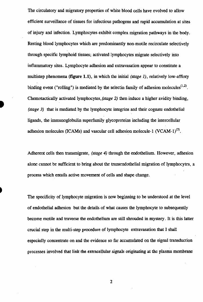
inflammatory sites. Lymphocyte adhesion and extravasation appear to constitute a
multistep phenomena (figure 1.1), in which the initial (stage 1), relatively low-affinty
binding event ("rolling") is mediated by the selectin family of adhesion molecules^^'^\
Chemotactically activated lymphocytes, (stage 2) then induce a higher avidity binding,
(stage 3) that is mediated by the lymphocyte integrins and their cognate endothelial
ligands, the immunoglobulin superfamily glycoproteins including the intercellular
adhesion molecules (ICAMs) and vascular cell adhesion molecule-1 (VCAM-1)^^\
Adherent cells then transmigrate, (stage 4) through the endothelium. However, adhesion
alone cannot be sufficient to bring about the transendothelial migration of lymphocytes, a
process which entails active movement of cells and shape change.
The specificity of lymphocyte migration is now beginning to be understood at the level
of endothelial adhesion but the details of what causes the lymphocyte to subsequently
become motile and traverse the endothelium are still shrouded in mystery. It is this latter
crucial step in the multi-step procedure of lymphocyte extravasation that I shall
especially concentrate on and the evidence so far accumulated on the signal transduction
processes involved that link the extracellular signals originating at the plasma membrane
Figure 1.1 : The four step model of lymphocyte transendothelial
migration.
The four step model of Ivmphocvte transendothelial migration.
A' *BLOOD
l y m p h o c y t e
: .T .t k u ir t /» t l« W p r 2 .A cli«tion3. Ami I aavlidKaiioa
4. Trw«»d©tiieE*l ntigjatioa
to the organisation of the cytoskeletal network which is involved in influencing cell shape
and thus motility.
1.1; Background
In 1875 Ranvier was the first to suggest that lymphoid cells were motile^*®. This
concept received enthusiastic support by others, however, objections were raised by
several research workers, including P. Ehrlich, according to whom the lymphocyte had
too little protoplasm to push the voluminous nucleus along^^ '^^^\
In 1921 Lewis and Webster and in 1923 Sabin clearly demonstrated lymphocyte motility
with intermittent stops and starts and the presence of a trailing cytoplasmic tail ^ ’ ^°\
The classic description of the morphology of moving lymphocytes was presented by
Lewis in 1931^^\ Lewis described the motile lymphocyte as a polarised asymmetric cell
in a configuration resembling that of a hand mirror with a thin advancing pseudopod, a
rounded area enclosing the anteriorly placed nucleus and a trailing tail of cytoplasm.
McFarland extended the studies of the cytoplasmic tail of the amoeboid lymphocyte and
termed this extension of cytoplasm, the uropod^^^'^^\ The uropod was demonstrated to
be a part of the cell not only associated with cell movement but with a variety of
lymphocyte interactions with the environment, including other cells. The uropod is
covered with microvilli and contains the golgi apparatus, mitochondria, rough
endoplasmic reticulum, centrioles, microfilaments and microtubules^^ '^’ ^ In contrast to
the uropod, the advancing edge of the lymphocyte, which was described by the early
investigators to be the most substrate adherent area of the cell, is generally devoid of
major cytoplasmic organelles.
The regulation of lymphocyte motility involves multiple control mechanisms, some of
which are only partly understood at present. Within the organism, lymphocytes must
move actively to specific sites in lymphoid and non-lymphoid tissues. This most likely
requires that lymphocyte motility is switched on and off and guided in a precise way
For a relatively long time, leukocyte motility and translocation, particularly chemo- and
haptotaxis, were studied preferentially using granulocytes and monocytes^^’ ^ \ This
was despite the obvious fact that motility must be important for the function of the
lymphocytes, both as recirculating cells and in immune responses. Thus, an abundance
of information was accrued on granulocyte migration and chemotaxis, whereas relatively
little was known about the corresponding events in lymphocytes.
A major reason why lymphocyte motility, migration, chemo- and haptotaxis did not
receive major attention until “later” was poor lymphocyte in vitro migration assays and
the non adhesiveness of lymphoid cells for noncellular substrates. Thus, lymphocytes
maintain their spherical suspension shape and show poor motile behaviour under
conditions in which fibroblasts and macrophages adhere and spread their cytoplasm over
substrates. Lymphocytes can exhibit motile forms in suspension without anchorage to
cellular and non cellular substrates. Lymphocyte motility is often determined in vitro
when the cells are nonadherent using the number of polarised cells as a measure of
motility^^^^\ However, it must be emphasised that the extension of pseudopods and
migration of cells are separable because pseudopod formation is not always followed by
migiation®^ '^^^ .
In conclusion, T lymphocyte motility depends on the capacity of the cells to form active
cell edges, the binding capacity of lymphocyte adhesion receptors, the availability of
adhesion ligands, and the capacity of the cells to de-adhere and perform repeated cycles
of motile events leading to translocation.
1.2: Lymphocyte - endothelial recognition.
To enter the various lymphoid tissues involved in recirculation, blood lymphocytes have
to cross the endothelial vascular lining (except in the spleen, where small penicillar
arterioles end in the parenchyma, thus allowing unhampered access of blood leukocytes).
The process of lymphocyte extravasation in lymphoid tissues occurs at specialised post
capillary vascular sites called high-endothelial venules (HEVs)^^\ Our understanding of
the mechanisms of this selectivity has been advanced by the discovery that naive and
memory lymphocytes prefer different recirculation pathw ays.W hen naive lymphocytes
encounter antigen, those lymphocytes with receptors specific for the antigen are
stimulated to expand clonally and are converted to memory lymphocytes that have
altered expression of adhesion receptors and circulatory p a tte rn s .F o r peripheral tissues
and lymph nodes, memory lymphocytes emigrate preferentially through tissue
endothelium, whereas naive lymphocytes enter the lymph node through HEVs.
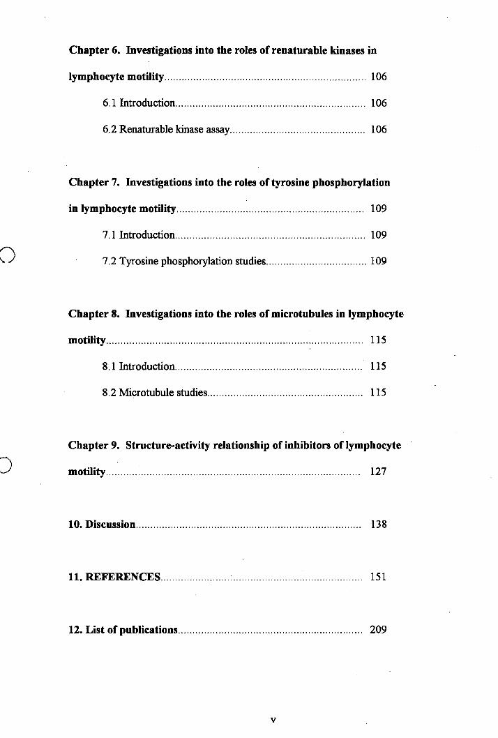
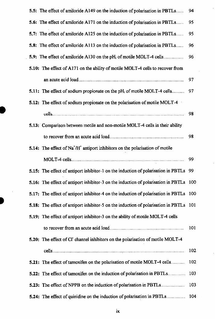
Recent research has started to identify the molecules involved in adhesion of
lymphocytes to HEVs and endothelium (figure 1.2). L-selectin on the surface of the
lymphocytes has been found to have various ligands, depending on the site in the
body^^°^ and recognises a carbohydrate epitope on several biochemically distinct
molecules synthesised by HEVs^^^ \ These include the proteins GlyCAM-1^^^^ and
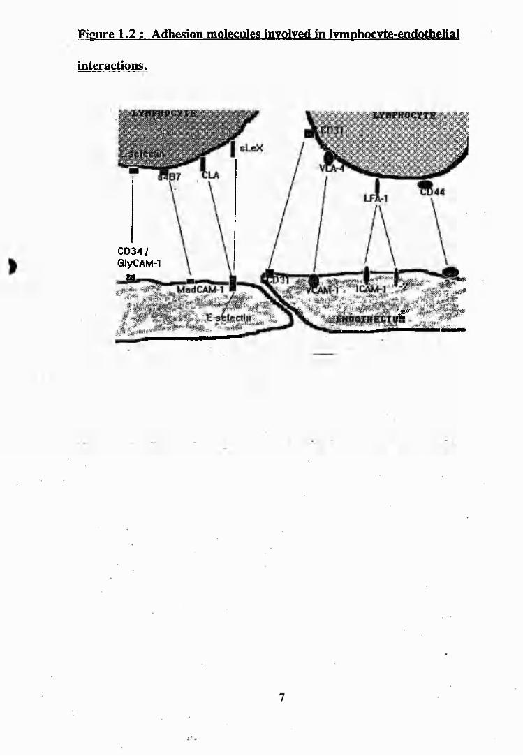
Figure 1.2 : Adhesion molecules involved in Ivmphocvte-endothelial
interactions.
CD34/ GIyCAM-1
o
r
CD34^. In mucosal lymphoid tissue, L-selectin (along with a^-integrins) binds to
MAdCAM-1 *’ and it has been shown in vitro that glycolipids can interact with selectins
in physiological flow conditions, thus contributing to rolling adhesions^^\ Recently it
has been shown that a^-integrins can mediate both rolling and adhesion in the multistep
recruitment of peripheral blood mononuclear cells in vivo and these interactions occur
independently of the selectins and P2 integrins^. L-selectin is shed following T-cell
activation^^^ and this may occur during interaction with endothelial cells to allow the T
cells to migrate. Recent work has shown that the cytoplasmic domain of L-selectin
interacts directly with the cytoplasmic actin binding protein a actinin and forms a
complex with vinculin and ta lin ^ \ The HEV ligands for L-selectin and other putative
homing receptors have been referred to as ‘vascular addressins’, signifying their role in
mediating the tissue specific adhesion of lymphocytes expressing the appropriate homing
receptors. The ligands for E-selectin (E-selectin is present on endothelia), include sialyl
lewis X (sleX), which is present on neutrophils and macrophages, and there is a similar
if not identical carbohydrate on a subset of memory T cells^ '^^. It has been found that
mice with null mutations in both endothelial selectins (P and E) develop a phenotype of
leukocyte adhesion deflciency^\ thus providing strong evidence for the functional
importance of selectins in vivo. Indeed, there is now direct evidence for the presence of
distinct E- and P-selectin ligands on T-lymphocytes and it has been suggested that y/ô T
cells may be preferentially recruited to inflammatory sites during the early stages of an
immune response when P-selectin is upregulated^.
The integrin LFA-1 on blood lymphocytes requires activation for binding to its
counterstructures ICAM-1 and ICAM-2, which are expressed on HEVs and endothelial
cells^^'^^\ Binding of I^selectin does not trigger activation of LFA-1, since lymphocytes
attach and roll in flow on purified peripheral node addressin identically, whether or not
purified ICAM-1 is present on the substrate. An additional stimulus is required before
they will arrest and strengthen adhesion through LFA-1^^\ Indeed, recent work has
shown that chemoattractant stimulation of neutrophils and lymphocytes, rolling on
immobilised peripheral node addressin (PNAd) and ICAM-1 results in rapid arrest and
firm sticking in vitro.
G protein-coupled receptors are required for lymphocyte recirculation and are likely to
provide the signals required to activate the adhesiveness of LFA-1. In relation to this,
some recent work has shown that transfecting fMLP receptors into lymphocytes andI
subsequently stimulating the cells with fMLP triggers rapid adhesion via VLA-4 and
shape change, which is pertussis toxin sensitive^^^\ Pertussis toxin profoundly depresses
lymphocyte recirculation via the lymphatics, which suggests that G-protein coupled
receptors of the class are required for lymphocyte emigration through the HEV^°\
This is seen in the condition known as ‘whooping cough’, whereby the infectious
bacterium (Bordetella), secretes copious amounts of pertussis toxin into the blood
system. One of the effects of this is the subsequent rise in the number of lymphocytes in
the blood stream due to their inability to traverse the endothelia. Despite the lack of
emigration, pertussis toxin treated lymphocytes bind normally to lymph node HEVs in
vitro. These findings provided the basis for an early proposal for a two step model in
which G protein coupled receptors function subsequent to binding of lymphocytes to
This theory has also been alluded to in more recent in vitro work^^\ which
implies that cultured HEVs may stimulate lymphocyte motility by two mechanisms: one
which is rapid and pertussis toxin sensitive and one which is slower, pertussis toxin
insensitive and dependent on lymphocyte adhesion to the HEVs.
Investigations into lymphocyte-endothelial interactions has shown that functionally
significant lymphocyte cell surface molecules (CD2, CD44, L-selectin and LFA-1) exist
as organised complexes in the cell membrane. Redistributions and associations between
them are triggered selectively by lymphocyte-endothelial cell contact^^\ The enormous
amount of research in this particular field has ultimately led to the discovery of other
important molecules involved in lymphocyte-endothelial adhesion. It is now becoming
clear that the interaction between VLA-4 on the lymphocytes and VCAM-1 is important
in both constitutive migration of lymphocytes into lymphoid organs and also in immune
mediated inflammation^'^^^. Another important molecule currently being investigated is
It has been suggested that in T cells, homophilic CD31 adhesion may be
primarily involved in transmigration of naive T cells and that its role is complementary
to that of ICAM-1^^^\ More evidence for the importance of CD31 was shown in a
recent paper which suggests that it has an important role in the extravasation of natural
killer (NK) T cells into tissues for constitutive surveillance and into sites of
inflammation^^^\ Cross linking of CD31 molecules induces cytoskeletal rearrangement
in human NK cells and this phenomenon is Mg '*', but not Ca^^ dependent, suggesting
the involvement of an integrin^^^\ Also both cell spreading and cytoskeletal
rearrangement, as well as CD31-mediated adhesion appears to be regulated by the
intracellular cAMP content^^^.
10
■ J
Another important adhesion molecule on T cells is cutaneous leukocyte-associated
antigen (CLA)^^\ although theories for its role have yet to become conclusive.
CD44 is a glycoprotein which is found on the surface of most leukocytes including
lymphocytes and has recently been found to be not necessary for normal lymphocyte
circulation. However it is required for extravasation into an inflammatory site involving
non-lymphoid tissue^^°\ In addition, recent evidence has demonstrated that CD44 and its
alternatively spliced isoforms (CD44R) endow some tumour cells with enhanced
metastatic ability^^^^\ Recent work has shown that in vitro there is a rolling interaction
between lymphoid cells and endothelial cells that is not selectin mediated but is in fact
mediated by
Antigen injected into the tissue of sensitized individuals induces localised accumulation
of lymphocytes. These lymphocytes (and those accumulating in tissues in autoimmune
diseases) are almost all memory cells^^ \ The phenotype of these cells is quite similar to
that of lymphocytes trafficking through these sites under basal conditions, suggesting that
the same molecular mechanisms that mediate basal trafficking may be up regulated in
inflammation. Accumulation of lymphocytes induced by specific antigen or by injection
of interferon y or TNFa is significantly inhibited by monoclonal antibodies (Mabs), to
either the LFA-la or the integrin a4 subunit ^ ' ^ and almost completely by a
combination of Mabs to LFA-la and Mabs to E-selectin and VCAM-1 also
inhibit lymphocyte accumulation in delayed type hypersensitivity in the skin^^\ However
recent research has shown that this is not the full story and that an ICAM-, ELAM- and
11
VCAM- independent modulation in the early phase of lymphocyte attachment to
endothelium, seems likely^^\
Therefore, emigration of lymphocytes through peripheral node HEVs, originally thought
to consist of two steps, has now been shown through recent evidence to require three
sequential area code signals (L-selectin tethering, chemoattractant activation and
subsequent integrin activation and binding), that are analogous to those involved in
neutrophil emigration from the blood stream^^\ Identification of a putative lymphocyte
chemoattractant secreted by peripheral lymph node HEVs and a chemoattractant receptor
that is predicted to be selectively expressed on the naive subset of lymphocytes that
recirculate through peripheral node HEVs will be the subject of intense research interest
in coming years.
In conditions such as chronic inflammation and cell mediated hypersensitivity,
lymphocytes make up a substantial part of local infiltrating leukocytes. In inflammation
the endothelium may exert functions in lymphocyte recruitment from the blood,
comparable to high endothelium in lymphoid tissues. In fact, HEV-like structures have
been described in various conditions of chronic inflammation, including autoimmune
lesions and immune reactivity around tumours^^ '^^\
Inflammation also affects traffic through the HEVs. Antigen injected into tissue, drains
to the regional lymph node and greatly increases blood flow to the node and naive
lymphocyte traffic through the HEV^^^. Furthermore, memory lymphocytes now appear
to enter the node directly; this is associated with induction of VCAM-1 on non-HEV
12
vascular endothélia within the node^^ . Entry is inhibited by Mab to the integrin a4
subunit, and this suggests a role for interaction of VCAM-1 with a4pl^^®\
Recent research has shown that the adhesion molecules topography on the surface of
leukocytes is a big factor in the outcome of an adhesion cascade^^^\ indeed various
adhesion molecules are enriched in the uropod region of the polarised lymphocyte,
particularly ICAM-1 and
The evidence so far, suggests that there are multiple adhesion molecules involved in
extravasation and that multiple signals are also required for directing activated
lymphocytes through the endothelia. Thus, a four step or area code model of leukocyte
emigration from the blood stream, established and validated in vitro and in vivo with
neutrophils^^^\ appears extendible to all subclasses of leukocytes, including lymphocytes
(figure 1.1). Combinatorial use of multiple adhesion and chemoattractant receptors in
the four step model^ with distinct distributions on leukocyte subsets, regulates selection
of the subclasses of leukocytes emigrating at inflammatory sites and the distinctive
recirculation behaviour of lymphocyte subsets.
1.3: Lymphocyte chemoattractants
Lymphocyte chemoattractants are interesting candidates for the stage 2 signal for
lymphocyte accumulation at inflammatory sites. Pertussis toxin treatment inhibits
lymphocyte emigration in response to antigen^^^\ Identification of lymphocyte
chemoattractants has been hampered by the low motility of lymphocytes compared with
that of monocytes or neutrophils. A number of chemokines, all of which were isolated
based on chemoattractive activity for neutrophils or monocytes or by cloning genes of
13
unknown function, have subsequentiy been tested and found to be chemoattractive for
lymphocyte subpopulations^^^’ ^
Chemokines, also known as intercrines, comprise a superfamily of small, secreted
proteins that mediate inflammation by inducing chemotaxis and activation of a variety of
inflammatory cells. Members of the chemokine superfamily possess a conserved
structural motif containing two cysteine pairs. Based on the arrangement of the cysteines
within this motif, chemokines are divided into two subfamilies. The first cysteine pair of
the C-X-C chemokines (a-intercrines), are separated by an intervening amino acid, while
the first cysteine residues of the C-C chemokines (P-intercrines), are adjacent. C-X-C
chemokines include IL-8 , MGSA, IP-10, ENA-78, platelet factor 4, platelet basic
protein and thromboglobulin. Members of the C-C chemokine subfamily include MLP-la
and MIP-ip, MCP-1, -2, -3, RANTES and 1-309. The two chemokine subfamilies
demonstrate 20-45% homology to each other at the amino acid level and are basic
heparin binding proteins.
Particular chemokines induce selective migration of leukocyte subsets which differ both
in phenotypic markers and activation state. This has led to the view that the cellular
composition at inflammatory sites depends on the combinatorial effects of multiple
chemokines, each with selective chemotactic activities. For example, while the C-C
chemokines RANTES, M IP-la and MIP-ip all induce monocyte migration, they have
distinct chemoattractant properties for lymphocytes. M IP-la induces the preferential
migration of activated CD8^ T cells and B cells (at higher concentrations the migration
of these cells seems to be diminished and the migration of CD4^ T cells is enhanced),
14
while MIP-ip selectively induces chemotaxis of activated CD4^ T cells ' ^ RANTES
induces migration of both activated and resting T cells, including, perhaps most
significantly, resting memory T cells (CD4^ and CD45R0^)^^^^^\ Furthermore, IL-8
acts as a chemoattractant for about 10% of human peripheral blood T lymphocytes
belonging to either the CD4 or CD8 subsets^^ '^^^ \ Greater proportions of polyclonally
activated, than of resting T lymphocytes, exhibit chemotactic responses IL-8^^^'^\
Recently, the C-X-C chemokine, IP-10, has been shown to induce chemotaxis of
activated, but not non-activated, human peripheral blood T lymphocytes^^^\ Phenotypic
analysis of the stimulated T cell population responsive to IP-10 demonstrated that
stimulated CD4^ and CD29^ T cells migrated in response to IP-10. This resembles the
biological activity of RANTES. Recent research has shown that recombinant human IP-
10 is capable of inducing human T cell migration in vivo and thus provides more
evidence for its role in inflammation^^^^.
This pattern of selective migration corresponds to the capacity of these chemokines to
enhance the adhesion of specific subsets of activated T . cells to DL-1 stimulated
endothelial cells^^^ M IP-la and MIP-ip augment the attachment of activated CD8^ and
CD4^ T cells respectively^^^\ It has now been reported that there is a new member of the
C-C chemokine family, termed MlP-ly^^^^, which is produced by dendritic cells and
recruits T cells before activation.
Moreover, differences in the kinetics of the expression between these chemokines may
further co-ordinate the regulation of the migration pattern and thus the composition of
the lymphocyte population at inflammatory sites, at any given time. Chemokines have
15

been shown to induce T cell adhesion to purified recombinant human adhesion molecules
and to extracellular matrix proteins, by stimulating the development of a high affinity
state in the integrin molecules^"^\ T. Springer’s lab have shown using a transendothelial
chemotaxis assay with HUVECS (human umbilical vein endothelial cells) on transwells
that only the C-C chemokines promote transendothelial chemotaxis of PBTLs and that
the C-C chemokines selectively recruit a memory subset of T lymphocytes^^"^ \ Also,
one of his latest papers shows that MCP-1, RANTES and MIP-ip induce T cell binding
to fibronectin but not ICAM-1, suggesting that the chemokines may be most important,
not in initiating integrin dependent firm adhesion of T cells to the vascular wall but
rather in subsequent adhesive interactions during migration into tissue '* ^
The endothelium may present chemoattractants to lymphocytes in a functionally relevant
way, as well as providing a permeability barrier that stabilises the chemoattractant
gradient. A new concept to emerge recently has been that of specialised chemokine
binding proteins that act as clearance receptors to remove chemotactic and inflammatory
peptides from the blood^^^ \ This reeeptor/protein is also found on endothelial eells and
thus it could potentially play a role of presenting chemokines to lymphocytes.
Since lymphocytes, responding to specific antigen in tissue, signal emigration of further
lymphocytes into the site, a chemoattractant was sought in material secreted by mitogen
stimulated mononuclear cells. Subsequent investigations revealed that MCP-1, previously
thought to be solely a monocyte chemoattractant, is also a lymphocyte chemoattractant^^^
to an activated subset of memory lymphocytes. There is a clear distinction between the
IL-8 and MCP-1 responsive T cell populations and that chemokine receptor expression
on T cells may be regulated with respect to lineage as well as cellular activation^'^^^
16
A model of selective chemotaxis has been proposed for the C-C chemokine MIP-ip and
other chemokines containing glycosaminoglycan binding sites^^\ In this model,
endothelial cells at inflammatory sites present CDS'*' T cells with a gradient of the
chemokine immobilised on endothelial surface proteoglycans, such as CD44. The bound
chemokine triggers functional activation of the lymphocyte integrins, enhancing
attachment to the vascular endothelium and migration through the vessel into the
surrounding tissue.
The chemokine receptors, like their ligands, form a family of structurally and
functionally related proteins. They are members of the superfamily of hepta-helical,
rhodopsin like, G-protein coupled receptors that can be defined by amino acid sequence
homologies^^^\ The C-C chemokines bind weakly, if at all, to human neutrophils.
Nevertheless, M IP-la and RANTES can induce small, transient elevations of
intracellular calcium that can be homologously and heterologously desensitised by MIP
l a and RANTES, but not by other stimuli, suggesting a shared neutrophil receptor^"^’ ^\
However, any functional importance is unclear, since M IP-la and RANTES do not
induce neutrophil chemotactic or microbicidal responses^^^.
To date, only the lymphocyte MIP-ip receptor (also known as the ACT-2 receptor)^^^
has been characterised biochemically, although the relationship of this protein to the
monocyte receptors is unknown. A distinct receptor for multiple C-C chemokines has
recently been cloned from monocytes^"^ \ This receptor, termed C-C CKR-1 induces a
rapid, transient increase in intracellular calcium, but the binding affinity is not
17
necessarily predictive of signal strength. While M IP-la binds to C-C CKR-1 with the
highest affinity and induces the strongest calcium signal, RANTES transmits a more
potent signal than MCP-1 and MIP-lp, which bind the receptor with higher affinities.
Indeed, there are now at least seven human chemokine receptors that bind or respond to
p-chemokines^^*^® . Recent research suggests that chemokines not only share receptors
but also signal transduction pathways. The signal transduction pathway of MCP-1,
RANTES and M IP-la are similar, involving pertussis toxin sensitive G-proteins, an
increase in intracellular calcium, a rapid activation of arachidonic acid release and
possibly protein kinase activation^^^^'^\
However, it is not only the recently discovered (and much publicised) chemokines that
are lymphocyte attractants, as other molecules such as interleukins have been found to be
chemoattractive for lymphocytes. For example, BL-l has been reported to be a potent
lymphocyte attractant in vitro^^^^\ Its release from the epidermis in disease or following
injury, may tlierefore constitute an important mechanism for the induction of pathological
lymphocyte infiltrates. Low level release of epidermal IL-1 under normal conditions may
also be responsible for physiological trafficking of lymphocytes in normal skin.
Recombinant BL-6 has also been shown to induce lymphocyte migration in vitro^^'^\
Other interleukins which have been reported to have chemotactic activity for T
lymphocytes include IL-10^“ ’ \ which is specific for CD8 T cells, IL-2 which is
reported to be specific for activated CD4^ T cells ^ ' and IL-15 which has just recently
been proven to be a chemoattractant for T lym phocytes^^'^^. Furthermore, IL-10
inhibits the IL-8 chemotactic response of CD4^, but not that of CD8 T cells, as well as
inhibiting B cell motility induced by IL-4^‘ ° . Another paper suggests that IL-1, IL-8 and
18
RANTES play important roles by inducing migration of T cells towards sites of
inflammation, whereas the T cell derived cytokines IL-2, IFN-y, IL-4, IL-10 and IL-13
seem to be important because of their modulatory effects on T lymphocyte
chemotaxis " .
Clinical research has shown that IL-2 mediates the regression of certain malignancies,
but clinical use is limited because of associated toxicities, including parenchymal
lymphocytic infiltration with multiple organ failure. Recent research has shown that IL-2
toxicity involves organ-specific TNF-a and RANTES production with increased ICAM-1
and VCAM-1 expression as potential mechanisms facilitating lymphocytic infiltration and
organ dysfunction^^\
Recently, a source of T cell chemoattractants has been shown to be neutrophils, which
upon stimulation with IL-8 release chemoattractants that mediate T-cell and monocyte
accumulation at sites of inflammation^°\
A lymphokine termed lymphocyte chemoattractant factor (LCF), which has no
significant homology to any previously described lymphocyte chemoattractants, has been
identified and cloned^^^^^ and membrane expression of CD4 functions to transmit the
migratory signal induced by LCF. However, LCF has now been termed as interleukin-16
and is secreted from serotonin stimulated CD8 '*’ T cells in vitro, therefore serotonin may
promote recruitment of CD4^ T cells via CD8^ T cells^^^ \ Eosinophils and CD4^ T
cells are preferentially recruited into sites of inflammation and in a recent publication it
was found that eosinophils are a source of two cytokines, IL-16 and RANTES, that are
19
chemotactic for both lymphocytes and eosinophils. Their data indicates that eosinophils
could contribute to the recruitment of CD4^ T cells and more eosinophils^^\ Also, it has
been found that CD4-lck coupling is essential for IL-16 induced T cell migration^^\
Many more T cell chemoattractants are being discovered lately such as recombinant
human growth hormone which is capable of inducing significant migration of resting and
activated human T cells and their subsets^\ A new chemokine, called Mig, which is of
the C-X-C family has been found and is likely to play a role in T cell trafficking. Also,
serum amyloid A has been shown to be a T cell chemoattractant^^, as well as
prostaglandin Ej and leukotriene 64^* .
It must also be noted that early research in the 1970’s and ‘80’s, reported that T
lymphocytes are responsive in a chemotactic manner to casein, C5a, f-met-leu-phe and
denatured proteins^^“ ’ ' ‘\ Also, P C. Wilkinson has quite recently shown that
staphylococcal enterotoxin B stimulates motility in T cells over a period of 72 h o u rs^ .
In summary, it is evident that there are many different types of chemoattractants for
lymphocytes. The diverse binding affinities and signalling potentials that each
chemoattractant possesses, as well as the differential expression of the chemoattractant
receptors on target cells, may regulate the combinatorial effect of multiple
chemoattractants on lymphocytes at localised sites of inflammation.
20
1.4î Signal transduction events
In the past few years major advances in our understanding of the signalling pathways
involved in cell motility have been achieved. Unfortunately, little of this work has been
done on lymphocytes, instead most cell motility research has tended to concentrate on
fibroblasts, slime moulds (Dictyostelium discoideum) and neutrophils. Thus some of the
literature reviewed here will incorporate relevant work done on neutrophils that can be
considered as similar to the events occurring in lymphocytes.
Polvphosphoinositides. intracellular calcium and protein k inase C.
Binding of chemoattractants and other agonists to receptors generates intracellular
signals^^^ '^^°\ leading to the alterations in the cytoskeleton involved in the motile
response. Among the many potential signalling events, the two that have received most
attention are alterations in polyphosphoinositides (ppis), such as phosphatidylinositol-
4.5-bisphosphate (PIP2) and changes in intracellular calcium concentration^^\ There is a
link between binding of chemoattractants to seven-transmembrane receptors and fluxes in
ppIs and intracellular calcium. Occupancy of the receptors leads to activation, in a G
protein dependent manner, of a phospholipase C (PLC), which is specific for
However, it must be noted that there are multiple potential ways of
regulating the phosphoinositol cycle in lymphocytes and these could also be involved in
the induction of m o t i l i t y T h e hydrolysis of PIP2 results in the generation of inositol
1.4.5-triphosphate (IP3) and diacylglycerol (DAG), which has been implicated as a
second messenger to induce shape change and altered actin polymerisation in
lymphocytes^^^^\ IP3 binds to specific receptors on intracellular organelles and induces
the liberation of sequestered calcium, while DAG in conjunction with calcium and
21
phosphatidyl serine, activates protein kinase C ( classical isotypes - a , P and y), which
has been reported to be involved in regulation of the actin network in lymphocytes^^^^\
Research in this lab has shown that activation of a serine /threonine kinase, which may
be a PKC isotype, is necessary for the constant shape changing required for motility of
lymphocytes^^^°\ Also, activation of a classical PKC isotype maintains lymphocytes in
the non-motile state and inhibition of the same PKC switches the cell to a constantly,
shape changing, locomotory phenotype^^^^\ This data suggests that the activation of a
classical PKC isotype maintains the lymphocytes in a non-motile state. Once this PKC
isotype has been inhibited, the cells would become motile with the activation of a second
serine threonine kinase (another PKC isotype or related kinase which is not inhibited by
the PKC inhibitors). A recent paper has shown the identification of a PKC substrate in B
cells, known as lymphocyte specific protein-1 (LSP-1), which is an intracellular calcium
binding protein that binds to F-actin and to the cytoskeleton^^^\
Neutrophil stimulation by N-formyl peptides induces the rapid and transient activation of
a group of ser/thr kinases^^'^°^\ These kinases exhibit the ability to be renatured after
polyacrylamide gel electrophoresis and retain their activation state under these
circumstances. Activation is inhibited by pertussis toxin, but is not induced by phorbol
myristate acetate (PMA) or blocked by staurosporine. Interestingly, activation of these
kinases is also blocked by wortmannin and LY294002, inhibitors of PI 3-kinase,
suggesting that the activities of the renaturable kinases may be dependent on the lipid
messengers generated by PI 3-kinase^^\ The renaturable kinases remain incompletely
characterised, with their structure and regulatory properties Still unknown. The
identification of neutrophil p21-activated kinases, as members of this group of
22
renaturable kinases^^^^\ suggests that low molecular weight GTP-binding proteins are
involved in the regulation of these signalling enzymes. The close correlation between
activation of the renaturable kinases and acute leukocyte stimulation by chemoattractants
makes it likely that they are participants in regulating early events in pathways leading to
activation of the respiratory burst, cytoskeletal assembly and motility.
It is not certain at present, whether lymphocyte motility requires increases in intracellular
calcium ([Ca '"'],). For reviews on the role of calcium in leukocyte motility see
refs. 177,178. It has been demonstrated in neutrophils that it is possible for the cells to
polymerise actin^^’®® and migrate in the presence of very low intracellular calcium levels
and where transient increases in [Ca^^], are buffered^®Also it has been shown that
neutrophils in response to chemoattractants can polymerise actin and polarise with very
low intracellular calcium levels^^^\ Investigations on lymphocytes have also shown that
Ca^^-mediated signals seem relatively unimportant in motility, whereas PKC mediated
signals are crucial^^^^\ Recent research has indicated that [Ca^^]| elevation rapidly causes
rounding and immobilization in T cells^^^\
There is also evidence to suggest that there is a close molecular interaction between
certain cytoskeletal proteins and a Gja-like protein^^°^\ Specifically, this association
appears to be required for the activation of PLC that results in IP3 production and
subsequent internal calcium release.
23
TL-2 and IL-15 signal transduction.
However, not all chemoattractants act through G protein linked receptors and thus there
are other alternative pathways to motility. For example, interleukin-2 which has been
shown to be a potent lymphocyte chemoattractant^®^\ has a receptor consisting of two
chains, a and the latter, which is associated with a number of protein tyrosine
kinases ® (PTK). IL-2 induces strong tyrosine phosphorylation of PI 3-kinase, Raf, She
(src homology 2 domain containing protein) and Vav in T cells^® '^\ as well as
activating p21 ras via a Products of the PI 3-kinase ^ ^ induced phosphorylation
of membrane ppis, such as PIP3, have been suggested as one possible signal for
induction of cytoskeletal changes^^^\ thus this could be one possible pathway (one of
many!) for the induction of motility in lymphocytes by IL-2. Recently it has been
discovered that both IL-2 and IL-15 which cause motility in T lymphocytes, have been
found to cause tyrosine phosphorylation of proteins termed Janus kinases 1 and 3 (JAK-1
and -3) and also of STAT3 and STAT5^^^’ ^ (signal transducer and activator of
transcription), which are members of the ST AT family of transcription factors,
downstream effectors of the JAK kinases. Also, another group found that IL-2 caused
tyrosine phosphorylation of STAT3 and that herbimycin A blocked the nuclear
translocation of STAT3^^\ IL-2 and IL-15 cause tyrosine phosphorylation of insulin
receptor substrates (IRS)-l and -2 in T cells and JAK-1 and JAK-3 associate with IRS-1
and -2 in T cells. This suggests that IRS-1 and -2 may be important docking molecules
recruited in response to IL-2 and IL-15 in T lymphocytes^^^\
24
IL-2 has also been found to induce expression, translocation and association of PKC-^ to
a structure coincident with the actin cytoskeleton. Furthermore, PKC-Ç has a role in
maintaining the integrity of the actin cytoskeletal structure in IL-2 stimulated cells^^^\
IL-8 which has been shown to be a chemoattractant for lymphocytes^^°'^ causes
activation of phospholipase-C and -D in T lymphocytes^®^.
Small molecular weight GTP-binding pro teins.
Recent cell motility research has focused on the pathway involving activation of the
small molecular weight GTP-binding proteins ^ ®'^ ’ ^ ’ ^ ras and rho in cytoskeletal
regulation. For example ras inhibition suppresses fibroblast migration towards PDGF-
gg(266) According to this schema, activation of ras via receptor coupled heterotrimeric
GTP-binding proteins or via, as yet, unidentified tyrosine kinases binding to growth
factor binding protein-2 (GRB-2) and SOS, leads to alterations in the cytoskeleton^^^'^\
Although unconfirmed in lymphocytes, these pathways seem to be highly conserved so
that data from other species and cell types are likely to be applicable. Research into ras
in lymphocytes has shown that it is activated within minutes upon the cell being
stimulated by mitogens and that this activation is apparently dependent upon PKC
activation^^^\ In particular, rho A (a member of the rho family), has been implicated in
growth factor induced formation of stress fibres and focal adhesions, whereas rac (a
member of the rho family of small molecular weight GTP binding proteins) has been
implicated in the formation of membrane ruffles^^’ . Also, it has been shown that rho
induced stress fibre formation is dependent on PKC activation and that rho-induced
activation of a tyrosine kinase is required for the formation of stress fibres^^\ Rho A
activation downstream of PKC is involved in LFA-1 activation and aggregation^^°^^
25
Furthermore, microinjection of antibodies to GRB-2, block growth factor induced
membrane ruffling and lamellipod formation in cultured epithelial cells^ ®\ thus linking
upstream events close to, or at the level of the receptor with the downstream events
resulting in cytoskeletal reorganisation. It must be stressed at this point that since
lymphocytes do not express as many stress fibres as other cell types, such as fibroblasts,
then if these small molecular weight G-proteins do play a role in lymphocyte motility, it
is most certainly through rac rather than rho. However, recent work has shown that in
lymphoid cells transfected with chemoattractant receptors, agonist stimulation activated
rhoA in seconds and inactivation of rho by C3 transferase exoenzyme blocked agonist
induced lymphocyte a4pl adhesion to VCAM-1, suggesting that rho participates in
signalling from chemoattractant receptors to trigger rapid adhesion in leukocytes^^^^\
Recently it has been reported that CDC42, another member of the rho family, triggers
the formation of filopodia, a third type of actin-based structure found at the cell
periphery. Activation of CDC42 in Swiss 3T3 cells leads to the sequential activation of
rac and then rho, suggesting a molecular model for the co-ordinated control of cell
motility by members of the rho family of GTPases^^\ Another possible mechanism for
the control of actin polymerisation by rho-like GTPases is suggested by the recent
identification of WASP, the protein implicated in the Wiskott-aldrich immunodeficiency
syndrome, as an effector of CDC42. Overexpression of WASP in a variety of cell lines
causes ectopic actin polymerisation at sites that are enriched in WASP and this
reorganisation of the actin cytoskeleton is CDC42 dependent^^^^\ In a separate study,
activation of CDC42 was also shown to cause F-actin reorganisation and co-localise with
the 85kDa regulatory subunit of PI 3-kinase^^^\
26
Between the initial receptor and the rho family member in the signal transduction
pathway, specific kinases may be required. For example, PI 3-kinase is required for the
activation of rac by the binding of agonists to tyrosine receptors^^'^^\ However, PI 3-
kinase is not required by agonists that induce ruffling via heterotrimeric G proteins, nor
is it required for induction of ruffling by A recent paper has shown that both
GTP- and GDP-bound rac-1 associate with phosphatidylinositol-4-phosphate 5-kinase in
vitro and in vivo. ¥l 3-kinase also bound to rac-1 and CDC42Hs, and these interactions
were GTP-dependent. This suggests that the effects of rho family members on the actin
cytoskeleton are mediated in part by phosphoinositide kinases^^^. Other data
demonstrates that rho regulates 4 ,5-PIP2 synthesis and indirectly, 4 ,5-PIP2 hydrolysis.
They also raise the possibility that PIP2 synthesis could mediate the effects of rho on the
actin cytoskeleton^. Another paper shows that the induction of arachidonic acid release
and leukotriene production is one of the major biochemical pathways by which rac can
influence the cytoskeleton^*^\
A ser./thr. protein kinase called protein kinase N is a target downstream of rho-GTP and
may therefore be also involved in m otility^\
Regulation of small molecular weight GTP-binding proteins
Immediately upstream of each rho family member, a guanine nucleotide exchange factor
(GEF) is apparently needed^^. The family of GEFs for rho family members share
common motifs, namely a Dbl homology region, which has GEF activity and a pleckstrin
homology domain, which can bind PIP2 and the py subunits of heterotrimeric G
proteins ^*®. A GEF can have specificity for a particular member of the rho family.
27
Thus, transfection of fibroblasts with Tiam-1, a GEF for rac and CDC42, stimulates
membrane ruffling presumably by activating rac^^^.
So far, the downstream elements of pathways that regulate cytoskeletal organisation have
not been defined. The list of activities stimulated by CDC42, rac and rho is long and
includes cascades of kinases that regulate gene transcription and cell growth^^^\ But,
none of these activities have been linked to F-actin rearrangements. CDC42 and rac
directly activate ser./thr. kinases of the p65^^ family (kinases homologous to STE20 of
yeast and pl20^^^ of rats)^^^ \ However, in neutrophils, inhibition of PI 3-kinase with
wortmannin inhibits chemoattractant activation of p65^"^ and NADPH oxidase, but does
not inhibit membrane ruffling^^^\ Thus, activation of this particular PAK is not needed
for membrane ruffling. A tyrosine kinase appears to be required downstream of rho for
the formation of stress fibres, as rho mediated induction of stress fiber formation in
Swiss 3T3 cells is inhibited by the tyrosine kinase inhibitor genesteW^\
Also interacting with the rho family are proteins which can negatively regulate their
activity by increasing the hydrolysis of their bound GTP; these negative regulators are
the GTPase activating proteins or GAPs ^ ' ^®\
A target of the B cell receptor - induced tyrosine phosphorylation is pl90^^°^\ a GAP for
rac and rho^^° \ These ras proteins are important regulators of the actin network^^'^,
suggesting that the tyrosine phosphorylation of p i90 may influence microfilament
behaviour. Interestingly, Vav, which has been implicated in regulating ras ® \ has
homologies to a GEF for rho in yeast, suggesting that it may also regulate rac and rho in
2 8
lymphocytes. It may be that the phosphorylations of p i90 and Vav lead to co-ordinated
actions on rac and rho, for example, by inhibiting p i90 action and stimulating Vav
action or vice versa. In addition, it seems that there are multiple functions for the
rhoGAP family members p i90 and bc/^^^ (product of the breakpoint cluster gene),
which may enable them to co-ordinate a network of signalling pathways linking protein
tyrosine kinases ^ ^ to different rho family proteins and other GTPases involved in
mediating organisation of the actin cytoskeleton in response to extracellular signals.
Another regulatory molecule in this schema is rhoGDI (GDP dissociation inhibitor - this
blocks the effects of GAPs and GEFs), which is an inhibitory GDP/GTP exchange
protein for the rho family, although it can interact with rac p21 also. It has been shown
that the rhoGDI protein is an integral part of the system that regulates cell motility in
fibroblasts^^ presumably through the microfilament system. More detailed data has
shown that the complexation of rhoGDI with both GDP and GTP bound forms of rac ^ ^
can be regulated by certain lipids generated in chemoattractant stimulated cells and thus
this would be a path by which chemoattractants can cause actin regulation^^^" \ Recently
a rho-GDI was identified that was specifically expressed in lymphocytes and is
downstream in the signalling cascade resulting from PKC activation^^^^\ In addition, a
lymphocyte protein was identified that has striking homology to a number of regulatory
rho-like proteins, that affect motility^^^^.
Thus, the evidence is quite strong that rho can regulate actin microfilament
organisation/assembly, although this has not been established in chemoattractant-
stimulatcd leukocytes. Polymerisation of neutrophil actin can be induced by guanine
nucleotides in permeabilised cells^®^\ and both rho and rac have been shown to regulate
29
the state of the actin cytoskeleton in mast cells ^® \ As with rho-induced actin assembly
in stress fibres, little is known about the mechanisms by which rac regulates actin
assembly associated with ruffling/cell motility, and there is no evidence yet that rac has
similar effects in chemoattractant-stimulated leukocytes. However, it seems quite likely
that these small molecular weight GTP-binding proteins and their regulatory counterparts
play a significant role in the signal transduction of lymphocyte motility.
Role of the second messenger cAMP.
The role of cAMP (cyclic adenosine mono-phosphate), in transduction of motility is
rather unclear so far, however recent research is indicating that an increase in
intracellular cAMP ([cAMPjJ concentration inhibits lymphocyte motility^^^^'^^\ and
affects their adhesiveness^^^\ Elevation of [cAMP], induces a decrease of cellular
filamentous actin and a stabilisation of microtubules^^^\ How increases in [cAMP],
modulate the cytoskeleton is unknown but it could be via control of putative actin
binding proteins, or it could be through intervention of transduction pathways that
control cytoskeleton organisation. For example, it has recently been shown that cAMP-
dependent protein kinase A (PKA) directly phosphofylates actin and reduces its
polymerisability. In contrast, protein kinase C mediated phosphorylation of monomeric
actin increases its polymerisability, thus having the opposite effect of PKA on actin^^^\
A recent study has shown that phosphodiesterase inhibitors inhibit the migration of
human T lymphocytes by increasing the [cAMP], concentration^^®^^
Lymphocytes and their preeursors are eells whose locomotor capacity varies at different
stages of maturation or activation^^^^\ A model has been proposed by P.C.
30
Wilkinson^^^^\ in which the two stages of lymphocyte locomotor activation can be seen
as follows:- (a) acquisition of locomotor capacity which is growth determined, occupies
a period of hours and may need expression of new genes; and (b) response by
polarisation and locomotion to a chemoattractant, similar to the response of neutrophils
and taking only minutes. These two stages can be distinguished pharmacologically. Two
immunosuppressant drugs, cyclosporin and FK506, specifically inhibit mitogen activated
lymphocyte growth, acting early in Gp These drugs inhibit the cell cycle related
acquisition of locomotor capacity in lymphocytes^^^ '^^®\ but have no effect on the
locomotor responses of already motile lymphocytes. Conversely, pertussis toxin has no
effect on the acquisition of locomotor capacity but does inhibit the immediate response of
lymphocytes to IL-8 and fetal calf serum^^^ \ their locomotion in filter assays^^^ and
their entry into lymphoid tissues^^^\ These observations suggest separate transduction
pathways, one mediated by a pertussis toxin-sensitive G protein for chemoattractant
induced lymphocyte motility; the other for growth activation and locomotor activation,
the pathway for which is probably not directly mediated by a pertussis toxin-sensitive G
protein.
Thus the second messengers involved in transduction of motility are beginning to emerge
but it is clear that much of it is yet to be discovered and that factors such as state of
lymphocyte activation and maturation are going to be important parameters in the signal
transduction pathways used.
31
1.5: Actin Modulation
Whatever the nature of the molecular signal or signals, exposure to chemoattractants
leads to highly ordered and spatially localised changes in the actin cytoskeleton that are
directly responsible for lamellar protrusion and cell motility^^^ '^^ '^^\ The control of the
cellular microfilament system is mediated by second messengers through various actin
binding proteins which have certain actin modifying functions. Actin polymerisation is
correlated with protrusive activity in almost all cell types along with filament cross
linking and filament severing.
There is very little literature published in the field of lymphocyte motility in connection
with actin modulation by second messenger systems. I have thus included reports of
systems that have been found in other cell types as it is thought that these systems are
fairly conserved throughout evolution, they are therefore, of relevance to lymphocytes.
Recent studies have highlighted the importance of thymosin-P4 in regulation of the
leukocyte cytoskeleton^^^^^^®\ These cells contain up to 250pM of this protein, which
quantitatively is sufficient to account for the majority of actin monomer sequestration.
Consistent with this function, increasing intracellular levels of thymosin-P4 by either
microinjection or by over expression in transfected cells reduces the amount of
filamentous actin by decreasing the effective cytosolic concentration of actin monomers.
This ultimately promotes monomer release from filament ends^^^ \ More importantly,
thymosin-P4 can release monomer rapidly, thus large amounts of monomer can be
released from this source in response to signals for filament assembly. There are two
other notes to add about this important actin monomer binding protein. First, thymosin-
P4 inhibits exchange of adenine nucleotide bound to actin monomer. Second, thymosin-
32
P4 has a 50-fold greater affinity for actin monomer bound to ATP (G-actin-ATP), than
for monomer bound to ADP (G-actin-ADP). Therefore, monomer release from
thymosin-P4 may be facilitated by exchange of ADP for ATP, by a local decrease in the
ATP/ADP ratio, resulting from the activity of second messenger systems that have ATP-
consuming activity. According to this scheme, the polymerisation of actin might in fact
involve addition of ADP-actin monomers to filaments^^^\
Profilins are a group of 15kDa molecular weight basic proteins^^^^’ "^ that are present in
two interconvertible states; a high affinity state that binds actin monomers tightly and a
low affinity state "*® that may function to sustain high rates of filament assembly at the
barbed end. Profilins can inhibit ATP hydrolysis by monomeric actin and speed
exchange of ADP for ATP, thus facilitating microfilament assembly^^^^. It is noteworthy
that membrane ppis including PIP and PIP2, lower the affinity of both forms of profilin
for G-actin^^^’ ^ and that the levels of these ppis are altered in response to
chemoattractants providing a potential mechanism for dynamic regulation of actin
assembly. The interaction between profilin and PIP2 prevents the hydrolysis of PIP2 by
the phosphorylated form of Phosphorylation on tyrosine (by a receptor
tyrosine kinase for example), of PLC-y, allows the lipase to overcome profilin inhibition
and to hydrolyse PIP2. While profilin seems to be able to regulate the activity of PLC-y
and to make it dependent upon tyrosine phosphorylation for activation, the hydrolysis of
PIP2 by activated PLC-y may, in fact regulate the interaction of profilin with actin^ '^ \ as
the subsequent binding of PIP2 to profilin inhibits the interaction between profilin and
actin. Therefore, PIP2 turnover may link receptor tyrosine kinase activation (or any other
system that can activate PLC-y) with actin network reorganisation, by modulating the
33
availability of profilin, as the concentration and distribution of PIP2 is altered in response
to chemoattractant stimulation. There is also evidence to suggest that profilin is
phosphorylated by PKC and that this phosphorylation is stimulated by PIP2 ^ . In
addition it has recently been shown that DAG, a product of PIP2 can directly enhance the
formation of actin nuclei at the membrane level by activating a nucleating protein
factor^^^\ which is yet to be characterised. Small GTP-binding proteins like ras also
have the ability to regulate inositol phospholipid metabolism^^^®\ It is possible that
regulation of the actin network by small GTP binding proteins requires specific
modulation of the local inositol phospholipid concentration at the membrane level^^^ \
The ability of proteins to bind to actin filaments and prevent monomer exchange is
termed capping and leukocytes contain several such proteW^^®\ Those that cap the
barbed ends result in net filament depolymerisation. Gelsolin, an 82kDa protein, is able
to bind to the barbed end of filaments and prevent monomer exchange as well as to sever
filaments in a calcium dependent manner^^^^\ Gelsolin is also able to bind actin
monomers, an interaction that is decreased by interactions with membrane ppIs ^^^V For
example, exposure of neutrophils to fMLP, decreases the number of gelsolin-G-actin
complexes^^^^\ A function of gelsolin that appears to be important in motility is its
ability to sever filaments in a calcium dependent manner^^^^\ Hence gelsolin is under
dual regulation: calcium promotes its binding to actin, its severing of actin filaments and
its blocking of monomer addition at the fast growing filament end - all effects leading to
actin depolymerisation and to the solution of a cross-liked actin gel. Therefore it follows
that the reversal of gelsolins tight binding to actin must be essential for assembly of the
pseudopodial network. Thus, ppis could be responsible for this reversal, with the
34
implication that gelsolin, which is regulated by the two known intracellular messengers
(calcium and an intermediate of the phosphoinositide cycle), and is thus positioned as an
integral component between second messenger systems and actin network regulation.
Other actin regulatory proteins include cofilin and destrin. Cofilin has the ability to bind
along the side of F-actin and to depolymerise F-actin in a pH-dependent manner. Various
ppis inhibit the actions of cofilin in a dose dependent manner, while IP3 has no effect on
them^^^ . Furthermore, in the same study destrin, a pH independent actin depolymerising
protein and deoxyribonuclease I, a G-actin-sequestering protein, were also functionally
inhibited by ppls. Thus it seems that the sensitivity to ppIs may be a common feature
among actin binding proteins which can regulate the state of actin polymerisation. In
recent years, a lymphocyte specific actin binding protein, termed LSP-1 was identified
that only binds F-actin and is thought to be involved in mediating cell motility^^^^\
However, this protein has now been found to be not lymphocyte specific but is present in
all human leukocytes^^^.
There are inevitably numerous more actin regulatory proteins and it must be stressed that
apart from the microfilament network, there is also the microtubule and intermediate
filament network which in some way are involved in cell movement^^^ '^^^\ Although the
general opinion is that fundamentally the initiation of shape change and motility is mostly
controlled by actin ^ ^ ’ as it has been shown that in lymphoma cells , a high level of actin
polymerisation is a prerequisite for the formation of pseudopodia and infiltration of the
cells into tissues ^^®\ Interestingly, rac has recently been found to interact with tubulin
and this may have a role in controlling changes in ceU morphology^^^\ Also, a new
35
unconventional myosin termed myosin IXb has been discovered with the highest levels in
peripheral blood T lymphocytes^*^>. The tail region was found to contain a putative
GTPase activating protein (GAP) domain of the rho/rac family of ras-like G proteins,
suggesting a role for this myosin in actin-based processes in lymphocytes.
The actin regulatory proteins just described may be regulated by several different
signalling pathways and stimulated pseudopod extension in lymphocyte motility will
undoubtedly involve crosstalk between specific receptors and signal transduction
systems.
36
1.6 î Aims of the project.
The aim of this project was to elucidate whether there is a final common signal
transduction pathway utilised by all agents that causes induction of motility in T
lymphocytes. Therefore, this is an investigation into the penultimate step of the “four
step model of lymphocyte transendothelial migration” (see fig. 1.1), in which the
lymphocyte first changes shape.
The first step in investigating this question was to develop a model whereby freshly
isolated peripheral blood T-lymphoeytes (PBTLs) eould be indueed into a motile state by
a number of agents. Therefore, agents will be tested for their ability to induce motility in
PBTLs and once a number of these have been found, then the second messenger
elements that they utilise will be examined in an attempt to observe whether there are
any common signal transduction elements. If any common second messengers are found,
then these would be contenders for part of a motility signal transduction pathway.
As well as investigating motility in PBTLs, a human lymphoid cell line, termed MOLT-4
cells will also be investigated as above. Two population variants of this cell line were
available, a motile and non-motile population and differences between the two
populations shall also be investigated.
37
2. Materials and Methods
38
2.1; Cells and media.
The MOLT-4 lymphoid cell line was obtained from ECACC (Porton Down, U.K.), and
maintained in growth media which constituted RPMI-1640 (Gibco BRL, Life
Technologies Ltd., Paisley, U.K.), with 10% fetal calf serum (heat inactivated,
mycoplasma and virus screened), lOmM HEPES buffer, 2mM L-glutamine, 50 lU/ml
penicillin and 50 pg/ml streptomycin, all obtained from Gibco. The cells were grown at
37°C. Motile and non-motile variants of MOLT-4 were isolated as described in ref. 131.
In isolating peripheral blood T lymphocytes, a medium consisting of all the above but
substituting the fetal calf serum with 2.5% human serum albumin (fraction V powder,
96-99% albumin. Sigma, U.K.), was used (2.5% HSA).
2.2: Solutions and buffers.
All materials from Sigma, U.K. unless otherwise stated.
Phosphate buffered saline (PBS) was prepared using deionised water (dH20 ) and PBS
Dulbecco A tablets (Unipath Ltd., Basingstoke, U.K.). The composition being, NaCl
(8g/L), KCl (0.2g/L), disodium hydrogen phosphate (1.15g/L), potassium dihydrogen
phosphate (0.2g/L). pH 7.3
Balanced salt solution (BSS) was prepared in dH20 by dissolving NaCl (8g/L), KCl
(0.4g/L), CaCl2 (0.14g/L), MgCl2.6H20 (0.2g/L), glucose (Ig/L), HEPES (2.388g/L)
and adjusting to pH 7.4. All constituents were obtained from BDH Ltd., Glasgow, U.K.
39
Laemmli’s sample buffer (Ix) : 62.5mM Tris-HCL, pH 6 .8 , 4% sodium dodecyl
sulphate (SDS) (BIO-RAD, U.K), 5% P-mercaptoethanol, 8.5% glycerol, 2.5mM
orthovanadate, lOmM paranitrophenylphosphate, 12^g/ml leupeptin, 12^g/ml aprotonin,
1.25mM PMSF, 0.025% bromophenol blue (BIO-RAD).
Western blot buffer : (2L) 70g glycine (BDH), 1.5g SDS, 24g tris-base, l,600mls
(IH2O and 400mls methanol (Fisher Scientific, Loughbrough, U.K)
Upper gel buffer : 500mls (IH2O, 30.25g tris-base, 2g SDS, 0.74g sodium EDTA, pH
6.8
Lower gel buffer : 500mls dH2 0 , 90.75g tris-base, 2g SDS, 0.74g sodium EDTA, pH
8.8
Runnmg buffer : 5L dH^O, 15g Tris-base, 72g glycine, 5g SDS.
Gel destain solution : IL (IH2O, IL methanol and 200ml glacial acetic acid.
Gel overlay solution : 20ml methanol, 200mg amido black, 80ml (IH2O.
Coomasie blue stain : 2.2g Coomasie blue, IL methanol, IL (IH2O and 200ml glacial
acetic acid.
40
2.3: General reagents.
AU reagents were from Sigma (U.K.) apart from the foUowing:
Bisindolyhnaleimide GF109203X (Bis.) (Calbiochem, U.K), Human recombinant
Interleukin-2, MIP-la, MIP-ip, RANTES, MCP-1, Interleukin-8 , Interleukin-la.
Interleukin-10, PDGF, EGF and TNFa are aU from R&D systems (U.K). Interleukin-15
(Peprotech, USA). AU amUoride compounds were from Research Biochemicals
International (USA).
2.4: Isolation of human peripheral blood T-Lymphocytes
(PBTLs).
Whole blood was obtained by venepuncture from healthy donors and anticoagulated by
mixing with heparin at approximately 1-2 U/ml whole blood. After dUution 1:1 with
PBS, 4ml volumes of the dUuted blood were layered onto 3ml of ficoll-paque (Pharmacia
Biotech, Sweden) in plastic, conical-based tubes. After centrifugation for 40 mins. at
400g, the mononuclear ceUs layer at the interface of the separation media was then
coUected by pasteur pipette and the mononuclear ceUs washed (z. e. resuspended in 20ml
PBS and centrifuged 5-10 minutes at 400g) twice. Typically 1-2x10^ mononuclear cells
were obtained per ml of blood. The ceUs were then resuspended at 1x10^ cells/ml in
2.5% human serum albumin (HSA). Then 1ml of the cell suspension was put in each
plastic eppendorf tube (1.5ml volume) and to this was added lOOpl of both mouse anti
human CD19 and CD 14 monoclonal antibodies (murine IgGlic, 200 tests/2ml, Serotec,
U.K.). The mixture was then mixed by rotation on a rotar wheel at 25 revolutions per
41
minute at room temperature for 30 minutes. The cells were washed twice in 2.5% HSA
and resuspended at 4xl0^cells/ml in 2.5% HSA. An equal volume of pre-washed goat
anti-mouse IgG coated Dynabeads (DYNAL, U.K) was added before mixing for 30
minutes at room temperature on a rotar wheel as before. The contaminating B cells and
monocytes with the attached magnetic beads were then removed using a Dyna-magnet
(DYNAL). The remaining T cells had a mean purity of 92% with <2% monocyte and B
cells as shown by fluoresence flow cytometry (data not shown). This method was
derived from reference 53
2.5: Cell polarisation assay.
The cells were resuspended at IxlOVml in the relevant media (10% PCS for MOLT-4
cells and 2.5% HSA for PBTLs) and 90pl of cell suspension was added to each well of a
96 well cluster plate. The plate was then incubated 20 minutes at 37°C, 5% CO2, before
addition of lOpl of a lOx working concentration of the test reagent. After which the plate
was incubated for 1 hour at 37°C, 5 %C02 . Cells were then fixed in a final concentration
of 3.7% formaldehyde/PBS and then assessed microscopically, under 400X
magnification, for the percentage of irregular, shape changed cells. The criterion for
non-shape changed cells was that at least three-quarters of the cell approximated to a
circle. Each experiment was set up in triplicate and results expressed as the mean %
shape changed cells ± standard error of the mean (SEM). Since some of the test reagents
were made up as stock solutions in dimethyl sulphoxide (DMSO), solvent controls were
incubated in all assays. With the data shown in the RESULTS section, the corresponding
concentrations of solvent were without effect.
This method was derived from and validated in previous literatwe ^ ® *^,
42
2 .6: Time-lapse video microscopy
Cells were suspended in the relevant media (5xl0^cells/ml), with ImM HEPES and
placed in the wells of 96-well cluster plates which were then sealed with tape to prevent
evaporation. The cluster plate was placed on the stage of a Zeiss Axiovert 35 microscope
and maintained at 37°C by means of a thermostatically controlled fan heater. For video
analysis a Panasonic WV-BL600 camera was used with a Panasonic time-lapse video
cassette recorder. Recordings were made over 1 hour and replayed at x 160.
2.7: Transmigration assay™.
For studies on PBTLs, Costar transwells (Cambridge, U.K.) of 24 well size with 6.5mm
diameter polycarbonate filters and a 3pm pore size were used. Freshly isolated PBTLs
were resuspended at 5x10^. cells/ml in 2.5% HSA media. Then, lOOpl of the cell
suspension was added to the upper chamber of a Costar transwell insert. The insert was
immediately placed in the well of a 24 well cluster plate containing 600pl of 2.5% HSA
with the relevant concentration of the test reagent. Control wells contained only 2.5%
HSA with no reagent. The plate was then incubated for 4 hours at 37°C, 5% CO2, after
which the inserts were removed and the cells in the lower wells were fixed in a final
concentration of 3.7% formaldehyde/PBS. The number of cells in the lower chamber was
determined with the use of a Neubauer counting chamber. For each test reagent
concentration, triplicate wells were set up and the results were expressed as the
percentage cells transmigrated compared to die initial number of cells added to the
transwell.
43
In experiments with MOLT-4 cells, the protocol was similar except inserts with 8pm
pores were used.
2.8: Intracellular calcium measurements^^^
Measurements of intracytoplasmic free Ca^ levels were performed with FURA-2/AM
(Molecular Probes, USA.). Freshly isolated PBTLs or MOLT-4 cells were washed twice
in BSS. The cells were then resuspended at 2.5 x lOVml in BSS with a 5pM final
concentration of FURA-2/AM and incubated at 37°C in a water bath for 45 minutes (in
the dark) with occasional mixing. FURA-2/AM was obtained as special packaging in
50pg aliquots, which were reconstituted in DMSO to produce a 5mM solution.. After
incubation the cells were washed once in BSS and resuspended at 1.5 x lOVml in BSS.
The cells were kept in a water bath at 37°C before the experiments were started. A 2ml
aliquot of the labelled cells was then transfered into a quartz cuvette and inserted into the
spectrometer. Fluorescence of the cellular suspension was monitored with a Perkin-
Elmer LS-50B luminescence spectrometer in quartz cuvettes thermostatically controlled
at 37°C. Fluorescence of the cellular suspension was first done with unlabelled cells to
correct experimental measurements for autofluorescence. The cell suspension was excited
alternately and 380 nm and the fluorescence measured at 510 nm. Ten nanometer slit
widths were used for both excitation and emission. After stabilization of the baseline,
stimuli were added in small volumes (typically 20pl).
Graphic representations of [Ca^ ]; were computed by using the equation:
44
[Ca ' li = 224 X (R - Rmin)/(Rm» - R) X Sf380/Sb380, previously published by
Grynkiewicz et Ru x and R jn were evaluated in ImM Ca^ - containing media
(BSS) by perforating the cells with lOpM ionomycin for R^^ followed by the addition of
an excess of EGTA at 5mM for R ûn-
2.9: Intracellular pH (pH,) measurements*^”*.
Measurements of pH, were performed with BCECF/AM (Molecular Probes, USA).
Freshly isolated PBTLs or MOLT-4 cells were washed twice in BSS and the cells then
resuspended at 2.5 x lOVml in BSS with a final concentation of 5pM BCECF/AM and
incubated at 37°C in a water bath (in the dark) for 30 minutes, with occasional mixing.
BCECF/AM was obtained as special packaging in 50pg aliquots, which were
reconstituted in DMSO to produce a 5mM solution. After incubation the cells were
washed once in BSS and resuspended at 1.5 x lOVml in BSS. The cells were then kept in
a water bath at 37°C before the experiments started. A 2ml aliquot of the labelled cells
was then transfered to a quartz cuvette and is inserted into the Perkin-Elmer LS-50B
luminescence spectrometer. Fluorescence of the cellular suspension was first done with
unlabelled cells to correct experimental measurements for autofluorescence. Excitation
wavelengths were adjusted alternatively to 440, or to 490 nm, while the emission
wavelength was set to 530 nm. Ten nanometer slit widths were used for excitation and
5nm slit Width used for emission.
After stabilization of the baseline, stimuli were added in small volumes (typically 20pl).
Calibration of pH; in cell suspensions in situ is difficult and erroneous, since it is nearly
impossible to change the buffer (and thereby the pH) witiiout losing or damaging tiie
45
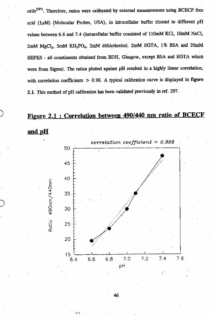
cells^^. Therefore, ratios were calibrated by external measurements using BCECF free
acid (IpM) (Molecular Probes, USA), in intracellular buffer titrated to different pH
values between 6 .6 and 7.4 (intracellular buffer consisted of llOmM KCl, lOmM NaCl,
2mM MgClz, 5mM KH2PO4, 2mM dithiothreitol. 2mM EGTA, 1% BSA and 20mM
HEPES - all constituents obtained from BDH, Glasgow, except BSA and EGTA which
were from S i^ a ) . The ratios plotted against pH resulted in a highly linear correlation,
with correlation coefficients > 0.98. A typical calibration curve is displayed in figure
2.1. This method of pH calibration has been validated previously in ref. 297.
Figure 2.1 : Correlation between 490/440 nm ratio of BCECF
and pH' c o r r e la t io n c o e f f i c ie n t = 0 .9 8 8
Eco
EcOG)
o0:
5 0
4 5
4 0
3 5
3 0
25
20
157.4 7.67.27.06.86.66 .4
pH
46
2.10: D-iwvo-Inositol 1.4.5-trisphosphate (IP,) assay*^^
Assays were conducted using the fHJIPj binding assay kit provided by Amersham
International following the instructions provided.
Fresh PBTLs were isolated and resuspended in 2.5% HSA at a concentration of
5xlOVml. 500pl of the cell suspension was then aliquoted into each 1.5ml eppendorf tube
and small volumes of the agonist were then added to the cells for 1 minute at 37°C. The
incubation was terminated by addition of 500pl ice cold 10% perchloric acid. After
leaving 10 minutes on ice, the samples were centrifuged for 5 minutes at 2,000g. 400pl
of the supernatant from each sample was transferred to a separate tube containing lOOpl
of lOmM EDTA, (pH 7.0).
The samples were neutralised by adding 300pl of a 1:1 (v v) mixture of Freon (1,1,2-
trichlorotrifluoroethane) and tri-n-octylamine, followed by vigorous mixing of the
separate phases on a vortex mixer. After centrifugation for 1 minute at 2000g, three
-phases were obtained. The upper phase was the neutralised sample plus all the water-
soluble components. A 400pl portion of the upper phase was removed for use in the IP3
binding assay kit, which is based on the ability of IP3 in the sample to displace fixed
amounts of pH]IP3 from the IP3 receptor. A standard curve was constructed in the range
0.2 to 25 pmol and displacement values obtained were converted to pmol IP3 by the use
of this curve.
2.11; Preparation of acrvlamide gels ” .
10% acrylamide mini gels were made as follows using a Biorad minigel apparatus.
47
To a 50ml tube is added 8ml of lower gel buffer, 13.1ml dH2 0 , 10.7ml 30% (w/v)
Acrylamide/Bis-acrylamide stock solution (Anachem,U.K.), 200|ils 10% Ammonium
persulphate and 20pls TEMED (BIO-RAD). After gentle mixing the solution was slowly
poured between the glass plates, up to the required mark. Then 200pl of gel overlay was
layered over the top and the gel left to set. Once the lower gel was set, the overlay was
washed off and the 5 % stacking upper gel was poured on. This was prepared by mixing
together, 5ml upper gel solution, 11.6ml dH2 0 , 3.3ml Acrylamide/Bis. solution, 200pl
ammonium persulphate and 200pl TEMED. Once the upper gel was poured on, the well-
comb was inserted and the gel left to set, after which the comb was removed and the
wells filled with running buffer.
2.12: Renaturable kinase assay.
Freshly isolated PBTLs were isolated and resuspended in 2.5% HSA. 250pl of the cell
suspension was then aliquoted into each 1.5ml eppendorf tube and incubated at 37°C for
20 minutes. To each tube 250pl of 2x the final working concentration of the test reagent
was then added, mixed well and the cells incubated for one hour at 37°C in a water bath.
After incubation the cells were washed twice in serum free RPMI-1640 media, the
supernatant aspirated and the cells solubilised in Laemmli sample buffer and boiled for 5
minutes. Samples were run on 10% (w/v) SDS-PAGE gels according to Laemmli^^®
along with rainbow molecular weight markers (High molecular weight range 14,300-
220,000 Da, from Amersham,U.K).
The gels were processed for renaturation essentially as described by Kameshita and
Fujisawa^^^ After washing SDS from the gels (20% isopropanol, 15mM Tris buffer pH
48
8.0) and dénaturation for 1 hour in 6M guanidine-HCL, samples were renatured
overnight at 4°C in 50mM Tris buffer pH 8.0, 50mM 2-mercaptoethanol and 0.04%
Tween-20. Following equilibration in phosphorylation buffer (lOmM HEPES pH 8.0,
2mM dithiothreitol, O.lmM EGTA, 5mM MgCl2 (BDH)) the gels were incubated with
[y^^PJATP (Amersham) (IpCi/ml) for 1 hour at room temperature. Finally, the gels were
washed extensively with 5% (w/v) trichloroacetic acid containing 1.0% (w/v) sodium
pyrophosphate and dried. Autophosphorylation was visualised by autoradiography using
Hyperfilm-MP from Amersham
2.13; Western blotting for tyrosine phosphorylation^^.
Freshly isolated PBTLs were isolated and resuspended in 2.5% HSA. 250pl of the cell
suspension was then aliquoted into each eppendorf and incubated at 37°C for 20 minutes.
To each tube 250|il of 2x the final working concentration of the test reagent was then
added, mixed well and the cells incubated for one hour at 37°C in a water bath. In
experiments in which the cells were pretreated with herbimycin A, 0.5pl of lO'^M
herbimycin A was added to the 250pl cell suspension to give a final concentration of 5 x
lO'^M and the cells incubated for 45 minutes at 37°C, after which the test reagent was
added as described above. After incubation the cells were washed twice in serum free
RPMI-1640 media, the supernatant aspirated and the cells solubilised in Laemmli sample
buffer and boiled for 5 minutes. Samples were run on 10% (w/v) SDS-PAGE gels
according to LaemmlP^ along with rainbow molecular weight markers (14,300-
220,000Da, Amersham).
49
After electrophoresis, the gels were washed in western blot buffer x3 every 10 minutes
whilst on a shaker (the BeUy Dancer - Stovall, Life Science Inc, USA.) and then the
proteins were transferred to nitrocellulose membranes (Hybond™ECL™, Amersham Life
Science, U.K.) using a Trans-Blot semi-dry transfer cell (BIO-RAD). Nonspecific sites
were blocked using 5% Bovine serum albumin in PBS/0.1% Tween-20 (PBS/tween) for
1 hour at room temperature. The membranes were washed 3 times in PBS/tween and
then the primary antibody - antiphosphotyrosine, (monoclonal IgG2b^ - clone 4G10),
(Upstate Biotechnology Incorporated, USA) was incubated with the membranes for 1
hour at a final dilution of 1/2000 in PBS/tween. The membranes were washed 3 times in
PBS/tween for a period of 1 hour and then incubated with a horseradish peroxidase-
labeUed sheep anti-mouse IgG (Amersham, U.K.) at a final dilution of 1/2000 in
PBS/tween. The membranes were washed 3 times in PBS/tween over a period of 1 hour
and the phosphotyrosine bands were revealed using the ECL detection system
(Amersham, U.K.) with ECL autoradiography film (Amersham, U.K.).
2.14: Immunofluorescence staining for actin and tubulin in
PBTLs<^>.
PBTLs were isolated as normal and treated with either lOpM Bis. or 50pM nocadazole
as described in section 2.14. After which the cells were fixed with 1% para
formaldehyde. The cells were then permeabilised by incubation at room ten^ierature with
0.1% lysophosphatidylcholine for 45 minutes on a rotary wheel.. The cells were then
washed xl in PBS/tween and resuspended in 400pl PBS/tween per treatment. Then 200pl
from each treatment was put into a separate eppendorf tube. Either of the anti-tubulin
antibodies was then added to the relevant tubes at a 1 in 1000 dilution (either, mouse
50
monoclonal antibody to polymerised p-tubulin, or depolymerised p-tubulin. Both from
Affiniti Research, Exeter, U.K.). Also the cells were stained for F-actin by the addition
of rhodamine-labelled phalloidin to a final concentration of 330nM. The cells were then
left at room temperature for one hour on a rotary wheel (slow speed 25 revs/min.). Cells
were then washed x3 in PBS/tween and resuspended in 200pl PBS/tween. The secondary
antibody (anti-mouse polyvalent immunoglobulins - FTTC conjugate) was then added to
each tube at a 1 in 500 dilution. The cells were incubated at room temperature on a
rotary wheel for one hour and then washed x4 in PBS/tween and resuspended in Citifluor
(Citifluor Ltd. London), after which slides were prepared. The cells were viewed using a
Zeiss Axioscop microscope equipped with epifluorescence and photographs taken using a
Nikon FX-35DX camera with Ilford PAN F 50 black and white film.
2.15; Measurement of Taurine
MOLT-4 cells were suspended at IxlOVml in 10% FCS media and incubated with
Taurine [1,2-^^C] (Amersham), O.lpCi/ml for 1.5 hours at 37°C. The cells were then
washed x2 and resuspended at IxlOVml in 0.5% FCS media. 1ml aliquots were put in
each eppendorf and the cells incubated at 37°C for 0 (control), 10, 20 or 30 minutes with
or without 0.5mls dHjO. The cells were then spun down in a microcentrifuge and 500pl
of the supernatant was collected (control) or 750pl from the hypotonically shocked cells.
The supernatant was then added to a scintillation vial along with 10ml Ultra gold
scintillation fluid (Packard, USA), and counted in a Packard 2500 TR liquid scintillation
analyzer. The cell pellets along with the rest of the supernatant were treated in the same
51
manner. The percentage efflux was calculated as follows [(DPM of supernatant of
sample x 2) 4- (total DPM of sample)] xlOO.
To test the effects of chloride channel blockers on taurine efflux, the protocol was
slightly different. The cells were labelled as above and then resuspended at 2xl0^/ml in
0.5% FCS media. 400|il of the cells were aliquoted into each eppendorf and to this was
added 400pl of either medium (control) or 2x final concentration of the channel blocker.
The cells were then incubated 5 minutes on a heating block after which they were
incubated for a further 20 minutes with or without (control) the addition of 400|il of
dHjO.
The percentage efflux was then calculated as above.
The chloride channel blockers tested were - NPPB [5-nitro-2-(3’phenylpropylamino)
benzoic acid], (LC laboratories, Woburn, MA. USA). Tamoxifen [[trans-2-[4-
(1,2,diphenyl-l-butenyl) phenoxy 1] -N, N-dimethy Icthylaminc] ], (Aldrich-chcmic),
Niflumic acid and Quinidine.
52

RESTJT.TS
53
Chapter 3
The Investigation for inducers of motility
Introduction
Before any investigations into the signal transduction mechanisms involved in PBTL
motility could be carried out, it was essential to first of all acquire a model in which
motility could be induced at will so that second messenger involvement could be
investigated. As well as using fresh human PBTLs, a leukaemia cell line was also used,
termed MOLT-4 cells. From this cell line, two sublines had previously been isolated,
termed motile and non-motile (see methods 2 .1).
Both PBTLs and non-motile MOLT-4 cells were used in experiments in which various
factors were tested for their effect at inducing motility. In initial experiments a simple
polarisation assay was used in place of a conventional transmigration assay. The degree
of lymphocyte polarisation or shape change correlates with the degree of motility
induced ^ ^ and the assay is a cheaper, better for screening and simpler assay than the
conventional transmigration assay.
Polarisation assay.
A summary of all the factors tested and their effects can be seen in Table 3.1. All the
factors tested were chosen because of reported effects on motility in lymphocytes or
other cell types in the literature. The chemokines M IP-la, MIP-lp, MCP-1 and IL-8
had minimal effects on PBTLs, causing polarisation to a maximum of 10% of the
population (all at lO'^M concentration), whereas RANTES had no effect whatsoever in
54
this system (all factors tested at concentration range of lO'^^M to lO'^M). The
chemokines had no polarisation effect at all on the non-motile MOLT-4 cells.
Substance P, vasoactive intestinal peptide (VIP), lysophosphatidic acid (LPA) and
platelet derived growth factor (PDGF), all caused polarisation in only 10% of the PBTL
population (all at lO'^M concentration) and had no effects on the non-motile MOLT-4
cells. IL -la, IL-10, epidermal growth factor and tumour necrosis factor a , all had no
effects on either PBTLs or non-motile MOLT-4 cells.
Although factors like MCP-1 were found to cause polarisation in 10% of the PBTL
population, it was decided that this would not provide sufficient sensitivity for
investigating the second messengers involved. However, 5 factors were found to cause
significant polarisation in PBTLs and the effects were significant enough to use these 5
as tools to induce motility in the subsequent investigations into the second messengers
involved.
Freshly isolated PBTLs are virtually all spherical, non-motile cells as can be seen in
figure 3.1. The biggest effect was seen with the protein kinase C inhibitor
bisindolyhnaleimide GF109203X (Bis), which caused up to 60% of the PBTL population
to polarise (figure 3.2), with a leading edge and a trailing uropod. IL-2, IL-15 and fetal
calf serum (FCS) also caused the PBTLs to change morphology in the same way as Bis.
Nocadazole, the microtubule disrupting agent caused shape change in 20 - 30% of
PBTLs, however, the morphology of the shape change was different from that induced
by the four other factors in that there was often no typical head or tail structures (fig.
3.3). Interleukins-2 and -15 were found to cause polarisation in 20 - 30% of the PBTLs
and FCS caused polarisation in 10 - 20% of the population.
55
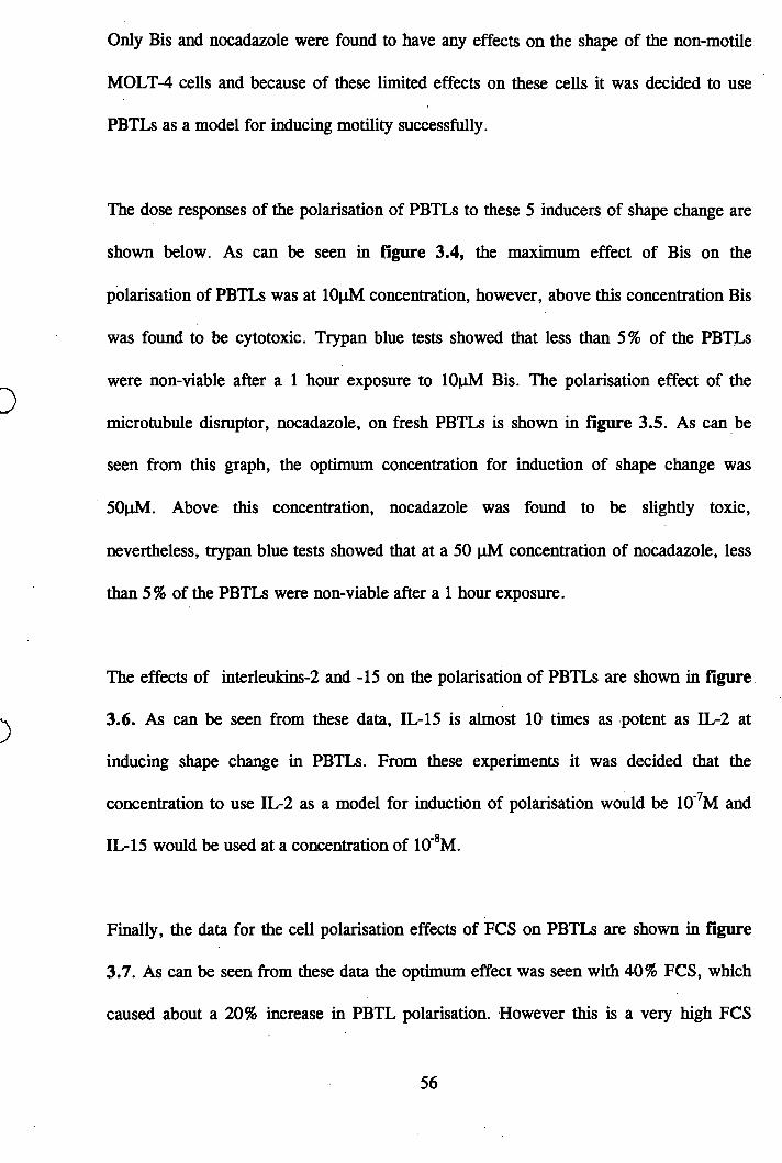
Only Bis and nocadazole were found to have any effects on the shape of the non-motile
MOLT-4 cells and because of these limited effects on these cells it was decided to use
PBTLs as a model for inducing motility successfully.
The dose responses of the polarisation of PBTLs to these 5 inducers of shape change are
shown below. As can be seen in figure 3.4, the maximum effect of Bis on the
polarisation of PBTLs was at lOpM concentration, however, above this concentration Bis
was found to be cytotoxic. Trypan blue tests showed that less than 5% of the PBTLs
were non-viable after a 1 hour exposure to lOpM Bis. The polarisation effect of the
microtubule disruptor, nocadazole, on fresh PBTLs is shown in figure 3.5. As can be
seen from this graph, the optimum concentration for induction of shape change was
50pM. Above this concentration, nocadazole was found to be slightly toxic,
nevertheless, trypan blue tests showed that at a 50 pM concentration of nocadazole, less
than 5% of the PBTLs were non-viable after a 1 hour exposure.
The effects of interleukins-2 and -15 on the polarisation of PBTLs are shown in figure
3.6. As can be seen from these data, IL-15 is almost 10 times as potent as IL-2 at
inducing shape change in PBTLs. From these experiments it was decided that the
concentration to use IL-2 as a model for induction of polarisation would be lO'^M and
IL-15 would be used at a concentration of lO'^M.
Finally, the data for the cell polarisation effects of FCS on PBTLs are shown in figure
3.7. As can be seen from these data the optimum effect was seen with 40% FCS, which
caused about a 20% increase in PBTL polarisation. However this is a very high FCS
56
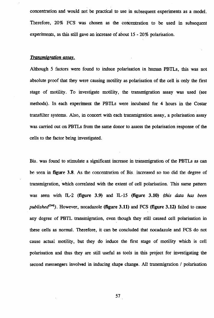
concentration and would not be practical to use in subsequent experiments as a model.
Therefore, 20% FCS was chosen as the concentration to be used in subsequent
experiments, as this still gave an increase of about 15-20% polarisation.
Transntieration assay.
Although 5 factors were found to induce polarisation in human PBTLs, this was not
absolute proof that they were causing motility as polarisation of the cell is only the first
stage of motility. To investigate motility, the transmigration assay was used (see
methods). In each experiment the PBTLs were incubated for 4 hours in the Costar
transfilter systems. Also, in concert with each transmigration assay, a polarisation assay
was carried out on PBTLs from the same donor to assess the polarisation response of the
cells to the factor being investigated.
Bis. was found to stimulate a significant increase in transmigration of the PBTLs as can
be seen in figure 3.8. As the concentration of Bis increased so too did the degree of
transmigration, which correlated with the extent of cell polarisation. This same pattern
was seen with IL-2 (figure 3.9) and IL-15 (figure 3.10) (this data has been
publishecP^). However, nocadazole (figure 3.11) and FCS (figure 3.12) failed to cause
any degree of PBTL transmigration, even though they still caused cell polarisation in
these cells as normal. Therefore, it can be concluded that nocadazole and FCS do not
cause actual motility, but they do induce the first stage of motility which is cell
polarisation and thus they are still useful as tools in this project for investigating the
second messengers involved in inducing shape change. All transmigration / polarisation
57
experiments were done in triplicate with 3 different donors and the results shown in
figures 3 are representative of 3 experiments.
Videomicroscopy of PBTLs treated with each of the five inducers of shape change,
confirmed the results of the transmigration assays, in that Bis, IL-2 and IL-15 caused the
cells to constantly change shape, whereas the FCS or nocadazole treated cells changed
shape initially but then remained frozen in this shape.
To summarise this stage of the project:- 5 factors were found to cause significant cell
polarisation in human PBTLs and these were Bis, nocadazole, IL-2, IL-15 and FCS.
However, only Bis., IL-2 and 11^15 caused transmigration of the cells across
nitrocellulose filters. Nevertheless, nocadazole and FCS can stiU be used to compare the
induction of motility (ie: polarisation) with the other three. Using these 5 factors as
tools, the next step was to analyse the second messengers induced to elucidate if there
were any common elements, which would then be contenders for part of a final common
pathway of motility.
58
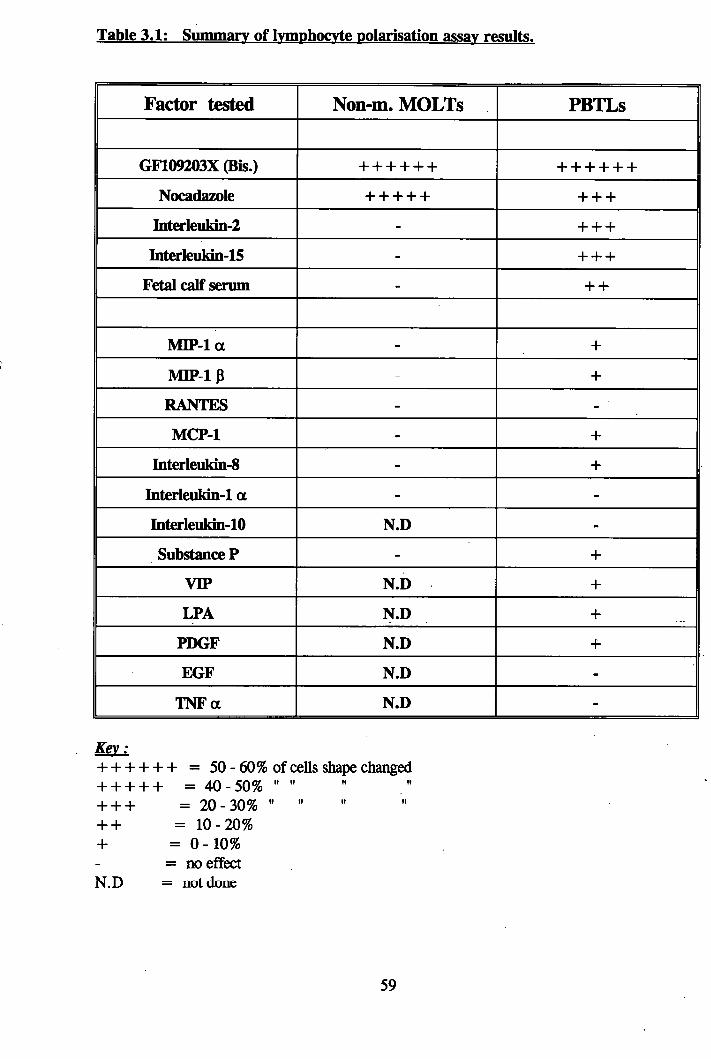
Table 3.1 : Summary of Ivmphocvte polarisation assay results.
Factor tested Non-m. MOLTs PBTLs
GF109203X (Bis.) + + + + + + + + + + + +
Nocadazole + -I- + + + + + +
Interleukin-2 - 4-4-4-
Interleukin-15 - 4-4-4-
Fetal calf serum - 4--H
MIP-1 a - 4-
MIP-IP 4-
RANTES - -
MCP-1 - 4-
Interleukin-8 - 4-
Interleukin-1 a - -
Interleukin-10 N.D -
Substance F - 4-
VIP N.D 4-
LPA N.D 4"
PDGF N.D 4-
EGF N.D -
TNFa N.D -
Ke\ :+ + + + + + = 50 - 60% of cells shape changed-H- + + + = 4 0 -5 0 % ..................+ + + = 20-30% " "+ + = 10 - 20%+ = 0 - 10%
= no effect N.D — not done
59

Figure 3.1: Freshly isolated Peripheral Blood T Lymphocytes (magnification x630).
O%»
• f
Figure 3.2: PBTLs treated with lOuM Bis, (magnification x630).
» #1
*
W
€
6 0
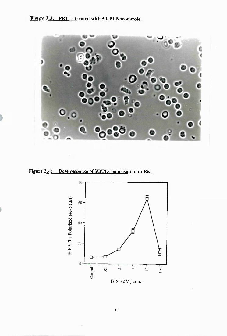
Figure 3.3: PBTLs treated with 50uM Nocodazole.
%
t%
Figure 3.4: Dose response of PBTLs polarisation to Bis.
Ico
"OOJc/3
UHCQCL
o S2 o
BIS. (uM) conc.
61
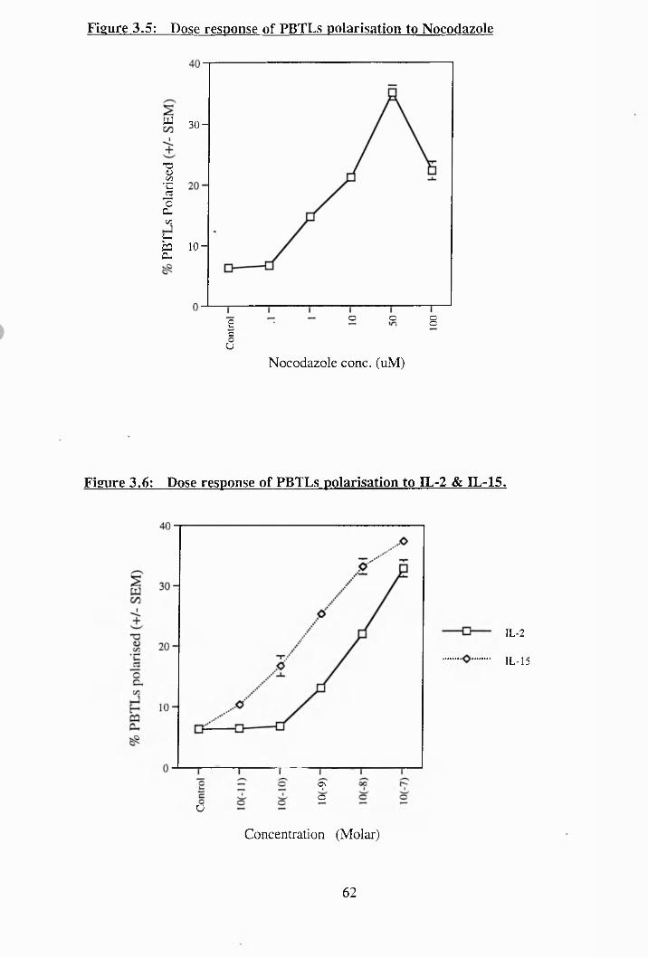
Figure 3.5: Dose response of PBTLs polarisation to Nocodazole
“ 30-
73 Uc/î
"CZ c3f£
C / D
H ,CQ 10 - CL
8o o2cU
Nocodazole conc. (uM)
Figure 3.6: Dose response of PBTLs polarisation to IL-2 & EL-15.
IL-2
IL-15
U9o
Concentration (Molar)
62
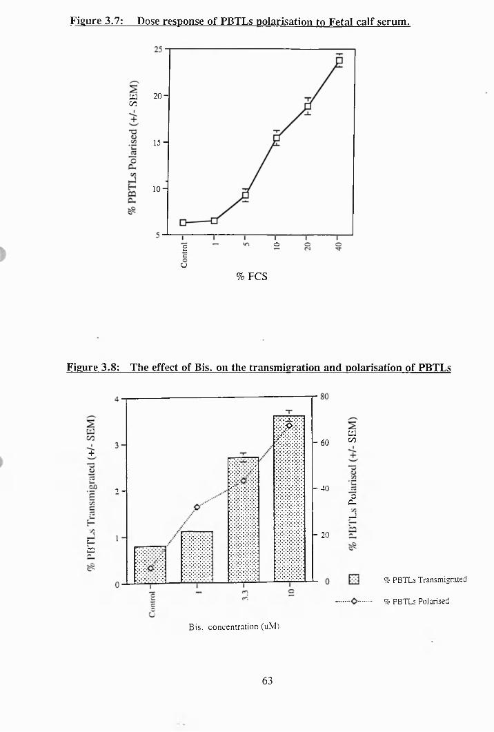
Figure 3.7: Dose response of PBTLs polarisation to Fetal calf serum.
w00
2 0 -
15-
1 0 -
o2cU
% PCS
Figure 3.8: The effect of Bis, on the transmigration and polarisation of PBTLs
- 60+
- 40? -
P*C2
- 20
a00
oC-u
% PBTLs Transmigrated
9c PBTLs Polarised
Bis. concentration (uM)
63
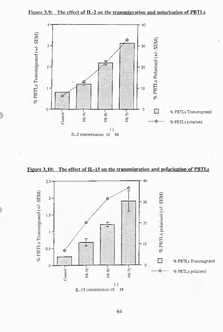
Figure 3.9: The effect of IL-2 on the transmigration and polarisation of PBTLs
+
o occ c
40
- 30
- 20
- 10
woc
£
C2C-
% PBTLs Transmigrated
% PBTLs polarised
( )IL-2 concentration 10 M
Figure 3.10: The effect of IL-15 on the transmigration and polarisation of PBTLs
2.5
■'m:
H 0.5
ac
40
- 30
- 20
- 10
00
% PBTLs Transmigrated
% PBTLs polarised
( )IL-15 concentration 10 M
64
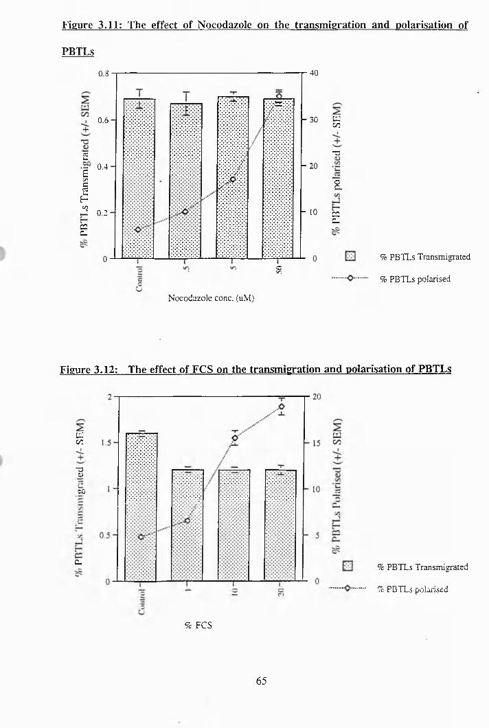
Figure 3.11: The effect of Nocodazole on the transmigration and polarisation of
PBTLs
0.8
0.6
£.g 0.4inC
IinU 0.2
C2C-
cV.
Nocodazole conc. (uM)
40
^ 30
- 20
- 10
tJ00
-a
a
C2C,
% PBTLs Transmigrated
% PBTLs polarised
Figure 3.12: The effect of FCS on the transmigration and polarisation of PBTLs
cu00
-3VCÛ
C_ % PBTLs Transmigrated
% PBTLs polarised
% FCS
65
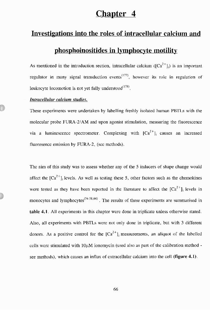
Chapter 4
Investigations into the roles of intracellular calcium and
phosphoinositides in lymphocyte motility
As mentioned in the introduction section, intracellular calcium ([Ca^^]j) is an important
regulator in many signal transduction events^^^ \ however its role in regulation of
leukocyte locomotion is not yet fully understood^
Intracellular calcium studies.
These experiments were undertaken by labelling freshly isolated human PBTLs with the
molecular probe FURA-2/AM and upon agonist stimulation, measuring the fluorescence
via a luminescence spectrometer. Complexing with [Ca^^Jj causes an increased
fluoresence emission by FURA-2, (see methods).
The aim of this study was to assess whether any of the 5 inducers of shape change would
affect the [Ca^^], levels. As well as testing these 5, other factors such as the chemokines
were tested as they have been reported in the literature to affect the [Ca^^], levels in
monocytes and lymphocytes^^" ’ ’ " . The results of these experiments are summarised in
table 4.1. All experiments in this chapter were done in triplicate unless otherwise stated.
Also, all experiments with PBTLs were not only done in triplicate, but with 3 different
donors. As a positive control for the [Ca^^]j measurements, an aliquot of the labelled
cells were stimulated with lOpM ionomycin (used also as part of the calibration method -
see methods), which causes an influx of extracellular calcium into the cell (figure 4.1).
66
Of the five inducers of polarisation tested, only Bis showed an effect on [Ca^^]; (figure
4.2). However, the increase in intracellular calcium observed seemed to be a fluorescent
artifact as the increase was not a typical calcium transient which rises and then
falls back to baseline (as in figure 4.7). As can be seen in fig 4.2, Bis was not
fluorescing in the abscence of PBTLs. Therefore, a possible answer was that once Bis
entered the cells it was binding to something intracellularly and this complex was
autofluorescing and giving a false signal in this system. To determine if this was indeed
the case, an experiment was carried out whereby, the PBTLs were labelled with FURA-
2/AM as usual and then before the experiment was run the cells were pretreated with
0.1% Triton X-100, which is a detergent which permeabilises the cells, thus allowing the
cells contents to be released into the extracellular media. Then the experiment is run and
Bis is added to the system. A representative experiment is shown in figure 4.3. As can
be seen. Bis is causing a response even with the cells pretreated with Triton, which
proves that Bis is not actually causing an increase in [Ca^^Jj, but is in fact fluorescing
non-specifically when it is in the presence of intracellular components of the cell.
Three of the chemokines, MIP-la, MIP-ip and MCP-1, seemed to exhibit very small
effects on the PBTLs [Ca^^], levels, as can be seen in figures 4.4 - 4.6. These small
effects could be due to the fact that the chemokines are specific for subsets of T cells and
these experiments contain large populations of all PBTLs. However, some clear results
were obtained by using non-motile MOLT-4 cells in this system. An example can be
seen in figure 4.7, in which 10 MCP-1 causes a transient increase in [Ca^^], of about
200 nM. It must be noted here also that MCP-1 has no polarisation effect at all on the
non-motile MOLT-4 cells. Similar [Ca^^]j fluxes in non-motile MOLT-4 cells were seen
67
with M IP-la, MIP-lp and RANTES, but these results were not as reproducible as the
MCP-1 effect. These 3 chemokines also had no effect on the polarisation of the MOLT
cells. Hence, it can be concluded from this data that an increase in [Ca^^], does not seem
to be essential for the induction of motility in lymphocytes.
Indeed, further experiments provided evidence for the theory that an increase in [Ca^^]j,
actually prevents the induction of motility. In the first set of these experiments, the
motile variant of the MOLT-4 cells were used in an experiment in which they were
exposed to various concentrations of ionomycin, a compound which permeabilises the
cell membranes and allows an influx of extracellular calcium into the cell. A
representative experiment can be seen in Figure 4.8. The motile MOLT-4 cells were
incubated with various concentrations of ionomycin for 30 minutes. As can be clearly
seen, as the concentration of ionomycin is increased, the cells become less polarised and
round up. The non-motile MOLT-4 cells were also tested for their response to ionomycin
exposure. As with the motile variant, the non-motile cells were incubated with various
concentrations of ionomycin (lO'^M to 10‘ ^M) for 30 minutes, but no effects on the
shape of the cells were observed (data not shown), thus indicating that an elevation in
[Ca^^li is not enough to cause shape change. Ionomycin was also tested to see whether
after induction of polarisation in PBTLs, ionomycin would reverse the shape change and
revert the cells back to a spherical, non-motile state. As can be seen in Figure 4.9, this
was indeed the result. Ionomycin reversed the polarisation induced by each of the five
factors after 30 minutes. Thus it seemed at this point that by increasing the [Ca^^Jj, this
in turn prevented motility.
68
The endosomal Ca^’*’- ATPase inhibitor, thapsigargin has been reported in the literature
to increase [Ca^^]j levels in lymphocytes^Thapsigargin was then tested to see if it,
like ionomycin reduced the extent of polarisation in motile MOLT-4 cells and prevented
the polarisation induced in PBTLs. Thapsigargin was found to have no effect on the
polarisation of motile MOLT-4 cells and no effect on their [Ca^^]j levels (data not
shown). In experiments in which PBTLs were pretreated with thapsigargin for 15
minutes and then stimulated with either of the five inducers of shape change for a further
45 minutes, it was observed that thapsigargin significantly prevented the induction of
polarisation in these cells (figure 4.10). To prove that thapsigargin does indeed cause an
increase in [Ca^^]j levels in PBTLs, experiments with FURA-2 labelled PBTLs were
carried out in which they were stimulated with lOuM thapsigargin. A representative
experiment can be seen in figure 4.11, in which thapsigargin induces a [Ca^^], increase
of about 135nM . However when the cells are exposed to 5mM EOT A for 5 minutes
before the experiment is started there is no increase in [Ca^^Jj, upon thapsigargin
stimulation. This shows that the majority of the increase in [Ca^^j; is not from the release
of Ca^’*’ from intracellular stores but a concomitant influx of Ca^^ from the extracellular
medium due to increases in plasma membrane Ca^^ permeability. It is thought that a
second messenger known as Ca^^ influx factor (GIF) is released or generated from the
endoplasmic reticulum or adjacent regions once the [Ca^^jj concentration in this
organelle falls beneath a critical level^^^ \ Further evidence to support the notion that an
increase in [Ca^^jj, actually prevents the induction of motility is shown in figure 4.12.
Again PBTLs that are pretreated with thapsigargin are unable to polarise upon
stimulation with the 5 inducers of shape change. However when the same experiment is
done again but the cells are pretreated with 5mM EGTA (to chelate the extracellular
69
Ca^^) before treatment with thapsigargin, then the PBTLs are able to polarise as normal,
thus indicating that the increase in [Ca^^]j induced by thapsigargin is of extracellular
origin.
Phosphoinositide studies.
In recent years phosphoinositides have been shown to play a key role in signal
transduction^^^ '^^^\ As mentioned in the introduction section, there are links between
alterations in polyphosphoinositides (ppl’s) and changes in [Ca^^], levels. Stimulation of
cell surface receptors initiates hydrolysis of a membrane-bound inositol lipid, which
produces at least two second messengers, - diacylglycerol (DAG) and inositol 1,4,5-
triphosphate (IP3). These messengers are generated by a membrane transduction process
comprising 3 main components: a receptor, a coupling G protein and phosphoinositidase
C. DAG acts by stimulating PKC whereas IP3 releases calcium from internal stores.
The first investigation into the role of ppl’s in the induction of motility was by assaying
IP3 production in PBTLs upon stimulation with inducers of shape change. This work was
done using the IP3 assay kit from Amersham (see methods). A representative experiment
can be seen in figure 4.13. It is clear from this data that there are no significant changes
in IP3 levels detected with this methodology when the PBTLs are stimulated by the
inducers of shape change. In these experiments the cells were stimulated with the
agonists for a standard time of one minute. From this data it seeems that IP3 is not
involved in a motility signal transduction pathway.
The involvement of the phosphoinositide 3-kinase (PI 3-kinase) in the induction of
motility was investigated by using specific inhibitors of the kinase. A fungal metabolite
70
known as wortmannin has proved to be a selective inhibitor of PI 3-kinase, if used at
sub-lOOnM concentration^PB TLs were preincubated with various concentrations of
wortmannin for 30 minutes and then stimulated with the 5 inducers of polarisation for 1
hour. A representative experiment can be seen in figure 4.14. Wortmannin has
prevented polarisation of the PBTLs but only at concentrations above lOOnM. It must be
noted at this point that wortmannin has an IC5 0 ~3nM, therefore at concentrations above
lOOnM, wortmannin is no longer specific for PI 3-kinase and affects other systems such
as phospholipase D, myosin light chain kinase and pleckstrin. Wortmannin was found to
have no effect at all on the motile MOLT-4 cells (data not shown).
Another specific PI 3-kinase inhibitor is a compound known as LY294002^^ '^\ which
inhibits PI 3-kinase activity with an IC5 0 of 1.4pM. This compound was also tested to see
whether it would prevent the induction of polarisation in PBTLs. PBTLs were
preincubated for 15 minutes in the presence of various concentrations of LY294002 and
then stimulated with the 5 inducers of polarisation for 1 hour. A representative
experiment can be seen in figure 4.15. The inhibitor was found to significantly inhibit
the induction of polarisation by all five inducers at concentrations as low as lOOnM.
Therefore these data suggest that PI 3-kinase may be involved in a motility pathway.
However, the LY294002 compound was found to have no effect whatsoever on the
polarisation of the motile MOLT-4 cells (data not shown). This seems to be a conflicting
result to the experiments with wortmannin, however the fact is that wortmannin is less
stable than LY294002 and this could explain why wortmannin did not inhibit motility.
71
When administered to animals, lithium induces subtle alterations in neural activity (for
example in manic-depressive illness and diurnal rhythms) and early development
(teratogenesis). Lithium is known to reduce the supply of inositol, the key substrate for
the phosphoinositide cascade by inhibiting some of the enzymes which hydrolyse the
inositol phosphates^^^^\ Thus lithium inhibits signal transduction indirectly by slowing
down the supply of the precursor lipid required to generate messengers such as IP3 and
DAG.
Therefore, as an additional approach to investigate the involvement of phosphoinositides
in motility, lithium chloride was tested for effects on the polarisation of PBTLs and
MOLT-4 cells. In figure 4.16 is a representative experiment in which PBTLs were
preincubated with lithium chloride for 30 minutes prior to stimulation with the various
inducers of polarisation for 1 hour. A concentration of lOOmM lithium was found to
inhibit polarisation by all five agonists, however at lower concentrations only the effects
of IL-2, IL-15 and FCS were blocked. Lithium chloride was found to have no effect
whatsoever on the polarisation of the motile variant of the MOLT-4 cells. Reports in the
literature suggest a concentration of lOmM for 30 minutes is sufficient to disrupt the
phosphoinositide cascade 181 ,1 9 9 )
To summarise this chapter; it was found that an increase in [Ca^^Jj was not essential for
the cells to polarise, in actual fact it was found that an increase in [Ca^^], levels
inhibited polarisation. PBTLs polarisation did not increase intracellular IP3 levels and the
role of PI 3-kinase in the signal transduction of motility was uncertain as one PI 3-kinase
72
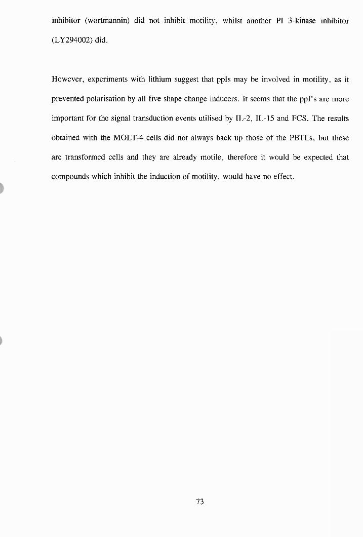
inhibitor (wortmannin) did not inhibit motility, whilst another PI 3-kinase inhibitor
(LY294002) did.
However, experiments with lithium suggest that ppis may be involved in motility, as it
prevented polarisation by all five shape change inducers. It seems that the ppl’s are more
important for the signal transduction events utilised by IL-2, IL-15 and FCS. The results
obtained with the MOLT-4 cells did not always back up those of the PBTLs, but these
are transformed cells and they are already motile, therefore it would be expected that
compounds which inhibit the induction of motility, would have no effect.
73
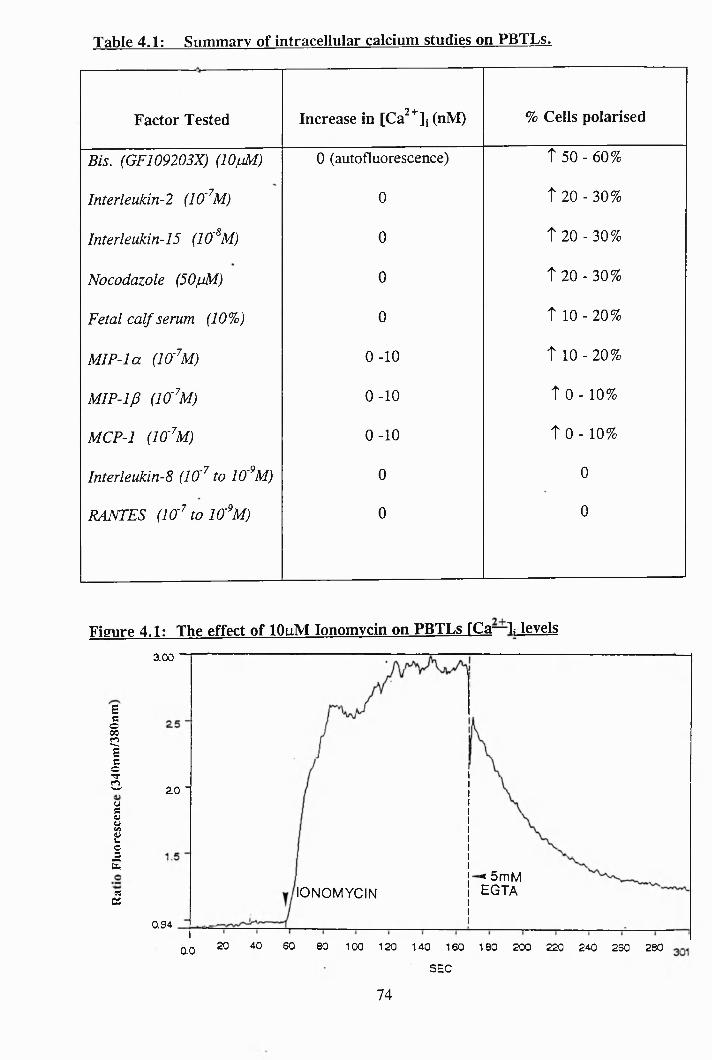
Table 4.1: Summary of intracellular calcium studies on PBTLs^---------------------------------------------
Factor Tested Increase in [Ca^^jj (nM) % Cells polarised
Bis. (GF109203X) (lOfM) 0 (autofluorescence) Î 50 - 60%
Interleukin-2 (10'^M) 0 Î 20 - 30%
Interleukin-15 (10'^M) 0 T20-30%
Nocodazole (50juM) 0 T 20 - 30%
Fetal calf serum (107o) 0 T 10 - 2 0 %
MIP-la (Iff^M) 0 -1 0 T 10 - 2 0 %
MIP-1J3 (la^M) 0 -1 0 t o - 10%
MCP-1 (lO'^M) 0 -1 0 t 0 - 10%
lnterleukin-8 (10'^ to lO'^M) 0 0
RANTES (la^tolO'^M) 0 0
Figure 4.1: The effect of 10uM Ionomycin on PBTLs FCa—L levels
Eco00ClEcoTf(2(j5aÎcsE«Pi
aoo
20 -
X 5m M EGTAIONOM YCIN
0.94
20 40 60 80 1 00 1 20 1 40 1 60 1 80 200 220 240 260 2800.0SEC
74
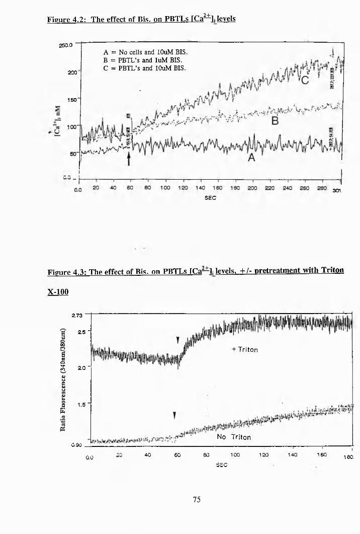
2+-Figure 4.2: The effect of Bis, on PBTLs [Ca—^ levels
25Q0
200
150
A = No cells and lOuM BIS. B = PBTL’s and luM BIS.C = PBTL’s and lOuM BIS.
301.SEC
Figure 4.3: The effect of Bis, on PBTLs ICa—L levels, + /- pretreatment with Triton
X-100
273
Ba25 -
o00
+ TritonBaoTf3 20 -
uuguao5Eo
I . I
No TritonÛ90
16040 80 100 120SO 180.0.0SEC
75
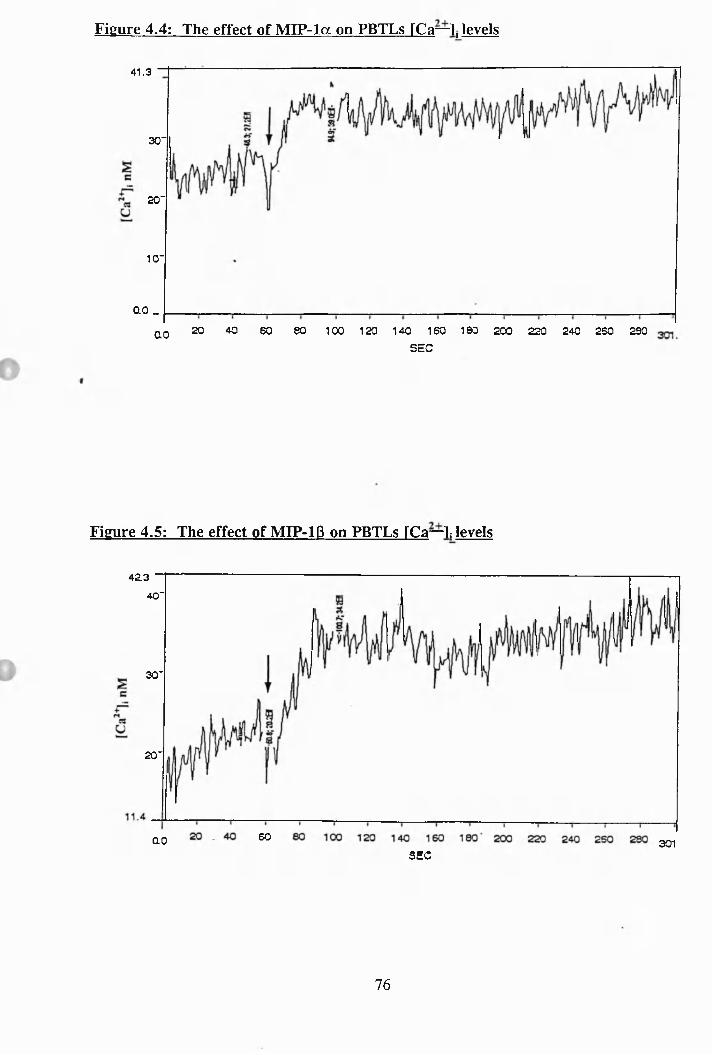
Figure 4.4: The effect of M IP -la on PBTLs FCa—1, levels
41.3
30“
20 “
10“
00 _
40 80 1 00 1 20 1 40 1 60 180 200 220 240 260 29020 60OOSEC
Figure 4.5: The effect of MIP-IB on PBTLs FCa—L levels
42.340“
30“
20“
60OO 301SEC
76
Figure 4.6: The effect of MCP-1 on PBTLs rCa—IJevels
eao
50"
U 40"
30"
24.9
00SEC
Figure 4.7: The effect of MCP-1 on non-motile MOLT-4 cells FCa—L levels
344.1
300"
250"
200"
150"
109.980 1 GO 120 1 40 1 60 1 80 200 220 240 260 28060402000
SEC
77
Figure 4.8: The effect of Ionomycin on the polarisation of motile MOLT-4 cells
I00
"3
18
i<u
80
60 -
4 0 -
2 0 -
T
T-Z3O
I
o
T3
Ionomycin conc.
Figure 4.9: The effect of Ionomycin on the polarisation of PBTLs
LUCO
TDCD(0
Oo_</)I— CO CL
CVI
(Sj
W-)pJj u.o
2 oc CNo c
E3 Control - No Ionomycin
H 0.1 uM Ionomycin
B l luM Ionomycin
^ lOuM Ionomycin
78
Figure 4.10: The effect of thapsigargin on the polarisation of PBTLs
60
W00
I
4*T3OJCO1OCLCOHS
[Ü3 No Thapsigargin
O .OluM Thapsigargin
H .luM Thapsigargin
^ luM Thapsigargin
lOuM Thapsigargin
79
Figure 4.11: The effect of thapsigargin on the FCa—]; levels of PBTLs
145.3
+10uM Thaps.1 0 0 "
50"
+ 5mM EGTA & lOuM T h ap s.
8.4
1 CO 120 140 1 c O60 600.0
Figure 4.12: The effect of thapsigargin on the polarisation of PBTLs pretreated
with 5mM EGTA
W 60
Et] No thapsigargin
O + lOuM Thapsigargin
+ lOuM Thapsigargin &5mMEGTA
80
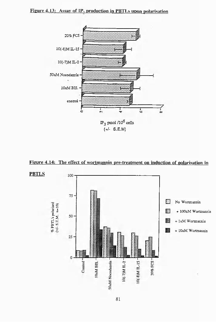
Figure 4.13: Assay of IP, production in PBTLs upon polarisation
20% FCS
10(-8)M IL-15
5Cu2yf Nocodazole
lOuM BIS,
concrol
c C4 00
IPg pmol /10° cells (+/- S.EM)
Figure 4.14: The effect of wortmannin pre-treatment on induction of polarisation in
PBTLS
2 cTc
g.- 1 ZPc c/i0.
+
No Wortmannin
+ lOOnM Wortmannin
+ luM Wortmannin
+ IGuM Wortmannin
81
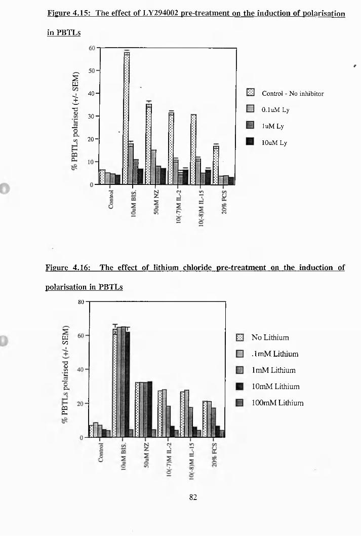
Figure 4.15: The effect of LY294002 pre-treatment on the induction of polarisation
in PBTLs
ÎJT3
30 H
â
40 -
2 0 -
NZ u2
cs
Control - No inhibitor
0.1 uM Ly
luM Ly
lOuM Ly
Figure 4.16: The effect of lithium chloride pre-treatment on the induction of
polarisation in PBTLs
80
W00
•SOO•d
â
2
No Lithium
.ImM Lithium
ImM Lithium
lOmM Lithium
lOOmM Lithium
82
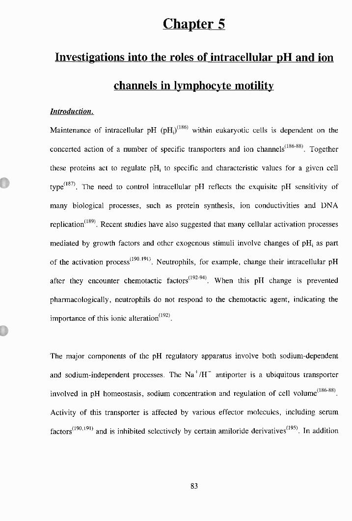
Chapter 5
Investigations into the roles of intracellular pH and ion
channels in lymphocyte motility
Introduction.
Maintenance of intracellular pH within eukaryotic cells is dependent on the
concerted action of a number of specific transporters and ion channels^ Together
these proteins act to regulate pHj to specific and characteristic values for a given cell
type^^^ \ The need to control intracellular pH reflects the exquisite pH sensitivity of
many biological processes, such as protein synthesis, ion conductivities and DNA
rep lication^R ecent studies have also suggested that many cellular activation processes
mediated by growth factors and other exogenous stimuli involve changes of pH, as part
of the activation process^^^^^^ \ Neutrophils, for example, change their intracellular pH
after they encounter chemotactic f a c t o r s ^ W h e n this pH change is prevented
pharmacologically, neutrophils do not respond to the chemotactic agent, indicating the
importance of this ionic alteration^
The major components of the pH regulatory apparatus involve both sodium-dependent
and sodium-independent processes. The Na"^/H^ antiporter is a ubiquitous transporter
involved in pH homeostasis, sodium concentration and regulation of cell volume^
Activity of this transporter is affected by various effector molecules, including serum
factors^^^ ’ ^ and is inhibited selectively by certain amiloride derivatives^^^^\ In addition
83
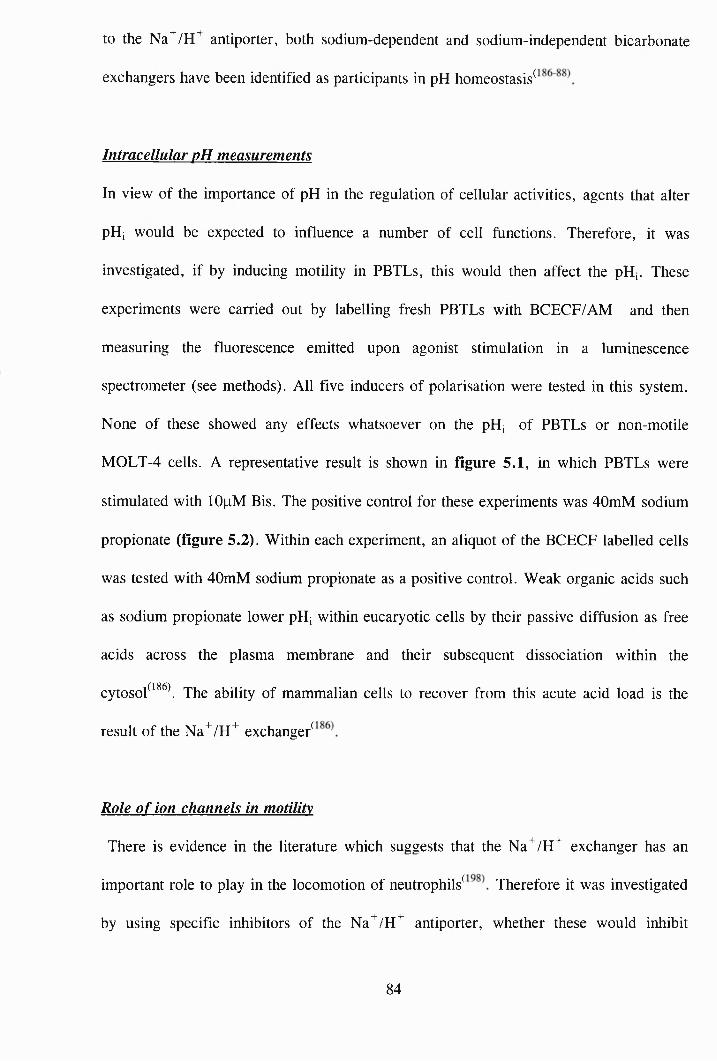
to the Na^/H"^ antiporter, both sodium-dependent and sodium-independent bicarbonate
exchangers have been identified as participants in pH homeostasis^
Intracellular pH measurements
In view of the importance of pH in the regulation of cellular activities, agents that alter
pHj would be expected to influence a number of cell functions. Therefore, it was
investigated, if by inducing motility in PBTLs, this would then affect the pH,. These
experiments were carried out by labelling fresh PBTLs with BCECF/AM and then
measuring the fluorescence emitted upon agonist stimulation in a luminescence
spectrometer (see methods). All five inducers of polarisation were tested in this system.
None of these showed any effects whatsoever on the pHj of PBTLs or non-motile
MOLT-4 cells. A representative result is shown in figure 5.1, in which PBTLs were
stimulated with lOpM Bis. The positive control for these experiments was 40mM sodium
propionate (figure 5.2). Within each experiment, an aliquot of the BCECF labelled cells
was tested with 40mM sodium propionate as a positive control. Weak organic acids such
as sodium propionate lower pHj within eucaryotic cells by their passive diffusion as free
acids across the plasma membrane and their subsequent dissociation within the
cytosol^^^^\ The ability of mammalian cells to recover from this acute acid load is the
result of the Na^/H^ exchange/
Role o f ion channels in motility
There is evidence in the literature which suggests that the Na^/H^ exchanger has an
important role to play in the locomotion of neutrophils^ Therefore it was investigated
by using specific inhibitors of the Na^/H^ antiporter, whether these would inhibit
84
motility in lymphocytes. The inhibitors used were amiloride and its de r i va t i ves^The
effect of these Na"""/H antiporter inhibitors on the polarisation of motile MOLT-4 cells
is shown in figure 5.3. The most potent compounds were amiloride 5-(N,N-
hexamethylane) (A130), amiloride 5-(N-methyl-N-isobutyl) (A149) and amiloride 5-(N-
ethyl-N-isopropyl) (A171), which virtually rounded up all the cells at lOOpM. The other
two inhibitors, amiloride 5-(N,N-dimethyl)-hydrochloride (A 125) and amiloride
hydrochloride (A ll3) were less potent as they only had effects at 500pM and above. In
these experiments, the cells were incubated with the inhibitors for a period of 1 hour. All
experiments in this chapter were done in triplicate and all experiments with PBTLs were
also done with 3 different donors, unless otherwise stated. In figures 5.4 - 5.8 are
representative results of experiments in which PBTLs were pre-incubated with the
particular amiloride derivative for 30 minutes and then the PBTLs were incubated with
an inducer of shape change for 1 hour. A130 (fig. 5.4), A149 (fig. 5.5) and A171 (fig.
5.6), all inhibited polarisation of the PBTLs at a concentration of lOOpM. A125
inhibited shape change at a higher concentration of 500uM (fig. 5.7) and A113 was the
least potent as it inhibited shape change significantly at a concentration of ImM (fig.
5.8).
To prove that the amiloride compounds were indeed preventing motility by inhibiting the
action of the Na'^/H^ antiporter, it was investigated whether the amilorides would
decrease the pHj of motile MOLT-4 cells. In theory, if the amilorides were indeed
blocking the antiports, this would lead to a build up of ions within the cell and a
decrease in pHj. A representative experiment can be seen in figure 5.9, in which BCECF
labelled motile MOLT-4 cells were stimulated with lOOpM A130 after 60 seconds.
85
Although there is an increase in fluorescence upon stimulation with A130, there is also
an increase in the negative control (ie: in the presence of no cells). This autofluorescence
was seen with all the amiloride derivatives. Attempts at the same experiment were made
using an alternative pHj probe termed SNARF-1 which works at different wavelengths to
BCECF. Although the amilorides were found not to autofluoresce at the wavelengths
used by SNARF-1 , this probe was found not to be sensitive enough to pick up any
changes in pHj (data not shown).
Therefore it could not be proved directly that the amilorides were blocking the antiports.
However an indirect way of investigating whether the antiport is being blocked is by
testing the ability of the cells to recover from an acute acid load. In figure 5.10 can be
seen a representative experiment in which control motile MOLT-4 cells were subjected to
a 40mM dose of sodium propionate which caused an acute drop in pHj which then
returned to normal over time due to the activity of the Na^/H^ antiporter which pumps
out the ions. However, MOLT-4 cells which have previously been treated for 5
minutes with lOOpM A171, have their recovery from the sodium propionate load
impaired resulting in a sustained acidification, suggesting that the antiporters have been
blocked. However, it must be noted at this point that these experiments were very
difficult to do and the result shown in fig. 5.10 is not very convincing, but there is a
better example of this type of experiment in fig. 5.19.
A question which arises from this data is whether the decrease in pHj caused by
inhibition of the antiporter is the reason that amiloride is blocking polarisation of the
cells. Thus, an experiment was set up whereby motile MOLT-4 cells were exposed to
various concentrations of sodium propionate and the resulting pHj measured and extent
86
of cell polarisation quantified. As can be seen in figure 5.11, as the concentration of
sodium propionate increases from ImM, at which there is no effect, to 40mM, the extent
of intracellular acidification increases. In figure 5.12 is a representative experiment in
which motile MOLT-4 cells are subjected to various doses of sodium propionate for 5,
15 and 30 minutes. A dose of ImM has little effect on cell polarisation as on pH,, but a
concentration of 20mM causes the cells to round up and to a further extent with 40mM.
This pattern correlates with the effects on pHj. It does seem however that the effects are
transient, with a more pronounced effect after 5 minutes but after 30 minutes exposure to
sodium propionate there is no effect on the cells polarisation, which is probably due to
the cells recovery from the acidification. This data suggests that it is the intracellular
acidification which is responsible for making the cells non motile.
Is there a difference in the pH, levels between motile and non motile MOLT-4 cells?
Also is there a difference in their ability to recover from an acute acid load? To answer
these questions a number of experiments were carried out whereby both motile and non-
motile MOLT-4 cells were subjected to 40mM sodium propionate and then the recovery
to basal pH; was measured over a 30 minute period. A representative result is shown in
figure 5.13. In this experiment there is a difference in basal levels of pH,, between the
two cell types, however this varied from one experiment to another, with the motile cells
exhibiting a higher pHj in one experiment and the non-motiles exhibiting a higher one in
another, therefore, very little can be read into this. Also, both types of MOLT-4 cells
were identical in their ability to recover from an acute intracellular acidification. Hence,
it can be concluded that pHj, is not an important difference between motile and non-
motile MOLT-4 cells.
87
As well as the amiloride compounds, a number of propietary compounds were tested
which are more potent than commercially available amiloride derivatives and with ICgo’s
for antiport inhibition of sub-pM concentrations in other experimental systems (N.
Matthews, personal communication). These inhibitors were termed inhibitor 1 , inhibitor
2, etc.. As in the previous experiment with the amiloride derivatives, the inhibitors were
incubated at 37°C with motile MOLT-4 cells for a period of 1 hour. A representative
experimental result can be seen in figure 5.14. As can be clearly seen, only inhibitor 3
had any effect on the motile MOLT-4 cells polarisation, rounding up all the cells at
lOOpM. The antiport inhibitors were then investigated in experiments with PBTLs, to see
if they could block the induction of shape change. PBTLs were incubated for 30 minutes
with the inhibitor being tested and then the PBTLs were stimulated by one of the
inducers of polarisation for 1 hour. Inhibitor 1 had some effects at reducing the extent of
polarisation (figure 5.15). Inhibitor 2 had no effect whatsoever (data not shown).
Inhibitor 3 was found to be the most effective of the six inhibitors, as it nearly blocked
the effects of the inducers of polarisation at a concentration of lOOpM (figure 5.16).
Inhibitor 4 was slightly more potent than inhibitor 1 (figure 5.17) and inhibitor 5 had a
slight effect at lOOpM concentration (figure 5.18). Inhibitor 6 had no effect at all (data
not shown).
From this data, inhibitor 3 proved to be the most potent compound and as with the
amiloride A171, it was investigated whether inhibitor 3 was actually blocking the
antiporter, by testing the ability of inhibitor 3 treated motile MOLT-4 cells to recover
from an acute acid load. A representative experiment is shown in figure 5.19, in which
the control cells (not treated with an antiporter inhibitor) recover from the acid load as
8 8
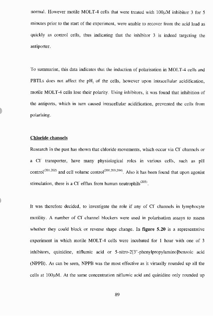
normal. However motile MOLT-4 cells that were treated with lOOpM inhibitor 3 for 5
minutes prior to the start of the experiment, were unable to recover from the acid load as
quickly as control cells, thus indicating that the inhibitor 3 is indeed targeting the
antiporter.
To summarise, this data indicates that the induction of polarisation in MOLT-4 cells and
PBTLs does not affect the pH; of the cells, however upon intracellular acidification,
motile MOLT-4 cells lose their polarity. Using inhibitors, it was found that inhibition of
the antiports, which in turn caused intracellular acidification, prevented the cells from
polarising.
Chloride channels
Research in the past has shown that chloride movements, which occur via Cl’ channels or
a Cr transporter, have many physiological roles in various cells, such as pH
control^^°^’ ° and cell volume control ° ' ^ ' " \ Also it has been found that upon agonist
stimulation, there is a Cl’ efflux from human neutrophils^^°^\
It was therefore decided, to investigate the role if any of Cl channels in lymphocyte
motility. A number of Cl channel blockers were used in polarisation assays to assess
whether they could block or reverse shape change. In figure 5.20 is a representative
experiment in which motile MOLT-4 cells were incubated for 1 hour with one of 3
inhibitors, quinidine, niflumic acid or 5-nitro-2 [3'-phenyIpropylamino]benzoic acid
(NPPB). As can be seen, NPPB was the most effective as it virtually rounded up all the
cells at lOOpM. At the same concentration niflumic acid and quinidine only rounded up
89
half the cells. Tamoxifen (trans-) which as well as being an anti-oestrogen, is a chloride
channel blocker and significantly inhibited motile MOLT-4 polarisation as can be seen in
figure 5.21. At a concentration of 12.5pM, tamoxifen inhibited all polarisation of the
motile MOLT-4 cells. Tamoxifen had the same potency at blocking induction of
polarisation in PBTLs, as can be seen in figure 5.22. This is a representative experiment
in which tamoxifen was incubated with the PBTLs for 15 minutes and then the cells were
stimulated with one of the five inducers of polarisation for 1 hour. Using this same
experimental protocol, the other Cl channel inhibitors were tested for their ability to
block polarisation in PBTLs. NPPB was effective at a concentration of lOOpM (figure
5.23), whereas quinidine (figure 5.24) and niflumic acid (figure 5.25) were less
effective, both only blocking the extent of polarisation by about 25%, at a lOOpM
concentration.
To determine if these chloride channel blockers were indeed targeting the chloride
channels, a number of experiments were done whereby MOLT-4 cells were labelled with
‘ C Taurine (see methods). It has been validated in the literature that the physiological
role of volume regulated chloride channels relates not only to their permeability to the
inorganic Cl’ but to their permeability to larger organic osmolytes such as taurine^^^ ’ ^
and so therefore the measurement of taurine efflux from hypotonically shocked, taurine
labelled cells is an indicator of volume regulated chloride channel activity. In figure
5.26 is a representative experiment in which both motile and non-motile MOLT-4 cells
were compared in terms of taurine efflux after hypotonic shock. As can be seen from this
data, taurine uptake by both types of cells were roughly the same and upon hypotonic
shock to increase the cell volume, the volume regulated chloride channels are activated
90
in both cell types. Also the volume regulated chloride channel (as measured by taurine
efflux) does not appear to be spontaneously activated in motile MOLT-4 cells. Upon
hypotonic shock, the amount of taurine released did not differ that much between 10
minutes and 30 minutes in the presence of water. In figure 5.27, is a representative
experiment, whereby motile MOLT-4 cells were treated with one of the Cf channel
blockers for 5 minutes and then the cells were hypotonically shocked to test whether the
blocker could indeed block the taurine efflux. In these experiments the cells were
hypotonically shocked for 20 minutes. As can be seen from fig. 5.27, the control cells
all released a small amount of taurine, however it was observed that tamoxifen (12.5pM)
seemed to cause some spontaneous activation (or toxicity) of the channels as even
without hypotonic shock the tamoxifen treated cells were releasing more taurine than
control cells. Upon hypotonic shock, the only compound which seemed to have any
effect was NPPB which blocked half of the taurine efflux. Niflumic acid and Quinidine
had no effect on blocking the taurine efflux and tamoxifen actually caused more taurine
efflux than control cells. The concentrations of the blockers used in these experiments
was the concentration that gave maximum inhibition of motility in motile MOLT-4 cells
(figures 5.20 and 5.21).
Therefore, this data suggests that blocking the volume regulated chloride channels does
not seem to be the mechanism by which these compounds (except tamoxifen) inhibit
motility and they must be targetting other elements of the cell machinary.
It was also found that none of the Cf channel blockers had any effect on the pHj of either
PBTLs or motile MOLT-4 cells (data not shown). Hence, these data suggest that the Cl
91
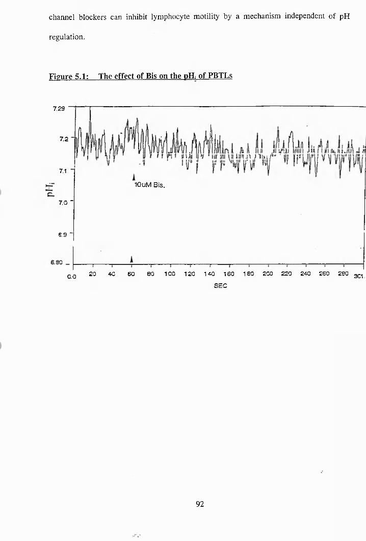
channel blockers can inhibit lymphocyte motility by a mechanism independent of pH
regulation.
Figure 5.1: The effect of Bis on the pH; of PBTLs
7.29
7.2
II ÙI
7.1
lOuM Bis.
7.0 -
6.9 -
80 ICO 120 1 40 1 60 1 80 200 220 240 260 280 3 .,20 40 600.0SEC
92
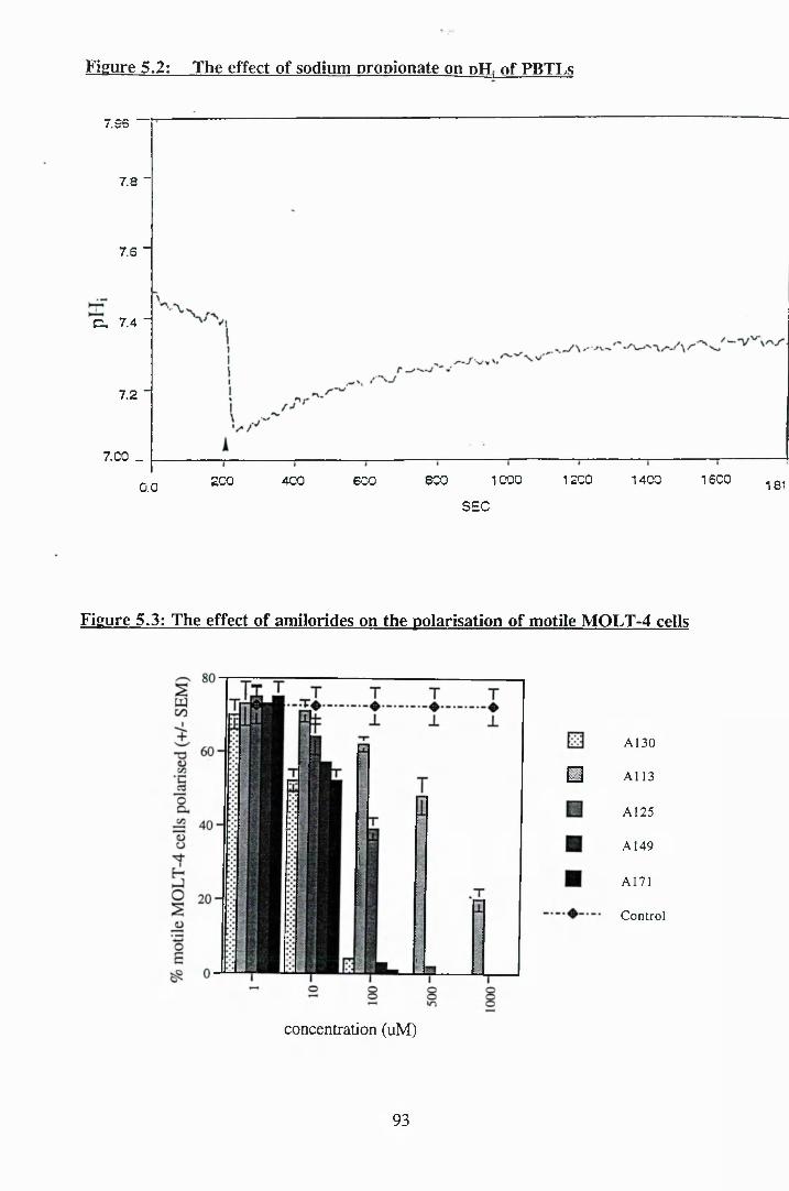
Figure 5.2: The effect of sodium propionate on dH, of PBTT.s
7.96
7.8 -
7.6 -
C . 7.4 -
7.2 -
7. CO _
16C01000 1200 1400800400200 600 1810.0SEC
Figure 5.3: The effect of amilorides on the polarisation of motile MOLT-4 cells
□A130
A113
A125
A149
A171
Control
concentration (uM)
93
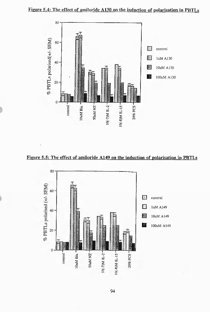
Figure 5.4: The effect of amiloride A130 on the induction of polarisation in PBTT s
a
•aDo n'C
-2oc-C/3
caCL
control
luM A130
lOuM A130
lOOuM A 130
C/) N csca 2S zo oin
c
oCN
Figure 5.5: The effect of amiloride A149 on the induction of polarisation in PBTLs
W 6 0 -
CO N5 2 j1 z3 so cm p
o'0?
E3 control
a luM A149
lOuM A149
H lOOuM A149
94
Figure 5.6: The effect of amiloride A171 on the induction of polarisation in PBTLs
CQ 20
N03 Z
S3
o
zoWl
control
luM A171
lOuM A171
lOOuM A171
SOQ
Figure 5.7: The effect of amiloride A125 on the induction of polarisation in PBTLs
"C 40
control
lOuM A125
lOOuM A125
500uM A 125
95
Figure 5.8: The effect of amiloride A113 on the induction of polarisation in PBTLs
m 60 control
luM A113
lOuM A1I3
lOOuM A1I3
ImM A1I3
Figure 5.9: The effect of amiloride A130 on the pH; of motile MOLT-4 cells
100.0
+ Cells
80"
No Cells® 6 0 -
lOOuMA13040-
31.2
20 40 60 80 100 120 1 6 014 00.0 188.jEC
96
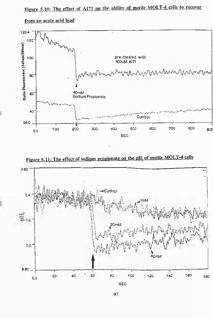
Figure 5.10: The effect of A171 on the ability of motile MOLT-4 cells to recover
from an acute acid load
129.4 -
120 'I
r
ao-f3sg
100
60"
■Apre-treated with lOOuM A171
N'l/40'
25.0
A40 mMSodium Propionate
Control
0.0 100 200 ' 300— I---------------------------------1—
400 500
SEC
600 700 600 905.
Figure 5.11: The effect of sodium propionate on the pH; of motile MOLT-4 cells
7.60
Control7.4
ImM
20mM
\,>rV7.0 -
40 mM
6.80 _16060 80 100 1404020 120 180QO
SEC
97
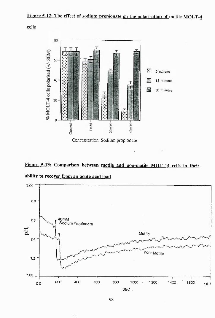
Fkure 5.12: The effect of sodium propionate on the polarisation of motile MOLT-4
cells
6 0 -
a 40
EiiiJ 5 minutes
P I 15 minutes
H 30 minutes
Concentration Sodium propionate
Figure 5.13: Comparison between motile and non-motile MOLT-4 cells in their
ability to recover from an acute acid load
7.96
7.8 -
7.6 ■^ Sodium Propionate
Motile7.4 -
7.2 "
7.00 _200 400 600 1000800 1200 1400 1 6 0 00.0 181'
SEC
98
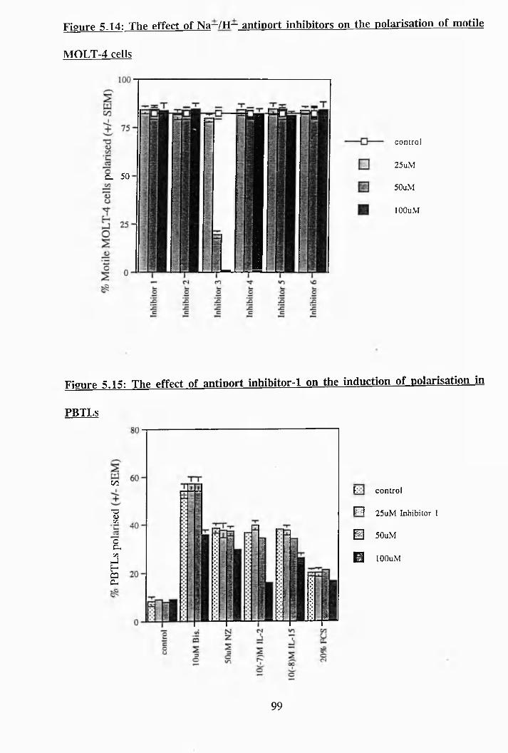
Figure 5.14: The effect of Na~/H— antioort inhibitors on the polarisation of motile
MOLT-4 cells
CL 50
control
25uM
50uM
lOOuM
Figure 5.15: The effect of antioort inhibitor-1 on the induction of polarisation in
PBTLs
cn
1a;caa.
I
k control
P i 25uM Inhibitor 1
@ 50uM
0 lOOuM
99
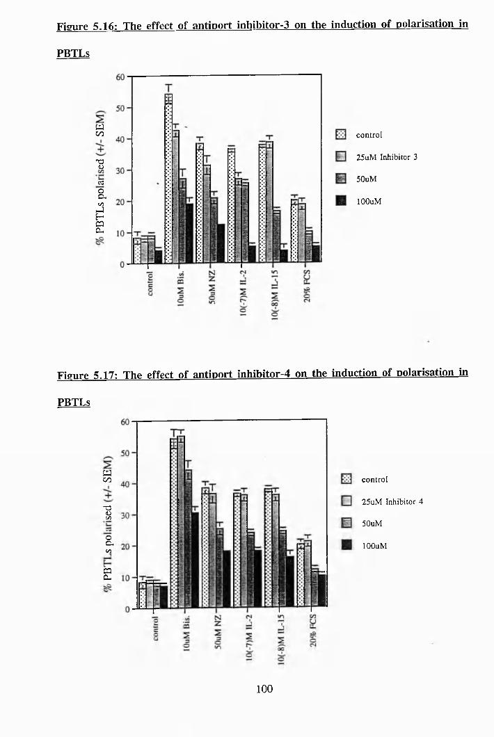
Figure 5.16: The effect of antioort inhibitor-3 on the induction of polarisation in
PBTLs
wCO
■Segagg
control
25uM Inhibitor 3
50uM
lOOuM
Figure 5.17: The effect of antiport inhibitor-4 on the induction of polarisation in
PBTLs
wCO
"3(ÜV]
aC /5
g
control
25uM Inhibitor 4
50uM
lOOuM
100
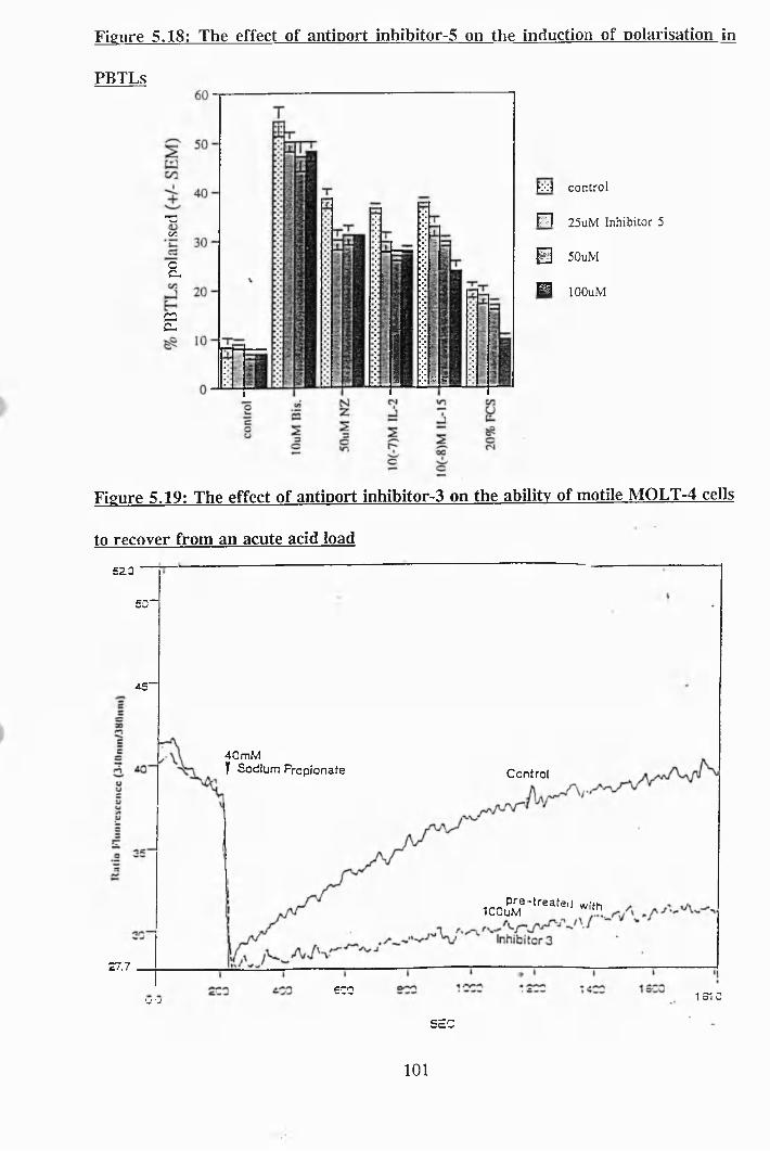
Figure 5,18: The effect of antiport inhibitor-5 on the induction of polarisation in
PBTLs
T3c/3
cc,
CÛc-
m *r
M control
P I 25uM Inhibitor 5
0 50uM
M lOOuM
Figure 5.19: The effect of antioort inhibitor-3 on the ability of motile MOLT-4 cells
to recover from an acute acid load
5 2 0
50“
4 5 “
40 mWT Sodium Propionate Control
^CoP--rea,e,i .,,hICOuM
27.7
600 1 3 1 0V -J
SEC
101
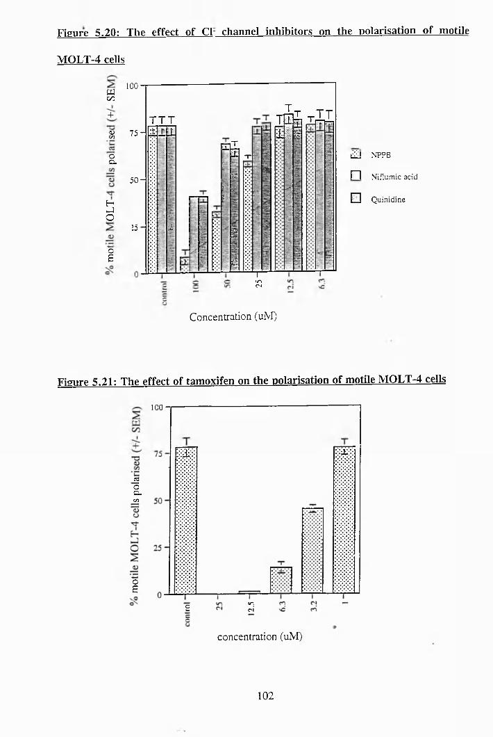
Figure 5.20: The effect of Cl~ channel inhibitors on the polarisation of motile
MOLT-4 cells
UJc/34-
H)CO
OCL
5O
o£
100
7 5 -
5 0 -
25
T T T• t j i î
2
FtITX T
isr
>I
g
£l
%
i f el \ i
TinrJ
a
i i J NPPB
r ~ l N iflum ic acid
B Q uin id ine
Concentration (uM)
Figure 5.21: The effect of tamoxifen on the polarisation of motile MOLT-4 cells
100
i3oC lc/5
OE
in.r i£
concentration (uNI)
102
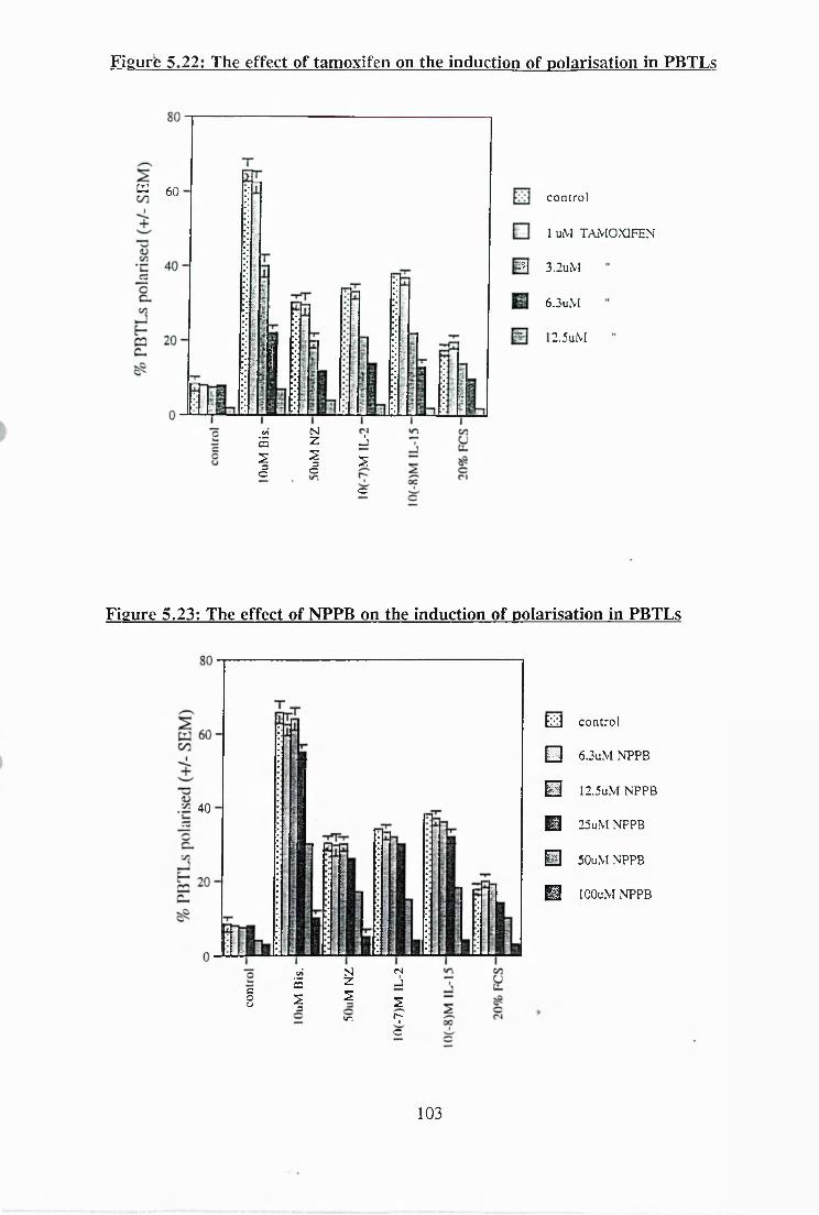
Figure 5.22: The effect of tamoxifen on the induction of polarisation in PBTLs
^ 60 control
1 uM TAMOXIFEN
m 3.2uM
6.3uM
2.5uM
to N5 Z J1 1 zC c
c
Figure 5.23: The effect of NPPB on the induction of polarisation in PBTLs
40
to" N fS
5 Z JcoCJ 1 z
s
iri rpc
EliJ control
0 6.3uM NPPB
0 I2.5uM NPPB
^ 25uM NPPB
@ 50uM NPPB
^ lOOuM NPPB
103
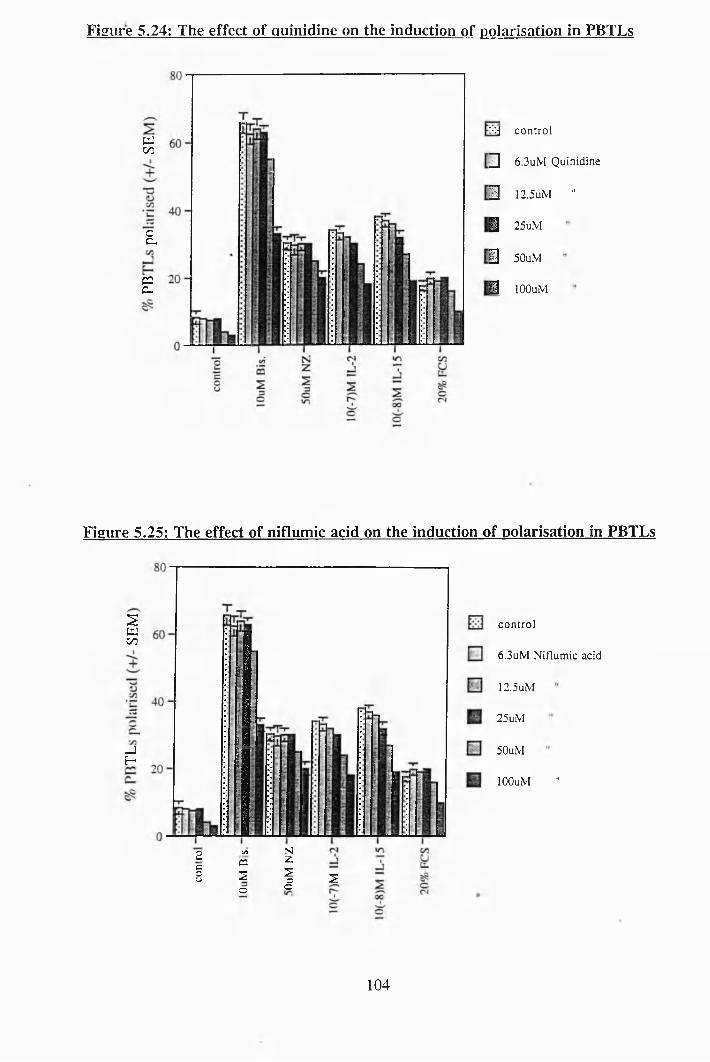
Figure 5.24: The effect of quinidine on the induction of polarisation in PBTLs
tu00
E
C2C_
xj control
^ 6.3uM Quinidine
Ü 12.5uM "
M 25uM
3 50uM
1 lOOuM
Figure 5.25: The effect of niflumic acid on the induction of polarisation in PBTLs
2tu00
-JE -
o c/3 Nc cs 2cs Z 1 S
c c
control
6.3uM Niflumic acid
12.5uM
25uM
50uM
lOOuM "
104
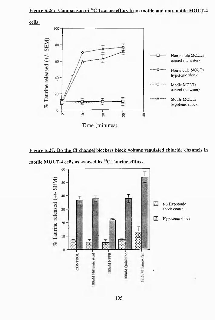
Figure 5.26: Comparison of Taurine efflux from motile and non-motile MOLT-4
cells.100
o omo
Non-motile MOLTs control (no water)
Non-motile MOLTs hypotonic shock
Motile MOLTs control (no water)
Motile MOLTs hypotonic shock
Tim e (minutes)
Figure 5.27: Do the Cl'channel blockers block volume regulated chloride channels in
motile MOLT-4 cells as assayed by Taurine efflux.
D 20
No Hypotonic shock control
F I Hypotonic shock
105
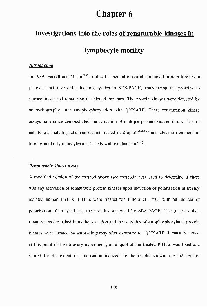
Chapter 6
Investigations into the roles of renaturahle kinases in
lymphocyte motility
Introduction
In 1989, Ferrell and Martin™, utilized a method to search for novel protein kinases in
platelets that involved subjecting lysates to SDS-PAGE, transferring the proteins to
nitrocellulose and renaturing the blotted enzymes. The protein kinases were detected by
autoradiography after autophosphorylation with [y^^PjATP. These renaturation kinase
assays have since demonstrated the activation of multiple protein kinases in a variety of
cell types, including chemoattractant treated neutrophils^^®^™ and chronic treatment of
large granular lymphocytes and T cells with okadaic acid^ ’®\
Renaturable kinase assay
A modified version of the method above (see methods) was used to determine if there
was any activation of renaturable protein kinases upon induction of polarisation in freshly
isolated human PBTLs. PBTLs were treated for 1 hour at 37°C, with an inducer of
polarisation, then lysed and the proteins separated by SDS-PAGE. The gel was then
renatured as described in methods section and the activities of autophosphorylated protein
kinases were located by autoradiography after exposure to [y^^PjATP. It must be noted
at this point that with every experiment, an aliquot of the treated PBTLs was fixed and
scored for the extent of polarisation induced. In the results shown, the inducers of
106
polarisation were found to induce shape change to the expected degree as shown in
chapter 1.
The results shown in figures 6.1 and 6.2 are representative of 4 experiments, all done
with different PBTL donors. In figure 6.1, the most prominent event is the Bis induced
increase in autophosphorylation of a 58kDa band (indicated by the arrow). This same
band was induced in all four experiments by Bis. Also in figure 6.1, there is a significant
increase in autophosphorylation of a doublet (indicated by the arrows), of 98 and 92kDa
by lL-2, lL-15 and nocadazole. This same doublet seems to be increased also in figure
6.2 by lL-2, Bis and PCS, however these were not found to be reproducible in further
experiments.
Collaborative work with C.Southern in this Institute has shown that activation of the
58kDa kinase by Bis occurs after 1 minute and increases up to a maximum level after 30
minutes. This time scale also correlates with the degree of polarisation of non motile
MOLT-4 cells induced by Bis. In the case of the 58kDa band, phosphoamino acid
analysis revealed autophosphorylation on threonine residues and recent work has
identified the 58kDa kinase as being mammalian ste20-like kinase-1 (MST-1) (personal
communication, C.Southern).
Hence, there is some evidence to suggest that this 58kDa kinase (MST-1) may be
involved in a motility pathway, however, it is not conclusive as lL-15, lL-2, PCS and
nocadazole do not seem to affect the kinase as Bis does.
107
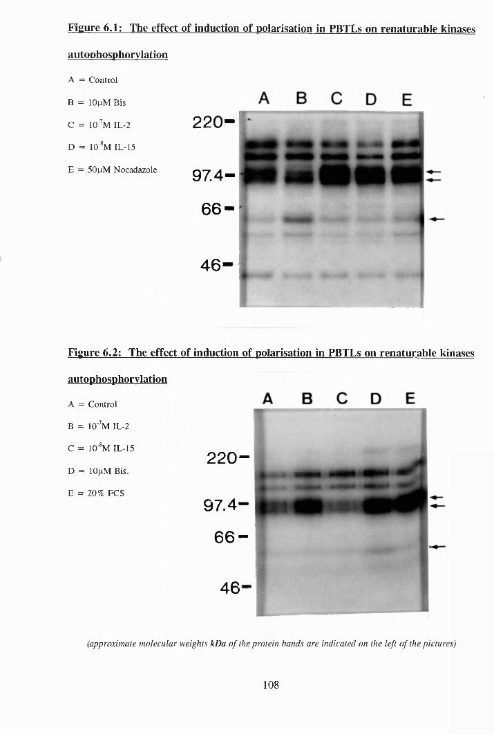
Figure 6.1: The effect of induction of polarisation in PBTLs on renaturable kinases
autophosphorvlation
A = Control
B = lOfiM Bis
C = lO'^M IL-2
D = lO 'V IL-15
E = 50|iM Nocadazole
220-Tr
97 .4 -
66 -
4 6 -
Figure 6.2: The effect of induction of polarisation in PBTLs on renaturable kinases
autophosphorvlation
A = Control
B = lO'^M IL-2
C = 10 V IL-15
D = 10|j,M Bis.
E = 20% PCS
220 -
9 7 .4 -
66 —
46
(approximate molecular weights kDa o f the protein bands are indicated on the left o f the pictures)
108
Chapter 7
Investigations into the roles of tyrosine phosphorylation
in lymphocyte motility
Introduction
Tyrosine phosphorylation is now recognised as a key mechanism by which cytokine
receptors and antigen receptors of lymphocytes initiate intracellular events^^^^\ also, the
phosphorylation of signalling proteins on tyrosine is essential for cellular regulation of
growth and differentiation.
Tyrosine phosphorylation studies
These experiments were undertaken (as detailed in methods section) by incubating
freshly isolated human PBTLs with one of the five inducers of polarisation at 37°C for
one hour. Then the cells were lysed and the proteins separated by SDS-PAGE, after
which the gels were subjected to Western blotting with the nitrocellulose blots being
probed for phosphotyrosine. With each experiment, an aliquot of the treated cells was
checked for extent of polarisation induced. In all experiments shown, there was a normal
degree of polarisation induced as shown in chapter 1.
As can be seen in figure 7.1 (representative of 3 experiments with 3 different donors) ,
only IL-2 caused any change in tyrosine phosphorylation levels in whole cell lysates of
treated PBTLs. There is a significant increase in tyrosine phosphorylation of a protein
band of 'llOkDa molecular weight (indicated by the arrow). There is unequal loading
109
of protein in the FCS treated lane, however other experiments showed FCS to have no
effect on tyrosine phosphorylation levels (data not shown). Later experiments with IL-15
showed it to have the same effect as IL-2, (figure 7.2), (representative of 3 experiments
with 3 different donors), in that it also caused tyrosine phosphorylation of a protein band
of 'llOkDa. To further test the involvement of tyrosine phosphorylation in lymphocyte
motility, experiments were carried out in which established tyrosine kinase inhibitors
were incubated with the PBTLs prior to them being induced to polarise by Bis, etc.
Genistein, tyrphostin A25 and tyrphostin A47, were all tested for inhibiting induction of
polarisation in PBTLs by all five inducers of polarisation, by incubating the cells with
the relevant inhibitor for 30 mins at 37°C and then stimulating the cells with one of the
inducers of polarisation for one hour at 37°C. The inhibitors were tested at
concentrations from lOOnM to ImM and all experiments were done in triplicate with
cells from 3 different donors. None of these inhibitors had any effect at preventing the
extent of polarisation induced (data not shown). However, similar experiments with the
tyrosine kinase inhibitor herbimycin A (IC5 0 = lp,M), proved otherwise (figure 7.3).
PBTLs were incubated with 5x10’ herbimycin A for 30 minutes (as done previously by
P.C.Wilkinson^^^^) and then stimulated with the inducers of polarisation for 1 hour at
37°C. As can be seen from figure 7.3, herbimycin A has significantly reduced the extent
of polarisation induced by Bis, nocadazole, IL-2, IL-15 and FCS. The experimental data
shown is representative of 3 experiments done with 3 different blood donors.
Thus, this data suggests that there is something common between the 5 different
treatments that induces polarisation, that is in turn inhibited by herbimycin A.
110
The next obvious experiment to do was to investigate whether IL-2 and IL-15 could still
cause tyrosine phosphorylation of the llOkDa protein if the cells were pretreated with
herbimycin A (thus reducing the extent of motility). Representative results of these
experiments are shown in figures 7.4 and 7.5. The experiments as usual were done 3
times with different blood donors. The cells were treated as in figure 7.3. As can be
seen in figure 7.4, herbimycin A has not prevented the tyrosine phosphorylation of the
llOkDa band by IL-2 or IL-15 as can be seen in figure 7.5. This data suggests that
herbimycin A is not preventing the llOkDa protein (pi 10) from being tyrosine
phosphorylated, so therefore herbimycin A must be inhibiting its target downstream from
the pi 10. This theory does not rule out the possibility that the pi 10 might be part of a
motility signal transduction pathway, common to both IL-2 and IL-15.
So what is the identity of the pi 10? Evidence from the literature suggests the closest
possibility is Janus kinase-3 ^ ' ' (JAK-3), which has a molecular weight of ~120kDa.
However, attempts to immunoprecipitate JAK-3 from PBTL cytoplasmic fractions with
anti-JAK-3 antibodies (see methods), proved unsuccessful (data not shown).
To summarise this chapter, no common changes in tyrosine phosphorylation levels of
PBTLs, were found to be induced by the 5 inducers of polarisation. However, IL-2 and
IL-15 were found to both cause an increase in tyrosine phosphorylation of pi 10, which
could be JAK-3. The tyrosine kinase inhibitor, herbimycin A was found to significantly
reduce the extent of polarisation induced by all five of the inducers, but it did not affect
the tyrosine phosphorylation of pi 10 by IL-2 and IL-15.
I l l
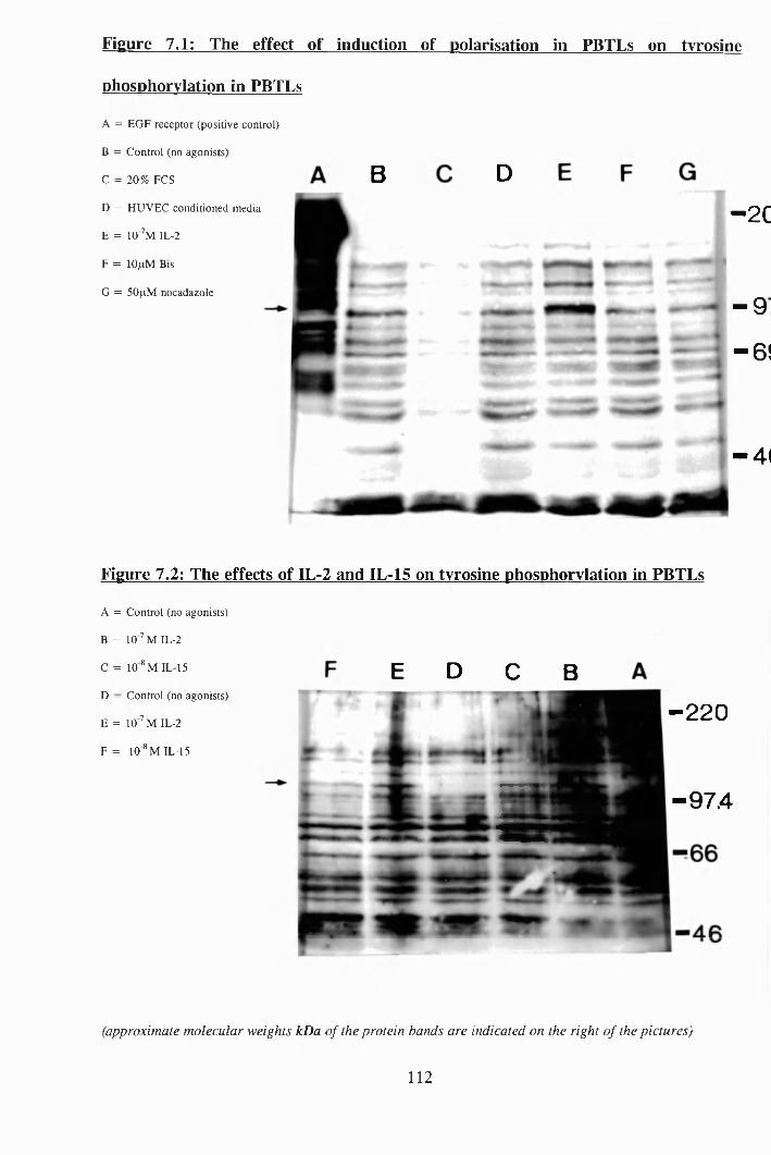
Figure 7.1: The effect of induction of polarisation in PBTLs on tyrosine
phosphorylation in PBTLs
A = EGF receptor (positive control)
B = Control (no agonists)
C = 20% FCS
D = HUVEC conditioned media
E = IO ’m IL-2
F = lOfxM Bis
G = 50fxM nocadazole
B D
-2C
- 9
" 6 !
-41
Figure 7.2: The effects of IL-2 and IL-15 on tyrosine phosphorylation in PBTLs
A = Control (no agonists)
B = lO '^M IL-2
C = 10 ® M IL-15
D - Control (no agonists)
E = IO ’ m IL-2
F = 10* M IL-15
E D C 8
-2 2 0
■ -9 7 4
(approximate molecular weights kDa of the protein bands are indicated on the right o f the pictures)
112
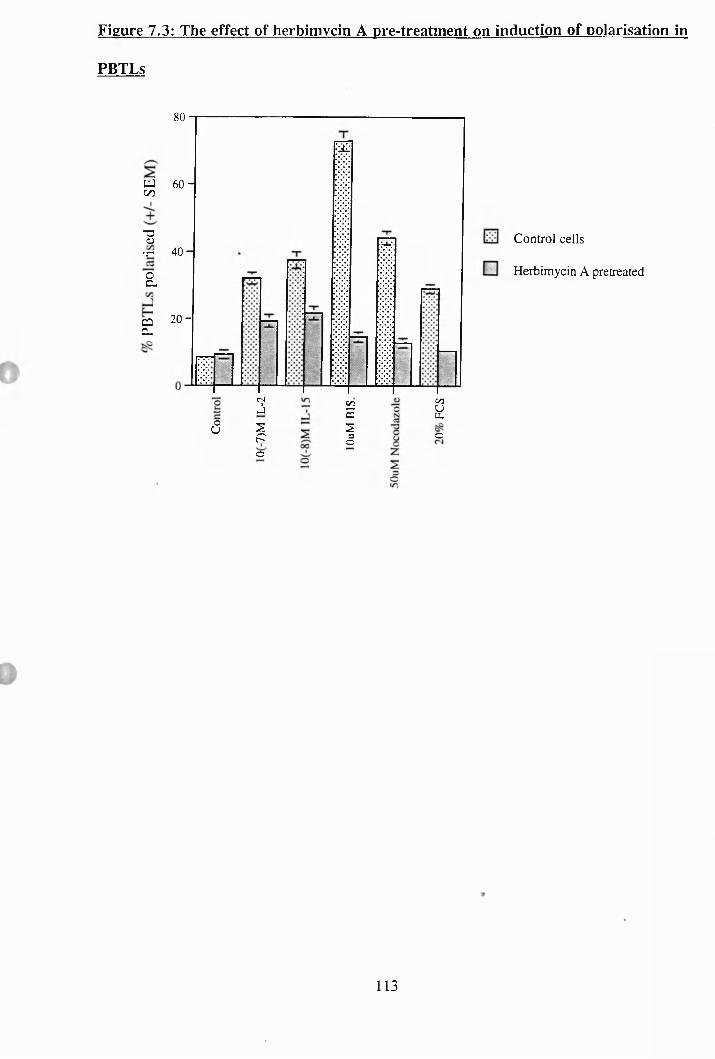
Figure 7.3: The effect of herbimycin A pre-treatment on induction of polarisation in
PBTLs
80
CQCL
60 -UJ 00
-o(U•r 40 H
oCL
2 0 -
Ô3spo
S22Ic
Control cells
Herbimycin A pretreated
gc
113
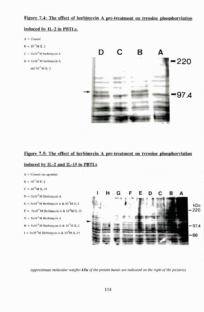
Figure 7.4: The effect of herbimycin A pre-treatment on tyrosine phosphorylation
induced by IL-2 in PBTLs.
A = Control
B = 10'’ M IL-2
C = 5x10'^ M herbimycin A
D = 5x10'^ M herbimycin A
and 10 IL-2
D B220
1^97.4
Figure 7.5: The effect of herbimycin A pre-treatment on tyrosine phosphorylation
induced by IL-2 and IL-15 in PBTLs
A = Control (no agonists)
B = IO ’ m IL-2
C = 10 ® M IL-15
D = 5x10'^ M Herbimycin A
E = 5x10'^ M Herbimycin A & lO'^M IL-2
F = 5x10^ M Herbimycin A & 10‘*M IL-15
G = 5x10'^ M Herbimycin A
H = 5x10 ’ M Herbimycin A & lO’ M IL-2
I = 5x10 M Herbimycin A & 10 ®M IL-15
I H
S5a*“
- 2 2 0
-97 .4
(approximate molecular weights kDa of the protein bands are indicated on the right of the pictures)
114
Chapter 8
Investigations into the roles of microtubules in
lymphocyte motility
Introduction
All the three major cytoskeletal fibres - microfilaments, microtubules and intermediate
filaments - and their associated proteins seem likely to contribute to the establishment of
cell polarity and the process of cell translocation across surfaces. As discussed in the
main introduction, the microfilament system is known to be intimately linked to the
mechanism of movement and force generation^ The contributions of the microtubule
and intermediate filament systems, however, are less clea/^^^\
Microtubules studies
As tools for investigating the importance of microtubules in lymphocyte motility, a
number of microtubule - directed drugs were utilised in polarisation assays with fresh
human PBTLs. Amongst the many microtubule - directed drugs, the taxol family are
unusual in that they stabilise cytoskeletal microtubules against depolymerisation^^^^ and
induce polymerisation and bundling. It is now known that taxol inhibits the dynamic
reactions at microtubule ends, suppressing both treadmilling and dynamic instability
A representative experiment can be seen in figure 8.1, in which PBTLs were incubated
with various concentrations of taxol for 30 minutes at 37°C and then stimulated with one
of the 5 inducers of polarisation for 1 hour. All experimental results in this chapter were
done in triplicate with 3 different blood donors. As can be seen from figure 8.1, taxol
has significantly inhibited the extent of polarisation induced, even at concentrations as
115
low as O.ljiM, This data suggests that microtubules must be remodelled, before shape
change can take place.
Vinblastine binding to tubulin occurs rapidly and binding is rapidly reversible. Beginning
with concentrations higher than approximately IpM, vinblastine depolymerises
microtubules by causing splaying and peeling of protofilaments at both microtubule ends
in Also unlike colchicine (described below), free vinblastine binds directly to
microtubule ends without first forming a complex with soluble tubulin^^^°\ Also
vinblastine does not become incorporated into the tubulin lattice of the microtubule, but
incorporates strictly at the microtubule ends^^^ \ As can be seen in figure 8.2, fresh
PBTLs incubated with various concentrations of vinblastine for 1 hour (at 37°C) exhibit
rounding up of the cells at high concentrations and did not induce polarisation. Trypan
blue tests showed that PBTLs incubated with lOOpM vinblastine were less than 10%
non-viable (data not shown).
In figure 8.3, can be seen a representative experiment whereby PBTLs were incubated
for 1 hour at 37°C in various concentrations of colchicine. As the concentration increases
to lOOpM the extent of polarisation increases. In contrast with vinblastine, which binds
directly to the ends of the microtubules, colchicine either cannot bind at all to
microtubule ends or it does so very inefficiently. Instead, it first binds to soluble tubulin
and forms a final-state tubulin-colchicine complex, which then incorporates at the
microtubule ends through a polymerisation-dependent pathway
116
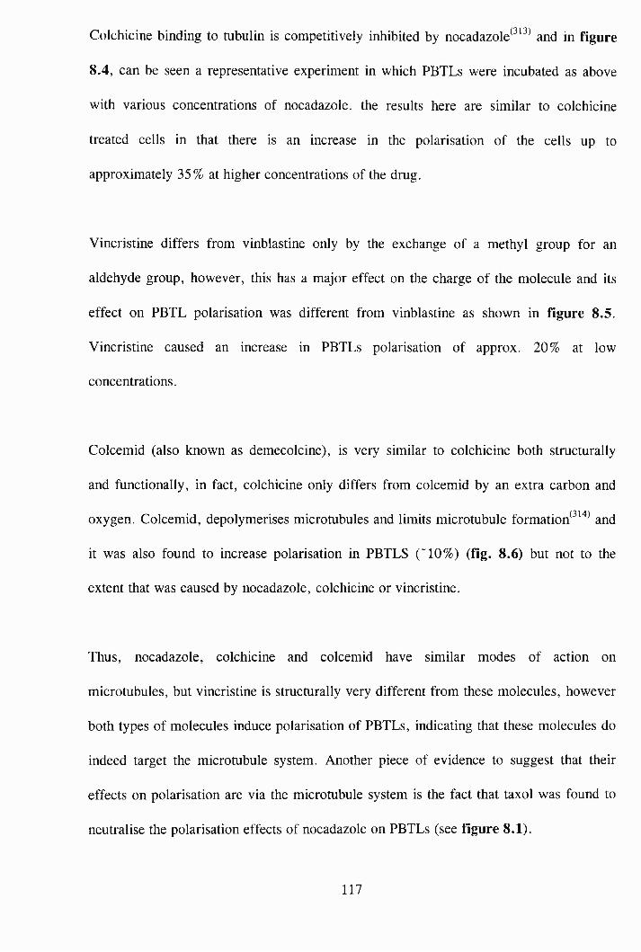
Colchicine binding to tubulin is competitively inhibited by nocadazole^^^^ and in figure
8.4, can be seen a representative experiment in which PBTLs were incubated as above
with various concentrations of nocadazole. the results here are similar to colchicine
treated cells in that there is an increase in the polarisation of the cells up to
approximately 35% at higher concentrations of the drug.
Vincristine differs from vinblastine only by the exchange of a methyl group for an
aldehyde group, however, this has a major effect on the charge of the molecule and its
effect on PBTL polarisation was different from vinblastine as shown in figure 8.5.
Vincristine caused an increase in PBTLs polarisation of approx. 20% at low
concentrations.
Colcemid (also known as demecolcine), is very similar to colchicine both structurally
and functionally, in fact, colchicine only differs from colcemid by an extra carbon and
oxygen. Colcemid, depolymerises microtubules and limits microtubule formation ^ " and
it was also found to increase polarisation in PBTLS ("10%) (fig. 8.6) but not to the
extent that was caused by nocadazole, colchicine or vincristine.
Thus, nocadazole, colchicine and colcemid have similar modes of action on
microtubules, but vincristine is structurally very different from these molecules, however
both types of molecules induce polarisation of PBTLs, indicating that these molecules do
indeed target the microtubule system. Another piece of evidence to suggest that their
effects on polarisation are via the microtubule system is the fact that taxol was found to
neutralise the polarisation effects of nocadazole on PBTLs (see figure 8.1).
117
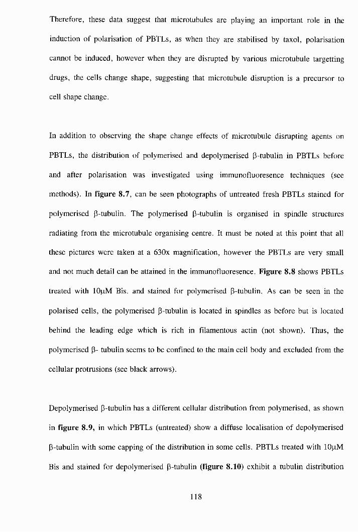
Therefore, these data suggest that microtubules are playing an important role in the
induction of polarisation of PBTLs, as when they are stabilised by taxol, polarisation
cannot be induced, however when they are disrupted by various microtubule targetting
drugs, the cells change shape, suggesting that microtubule disruption is a precursor to
cell shape change.
In addition to observing the shape change effects of microtubule disrupting agents on
PBTLs, the distribution of polymerised and depolymerised (3-tubulin in PBTLs before
and after polarisation was investigated using immunofluoresence techniques (see
methods). In figure 8.7, can be seen photographs of untreated fresh PBTLs stained for
polymerised (3-tubulin. The polymerised P-tubulin is organised in spindle structures
radiating from the microtubule organising centre. It must be noted at this point that all
these pictures were taken at a 630x magnification, however the PBTLs are very small
and not much detail can be attained in the immunofluoresence. Figure 8.8 shows PBTLs
treated with lOpM Bis. and stained for polymerised P-tubulin. As can be seen in the
polarised cells, the polymerised p-tubulin is located in spindles as before but is located
behind the leading edge which is rich in filamentous actin (not shown). Thus, the
polymerised p- tubulin seems to be confined to the main cell body and excluded from the
cellular protrusions (see black arrows).
Depolymerised p-tubulin has a different cellular distribution from polymerised, as shown
in figure 8.9, in which PBTLs (untreated) show a diffuse localisation of depolymerised
P-tubulin with some capping of the distribution in some cells. PBTLs treated with lOpM
Bis and stained for depolymerised p-tubulin (figure 8.10) exhibit a tubulin distribution
118
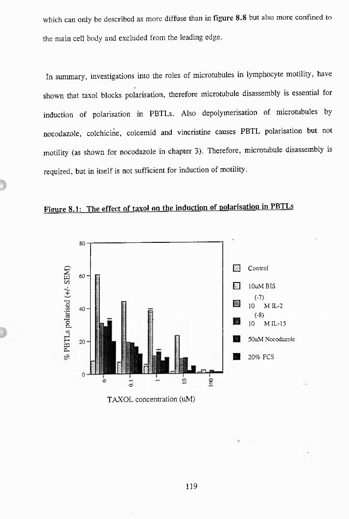
which can only be described as more diffuse than in figure 8.8 but also more confined to
the main cell body and excluded from the leading edge.
In summary, investigations into the roles of microtubules in lymphocyte motility, have
shown that taxol blocks polarisation, therefore microtubule disassembly is essential for
induction of polarisation in PBTLs. Also depolymerisation of microtubules by
nocodazole, colchicine, colcemid and vincristine causes PBTL polarisation but not
motility (as shown for nocodazole in chapter 3). Therefore, microtubule disassembly is
required, but in itself is not sufficient for induction of motility.
Figure 8.1: The effect of taxol on the induction of polarisation in PBTLs
È 6 0 -
"O(U 40 -
ic-
8ooo
[xj Control
B lOuM BIS
_ (-7)H 10 M IL-2
(-8)M 10 MIL-15
H 50uM Nocodazole
■ 20% FCS
TAXOL concentration (uNI)
119
Figure 8.2: The effect of vinblastine on the polarisation of PBTLs
wC /D
03"oP -cr
s
12.5
1 0 -
7 .5 -
5 -
2 .5 -
T.T
T
LL-
T"o 8
Vinblastine concentration (uM)
Figure 8.3: The effect of colchicine on the polarisation of PBTLs
40
IC /D
■a
C3I
C / 3
S
3 0 -
2 0 -
1 0 -
(NVO 8
Colchicine concentration (uM)
120
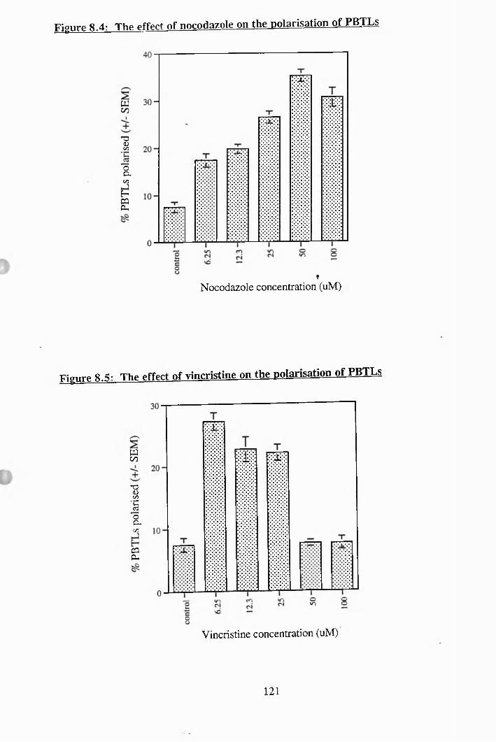
Figure 8.4: The effect of nocodazole on the polarisation of PBTLs
w00
'a<Dc/3
oCUc/5
HS
Nocodazole concentration (uM)
Figure 8.5: The effect of vincristine on the polarisation of PBTLs
woo
T3Dc /3•CC3"oC-
C/3
g
Vincristine concentration (uM)
121
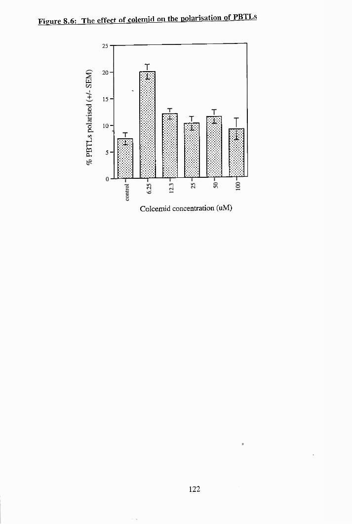
Figure 8.6: The effect of colemîd on the polarisation of PBTLs
wGO
T3(Ücd
&
;2
25
2 0 -
1 5 -
10 -
5 -
T
±
<N
T
Colcemid concentration (uM)
122
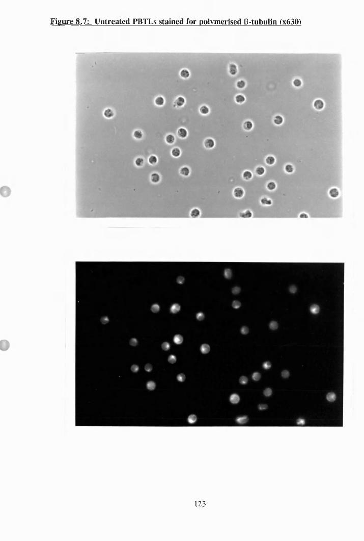
Figure 8.7: Untreated PBTLs stained for polymerised B-tubulin (x630)
123
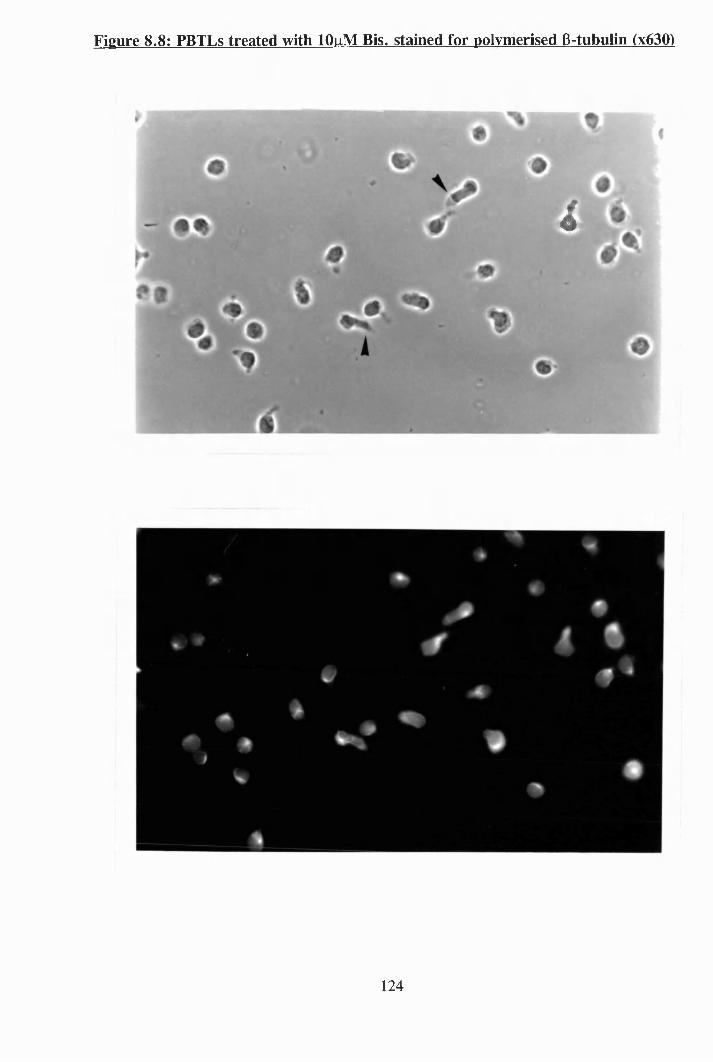
Figure 8.8: PBTLs treated with lOuM Bis, stained for polymerised B-tubulin (x630)
i
124
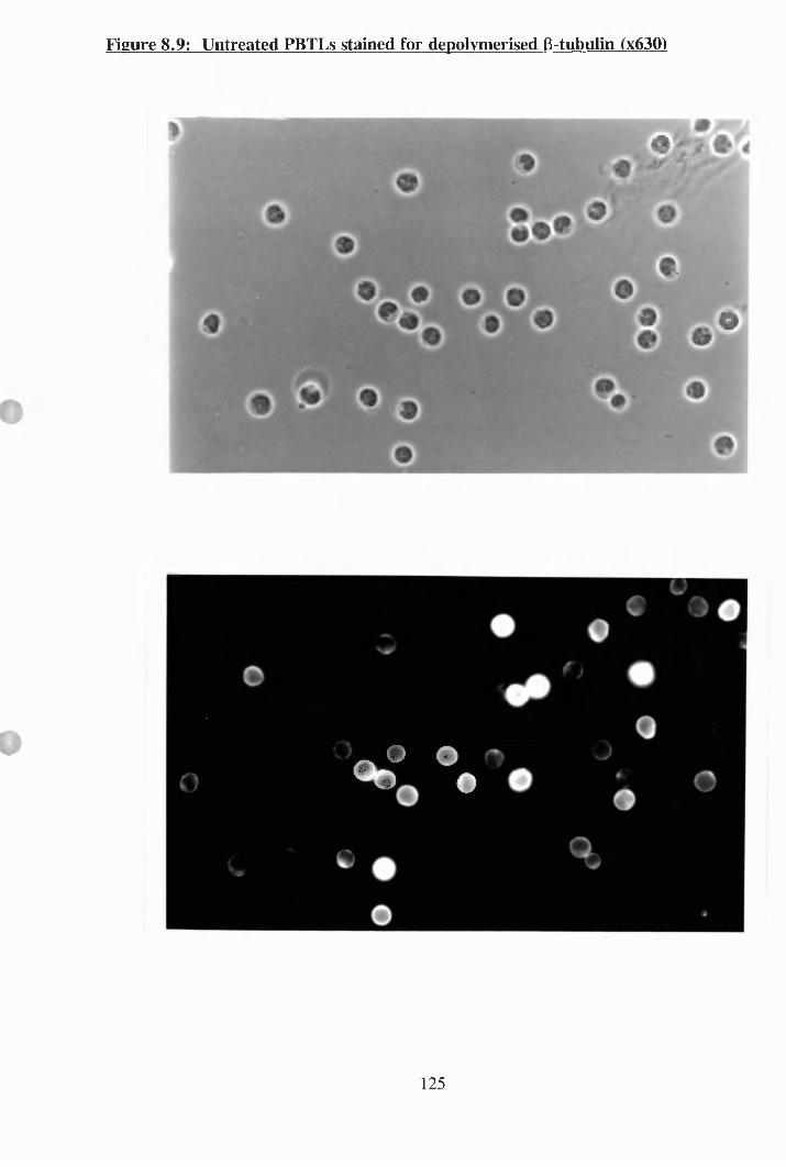
Figure 8.9: Untreated PBTLs stained for depolvmerised B-tubulin (x630)
#
@O'
«te® %
125
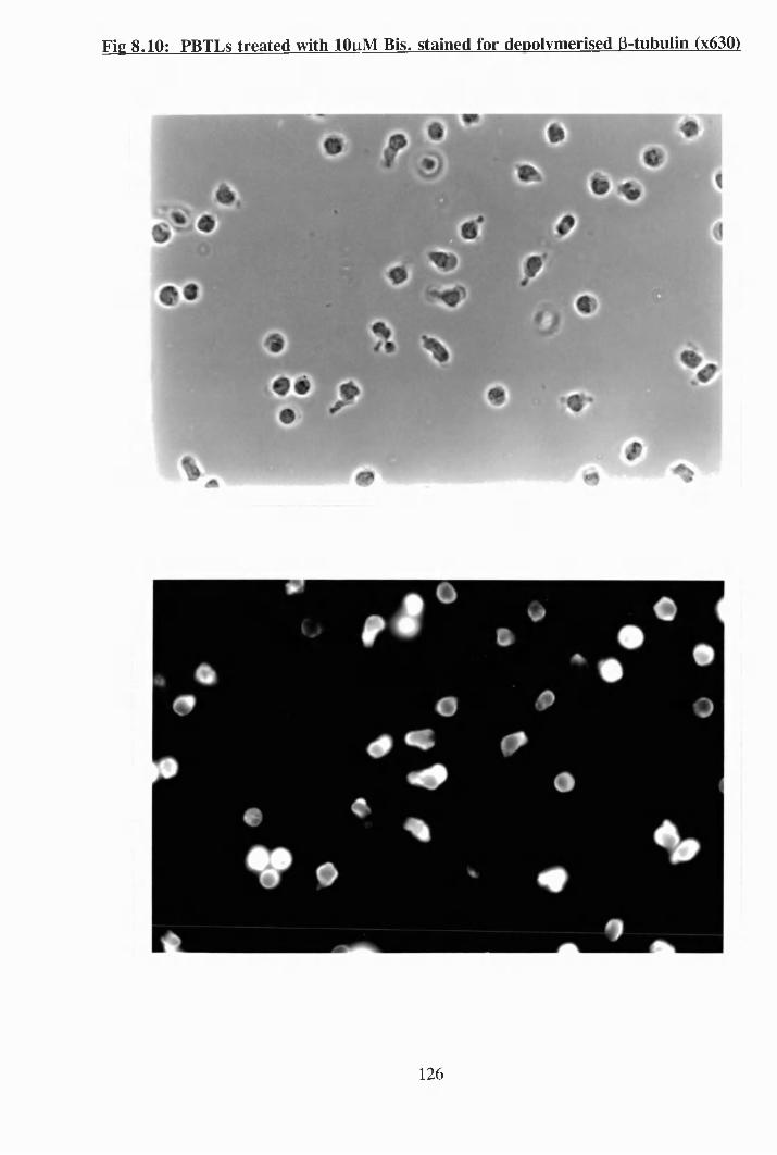
Fig 8.10: PBTLs treated with lOuM Bis, stained for depolvmerised B-tubulin (x630)
126
Chapter 9
Stnicture-activity relationship of inhihitors of
lymphocyte motility.
During this project a number of compounds were found to have inhibitory effects on
lymphocyte motility as measured by the polarisation assay. Most of these compounds are
different from each other in their proposed targets (ie: herbimycin A is a tyrosine kinase
inhibitor, whereas taxol is a microtubule stabiliser), however, they have all been found
in this study to have one thing in common, which is the capability to inhibit, to varying
degrees, the polarisation of PBTLs by shape change inducers. Therefore, it was decided
to investigate whether there were any structural similarities between all these compounds
which might be the reason for their common effects on PBTLs.
In figure 9.1 is shown the chemical structure of ionomycin which is a calcium ionophore
and was found to significantly prevent the induction of shape change in PBTLs at a
concentration of 10|iM as seen in fig. 4.9. Thapsigargin, the Ca^^-ATPase inhibitor was
also found to inhibit PBTL polarisation at a concentration of lOpM (figure 4.12), and its
chemical structure is depicted in figure 9.2. The structure of these compounds are very
different and it seems unlikely that they have a common mechanism.
All the amiloride compounds (Na'"'/H'^ antiport blockers) which were tested in chapter 5
for their ability to prevent PBTL polarisation are depicted in figure 9.3 (see chapter 5
for full names). Their order of potency in inhibiting polarisation was
127
A171 > A149> A130> A125> A113, which is consistent with their known effects on
antiport blockade^^^^\ As can be seen from their chemical structures, they all
have an aromatic core with amino groups in side chains.
In fîgure 9.4 can be seen the structures of the chloride channel blockers, NPPB,
niflumic acid, quinidine and tamoxifen. From the data shown in chapter 5, the
compounds NPPB and tamoxifen were the most potent polarisation inhibitors, however
their structures are dissimilar and it may be that their effect on chloride channels are by
different mechanisms (eg: direct blockade versus inhibition of a regulatory molecule).
The microtubule targetting drugs, taxol, vinblastine and vincristine are large polycyclic
compounds and their structures are shown in figure 9.5. Taxol was found to be very
effective at inhibiting polarisation in PBTLs (see fîgure 8.1) and vinblastine was found
to round up freshly isolated PBTLs. Vincristine actually caused polarisation in PBTLs
but its structure is shown here to show how similar vinblastine and vincristine are, as
they differ only by a methyl group for an aldehyde group.
Wortmannin and LY294002 have been reported in the literature to be potent PI 3-kinase
inhibitors and were shown in figures 4.14 and 4.15 to inhibit PBTL polarisation.
However as mentioned in chapter 4, wortmannin inhibited polarisation at concentrations
which are non-specific for PI 3-kinase. Their structures are shown in figure 9.6 and as
you can see there is very little similarity between their structures.
128
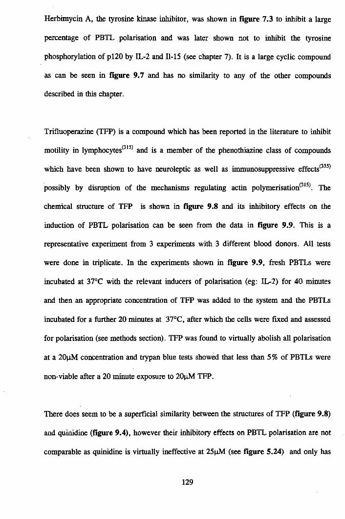
Herbimycin A, the tyrosine kinase inhibitor, was shown in figure 7.3 to inhibit a large
percentage of PBTL polarisation and was later shown not to inhibit the tyrosine
phosphorylation of pl20 by IL-2 and 11-15 (see chapter 7). It is a large cyclic compound
as can be seen in figure 9.7 and has no similarity to any of the other compounds
described in this chapter.
Trifluoperazine (TFP) is a compound which has been reported in the literature to inhibit
motility in lymphocytes^^^^ and is a member of the phenothiazine class of compounds
which have been shown to have neuroleptic as well as immunosuppressive effects ^ ^
possibly by disruption of the mechanisms regulating actin polym erisation^^The
chemical structure of TFP is shown in figure 9.8 and its inhibitory effects on the
induction of PBTL polarisation can be seen from the data in figure 9.9 This is a
representative experiment from 3 experiments with 3 different blood donors. All tests
were done in triplicate. In the experiments shown in fîgure 9.9, fresh PBTLs were
incubated at 37°C with the relevant inducers of polarisation (eg: lL-2) for 40 minutes
and then an appropriate concentration of TFP was added to the system and the PBTLs
incubated for a further 20 minutes at 37°C, after which the cells were fixed and assessed
for polarisation (see methods section). TFP was found to virtually abolish all polarisation
at a 20nM concentration and trypan blue tests showed that less than 5 % of PBTLs were
non-viable after a 20 minute exposure to 20pM TFP.
There does seem to be a superficial similarity between the structures of TFP (fîgure 9.8)
and quinidine (figure 9.4), however their inhibitory effects on PBTL polmsation are not
comparable as quinidine is virtually ineffective at 25pM (see figure 5.24) and only has
129
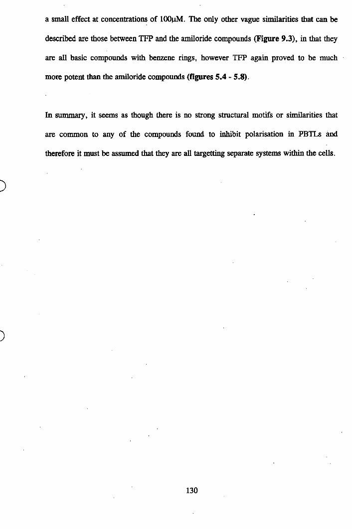
a small effect at concentrations of lOOpM. The only other vague similarities that can be
described are those between TFP and the amiloride compounds (Figure 9.3), in that they
are all basic compounds with benzene rings, however TFP again proved to be much
more potent than the amiloride compounds (figures 5.4 - 5.8).
In summary, it seems as though there is no strong structural motifs or similarities that
are common to any of the compounds found to inhibit polarisation in PBTLs and
therefore it must be assumed that they are all targetting separate systems within the cells.
130
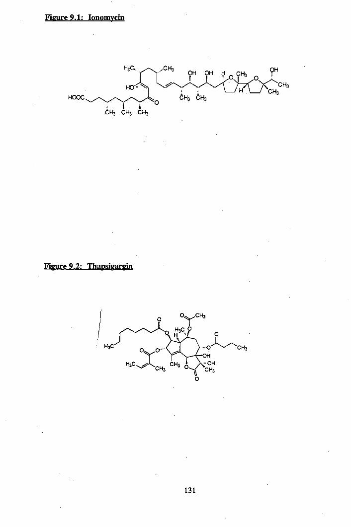
Figure 9.1: lonomvcin
QHOH OH
HOOC
Figure 9.2; Thapsigargin
,0- ........CH3
131
Figure 9.3: Amiloride compounds
- 0V , II N H i
»-;xXr(CHifnCHCHi
N = C — N H ]
/
A - 149 ^ 1 3 0
N H jX . Ï
/ N N NH j M:N N NH2 . IK'I(CH3)2CH
AI 1.1A-I7 I
(C H j )2 N ^ N N H i
• MCI
A-I2.S
132
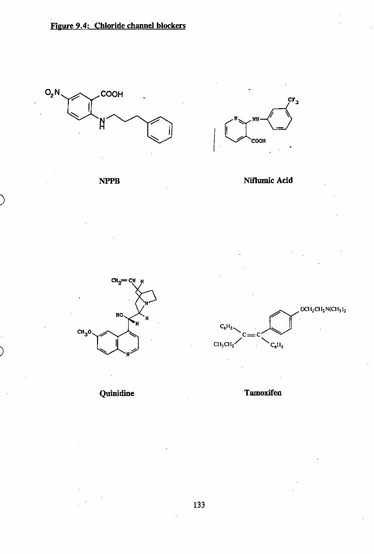
Figure 9.4: Chloride channel blockers
COOHCF,
NH
COOH
NPPB Niflumic Acid
HO
CH,0
Oai^CH.NCCH))!
^ C = C -
Quinidine Tamoxifen
133
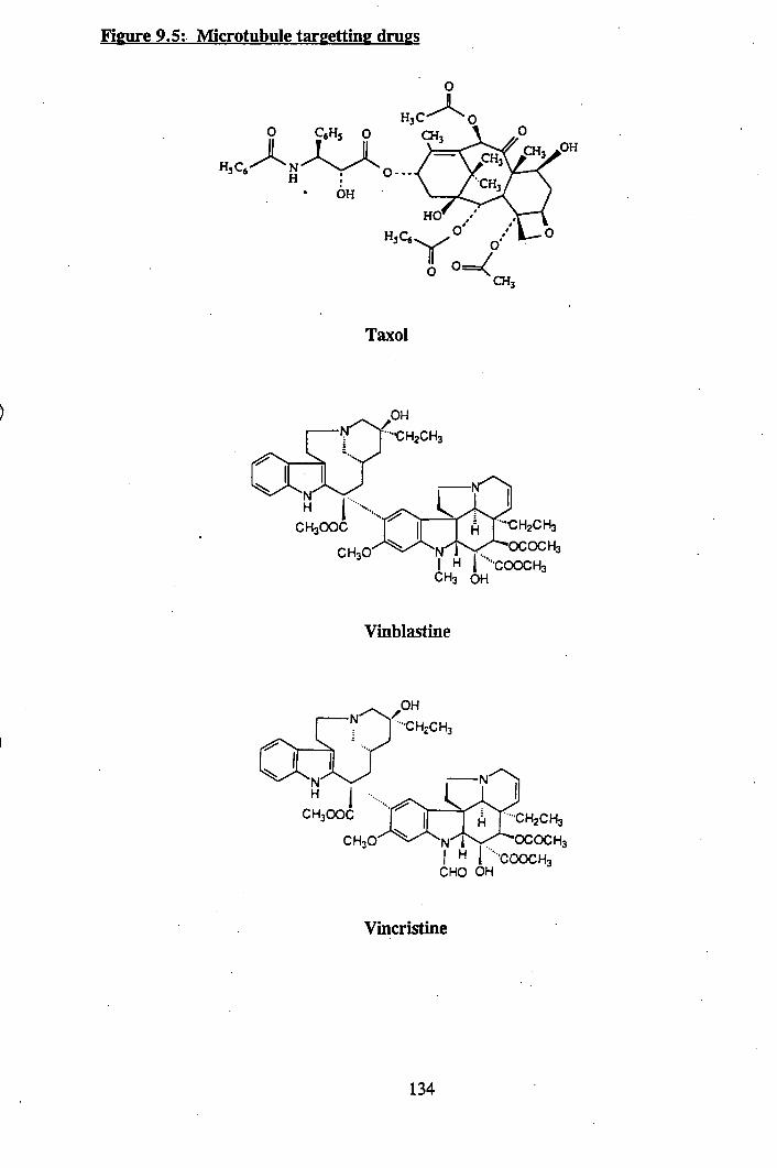
Figure 9.5: Microtubule targetting drugs
OH
H3Q5
Taxol
•••■CH2CH3
CH3OOC Â J-CH2CH3OCOCH3
H 1 ""'COOCH3 CH3 OH
Vinblastine
•••CH2CH3
CH300ÇCH30
 r-CH2CH3N^l^YT'^OCOCHaI H I ■■•-COOCH3
CHO OH
Vincristine
134
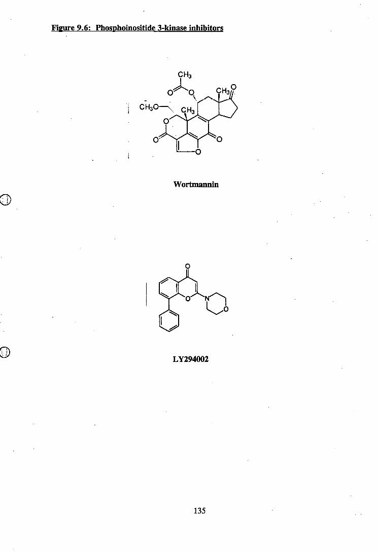
Figure 9.6: Phosphoinositide 3-kinase inhibitors
Wortmannin
LY294002
135
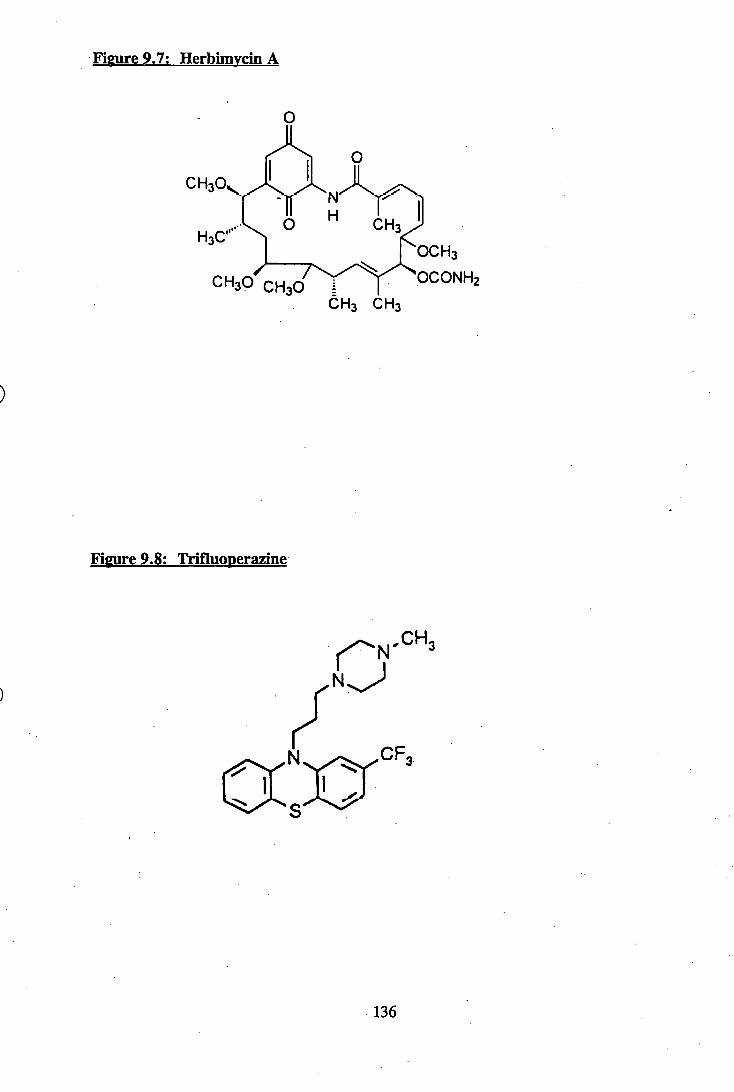
Figure 9.7: Herbimvcin A
ACHaO^
H3C...O CH3
^OCHa
CH3O' CH3O T TÔH3 CHa
Figure 9.8: Trifluoperazine
CH
CF
136
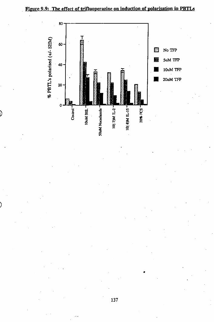
Figure 9.9: The effect of trifluoperazine on induction of polarisation in PBTLs
00
•gcn
i1
S
NoTFP
B SuMTFP
■ lOuMTFP
■ 20uMTFP
137

10. Discussion
»
138
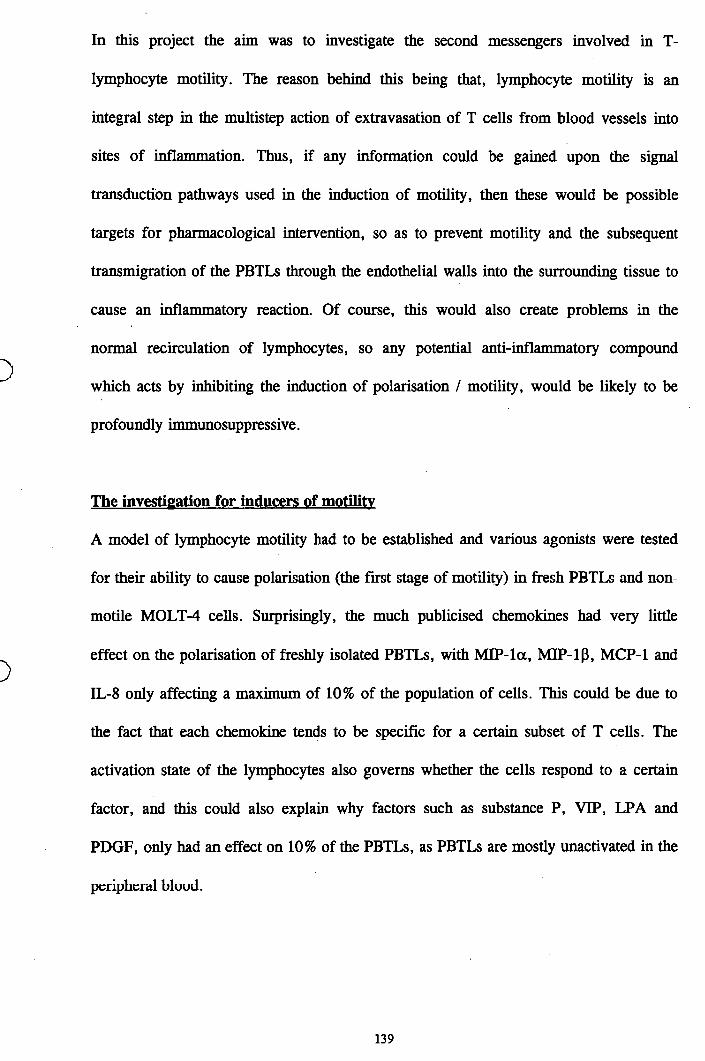
In this project the aim was to investigate the second messengers involved in T-
lymphocyte motility. The reason behind this being that, lymphocyte motility is an
integral step in the multistep action of extravasation of T cells from blood vessels into
sites of inflammation. Thus, if any information could be gained upon the signal
transduction pathways used in the induction of motility, then these would be possible
targets for pharmacological intervention, so as to prevent motility and the subsequent
transmigration of the PBTLs through the endothelial walls into the surrounding tissue to
cause an inflammatory reaction. Of course, this would also create problems in the
normal recirculation of lymphocytes, so any potential anti-inflammatory compound
which acts by inhibiting the induction of polarisation / motility, would be likely to be
profoundly immunosuppressive.
The investigation for inducers of motilitv
A model of lymphocyte motility had to be established and various agonists were tested
for their ability to cause polarisation (the first stage of motility) in fresh PBTLs and non
motile MOLT-4 cells. Surprisingly, the much publicised chemokines had very little
effect on the polarisation of freshly isolated PBTLs, with M IP-la, MIP-ip, MCP-1 and
IL-8 only affecting a maximum of 10% of the population of cells. This could be due to
the fact that each chemokine tends to be specific for a certain subset of T cells. The
activation state of the lymphocytes also governs whether the cells respond to a certain
factor, and this could also explain why factors such as substance P, VIP, LPA and
PDGF, only had an effect on 10% of the PBTLs, as PBTLs are mostly unactivated in the
peripheral blood.
139
The MOLT-4 cells proved to be unresponsive to all of the factors tested except, Bis. and
nocadazole. The reasons for this could be that either the MOLT-4 cells do not possess
the receptors for any of these factors or it could be that pieces of the signal transduction
machinery needed to relay the signals are missing or unable to function (which is a
strong possibility, since these are transformed cells). For example, although the MOLT-4
cell line is classified as a T cell lymphoblastic leukaemia it is actually negative for the
CD3 marker^^^^\ Hence, it was decided to concentrate on using PBTLs as a model for
induction of motility in lymphocytes as five factors were found to cause significant
polarisation in PBTLs, these being. Bis - a PKC inhibitor, nocadazole- a microtubule
disrupting agent, FCS - a mixture of unknown quantities of proteins and growth factors
and finally the two interleukins-2 and -15, which are physiological agents involved in T
cell regulation.
However, cell polarisation is only an indicator of motility as it does not always lead to
cell locomotion. Therefore, these five factors were tested in transmigration assays and
IL-2, IL-15 and Bis., were found to cause significant transmigration across
polycarbonate filters, whereas FCS and nocadazole had no effect whatsoever.
Nevertheless, FCS and nocadazole were still useful as tools for inducing polarisation in
PBTLs as this is the crucial first step in motility - cells must change shape before they
can become motile, therefore, nocadazole and FCS presumably use the same intracellular
machinery as Bis. IL-2 and 11^15 to cause shape change
Rùle of intracellular calHum
Having established a model for the induction of motility in PBTLs, the next step was to
investigate whether these 5 inducers of shape change utilised any common second
140
messengers, that would then be contenders for involvement in a motility signal
transduction pathway. It was found that none of the 5 factors caused any significant
increases in [Ca^^]j that could be detected with the system used. Therefore, there could
have been very small local fluctuations in [Ca^^]; within the cells that escaped detection.
Also tested for effects on PBTLs levels, were some of the chemokines which
have been reported in the literature to cause transient [Ca^^]| increases. However, MIP-
la , MIP-ip and MCP-1 had minimal effects on PBTLs in this system, with only an
increase of about lOnM [Ca """]; being detected. This could be due to the fact that within
each experiment there are 2x10^ PBTLs and only a subpopulation of these will respond
to the chemokine in question, thus diluting the signal. Interleukin-8 and RANTES were
found to have no effect whatsoever, but interestingly the non-motile MOLT -4 cells
responded dramatically to MCP-1 and a few of the other P-chemokines with classical
[Ca^^ji transients, proving that this system does indeed work, but more importantly
showing that a physiological factor such as MCP-1 which causes a [Ca^^], transient
increase does not affect the morphology of the cells.
Further investigations into [Ca^^]; and motility showed that an increase in [Ca^^j^
actually inhibits polarisation. First of all motile MOLT-4 cells and PBTLs that had
previously been treated to cause polarisation were found to round up on exposure to
ionomycin. At low concentrations (0.5pM), the calcium ionophore ionomycin
preferentially inserts in the membrane of the intracellular stores ^ ^ and causes an
increase in [Ca^^]; levels due to emptying of intracellular calcium stores. At higher
concentrations (lOpM), the ionophore ionomycin, inserts into the cell membrane causing
an influx of extracellular calcium into the cytosol. Thapsigargin treated PBTLs were
unable to polarise upon exposure to the inducers of shape change due to the increase in
141
[Ca ^]i levels elicited by the Ca^^-ATPase inhibitor. These results are supported by other
workers^^°^’ ®\ who have also found that a [Ca^^], elevation in T-lymphocytes causes
rounding up and immobilisation of the cells.
The [Ca^^]i dependence of lymphocyte motility is opposite to that described for other
leukocytes where [Ca^^]. increases are associated with increased motility. For example,
[Ca^^]i elevation speeds up neutrophil migration on various substrates^^^^\ via a
calcineurin dependent mechanism^^^°\ Since stimuli (ionomycin and thapsigargin) that
led to a rise also provoked a rounding of the lymphocyte, then there must be a
link between the cytoskeleton and calcium. Fragmentation of filamentous actin by
calcium-dependent proteins (such as gelsolW^^^ ) can explain some of these events. In
neutrophils, a Ca^^-induced depolymerisation of actin has been shown^^^\ A correlation
between [Ca^^ji augmentation and retraction of protrusions has been described in other
cell types, for example cytotoxic T cells after interaction with a target cell^^^ \ or
endothelial cells stimulated with thrombin^^ '^ However, this is not necessarily true in all
cell types. For instance, in neuroblastoma cells, although lysophosphatidic acid elicits
both a [Ca^^ji rise and a neurite retraction, in this case the two events are not causally
related^^^\
Local signalling events at the leading edge may be responsible for protrusion of a
pseudopod by regulating actin-binding proteins. Although the studies with Ca -depleted
cells show that changes in [Ca^^], are not a required part of the signal at the leading
edge, localised brief increases in [Ca '^Jj could play an auxiliary role in protrusion by
generating additional actin-nucleation sites and by breaking crosslinks. With increased
ability to measure [Ca^^], while observing morphological changes, and with improved
142
methods to manipulate [Ca^^],, it should be possible to determine whether such local
[Ca^^]i transients do play a functional role.
Role of phosphoinositides
The first set of experiments which investigated the role of phosphoinositides in
lymphocyte motility were assays of IP3 levels within the cells. IP3 is an important second
messenger as it can signal the endoplasmic reticulum to release its stores of Ca^^ and
thus elevate the [Ca^^], levels. It was found that none of the five inducers of shape
change had any effects on the IP3 levels after an arbitrary one minute period. The results
from experiments whereby inhibitors of PI 3-kinase were used were unclear as
w ortmannin was only inhibiting polarisation in PBTLs at concentrations at which is
unspecific for PI 3-kinase. However, another PI 3-kinase inhibitor, LY294002 was
found to significantly block the induction of motility in PBTLs at low concentrations.
W ortmannin is known to be less stable than LY294002 and this could explain why it had
less of an effect than LY294002, which then lead to the conclusion that PI 3-kinase could
possibly be involved in the signal transduction of motility. Further evidence for the role
of phosphoinositides in motility came from the experiments involving lithium chloride,
which was found to inhibit the induction of shape change by IL-2, IL-15 and FCS (and
Bis. and nocadazole, but at higher concentrations.). These results are not conclusive but
they do suggest that phosphoinositides could be playing a role in motility signal
transduction
Although no direct role for PI 3-kinasc in actin polymerisation has been demonstrated, it
is neeessary for some forms of eell motility and adherence. PDGF receptor mutants that
do not bind PI 3-kinase, do not ruffle or undergo chemotaxis in response to
143
It has also been found that both PDGF receptor mutants that do not bind
PI 3-kinase and wortmannin inhibit binding of GTP to rac in response to PDGF^^^\
These data place PI 3-kinase upstream of rac in fibroblasts and, consistent with this idea,
injection of fibroblasts with V12 rac circumvents inhibition of ruffling by
wortmannin^^^^\ Finally, although the biochemical mechanism is obscure, there is
growing evidence both from receptor mutants and from inhibitors that PI 3-kinase is
required for stimulus-dependent activation of integrins and cell adherence^^^®’ ^ ^
Role of intracellular pH and ion channels
Induction of shape change in PBTLs was found to have no effect on the intracellular pH
of the cells as measured in this system. However, a decrease in pHj caused rounding up
of motile MOLT-4 cells and blocked polarisation in PBTLs. One of the main pHj
regulatory mechanisms is the Na'"'/H'*' antiporter and blockage of these was found to
cause inhibition of motility in PBTLs and motile MOLTs.
Cytosolic pHj is a candidate to regulate cell motility, since certain steps in the actin
polymerisation sequence and the binding of actin filaments to membrane-anchoring
proteins are pH-dependent events^^^°\ Indirect observations are consistent with this
notion: the ability of neutrophils to polarise and perform chemotaxis is reduced when the
extracellular pH (pH^) is made more acidic, which is expected to lower pH/^^\ More
importantly, it is possible to induce cytoskeletal reorganisation in neutrophils in a
receptor-independent manner by the addition of weak electrolytes, which can modify pHj
at constant pHg^^^\ Therefore, pH, must be given consideration as a regulator and
possible mediator of cell shape change and chemotaxis.
144
In a recent publication^^^^ neutrophil spreading on adhesive substrates caused a rapid
and sustained cytosolic alkalinisation. This pHj increase was prevented by the omission
of external Na'*’, suggesting that it results from the activation of Na‘‘‘/H''’ exchange. It
was also found that neutrophil motility was prevented by selectively blocking the NHE-1
isoform of the Na'^'/H''' antiporter. Support for the results obtained in this project were
also observed in that neutrophil spreading was strongly inhibited when pHj was clamped
at acidic values^^^ \ Interestingly, the inhibition of neutrophil shape change required pre
acidification of the cells ^ ^ , since neutrophils spread normally when acidified shortly
after contact with the substrate. This suggests that the pH-sensitive step is an early event,
and that adherence and spreading, once initiated, can proceed independently of pH .
It has been shown that there are interactions between actin filaments and the Na'*'/H'^
antiporte/^^^\ Also, microtubules have been suggested to have links with the Na’ /H'*’
antiporter, which were shown to be regulated in a mechanosensitive manner in
lymphocytes^^^^\ In addition, it has been shown in human B-lymphoid cells that the
cAMP-mediated signal transduction pathway and pertussis toxin-sensitive GTP-binding
proteins act synergistically to regulate amiloride-sensitive sodium channels^^ '^^
Thus, it would seem that the the Na'*'/!!''’ antiporters play a vital role in lymphocyte
motility, probably due to its role in pH, regulation which has effects on all enzymes
within the cell and also it would seem through its interactions with the cytoskeleton.
Investigations into the roles of chloride channels in lymphocyte motility showed that
various chloride channel blockers could inhibit PBTLs and motile MOLTs polarisation
effectively, however it was found that the blockers were not actually exerting their
145
motility inhibitory effects via chloride channel blockage but through other mechanisms
which are unknown. Therefore, the role of chloride channels in lymphocyte motility
remains unclear at this point, however it can be assumed that they would play at least
some minor role in the complicated entanglement that is the signal transduction
regulation of motility, as chloride channels are important in cell volume
control^°^'^°^'^^\ as well as pH; control via the CI /HCO3 antiport exchanger^°^'^^\ A
CI /HCO3 antiport exchanger, similar to the band 3 protein in the membrane of red
blood cells, is thought to play an important part in pH; regulation in many nucleated
cells. Like the Na'"'/H'^ exchanger, the CI /HCO3 antiport exchanger is regulated by pH;,
but in the opposite direction. Its activity increases as pH; rises, increasing the rate at
which HCO3 is ejected from the cell in exchange for Cl’, thereby decreasing pH;
whenever the cytosol becomes too alkaline. Also, it must be noted at this point that the
importance of chloride channels in motility may not be just due to their physiological
role in Cl' transport but to their permeability to larger organic osmolytes such as
The role of renaturahle kinases
Investigations into the role of renaturahle kinases in PBTL motility, showed that of the
five inducers of motility, only Bis. seemed to have any effects, in that it repeatedly
activated a renaturahle autophosphorylating kinase of molecular weight 58kDa.
Collaborative work in the same laboratory has identified this 58kDa kinase to be MST-1
(mammalian Ste20-like), a serine/threonine protein kinase. Little is known about this
kinase at the current time, however its role in the signal transduetion of motility in
PBTLs cannot be considered too important as the other four inducers of motility failed to
activate it in this assay.
146
The role of tvrosine phosphorylation
In this study it was found that of the five inducers of polarisation in PBTLs only IL-2
and IL-15 caused tyrosine phosphorylation of proteins that were detected in this system.
Both IL-2 and IL-15 were found to cause tyrosine phosphorylation of a protein of
molecular weight approximately llOkDa (pi 10). Of the tyrosine kinase inhibitors used,
only herbimycin A was found to block polarisation of PBTLs and more importantly it
was capable of inhibiting shape change in PBTLs by all five of the inducers of motility
effectively. This suggested that herbimycin A was targeting a point in the signal
transduction system that was utilised by all five factors. Similar results have been
obtained with neutrophils pre-treated with herbimycin A, in that it blocks their
chemotactic response to fMLP^^^ . It should be noted at this point however, that
herbimycin A is not necessarily blocking shape change by inhibition of a tyrosine kinase,
it could be due to other adverse effects. Further evidence to suppport this notion was
obtained in the experiments in whieh it was found that PBTLs pre-treated with
herbimycin A before exposure to IL-2 or IL-15 were still found to cause tyrosine
phosphorylation of pi 10. Thus, if herbimycin A is indeed inhibiting polarisation by
targeting a tyrosine kinase then it must be downstream from the pi 10 protein.
Identification of the p i 10 protein has been unsuccessful. Evidence from the literature
would suggest the most likely candidate to be Janus kinase-3 (JAK-3), as this protein has
been shown to be tyrosine phosphorylated by both IL-2 and IL-15 in T cells ^ ^ and has a
molecular weight of approximately 120kDa. However, experiments to immunoprecipitate
the p i 10 protein using anti-JAK-3 antibodies proved unsuccessful. Therefore, it is
uncertain at this point whether the p i 10 protein is indeed JAK-3, however, whatever the
147
identity of the protein it is possible that it is part of a motility signal transduction
pathway utilised by IL-2 and IL-15.
Indeed, it has been shown in neutrophils treated with chemotactic agents such as fMLP,
that they also cause tyrosine phosphorylation of a protein of molecular weight
120kDa ^ ^ - (identity unknown at time of publication).
Role of microtubules
To evaluate the involvement of microtubules in PBTL polarisation, a number of
microtubule targeting drugs were used on the PBTLs. From these results it was clear that
before the PBTLs could change shape, the microtubular system must be rearranged. This
was deduced from experiments with taxol. The effect of taxol at the molecular level is
opposite to that of the other microtubule targeting drugs used, such as colchicine. Taxol
stabilises microtubules in a polymerised state^^^\ Due to this effect, taxol promotes
polymerisation of free microtubules not associated with any organising centers, so that
gradually the system of microtubules radiating from the perinuclear center is replaced by
numerous aggregates of free microtubules^^^^\ This disintegration of the microtubule
system was found to be accompanied by the inabilty of the PBTLs to polarise in réponse
to the five inducers of shape change. Also, it was found that agents such as colchicine
and nocadazole which disrupt microtubules, could cause shape change in PBTLs
themselves.
Immunofluorescence photographs of unactivated PBTLs stained for polymerised and
depolymerised P-tubulin show that they have different cellular distribution profiles but in
polarised PBTLs both types of tubulin seemed to be excluded from the leading edge of
148
the cell which is rich in F-actin microfilaments forming a cortical meshwork. Indeed, it
has been found that polarised motile fibroblasts are characterised by trailing processes
rich in microtubules^^^*^
What then is the role of microtubules in PBTLs motility? Unfortunately, there is little
published work on this subject concerning PBTLs, however there are theories on the
subject. One hypothesis, originally proposed in is that the microtubules
transport new membrane and cortical components from the golgi apparatus to the leading
edge. One reason that the microtubules need to be disrupted prior to polarisation is a
structural one, in that the intracellular scaffolding has to be re-arranged before the cell
can change shape. Also, it could be that the disruption of the microtubule network allows
the release of second messengers that are bound to the microtubules, so that they are then
free to take part in signal transduction.
Therefore, it would seem that PBTL polarisation is stabilised first by reorganisation of
the actin cortex induced by the extension of pseudopods; in the next stage polarisation is
enhanced and further stabilised by the microtubule-dependent redistribution of organelles
Inhibitors of lymphocyte polarisation
After assessing the chemical structures of all the compounds which inhibited PBTL
polarisation, it was found that there were no significant structural similarities between
any of the compounds that may explain their common effects on PBTLs polarisation.
Therefore, it must be assumed that they are all targeting separate systems within the
cells.
149
Conclusion
Thus, five different factors were found to cause significant polarisation in PBTLs, but no
common second messenger elements were found to be utilised by the five. However, a
number of pharmacological agents were found, that prevented induction of polarisation in
PBTLs by all five factors and these it would seem may be targeting unknown second
messenger elements involved in the signal transduction of T-lymphocyte motility, or the
intracellular motility machinery itself.
150

11. References
»
151
1. Butcher^.C : Leukocyte-endothelial cell recognition: Three (or more) steps to
specificity and diversity. (Revievy), Cell (1991), 67,1033-1036
2. Lasky,L.A ; Selectins: Interpreters of cell specific carbohydrate information during
infiammation.(Review), Science (1992), 258,964-969
3. Springer,T.A : Traffic signals for lymphocyte recirculation and leukocyte
emigration: The multistep paradigm. (Review). Cell (1994), 76,301-314
4. Duijyestijm,A. and Hamman,A : Mechanisms and regulation of lymphocyte
migration.(Review), Immunol Today. (1989), 10,23-37
5. Mackay,C.R : Migration pathways and immunologic memory among T
lymphocytes. (Review), Semin. Immunol (1992), 4, 51-58
6 . Sprent,J : T and B memory cells.(Review), Cell (1994),76,315-322
7 Baumhueter,S., Singer,M.S., Henzel,W., Hemmerich,S., Renz,M.,
Rosen,S.D., and Lasky,L.A : Binding of L-selectin to the vascular sialomucin
CD34. Science (1993), 262,436-442
152
8. Hamman,A., Andrew,DP., Jablonski-WestrichJ)., Holzmann,B., and
Butcber,£.C. : The role of a4 integrins in lymphocyte homing to mucosal
tissues in vivo . J Immunol. (1994), 152, 3282-3293
9. Zimmerman,G.A., Prescott,S.M., and McIntyre,M. : Endothelial cell
interactions with granulocytes: Tethering and signalling molecules. (Review),
Immunol. Today (1992), 13,93-98
10. Spangrude,G.J., Braaten,B.A., and Daynes,R.A : Molecular mechanisms of
lymphocyte extravasation: Studies of two selective inhibitors of lymphocyte
recirculation. J./mmwwo/. (1984), 132,354-362
11. FreemontC.J., Jones,C., Bromley.M. and Andrew,?. : Changes in vascular
endothelium related to lymphocyte collections in diseased synovia. Arthritis.
Rheum. (1983), 26, 1427-1433
12. Freemont,C.J. : The small blood vessels in areas of lymphocytic infiltration
around malignant neoplasms. Br. J. Cancer (1982), 46, 283-288
13. Mackay,C.R. ,Marston,W.L, DudIer,L., Spertini,0,. Tedder,T.F., and
Hein,W.R. Tissue specific migration pathways by phenotypically distinct
subpopulations of memory T cells. Eur. J. Immunol. (1992), 22, 887-895
153
14. ChisolmJP L., Williams,C.A., and Lobb,R.R : Monoclonal antibodies to the
integrin a4 subunit inhibit the murine contact hypersensitivity response. Eur. J.
Immunol. (1993), 23,682-688
15. Scbeynius,A., Camp,R.L, and Pure,E. : Reduced contact sensitivity reactions
in mice treated with monoclonal antibodies to leukocyte function associated
molecule-1 and intercellular adhesion molecule-1. J. Immunol. (1993), 150,
655-663
16. MuUigan,M.S, and lssekutz,T.B. : Dual inhibition of VLA-4 and LFA-1
maximally inhibits cutaneous delayed type hypersensitivity induced inflammation.
Am. J. Pathol. (1994), 144,1008-1015
17. Mackay,C.R., Marston,W.L., and Dudler,L.: Altered patterns of T cell
migration through lymph nodes and skin following antigen challenge. Eur. J.
Immunol. (1992), 22,2205-2210
18. Issekutz,T.B. : Inhibition of in vivo lymphocyte migration to inflammation and
homing to lymphoid tissues by the TA-2 monoclonal antibody: a likely role for
VLA-4 in vivo. J. Immunol. (1993), 147,4178-4184
19. AlbeIda,S.M., Wayne Smitb,C., and W ardf A.: Adhesion molecules and
inflammatory injury. FASEB. J. (1994),8, 504-512
154
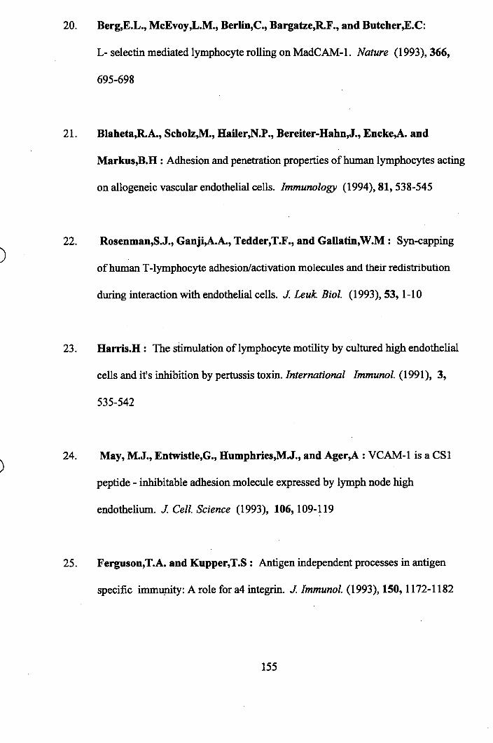
20. Berg,E L., McEvoy,L.M., Berlin,C., Bargatze,R.F., and Butcher,E.C:
L- selectin mediated lymphocyte rolling on MadCAM-1. Nature (1993), 366,
695-698
21. Blaheta,R.A., Scholz,M., Hailer,N.P., Bereiter-Hahn,J., Encke,A. and
Markus,B.H : Adhesion and penetration properties of human lymphocytes acting
on allogeneic vascular endothelial cells. Immunology (1994), 81, 538-545
22. Rosenman,S.J., Ganji,A.A., Tedder,T.F., and Gallatin,W.M : Syn-capping
of human T-lymphocyte adhesion/activation molecules and their redistribution
during interaction with endothelial cells. J. Leuk. Biol. (1993), 53, 1-10
23. Harris.H : The stimulation of lymphocyte motility by cultured high endothelial
cells and it's inhibition by pertussis toxin. International Immunol. (1991), 3,
535-542
24. May, M.J., Entwistle,G., Humphries,M.J., and Ager,A : VCAM-1 is a CSl
peptide - inhibitable adhesion molecule expressed by lymph node high
endothelium. J. Cell. Science (1993), 106, 109-119
25. Ferguson,T.A. and Kupper,T.S : Antigen independent processes in antigen
specific immunity: A role for a4 integrin. J. Immunol. (1993), 150, 1172-1182
155
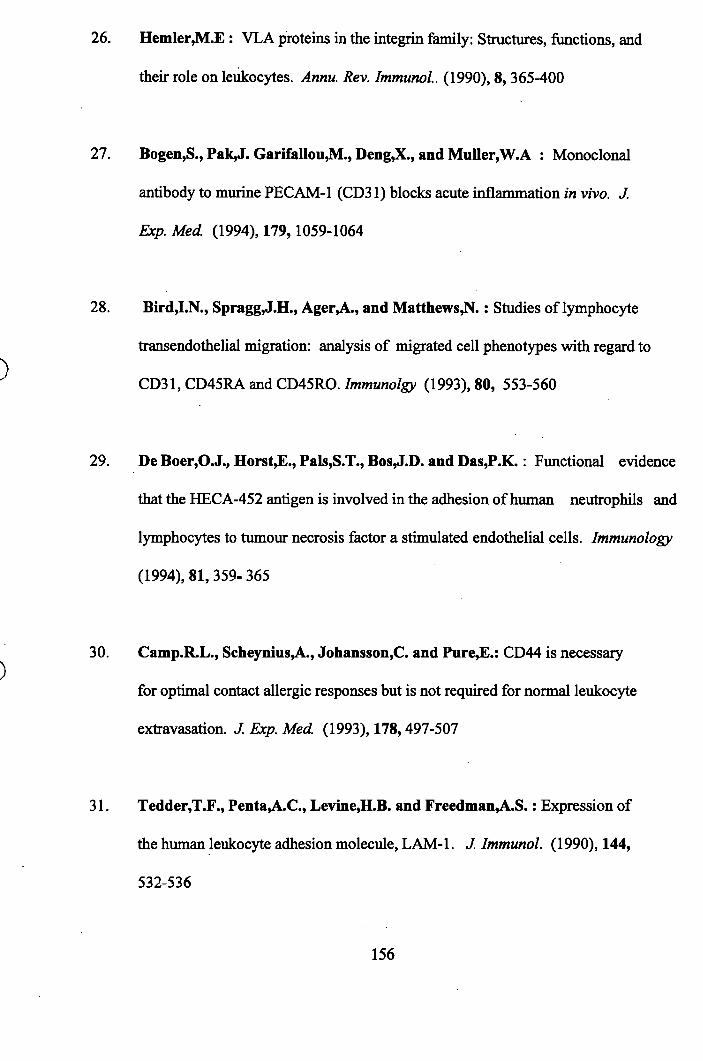
26. Hemler,M.£ : VLA proteins in the integrin family: Structures, functions, and
their role on leukocytes. Annu. Rev. Immunol. (1990), 8, 365-400
27. Bogen,S., Pak,J. Garifallou,M., Deng,X., and Muller,W.A : Monoclonal
antibody to murine PEC AM-1 (CD 31) blocks acute inflammation in vivo. J.
Exp. Med. (1994), 179,1059-1064
28. BirdJl.N., Spragg,J.H., Ager,A., and Matthews,N. : Studies of lymphocyte
transendothelial migration: analysis of migrated cell phenotypes with regard to
CD31, CD45RA and CD45R0. JmmwWgy (1993), 80, 553-560
29. De Boer,O.J., Horst,£., Pals,S T., Bos,J.D. and Das,P.K. : Functional evidence
that the HECA-452 antigen is involved in the adhesion of human neutrophils and
lymphocytes to tumour necrosis factor a stimulated endothelial cells. Immunology
(1994), 81,359-365
30. Camp.R.L., Scheynius,A., Johansson,C. and Pure,£.: CD44 is necessary
for optimal contact allergic responses but is not required for normal leukocyte
extravasation. J. Exp. Med (1993), 178,497-507
31. Tedder,T.F., Penta,A.C., Levine J1.B. and Freedman,A.S. : Expression of
the human leukocyte adhesion molecule, LAM-1. J. Immunol. (1990), 144,
532-536
156
32. Picker,L.J : The neutrophil selectin LECAM-1 presents carbohydrate ligands to
the vascular selectins ELAM-1 and GMP-140. (1991), Cell 66,921-933
33. Shimuzu,Y., Shaw,S., Graber,N., Gopal,T.V., Horgan,K.J., Van
Seventer,G.A., and Newman,W. : Activation- independent binding of human
memory T cells to adhesion molecule ELAM-1. Nature (1991), 349,799-803
34. PickerJLJ., Kishimoto,T.K., Smith,C.W., Warnock,R.A. and Butcher,£.C.:
ELAM-1 is an adhesion molecule for skin-homing T cells. Nature (1991), 349,
796 -799
35. Brandley,B.K., Swiedler,S.J., and RobbinsJ*.W. : Carbohydrate ligands of
the LEC cell adhesion molecules (minireview). (1990), Cell, 63,861-863
36. Honda,S., Campell,J.J., Andrew,D.P., EngeIhardt,B., Butcher^yA.,
Wamock,R.A., Ye,R.D., and Butcher,E.C. : Ligand induced adhesion to
activated endothelium and to vascular cell adhesion molecule-1 in lymphocytes
transfected with the N-formyl peptide receptor(1994), J. Immunol. 152,4026-31
37. Spangrude,G J . , Sacchi,F.., Hill,H.R., Van Epps,D.E., and Daynes JLA:
Inhibition of lymphocyte and neutrophil chemotaxis by pertussis toxin. J.
Immunol. (1985), 135,4135-4143
157
38. Oppenheim,J.J., Zachariae,C.O.C., Mukaida,N., and Matsushima,K.:
Properties of the novel proinflammatory supergene "intercrine" cytokine family.
Annu. Rev. Immunol. (1991), 9, 617-648
39. Miller,M.D. and Krangel,M.S.: Biology and biochemistry of the chemokines: a
family of chemotactic and inflammatory cytokines. CRC. Crit. Rev. Immunol.
(1992), 12, 17-46
41. Schall,T.J., Bacon^C, Camp,R.D.R., Kaspari,J.W., and GoeddalJl.V. :
Human MIP-la and MIP-lp chemokines attract distinct populations of
lymphocytes. J. Exp. Med. (1993), 177,1821-1825
42. TanbJ).D., Conlon,K., Lloyd,A.R., Oppenheim,J.J, and Kelvin J>.J. :
Preferential migration of activated CD4^ and CD8 T cells in response to MIP-la
and MIP-1 p. Science (1993), 260, 355-340
43. SchaIl,T.J., Bacon,K., Toy,K.J., and Goeddel,D.V.: Selective attraction of
monocytes and T lymphocytes of the memory phenotype by cytokine RANTES.
Nature, (1990), 347,669-673
158
44. Tanaka,Y., Adams,D.H., Hubscher,S., Hirano,H., Siebenlist,U., and
Shaw,S.; T cell adhesion induced by proteoglycan - immobilised cytokine MIP-1 p.
Aa/wre, (1993), 361,79-82
45. Murphy,P.M. : The molecular biology of leukocyte chemoattractant receptors.
Annu. Rev. Immunol. (1994), 12, 593-633
46. Gao,J.L., Kuhns,D.B., Lee Tiffany,H., M cD erm ottL i,X ., Francke,U.,
and Murphy,P.M., : Structure and functional expression of the human
macrophage inflammatory protein la / RANTES receptor. J. Exp. Med. (1993),
177, 1421-1427
47. McCoIlyS.R., Hachicha,M., Levasseur,S., Neote,K. and Schall,T.J.:
Uncoupling of early signal transduction events from effector function in human
peripheral blood neutrophils in response to recombinant MIP-la and MIP- Ip.
J. Immunol. (1993), 150,4550-4560
48. Napolitano,M., Seamon,K.B. and Leonard, W.J. : Identification of cell surface
receptors for the Act - 2 cytokine. J. Exp. Med. (1990), 172,285-289,
49. Neote^C, DiGregorio, Mak,J.Y, Horuk,R., and Schall,T.J.: Molecular cloning,
functional expression and signalling characteristics of a C-C chemokine receptor.
Cell, (1993), 72, 415-425
159
50. Leonard^J., Skeel^., Yoshimura,T., N oer^ , Kutvirt,S. and Van
EppsJ).: Leukocyte specificity and binding of human neutrophil
attractant/activating protein 1. J. Immunol (1990), 144,1323-30
51. Larsen,C.G., Anderson,A.O., Appellad^., Oppenheim,J.J., and Matsushima,K.
Neutrophil activating protein ( NAP-1) is also chemotactic for T- lymphocytes.
Science (1989), 243,1464-66
52. Camp,R. : Lymphocyte chemotaxis. Second Int. Symp. on Chemotactic Cytokines.
ed. J. Westwick et al (1990). 27-52. Human press
53. CarrdVI.W., Roth,S.J., Luther,£., Rose,S.S., and Springer,T.A.: Monocyte
chemoattractant protein 1 acts as a T- lymphocyte chemoattractant. P.N.A.S USA
(1994), 91, 3652-3656
54. Sozzani,S., MolinodVI., LocatidVi., Luini,W., Cerletti,C., Vecchi,A and
Mantovani,A.: Receptor activated calcium influx in human monocytes exposed
to MCP-1, and related cytokines. J. Immunol (1993), 150, 1544-1548
55. Sozzani,S., Luini,W., MolinodVI., Jilek,?., Bottazzi,B., Cerletti,C.,
Matsushima,K. and Mantovani,A.: The signal transduction pathway involved
160
in the migration induced by a monocyte chemoattractant. J. Immunol (1991),
147,2215-2220
56. RoUins,B.J., Walz,A., and Baggiolini,M.: Recombinant human MCP-1/ JE
induces chemotaxis, calcium flow and the respiratory burst in human monocytes.
Blood (1991), 78,1112-1120
57. Locati,M., Zhou,D., Luini,W., Evangelista,V., Mantovani,A., and
Sozzani,S.: Rapid induction of arachadonic acid release by MCP-1 and related
chemokines. JB iol Chem. (1994), 269,4746-4753
58. Kunaf., Reddigari,S.R., Schall,T.J., Rucinski,D., ViksmandM.Y., and
Kaplan,A.P.: RANTES, a monocyte and T lymphocyte chemotactic cytokine
releases histamine firom human Basophils. J. Immunol (1992), 149,630-635
59. Bacon,K.B., WestwicM- and Camp,R.D.: Potent and specific inhibition of
IL-8, IL -la and IL-lp induced lymphocyte migration by calcium channel
antagonists. Biochem. & Biophys. Res. Communications. (1989), 165, 349-54
60. Hunninghake,G.W., Glaz:er,A.J., MonickdVI.M and Dinarello,C.A. : IL-1
is a chemotactic factor for human T-lymphocytes. American Rev. o f
Resp. Diseases. (1987), 135,66-71
161
61. Miossec,P., Yu,C L. and Ziff^Vf. : Lymphocyte chemotactic activity of human
\LA. J. Immunol (1984), 133,2007-11
62. Bacon,K., Gearing,A. and Camp,R.D : Induction of in vitro human
lymphocyte migration by IL-3, EL-4 and IL-6 . Cytokine. (1990), 2(2), 1-6
64. Sozzani,S., Zhou,D., Locati,M., Rieppi,M., Proostf., Magazin,M., Vita,N.,
Van Damme,J., and Mantovani,A. : Receptors and transduction pathways for
monocyte chemotactic protein -2 and -3. J. Immunol (1994), 152, 3615-3621
65. Taub,D.D., LIoyd,A.R., Conlon,K., Ming-wang,J., OrtaIdo,J.R.,
Harada,A., Matsushima,K., KeMndD.J., and Oppenheim,J,J. : Recombinant
human interferon inducible protein 10 is a chemoattractant for human monocytes
and T lymphocytes and promotes T cell adhesion to endothelial cells. J. Exp.
Med. (1993), 177, 1809- 1814
66. Jinquan,T., Larsen,C.G., Gesser,B., Matsushima^C, and Thestrup-
Penderson, K. : Human IL-10 is a chemoattractant for CD8 T lymphocytes and
an inhibitor of IL-8 induced CD4^T lymphocyte migration. J. Immunol (1993),
151,4545-4551
67. KomBeld,H., Berman,J.S., Beer,D.J., and Center,D.M. : Induction of human T
lymphocyte motility by IL-2. J. Immunol (1985), 134, 3887-3891
162
68 . Wilkinson,P.C. and Newman,!. : Identification of IL-8 as a locomotor
attractant for activated human lymphocytes in mononuclear cell cultures with anti-
CD3 or purified protein derivative of Mycohacterium tuberculosis. J. Immunol.
(1992), 149, 2689-2694
69. Larsen,C.G., Anderson,A.O., AppeIla,E., Oppenheim,J.J. and
MatsushimaJK.: The neutrophil - activating protein (NAP-1) is also chemotactic
for T lymphocytes. Science (1989), 243, 1464-1466
70. Cruikshank,W.W., Greenstein,J.L., Theodore,A.C. and Center,D.M.
Lymphocyte chemoattractant factor induces CD4 - dependent intracytoplasmic
signalling in lymphocytes. J. Immunol. (1991), 146,2928-2934
71. Cruikshank,W. W., Center J).M., Nisar,N., Wu,M., Natke3**
Theodore,A.C., and KomfieId,H. : Molecular and functional analysis of a
lymphocyte chemoattractant factor : Association of biologic function with CD4
expression. P.N.A.S. USA. (1994), 91, 5109-5113
72. Zacharie,C.O., Jinquan,T., Nielsen,V., Kaltoft,K., and Thestrup-
Pedersen,K. : Phenotypic determination of T lymphocytes responding to
chemotactic stimulation from fMLP, EL-8, IL-10 and epidermal lymphocyte
chemotactic factor. Arch. Dermatol. Res. (1992), 284(6), 333-338
163
73. Taub^J.D., Tsarfaty,G., Lloyd,A.R., Durum,S.K., Fongo,D.L. and
Murphy,W. : Growth hormone promotes human T cell adhesion and migration
to both human and murine matrix proteins in vitro and directly promotes
xenogeneic engraftment. J. Clinical Invest. (1994), 94,293-300
74. Stossel,T.P. : From signal to pseudopod. How cells control cytoplasmic actin
assembly. J. Biol. Chem. (1989), 264,18261-18264
75. Coggeshall,K.M. and Altman,A. : Stimulation of PIP; hydrolysis by aliminium
fluoride in resting T cell subsets of normal and autoimmune - prone Ipr mice.
J. Immunol. (1989), 143,780-786
76. Goldsmith,M.A., Desai,D.M., Schultz,T. and Weiss,A. :
Function of a heterologous muscarinic receptor in T cell antigen receptor signal
transduction mutants. J. Biol. Chem. (1989), 264,17190-17197
77. Inokuchi,S. and Imboden,J.B. ; Antigen receptor mediated regulation of
- sustained polyphosphoinositide turnover in a human T- cell line. Evidence for
a receptor - regulated pathway for production of PIP;. J. Biol. Chem. (1990)
265, 5983-5989
78. Desai,D.M., NewtonJVl.E., KadIecek,T. and Weiss^A. : Stimulation of the
164
phosphatidylinositol pathway, can induce T cell activation. Nature (1990), 348,
66-69
79. SklarJU A., Omann,G.M. and Painter,R.C. : Relationship of actin
polymerisation and depolymerisation to light scattering in human neutrophils:
dependence on receptor occupancy and intracellular calcium. J. Cell Biol
(1985), 101,1161-1166
80. Downey,G.P., Chan,C., Trudel,S. and Grinstein,S. : Actin assembly in
electropermeabilised neutrophils: role of intracellular calcium. J. Cell Biol
(1990), 110,1975-82
81. Zigmond,S.H., Slonczewski,J.L., WildedM.W. and CarsonidVl.
Polymorphonuclear leukocyte locomotion is insensitive to lowered cytoplasmic
calcium levels. Cell Motil Cytoskel (1988), 9,184-89
82. Wilkinson,P.C and Newman,!. : Chemoattractant activity of IL-2 for human
lymphocytes : a requirement fot the IL-2 receptor b chain. Immunology (1994)
82,134-9
83. Waldmann,T.A. : The interleukin 2 receptor (minireview).
J. Biol Chem. (1991) 266,2681-2684
165
84. Fung,M.R., Scearce,R.M., HofiTman,J.A., Peffer,N.J., Hammes,S.R. and
Hosking,J.B. : Tyrosine and serine/threonine kinases associate with the
functional beta subunit of the human IL-2 receptor. J. Immunol. (1991),
147,1253-1257
85. RemiI1ard,B., PetrilIo,R., Maslin.ski,W., Tsudo,M., Strom,T.B., Cantley,l<.
and Varticovski : IL-2 receptor regulates activation of phosphatidylinositolI
3-kinase. J. Biol. Chem. (1991), 266,14167-70
86 . Augustine,J.A., Sutor,S.L. and Abraham,R.T. : IL-2 and polymavirus middle
T antigen - induced modification of phosphatidylinositol 3-kinase activity in
activated T lymphocytes. Mol. Cell. Biol. (1991), 11,4431-40
87. Merida J. Diez,£. and Gaulton,G.N.. : IL-2 binding activates a tyrosine
phosphorylated phosphatidylinositol 3-kinase. J. Immunol (1991) 147,2202-7
88. Tumer3*y Rapp,U., App,H., Greene,H., Dobashi,K. and Reed,J.
IL-2 induces tyrosine phosphorylation and activation of p72-74 Raf-1 kinase
in a T cell line. P.N.A.S. USA (1991), 88,1227-31
89. Evans,G.A., Howard,O.M., Erwin,R. and Farrar,W.L. : IL-2 induces
tyrosine phosphorylation of the Vav protooncogene product in human T cells:
Lack of requirement for the tyrosine kinase Ick. Biochem. J (1993), 294,
166
339-42
90. BumsJL A., Karnitz,L.M., Sutor,S.L. and AbrahamJL'l'* • IL-2 induced
tyrosine phosphorylation of p52 she in T lymphocytes. J. Mol Biol (1993)
268,17659-61
91. Eberle,M., Traynor-Kaplan,A.E., SklardL.A. and Norgaurer,J. : Is there a
relationship between phosphatidylinositol triphosphate and F- actin
polymerisation in neutrophils ? J. Biol Chem. (1990), 265,16725-28
92. PeppelenboschdVf.P., TertooIen,L.G.J., Hage,W.J., and De Laat,S.W. :
Epidermal growth factor - induced actin remodelling is regulated by
5 - lipoxygenase and cycloygenase products. Cell (1993), 74, 565-75
93. Batzer,A., DaIey,R., Yajnik,W., SkolnikJF., Chardin,P., Bar-SagiJ).,
Margolis,B. and Schlessinger,J. : Guanine - nucleotide releasing factor hSOSl
binds to Grb-2 and links receptor tyrosine kinases to Ras signalling. Nature
(1993) 363, 85-88
94. Rozakis-Adcock,M, Femley,R., Wade,!., Pawson,T. and BotwellJ)* :
The SH2 and SH3 domains of mammalian Grb2 couple the EOF receptor to the
Ras activator mSOSl. Nature (1993), 363, 83-85
167
95. Downward,!., Graves,!.D., W am ef .H., Rayter,S. and Cantrell J). A. :
Stimulation of p21 ras upon T - cell activation. Nature (1990), 346, 719-23
96. Ridley,A.!. and Hall,A. : The small GTP-binding protein Rho regulates the
assembly of focal adhesions and actin stress fibres in response to growth factors.
Cell (1992), 70,389-99
97. Ridley,A.!., PatersonJI.F., !ohnston,C.L., Diekmann,D. and HaU,A. :
The small GTP - binding protein Rac regulates growth factor induced membrane
ruffling. Cell (1992), 70,401-10
98. MatuokaJC, Shibasaki,F., ShibatadVl. and Takenawa,T. : Ash/Grb -2 , a
SH2/SH3 containing protein, couples to signalling for mitogenesis and
cytoskeletal reorganisation by EGF and PDGF. EMBO.J (1993), 12,3467-73
99. Ridley,A.!. and Hall,A. ; Signal transduction pathways regulating Rho -
mediated stress fibre formation: requirement for a tyrosine kinase.
EMBO.J. (1994), 13,2600-10
100. NakanishiJB., Brewer,K.A. and £xton,!.H. : Activation of the z isozyme
of protein kinase C by phosphatidylinositol 3,4,5 - triphosphate.
J. Biol. Chem. (1993), 268, 13-16
168
101. GolddVl.R., Crowley, M.T., Martin,G.A., McCormick,F. and
Defranco,A.L. : Targets of B lymphocyte antigen receptor signal transduction
include the p21 ras GTPase - activating protein (GAP) and the two GAP
associated proteins. J. Immunol, {\992i) 150,377-86
102. Settleman,!., Albright,C.F., Foster,L.C., and WeinbergdELA. :
Association between GTPase activators for Rho and Ras families.
Nature, (1992), 359,153-154
103. GnlblnsJE., Coggeshall,K.M., Baier,G., Katzau,S., Bum,?, and
Altman,A. : Tyrosine kinase stimulated guanine - nucleotide exchange activity
of Vav in T cell activation. Science (1993) , 260, 822-825
104. Ridley ,A.J., Self,AJ., Kasmi,F., Paterson,H.F., Hall,A., Marshall,C.J.
and Ellis,C. : Rho family GTPase activating proteins p i90, her and rhoGAP
show distinct specificities m vzTro and in vzvo. EMBO.J. (1993), 12, 5151-60
105. Schlessinger,!. : How receptor tyrosine kinase activates Ras. (review).
TIBS (1993) 18,273 - 75
106. Bourguignon J^.Y.W., Walker,G. and Huang,H.S. : Interactions between
a lymphoma membrane associated Guanosine 5' - triphosphate binding protein
169
and the cytoskeleton during receptor patching and capping. J. Immunol (1990),
144,2242-52
107. T ommaga,TSugie,K., HiratadVf., Morii,N., Fukata,!., Uchida,A.,
lmura,H. and Nanimiya,S. : Inhibition of PMA induced LFA-1 dependent
lymphocyte aggregation by ADP ribosylation of the small molecular weight
GTP binding protein, rho. J. Cell Biol (1993), 120,1529-37.
108. Downward,!., Graves,!, and Cantrell^)* • The regulation and function of
p21 ras in T cells. Immunology Today (1992), 13, 89-92
109. Hall.A. : Ras related GTPases and the cytoskeleton. Mol Biol o f the Cell
(1992), 3,475 - 79
110. Bokoch,G.M. : Emerging concepts in the Ras superfamily of GTP - binding
proteins. FASEB. J. (1993), 7, 750-759
111. Chavrier^P., Gorvel,!.P. and Bertoglio,!. : An immunologists look at the Rho
and Rab GTP - binding proteins. Immunolgy Today (1993), 14,440-446
112. BoguskidVl,S. and McCormick,?. ; Proteins regulating Ras and it's relatives.
Nature (1993), 366,643-646
170
113. Sasaki,T., Kato,M. and Takai,Y. : Consequences of weak interactions of rho
GDI with the GTP - bound forms of rho p21 and rac p21. J. Biol. Chem. (1993)
268,23959-63
114. Chuang,T.H., Bohl,B.P. and Bokoch,G.M. : Biologically active lipids are
regulators of Rac - GDI complexation. J. Biol. Chem. (1993), 268,26206-11
115. Scherle,P., Behrens,T. and StandtJ^ M. : Ly - GDI, a GDP - dissociation
inhibitor of the rhoA GTP binding protein, is expressed preferentially in
lymphocytes. P.N.A.S. USA. (1993), 90,7568-72
116. Habets,G.M., SchoItes,E.H.M., Zuyogeest,D., Van der Kammen,R.A,
Stam,J.C., Bems,A. and CoIlard,J.G. : Identification of an invasion
inducing gene, Tiam-1, that encodes a protein with homology to GDP-GTP
exchangers for rho - like proteins. Cell (1994), 77, 537-49
117. CantrellJ). : G proteins in lymphocyte signalling. Curr. Opin. Immunol. (1994),
6, 380-384
118. Cantley,L.C., Auger^CR., Carpenter,C., Duckworth,B., Graziani,A,
Kapellar,R. and Soltoff,S. : Oncogenes and signal transduction. Cell (1991),
64,281-302
171
119. Ullrich,A. and Schlessinger,!. : Signal transduction by receptors with tyrosine
kinase activity. Cell, (1990), 61,203-212.
120. Taga,T. and Kishimoto,T. : Cytokine receptors and signal transduction.
FASEB. J. (1993), 7,3387-96
121. Oppenheimer-Marks,N., Kavanaugh,A.F. and LipskydP E. :
Inhibition of the transendothelial migration of human T lymphocytes by
prostaglandin E;. J. Immunol. (1994), 152,5703-5710
122. Valitutti,S., Dessing,M. and Lanzavecchia,A. : Role of cAMP in
regulating cytotoxic T lymphocyte adhesion and motility. Eur. J. Immunol.
(1993), 23,790-95
123. Freitas,Â.A. and Bognacki,!. : The role of cell locomotion in lymphocyte
migration. Immunology. (1979), 36,247-253
124. Van £ppsJ).E. ; Suppression of human lymphocyte migration by PGE;.
Inflammation, (1981), 5,81-90
125. To,S,S,T. and SchreiberJ^. : Effect of leukotriene b4 and prostaglandin E; on
the adhesion of lymphocytes to endothelial cells. Clin. Exp. Immunol. (1990),
81,160-165
172
126. Cook,S.J. and McCormick,F. : Inhibition by cAMP of Ras - dependent
activation of Raf. Science, (1993), 262, 1069-1073
127. Wilkinson,P.C. and Watson,E.A : FK506 and pertussis toxin distinguish
growth - induced locomotor activation from attractant - stimulated locomotion in
human blood lymphocytes. Immunology, (1990), 71,417-22
128. Wilkinson,P.C. and Higgins,A. : Cyclosporin A inhibits mitogen activated but
not phorbol ester - activated locomotion of human lymphocytes. Immunology,
(1987), 61,311-316
129. Wilkinson,P.C. : The locomotor capacity of human lymphocytes and it's
enhancement by cell growth. Immunology, (1986), 57,281-89
130. Thorp,K.M., Southem,C. and Matthews,N. : Effect of Serine/Threonine
kinase inhibitors on motility of human lymphocytes and U937 cells.
Immunology, (1994), 81, 546-50
131. Southem,C., Wilkinson,P.C., Thorp JCM., Henderson,L.K., NemecdM.
and Matthews,N. : Inhibition of protein kinase C results in a switch from a non-
' motile to a motile phenotype in diverse human lymphocyte populations.
Immunology, (1995), 84,326-332
173
132. Phatak,P.D., Packman,C.H. and LichtmandVl.A. : Protein kinase C
modulates actin confirmation in human T lymphocytes. J. Immunol. (1988),
141,2929-34
133. Stossel,T.P. : The mechanical responses of white blood cells. Inflammation :
Basic principles and clinical correlates. (1992). Second edition (Raven press)
Chapter 24,459-470
134. Stossel,T.P. : On the crawling of animal cells. Science, (1993), 260, 1086-1091
135. Condeelis J . : Life at the leading edge : The formation of cell protrusions.
Annu. Rev. Cell Biol. (1993), 9,411-44
136. Goldschmidt - CIermont,P.J., Furman,M.I., Wachsstock,D., Safer J).
Nachmias,y.T. and PoUard,T.D. : The control of actin nucleotide exchange
by Thymosin p4 and Profilin. A potential regulatory mechanism for actin
polymerisation in cells. Mol. Biol. Cell (1992), 3,1015-24
137. Safer,D. and Nachmias,V.T. : Beta Thymosins as actin binding peptides.
Bioessays (1994), 16, No.7., 473-480
138. Nachmias,V.T. : Small actin binding proteins : The p- Thymosin family.
174
Curr. Opin. Cell Biol, (1993), 5, 56-62
139. Sanders^H C. : Thymosin p4 is a potent regulator of actin polymerisation in
living cells. P.N.A.S. USA. (1992), 89,4678-82
140. Carlier,M.F., Jean,C., Rieger,K.J., Lenfent^M. and PantaloniJ). :
Modulation of the interaction between G - actin and Thymosin P4 by the
ATP/ADP ratio : Possible implication in the regulation of actin dynamics.
P.N.A.S. USA. (1993), 90, 5034-38
141. Machesky,L.M. and Pollard,T.D. : Profilin as a potential mediator of
membrane - cytoskeletal communication. Trends in Cell Biol. (1993), 3, 381-386
142. Sohn,R.H. and Goldschmidt - Clermont!*.!. : Profilin ; At the crossroads of
signal transduction and the actin cytoskeleton. Bioessays, (1994), 16, No.7, 465-
472
143. Southwick,F.S. and Young,C.L. : The actin released fi'om profilin - actin
complexes is insufficient to account for the increase in F- actin in chemoattractant
- stimulated polymorphonuclear leukocytes. J. Cell Biol (1990), 110, 1965-73
144. Lassing!. and Lindberg,V. : Specific interaction between phosphatidylinositol
4,5 - bisphosphatc and profilactin. Nature, (1985), 314,472 - 74
175
145. MacheskyJL.M., Goldschmidt - Clermont,P.J., and Pollard,T.D. :
The affinities of human platelet and Acanthamoeba Profilin isoforms for
polphosphoinositides account for their relative abilities to inhibit phospholipase
C. Cell Regul. (1990), 1,937 - 50
146. Goldschmidt - Clermont!*.!., Machesky!j.M., Baldassare,!.!. and
PoUard,T.D. : The actin binding protein profilin binds to PIP; and inhibits it's
hydrolysis by PLC. Science,(1990), 247, 1575 - 78
147. Goldschmidt - Clermont!*.!, and !anmey,P.A. : Profilin ; a weak CAP
for actin and Ras. Cell, {1991), 66,419-21
148. Sharifr,A. and Luna!^.!. : Diacylglycerol - stimulated formation of actin
nuclcation sites at plasma membranes. Science, (1992), 256,245 - 248
149. Heldman,A.W. and Goldscmidt - Clermont,P.!. : Cell signalling and motile
activity. Cell Behaviour : adhesion and motility. SEE Symosia No.47 (1993),
317-331
150. Weeds,A., and Maciver,S. : F - actin capping proteins. Curr. Opin.Cell Biol.
(1993), 5, 63-69
176
151. Kwiatkowski!). J., Janmey!*.A. and Yin,H.L. : Identification of critical
fimctional and regulatory domains in gelsolin. J. Cell Biol (1989), 108, 1717 -
26
152. Janmey,P.A. and Stossel,T.P. : Gelsolin - polyphosphoinositide interaction.
Full expression of gelsolin - inhibiting function by polyphosphoinositides in
vesicular form and inactivation by dilution, aggregation or masking of the
inositol head group. J. Biol Chem. (1989), 264,4825-31
153. Howard,T., Chaponnier,C., Y in!l. and StosseI,T.P. : Gelsolin actin
interaction and actin polymerisation in human neutrophils. J. Cell Biol. (1990),
110, 1983 -93
154. Cunningham,C.C., Stossel,T.P. and Kwiatkowskl4).J. : Enhanced motility
inNIH 3T3 fibroblasts that overexpress gelsolin. Science, (1991), 251,1233 -
1236
155. Horuk,R., Zi-xuan,W., Peiper,S.C. and Hesselgesser,!. : Identification and
characterisation of a promiscuous chemokine - binding protein in a human
erythroleukemic cell line. J. Biol Chem. (1994), 269, 17730-33
156. Yonezawa,N., Nishida!^., lidaJC, Yahara!. and Sakai^I. :
Inhibition of the interactions of Cofilin,Destrin and Deoxyribonuclease I with
177
actin by phosphoinositides. J. Biol. Chem. (1990), 265, 8382-86
157. Downey,G.P. : Mechanisms of leukocyte motility and chemotaxis.
Current Opin. in Immunol. (1994), 6,113-24
158. Verschueren!!., Van der Taelen!., Dewit^T., De Braekleer,!. and
Baetselier!*.D. : Metastatic competence of BW5147 T- lymphoma cell lines is
correlated with in vitro invasiveness, motility and F - actin content.
J. Leuk Biol. (1994), 55, 552-560
159. Schliwa,M. and Honer,B. : Microtubules, centrosomes and intermediate
filaments in directed cell movement. Trends in Cell Biol. (1993), 3, 377-482
160. W am !L : Cytoskeletal changes associated with cell motility.
Cell Behaviour : adhesion and motility. SEE Symosia No.47 (1993) p325-330
161. Bershadsky,A.D. and Vasiliev,J.M. : Mechanisms of regulation of
pseudopodial activity by the microtubule system. Cell Behaviour : adhesion and
motility. SEEjSymposiaNo.47 (1993) p353-370
162. Keller, H.U., Niggli,V. and Zimmermanni,A. : Diacylglycerols and PMA
induce actin polymerisation and distinct shape changes in lymphocytes.
J. Cell Science (1989), 93,457-65
178
163. Jongstra - Bilen,!., Janmeyf A., Hartwig,J.H., Galea,S. and Jongstra,!.
The lymphocyte - specific protein LSPl binds to F - actin and to the cytoskeleton
through it's COOH - terminal basic domain. J. Cell Biol (1992), 118,1443-53
164. Oppenheimer - Marcks,N., Dayis,L.S. and Lipsky,P.E : Human T
lymphocyte adhesion to endothelial cells and transendothelial migration.
J. Immunol (1990), 145,140 - 148
165. El - Naggar,A.L., Van Epps!>.E. and Williams^LC. : Human blood B and T
lymphocyte locomotion in response to casein, C5a and f-met-leu-phe.
Cell Immunol (1980), 56,365-370
166. El - Naggar,A.L., Van Epps!>.E. and Williams,R.C. : Effect of culturing on
the human lymphocyte locomotor response to casein, C5a and f-met-leu-phe.
Cell Immunol (1981), 60,43-48
167. Hansson,A., Skoglund,G., Lassing!., Lindburg,U. and Ingelman -
SundbergdH. : Protein kinase C dependent phosphorylation of profilin is
specifically stimulated by PIP;. Biochem. and Biophys. Res. Comm. (1988),
150, No2. 526-531
168. Ohta,Y., Akiyama,T., Nishida!^. and Sakai^l. : Protein kinase C and cAMP
- dependent protein kinase induce opposite effects on actin polymerisability.
179
FEES Letters. (1987), 222, No2,305-10
169. Ratner,S. and Lichlyter!). : Signal transduction in the mitogen - induced
stimulation of lymphocyte motility in extracellular matrix. FASEB J. (1992), 6,
A6510 (abstract).
170. Clinchy,B., BjorcigP., PauUe,S. and MoUer,G. : Interleukin -10 inhibits
motility in murine and human B lymphocytes. Immunology (1994), 82,376-82
171. . WilkinsonJP.C., Roberts,!.A., Russel^L!. and Mcloughlin^Vl :
Chemotaxis of mitogen - activated human lymphocytes and the effects of
membrane - active enzymes. Clin. exp. Immunol. (1976), 25,280-284
172. Kapellar,R. and Cantley!j.C. : Phosphatidylinositol 3-kinase
Bioessays (1994), 16, No.8, 565-573
173. Izquierdo,M. and Cantrell!). A : Protein tyrosine kinases couple the
interleukin - 2 receptor to p21™. Eur.J. Immunol. (1993), 23, 131-135
174. Berg!:.L., RobinsondVl.K., Wamock!!.A and Butcher,£.C. ;
The human peripheral lymph node vascular addiessin is a ligand for LECAM-1,
the peripheral lymph node homing receptor. J: Cell Biol. (1991), 114,343-49
180
)
175. LaskyJjA., Singer,M.S., Dowbenko!)., Imai,Y., Henzel,\V.J., Grimley,C.,
Fennie,C., GUletJ^., Watson,S.R. and Rosen,S.D. : An endothelial ligand
for L-selectin is a novel mucin - like molecule. Cell (1992), 69,927-38
176. Ager^V.. : Lymphocyte recirculation and homing : roles of adhesion molecules
and chemoattractants. Treruis in Cell Biology (1994), 4,326-330
177. Clapham!).£. : Calcium signalling. Cell, (1995), 80,259-268
2+178. Maxfield!LF. : Regulation of leukocyte locomotion by Ca
Trends in Cell Biology. (1993), 5,386-341
179. Sharfa,O.B., Foder,0., Thastrup,B., Hoffman,!., MoUer!^., Ryder^CD.,
!acobson,E. and Langhoff!:. : Effect of thapsigargin on cytoplasmic calcium and
proliferation of human lymphocytes in relation to AIDS. Biochem. Biophys. Acta.
(1988), 972,257-64
180. Clapham!).E. ; A mystery new influx factor ? Nature, (1994), 364,763-64
181. Berridge,M.!. and Irvine^LF. ; Inositol phosphates and cell signalling.
Nature, (1989), 341,197-201
182. KapcUar^. and CantlcyJ..C. : Phosphatidylinositol 3-kinase. Bioessays (1994)
181

16, No.8, 565-571
183. Hazeki,0. : Wortmannin as a unique probe for an intracellular signalling protein,
phosphoinositide 3-kinase. TIBS, (1995), 20, 303-306
184. Vlahos,C.J., Matter,W.F., Hui^CY. and Brown^LF. : A specific inhibitor of
phosphatidylinositol 3-kinase, 2-(4-morpholinyl)-8-phenyl-4H-l -benzopyran-4one
(LY294002). J.Biol Chem. (1994), 269, 5241-5248
185. Berridge,M.J., Downes,C.P. and HanleydM.R. : Calcium signalling. Biochem, J.
(1982), 206, 587-595
186. Boron,W.F. : Transport of IT and of ionic weak acids and bases. J. Membr.
Biol. (1983), 72, 1-6
187. RoosA* and Boron,W.F. : Intracellular pH. Physiol. Rev. (1981), 61,296-302
188. Mahus4.H. : Regulation of intracellular pH in eukaryotic cells. Biochem. J.
(1988), 250, 1-5
189. Busa,W.B. : Mechanisms and consequences of pH mediated cell regulation.
Annu. Rev. Physiol. (1986), 48, 389-395
182
190. GrinsteiiiyS. and RotsteinA* : Mechanisms of regulation of the Na^/lT
exchanger. J. Membr. Biol. (1986), 90,1-6
191. Moolenaar,W.H. : Effects of growth factors on intracellular pH regulation.
Annu. Rev. Physiol. (1986), 48, 363-367
192. Simchowitz!j. and Cragoe!^.J. : Regulation of human neutrophil chemotaxis
by intracellular pH. J. Biol. Chem. (1986), 261,6492-95
193. Grinstein,S. and Funiya,W. : Characterisation of the amiloride-sensitive
Na^/H^ antiport of human neutrophils. Am. J. Physiol. (1986), 250, PC283
194. Molski,T., Naccache!*., Volpi^M., Wolpert!^.M. and Sha’afi,R. :
Specific modulation of intracellular pH of rabbit neutrophils by chemotactic
factors. Biochem. Biophys. Res. Commun. (1980), 94, 508-512
195. Simchowitz^j. and Cragoe!^.J. : Inhibition of chemotactic factor activated
Na^/IT exchanger in human neutrophils by analogs of amiloride: structure activity
relationships in the amiloride series. Mol. Pharmacol. (1986), 30, 112-117
196. HsuA'S and Becker^I L. : Volume changes induced in rabbit
polymorphonuclear leukocytes by chemotactic factors and cytochalasin B.
Am. J. Pathol. (1975), 81, 1- 14
183
197. 0 ’Flaherty,J.T., Kreutzer!).L. and Ward,?.A. : Neutrophil aggregation and
swelling induced by chemotactic factors. J. Immunol (1977), 119,232-237
198. Rosengren,S., Henson,?, and Worthen,G.S. : Migration associated volume
changes in neutrophils facilitate the migratory process in vitro.
Am. J. Physiol (1994), 67, C1623-C1632
199. FahydVI. and Kane, M.T. : Effects of lithium chloride on reabbit blastocyst
expansion and accumulation of phosphoinositides and inositol phosphates.
J. Reprod Fertil (1994), 100,347-352
200 . SimchowitzJL* and Cragoe!^.!. : Inhibition of chemotactic factor activated
Na /KT exchanger in human neutrophils by analogs of amiloride: structure activity
relationships in the amiloride series. Mol Pharmacol (1986), 30,112-118
201. Hoffmann!^.K. and SimonsenA O. : Membrane mechanisms in volume and
pH regulation in vertebrate cells. Physiol Rev. (1989), 69, 315-382
202. SimchowitzA* and RootA* : Regulation of intracellular pH in human
neutrophils. J. Gen. Physiol (1985), 85,443-450
184
203. Worrel,R.T,, Butt^.G.y Cliff,W.H. and FrizzeD,R.A. : Volume sensitive
chloride conductance in human colonic cell line T84. Am. J Physiol (1989)
256,0111-15
204. Valverde,M.A., Diaz,M., Sepulveda,F.V., GiU,D.R. and ffiggins,C.F. :
Volume regulated chloride channels associated with the human multidrug
resistance P-glycoprotein. Nature, (1992), 355 , 830-833
205. Shimizu,Y., DanieIs,R.H., EImore,M.A., Finnen,M.J., HU1,M.E. and
Lackie J.M . : Agonist stimulated chloride efflux from human neutrophils.
Biochem.Pharm. (1993), 45, 1743-1751
206. Ferrell,J.E. and Martin,G.S. : Renaturable kinase detection by SDS PAGE. J.
Biol Chem. (1989), 264,20723-20729
207. Ding,J. and Badawey,J.A. : Stimulation of neutrophils with a chemoattractant
activates several novel protein kinases that can catalyse the phosphorylation of
peptides derived from the 47-kDa protein component of the phagocyte oxidase
and myristoylated alanine - rich C kinase substrate. J. Biol Chem. (1993)
268, 17326-17333
185
208. Ding,J. and Badawey,J.A. : Neutrophils stimulated with a chemotactic peptide
or a phorbol ester, exhibit different alterations in the activities of a battery of
protein kinases. J. Biol. Chem. (1993), 268,5234-5240.
209. Grinstem,S. and Furya,W. : Chemoattractant induced tyrosine phosphorylation
and activation of microtubule - associated protein. J. Biol. Chem. (1992),
267,18122-18125
210. McVicarJ).W., Mason,A.T., Bere,E.W. and Ortaldo,J.R. : Activation of
peripheral large granular lymphocytes with okadaic acid.
Eur. J. Immunol. (1994), 24,165-170
211. Defranco,A.L. : Signalling pathways activated by protein tyrosine phosphorylation
in lymphocytes. Curr. Op. Immunol. (1993), 6,364-371
212. Fujii,M., Nakagawa,Y., Schindler,!!., Kawahara,A., Mori,H., Groner,B.,
IhIe,J., Minami,Y., Miyazaki,T. and Taniguchi,T. : activation of STAT 5 by
IL-2 requires a carboxyl - terminal region of the IL-2 receptor p chain but is not
essential for the proliferative signal transmission. PNAS. USA (1995), 92,
5482-5486
186
213. Johnston,!., Bacon,C., Rees,R, Kaplan,D., Shibuya,K., Ortaldo,!.,
Gupta,S., Chen,Y. and O’Shea,!. ; Tyrosine phosphorylation and activation of
STAT 5, STAT 3 and Janus kinases by interleukins 2 and 15.
PNAS USA, (1995), 92, 8705-8709
214. Johnston,!., Kawamura,M., Kirken,R., Blake,T., Ortaldo,!., McVicarJ).
and O’Shea,!. : Phosphorylation and activation of the JAK-3 janus kinase in
response to interleukin-2. Nature, (1994), 370,151-155
215. Am elJ. and WadeJLH. : How does taxol stabilise microtubules.
Curr. Biology (1995), 5,No.8, 900-908
216. Ranv:er,L. : F.Savy, Paris. (1875), 640-650
217. Henderson,M, : Review of motility of leukocytes of blood, with few additional
observations. Anat. Rec. (1928), 38, 71-85
218. Harris, H. : Role of chèmotaxis in inflammation. Physiol.Rev. (1954), 34, 529-35
219. Lewis,N.H. and Webster,L.T. : Migration of lymphocytes in plasma cultures of
human lymph nodes. J. Exp. Med (1921), 33,261-230
220. Sabin,F. : Studies of living human blood cells. Bull. John Hopkins Hosp. (1923)
187
)
34,277-86
221. Lewis,W.H. ; locomotion of lymphocytes. Bull John Hopkins Hosp. (1931), 49,
29-37
222. McFarland,W. and Heilman!).H. : Lymphocytes foot apendage, its role in
lymphocyte function and immunological reactions. Nature, (1965), 205, 887-901
223. McFarland,W. and Heilman,?. : Functional anatomy of the lymphocyte in
immunological reactions in vitro. J. Exp. Med. (1966), 124,251-257
224. McFarIand,W. and Schechter,G.P. : The lymphocyte in immunological reactions
in vitro: ultrastructural studies. Blood. (1970), 35,683-690
225. Roscnthal,A.S. and Rosenstreich,D.I. : The lymphocyte uropod: a specialised
surface site for immunologic recognition. Biomembranes, (1974), 5 ,1-7
226. Haston,W.S. and Wilkinsonf .C. : Locomotion and chemotaxis of leucocytes :
gradient perception and locomotor capacity. Curr. Opin. Immunology, (1988),
I, 5-12
227. ParrotJ).M. and Wilkinson,P.C. : Lymphocyte locomotion and migration.
Prog. Allergy, (1981), 28, 193-200
188
228. Condeelis,!. : Life at the leading edge: the formation of cell protrusions.
Annu. Rev. Cell Biol. (1993), 9,411-42
229. Marks,?, and Maxfield,F. : Transient increases in cytosolic free calcium appear
to be required for the migration of adherent human neutrophils. J. Cell. Biol.
(1990), 110,43-52
230. McEver,R.P., Moore,K.L. and CummingsJLD. : Leukocyte trafficking
mediated by selectin-carbohydrate interactions. J. Biol. Chem. (1995), 270,
11025-11028.
231. AlonJR., Feizi,T., Yuen,C., FuhlbriggeJR.C. and Springer,T.A. :
Glycolipid ligands for selectins support leukocyte tethering and rolling under
physiologic flow conditions. J. Immunol. (1995), 154, 5356-5366
232. PavalkoJF.M., WalkerJ).M., GrahamJ^., GoheenJVI., Doerschuk,C.M. and
Kansas,G.S. : The cytoplasmic domain of L-selectin interacts with cytoskeletal
proteins via a-actinin: receptor positioning in microvilli does not require
interaction with a-actinin. J! Cell. Biol. (1995), 129,1155-65
233. Lawrence,M.B., Berg,E.L., ButcherJE.C. and Springer,?.A. : Rolling of
189
lymphocytes and neutrophils on peripheral node addressin and subsequent arrest
on ICAM-1 in shear flow. Eur. J. Immunol. (1995), 25, 1025-30
234. ZocchiJM.R., Ferrero,E., Leone,B.E., Roveref., Toninelli,E. and FardiJL
CD31/PECAM-1 - driven chemokine independent transmigration of human T
lymphocytes. Eur. J. Immunol. (1996), 26, 759-767
235. Berman,M.E., Xie,Y. and Muller,W.A. : Roles of CD31 in NK cell,
transendothelial migration and P2 integrin activation. J. Immunol. (1996), 156,
1515-1524
236. Poggi,A., PanzeriJM. and Zocch:JI.R. : CD31 triggered rearrangement of the
actin cytoskeleton in human natural killer cells. Eur. J. Immunol. (1996), 26
817-824
237. Andrian,!!., Hasslen,S., Nelson,R.D., Erlandsen,S. and Butcher,E.C. :
A central role for microvillous receptor presentation in leukocyte adhesion under
flow. Cell (1995), 82,989-999
238. Sanchez Madrid,?. : Cellular polarisation induced by chernokines, a mechanism
for leukocyte recruitment. Immunology Today (1996), 17, 127-132
190
239. Taub,D.D., LangoJ). and Murphy,W.J. : Human IP-10 induces mononuclear
cell infiltration in mice and promotes the migration of human T lymphocytes into
the peripheral tissues of SCID mice. Blood (1996), 87, 1423-1431
240. Lloyd,A.R., Oppenheim,J.J., K e l v i n a n d TaubJ).D. : Chemokines regulate
T cell adherence to recombinant adhesion molecules and extracellular matrix
proteins. J. Immunol (1996), 156, 932-938
241. Roth,S.J., CarrJVl.W. and Springer,?.A. : C-C chemokines, but not the the
C-X-C chemokines interleukin-8 and interferon-y inducible protein-10, stimulate
transendothelial chemotaxis of T lymphocytes. Eur. J. Immunol (1995), 25,
3482-3488
242. CarrJVI.W., Alon,R. and Springer,?.A. : The C-C chemokine MCP-1
differentially modulates the avidity of Pi and Pj integrins of T lymphocytes.
Immunity, (1996), 4, 179-187
243. Qin,S., Larosa,G., Campbell,!., Kassam,N., ShiJL, ZengJL., Butcher,E.C.
and Mackay,C.R- : Expression of monocyte chemoattractant protein-1 and
interleukin-8 receptors on subsets of T cells: correlation with transendothelial
chemotactic potential. Eur. J. Immunol (1996), 26, 640-647
191
244. Mclnnes Mughales,!., FieldJVl., Leung,B.P., Huang,F.P., DixonJLJ.,
SturrockJR.D., Wilkinson,P.C. and Liew,F.W. : The role of interleukin-15 in T
cell migration and activation in rheumatoid arthritis. Nature Medecine (1996)
2,175-182
245. Wilkinson JP.C. and Liew,F.Y. : Chemoattraction of human blood T
lymphocytes by interleukin-15. J. Exp. Med (1995), 181, 1255-1259
246. Giri,J., Kumaki,S., FriendJD., Loomis,A., ShanebeckJX., Dubose,R.,
Cosman,D., Park,L.S. and AndersonJD.M. : Identification and cloning of a
novel IL-15 binding protein that is structurally related to the a chain of the IL-2
receptor. EMBO (1995), 14, 3654-3663
247. Jinquan,?., Deleuran,B., Gesser,B., M aare^l., Larsen,C.G. and Thestrup-
Pederson,!. ; Regulation of human T lymphocyte chemotaxis in vitro by T cell
derived cytokines IL-2, IFN-y, IL-4, IL-10 and IL-13. J. Immunol. (1995), 154,
3742-3752
248. Andersen»!., Lentsch,A., Miller,F. and Edwards JH. : The role of cytokines,
adhesion molecules and chemokines in IL-2 induced lymphocytic infiltration.
J. Clin. Invest. (1996), 97,1952-1959
192
249. Lo de Jong,!., FarnerJ^., WidmerJVf. and Sondel,P.M. : Interaction of
interleukin-15 with the shared IL-2 receptor p and y subunits. J. Immunol (1996),
156, 1339-1348
250. TaubJ).D., AnverJVI., Oppenbeim,!.!. and Murphy,W.!. : T lymphocyte
recruitment by IL-8. J. Clin. Invest. (1996), 97, 1931-1941
251. Laberge,S., Cruikshank,W., Beer,D. and Center J5.M. : Secretion of IL-16
from serotonin stimulated CD8 T cells in vitro. J. Immunol (1996), 156,
310-315
252. Lim,K., Wan,H., Bozza,P., ResnickJVl., WongJ)., Cruikshank,W.,
Komfîeldjl., CenterJ).M. and WellerJP.F. : Human eosinophils elaborate the
lymphocyte chemoattractants IL-16 and RANTES. J. Immunol (1996), 156,
2566-2570
253. Ryan,T.C., Cruikshank,W., KomfîeldJH., Collins,?, and CenterJ).M. :
The CD4 - associated tyrosine kinase p56‘‘ is required for lymphocyte
chemoattractant factor induced T lymphocyte migration. J. Biol Chem. (1995),
270,17081-17086
254. LiaoJP., Rabin,R.L., Yannelli,!.R., KoniarisJ^.G. and Farber,!.M :
Human Mig chemokine: Biochemical and functional characterisation.
193
J. Exp. Med. (1995), 182,1301-1314
255. Xu,L., BadolatoJL, Murphy,W.J., LongoJD.L., AnverJVI., Hale,S.,
Oppenheim,J.J. and Ming Wang,!. : A novel biologic function of Serum
Amyloid A: induction of T lymphocyte migration and adhesion.
J. Immunol. (1995), 155,1184-1190
256. LeppertJ). : Prostaglandin E2 and Leukotriene B4 as T cell chemoattractants.
FASEB (\995\ 9,1474 - 79
257. Newman,!, and WilkinsonJ^.C. : The bacterial superantigen staphylococcal
enterotoxin B stimulates lymphocyte locomotor capacity during culture in vitro.
Immunology (1996), 87,428-433
258. Carbello,E., ColomerJ)., BlackshearJP. and Gil,!. : Characterisation and
purification of a protein kinase C substrate in human B cells.
J. Immunol. (1996), 156, 1709-1713
259. Lepidijl., Zaffran,Y., Ansaldi,!., Mege,!. and Capo,C. : Morphological
polarisation of human polymorphonuclear leukocytes in response to three different
chemoattractants: an effector response independent of calcium rise and tyrosine
kinases. J. Cell Science (1995), 108,1771-1778
194
260. Brun,G. : Interleukin-2 induced translocation of STAT-3. J. Biol Chem. (1995),
270,11628-31
261. Johnston,!.A., WangJu.M., HansonJË.P., Sun,X., WhiteJH., Oakes,S.A.,
Pierce,!, and O’Shea,!.!. : Interleukins 2,4,7 and -15 stimulate tyrosine
phosphorylation of insulin receptor substrates 1 and 2 in T cells. J. Biol. Chem.
(1995), 270,28527-28530
262. Gomez,!., BonayJP., Pitton,C., Garcia,A., Silva,A., FresnoJH., Alvarez,F.
and Rebello,A. : Physical association and functional relationship between protein
kinase CÇ and the actin cytoskeleton. Eur. J. Immunol. (1995), 25,2673-2678
263. SymonsJM. : Rho family GTPases: the cytoskeleton and beyond. TIBS (1996),
21,178-82
264. Ding,!, and Badawey J.A. : Wortmannin and 1-butanol block activation of a
novel family of protein kinases in neutrophils. FEBS Lett. (1994), 348,149-54
265. Knaus,U.G., Morris,S., ChemoffJ. and Bokoch,G.M. : Regulation of human
leukocyte p21-activated kinases through G protein-coupled receptors. Science
(1995), 269,221-223
195
266. Kundra,V., Anand-Apte3<» FeigJj. and Zetter,B.R. : The chemotactic réponse
to PDGF-BB: evidence of a role for ras. J. Cell Biol (1995), 130,725-731
267. Laudanna,C., Campbell,!.!, and Butcher JE.C. : Role of rho in
chemoattractant-activated leukocyte adhesion through integrins. Science (1996),
271,981-86
268. Nobes,C.D. and Hall,A. : Rho, rac and CDC42 GTPases regulate the assembly
of multimolecular focal complexes associated with actin stress fibres, lamellipodia
and filopodia. Cell (1995), 81,53-62
269. SymonsJM., Derry,!., KarlackJB., !iang,S., McCormickJB'., Francke,V. and
Abo,A. : Wiskott-Aldrich syndrome protein: a novel effector for the GTPase
CDC42HS, is implicated in actin polymerisation. Cell (1996), 84, 723-734
270. DutartreJI., Davoust,!., Gorvel,!.?. and ChavrierJP. : Cytokinesis arrest and
redistribution of actin-cytoskeleton regulatory components in cells expressing the
rho-GTPase CDC42Hs. J. Cell Science (1996), 109, 367-377
271. Bokoch,G.M., Vlahos,C.!., Wang,Y., Knaus,U. and Traynor-Kaplan,A. :
Rac GTPase interacts specifically with phosphatidylinositol 3-kinase.
Biochem. J. (1996), 315,775-779
196
272. Hawkins,?.?., Qiu,R., StokoeJ)., Cooke,F., Walters^!., Wennstrom,R.,
Evans,?., SymonsJH. and Stephens,?. : PDGF stimulates an increase in GTP-
rac via activation of phosphoinositide 3-kinase. Curr. Biol. (1995), 5,393-403
273. Vlahos,C.J., Matter,W.F., Brown,R.F., Heyworth,R.G., Ye,R., M arderf.,
Schel,A., Rothfuss JK. J. : Investigation of neutrophil signal transduction using a
specific inhibitor of phosphatidylinositol 3-kinase. J. Immunol. (1995), 154,
2413-2422
274. Ding,!., Vlahos,C.J., LiuJR., Brown,R.F. and Badwey,J.A. : Antagonists of
phosphatidylinositol 3-kinase block activation of several novel protein kinases in
neutrophils. J Biol. Chem. (1995), 270,11684-11691
275. Hall,A. : Small GTP-binding proteins and the regulation of the actin cytoskeleton
Annu. Rev. Cell Biol. (1994), 10, 31-54
276. SymonsJVi. : The rac and rho pathways as a source of drug targets for ras-
mediated malignancy. Curr. Opin. Biotechnol. (1995), 6 , 668-674
277. Michiels,F., Habets,G., Stam J.C. and ColIard,J.G. ; A role for rac in Tiam- 1
induced membrane ruffling and invasion. Nature (1995), 375, 338-340
197
278. Lamarche,N. and HallA^. : GAPs for rho-related GTPases. Trends Genet.
(1994), 10,346-440
279. ToliasJK.F., CantleyJ^.C. and Carpenter,C.L. : Rho family GTPases bind to
phosphinositide kinases../ Biol. Chem. (1995), 270,17656-17659
280. Chong,L.D., Traynor-Kaplan,A., Bokoch,G.M. and SchwartzJVI.A. :
The small GTP-binding protein rho regulates a phosphatidylinositol 4-phosphate
5-kinase in mammalian cells. Cell (1994), 79, 507-513
281. Peppelenbosch JM., QiuJL, T ertoolen JL. J., McCormick,F., Hall,A.,
SymonsJVl. and Bos,J.L. : Rac mediates growth factor-induced arachadonic acid
release. Cell (1995), 81, 849-856
282. Watanbe,G., Saito,Y. Morii,N., Ono,Y. and Narumino,S. : Protein kinase N
and PKN-related protein Rhophillin as targets of small GTPase rho. Science
(1996), 271, 645-50
283. Therrien,S. and NaccacheJP.H. : Guanine nucleotide induced polymerisation of
actin in electropermeabilised human neutrophils. J. Cell. Biol. (1989), 109,
1125-36
198
284. Norman,J.C., PriceJL.S., R i d l e y H a I l , A . and Koffer,A. : Actin filament
organisation in activated mast cells is regulated by heterotrimeric and small
GTP binding proteins. J. Cell Biol (1994), 126, 1005-20
285. Lidington,E., Dominguez JH., Feny3*» and Rose,M.L. : Inhibition of the
transendothelial migration of human lymphocytes but not monocytes by
phosphodiesterase inhibitors. Clin. Exp. Immunol (1996), 104,66-71
286. Bacon,K.B., RomoJL., Life,?., TaubJ).D., ?remack,B.A., Wells,?.,
Schall,?.J. and ?ower,C.A. : IL-8 induced signal transduction in T
lymphocytes involves receptor-mediated activation of phospholipases C and D.
J. Immunol (1995), 154,3654-3666.
287. Yao,L., Guerrero,A. and Howard,?.H. : The actin binding protein,
lymphocyte specific protein-1, is expressed in human leukocytes and human
myeloid and lumphoid cell lines. J. Immunol (1995), 155,3563-3569
288. Best,A., Ahmed,S., Kozma,R. and Lim!^. : The ras related GTPase racl binds
tubilin. J. Biol Chem. (1996), 271, 3756-3762
289. Wirth,!., Jensen^CA., ?ost,?.L., Bement,W. and MoosekerJVl.S. :
199
Human myosin-IXb, an unconventional myosin with a chimerin-like rho/rac
GTPase-activating protein domain in its tail. J. Cell Science (1996), 109,653-
661
290. Arch, R., WirthJM., Hoffmann,H., Ponta,S., Matzuka,?., Herrlich,?. and
Zoller JM. : Participation in normal immune responses of a metastasis-inducing
splice variant. Science (1992), 257,682-89
291. SyJVl., Guo,Y. and Stamenkovic^. : Distinct effects of two CD44 isoforms on
tumour growth in vivo. J. Exp. Med (1991), 174, 859-65
292. Gunthert,U., HofihnannJM., Rudy,W., Reber,S., Zoller,M., Haumann^.,
Matzku,S., Wenzel,A., PontaJS. and Herrlich,?. : A new variant of
glycoprotein CD44 confers metastatic potential to rat carcinoma cells. Cell
(1991), 65,13-23
293. Proost,?., Wuysts,A. and Van Damme,!. : Human monocyte chemotactic
proteins-2 and -3 : structural and functional comparison with MCP-1. J. Leuk.
Biol. (1996), 59,67-74
294. Deng, H., LiuJR., Ellmeir,W., BurkhartJVf., M arziof., Sutton,R.,
Davis,C., Peiper,S., Schall,?., Lillmann!). and Landau,N. : Identification of a
major co-receptor for primary isolates of HIV-1. Nature (1996), 381 661-64
200
295. Raport,C., Chantry,D., Eddy JR., Shows,?, and Godiska,R. : New members
of the chemokine receptor gene family. J. Leuk Biol (1996), 59,18-23
296. Grynkewicz,G., Poenie,M. and ?s:en,R. : A new generation of Ca^ indicators
with greatly improved fluoresence properties. J. Biol Chem. 260,3440-45
297. Guse,A., Roth,E. and £mmrich,F. : Calcium release and entry induced by
rapid cytosolic alkalinization in Jurkat T-lymphocytes. Biochem. J. (1994),
301,83-88
298. Laemmli,U. : Cleavage of structural proteins during the assembly of the head of
bacteriophage T4. Nature (1970), 227,680-685
299. Kameshita!. and Fujisawa,H. : A sensitive method for detection of calmodulin-
dependent protein kinase II activity in sodium dodecyl sulphate-polyacrylamide
gel! jBioc/zem. (1989), 183, 139-143
300. Kirk,!, and Kirk^K. : Inhibition of volume-activated I" and Taurine efflux from
HeLa cells by P-glycoprotein blockers correlates with calm odulin inhibition.
J. Biol Chem. (1994), 269,29389-29394
201
301. LewisJR., Bursell.J. and KirkJK. : Anion selectivity of the swelling-activated
osmolyte channel in eel erythrocytes. J. Membr. Biol (1996), 149, 103-111
302. Kirk^C and Kirk,!. : Volume regulatory taurine release from a human lung
cancer cell line - evidence for amino acid transport via a volume-activated chloride
channel. Febs. Letters. (1993), 33, 153-158
303. Negulescu,?., Khan,A., Kerschbaum,H. and CahalanJM. : Polarity of T cell
shape, motility and sensitivity to antigen. Immunity, (1996), 4,421-430
304. Bullard!)., KunkelJE., Kubo,H., HicksJVl., Doyle,N., Ley,K. and
Beaudet,A. : Infectious susceptibility and severe deficiency of leukocyte rolling
and recruitment in E-selectin and P-selectin double mutant mice.
J. Exp. Med. (1996), 183,2329-2336
305 DeGrenadle,H., Estess,P., Picker!^, and SiegelmanJM. : CD44 and its ligand
hyaluronate mediate rolling under physiological flow : A novel lymphocyte-
endothelial cell primary adhesion pathway. J. Expe. Med. (1996), 183, 1119-
1130
306. MobamadzadebJSl., Poltorak,A., Bergstresser,P., BeutlerJR. and
Takashima^A. : Dendritic cells produce Macrophage Inflammatory Protein-ly, a
new member of the CC chemokine family. J. Immunol (1996), 156,3102-3106
202
307. Diacovo,?., Roth,S., Morita,C., BrennerJM. and Springer,T.A. :
Interactions of human o/p and y/ô T lymphocyte subsets in shear flow with E-
selectin and P-selectin. J. Exp. Med. (1996), 183, 1193-1203
308. Johnston,B., Issekutz,T.B and Kubes,?. : The a^-integrin supports leukocyte
rolling and adhesion in chronically inflamed postcapillary venules in vivo.
J. Exp. Med. (1996), 183, 1995-2006
309. JordanJVI.A., Toso,R., Thrower!), and Wilson!^. : Mechanism of mitotic
block and inhibition of cell proliferation by taxol at low concentrations.
P.N.A.S USA (1993), 90,9552-6
310. Jordan JVl., Margolis!L, Himes!L and Wilson!^. : Identification of a distinct
class of vinblastine binding sites on microtubules. J. Mol. Biol.{\9%6), 187,61-73
311. Jordan,M. Wilson!^. : Kinetic analysis of tubulin exchange at microtubule ends
at low vinblastine concentrations. Biochemistry, (1990), 29,2730-2739
312. Skoufias!). and Wilson!j. : Mechanisms of inhibition of microtubule
polymerisation by colchicine: Inhibitory potencies of unliganded colchicine and
tubulin-colchicine complexes.. Biochemistry, (1993), 31,738-746
203
313. Hoebeke^f., Van Nijen,G. and De BrabanderJM. : Interaction of nocadazole, a
new anti-tumoral drug, with rat brain tubulin. Biochem. Biophys. Res.
Commun., (1973), 69,319-324
314. Urbani!^. : Dissociation of nuclear and cytoplasmic cell cycle, progression by
drugs employed in cell synchronization. Exp. Cell Res. (1995), 219, 159-68
315. Matthews,N., Franklin JR. J. and Kendrick!). A. : Structure-activity
relationships of phenothiazines in inhibiting lymphocyte motility as determined
by a novel flow cytometric assay . Biochemical Pharmacology, (1995), 50,1053-
1061
316. Tsang!C, Syed,V., Kashmiri,S., Calvo,B., Shu JL., Nieroda,C., Greiner,J.
and Shlom,J. : A human T cell line engineered to secrete chimeric monoclonal
antibody. J Immunotherapy, (1993), 13, 143-152
317. Merrit!F.E. and Rink,T.J. : Regulation of cytosolic free calcium in Fura-2
loaded rat parotid acinar cells. J. Biol. Chem. (1987), 262,17362-17369
318. Donnadieu,E., Bismuth,G. and Traitman,A. : Antigen recognition by helper T
cells elicits a sequence of distinct changes of their shape and intracellular calcium.
Current Biology, (1994), 4, 584-595
204
319. Mandeville,J., GhoshJL and Maxfield!'. : Intracellular calcium levels correlate
with speed and persistant forward motion in migrating neutrophils. Biophys. J.
(1995), 68,1207-1217
320. Hendey,B., Klee,C. and MaxfieldJP. : Inhibition of neutrophil chemokinesis by
inhibitors of calcineurin. Science, (1992), 258,296-299
321. Mclaughlin!*., Gooch,J. and Weeds,A. : Structure of gelsolin segment 1-actin
complex and the mechanism of filament severing. Nature, (1993), 364, 685-692
322. Downey,G.P., Chan,C.K., Trudel,S. and Grinstein,S. : Actin assembly in
electropermeabilised neutrophils : role of intracellular calcium. J. Cell Biol.
(1990), 110, 1975-1982
323. Poenie,M., Tsicn^L and Schmitt-Verhulst,A. : Sequential activation and lethal
hit measured by [Ca ]j individual cytolytic T cells and targets. EMBO. J. (1987),
6,2223-2232
324. Sago,H. and Linuma,K. : Cell shape change and cytosolic Ca^ in human
umbilical -vein endothelial cells stimulated with thrombin. Thromb. Haemostas,
(1992), 67, 331-334
325. Wennstrom,S., Siegbahn,A., Yokote^C, Heldin,C. and Clacsson,W.L. :
205
Membrane ruffling and chemotaxis transduced by the PDGF beta-receptor require
the binding site for phosphatidylinositol 3’ kinase. Oncogene, (1994), 9,651-660
326. Kundra,V., Escobedo J ., KimJM., Rhee,S., WilliamsJL. and Zetter,B.R. :
Regulation of chemotaxis by the platelet derived growth factor receptor-beta.
Nature, (1994), 367,474-476
327. Nobes,C.D., Hawkins!*., Stephens!^, and Hall,A. : Activation of the small
GTP-binding proteins rho and rac by growth factor receptors. J. Cell Science,
(1995), 108,225-233
328. Kinashi,T., Escobedo^l., Williams!., and Springer,T.A. : Receptor tyrosine
kinase stimulates cell-matrix adhesion by phosphatidylinositol 3-kinase and
phospholipase C-gamma 1 pathways. Blood, (1995), 86,2086-2090
329. Serve!!., YeeJV., Stella,G., TanJ. and Besmer!). : Differential roles of PI
3-kinase and Kit tyrosine 821 in Kit receptor-mediated proliferation, survival and
cell adhesion in mast cells. EMBO, J. (1995), 14,473-483
330. Edmonds,B., Murray J . and Condeelis,J. : pH regulation of the F-actin binding
properties of Dictyostelium elongation factor la. J. Biol. Chem. (1995), 270,
15222-15230
206
331. Demaurex,N., Downey,G., Waddell,T. and Grinstein,S. : Intracellular pH
regulation during spreading of human neutrophils. J. Cell Biol (1996), 133,
1391-1402
332. CantielloJM. : Role of the actin cytoskeleton on epithelial Na^ channel
regulation. Kidney Int., (1995), 48, 970-984
333. Achard^l.M., Bubien,J., BenosJ).J. and WamockJ). : Stretch modulates
amiloride sensitivity and cation selectivity of sodium channels in human B
lymphocytes. Am. J. Physiol (Cell Physiology), (1996), 270, C224-C324
334. Bubien^l., JopeJLS. and WarnocM). : G proteins modulate amiloride- sensitive
sodium channels. J. Biol. Chem. (1994), 269,17780-17783
335. RoudebushJLE., Berry!*., LaymanJV., Butler!., and Byrant,H. :
Dissociation of immunosuppression by chloropromazine and trifluoperazine fi-om
pharmacological activities as dopamine antagonists. Int. J. Immunopharmacol.
(1991), 13,961-968
336. Rollet,E., Caon,A., Roberge,C., Liao,N., McColl,S. and Naccache!>. :
Tyrosine phosphorylation in activated human neutrophils. J. Immunol (1994),
153, 353-63
207
337. YasuiJK., YamazakiJM., Tsuno,T. and Komiyama,A. : Signal transduction
pathway in human polymorphonuclear leukocytes for chemotaxis induced by a
chemotactic factor. J. Immunol (1994), 152, 5922-30
338. Chen,W.T. : Mechanism of retraction of the trailing edge during fibroblast
movement. J. Cell Biol (1981), 90, 187-200
339. Kupfer,A., Louvard!). and Singer,S.J. : Polarisation of the golgi apparatus
and the microtubule - organising centre in cultured fibroblasts at the edge of an
experimental wound. PNAS. USA (1982), 79,2603-2607
340. Schiff,?. and Horwitz,S. : Taxol stabilises microtubules in mouse fibroblast
cqWs PNAS USA (1980), 77,1561-1565
341. Debrabander,M., Gcvcns,G., Neydens,R., WillebrordsJL and Demay J . :
Taxol induces the assembly of free microtubules in living cells and blocks the
organising capacity of the centrosomes and kinetochores. PNAS. USA (1981),
78, 5608-5612
342. Gouy,H., DeterreJ*., Debre,?. and Bismuth,G. : Cell calcium signalling via G^i
cell surface gangliosides in the human Jurkat T cell line. J. Immunol. (1994), 152,
3271-3281
208
343. Shivnan^i. and Alexander!).R : Protein kinase C activation inhibits TCR -
mediated calcium influx but mot inositol triphosphate production in HPB-ALL T
cells. J. Immunol (1995), 152, 1146-56
344. Rodionov,V., Tanaka^:., Bershadsky,A., Vasiliev J . and Gelfand,V. :
Microtubule - dependent control of cell shape and pseudopodial activity is inhibited
by the antibody to kinesin motor domain. J. Cell Biol (1993), 123, 1811-1820
209
12. List of publications
McInnes4*B., Mughales^.A., Field,M., Leung,B.P., H u a n g ,F DixonJLJ.,
Sturrock3.D., WiIkmsoii,P.C. and Liew,F.Y. : The role of interleukin-15 in T
cell migration and activation in rheumatoid arthritis. Nature Medicine (1996), Vol
2, No.2,175-182
Diion.R.J.. Southern,C. and Matthews,N. : Signal transduction pathways
involved in induction of lymphocyte motility. Inflammation Research, (1995),
44 sup.3, pS292
210
Related Documents