27 Filling Defects in the Jejunum and Ileum

27 filling defects in the jejunum and ileum
Aug 18, 2015
Welcome message from author
This document is posted to help you gain knowledge. Please leave a comment to let me know what you think about it! Share it to your friends and learn new things together.
Transcript

27 Filling Defects in the Jejunum and Ileum
CLINICAL IMAGAGINGAN ATLAS OF DIFFERENTIAL DAIGNOSIS
EISENBERG
DR. Muhammad Bin Zulfiqar PGR-FCPS III SIMS/SHL

• Fig GI 27-1 Leiomyoma of the jejunum (arrow).31

• Fig GI 27-2 Hemangiomatosis of the small bowel and mesentery. Characteristic phleboliths are associated with multiple filling defects in the small bowel.

• Fig GI 27-3 Peutz-Jeghers syndrome. Multiple small bowel hamartomas are present in a patient with mucocutaneous pigmentation.32
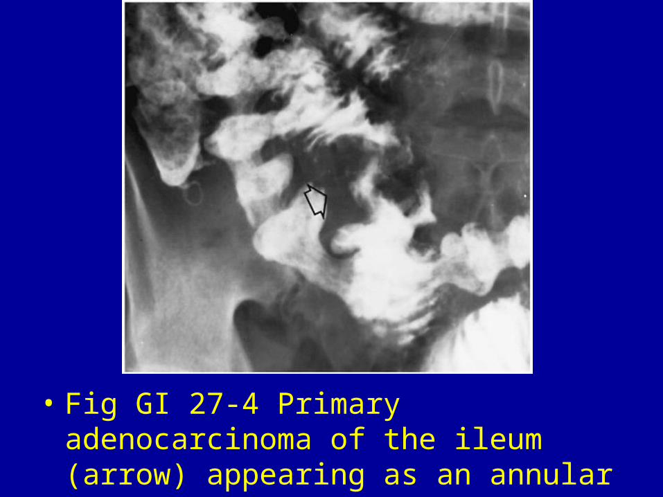
• Fig GI 27-4 Primary adenocarcinoma of the ileum (arrow) appearing as an annular constricting lesion.

• Fig GI 27-5 Lymphoma. Note the large, bulky, irregular lesion (arrow).

• Fig GI 27-6 Lymphoma. Multiple large irregular masses.

• Fig GI 27-7 Leiomyosarcoma. Large, bulky, irregular lesion (arrows).

• Fig GI 27-8 Metastatic hypernephroma. Multilobulated nodular mass in the proximal jejunum.

• Fig GI 27-9 Carcinoid tumor (arrow).

• Fig GI 27-10 Gallstone ileus. The obstructing stone (white arrows) in the jejunum is associated with evidence of barium in the biliary tree (black arrow).

• Fig GI 27-11 Ascaris. The linear intestinal tract of the roundworm is filled with barium (arrow).32

• Fig GI 27-12 Nodular lymphoid hyperplasia. Large filling defects suggest multiple polypoid masses.

• Fig GI 27-13 Phytobezoar. Large, irregular, proximal jejunal filling defect containing barium within the interstices of the lesion. Note the second bezoar in the stomach.33

• Fig GI 27-14 Crohn's disease. Multiple polypoid lesions in the distal jejunum and proximal ileum show both smooth and lobulated contours.34
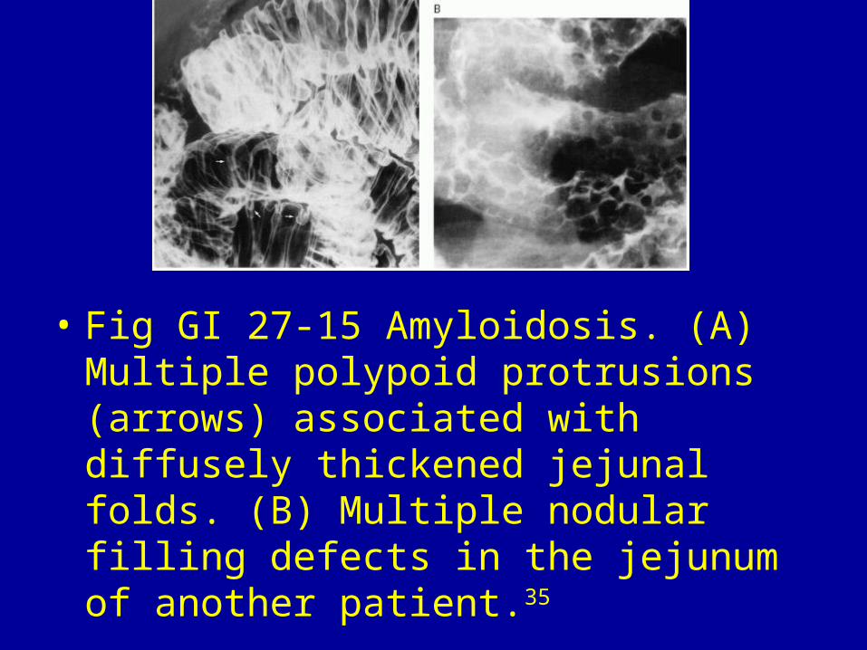
• Fig GI 27-15 Amyloidosis. (A) Multiple polypoid protrusions (arrows) associated with diffusely thickened jejunal folds. (B) Multiple nodular filling defects in the jejunum of another patient.35


Related Documents