2.7 Duodenum and pancreas 2.8 Kidneys, suprarenal glands and ureters 2.9 Posterior abdominal wall and diaphragm Abdomen 4 Albert van Schoor GNK 288 (SA4 Anatomy dissection)

2.7 Duodenum and pancreas 2.8 Kidneys, suprarenal glands and ureters 2.9 Posterior abdominal wall and diaphragm Abdomen 4 Albert van Schoor GNK 288 (SA4.
Dec 14, 2015
Welcome message from author
This document is posted to help you gain knowledge. Please leave a comment to let me know what you think about it! Share it to your friends and learn new things together.
Transcript
2.7 Duodenum and pancreas2.8 Kidneys, suprarenal glands and ureters2.9 Posterior abdominal wall and diaphragm
Abdomen 4
Albert van Schoor
GNK 288 (SA4 Anatomy dissection)

2.7 Duodenum and pancreas
2.7.1 Surface anatomy
2.7.1 Structure
2.7.3 Blood supply, nerve supply
and lymph drainage

2.7.1 Surface anatomy
• Review the surface anatomy of the duodenum and pancreas
• Name the vertebral heights of the various parts of the pancreas
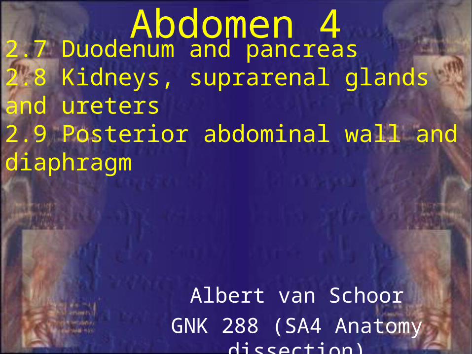
1st part• Approx. 5 cm
long• Anterolateral
of body of L1• On the
transpyloric line
2.7.1 Surface Anatomy

2nd part• Approx. 7-10
cm long• Descends
along the right sides of L1-L3
2.7.1 Surface Anatomy

3rd part• Approx. 6-8
cm long• Horisontal• Crosses the
body of L3
2.7.1 Surface Anatomy
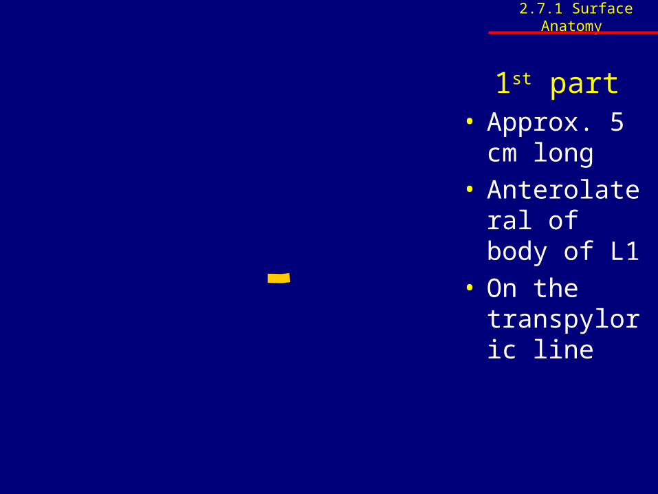
4th part• Approx. 5cm
cm long• Begins at the
left of L3• Rises superior
as far as the superior border of L2
2.7.1 Surface Anatomy

T12
L2
L1L1
L2
L3
2.7.1 Surface Anatomy
2.7.2 Structure
• Identify and briefly describe the four parts of the duodenum and the structures opening into it

1st part• 1st 5cm from
gastroduodenal junction
• Anterior:– Liver– Gallbladder
• Posterior:– Ant. Border of
omental foramen (gastroduodenal artery)
2.7.2 Structure

2nd part• 7-10cm long• Vertical• Curves around
head of pancreas• Anterior:
– Transverse colon
• Posterior:– Right kidney and
ureter
• Posteromedial:– Major and minor
duodenal papillae
2.7.2 Structure
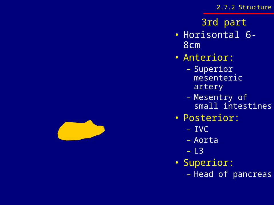
3rd part• Horisontal 6-8cm• Anterior:
– Superior mesenteric artery
– Mesentry of small intestines
• Posterior:– IVC– Aorta– L3
• Superior:– Head of pancreas
2.7.2 Structure

4th part• Runs vertical for
2.5 - 5cm to duodenojejenal junction
• Suspensory ligament of duodenum (Lig. of Treitz) extend from right crus of diaphragm
• Suspends duedenojejenal junction
2.7.2 Structure

2.7.2 Structure
• Identify and schematically illustrate the different parts (regions) and duct system of the pancreas
• Identify and list the general and peritoneal relations of the four parts of the duodenum
• Identify and briefly discuss the relations of the pancreas to the spleen, duodenum, stomach and transverse colon and peritoneum
• Identify the root of the transverse colon

Major pancreatic duct of Wirsung
Accessory pancreatic duct of Santorini
2.7.2 Structure
2.7.2 Structure
• Identify and briefly discuss the relations of the pancreas to the spleen, duodenum, stomach and transverse colon and peritoneum
• Identify the root of the transverse colon

• Superior:– Splenic artery– Common hepatic
artery• Posterior:
– IVC– Aorta– Sup. mesenteric
vessels– Crurae of diaphragm– Coeliac plexus – Left kidney
• Anterior:– Stomach– Transverse mesocolon– Omental bursa
2.7.2 Structure

2.7.3 Blood supply and lymph drainage
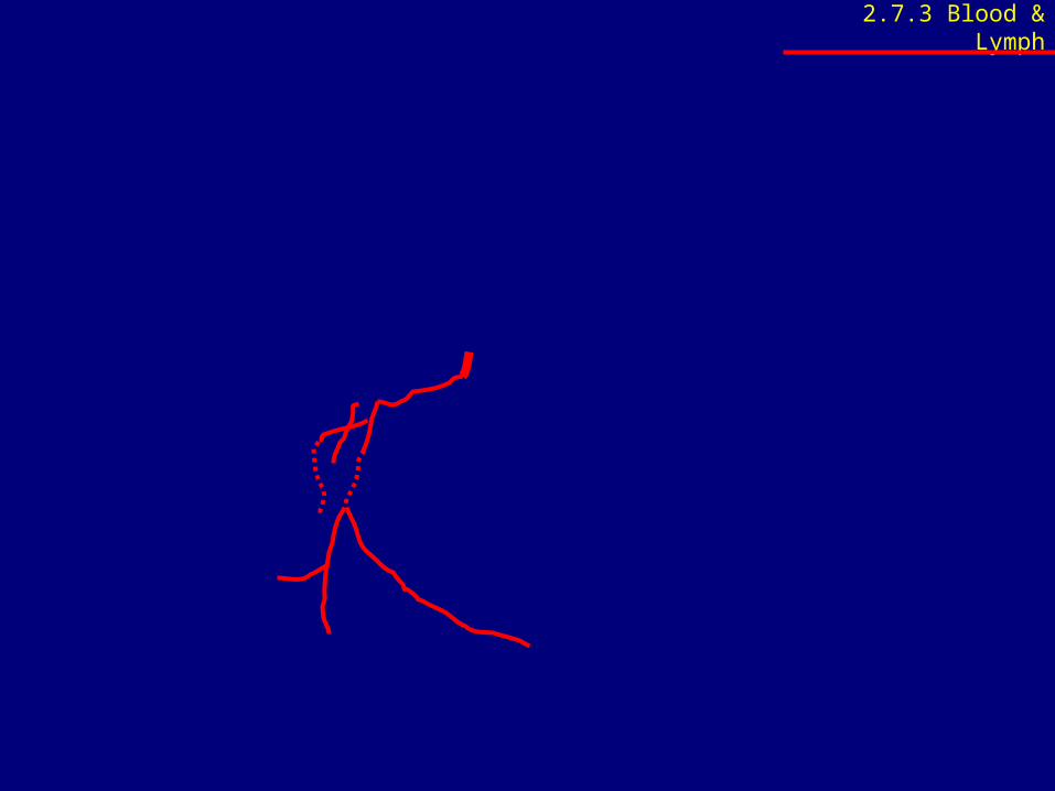
• Identify, trace and list the arterial supply and venous drainage of the duodenum and pancreas
• Give a broad overview of the major lymph node groups into which the upper abdominal organs drain
2.7.3 Blood & Lymph

• Veins follows the arteries
• Drains into portal vein either directly or indirectly through:– Superior mesenteric vein– Splenic vein
2.7.3 Blood & Lymph

2.8 Kidneys, suprarenal glands and ureters
2.8.1 Surface anatomy
2.8.2 Structure
2.8.3 Blood supply, nerve supply
and lymph drainage
2.8.4 Radiographic anatomy
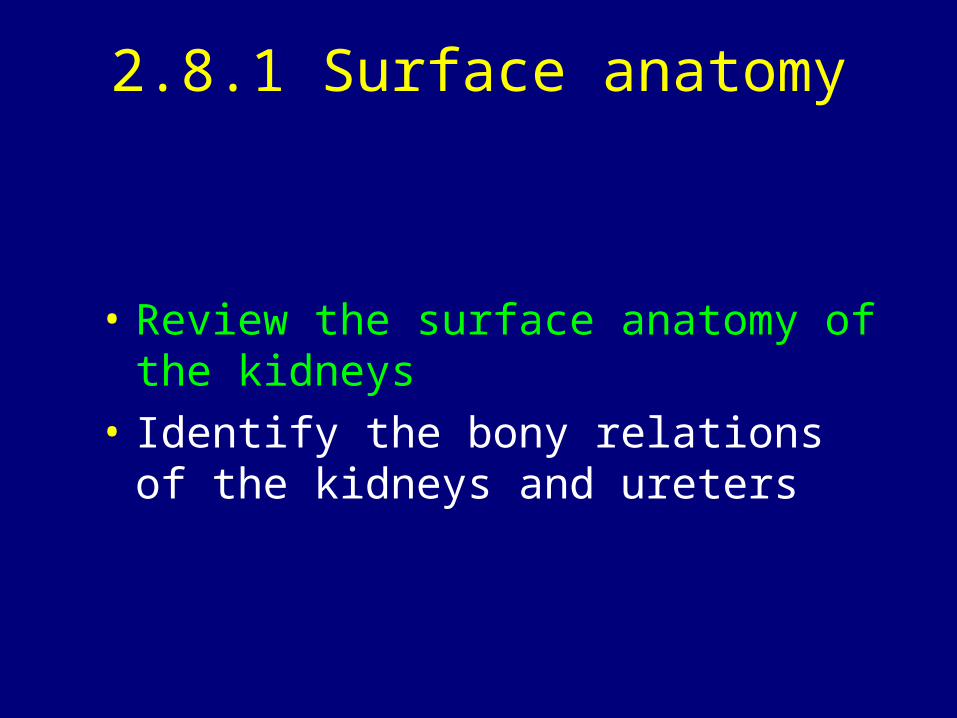
2.8.1 Surface anatomy
• Review the surface anatomy of the kidneys
• Identify the bony relations of the kidneys and ureters
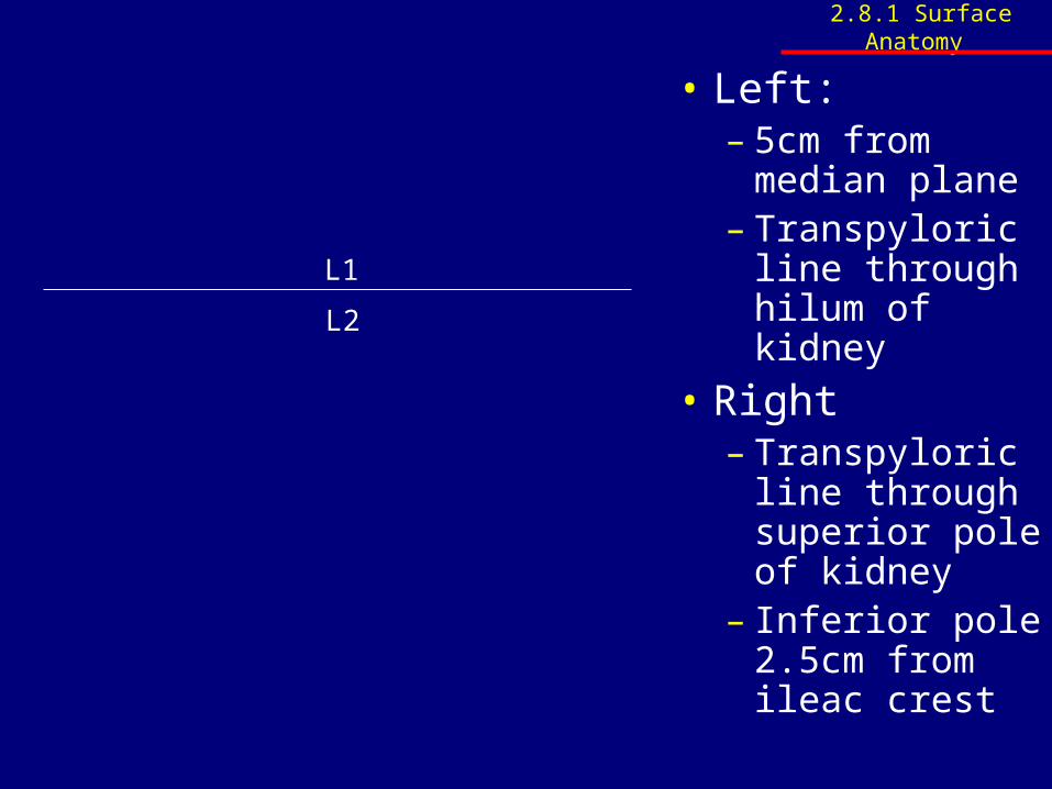
• Left:– 5cm from
median plane– Transpyloric
line through hilum of kidney
• Right– Transpyloric
line through superior pole of kidney
– Inferior pole 2.5cm from ileac crest
L1
L2
2.8.1 Surface Anatomy

T12 T12
T11T11
Psoas MajorQuadratus LumborumTransverse Abdominis
2.8.1 Surface Anatomy
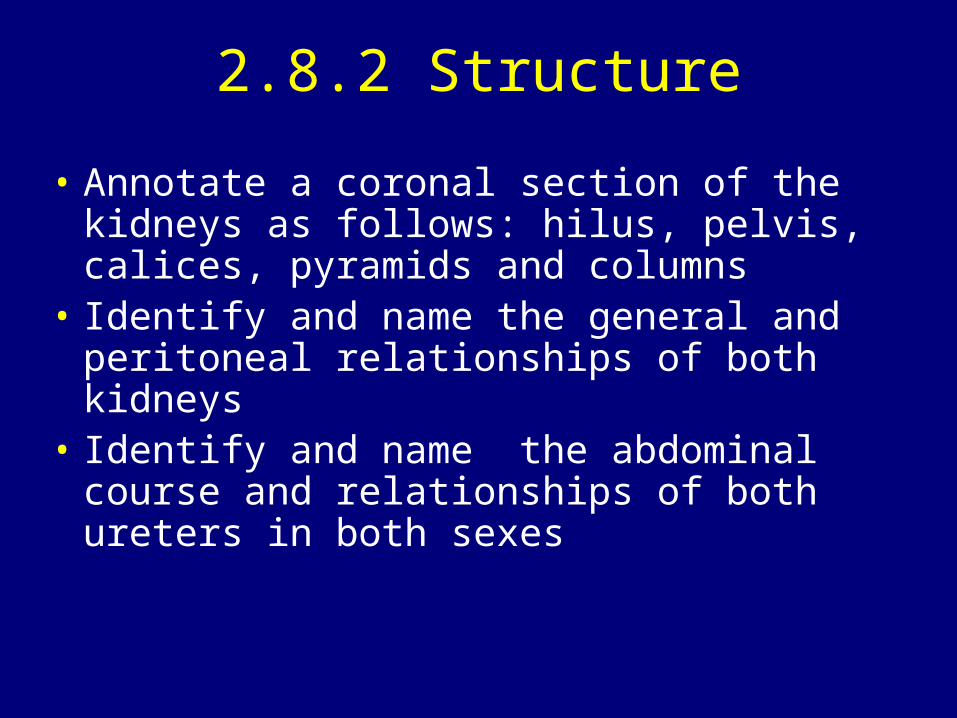
2.8.2 Structure
• Annotate a coronal section of the kidneys as follows: hilus, pelvis, calices, pyramids and columns
• Identify and name the general and peritoneal relationships of both kidneys
• Identify and name the abdominal course and relationships of both ureters in both sexes
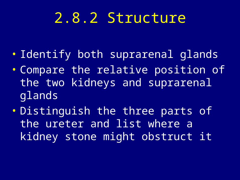
2.8.2 Structure
• Identify both suprarenal glands
• Compare the relative position of the two kidneys and suprarenal glands
• Distinguish the three parts of the ureter and list where a kidney stone might obstruct it

2.8.2 Structure

• Approximately 25cm long• Urine moves by means of peristaltic movements• 3 Components:
– Abdominal: adjacent to transverse processes of lumbar vertebrae L2-L5
– Pelvic: enters true pelvis, continues medial, over internal iliac artery, and anteromedial towards bladder
– Intramural: short part between 2 layers of bladder wall. Serves as a mechanical valve to prevent reflux of urine
2.8.2 Structure

• Potential constrictions:– Renal pelvis– Where ureter crosses
the internal iliac artery– Between the 2 muscle
layers of the bladder wall
2.8.2 Structure

2.8.3 Blood supply, nerve supply and lymph drainage
• Trace, identify and compare the course and relations of both renal arteries and veins
• Identify, trace and name the arterial supply and venous drainage of both suprarenal glands
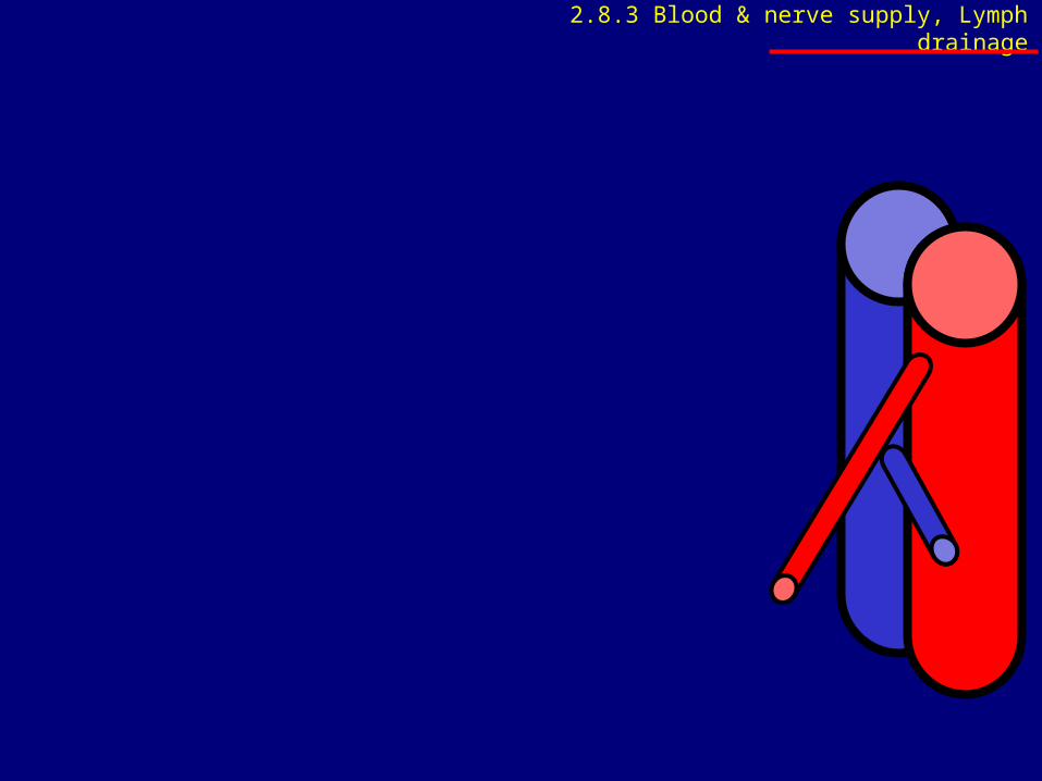
2.8.3 Blood & nerve supply, Lymph drainage

2.8.4 Radiographic anatomy
• Identify the following on a urogram (or IVP): – Kidneys – ureters, – bladder, – major and minor calyces, – pelvis of kidney, and – relation of the ureters to the transverse processes of
the lumbar vertebrae
www.up.ac.za/academic/medicine/anatomy/current/sa4/week01e.html#radio

2.9 Posterior abdominal wall and diaphragm
2.9.1 Abdominal aorta2.9.2 Abdominal inferior vena cava2.9.3 Lymph drainage2.9.4 Nerve structures2.9.5 Fascia2.9.6 Muscles2.9.7 Diaphragm2.9.8 Osteology

2.9.1 Abdominal aorta
• Identify the course, relationships and branches of the abdominal aorta

• 3 unpaired to viscera (anterior)– Coeliac trunk– Sup. mesenteric– Inf. mesenteric
• 3 paired to viscera (lateral)– Suprarenal– Renal – Gonadal
• 5 paired (lateral)– Inferior phrenic– 4 pairs of lumbar
• 2 paired terminal branches– Common iliac – Median sacral
2.9.1 Abdominal aorta

2.9.2 Abdominal inferior vena cava
• Briefly discuss and identify the course, relations and tributaries of the inferior vena cava
• Identify the different drainage of the left and right gonadal veins
2.9.3 Lymph drainage
• Give a broad overview of the lymph drainage of the abdomen

2.9.4 Nerve structures
• Identify and briefly describe the sympathetic trunk and where possible the plexuses

• 2 parallel nerve cords • Extending on either
side of the vertebral column from the base of the skull to the coccyx.
• 2 sympathetic trunks come together anterior to the coccyx to form the ganglion impar.
2.9.4 Nerve structures

2.9.4 Nerve structures
• Identify and discuss the distribution and root values of the following nerves of the lumbar plexus: – Subcostal, – iliohypogastric, – ilioinguinal, – lateral cutaneous nerve of the thigh, – genitofemoral, – femoral and – obturator nerves
• Give an overview of the autonomic nerve supply of the abdomen

• Splanchnic nerves– Thoracic (Coeliac plexus)
– Lumbar (Intermesenteric / aortic & superior hypogastric plexus)
– Sacral (Inferior hypogastric plexus)
– Pelvic* (Inferior hypogastric plexus)
* Both sympathetic and parasympathetic
Autonomic
Sympathetic Paraympathetic
• Vagus nerve (X) ~ cranial outflow– Foregut & midgut
• S2-S4 ~ abdominal outflow– Hindgut
• Intermesenteric / aortic plexus
• Superior & inferior mesenteric plexuses
• Superior & inferior hypogastric plexuses
2.9.4 Nerve structures

2.9.5 Fascia
• Identify the layers of the thoracolumbar fascia
• Identify the psoas and iliac fascia

2.9.6 Muscles
• Identify and briefly discuss the major attachments, relationships to organs and nerves, as well as the nerve supply of the following muscles: – psoas major, – iliacus and – quadratus lumborum

Muscle Origin Insertion Innervation
Psoas major
Lateral surface of bodies and intervertebral discs of
T12 and L1 to L5, transverse processes of
the lumbar vertebrae
Lesser trochanter of
the femur Anterior rami of L1 to L3
Quadratus Lumborum
Transverse process of L5, iliolumbar ligament, and
iliac crest
Transverse processes of
L1-L4 and inferior border
of rib 12
Anterior rami of T12 and L1 to L4
Iliacus
Upper two-thirds of iliac fossa, anterior sacro-iliac and iliolumbar ligaments, and upper lateral surface
of sacrum
Lesser trochanter of
femur
Femoral nerve (L2 - L4)
2.9.6 Muscles

2.9.7 Diaphragm
• Review the surface anatomy of the diaphragm
• Identify and name it's attachments, blood supply and nerve supply

1a) Vertebral ~ crura
• Right Crus– L1-L3
• Left Crus– L1-L2
2.9.7 Diaphragm
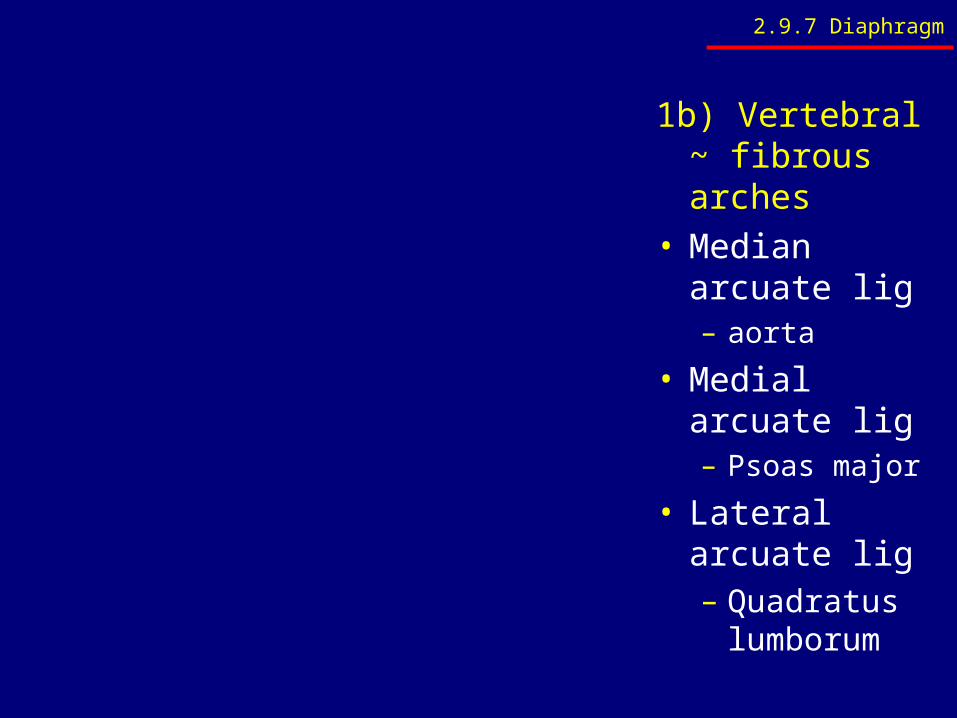
1b) Vertebral ~ fibrous arches
• Median arcuate lig– aorta
• Medial arcuate lig– Psoas major
• Lateral arcuate lig– Quadratus
lumborum
2.9.7 Diaphragm

2) Costal• Ribs 7-12
3) Sternal
• 2 small attachments to xiphisternum
2.9.7 Diaphragm
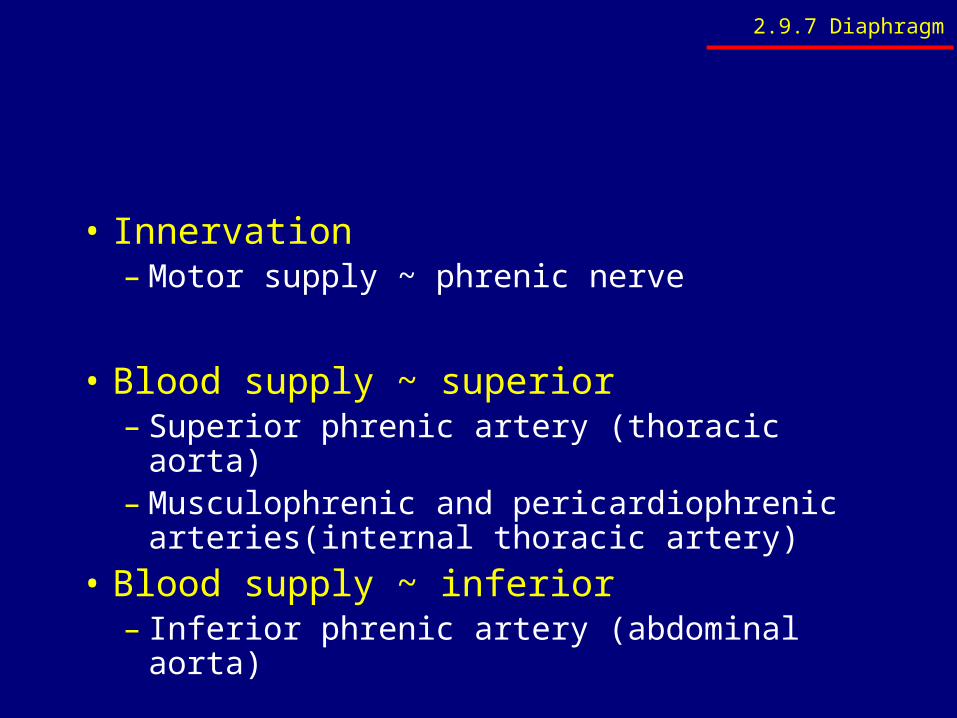
• Innervation– Motor supply ~ phrenic nerve
• Blood supply ~ superior– Superior phrenic artery (thoracic aorta)– Musculophrenic and pericardiophrenic
arteries(internal thoracic artery)
• Blood supply ~ inferior– Inferior phrenic artery (abdominal aorta)
2.9.7 Diaphragm
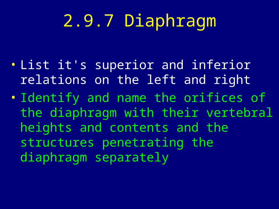
2.9.7 Diaphragm
• List it's superior and inferior relations on the left and right
• Identify and name the orifices of the diaphragm with their vertebral heights and contents and the structures penetrating the diaphragm separately

T8
T10
T12
2.9.7 Diaphragm

2.9.7 Diaphragm
• Identify and name the arcuate ligaments of the diaphragm and the structures related posterior to them
• Identify and name the crura of the diaphragm in relation to the oesophageal hiatus

2.9.8 Osteology
• Identify and briefly discuss the bony elements of the posterior abdominal wall
Related Documents