Small Molecule Therapeutics Reversal of Chemoresistance in Ovarian Cancer by Co-Delivery of a P-Glycoprotein Inhibitor and Paclitaxel in a Liposomal Platform Yilin Zhang 1 , Shravan Kumar Sriraman 2 , Hilary A. Kenny 1 , Ed Luther 2 , Vladimir Torchilin 2 , and Ernst Lengyel 1 Abstract The overexpression of permeability-glycoprotein (P-gp), an ABC transporter involved in the cellular exclusion of chemo- therapeutic drugs, is a major factor in paclitaxel-resistant ovarian cancer. However, in clinical trials, co-administration of P-gp inhibitors and anticancer drugs has not resulted in the efficient reversal of drug resistance. To improve administra- tion, we encapsulated the third-generation P-gp inhibitor tariquidar (XR-9576, XR), alone or in combination with pac- litaxel (PCT) in liposomes (LP). After optimization, the lipo- somes demonstrated favorable physicochemical properties and the ability to reverse chemoresistance in experiments using chemosensitive/chemoresistant ovarian cancer cell line pairs. Analyzing publicly available datasets, we found that overexpression of P-gp in ovarian cancer is associated with a shorter progression-free and overall survival. In vitro, LP(XR) significantly increased the cellular retention of rhodamine 123, a P-gp substrate. LP(XR,PCT) synergistically inhibited cell viability, blocked proliferation, and caused G 2 –M arrest in paclitaxel-resistant SKOV3-TR and HeyA8-MDR cell lines overexpressing P-gp. Holographic imaging cytometry revealed that LP(XR,PCT) treatment of SKOV3-TR cells induced almost complete mitotic arrest, whereas laser scanning cytometry showed that the treatment induced apoptosis. In proof-of- concept preclinical studies, LP(XR,PCT), when compared with LP(PCT), significantly reduced tumor weight (43.2% vs. 16.9%, P ¼ 0.0007) and number of metastases (44.4% vs. 2.8%, P ¼ 0.012) in mice bearing orthotopic HeyA8-MDR ovarian tumors. In the xenografts, LP(XR,PCT) efficiently induced apoptosis and impaired proliferation. Our findings suggest that co-delivery of a P-gp inhibitor and paclitaxel using a liposomal platform can sensitize paclitaxel-resistant ovarian cancer cells to paclitaxel. LP(XR,PCT) should be considered for clinical testing in patients with P-gp–overexpressing tumors. Mol Cancer Ther; 15(10); 2282–93. Ó2016 AACR. Introduction Ovarian cancer is the deadliest gynecologic malignancy in developed countries. It has a 5-year survival rate of approxi- mately 40% and accounts for 6% of all cancer-related deaths in women in the United States (1, 2). The current standard of care includes debulking surgery and subsequent adjuvant chemo- therapy with paclitaxel and carboplatin. Although combination chemotherapy clearly prolongs the lives of patients with ovar- ian cancer, the disease recurs in 80% of cases because the tumor cells develop drug resistance (3, 4). A modest increase in survival has been achieved with intraperitoneal chemotherapy, but the complications associated with peritoneal catheters currently impede their widespread clinical adoption (5). Since the discovery of multidrug resistance (MDR) about 4 decades ago (6), tremendous efforts have been made to eluci- date its mechanisms. So far, three major mechanisms of MDR have been identified and explored: (i) plasma membrane ATP- binding cassette (ABC) transporters extrude drugs, preventing intracellular accumulation (4, 7, 8); (ii) transmembrane impor- ters regulate the entry of water-soluble drugs (9, 10); and (iii) a constellation of contributing factors such as epigenetic mod- ifications (11, 12), miRNAs (13, 14), and elements of the tumor microenvironment, which work together to induce drug resis- tance (15, 16). Permeability-glycoprotein (P-gp, MDR1, ABCB1; refs. 17, 18) and multidrug resistance associated-protein 1 (MRP1, ABCA1; ref. 4) are 2 important ABC transporters associated with drug resistance in ovarian cancer. P-gp was the first ABC member found to confer chemoresistance and is still seen as one of the most promising targets for the reversal of chemoresistance (17). The overexpression of P-gp is associated with reduced progres- sion-free and overall survival in several cancer types, including ovarian cancer (19–22), most probably because it contributes to the cellular exclusion of a broad spectrum of chemothera- peutic drugs, including taxanes, doxorubicin, and vinca alka- loids (8). The energy-dependent efflux of the chemotherapeutic drugs out of the cancer cell reduces the intracellular 1 Department of Obstetrics and Gynecology/Section of Gynecologic Oncology, University of Chicago, Chicago, Illinois. 2 Center for Phar- maceutical Biotechnology and Nanomedicine, Northeastern Univer- sity, Boston, Massachusetts. Note: Supplementary data for this article are available at Molecular Cancer Therapeutics Online (http://mct.aacrjournals.org/). Y. Zhang and S.K. Sriraman contributed equally to this article. Corresponding Author: Ernst Lengyel, Department of Obstetrics and Gynecol- ogy, University of Chicago, 5841 South Maryland Avenue, Chicago, IL 60637. Phone: 773-834-0563; Fax: 773-702-5161; E-mail: [email protected] doi: 10.1158/1535-7163.MCT-15-0986 Ó2016 American Association for Cancer Research. Molecular Cancer Therapeutics Mol Cancer Ther; 15(10) October 2016 2282 Downloaded from http://aacrjournals.org/mct/article-pdf/15/10/2282/1850443/2282.pdf by guest on 30 May 2022

Welcome message from author
This document is posted to help you gain knowledge. Please leave a comment to let me know what you think about it! Share it to your friends and learn new things together.
Transcript
Small Molecule Therapeutics
Reversal of Chemoresistance in Ovarian Cancerby Co-Delivery of a P-Glycoprotein Inhibitorand Paclitaxel in a Liposomal PlatformYilin Zhang1, Shravan Kumar Sriraman2, Hilary A. Kenny1, Ed Luther2,Vladimir Torchilin2, and Ernst Lengyel1
Abstract
The overexpression of permeability-glycoprotein (P-gp), anABC transporter involved in the cellular exclusion of chemo-therapeutic drugs, is a major factor in paclitaxel-resistantovarian cancer. However, in clinical trials, co-administrationof P-gp inhibitors and anticancer drugs has not resulted in theefficient reversal of drug resistance. To improve administra-tion, we encapsulated the third-generation P-gp inhibitortariquidar (XR-9576, XR), alone or in combination with pac-litaxel (PCT) in liposomes (LP). After optimization, the lipo-somes demonstrated favorable physicochemical propertiesand the ability to reverse chemoresistance in experimentsusing chemosensitive/chemoresistant ovarian cancer cell linepairs. Analyzing publicly available datasets, we found thatoverexpression of P-gp in ovarian cancer is associated with ashorter progression-free and overall survival. In vitro, LP(XR)significantly increased the cellular retention of rhodamine123, a P-gp substrate. LP(XR,PCT) synergistically inhibited
cell viability, blocked proliferation, and caused G2–M arrestin paclitaxel-resistant SKOV3-TR and HeyA8-MDR cell linesoverexpressing P-gp. Holographic imaging cytometry revealedthat LP(XR,PCT) treatment of SKOV3-TR cells induced almostcomplete mitotic arrest, whereas laser scanning cytometryshowed that the treatment induced apoptosis. In proof-of-concept preclinical studies, LP(XR,PCT), when compared withLP(PCT), significantly reduced tumor weight (43.2% vs.16.9%, P ¼ 0.0007) and number of metastases (44.4% vs.2.8%, P ¼ 0.012) in mice bearing orthotopic HeyA8-MDRovarian tumors. In the xenografts, LP(XR,PCT) efficientlyinduced apoptosis and impaired proliferation. Our findingssuggest that co-delivery of a P-gp inhibitor and paclitaxel usinga liposomal platform can sensitize paclitaxel-resistant ovariancancer cells to paclitaxel. LP(XR,PCT) should be considered forclinical testing in patients with P-gp–overexpressing tumors.Mol Cancer Ther; 15(10); 2282–93. �2016 AACR.
IntroductionOvarian cancer is the deadliest gynecologic malignancy in
developed countries. It has a 5-year survival rate of approxi-mately 40% and accounts for 6% of all cancer-related deaths inwomen in the United States (1, 2). The current standard of careincludes debulking surgery and subsequent adjuvant chemo-therapy with paclitaxel and carboplatin. Although combinationchemotherapy clearly prolongs the lives of patients with ovar-ian cancer, the disease recurs in 80% of cases because the tumorcells develop drug resistance (3, 4). A modest increase insurvival has been achieved with intraperitoneal chemotherapy,
but the complications associated with peritoneal catheterscurrently impede their widespread clinical adoption (5).
Since the discovery of multidrug resistance (MDR) about 4decades ago (6), tremendous efforts have been made to eluci-date its mechanisms. So far, three major mechanisms of MDRhave been identified and explored: (i) plasma membrane ATP-binding cassette (ABC) transporters extrude drugs, preventingintracellular accumulation (4, 7, 8); (ii) transmembrane impor-ters regulate the entry of water-soluble drugs (9, 10); and (iii) aconstellation of contributing factors such as epigenetic mod-ifications (11, 12), miRNAs (13, 14), and elements of the tumormicroenvironment, which work together to induce drug resis-tance (15, 16).
Permeability-glycoprotein (P-gp, MDR1, ABCB1; refs. 17, 18)and multidrug resistance associated-protein 1 (MRP1, ABCA1;ref. 4) are 2 important ABC transporters associated with drugresistance in ovarian cancer. P-gp was the first ABC memberfound to confer chemoresistance and is still seen as one of themost promising targets for the reversal of chemoresistance (17).The overexpression of P-gp is associated with reduced progres-sion-free and overall survival in several cancer types, includingovarian cancer (19–22), most probably because it contributesto the cellular exclusion of a broad spectrum of chemothera-peutic drugs, including taxanes, doxorubicin, and vinca alka-loids (8). The energy-dependent efflux of the chemotherapeuticdrugs out of the cancer cell reduces the intracellular
1Department of Obstetrics and Gynecology/Section of GynecologicOncology, University of Chicago, Chicago, Illinois. 2Center for Phar-maceutical Biotechnology and Nanomedicine, Northeastern Univer-sity, Boston, Massachusetts.
Note: Supplementary data for this article are available at Molecular CancerTherapeutics Online (http://mct.aacrjournals.org/).
Y. Zhang and S.K. Sriraman contributed equally to this article.
Corresponding Author: Ernst Lengyel, Department of Obstetrics and Gynecol-ogy, University of Chicago, 5841 South Maryland Avenue, Chicago, IL 60637.Phone: 773-834-0563; Fax: 773-702-5161; E-mail: [email protected]
doi: 10.1158/1535-7163.MCT-15-0986
�2016 American Association for Cancer Research.
MolecularCancerTherapeutics
Mol Cancer Ther; 15(10) October 20162282
Dow
nloaded from http://aacrjournals.org/m
ct/article-pdf/15/10/2282/1850443/2282.pdf by guest on 30 May 2022
accumulation of otherwise effective therapies, requiring ahigher dose to achieve an anticancer effect. Therefore, inhibit-ing P-gp to increase the length of time a drug remains inside thecell represents a reasonable strategy for the reversal of che-moresistance. Downregulating P-gp expression with RNA inter-ference or blocking P-gp activity with inhibitors has beenshown to sensitize chemoresistant cancer cells to chemotherapyin various cancers (23). Three generations of P-gp inhibitorshave been identified. The first 2 generations of P-gp modula-tors, such as cyclosporine A and verapamil, function by com-petitively binding substrates at the transmembrane domain;but, although they successfully compete for substrate binding,they display low specificity and significant side effects (7).Tariquidar (XR9576), a third-generation inhibitor (24), is anoncompetitive inhibitor of P-gp which impedes drug efflux byblocking the transition of P-gp to an open conformation (25).However, clinical trials with tariquidar had to be terminatedearly because of the ability of inhibitor to block normalphysiologic functions of P-gp. P-gp, when highly expressed,acts to maintain the blood–brain barrier and exclude toxinsfrom normal tissues, notably those of the intestines, kidney,and spleen (as reviewed in ref. 26).
To limit the effects of tariquidar on normal tissue, as well asovercome its unfavorable pharmacokinetic properties, such aslow water solubility and short serum half-life (4, 10), aneffective systemic delivery approach with improved pharmaco-kinetic profiling and tumor targetability is necessary. Encapsu-lating tariquidar and paclitaxel in nanoparticles represents onesuch strategy, as nanoparticles are known to passively accumu-late at tumor sites due to enhanced permeability and retention(EPR) caused by leaky malignant tumor vasculature (27). Inaddition, nanoparticles confer more favorable pharmacokine-tic profiles to the encapsulated substances (28). Indeed, poly(D,L-lactide-co-glycolide) (PLGA)-formulated nanoparticlesdelivering paclitaxel and tariquidar were reported to efficientlyreduce subcutaneous mammary tumor growth in a paclitaxel-resistant mouse model (29).
In view of the important role of P-gp in ovarian cancerchemotherapy resistance in general and paclitaxel resistancespecifically and given that tariquidar has been established as anefficient P-gp inhibitor, our goal was to determine whethertariquidar/paclitaxel nanoparticles are a viable strategy thatshould be further developed for treatment of paclitaxel-resis-tant ovarian cancer. We hypothesized that co-administration ofthe P-gp inhibitor and paclitaxel using a liposomal platformcould achieve synergistic effects, increasing the retention ofpaclitaxel in chemotherapy-resistant ovarian cancer, therebysignificantly reducing the effective dose of paclitaxel requiredto kill tumor cells. Using paclitaxel-sensitive/resistant cell linepairs, we report here that liposomes encapsulating both tar-iquidar and paclitaxel increase the cytotoxicity of paclitaxel andeffectively inhibit chemoresistant ovarian cancer tumor growthin a preclinical model.
Materials and MethodsReagents
Egg phosphatidylcholine (ePC), cholesterol, and a hand-held mini extruder were purchased from Avanti Polar Lipids.1,2-Distearoyl-sn-glycero-3-phosphoethanolamine-N-[methoxy(polyethylene glycol)-2000] (DSPE-PEG2000) and 1,2-dioleoyl-
3-trimethylammonium-propane (DOTAP) were purchasedfrom Corden Pharma International. Tariquidar and paclitaxelwere obtained from Medkoo Biosciences and LC Labs, respec-tively. Rhodamine 123 was purchased from Sigma-Aldrich.CellTiter-Blue Cell Viability Assay was obtained from Promega.The X-Bridge C18 column (4.6 � 250 mm2, 5 mm id) was fromWaters. Hoechst 33342, Yo-Pro, and propidium iodide (PI) werepurchased from Life Technologies. Fluorescein isothiocyanate(FITC)-labeled P-gp (UIC-2), phycoerythrin (PE)-labeled MRP-1(QCRL-1), b-tubulin (G-8), and goat anti-mouse IgG-FITC anti-bodies were purchased from Santa Cruz Biotechnology.CDC25C (5H9), p21 (12D1), GAPDH (14C10), phosphorylat-ed CDC2 (Tyr15, 10A11), and total CDC2 antibodies wereobtained from Cell Signaling Technology, whereas cyclin B1(S126) and P-gp antibodies were purchased from Abgent.Cleaved caspase-3 (Asp175) antibody was from Cell SignalingTechnology. Ki-67 antibody (SP6) and high-performance liquidchromatography (HPLC)-grade acetonitrile were from ThermoFisher Scientific.
Cell cultureSKOV3ip1 andHeyA8 cells were provided by Dr. GordonMills
(MD Anderson Cancer Center, Houston, TX). HeyA8-MDR was agift from Dr. Anil Sood (MD Anderson Cancer Center; ref. 30).SKOV3-Taxol–resistant cells (TR) were provided by Dr. ZhenfengDuan (MGH, Boston, MA; ref. 31). Tyk-nu cells were provided byDr. Kenjiro Sawada (Osaka University, Osaka, Japan; ref. 32). TheTyk-nu carboplatin-resistant (R) cell line (33)was purchased fromJCRB cell bank. Tyk-nu and Tyn-nu-R cells were cultured in Eagleminimum essential medium with 15% FBS, 1% penicillin/strep-tomycin, and 1% L-glutamine (34). All other human ovariancancer cell lines were maintained in DMEM with 10% FBS and1% penicillin/streptomycin solution at 37�C with 5% CO2. Allcell lines were authenticated using the commercial service, Cell-Check (IDEXX Bioresearch). Samples were confirmed to be ofhuman origin and no mammalian interspecies contaminationwas detected. The alleles for 9 short tandem repeat (STR) markerswere determined and the results were compared with the profilesfrom DSMZ, ATCC, JCRB, and RIKEN STR databases.
Preparation of liposomesLiposomes were synthesized by the thin-film hydration meth-
od (35). Briefly, a lipid film of ePC, cholesterol, and DOTAPmixed at a mole ratio of 64%:30%:6% with tariquidar andpaclitaxel (equimolar amounts at a 1% w/w ratio to the totallipid) was prepared by removal of organic solvents using rotaryevaporation followed by freeze-drying overnight. The lipid filmwas hydrated with PBS (pH ¼ 7.4) to a 5 mg/mL lipid concen-tration and then extruded through 200-nm polycarbonate mem-branes. The nonencapsulated, insoluble, hydrophobic drug wasthen removed by syringe filtration (0.22-mm filter). A film of thePEG2000-DSPE at a mole ratio of 2% (to the total lipid in theliposome) was hydrated with the liposomal solutions and wasconstantly stirred at 37�C overnight. For in vivo studies, the lipidfilm was rehydrated to a 40 mg/mL lipid concentration.
Physicochemical characterization of liposomesParticle size and zeta potential were measured using an N4
Coulter Particle Size Analyzer and Zetaplus (Brookhaven Instru-ments Corporation), respectively. The particle size and surfacemorphology was further confirmed with a uranyl acetate stain
Tariquidar and Paclitaxel Therapy in Ovarian Cancer
www.aacrjournals.org Mol Cancer Ther; 15(10) October 2016 2283
Dow
nloaded from http://aacrjournals.org/m
ct/article-pdf/15/10/2282/1850443/2282.pdf by guest on 30 May 2022
using transmission electron microscopy (TEM). The liposomaldrug concentrations were determined by reverse phase HPLCusing an X-bridge C18 column on a Hitachi Elite LaChromHPLCsystem. A mixture of 10 mmol/L ammonium acetate buffer(pH ¼ 4; 40%) and acetonitrile (60%) was used as the mobilephase with a 1-mL/minute flow rate. The tariquidar had a reten-tion time of about 3.2minutes whereas paclitaxel had a retentiontime of about 5.3 minutes. Detection of both drugs was carriedout using a UV detector (228 nm). Liposomal drug concentra-tions were calculated by comparison against a standard curve ofeach drug (0–20 mg/mL). The characterization of drug-loadedliposomes is described in Supplementary Table S1.
Kaplan–Meier survival analysisWe performed Kaplan–Meier survival analyses on the basis of
P-glycoprotein (P-gp/ABCB1 gene) expression in patients withovarian cancer using the Gene Expression Omnibus (GEO) andThe Cancer Genome Atlas (TCGA) database. The cut-off level ofexpression dividing the high or low groups was determined by analgorithm of the Kaplan–Meier plotter (36, 37). Accession num-bers for gene expression datasets were: GSE-14764, -15622,-18520, -19829, -23554, -26193, -26712, -27651, -30161,-3149, -51373, -9891, and TCGA. The expression range of theP-gp probe (209994_s_at) was 2 to 1,918. Progression-free sur-vival (PFS) was calculated for each dataset (n > 1,000; follow-upfor 15 years) with the cut-off level of 43. Overall survival (OS)rate was assessed for each dataset (n ¼ 1,339; follow-up for 20years) with a cutoff level of 64. The expression range of theMRP1probe (205887_x_at) was 11 to 1,815, and the expression rangeof the MRP3 probe (208161_s_at) was 3 to 5,163. PFS wascalculated for each dataset (n ¼ 1,056; follow-up for 15 years)with the cut-off level of 393 (MRP1) and 101 (MRP3).
Flow cytometry analysis of P-gp and MRP1 expressionHeyA8, HeyA8-MDR, SKOV3, SKOV3-TR, Tyk-nu, and Tyk-nu-
R cells were harvested using nonenzymatic CellStripper solution(Corning Inc.) to preservemembrane integrity. After washing, thecells (5�105)were stainedwith10mLof either FITC-labeled anti–P-gp antibody or PE-labeled anti-MRP1 antibody on ice for 30minutes. After spinning down, cells were resuspended in HBSSwith 1% FBS and expression was analyzed by an LSR II Fortessacell analyzer (BD Biosciences; ref. 38). The data were processed byFlowJo software (Tree Star Inc).
Rhodamine 123 exclusion assayHeyA8/HeyA8-MDR and SKOV3ip1/SKOV3-TR cells were
treated with free tariquidar (140 nmol/L, XR) or liposomaltariquidar (14 nmol/L, XR) for 48 hours. The cells were thentrypsinized, washed, and stained with 1 mmol/L of rhodamine123, a fluorescent substrate of P-gp, in an incubator for 30minutes (35). The cells were spun down, resuspended in ice-cold HBSS with 1% FBS, and analyzed by the LSR II Fortessa cellanalyzer. The data were processed by FlowJo software.
Laser scanning cytometryTo measure apoptosis, SKOV3-TR cells (3� 103) were seeded
on black 96-well plates 24 hours prior to the experiment.Liposomes were sterile filtered through 0.2-mm filters andincubated with the cells for 24 hours and washed off. After afurther 24 hours, 3 fluorescent dyes [Hoechst 33342 (5 mg/mL),Yo-Pro (0.063 mg/mL), and PI (1 mg/mL)] were added to stain
live cell nuclei, cells in early apoptosis (slight membranepermeability), and late apoptosis/necrosis, respectively. Afterincubation at 37�C for 30 minutes, the cells were analyzedin situ using the iCyte laser scanning cytometer (CompuCyteCorp.; refs. 39, 40). Excitation/emission wavelengths used were405/440 nm with a 30-nm bandwidth for Hoechst, 488/515nm with a 30-nm bandwidth for Yo-Pro, and 488/635 nm forPI. All data analyses were carried out using the iCyte Software(Version 3.4). Alternatively, DNA content distributions weremeasured by PI staining. Upon treatment with liposomes(50 nmol/L paclitaxel; 40 nmol/L XR) for 18 hours, the cellswere permeabilized by 70% ethanol overnight, stained withPI/RNase buffer (BD Biosciences), and detected by an LSR IIFortessa cell analyzer. The cell-cycle distributions were furtherprocessed by FlowJo software (38).
In vitro cytotoxicityCells (3 � 103) were seeded in each well of a 96-well plate 24
hours prior to the experiments. Liposomes (50 or 100 nmol/Lpaclitaxel) were incubated with the cells for 7 days. Cell viabilitywas then measured using a CellTiter-Blue Cell Viability Assaykit. The living cells are able to convert resazurin, a redox dye, tofluorescent resorufin, whereas the dying or dead cells couldnot. The fluorescent signals were detected via a Synergy HTplate reader (Bio-Tek) with an excitation wavelength of 530 nmand emission of 590 nm.
Colony formation assaysCell proliferation upon liposome treatment was examined
by two types of colony formation assays: monolayer and softagar (41). For monolayer colony formation, cells were seededonto 24-well plates and treated with free or liposomal drugs(100 nmol/L paclitaxel; 90 nmol/L XR) for 7 days beforefixation, crystal violet staining and imaging (G-box, Syngene).For soft agar colony formation, the cells mixed with 0.4% agar/medium were seeded onto 0.8% agar/medium support in6-well plates and treated with the liposomes (100 nmol/Lpaclitaxel) for 40 days with the media changed every week.Cells were fixed, stained with crystal violet, and imaged with theG-Box bioimaging system.
Scratch assayHeyA8-MDR/HeyA8, SKOV3-TR/SKOV3-TR (3 � 103) cells
were seeded in a 96-well plate. Scratches were generated usinga 96-pin wound maker (Essen BioScience) and cells were treatedwith free tariquidar (XR), paclitaxel, LP(XR), LP(PCT), or LP(XR,PCT) (50 nmol/L paclitaxel; 40 nmol/L XR) or not treated. Thecells were imaged in real-time every 2 hours using an IncuCytesystem (Essen BioScience) with phase-contrast microscopy at a10�magnification (42, 43).Changes in confluence of thewound/scratch were quantified by Zoom software (Essen BioScience).
Holographic imaging cytometrySKOV3-TR (2 � 105) cells were seeded in T-25 flasks for 48
hours prior to the experiment. The cells were incubated withliposomes (100 nmol/L paclitaxel) and imaged every 5 minutesfor 48 hours using the holographic imaging cytometer Holo-Monitor M4 (Phase Holographic Imaging AB), which employsa low-power 635 nm red laser diode to create an interferencepattern (hologram) that is reconstructed by software intoimages. HoloStudio software (Phase Holographic Imaging AB)
Zhang et al.
Mol Cancer Ther; 15(10) October 2016 Molecular Cancer Therapeutics2284
Dow
nloaded from http://aacrjournals.org/m
ct/article-pdf/15/10/2282/1850443/2282.pdf by guest on 30 May 2022
was used for cellular segmentation, calculating quantitativefeatures (optical thickness and volume), and individual celltracking. Thirty-five cells were randomly chosen to quantitatethe frequency of mitotic events. A cell volume greater than2,500 mm3 or a thickness greater than 8 mmwas chosen as cutoffvalues for mitotic activity. To visualize the whole dynamicprocess, 4 dimensional (x-position, y-position, cell thicknesscoded as brightness over time) projections of the hologramswere generated with the cell thickness threshold adjusted toshow the mitotic cells (44, 45).
Immunofluorescence of b-tubulinSKOV3-TR (2 � 104) cells were seeded onto coverslips placed
in 6-well plates. After 24 hours, the cells were treated withliposomes (200 nmol/L paclitaxel) for 24 hours. After fixation,the cells were permeabilized with cold methanol, blocked with3% BSA in PBS, and stained with the b-tubulin primary anti-body (1:20) overnight at 4�C. The cells were then stained withFITC-labeled secondary antibodies (1:30) for 2 hours, 5 mmol/Lof Hoechst 33342 for 30 minutes at room temperature, andimaged by laser scanning confocal microscopy (Zeiss).
In vivo studiesPaclitaxel-resistant ovarian cancer Hey8-MDR (1 � 106)
cells were injected intraperitoneally into 5- to 6-week-oldathymic female nude mice. Eight days later, the mice wererandomized into 3 groups (5 mice/group) and injected intra-peritoneally with LP(PCT), LP(XR,PCT) (1.5 mg/kg paclitaxel;�1 mg/kg XR) or PBS 3 times per week. The injection wasrepeated 5 times. Body weights were measured 4 times within20 days. The mice were euthanized 2 days after the fifth
treatment and the tumor weight and numbers were recorded.The animal studies were repeated twice. Tumors failed to formin 1 and 2 mice in the LP(XR,PCT) and LP(PCT) treatmentgroups, respectively; these mice were therefore excluded fromthe assessment. The tumors were fixed in formalin, paraffin-embedded, and sectioned for subsequent immunohisto-chemical and hematoxylin and eosin (H&E) staining. Immu-nohistochemical staining of human ovarian tumor sectionswas performed to assess markers of apoptosis (cleaved cas-pase-3, Asp175, 1:20) and proliferation (Ki67, SP6, 1:300).The slides (5 slides/group) were imaged at 200� magnifica-tion from three random fields (46); stained cells were quanti-fied using NIH ImageJ software (28).
Statistical analysisData were expressed as means � SEM. Statistical analyses
were performed using two-tailed t test or ANOVA (47, 48)with GraphPad Prism. Significance was determined by P < 0.05(�), P < 0.01 (��), and P < 0.001 (���).
ResultsP-gp overexpression is associated with poor prognosis inpatients with ovarian cancer
Several reports have indicated that P-gp plays an importantbiologic and prognostic role in the acquisition of MDR in awide range of human cancers, including ovarian cancer. How-ever, the number of patients in these studies was small(19–22). We, therefore, interrogated two publicly availabledatasets to determine whether P-gp mRNA expression is asso-ciated with the survival of ovarian cancer patients (Fig. 1,
1.0
0.8
0.6
0.4
0.2
0.0
1.0
0.8
0.6
0.4
0.2
0.0
1.0
0.8
0.6
0.4
0.2
0.0
1.0
0.8
0.6
0.4
0.2
0.0
0 50
ExpressionLow
HR = 1.4 (1.2−1.62)log-rank P = 1.3e-05
HR = 1.21 (1.04−1.41)log-rank P = 0.012
HR = 1.11 (0.95−1.29)log-rank P = 0.19
HR = 1.2 (1.04−1.4)log-rank P = 0.016
High
ExpressionLowHigh
ExpressionLowHigh
ExpressionLowHigh
100Progression-free survival time (months) Overall survival time (months)
Progression-free survival time (months)Progression-free survival time (months)
ABCB1/P-gp
ABCC1/MRP1 ABCC3/MRP3
ABCB1/P-gpA B
Sur
viva
l pro
babi
lity
Sur
viva
l pro
babi
lity
Sur
viva
l pro
babi
lity
Sur
viva
l pro
babi
lity
150
0 50 100 150 0 50 100 150
0 50 100 150 200
C D
Figure 1.
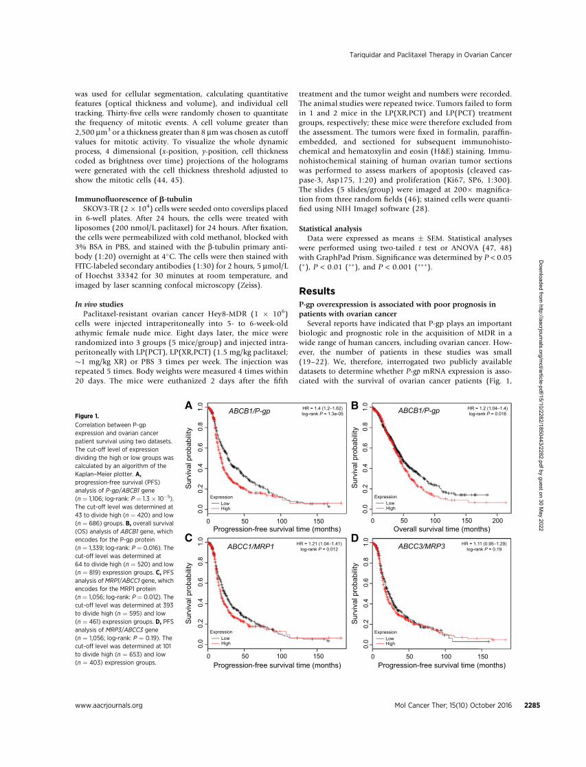
Correlation between P-gpexpression and ovarian cancerpatient survival using two datasets.The cut-off level of expressiondividing the high or low groups wascalculated by an algorithm of theKaplan–Meier plotter. A,progression-free survival (PFS)analysis of P-gp/ABCB1 gene(n ¼ 1,106; log-rank: P ¼ 1.3 � 10�5).The cut-off level was determined at43 to divide high (n ¼ 420) and low(n ¼ 686) groups. B, overall survival(OS) analysis of ABCB1 gene, whichencodes for the P-gp protein(n ¼ 1,339; log-rank: P ¼ 0.016). Thecut-off level was determined at64 to divide high (n ¼ 520) and low(n ¼ 819) expression groups. C, PFSanalysis of MRP1/ABCC1 gene, whichencodes for the MRP1 protein(n ¼ 1,056; log-rank: P ¼ 0.012). Thecut-off level was determined at 393to divide high (n ¼ 595) and low(n ¼ 461) expression groups. D, PFSanalysis of MRP3/ABCC3 gene(n ¼ 1,056; log-rank: P ¼ 0.19). Thecut-off level was determined at 101to divide high (n ¼ 653) and low(n ¼ 403) expression groups.
Tariquidar and Paclitaxel Therapy in Ovarian Cancer
www.aacrjournals.org Mol Cancer Ther; 15(10) October 2016 2285
Dow
nloaded from http://aacrjournals.org/m
ct/article-pdf/15/10/2282/1850443/2282.pdf by guest on 30 May 2022
TCGA, GEO datasets; refs. 7, 37). Kaplan–Meier analysis ofmore than 1,000 patients showed that high expression of P-gpis significantly correlated with shorter PFS (n ¼ 1,106; log-rank:P¼ 1.3� 10�5, HR¼ 1.4, Fig. 1A) and OS (n¼ 1,339; log-rank:P ¼ 0.016, HR ¼ 1.2, Fig. 1B). Expression of MRP1, whichconfers resistance to vinca alkaloids, but not to taxanes, wasassociated with reduced PFS (P ¼ 0.012, HR ¼ 1.21, Fig. 1C),but not with OS (data not shown). In contrast, the expressionofMRP3 (ABCC3), another ABC transporter, was not correlatedwith PFS (P ¼ 0.19, HR ¼ 1.11, Fig. 1D). This is consistentwith results from a previous study that analyzed the correla-tion of the expression of P-gp, MRP1, and MRP3 with PFS usingimmunohistochemical staining in 111 patients with ovariancancer (19).
Preparation and characterization of liposomes loaded withtariquidar and paclitaxel
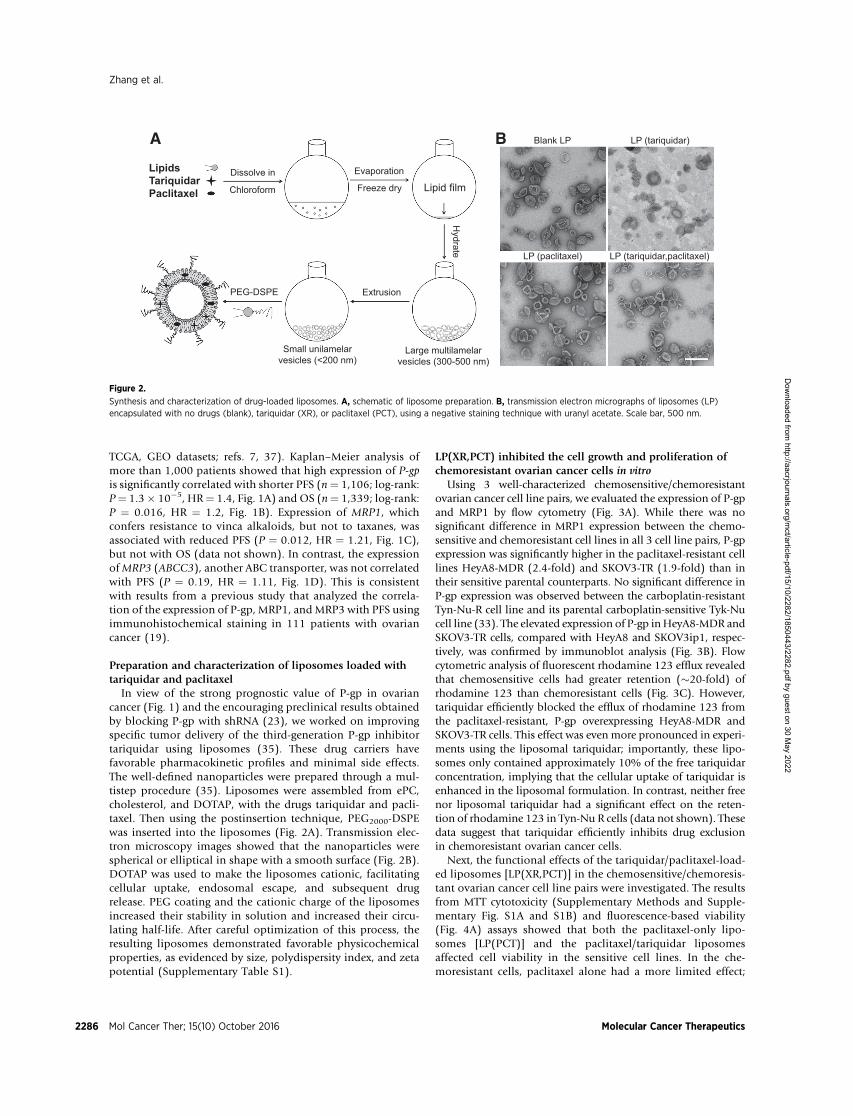
In view of the strong prognostic value of P-gp in ovariancancer (Fig. 1) and the encouraging preclinical results obtainedby blocking P-gp with shRNA (23), we worked on improvingspecific tumor delivery of the third-generation P-gp inhibitortariquidar using liposomes (35). These drug carriers havefavorable pharmacokinetic profiles and minimal side effects.The well-defined nanoparticles were prepared through a mul-tistep procedure (35). Liposomes were assembled from ePC,cholesterol, and DOTAP, with the drugs tariquidar and pacli-taxel. Then using the postinsertion technique, PEG2000-DSPEwas inserted into the liposomes (Fig. 2A). Transmission elec-tron microscopy images showed that the nanoparticles werespherical or elliptical in shape with a smooth surface (Fig. 2B).DOTAP was used to make the liposomes cationic, facilitatingcellular uptake, endosomal escape, and subsequent drugrelease. PEG coating and the cationic charge of the liposomesincreased their stability in solution and increased their circu-lating half-life. After careful optimization of this process, theresulting liposomes demonstrated favorable physicochemicalproperties, as evidenced by size, polydispersity index, and zetapotential (Supplementary Table S1).
LP(XR,PCT) inhibited the cell growth and proliferation ofchemoresistant ovarian cancer cells in vitro
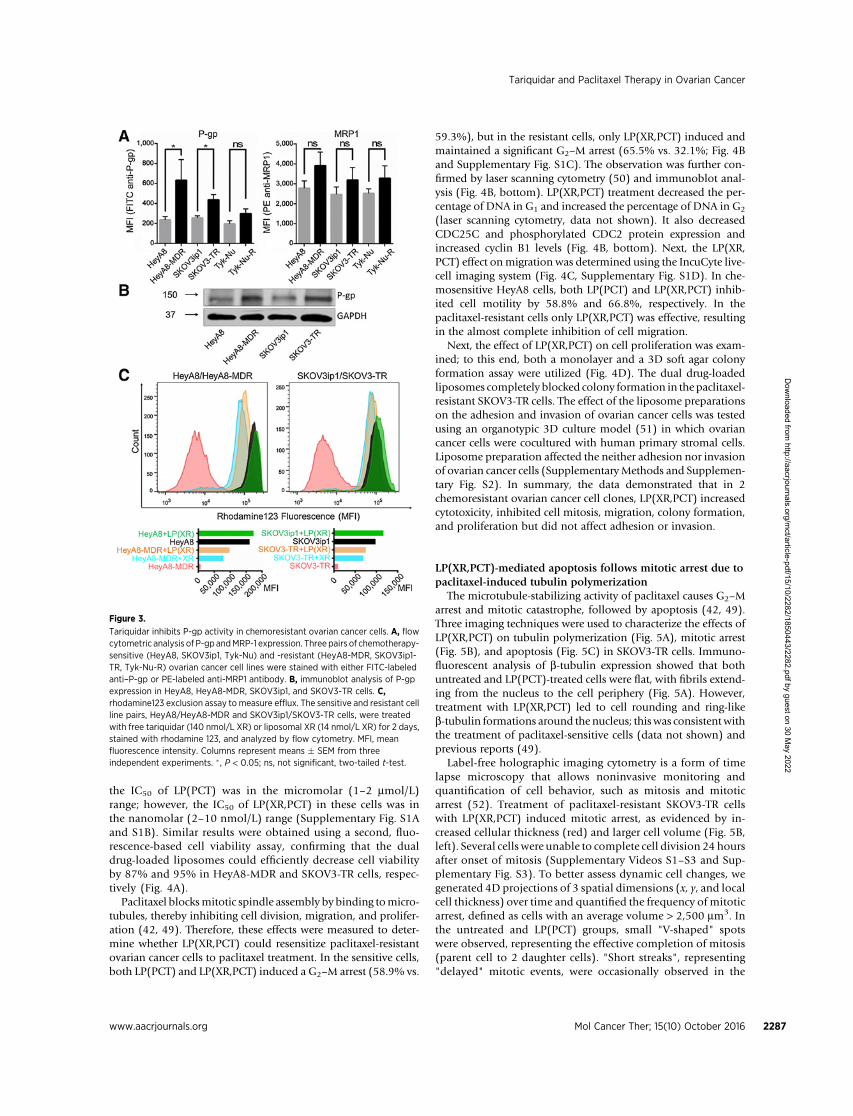
Using 3 well-characterized chemosensitive/chemoresistantovarian cancer cell line pairs, we evaluated the expression of P-gpand MRP1 by flow cytometry (Fig. 3A). While there was nosignificant difference in MRP1 expression between the chemo-sensitive and chemoresistant cell lines in all 3 cell line pairs, P-gpexpression was significantly higher in the paclitaxel-resistant celllines HeyA8-MDR (2.4-fold) and SKOV3-TR (1.9-fold) than intheir sensitive parental counterparts. No significant difference inP-gp expression was observed between the carboplatin-resistantTyn-Nu-R cell line and its parental carboplatin-sensitive Tyk-Nucell line (33). The elevated expression of P-gp inHeyA8-MDR andSKOV3-TR cells, compared with HeyA8 and SKOV3ip1, respec-tively, was confirmed by immunoblot analysis (Fig. 3B). Flowcytometric analysis of fluorescent rhodamine 123 efflux revealedthat chemosensitive cells had greater retention (�20-fold) ofrhodamine 123 than chemoresistant cells (Fig. 3C). However,tariquidar efficiently blocked the efflux of rhodamine 123 fromthe paclitaxel-resistant, P-gp overexpressing HeyA8-MDR andSKOV3-TR cells. This effect was even more pronounced in experi-ments using the liposomal tariquidar; importantly, these lipo-somes only contained approximately 10% of the free tariquidarconcentration, implying that the cellular uptake of tariquidar isenhanced in the liposomal formulation. In contrast, neither freenor liposomal tariquidar had a significant effect on the reten-tion of rhodamine 123 in Tyn-Nu R cells (data not shown). Thesedata suggest that tariquidar efficiently inhibits drug exclusionin chemoresistant ovarian cancer cells.
Next, the functional effects of the tariquidar/paclitaxel-load-ed liposomes [LP(XR,PCT)] in the chemosensitive/chemoresis-tant ovarian cancer cell line pairs were investigated. The resultsfrom MTT cytotoxicity (Supplementary Methods and Supple-mentary Fig. S1A and S1B) and fluorescence-based viability(Fig. 4A) assays showed that both the paclitaxel-only lipo-somes [LP(PCT)] and the paclitaxel/tariquidar liposomesaffected cell viability in the sensitive cell lines. In the che-moresistant cells, paclitaxel alone had a more limited effect;
A
LipidsTariquidarPaclitaxel
Dissolve in
Chloroform Freeze dry
Evaporation
Lipid film
Blank LP LP (tariquidar)
LP (tariquidar,paclitaxel)LP (paclitaxel)
Extrusion
Hydrate
PEG-DSPE
Small unilamelarvesicles (<200 nm)
Large multilamelarvesicles (300-500 nm)
B
Figure 2.
Synthesis and characterization of drug-loaded liposomes. A, schematic of liposome preparation. B, transmission electron micrographs of liposomes (LP)encapsulated with no drugs (blank), tariquidar (XR), or paclitaxel (PCT), using a negative staining technique with uranyl acetate. Scale bar, 500 nm.
Zhang et al.
Mol Cancer Ther; 15(10) October 2016 Molecular Cancer Therapeutics2286
Dow
nloaded from http://aacrjournals.org/m
ct/article-pdf/15/10/2282/1850443/2282.pdf by guest on 30 May 2022
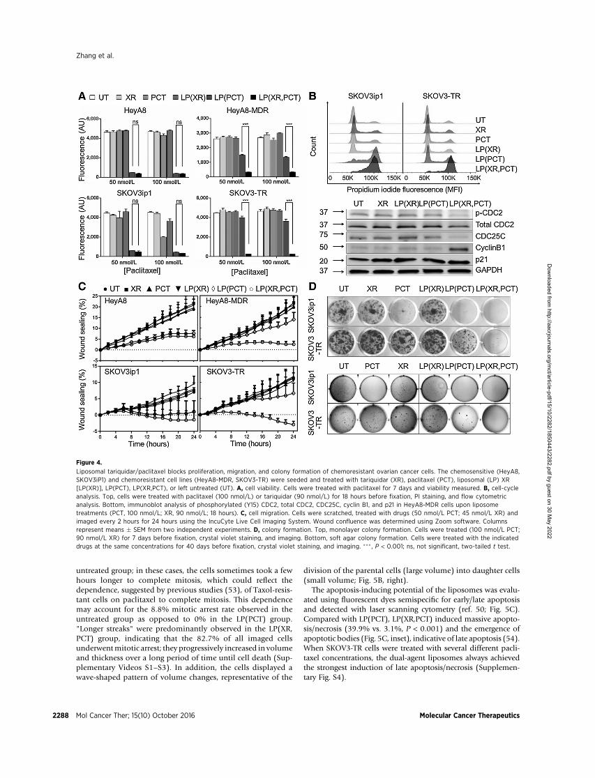
the IC50 of LP(PCT) was in the micromolar (1–2 mmol/L)range; however, the IC50 of LP(XR,PCT) in these cells was inthe nanomolar (2–10 nmol/L) range (Supplementary Fig. S1Aand S1B). Similar results were obtained using a second, fluo-rescence-based cell viability assay, confirming that the dualdrug-loaded liposomes could efficiently decrease cell viabilityby 87% and 95% in HeyA8-MDR and SKOV3-TR cells, respec-tively (Fig. 4A).
Paclitaxel blocksmitotic spindle assembly by binding tomicro-tubules, thereby inhibiting cell division, migration, and prolifer-ation (42, 49). Therefore, these effects were measured to deter-mine whether LP(XR,PCT) could resensitize paclitaxel-resistantovarian cancer cells to paclitaxel treatment. In the sensitive cells,both LP(PCT) and LP(XR,PCT) induced a G2–M arrest (58.9% vs.
59.3%), but in the resistant cells, only LP(XR,PCT) induced andmaintained a significant G2–M arrest (65.5% vs. 32.1%; Fig. 4Band Supplementary Fig. S1C). The observation was further con-firmed by laser scanning cytometry (50) and immunoblot anal-ysis (Fig. 4B, bottom). LP(XR,PCT) treatment decreased the per-centage of DNA in G1 and increased the percentage of DNA in G2
(laser scanning cytometry, data not shown). It also decreasedCDC25C and phosphorylated CDC2 protein expression andincreased cyclin B1 levels (Fig. 4B, bottom). Next, the LP(XR,PCT) effect onmigration was determined using the IncuCyte live-cell imaging system (Fig. 4C, Supplementary Fig. S1D). In che-mosensitive HeyA8 cells, both LP(PCT) and LP(XR,PCT) inhib-ited cell motility by 58.8% and 66.8%, respectively. In thepaclitaxel-resistant cells only LP(XR,PCT) was effective, resultingin the almost complete inhibition of cell migration.
Next, the effect of LP(XR,PCT) on cell proliferation was exam-ined; to this end, both a monolayer and a 3D soft agar colonyformation assay were utilized (Fig. 4D). The dual drug-loadedliposomes completely blocked colony formation in the paclitaxel-resistant SKOV3-TR cells. The effect of the liposome preparationson the adhesion and invasion of ovarian cancer cells was testedusing an organotypic 3D culture model (51) in which ovariancancer cells were cocultured with human primary stromal cells.Liposome preparation affected the neither adhesion nor invasionof ovarian cancer cells (Supplementary Methods and Supplemen-tary Fig. S2). In summary, the data demonstrated that in 2chemoresistant ovarian cancer cell clones, LP(XR,PCT) increasedcytotoxicity, inhibited cell mitosis, migration, colony formation,and proliferation but did not affect adhesion or invasion.
LP(XR,PCT)-mediated apoptosis follows mitotic arrest due topaclitaxel-induced tubulin polymerization
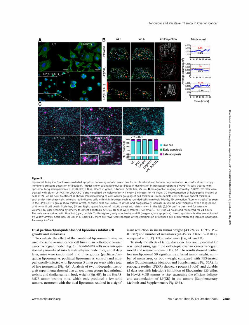
The microtubule-stabilizing activity of paclitaxel causes G2–Marrest and mitotic catastrophe, followed by apoptosis (42, 49).Three imaging techniques were used to characterize the effects ofLP(XR,PCT) on tubulin polymerization (Fig. 5A), mitotic arrest(Fig. 5B), and apoptosis (Fig. 5C) in SKOV3-TR cells. Immuno-fluorescent analysis of b-tubulin expression showed that bothuntreated and LP(PCT)-treated cells were flat, with fibrils extend-ing from the nucleus to the cell periphery (Fig. 5A). However,treatment with LP(XR,PCT) led to cell rounding and ring-likeb-tubulin formations around the nucleus; thiswas consistentwiththe treatment of paclitaxel-sensitive cells (data not shown) andprevious reports (49).
Label-free holographic imaging cytometry is a form of timelapse microscopy that allows noninvasive monitoring andquantification of cell behavior, such as mitosis and mitoticarrest (52). Treatment of paclitaxel-resistant SKOV3-TR cellswith LP(XR,PCT) induced mitotic arrest, as evidenced by in-creased cellular thickness (red) and larger cell volume (Fig. 5B,left). Several cells were unable to complete cell division 24 hoursafter onset of mitosis (Supplementary Videos S1–S3 and Sup-plementary Fig. S3). To better assess dynamic cell changes, wegenerated 4D projections of 3 spatial dimensions (x, y, and localcell thickness) over time and quantified the frequency of mitoticarrest, defined as cells with an average volume > 2,500 mm3. Inthe untreated and LP(PCT) groups, small "V-shaped" spotswere observed, representing the effective completion of mitosis(parent cell to 2 daughter cells). "Short streaks", representing"delayed" mitotic events, were occasionally observed in the
Figure 3.
Tariquidar inhibits P-gp activity in chemoresistant ovarian cancer cells. A, flowcytometric analysis of P-gp andMRP-1 expression. Three pairs of chemotherapy-sensitive (HeyA8, SKOV3ip1, Tyk-Nu) and -resistant (HeyA8-MDR, SKOV3ip1-TR, Tyk-Nu-R) ovarian cancer cell lines were stained with either FITC-labeledanti–P-gp or PE-labeled anti-MRP1 antibody. B, immunoblot analysis of P-gpexpression in HeyA8, HeyA8-MDR, SKOV3ip1, and SKOV3-TR cells. C,rhodamine123 exclusion assay to measure efflux. The sensitive and resistant cellline pairs, HeyA8/HeyA8-MDR and SKOV3ip1/SKOV3-TR cells, were treatedwith free tariquidar (140 nmol/L XR) or liposomal XR (14 nmol/L XR) for 2 days,stained with rhodamine 123, and analyzed by flow cytometry. MFI, meanfluorescence intensity. Columns represent means � SEM from threeindependent experiments. � , P < 0.05; ns, not significant, two-tailed t-test.
Tariquidar and Paclitaxel Therapy in Ovarian Cancer
www.aacrjournals.org Mol Cancer Ther; 15(10) October 2016 2287
Dow
nloaded from http://aacrjournals.org/m
ct/article-pdf/15/10/2282/1850443/2282.pdf by guest on 30 May 2022
untreated group; in these cases, the cells sometimes took a fewhours longer to complete mitosis, which could reflect thedependence, suggested by previous studies (53), of Taxol-resis-tant cells on paclitaxel to complete mitosis. This dependencemay account for the 8.8% mitotic arrest rate observed in theuntreated group as opposed to 0% in the LP(PCT) group."Longer streaks" were predominantly observed in the LP(XR,PCT) group, indicating that the 82.7% of all imaged cellsunderwentmitotic arrest; they progressively increased in volumeand thickness over a long period of time until cell death (Sup-plementary Videos S1–S3). In addition, the cells displayed awave-shaped pattern of volume changes, representative of the
division of the parental cells (large volume) into daughter cells(small volume; Fig. 5B, right).
The apoptosis-inducing potential of the liposomes was evalu-ated using fluorescent dyes semispecific for early/late apoptosisand detected with laser scanning cytometry (ref. 50; Fig. 5C).Compared with LP(PCT), LP(XR,PCT) induced massive apopto-sis/necrosis (39.9% vs. 3.1%, P < 0.001) and the emergence ofapoptotic bodies (Fig. 5C, inset), indicative of late apoptosis (54).When SKOV3-TR cells were treated with several different pacli-taxel concentrations, the dual-agent liposomes always achievedthe strongest induction of late apoptosis/necrosis (Supplemen-tary Fig. S4).
Figure 4.
Liposomal tariquidar/paclitaxel blocks proliferation, migration, and colony formation of chemoresistant ovarian cancer cells. The chemosensitive (HeyA8,SKOV3iP1) and chemoresistant cell lines (HeyA8-MDR, SKOV3-TR) were seeded and treated with tariquidar (XR), paclitaxel (PCT), liposomal (LP) XR[LP(XR)], LP(PCT), LP(XR,PCT), or left untreated (UT). A, cell viability. Cells were treated with paclitaxel for 7 days and viability measured. B, cell-cycleanalysis. Top, cells were treated with paclitaxel (100 nmol/L) or tariquidar (90 nmol/L) for 18 hours before fixation, PI staining, and flow cytometricanalysis. Bottom, immunoblot analysis of phosphorylated (Y15) CDC2, total CDC2, CDC25C, cyclin B1, and p21 in HeyA8-MDR cells upon liposometreatments (PCT, 100 nmol/L; XR, 90 nmol/L; 18 hours). C, cell migration. Cells were scratched, treated with drugs (50 nmol/L PCT; 45 nmol/L XR) andimaged every 2 hours for 24 hours using the IncuCyte Live Cell Imaging System. Wound confluence was determined using Zoom software. Columnsrepresent means � SEM from two independent experiments. D, colony formation. Top, monolayer colony formation. Cells were treated (100 nmol/L PCT;90 nmol/L XR) for 7 days before fixation, crystal violet staining, and imaging. Bottom, soft agar colony formation. Cells were treated with the indicateddrugs at the same concentrations for 40 days before fixation, crystal violet staining, and imaging. ��� , P < 0.001; ns, not significant, two-tailed t test.
Zhang et al.
Mol Cancer Ther; 15(10) October 2016 Molecular Cancer Therapeutics2288
Dow
nloaded from http://aacrjournals.org/m
ct/article-pdf/15/10/2282/1850443/2282.pdf by guest on 30 May 2022
Dual paclitaxel/tariquidar-loaded liposomes inhibit cellgrowth and metastasis
To evaluate the effect of the combined liposomes in vivo, weused the same ovarian cancer cell lines in an orthotopic ovariancancer xenograft model (Fig. 6). HeyA8-MDR cells were intraper-itoneally inoculated into female athymic nude mice, and 8 dayslater, mice were randomized into three groups (paclitaxel/tari-quidar liposomes vs. paclitaxel liposomes vs. control) and intra-peritoneally injected with liposomes 3 times per weekwith a totalof five treatments (Fig. 6A). Analysis of two independent xeno-graft experiments showed that all treatment groups had minimaltoxicity and similar gains in body weight (Fig. 6B). In the HeyA8-MDR tumor–bearing mice, which only produced a few solidtumors, treatment with the dual liposomes resulted in a signif-
icant reduction in mean tumor weight (43.2% vs. 16.9%, P ¼0.0007) and number of metastases (44.4% vs. 2.8%, P ¼ 0.012),compared with LP(PCT)-treated mice (Fig. 6C and D).
To study the effects of tariquidar alone, free and liposomal XRwas tested using again the orthotopic ovarian cancer xenograftmodel and regimen shown in Fig. 6A. The results showed neitherfree nor liposomal XR significantly affected tumor weight, num-ber of metastases, or body weight compared with PBS-treatedmice (Supplementary Methods and Supplementary Fig. S5A). Insurrogate studies, LP(XR) showed a potent (3-fold) and durable(2 days post fifth injection) inhibition of Rhodamine 123 effluxin HeyA8-MDR tumors ex vivo, suggesting the efficient deliveryand accumulation of LP(XR) in the tumors (SupplementaryMethods and Supplementary Fig. S5B).
Figure 5.
Liposomal tariquidar/paclitaxel–mediated apoptosis following mitotic arrest due to paclitaxel-induced tubulin polymerization. A, confocal microscopy.Immunofluorescent detection of b-tubulin. Images show paclitaxel-induced b-tubulin dysfunction in paclitaxel-resistant SKOV3-TR cells treated withliposomal tariquidar/paclitaxel [LP(XR,PCT)]. Blue, Hoechst; green, b-tubulin. Scale bar, 25 mm. B, holographic imaging cytometry. SKOV3-TR cells weretreated with either LP(PCT) or LP(XR,PCT) and visualized by HoloMonitor M4 every 5 minutes for 48 hours. 3D representation of holographic images ofcells at 24- or 48-hour treatment is shown. Pseudocoloring of cells allows gauging of cell thickness. Green depicts cells with low optical thickness,such as flat interphase cells, whereas red indicates cells with high thickness such as rounded cells in mitosis. Middle, 4D projection. "Longer streaks" as seenin the LP(XR,PCT) group show mitotic arrest, as these cells are unable to divide and progressively increase in volume and thickness over a long periodof time until cell death. Scale bar, 25 mm. Right, quantification of mitotic arrest with data shown in the left (2,500 mm3, a threshold for averagevolume). C, laser scanning cytometry to detect apoptosis. SKOV3-TR cells were treated (150 nmol/L, PCT) for 24 hours and recovered for 24 hours.The cells were stained with Hoechst (cyan, nuclei), Yo-Pro (green, early apoptosis), and PI (magenta, late apoptosis). Insert, apoptotic bodies are indicatedby yellow arrows. Scale bar, 50 mm. In LP(XR,PCT), there are fewer cells because of the combination of reduced cell proliferation and induced apoptosis.Two-way ANOVA.
Tariquidar and Paclitaxel Therapy in Ovarian Cancer
www.aacrjournals.org Mol Cancer Ther; 15(10) October 2016 2289
Dow
nloaded from http://aacrjournals.org/m
ct/article-pdf/15/10/2282/1850443/2282.pdf by guest on 30 May 2022
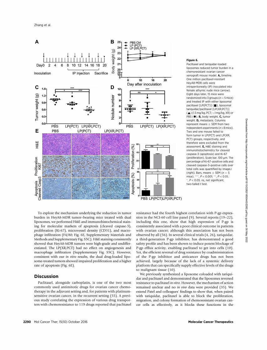
To explore the mechanism underlying the reduction in tumorburden in HeyA8-MDR tumor–bearing mice treated with dualliposomes, we performed H&E and immunohistochemical stain-ing for molecular markers of apoptosis (cleaved caspase-3),proliferation (Ki-67), microvessel density (CD31), and macro-phage infiltration (F4/80; Fig. 6E, Supplementary Materials andMethods and Supplementary Fig. S5C).H&E staining consistentlyshowed that HeyA8-MDR tumors were high-grade and undiffer-entiated. The LP(XR,PCT) had no effect on angiogenesis andmacrophage infiltration (Supplementary Fig. S5C). However,consistent with our in vitro results, the dual drug-loaded lipo-some-treated tumors showed impaired proliferation and a higherrate of apoptosis (Fig. 6E).
DiscussionPaclitaxel, alongside carboplatin, is one of the two most
commonly used antimitotic drugs for ovarian cancer chemo-therapy in the adjuvant setting and, for patients with platinum-sensitive ovarian cancer, in the recurrent setting (55). A previ-ous study correlating the expression of various drug transpor-ters with chemoresistance to 119 drugs reported that paclitaxel
resistance had the fourth highest correlation with P-gp expres-sion in the NCI-60 cell line panel (9). Several reports (19–22),including this one, show that high expression of P-gp isconsistently associated with a poor clinical outcome in patientswith ovarian cancer; although this association has not beenobserved by all (56). In several clinical trials (4, 26), tariquidar,a third-generation P-gp inhibitor, has demonstrated a goodsafety profile and has been shown to induce potent blockage ofP-gp efflux activity, enabling paclitaxel to get into cells (10).Yet, the efficient reversal of drug resistance by coadministrationof the P-gp inhibitor and anticancer drugs has not beenachieved, largely because of the lack of a systemic deliveryplatform that can specifically supply effective levels of the drugsto malignant tissue (10).
We previously synthesized a liposome coloaded with tariqui-dar and paclitaxel and demonstrated that the liposomes reversedresistance to paclitaxel in vitro.However, themechanism of actionremained unclear and no in vivo data were provided (35). Weextend Patel and colleagues' findings to show that, when pairedwith tariquidar, paclitaxel is able to block the proliferation,migration, and colony formation of chemoresistant ovarian can-cer cells as effectively, as it blocks these functions in the
Figure 6.
Paclitaxel and tariquidar-loadedliposomes reduced tumor burden in achemoresistant ovarian cancerxenograft mouse model. A, timeline.One million paclitaxel-resistantHeyA8-MDR cells wereintraperitoneally (IP) inoculated intofemale athymic nude mice (arrow).Eight days later, 15 mice wererandomized into 3 groups (n¼ 5mice)and treated IP with either liposomalpaclitaxel [LP(PCT)] (&), liposomaltariquidar/paclitaxel [LP(XR,PCT)](~) (1.5mg/kg, PCT;�1 mg/kg, XR) orPBS (*). B, body weight. C, tumorweight. D, metastasis. Columnsrepresent means � SEM from twoindependent experiments (n>8mice).Two and one mouse failed toform tumor in LP(PCT) and LP(XR,PCT) groups, respectively, andtherefore were excluded from theassessment. E, H&E staining andimmunohistochemistry for cleavedcaspase-3 (apoptosis) and Ki-67(proliferation). Scale bar, 100 mm. Thepercentage of Ki-67–positive cells andcleaved caspase-3–positive cells overtotal cells was quantified by ImageJ(right). Bars, means � SEM (n ¼ 5mice). ��� , P < 0.001; ��, P < 0.01;� , P < 0.05. ns, not significant,two-tailed t test.
Zhang et al.
Mol Cancer Ther; 15(10) October 2016 Molecular Cancer Therapeutics2290
Dow
nloaded from http://aacrjournals.org/m
ct/article-pdf/15/10/2282/1850443/2282.pdf by guest on 30 May 2022
chemosensitive parental cells. To facilitate the assessment oftherapeutic efficacy of this formulation in animal studies, wesuccessfully increased the loading amounts of both tariquidar andpaclitaxel by approximate 20-fold without affecting physico-chemical features of the liposomes. Experiments using the lipo-somes with preclinical mouse models confirmed these results invivo, showing that tariquidar resensitizes chemoresistant ovariancancer cells to paclitaxel treatment, allowing paclitaxel to induceapoptosis in established tumors. This is consistent with previousin vitro results using the P-gp substrate doxorubicin. The IC50 fordoxorubicin was much lower in the presence of tariquidar andinhibition of cell growth persisted, despite removal of tariquidarfrom the media (57).
The dual encapsulation of tariquidar and paclitaxel into nano-particles ensures that they will be internalized by ovarian cancercells simultaneously and that the upload of both agents willinvolve the same pharmacokinetic profiles. To evaluate themech-anism of action of tariquidar in the liposome formulation, weemployed advanced microscopic live-cell imaging approaches,such as kinetic cell monitoring (IncuCyte), laser scanning cyto-metry (iCyte), andholographic imaging cytometry (HoloMonitorM4). Because of its intrinsic ability to directly quantify phase shiftand optical path length, holographic imaging circumvents opticalartifacts, such as the shading-off effects and spurious asymmetryassociated with the most widely used contrast-generating techni-ques (e.g., phase contrast and differential interference contrast) inoptical microscopy (58). This label-free quantitative techniqueallowed us to follow the development of mitotic arrest in real-time. It permitted us to conclude that tariquidar treatment ofpaclitaxel-resistant cells resensitized the cells to paclitaxel, allow-ing the drug to induce almost complete mitotic arrest.
We also note that ovarian cancer may be particularly suitablefor nanoparticle-facilitated MDR studies and treatment, becauseas many as 80% of epithelial ovarian tumors develop MDR. Thedual drug-loaded liposomes increased the specific delivery ofdrugs to tumor cells and allowed tariquidar to become signif-icantly more efficient in enabling paclitaxel to inhibit prolifer-ation and colony formation, and promote cell-cycle arrest. Thefact that the liposome platform significantly reduced tumorburden and metastasis in our mouse model suggests that wehave identified an efficient vehicle for the delivery of paclitaxeland tariquidar specifically to the tumor for the reversal of MDRin chemoresistant ovarian cancer. Dual drug-loaded nanopar-ticles may be the next step toward bringing a P-gp inhibitor tothe clinic.
Although MDR is especially common in ovarian cancer,appropriate patient selection is important if LP(XR,PCT) is tosucceed as a therapeutic in chemoresistant ovarian cancer. In arecent study using whole-genome characterization of chemore-
sistant ovarian cancer (20), we reported a fusion protein, inwhich P-gp expression was driven by the promoter and exon 1of SLC25A40 (a mitochondrial carrier protein) resulting in afused transcript. This protein was detected in 8% of all recur-rent, chemoresistant tumors that had been treated with pacli-taxel, but not in chemosensitive tumors. Because paclitaxel cancause double strand breaks, it is probable that these geneticrearrangements were drug-induced (59). Given that clinicalstudies of P-gp inhibitors in unselected cases have had mixedresults (60), selection of patients with P-gp overexpression orthe SLC25A40/P-gp fusion protein may identify a patientsubgroup that is more likely to respond to liposome-encapsu-lated tariquidar and paclitaxel.
Disclosure of Potential Conflicts of InterestNo potential conflicts of interest were disclosed.
Authors' ContributionsConception and design: Y. Zhang, S.K. Sriraman, V. Torchilin, E. LengyelDevelopment ofmethodology: Y. Zhang, S.K. Sriraman, E. Luther, V. Torchilin,E. LengyelAcquisition of data (provided animals, acquired and managed patients,provided facilities, etc.): Y. Zhang, S.K. Sriraman, E. Luther, V. TorchilinAnalysis and interpretation of data (e.g., statistical analysis, biostatistics,computational analysis): Y. Zhang, S.K. Sriraman, E. Luther, E. LengyelWriting, review, and/or revision of the manuscript: Y. Zhang, S.K. Sriraman,H.A. Kenny, V. Torchilin, E. LengyelAdministrative, technical, or material support (i.e., reporting or organizingdata, constructing databases): E. LengyelStudy supervision: H.A. Kenny, V. Torchilin, E. Lengyel
AcknowledgmentsWe thank Gail Isenberg (University of Chicago), Dr. Karen Watters (Uni-
versity of Chicago), and Dr. Elena Holden (Executive Strategic Advisory) forediting this article. The authors also acknowledge the help of Dr. Marion Curtisin validating cell lines, Dr. Mark Eckert in survival analysis, and Jiayi Pan forliposome preparation. We thank the human tissue resource center and flowcytometry core facilities at the University of Chicago for their experttechnical support. We would also like to thank William Fowle, Dr. RajivKumar, and Dr. Srinivas Sridhar at Northeastern University for the phys-icochemical nanocharacterization.
Grant SupportThis work was supported by NCI Alliance for Nanotechnology through a
Cancer Nanotechnology Platform Partnership U01CA151461, a National Can-cer Institute grant (NCI-R01 CA111882 to E. Lengyel) and by an NIH grant(U54CA151881 to V. Torchilin).
The costs of publication of this articlewere defrayed inpart by the payment ofpage charges. This article must therefore be hereby marked advertisement inaccordance with 18 U.S.C. Section 1734 solely to indicate this fact.
Received December 22, 2015; revised June 23, 2016; accepted July 13, 2016;published OnlineFirst July 27, 2016.
References1. Siegel RL, Miller KD, Jemal A. Cancer statistics, 2015. CA Cancer J Clin
2015;65:5–29.2. Lengyel E. Ovarian cancer development and metastasis. Am J Pathol
2010;177:1053–64.3. Hartmann LC, Lu KH, Linette GP, Cliby WA, Kalli KR, Gershenson D,
et al. Gene expression profiles predict early relapse in ovarian cancerafter platinum-paclitaxel chemotherapy. Clin Cancer Res 2005;11:2149–55.
4. Leonard GD, Fojo T, Bates SE. The role of ABC transporters in clinicalpractice. Oncologist 2003;8:411–24.
5. Armstrong DK, Bundy B, Wenzel L, Huang H, Baergen R, Lele S, et al.Intraperitoneal cisplatin and paclitaxel in ovarian cancer. N Engl J Med2006;353:34–43.
6. Dano K. Active outward transport of daunomycin in resistant Ehrlichascites tumor cells. Biochim Biophys Acta 1973;323:466–83.
7. Hedditch EL, Gao B, Russell AJ, Lu Y, Emmanuel C, Beesley J, et al. ABCAtransporter gene expression and poor outcome in epithelial ovarian cancer.J Natl Cancer Inst 2014;106: pii: dju149.
8. Sharom FJ. ABC multidrug transporters: structure, function and role inchemoresistance. Pharmacogenomics 2008;9:105–27.
Tariquidar and Paclitaxel Therapy in Ovarian Cancer
www.aacrjournals.org Mol Cancer Ther; 15(10) October 2016 2291
Dow
nloaded from http://aacrjournals.org/m
ct/article-pdf/15/10/2282/1850443/2282.pdf by guest on 30 May 2022
9. Huang Y, Anderle P, Bussey KJ, Barbacioru C, Shankavaram U, Dai Z, et al.Membrane transporters and channels: Role of the transportome in cancerchemosensitivity and chemoresistance. Cancer Res 2004;64:4294–301.
10. Szakacs G, Paterson JK, Ludwig JA, Booth-Genthe C, Gottesman MM.Targeting multidrug resistance in cancer. Nat Rev Drug Discov 2006;5:219–34.
11. Su HY, Lai HC, Lin YW, Liu CY, Chen CK, Chou YC, et al. Epigeneticsilencing of SFRP5 is related tomalignant phenotype and chemoresistanceof ovarian cancer through Wnt signaling pathway. Int J Cancer 2010;127:555–67.
12. Rizzo S, Hersey JM, Mellor P, Dai W, Santos-Silva A, Liber D, et al. Ovariancancer stem cell-like side populations are enriched following chemother-apy and overexpress EZH2. Mol Cancer Ther 2011;10:325–35.
13. Boyerinas B, Park SM,MurmannAE,GwinK,Montag AG, ZillhardtM, et al.Let-7 modulates acquired resistance of ovarian cancer to taxanes via IMP-1mediated stabilization of MDR1. Int J Cancer 2012;130:1787–97.
14. Frederick PJ, GreenHN,Huang JS, EggerME, FrieboesHB,GrizzleWE, et al.Chemoresistance in ovarian cancer linked to expression of microRNAs.Biotech Histochem 2013;88:403–9.
15. Loessner D, Stok KS, Lutolf MP, Hutmacher DW, Clements JA, Rizzi SC.Bioengineered 3D platform to explore cell-ECM interactions and drugresistance of epithelial ovarian cancer cells. Biomaterials 2010;31:8494–506.
16. Thibault B, Castells M, Delord JP, Couderc B. Ovarian cancer microenvi-ronment: implications for cancer dissemination and chemoresistanceacquisition. Cancer Metastasis Rev 2014;33:17–39.
17. Juliano RL, Ling V. A surface glycoprotein modulating drug permeabilityin Chinese hamster ovary cell mutants. Biochim Biophys Acta 1976;455:152–62.
18. Shen DW, Fojo A, Chin JE, Roninson IB, Richert N, Pastan I, et al. Humanmultidrug-resistant cell lines: increased mdr1 expression can precede geneamplification. Science 1986;232:643–5.
19. Sedlakova I, Laco J, Caltova K, Cervinka M, Tosner J, Rezac A, et al. Clinicalsignificance of the resistance proteins LRP, Pgp,MRP1,MRP3, andMRP5 inepithelial ovarian cancer. Int J Gynecol Cancer 2015;25:236–43.
20. Patch AM, Christie EL, Etemadmoghadam D, Garsed DW, George J, Fere-day S, et al. Whole-genome characterization of chemoresistant ovariancancer. Nature 2015;521:489–94.
21. Penson RT, Oliva E, Skates SJ, Glyptis T, Fuller AFJr, Goodman A, et al.Expression of multidrug resistance-1 protein inversely correlates withpaclitaxel response and survival in ovarian cancer patients: a study in serialsamples. Gynecol Oncol 2004;93:98–106.
22. Zajchowski DA, Karlan BY, Shawver LK. Treatment-related protein bio-marker expression differs between primary and recurrent ovarian carcino-mas. Mol Cancer Ther 2012;11:492–502.
23. Duan Z, Brakora KA, Seiden MV. Inhibition of ABCB1 (MDR1) andABCB4 (MDR3) expression by small interfering RNA and reversal ofpaclitaxel resistance in human ovarian cancer cells. Mol Cancer Ther2004;3:833–8.
24. RoeM, Folkes A, Ashworth P, Brumwell J, Chima L,Hunjan S, et al. Reversalof P-glycoprotein mediated multidrug resistance by novel anthranilamidederivatives. Bioorg Med Chem Lett 1999;9:595–600.
25. Loo TW, Clarke DM. Tariquidar inhibits P-glycoprotein drug efflux butactivates ATPase activity by blocking transition to an open conformation.Biochem Pharmacol 2014;92:558–66.
26. Fox E, Bates SE. Tariquidar (XR9576): a P-glycoprotein drug efflux pumpinhibitor. Expert Rev Anticancer Ther 2007;7:447–59.
27. Kim BY, Rutka JT, Chan WC. Nanomedicine. N Engl J Med 2010;363:2434–43.
28. Zhang Y, Kenny HA, Swindell EP, Mitra AK, Hankins PL, Ahn RW, et al.Urokinase plasminogen activator system targeted delivery of nanobins as anovel ovarian cancer therapeutics. Mol Cancer Ther 2013;12:2628–39.
29. Patil Y, Sadhukha T,Ma L, Panyam J. Nanoparticle-mediated simultaneousand targeted delivery of paclitaxel and tariquidar overcomes tumor drugresistance. J Control Release 2009;136:21–9.
30. Thaker PH, Yazici S, Nilsson M, Yokoi K, Tsan RZ, He J, et al. Antivasculartherapy for orthotopic human ovarian carcinoma through blockade of thevascular endothelial growth factor and epidermal growth factor receptors.Clin Cancer Res 2005;11:4923–34.
31. Duan Z, Feller AJ, Penson RT, Chabner BA, Seiden MV. Discovery ofdifferentially expressed genes associated with paclitaxel resistance using
cDNA array technology: analysis of interleukin (IL) 6, IL-8, and monocytechemotactic protein 1 in the paclitaxel-resistant phenotype. Clin CancerRes 1999;5:3445–53.
32. Yoshiya N. [Establishment of a cell line from human ovarian cancer(undifferentiated carcinoma of FIGO classification) and analysis of itscell-biological characteristics and sensitivity to anticancer drugs]. NihonSanka Fujinka Gakkai Zasshi 1986;38:1747–53.
33. Yoshiya N, Adachi S, Misawa Y, Yuzawa H, Honda T, Kanazawa K, et al.[Isolationof cisplatin-resistant subline fromhumanovarian cancer cell lineand analysis of its cell-biological characteristics]. Nihon Sanka FujinkaGakkai Zasshi 1989;41:7–14.
34. Kenny HA, Lal-Nag M, White EA, Shen M, Chiang CY, Mitra AK, et al.Quantitative high throughput screening using a primary human three-dimensional organotypic culture predicts in vivo efficacy. Nat Commun2015;6:6220.
35. Patel NR, Rathi A, Mongayt D, Torchilin VP. Reversal of multidrug resis-tance by co-delivery of tariquidar (XR9576) and paclitaxel using long-circulating liposomes. Int J Pharm 2011;416:296–9.
36. Matsuo Y, Park JH, Miyamoto T, Yamamoto S, Hisada S, Alachkar H, et al.TOPK inhibitor induces complete tumor regression in xenograft models ofhuman cancer through inhibition of cytokinesis. Sci Transl Med 2014;6:259ra145.
37. Gyorffy B, Lanczky A, Szallasi Z. Implementing an online tool forgenome-wide validation of survival-associated biomarkers in ovarian-cancer using microarray data from 1287 patients. Endocr Relat Cancer2012;19:197–208.
38. Lengyel E, Litchfield LM, Mitra AK, Nieman KM, Mukherjee A, Zhang Y,et al. Metformin inhibits ovarian cancer growth and increases sensitivity topaclitaxel in mouse models. Am J Obstet Gynecol 2014;212:479.e1–10.
39. Sriraman SK, Geraldo V, Luther E, Degterev A, Torchilin V. Cytotoxicity ofPEGylated liposomes co-loaded with novel pro-apoptotic drug NCL-240and the MEK inhibitor cobimetinib against colon carcinoma invitro. JControl Release 2015;220:160–8.
40. Bedner E, Li X, Gorczyca W, Melamed MR, Darzynkiewicz Z. Analysis ofapoptosis by laser scanning cytometry. Cytometry 1999;35:181–95.
41. Mitra AK, Chiang CY, Tiwari P, Tomar S, Watters KM, Peter ME, et al.Microenvironment-induced downregulation of miR-193b drives ovariancancer metastasis. Oncogene 2015;34:5923–32.
42. Ruttala HB, Ko YT. Liposomal co-delivery of curcumin and albumin/paclitaxel nanoparticle for enhanced synergistic antitumor efficacy. Col-loids Surf B Biointerfaces 2015;128:419–26.
43. Liu Z, Yang X, Li Z, McMahon C, Sizer C, Barenboim-Stapleton L, et al.CASZ1, a candidate tumor-suppressor gene, suppresses neuroblastomatumor growth through reprogramming gene expression. Cell Death Differ2011;18:1174–83.
44. Minetti C, Podgorski T, Coupier G, Dubois F. Fully automated digitalholographic processing for monitoring the dynamics of a vesicle suspen-sion under shear flow. Biomed Opt Express 2014;5:1554–68.
45. Di Caprio G, El Mallahi A, Ferraro P, Dale R, Coppola G, Dale B, et al. 4Dtracking of clinical seminal samples for quantitative characterization ofmotility parameters. Biomed Opt Express 2014;5:690–700.
46. NiemanKM, KennyHA, Penicka CV, Ladanyi A, Buell-Gutbrod R, ZillhardtM, et al. Adipocytes promote ovarian cancer metastasis and provide energyfor rapid tumor growth. Nat Med 2011;17:1498–503.
47. Alin A, Kurt S. Testing non-additivity (interaction) in two-way ANOVAtables with no replication. Stat Methods Med Res 2006;15:63–85.
48. Sasaki H, Sheng Y, Kotsuji F, Tsang BK. Down-regulation of X-linkedinhibitor of apoptosis protein induces apoptosis in chemoresistant humanovarian cancer cells. Cancer Res 2000;60:5659–66.
49. AhmedAA,Mills AD, IbrahimAEK, Temple J, BlenkironC,ViasM, et al. Theextracellular matrix protein TGFBI induces microtubule stabilization andsensitizes ovarian cancers to paclitaxel. Cancer Cell 2007;12:514–27.
50. Darzynkiewicz Z, Smolewski P, Holden E, Luther E, HenriksenM, FrancoisM, et al. Laser scanning cytometry for automation of the micronucleusassay. Mutagenesis 2011;26:153–61.
51. Kenny HA, Chiang CY, White EA, Schryver EM, Habis M, Romero IL, et al.Mesothelial cells promote early ovarian cancer metastasis through fibro-nectin secretion. J Clin Invest 2014;124:4614–28.
52. Falck Miniotis M, Mukwaya A, Gjorloff Wingren A. Digital holographicmicroscopy for non-invasive monitoring of cell cycle arrest in L929 cells.PLoS One 2014;9:e106546.
Zhang et al.
Mol Cancer Ther; 15(10) October 2016 Molecular Cancer Therapeutics2292
Dow
nloaded from http://aacrjournals.org/m
ct/article-pdf/15/10/2282/1850443/2282.pdf by guest on 30 May 2022
53. Goncalves A, Braguer D, Kamath K, Martello L, Briand C, Horwitz S,et al. Resistance to Taxol in lung cancer cells associated withincreased microtubule dynamics. Proc Natl Acad Sci U S A 2001;98:11737–42.
54. Ashush H, Rozenszajn LA, Blass M, Barda-Saad M, Azimov D, Radnay J,et al. Apoptosis induction of humanmyeloid leukemic cells by ultrasoundexposure. Cancer Res 2000;60:1014–20.
55. Fleming GF, Seidman JD, Lengyel E. Epithelial ovarian cancer. In:BarakatRR, Berchuck A,MarkmannM, Randall ME, editors. Principles and practiceof gynecologic oncology. Philadelphia, PA: LippincottWilliams &Wilkins;2013. p.757–848.
56. Izquierdo MA, van der Zee AG, Vermorken JB, van der Valk P, Belien JA,Giaccone G, et al. Drug resistance-associated marker Lrp for prediction
of response to chemotherapy and prognoses in advanced ovariancarcinoma. J Natl Cancer Inst 1995;87:1230–7.
57. Mistry P, Stewart AJ, DangerfieldW,Okiji S, Liddle C, BootleD, et al. In vitroand in vivo reversal of P-glycoprotein-mediated multidrug resistance by anovel potent modulator, XR9576. Cancer Res 2001;61:749–58.
58. Marquet P, Depeursinge C, Magistretti PJ. Exploring neural cell dynam-ics with digital holographic microscopy. Annu Rev Biomed Eng 2013;15:407–31.
59. Huff LM, Lee JS, Robey RW, Fojo T. Characterization of gene rearrange-ments leading to activation of MDR-1. J Biol Chem 2006;281:36501–9.
60. Abraham J, Edgerly M, Wilson R, Chen C, Rutt A, Bakke S, et al. A phase Istudy of the P-glycoprotein antagonist tariquidar in combination withvinorelbine. Clin Cancer Res 2009;15:3574–82.
www.aacrjournals.org Mol Cancer Ther; 15(10) October 2016 2293
Tariquidar and Paclitaxel Therapy in Ovarian Cancer
Dow
nloaded from http://aacrjournals.org/m
ct/article-pdf/15/10/2282/1850443/2282.pdf by guest on 30 May 2022
Related Documents