21 Enhancing Ventricular Margins on Computed Tomography

21 enhancing ventricular margins on computed tomography
Aug 18, 2015
Welcome message from author
This document is posted to help you gain knowledge. Please leave a comment to let me know what you think about it! Share it to your friends and learn new things together.
Transcript

21 Enhancing Ventricular Margins on Computed
Tomography

CLINICAL IMAGAGINGAN ATLAS OF DIFFERENTIAL DAIGNOSIS
EISENBERG
DR. Muhammad Bin Zulfiqar PGR-FCPS III SIMS/SHL
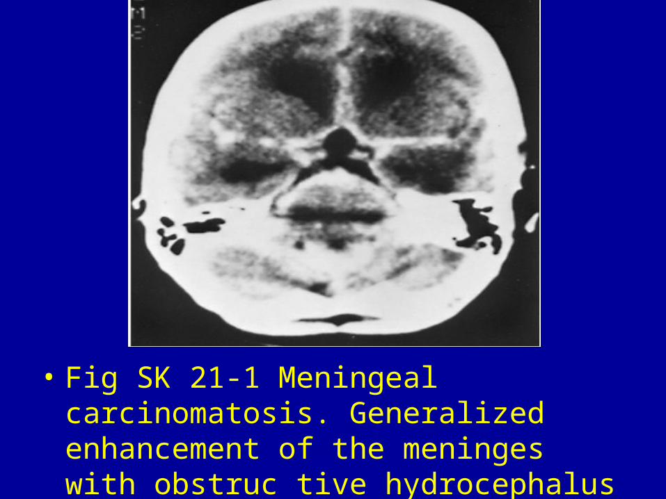
• Fig SK 21-1 Meningeal carcinomatosis. Generalized enhancement of the meninges with obstruc tive hydrocephalus

• Fig SK 21-2 Histiocytic lymphoma. Homogeneously enhancing lesions (arrows) deep in the brain associated with enhancement of ventricular margins.

• Fig SK 21-3 Subependymal metastases. Multiple enhancing ependymal nodules (arrows) in a patient with posterior fossa ependymoblastoma and hydrocephalus.1
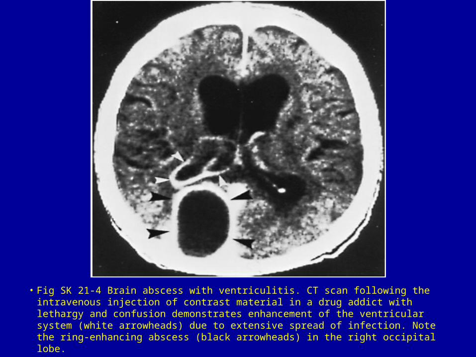
• Fig SK 21-4 Brain abscess with ventriculitis. CT scan following the intravenous injection of contrast material in a drug addict with lethargy and confusion demonstrates enhancement of the ventricular system (white arrowheads) due to extensive spread of infection. Note the ring-enhancing abscess (black arrowheads) in the right occipital lobe.

• Fig SK 21-5 Pneumococcal meningitis. (A) Noncontrast scan shows dilatation of the temporal horns of the lateral ventricles (arrowheads). (B) After the intravenous injection of contrast material, there is enhancement of the meninges in the basal cisterns (arrowheads), reflecting the underlying inflammation due to meningitis. The hydrocephalus in meningitis is due to blockage of the normal flow of cerebrospinal fluid by inflammatory exudate at the level of the aqueduct and the basal cisterns.


Related Documents