20 th ANNUAL CONFERENCE Socie of Anaesthesiologisʦ of Nepal (SANCON) OVERCOMING CHALLENGES IN ANAESTHESIA 16th March, 2019 Abstracts

Welcome message from author
This document is posted to help you gain knowledge. Please leave a comment to let me know what you think about it! Share it to your friends and learn new things together.
Transcript
20th ANNUAL CONFERENCE
Society of Anaesthesiologists of Nepal (SANCON)
OVERCOMING CHALLENGES IN ANAESTHESIA
16th March, 2019
Abstracts
20thANNUAL CONFERENCE
Society of Anaesthesiologists of Nepal (SANCON)
OVERCOMING CHALLENGES IN ANAESTHESIA
16th March, 2019
Society of '$. Anaesthesiologists f � 1
f N I � SA &o epa �
1987 6' 'l:.sn-,es,Ol-cP
Abstracts

ABSTRACTS
20TH ANNUAL CONFERENCE OF THE SOCIETY OF ANAESTHESIOLOGISTS OF NEPAL
(SANCON)
Table of Contents
S.No. Topic Author Page
No.
1 Overcoming challenges in anaesthesia Prof. Dr. Bishwas
Pradhan
1-2
2 Overcoming challenges in anaesthesia-
Chinese perspective
Prof. Jin Liu 4
3 Regional anaesthesia in Japan: Past,
present, and future
Shinichi Sakura 5
4 Life of Professor Thomas Joseph
McCaughey
Dr. Shambhu Acharya 6-7
5 Overcoming challenges in Anaesthesia- Sri
Lankan perspective
Dr. Asoka Gunaratne 8
6 Principles of quality improvement and
patient safety
Dr. Apurb Sharma 9
7 Persistent pain in cancer patients Dr. Balkrishna Bhattarai 10-11
8 Tramadol- are we prescribing
appropriately?
Dr. Anil Shrestha 12
9 The journey on developing pain
management services in Nepal
Prof. Dr. Roshana
Amatya
13
10 Off-pump cardiac surgery: Anaesthetic
considerations
Dr. Deepak K. Tempe 14-17
11 Delirium after cardiac surgery Dr. Smriti Mahaju
Bajracharya
18-19
12 NIRS in perioperative cardiac surgery Dr. Priska Bastola 20
13 Management of pulmonary hypertension in
children post-cardiac surgery
Dr. Mohamed Hassan
Ariff
21
14 Obstetric anaesthesia: up-to-date Dr. Yoshimi Inagaki 22
15 Death in OT: What to do? Dr. Sanjya Agrawal 23-29
16 An update on perioperative anaphylaxis and
national audit
Dr. Shambhu Acharya 30-31
17 Opioid free anaesthesia: Is it possible?- A
review
Dr. Sanjaya Poudel 32
18 Enhanced recovery after surgery Dr. Nishkarsh Gupta 33-34
19 NICE and warm- putting NICE into practice Dr. M. Puchakayala, Dr.
S. Bhattacharya
35-36
20 Challenges in management of septic shock;
do we need to change our focus?
Prof. Dr. B.D. Jha 37
21 Hemodynamic monitoring for critically ill
patient- an approach
Dr. Lalit K. Rajbanshi 38
22 Role of stellate ganglion block for the relief
of sympathetically maintained pain
Dr. Ujma Shrestha, Dr.
Baburaja Shrestha
39
23 Perioperative duloxetine as part of
multimodal analgesia regime reduces
Dr. Nishith Govil 40-41
20TH ANNUAL CONFERENCE OF THE SOCIETY OF ANAESTHESIOLOGISTS OF NEPAL
(SANCON)
postoperative pain in lumbar discectomy: a
randomized, triple blind, placebo-controlled
trial
24 New frontier of pain management in
thoracic region: ultrasound guided newer
blocks
Dr. Rakesh Kumar 42
25 Ultrasound guided stellate ganglion block-
is it the holy grail?
Prof. Dr. Jyotsna Punj 43-44
26 Near misses during awake craniotomy Dr. Hemanshu
Prabhakar
45
27 Ketamine: myths, controversies and
emerging evidences in neuroanaesthesia
Dr. Ritesh Lamsal 46
28 Anaesthesia for neuroendoscopic
procedure
Dr. Sanjay Agrawal 47-50
29 Evidences in traumatic brain injury Dr. Navindra Raj Bista 51-52
30 Anaesthesia for cardiac transplantation- an
update
Dr. S. Ponnambala
Namasivayam
53
31 Anaesthesia for liver transplant: an update Dr. Mrityunjay Kumar 54-61
32 Liver donor liver transplantation Dr. Yoon Ji Choi 62
33 Case series of external iliac artery
dissection in renal transplant recipient
Dr. Renu Gurung 63-64
34 PECs block: just some fancy ultrasound
guided intervention or a boon for breast
surgery patients?
Dr. Prakash Maden
Limbu
65
35 Spine: sonoanatomy and real time epidural Dr. Manoj Kamal 66
36 Role of regional anaesthesia in enhanced
recovery after surgery (ERAS)
Dr. Hemant Adhikari 67
37 Comparison of safe apnea duration of
continuous positive airway pressure versus
normal pre-oxygenation during general
anaesthesia
Dr. Kundu Shrestha, Dr.
Ravi Ram Shrestha, Dr.
Anuj Jung Karki
68
38 Comparison of intraperitoneal instillation of
ropivacaine with and without tramadol for
post-operative analgesia in laparoscopic
cholecystectomy
Dr. Anshu Kumari, Dr.
Bikal Ghimire, Dr. Binita
Acharya, Dr. Anil
Shrestha
69-70
39 Comparison of prophylactic low dose
ketamine and ondansetron in prevention of
intraoperative shivering after spinal
anaesthesia
Dr. S. Bista, Dr. S.
Manandhar, Dr. A.
Pokharel
71
40 A comparative study of hemodynamic
changes during orotracheal intubation using
video laryngoscope and direct
laryngoscope
Dr. Sarobar Upadhyaya,
Dr. Laxmi Pathak
72
41 Sonographic measurement of optic nerve
sheath diameter pre and post carbon
dioxide pneumoperitoneum in patients
Dr. Manan Karki, Dr.
Babu Raja Shrestha
73
20TH ANNUAL CONFERENCE OF THE SOCIETY OF ANAESTHESIOLOGISTS OF NEPAL
(SANCON)
undergoing laparoscopic cholecystectomy
under general anaesthesia
42 Comparison of dexmedetomidine and
fentanyl for attenuation of the hemodynamic
response to laryngoscopic endotracheal
intubation
Dr. K. Dhakal, Dr. S.
Sapkota, Dr. S. Shah
74-75
43 Ultrasound for management of airway Dr. Rakesh Garg 76-77
44 Role of endotracheal tube size on nasal and
laryngeal morbidity during awake
nasotracheal intubation: a randomized
controlled trial
Dr. Stalin Vinayangam,
Dr. Thirumurugan
Arikrishnan, Dr. Pankaj
Kundra, Dr. Sunil Kumar
Saxena
78
45 Emergency laparotomy: improving patient
outcome
Dr. Ramesh K Khoju
Shrestha
79-80
46 NEWS scoring system in emergency
abdominal surgeries
Dr. Ritu Pradhan 81
47 Life-saving peripheral nerve blocks in
trauma
Lt Col (Dr.) Krishna
Prasad
82-83
48 Anaesthesia in mobile surgical camps Dr. Bishwo Ram Amatya 84
49 Recent NPO guidelines and perioperative
hydration in paediatrics
Prof. Dr. Shanta Sapkota 85
50 Anaesthetic neurotoxicity in paediatric
patients
Dr. Yuichiro Toda 86
51 Subrachnoid block as a sole anaesthesia
for high risk former preterm infants
Dr. Anju Gupta 87-88
52 Comparative study of crystalloid (ringer’s
lactate) and colloid (hydroxy ethyl starch)
as preloading fluids in prevention of spinal
hypotension in patients undergoing lower
limb surgeries
Dr. Abdulla Ilyas, Dr.
Renu Gurung, Dr.
Madindra Basnet, Dr.
Priska Bastola
89-90
53 An ultrasound guided identification of level
of lumbar puncture used for subarachnoid
block in elective cesarean delivery
Dr. A.P. Tiwari, Dr. Bidur
Baral, Dr. A.B. Shrestha,
Dr. R. Pradhan
91
54 Single versus double syringe technique for
intrathecal administration of bupivacaine
and fentanyl to prevent hypotension in
patients undergoing elective caesarean
section
Dr. S. Shrestha, Dr. J.N.
Pokharel, Dr. T. Gurung
92
55 To study the perfusion index derived from
pulse oximeter in predicting hypotension
during spinal anaesthesia for cesarean
section
Dr. Sunti Barahi, Dr.
Shyam Krishna Maharjan
93
56 Ultrasound versus chest x-ray for
confirmation of central venous catheter tip
position: a comparative study
Dr. Lokendra Narayan
Mandal, Dr. Bashu Dev
Parajuli, Dr. Amit
Sharma Bhattarai, Dr.
94-95
20TH ANNUAL CONFERENCE OF THE SOCIETY OF ANAESTHESIOLOGISTS OF NEPAL
(SANCON)
Subhash Prasad
Acharya
57 Effectiveness of dexamethasone as an
adjuvant to bupivacaine in supraclavicular
brachial plexus block
Dr. P.K. Jha, Dr. S.
Sapkota, Dr. B.L. Shah
96
58 Fontan physiology: anaesthetic implication
for electrophysiological study and catheter
ablation: a case report
Dr. K. Harikrishnan 97-98
59 Airway diversity encountered in paediatric
ophthalmic anesthesia
Dr. Kanchan Prakash
Poudyal
99
60 Anaesthetic management of eclampsia in
remote high altitude: an audit study in 14
cases
Dr. R. Bhattarai, Dr. R.
Khapung, Dr. R. Shah,
Dr. K. Pyakurel
100
61 Intraoperative accidental bronchus rupture
in infant during oesophageal repair:
anaesthesiologist’s nightmare
Dr. Lokvendra Singh
Budania
101
62 Laparoscopic surgery in patient with
coronary artery disease
Dr. Rosi Pradhan 102
63 Perioperative management of carotid body
tumor: an anaesthetic challenge
Dr. Vamsidhar Chamala 103-104
64 Congenitally corrected transposition of
great arteries with complete heart block in a
parturient for emergency LSCS:
anaesthetist caught off guard
Dr. Yogesh K Gaude 105-106
65 Hemorrhagic shock in an emergency LSCS
done for fetal bradycardia due to uterine
rupture
Dr. Nikita Gurung, Dr.
Rishabh Ravi, Dr.
Banashree Dutta, Dr.
Navindra Raj Bista
107-108
66 Anaesthetic management of a patient with
sickle cell anaemia with dilated
cardiomyopathy undergoing caesarean
section
Dr. Sharmila Gurung, Dr.
Anand Agrahari
109
67 Anaesthetic managemtn of a patient with
xeroderma pigmentosa
Dr. Allen Suwal, Dr.
Bashudev Parajuli
110
68 Spinal anaesthesia for caesarean delivery in
a lady with situs inversus with dextrocardia
Dr. Ashmita Maharjan,
Dr. Baburaja Shrestha
111
69 Management of a case of intraoperative
anaphylactic shock in a patient scheduled
for excision of mass in right parieto-occipital
region with split skin graft (SSG)
Dr. Banashree Duta, Dr.
Megha Koirala, Dr. Binita
Acharya
112
70 Management of airway via fiberoptic
intubation in a patient with suicidal cut
throat injury
Dr. Keshav Adhikari, Dr.
Megha Koirala, Dr. Pujan
Balla, Dr. Semanta Dahal
113
71 Unsuccessful nasal intubation with
fiberoptic bronchoscope in a patient with
severe TMJ ankylosis
Dr. R. Shakya, Dr. B.
Gautam, Dr. M. Karki
114-115
20TH ANNUAL CONFERENCE OF THE SOCIETY OF ANAESTHESIOLOGISTS OF NEPAL
(SANCON)
72 Comparison of bupivacaine and ropivacaine
in caudal block for post-operative analgesia
in paediatric population undergoing
herniotomy
Dr. Rishabh Ravi 116-117
73 Severe mitral stenosis for emergency
laparotomy for ruptured ectopic pregnancy-
a case report
Dr. Prashant Bidari, Dr.
Bipin Karki
118
74 Anaesthesia for a case of 24 years
primigravida at 35 weeks of gestation with
pentalogy of fallot planned for elective
LSCS
Dr. Hari Prasad Gyawali,
Dr. Navindra Raj Bista
119
75 Ultrasound guided estimation of skin to
subarachnoid space depth in patients
scheduled for elective surgeries under
spinal anesthesia
Dr. S. Devkota, Dr. B.
Baral, Dr. P. Poudel
120
76 Anaesthesia for caesarean section in
patients with rheumatic heart disease
Dr. Anup Uprety, Dr.
Bashudev Parajuli
121
77 Spine surgery during early infancy and
anaesthetic concern
Dr. B. Baral, Dr. B.
Gautam, Dr. S. Barahi
122
78 Anaesthetic management of a case of
juvenile respiratory papillomatosis: an
experience
Dr. Abinash Dhoj Joshi,
Dr. Bashu Dev Parajuli
123

1
20TH ANNUAL CONFERENCE OF SOCIETY OF ANESTHESIOLOGISTS OF NEPAL (SANCON)
ABSTRACT
Since the successful documented demonstration of ether anesthesia in 16 October
1846 in Masachussets Hospital, Boston, anesthesiology field has traversed a long
way developing practices, adopting modern technologies to provide safe anesthesia.
Anesthesiologists realized long time back that clinical practice is not enough for
development of the specialty which opened new horizon of academic area integrating
field of research that complemented dream and vision of providing safer anesthesia
across the world.
There are several milestones in the development of anesthetic drugs and agents that
made anesthesia more predictable and safer. Memory lanes in the field of intravenous
induction agents will take us to 1930 when sodium thiopental was discovered and
was first tried in human volunteers. This was an era when a classical step of induction
of anesthesia which was evident during Ether anesthesia induction was abolished.
First challenge of dream of smooth induction was met. Later on development of
Etomidate, Ketamine and Propofol in 1960, 1962 & 1986 respectively addressed
many challenges of anesthesiologists in safety issues in intravenous induction agents.
Similarly morphine, pethidine, fentanyl solved analgesic component of balanced
anesthesia during 1804, 1939 and 1960. 1846, 1955, 1970, 1990 introduced ether,
halothane, isoflurane and sevoflurane in inhalational agent’s forum addressing several
challenges for anesthesiologists. Similarly anesthesia delivery system developed from
Schimmelbusch mask to modern anesthesia delivery work. Monitoring area took
a huge leap from sphygmomanometer in 1881 to state of art patient monitoring
Overcoming challenges in Anaesthesia
Prof. Dr. Bishwas PradhanHead, Department of Cardiothoracic & Vascular Anesthesiology
Manmohan Cardiothoracic Vascular & Transplant CenterMaharajgunj Medical Campus, Institute of Medicine
Tribhuvan University

2
20TH ANNUAL CONFERENCE OF SOCIETY OF ANESTHESIOLOGISTS OF NEPAL (SANCON)
systems. All those developments were phenomenal in addressing challenges to
decrease morbidity and mortality due to anesthetic reasons. Though Nepalese
history of clinical anesthesia is very short with first anesthesiologist giving anesthesia
only in 1956, all the latest developments in this field is already being used in Nepal.
Though it’s not clear when and where first academic program was started globally,
post graduate program in Nepal started in 1985 contributing in the development
of almost 45 MD postgraduate anesthesiologists per year in current scenario.
The number of anesthesiologists in Nepal is 1:100,000 populations which is still
far from WHO standard of 5:100,000. Settling in more lucrative jobs outside the
country, entering in different subspecialties like critical care, pain and palliative
medicine leaving the role as anesthesiologist in operation theatres, limited post
graduate programs and expanding surgical areas may contribute to less number
of anesthesiologists worldwide.
Development of societies of anesthesiologists globally developed a forum to
discuss and solve common problems and support societies from less affluent
countries academically and clinically providing opportunities for the same.
Likewise, it’s a continuum process that we are always challenged to provide services
to the rapidly developing surgical and allied areas in and outside operation theaters
and hence should make ourselves up-to-date and develop vision to overcome
challenges in the field of anesthesia to provide anesthesia services by competent
anesthesiologists only.

3
20TH ANNUAL CONFERENCE OF SOCIETY OF ANESTHESIOLOGISTS OF NEPAL (SANCON)
ABSTRACT
I am an anesthesiologist from Korea and I would like to talk about the development and challenges of the Korean Society of Anesthesiologists of the Republic of Korea in the South of the Korean Peninsula in East Asia.
Initially, the “Korean Society of Anesthesiologists” was founded by 9 doctors on November 10, 1956 and the area of the Anesthesiology Society was nothing but anesthesia in the operating room. However, for the past 62 years, the Korean Society of Anesthesiologists has established a large-scale scientific society with more than 5,000 professionals and 15 sub-special associations. The 2018 conference (Koreanesthesia 2018) was held as an international conference with 250 international participants from 26 countries. In addition, we are playing a pivotal role in general medical care including pre-operative and post-operative patient care, acute or chronic pain management, intensive care management, surgical out-patient anesthesia, sedation, and hospice.
However, we also face some problems as the medical environment changes.
Firstly, the ‘legalization of anesthesia nurses’ as a part of the professional nursing system in 2017 is causing problems. This is controversial because the nurse can administer anesthesia without anesthesiologists, which is directly related to the safety of the patient and the scope of the nurse’s medical care.
Secondly, the 80-hour work problem of the resident under ‘the special law of the resident’ has improved the quality of life of the residents, but it causes various problems because this increases the loading of specialists and shortens training time.
Third, the concentration of the anesthesiologist in the metropolitan area causes problems due to imbalance such as failure to meet the needs of the anesthesiologist in the province or decrease in income due to oversupply in the metropolitan area.
However, we will do our best to solve these challenges and develop social and anesthetic fields.
Overcoming Challenges in Anaesthesia - Korean Perspective
Dr.Yoon Ji Choi

4
20TH ANNUAL CONFERENCE OF SOCIETY OF ANESTHESIOLOGISTS OF NEPAL (SANCON)
ABSTRACT
Today, there are three major challenges in anesthesia in China. The first is that china
has the biggest population (1.4 billion) in the world and we now provide about 70 million
anesthesia a year. The second is that the needs for anesthesia service growth rapidly,
more than 10% a year in terms of anesthesia cases. The third is the big gap in anesthesia
quality between 2000 big hospitals and small hospitals. In order to overcoming these
challenges., The government, CSA and CAA made a plan to increase number and quality
of anesthesiologists in near future. We will increase anesthesiologists from 90,000 today
to 160,000 by 2030; adjust the ratio of surgeons : anesthesiologists to 3:1. By that time,
the density of anesthesiologists will be 1.2 per 10,000 population. We need 300,000
anesthesiologists in 2050. Over last 5 years, China has established a national standard
residency training system (3 year training) for all medical specialties and now, we have 309
anesthesia residency training programs in China and we plan to expand the programs to
1000 by 2030. About 20% of residency graduates will take one of 7 subspecialty training
(2 years) in anesthesiology: cardiothoracic anesthesia, pediatric anesthesia, intensive
care, pain, OB-GYN anesthesia, cardiopulmonary perfusion, and advanced general
anesthesia. By requiring all medical graduates taking the national standard residency
training,
China will iron out the difference in routing anesthesia service between city and countryside
in the near future.
Overcoming challenges in anesthesia -Chinese perspective
Jin Liu, M.D., Professor and Chairman,Department of Anesthesia and CCM, West China Hospital, Sichuan UniversityThe Honorary President, Chinese Society of Anesthesiology (CSA) and Chinese
Association of Anesthesiologists (CAA)

5
ABSTRACT
There is growing demand for regional anaesthesia due to early ambulation, short hospital stays, and increased focus on patient satisfaction. Thus, regional anaesthesia now appears to be a requisite skill for all anaesthesiologists. The advent of ultrasound-guided regional anaesthesia has led to the development of a number of new blocks. Recent research has shown that ultrasound can effectively and safely facilitate neuraxial anaesthesia and peripheral nerve blocks. However, patients receiving regional anaesthesia during their perioperative period are still in the minority.
In the past, regional anaesthesia was not popular in Japan because there was a lack of a proper educational system. There was insufficient opportunity to obtain the knowledge necessary for regional anaesthesia.
Under such circumstances, the Japanese Society of Regional Anesthesiologists (JSRA) was established in 2013. The first meeting was held in 2014. Each annual meeting consists of educational lectures and hands-on workshops. In addition, JSRA will start the Japanese Regional Anesthesia Certificate Examination this year.
Despite the positive achievements of JSRA, there are still a number of problems. First, not many institutions have a proper environment to provide regional anaesthesia. This includes specialized block rooms as well as support from administrators, surgeons and/or nurses. Second, not many institutions have ultrasound machines of good quality. Third, conducting regional anaesthesia is still considered as an extra, time-consuming service. Fourth, there are not enough qualified experts who can teach in each hospital where major surgeries are conducted. And finally, there is no educational institution that offers a regional anaesthesiology and acute pain medicine fellowship program.
In this presentation, I will sketch a brief history of regional anaesthesia in Japan and summarize recent progress in anaesthesia. I’ll also highlight major problems we still face and offer potential solutions as to how we can provide better pain management by using regional anaesthesia.
Regional Anaesthesia In Japan:Past, Present And Future
Shinichi SakuraSurgical Center, Department of Anesthesiology, Shimane University Hospital
89-1, Enya-cho, Izumo City, 6938501 Japan81-853-20-2295

6
20TH ANNUAL CONFERENCE OF SOCIETY OF ANESTHESIOLOGISTS OF NEPAL (SANCON)
Professor Thomas Joseph McCaughey was born on 3 December 1925 in Fintona,
County Tyrone in Northern Ireland. His parents were Thomas James McCaughey and
Margaret McSorley-McCaughey and they had six children: Nan, Tom, Gerry, Ita, Vera and
Sheelagh.
Tom married twice and had six children with his first wife Suzanne: Marie -France
(deceased in infancy), Danny, Gerry, Paul, Tom and Dominique.
He went to St Column’s college in Derry. He graduated as a doctor from University College
in Dublin in 1950. After graduation he worked at Hackney Hospital in London and then
did several locum jobs in England.
Tom moved to Newfoundland, Canada in 1954 and then became Head of Department of
Anaesthesiology at Winnipeg Children’s Hospital, a position he held for 13 years. He was
a leading paediatric cardiac anaesthesiologist there.
He subsequently moved to Montreal in 1970 and became Head of Department of
Anaesthesiology at Montreal General Hospital and Co-chair of Anaesthesia at McGill
University. He then worked in Community Hospitals in Hull, Shawville, Buckingham and
Maria in Canada.
Life of professor thomas joseph mccaughey
Dr Shambhu Acharya, MD with Honours (USSR), FCARCSI (Dublin), FRCA, DEAA, MSc in Pain Management (Cardiff),
FFPMRCA, PG Cert in Med Education (Dundee)Consultant Anaesthetist, Aintree University Hospital, Liverpool, UK
Email: [email protected] Telephone: +441515296215

7
Tom’s time in Nepal:
Tom and his second wife Theodora visited Nepal and trekked to Arun River in December
1983.
1983: Shanta Bhavan Hospital. Here he met Dr Chris Ward, the author of “Anaesthetic
Equipment”.
1985: Quoting Prof Maltby: He converted the DA program into a triumph.
1987-88: One full year in Nepal as a preceptor for DA program
1990-93: Tom shared 6 months of work with professors Sandison, Maltby, and Tweed
1994: Tom conducted feasibility study for MD program
1996: MD program was started; Tom remained in Nepal until 2000.
After that he visited Nepal many more times.
Tom was an excellent teacher, keen observer and an effective facilitator. He was always
available to his students and colleagues. He was a very caring, compassionate and
spiritual person and regularly visited church every Sunday.
Throughout his career Tom published many academic papers and in 2000 was awarded
the Canadian Anaesthesiologists Society’s Highest Honour, the Gold Medal.
Late in life, he dedicated himself to writing, both fiction and non-fiction, publishing “Sick
and You Cared” (2005), “When Miss Nepal Vanished” (2007), “Maoist in the House”
(2007) and “Disdaining the reins: my life to fifty-five” (2012).
Apart from Nepal Tom also worked and taught in Uganda.
Tom died on 20 December 2013 peacefully in Canada, surrounded by his family..

8
20TH ANNUAL CONFERENCE OF SOCIETY OF ANESTHESIOLOGISTS OF NEPAL (SANCON)
Dr Asoka GunaratnePresident
College of Anaesthesiologists and Intensivits of Sri Lanka
In 1981 the Post Graduate Institute of Medicine of the University of Colombo commenced its MD
programme in Anaesthesiology and board certification. In 2012 the board of study initiated a new
programme for Board Certification in Critical Care Medicine. However we are still unable to provide
consultant cover for each and every theatre list in the country. We should be able to meet this
demand in the next few years.
In anaesthesia, our auditing is very poor and we have no audit departments. We also need well-
established day case units. At present we try to provide this service within the existing infrastructure
which is far short of the ideal.
When it comes to Critical Care, we lack proper outreach teams which should be multi-disciplinary.
Our Intensive care Units are not designed by medical architects. We also have a severe shortage
of nurses. We still don’t have ICU follow up clinics and clear ICU admission and discharge criteria.
In Pain Medicine we need better organized acute pain services established in every hospital
undertaking surgery and a chronic pain programme at least in all provinces with follow up clinics.
We have no electronic data storage system or data link. The patients are often ignorant about
their medical condition and the clinicians are dependent on the diagnosis cards. Lack of a proper
GP referral system and patient migration to any hospital at their will are major problems in our
country. Health expenditure which is 4.9% of the GDP is totally inadequate. Total lack of any guide
line as to what clinical procedure can be undertaken where a major setback in our free health
service is. Ministry policy of making appointments purely on seniority basis and not recognizing
special training in a particular field either locally or during their training abroad has wasted a
lot of good human resource. Also the ministry policy of appointing consultants to places where
there is no infrastructure has wasted valuable human resource. Politically made decisions to open
up new stations have resulted in mal distribution of consultant services in the ministry. Culture of
being resistance to change and the trade unions becoming a deciding factor is detrimental to our
progress.
Overcoming challenges in Anaesthesia-Sri Lankan Perspective

9
ABSTRACT
Patient safety and quality healthcare services are becoming more and more relevant
these days in a country like Nepal. Quality improvement and safety implementation
are proven tools to improve patient care and outcomes. The new constitution
of Nepal reaffirms the nation’s commitment to providing a high-quality universal
health service. However, a proper process of implementing the safety and quality
are lacking mainly due to fear of an increase in cost, probably unwillingness of
the leadership of the health care systems and more importantly due to lack of
knowledge of the processes and training programs. This presentation will focus
on the core principles of quality improvement and patient safety, the position of
Anesthesiologists as a leader, creating a culture, methods of identifying weaknesses
in the system, scientific approach of creating, implementing and then sustaining
changes. The value aspect of quality and cost of quality will also be discussed.
Keywords: Healthcare Quality; Patient safety; PDSA cycles; Quality improvement
project; System approach
Principles of quality improvement and patient safety
Apurb Sharma, MDNepal Mediciti, Sainbu, Bhaisepati, Lalitpur 44700, Nepal
Fellow of Safety, Quality, Informatics and Leadership Course 2018-2019, Harvard Medical School, Boston MA, USA

10
20TH ANNUAL CONFERENCE OF SOCIETY OF ANESTHESIOLOGISTS OF NEPAL (SANCON)
ABSTRACT
Pain is one of the major concerns in any patient suffering from cancer. It does not only
cause suffering in untreated patients and those undergoing treatment but also adversely
affects the recovery and rehabilitation of the patient even if the disease is appropriately
treated. Further, it has massive negative influence on the patient’s quality of life, mental
wellbeing and employment status. Persistent pain in cancer patients is quite complex in
pathophysiology and very challenging to treat. The pain may be because of the disease
itself and/or associated with the treatment or intervention received viz. chemotherapy-
induced, radiotherapy induced and/or persistent postsurgical pain. The issue is further
complicated by existence of different barriers in our surrounding that often hinder the
optimal management of pain in cancer patients. Current views on pathophysiology and
treatment of persistent pain in cancer patients will be discussed.
Persistent pain in cancer patients
Dr Balkrishna Bhattarai Department of Anaesthesiology and Critical Care, BPKIHS, Dharan,
NepalEmail: [email protected]

11
Topic 1 Predicting and preventing persistent
postsurgical pain
Although numerous studies have been published on the subject in recent years,
persistent postsurgical pain is still a highly underestimated problem. This lecture
puts the spotlight on risk factors and prevention of chronic pain and offers an
alternative view of its origin.
Topic 2 Opioid free anaesthesia
Over decades, opioids have been regarded as a cornerstone of modern anesthesia.
Looking at the problems we generate by administering them we should start asking
ourselves whether this should still be the case…
Biography
Roman Zuercher is an alumnus of the University of Basel, Switzerland. As the
former medical head of the orthopedic, obstetric and gynecological operating
theaters, he has established ultrasound-guided regional anesthesia more than a
decade ago as a standard practice at the University of Basel. He is particularly
interested in the advancement of continuous nerve block techniques for pre- and
postoperative pain control, regional anesthesia procedures to reduce chronic pain
following surgical interventions as well as new concepts in anesthesia for cancer
surgery. At the moment he is the Director of the Regional Anesthesia Program and
Head of the Ambulatory Center at the Bethesda Hospital in Basel, Switzerland.
Roman is a dedicated clinical anesthesiologist with a keen interest in promoting and
teaching regional anesthesia. His extracurricular interests include family outings
with his wife Corinne and his three children in the mountains of his Swiss homeland.
Dr Roman Zurcher Abstracts

12
20TH ANNUAL CONFERENCE OF SOCIETY OF ANESTHESIOLOGISTS OF NEPAL (SANCON)
ABSTRACT
Tramadol is one of the most common analgesic drug. It is an opioid analgesic licensed
for use in moderate to severe pain. It is commonly used as a step 2 option of the World
Health Organization (WHO) analgesic ladder. Tramadol is a synthetic, atypical, centrally-
acting analgesic that binds to the μ-opioid receptors and also inhibits the reuptake of
serotonin and noradrenaline, resulting in both opioid and antidepressant-like effects.
Tramadol is associated with less risk of respiratory depression and constipation than
other opioids but has an increased risk of serotonin toxicity. Although it is associated with
fewer of the typical opioid adverse effects, other effects such as nausea, vomiting and
dizziness are common, and can be problematic.
Tramadol was said to have low risk for abuse, so it was initially approved by US FDA
as a non controlled analgesic in 1995 but now has placed tramadol into schedule IV
of the Controlled Substances Act effective August 18, 2014. Tramadol was also freely
available in drug stores in Nepal and was commonly used for various pain conditions
when analgesics like paracetamol and NSAIDS were not effective. It was also commonly
used for postoperative pain management. However, stating the reason that the abuse of
tramadol was found to be massively increased, DDA of Government of Nepal has also
placed tramadol into controlled drug list effective from 31st August 2018
Tramadol is being used for various acute and chronic pain conditions. It is also been used
for patients with different conditions, age or character and also been used singly or in
combination with different medications however the effect may not be same for all and
sometimes may land into serious adverse problem. We need to be more appropriate in
using tramadol in different patient, different condition and with different other drugs.
Tramadol- are we prescribing appropriately?
Dr. Anil ShresthaAssoc. Professor and Head
Department of AnaesthesiologyTribhuvan University Teaching Hospital
Maharajgunj Medical Campus, Institute of Medicine KathmanduPhone -9851069727, E-mail – [email protected]

13
ABSTRACT
INTRODUCTION: Pain is a universal phenomenon with no discriminations. Pain is one
of the most common symptom experienced at some point during the course of many
illnesses.
Pain medicine is a branch of employing an interdisciplinary approach for erasing the
suffering and improving the quality of life of those living with chronic pain.The science of
pain management has been rapidly developing in western world. Hence with the urgent
need to bring together clinicians, healthcare provider, scientists and policy makers
our pursuit to seek better pain services in Nepal started one and half decade ago in
many tertiary level teaching institutes. The issues such as lack of human resources
and lack of advanced skills, lack of proper infrastructure, reliable medical supplies and
lack of recognition to pain services by the concerned authorities is an ever persisting
problem that needs lot of efforts from the health system of the country. Inspite of all the
difficulties, we have been able to provide dedicated services to manage acute pain,
chronic pain conditions and complex pain disorders in an multidisciplinary manner only
in few tertiary level teaching institutes.Whatsoever it does not fulfill the need of pain
service in the country.
CONCLUSION: Pain services are ever evolving and each patient needs precision care
which makes the service and the outcome so diverse. Realising the complexity and
difficulty of management of chronic pain disorders, we need to keep together clinicians,
health care providers and policy makers to find better solution for better pain service
and to implement the future directives, which will be highlighted in this paper.
The journey on developing pain management services in nepal
Prof. Dr. Roshana amatyaDepartment of anesthesiology
Tu teaching hospitalInstitute of medicine
NepalCorrespondence @ [email protected], 9818449355

14
20TH ANNUAL CONFERENCE OF SOCIETY OF ANESTHESIOLOGISTS OF NEPAL (SANCON)
ABSTRACT
Coronary artery disease (CAD) is a leading cause of death and contributes significantly to
health care resource utilization. It is well known that compared with medical management
alone, conventional coronary artery bypass surgery (CCAB) prolongs life and reduces
symptoms. Therefore, CCAB has been the standard surgical treatment for CAD. However,
there are adverse clinical consequences associated with CCAB that have largely been
attributed to the cardiopulmonary bypass (CPB) circuit, hypothermic cardiac arrest, aortic
cannulation and cross clamping. The off-pump beating heart surgery (OPCAB) without
exposure to CPB circuit and percutaneous coronary intervention with stenting have
emerged as safe alternatives to CCAB. Although, OPCAB was first developed in 1960s, the
wider application of this technique has been possible in late 90s, when the tissue stabilizers,
which utilize suction technology were introduced. With further improvements such as the
apical positioners and intracoronary shunts, complete revascularisation of all the coronary
vessels during OPCAB became a reality. The OPCAB is technically more demanding and
the incidence of OPCAB in a given centre can vary from 0-100% and is mostly determined
by the surgical expertise.
Initial reports were mainly based on non-randomised trials on low-risk patients undergoing
single-vessel or double-vessel bypass. These demonstrated significant benefits of
OPCAB over CCAB. However, they suffered from the potential risk of unbalanced baseline
characteristics leading to biases in favour of OPCAB. More recently randomized trials have
been published. But for one trial that reported a reduction in graft patency rates at 3 months
, most others reported favourable outcome with OPCAB. The lack of power of individual
trials to adequately explore important effects on clinically relevant outcomes such as death,
Off-pump cardiac surgery: Anaesthetic considerations
Dr. Deepak K. TempeProfessor of Excellence
Department of Anaesthesiology and Intensive Care G.B. Pant Institute of Postgraduate Medical Education and Research
Former Dean, Maulana Azad Medical CollgeNew Delhi, India

14
stroke and myocardial infarction can be overcome by using meta-analysis (stastical
aggregation of randomized trials) to detect potential differences in clinical outcomes. One
such meta-analysis has shown that no significant differences were found for 30-day
mortality, myocardial infarction, stroke, renal dysfunction, intraaortic balloon pump, wound
infection, re-thoracotomy, or reintervention. However, OPCAB significantly decreased
atrial fibrillation, transfusion, inotropic requirements, respiratory infections, ventilation time,
intensive care unit stay and hospital stay. Patency and neurocongnitive function results
were inconclusive. In-hospital and 1 year direct costs were generally higher for CCAB.
Thus, this meta-analysis has demonstrated selected short-term and mid-term clinical
and resource outcome benefits with OPCAB in comparison to CCAB. The never ending
debate whether OPCAB is better than CCAB continues to date. The current literature
suggests, and the consensus is that the short-term outcome of OPCAB is comparable
and at times better than CCAB, However, CCAB provides survival benefit in the long
term. A recent paper has substantiated these conclusions and shown that OPCAB was
associated with higher mortality compared with CCAB at 10 years. It was associated with
higher risk of incomplete revascularization, and higher rates of repeat revascularization.
The anaesthetic technique has evolved to match the progress in the techniques of the
OPCAB surgery. When the tissue stabilizers were not present, minithoracotomy was
performed to accomplish beating heart surgery. The use of beta-blockers to decrease
the heart rate, one lung ventilation and anaesthetic technique directed to achieve early
extubation were the prominent features of the anaesthetic management. With the
availability of tissue stabilizers, the need for decreasing the heart rate and one lung
ventilation is no longer required. The changes in the anaesthetic techniques that have
emerged in a patient undergoing OPCAB are summarized in the table.
Table: Anaesthetic techniques used in OPCAB. These are mainly directed towards
achieving early extubation, which entails more intensive pain relieving measures in the
postoperative period
• Reductioninthedosageofopoids
• Useofshorteractingopioids
• Administrationofopioidsintheformofaninfusion
• Maintenanceofanaesthesiawithinhalationalagentsorpropofol

16
20TH ANNUAL CONFERENCE OF SOCIETY OF ANESTHESIOLOGISTS OF NEPAL (SANCON)
• Useofthoracicepiduralanalgesia
• Useofintrathecalopioids
• Intensivemonitoringandmaintenanceofhaemodynamics
• Earlyextubation
• Intensivepainmanagementinthepostoperativeperiod
With the improvement in tissue stabilizers (especially the apical positioners), intracoronary
shunts, as well as the surgical expertise, the haemodynamic course is not as turbulent
as it used to be and the use of inotropes to maintain the haemodynamics has certainly
gone down. Likewise, the regular use of ischaemic preconditioning is declining and
the groups describing large series of OPCAB do not report its usage. Pharmacological
preconditioning with the help of volatile anaesthetics can be employed to limit the
adverse effects of ischaemic myocardial damage.
Myocardial ischaemia during OPCAB: The protection of the myocardium from ischaemia
is one of the main problems that the anaesthesiologists have faced. The period during
which, the distal anastomosis is performed constitutes the ischaemic interval. The use of
intracoronary shunts provides blood flow to the distal myocardium. However, the blood
flow through the intracoronary shunt depends on the degree of narrowing of the native
coronary artery. Further, it also depends on the haemodynamic status at that moment.
The mean arterial pressure and cardiac output have been shown to decrease during
this period, thus compromising the flow across the shunt. It is therefore, important to
understand the principles of mechanical and pharmacological assistance to reduce
myocardial ischaemia during OPCAB.
In conclusion, the OPCAB has demonstrated significant short term benefits. However,
the OPCAB technique is more technically demanding and the long-term graft patency
using this technique is inferior to CCAB. Also, few other issues that need to be looked into
are its utility in the high-risk population (which is most likely to benefit due to avoidance
of CPB) and the conversion rate to CCAB and the results of such converted operations.
There is no denying that the anaesthesiologists and the surgeons need to be proficient
in both on- and off-pump techniques and individual patient’s best interest should be one
of the important parameters in the selection of the technique.

17
References: 1Khan NE, De Souza A, Mister R, et al. A randomized comparison of off-pump multivessel coronary artery bypass surgery. N Engl J Med 2004;350:21-282Cheng DC, Bainbridge D, Martin JE, Novick RJ. Does off-pump coronary artery bypass reduce mortality, morbidity, and resource utilization when compared with conventional coronary artery bypass? A meta-analysis of randomized trials. Anesthesiology 2005;102:188-2033Davierwala PM. Current outcomes of coronary artery bypass grafting: evidence from real world practice. J Thorac Dis 2016;8(suppl 10):S772-S7864Chikwe J, Lee T, Itagaki S, et al. Long-term outcomes after off-pump versus on-pump coronary artery bypass grafting by experienced surgeons. J Am Coll Cardiol 2018;72:1478-86

18
20TH ANNUAL CONFERENCE OF SOCIETY OF ANESTHESIOLOGISTS OF NEPAL (SANCON)
ABSTRACT
Delirium is a common problem that occurs after cardiac surgery. The incidence of delirium
after cardiac surgery is estimated to be 26-52%, with a significant percentage being
hypoactive delirium1-4.Postoperative delirium has been associated with higher hospital
costs, longer lengths of hospital stay, increased likelihood of institutionalization, increased
risk for dementia, and increased morbidity and mortality.5-7 Recognizing delirium and
those at risk can reduce the impact of delirium through targeted interventions and risk
reduction[8,9].Despite extensive research, acute confusion states after cardiac surgery
remain a subject of great importance and controversy. The profound impact of psychotic
disturbances on postoperative outcomes was noted in numerous studies.
This review highlights the epidemiology, peri-operative risk factors, tools to assist in
diagnosing delirium, and current pharmacological and non-pharmacological therapy
options.Understanding the frequency of delirium and the complications associated with it
can help guide future research and resource allocation.
REFERENCE 1. Rudolph J, Inouye S, Jones R, et al. Delirium: An independent predictor of functionaldecline after cardiac surgery. J Am Geriatr Soc. 2010;58:643–649. [PMC free article][PubMed]2. Rudolph JL, Jones RN, Levkoff SE, et al. Derivation and validation of a preoperativeprediction rule for delirium after cardiac surgery. Circulation. 2009;119(2):229–36.[PMC freearticle] [PubMed]3. Schoen J, Meyerrose J, Paarmann H, et al. Preoperative regional cerebral oxygen saturationis a predictor of postoperative delirium in on-pump cardiac surgery patients: a prospective
Delirium after cardiac surgery
Smriti mahaju Bajracharya, MDDepartment of cardiac anesthesia and ICU, Shahid Gangalal National
heart Center, Bansbari, Kathamandu

19
observational trial. Crit Care. 2011;15(5):R218. [PMC free article][PubMed]4. Plaschke K, Fichtenkamm P, Schramm C, et al. Early postoperative delirium after open-heart cardiac surgery is associated with decreased bispectral EEG and increased cortisol andinterleukin-6. Intensive Care Med. 2010;36(12):2081–9. [PubMed]5.Greene NH, Attix DK, Weldon BC, Smith PJ, McDonagh DL, Monk TG.Measuresof executive function and depression identify patients at risk for postoperative delirium.Anesthesiology. 2009;110(4):788–795.6.Franco K, Litaker D, Locala J, Bronson D. The cost of delirium in the surgical patient.Psychosomatics. 2001;42(1):68–73.7.Kat MG, Vreeswijk R, de Jonghe JF, et al. Long-term cognitive outcome of delirium inelderly hip surgery patients: a prospective matched controlled study over two and a halfyears. Dement Geriatr Cogn Disord. 2008;26(1):1–8.8. Inouye SK. Prevention of delirium in hospitalized older patients: Risk factors and targetedintervention strategies. Ann Med 2000;32:257-263. PubMed Abstract9. Marcantonio ER, Flacker JM, Wright RJ et al. Reducing delirium after hip fracture: Arandomized trial. J Am Geriatr Soc 2001;49:516-522.

20
20TH ANNUAL CONFERENCE OF SOCIETY OF ANESTHESIOLOGISTS OF NEPAL (SANCON)
ABSTRACT
Cerebral oximetry based on Near-Infrared Spectroscopy (NIRS) is a noninvasive technology
that can monitor the regional oxygen saturation of the frontal cortex. Intraoperatively, it
provides continuous information about brain oxygenation, it becomes the sentinel organ
indexing overall organ perfusion and injury. One of the major concerns during intraoperative
fall in perfusion pressure is the risk of tissue ischemia, hence this ischemia not detectable at
the systemic level is considered a major contributor to postoperative morbidity and mortality.
To limit this gap, between recognition of problematic oxygenation in systemic and tissue
level, non invasive techniques capable for continuous assessment of tissue oxygenation
were introduced. Among them, the most promising and broadly spread technique is near-
infrared spectroscopy (NIRS). As neurological outcome remains problematic in cardiac
surgery, many anesthesiologists working in these fields use the technology for brain
perfusion assessment.
Normal rSO2 values, prior to the induction of general anesthesia, range from 60% to 80%.
Although lower values (55-60%) for cardiac surgery patients breathing room air are not
considered atypical, these patients deserve a higher degree of attention. Besides cerebral
oxygenation, baseline cerebral oximetry values reflect a patient’s overall cardiopulmonary
function and systemic oxygen needs. It is known that cerebral metabolic rate is coupled
to oxygen delivery. Cerebral blood flow is modulated in the presence of decreased oxygen
delivery due to decreased arterial oxygen content. A diversion of flow towards the central
compartment is apparent during circulatory distress, to maintain perfusion/oxygenation
of vital organs. Cerebral blood flow is preserved at the expense of relative systemic
hypoperfusion, hence a low rSO2 reflects significant systemic circulatory compromise. The
NIRS-monitored brain becomes an index organ where, beyond alterations of local factors,
alone or in combination, derangements of systemic factors are reflected.
KEY word: Cerebral oximetry, cardiac, surgery, anesthesia, monitor, brain
Nirs in perioperative cardiac surgery
Priska BastolaLecturer, Manmohan Cardiaothoracic Vascular and Transplant center, Institute of Medicine, Tribhuwan
University, Maharajgunj, Kathmandu, [email protected]

21
PHYSIOLOGY: Normal mean pulmonary artery pressures ranges around 15mmHg. A
mean of >25mmHg at rest or 30mmHg under stress is accepted as pulmonary hypertension.
This elevated pressure and resistance will lead to progressive RV hypertrophy and failure
depending on the duration and severity of the disease.
In the perioperative period, pulmonary hypertension most commonly occurs as a result
of hypoxia, left to right shunts, precapillary and post capillary causes (LV Failure, Mitral
stenosis, obstructive pulmonary venous diseases). Symptomatic therapy should start by
reducing factors that stimulate or accentuate pulmonary hypertension.
MANAGEMENT: The approach to the patient with perioperative pulmonary hypertension
can be looked at from my simple way of tackling the problem using the Alphabetical
approach.
A Airway manipulation. This involves correction of hypoxia, hypercarbia, high airway
pressures etc. and institution of different modes of ventilation (high frequency
ventilation, reverse I:E ratios).
B Biochemical correction of acidosis, hypomagnesaemia, hypokalaemia etc.
C Correctable cardiac lesions e.g. Residual shunts, MAPCAs, etc.
D Drugs
1) direct vasodilators – calcium channel blockers, tolazoline, hydrallazine
2) cAMP augmenters – adrenaline, isoprenaline, PGE1, PGI2, adenosine, bipyridine
derivatives (amrinone, milrinone, sildanefil)
3) cGMP augmenters – nitric oxide, sodium nitroprusside, glyceryl trinitrate
4) Newer agents - sildanefil, bosentan
5) Others –Ca channel blockers, Mg,
E ECMO
F Failure Prevention especially RVF – Atrial septostomy, not closing/creating PFO
during surgery, not closing the chest after surgery.
Cardiac AnaesthesiaManagement Of Pulmonary Hypertension In
Children Post Cardiac Surgery
MOHAMED HASSAN ARIFFConsultant Anaesthesiologist and Intensivist
National Heart Institute, [email protected]

22
20TH ANNUAL CONFERENCE OF SOCIETY OF ANESTHESIOLOGISTS OF NEPAL (SANCON)
ABSTRACT
Anaesthetic management for caesarean section has been changing. First, spinal anesthesia
(SA) or epidural anesthesia (EA) alone was performed and second, combination of epidural
and spinal anesthesia (CESA) became a popular anaesthesia method. Recently, spinal
anaesthesia with opioids (morphine and fentanyl) has been spreading internationally. CESA
is superior to SA or EA on stability of anaesthesia during surgery and postoperative pain
relief. However, its technique is complexed. On the other hand, SA using morphine 0.1
mg and fentanyl 0.05 mg with hyperbaric bupivacaine 2.0 – 3.0 mL has both stability of
anaesthesia by fentanyl and postoperative pain relief by morphine. General anaesthesia (GA)
is seldom selected for elective caesarean section. Prognosis of newborn is controversial in
GA; similar prognosis to regional anesthesia (RA) (1) and worse prognosis to RA (2). In GA,
the problem is airway management, particularly difficult mask ventilation and/or tracheal
intubation. To avoid this accident, high flow humidified nasal oxygen therapy is applied for
pregnant women before induction of GA (3).
The considerable issue during caesarean section is massive obstetric hemorrhage. When
massive obstetric hemorrhage occurs, an anesthesiologist becomes a commander to
operate the bundle for maternal safety. The check-list and protocols for maternal safety should
be prepared and simulation according a scenario of massive obstetric hemorrhage should
be performed periodically among medical staffs including surgeons, anaesthesiologists,
nurses and clinical engineers in the operating room (4).
References1. Regional versus general anaesthesia for caesarean section. Cochrane Systematic Review -
Intervention: 17 October 20122. Sao Paulo Med J. 2015; 133(3):227-343. Anaesth Intensive Care 2018; 46:36-41.4. http://www.anesth.co.jp/guide/pdf/guideline_Sanka_kiki.pdf
Obstetric anaesthesia; up-to-date
Yoshimi Inagaki, MD, PhDDepartment of Anesthesiology and Critical Care Medicine
Tottori University Faculty of Medicine36-1 Nishicho, Yonago, Tottori 683-8504, JapanPhone: +81-859-38-6651, FAX: +81-859-6657
E-mail: [email protected]

23
Death on the table is fortunately not a common event. In a survey conducted by Lunn &
Mushin 1 in 166 surgical patients died within 6 days, only 1 in 10,000 was actually due to the
effects of the anesthesia alone. The survey also showed that the causes have not changed
significantly during the last 30 years in-spite of marked changes in anaesthetic technology.
[ In another study of peri-operative deaths, the most common (56%) cause of perioperative
death was disease/injury for which the operation was done, followed by shock & inevitable
risks of the operation (30%).
Common Reasons for a Patient to Die During Surgery
The most common causes of a patient death during surgery includes:
• Surgical/patientrelatedfactors
• factorsrelatedtoanaesthesia
• Uncontrolledbleeding/pulmonaryembolism
• Equipmentfailure
Anaesthesia related factors: anaesthesia related variables includes hypersensitivity or adverse
effects of anaesthetic agents ,Improper techniques & equipments, lack of experience, gross
negligence in precautions, careless in method, accidents during intubation/bronchoscopy,
over dose of drugs and improper pre-anaesthetic medications.
Simple anaesthetic management principles seem to have a major effect on peri-operative
mortality. The routine use of an equipment checklist, direct availability of an anesthesiologist
to help lend a hand or troubleshoot when needed, the use of full-time compared with part-
time anesthesia team members, the presence of two members of the anesthesia team at
emergence, and reversal of muscle relaxants at the end of anesthesia had dramatic, positive
effects that were associated with reduced perioperative mortality within 48 h after surgery
and anesthesia.
Liability of an Aneasthetist and Medico- Legal Aspects Related To Anaesthetic
Deaths
When death occurs during a surgical procedure performed under anesthesia, the surgeon
or anesthetists should at once report the matter to police for holding an inquest. As per
Death in OT: What to do?
Dr. Sanjay Agrawal, Dept. of Anaesthesiology, AIIMS Rishikesh

24
20TH ANNUAL CONFERENCE OF SOCIETY OF ANESTHESIOLOGISTS OF NEPAL (SANCON)
Sec.39 CrPC all deaths occurring in due course of surgery and anesthesia should be treated
as unnatural deaths and should be reported to the police. Failing of which the doctor can
be punished under Section 202 IPC for intentional omission to give information of offence to
police by the person who is bound to inform [
Medico-legal Aspects related to Anaesthetic Deaths and liability of anaesthesiologists:
• Whendeathoccursduringasurgicalprocedureperformedunderanesthesia,thesurgeon
or anesthetists should at once report the matter to police for holding an inquest.
• AsperSec.39CrPCalldeathsoccurringinduecourseofsurgeryandanesthesiashould
be treated as unnatural deaths and should be reported to the police. Failing of which the
doctor can be punished under Section 202 IPC for intentional omission to give information
of offence to police by the person who is bound to inform.
Anaesthesiologist duty in Anaesthetic Practices:
• Anesthesiologistmustattendthepatientadaybeforesurgery,doPre-anestheticcheck-
up and investigate the patient for any alarming situations if required. Before consent,
anesthetist must explain the procedure of anesthesia, type and nature of anesthetic agent,
its side effects, complications and risks involved in the procedure clearly to the patient in
local language, so that he can understand the nature and consequences of giving consent.
• Informed Consent: Before administration of anesthesia, the anesthetist must take
the consent in writing from the patient or his legal guardian or parents if he or she is
unconscious or below 18 years of age. It is the ultimate right of the patient to accept or
refuse the medication. Nothing should be decided against the patient’s will.
• Reasonable Degree of Skill: He must apply reasonable degree of skill and care in the
selection of anesthetic agent and the procedure. It is the duty of the hospital management
to provide adequate and trained hands. They must provide all necessary latest functioning
equipment. Trainee should be regularly supervised by the seniors. Anesthetic must adhere
to standard practice and follow the protocols of the institution.
• Precaution and Defense: Anesthetist should update his professional knowledge all the
time, keep full and accurate records of his patients. He must check the instruments prior
to use do the sensitivity test for a drug known to cause anaphylactic reactions and do not
leave patient till recovered from effect of anesthesia.

25
Steps to be taken after death in OT:
(“Catastrophes in Anaesthetic Practice - Handling the aftermath” GUIDELINES OF
THE AAGBI AND THE ASA)
Steps to take immediately after the event
1. Breathe, curse, pray, sit down…take a moment to regain your composure
2. Records
• If possible, designate one person during the resuscitation to keep a record of
the sequence of events, including personnel involved, times, drugs and fluids
used, interventions and procedures performed, and the outcomes
• After the event, make accurate, detailed notes on the anaesthetic chart of the
anaesthetic given and the events as they occurred
• No alterations should be made to the original notes, if any additions or
amendments need to be made, these should be recorded separately, signed,
timed and dated
• Ideally, details of the preoperative discussion with the patient should have
been documented – including risks of the anaesthesia and consent for regional
techniques.
• Make a photocopy of the anaesthetic chart, copies of relevant investigations
for your personal record, as well as a personal set of notes detailing the event
“The personal notes should include every detail of the routine followed for this
patient – when the patient was first seen, by whom, what was prescribed,
investigations and results, anaesthetic plan –
From a medico-legal point of view: make no assumptions, and the more detail the
better.
3. Supporting the Anaesthetist
In the period immediately following the death, aspects that our anaesthetic
colleague may need assistance with are:
• Inform the senior registrar / consultant on duty
• Quickly review the case and go over the sequence of events that transpired
while still fresh in one‟s memory
• Help to complete documents and make appropriate patient notes

26
20TH ANNUAL CONFERENCE OF SOCIETY OF ANESTHESIOLOGISTS OF NEPAL (SANCON)
• Help from a senior to speak to the patient‟s family
• Depending on the circumstances a decision will need to be made together
with the anaesthetist involved and the senior whether or not they are fit to
complete their slate/call or whether they need to be relieved of their duties
4. Dealing with the patient
• Any death occurring whilst under the influence of anaesthesia constitutes a
procedure-related death6, and will necessitate further investigation and post-
mortem
• All lines, tubes, drains and other equipment connected to the patient must be
left in place, and a detailed description should be made thereof. If any doubt
exists regarding the position of the endotracheal tube, this should be checked
and recorded by a second anaesthetist.
• Documentation should be completed as soon as possible to expedite the
process and to facilitate transfer of the body to the mortuary
5. Communicating with the relatives
• Whenever possible, such news should be communicated to the family in
person. You may need to contact the family telephonically, inform them
that a serious complication has occurred and ask them to come to the
hospital to speak in person. Try to avoid disclosing the news of the death
over the telephone.
• Find a quiet, comfortable room to sit down with the family. The initial
meeting will involve informing them what has occurred, and answering any
of their immediate questions.
• Never speak to the family alone, ideally you and the surgical colleague
involved should speak to them together, including a member of the nursing
team and an interpreter if necessary.
• Before the meeting, you and the surgeon should decide jointly on what
information to disclose. Offering conflicting versions of events creates
mistrust and such miscommunication could be the root of possible
litigation.
• If the cause of death is known, then this should be explained in simple
terms. If no cause has been determined yet, do not speculate or offer an
opinion – rather inform them that the matter is under investigation.

27
• Be empathetic. Offering an apology does not imply fault.
• The family will likely need time to process the news, don‟t give too much detailed
information initially, but rather schedule a second meeting, if necessary, to answer
further questions.
• Inform them of the procedure that will follow regarding a post-mortem and
whom they can liaise with to enquire when the body will be released for funeral
arrangements
6. Documentation to Complete
• A perioperative death in theatre mandates the completion of a GW24/7 form.
• For an unnatural death in the Intensive Care Unit (KEH and IALCH), we complete
an “Unnatural death form” and standard discharge summary.
• These forms go through to the Forensic Pathologist. The purpose of these forms
is to provide as much detail as possible to assist the Forensic Pathologist and
inquest Magistrate in understanding the events that transpired and in making their
findings. Upon completion of their investigation, the Forensic Pathologist will issue
a Notification of Death form (DHA-1663).
Subsequent Actions
1. Equipment and drugs
• If there is any suspicion of malfunctioning equipment in the theatre or drug
irregularities, this may warrant further investigation. A decision will need to be
made in conjunction with theatre matron whether to take the theatre or individual
equipment out of commission until such time that its safety can be verified by
medical equipment maintenance personnel, manufacturers or toxicologists.
2. Debriefing the theatre team
• Ideally all members of the theatre team (including nursing and technical staff)
involved in the case should be debriefed as soon as is possible or convenient after
the event. Having a short, even informal discussion together of the events that
transpired, in an open honest manner could go a long way in gaining information,
feedback, relieving anxiety, blame; and in maintaining the camaraderie of the
theatre teams we work with each day.

28
20TH ANNUAL CONFERENCE OF SOCIETY OF ANESTHESIOLOGISTS OF NEPAL (SANCON)
3. Communicating with the media
• Following the intraoperative death of patient, there may be scenarios in which the
media may be involved and approach the hospital staff for statements. A nominated
hospital representative should be the only person liaising with the media and all
enquiries should be directed to this person
The Role of the Anaesthetic Department
• Departments should be prepared to exercise flexibility and a commitment to
providing support to the anaesthetist who may be stressed or emotionally
traumatized after the event. Aside from the personal wellbeing of the anaesthetists
themselves, the guidelines highlight that a “stressed anaesthetist will be more
prone to making errors”4, which helps neither the department nor the subsequent
patients who come under their care.
• In the immediate time period following the event, it may be necessary to arrange
for someone to take over the anaesthetist’s duties or complete his/her call should
s/he feel unable to do so.
• An informal debriefing following the event is strongly suggested, where necessary a
trusted senior colleague should be assigned to mentor and provide support to the
anaesthetist for as long as they may need it. This could involve follow-ups with the
anaesthetist (formal or informal) in the weeks following the event.
• At a later stage, review of the case in a departmental Morbidity and Mortality
meeting may be a useful learning tool.
• Provide retraining, if needed, in a particular skill that the anaesthetist had
Conclusion
• Morbidity and mortality during anesthesia has been markedly reduced due to better
understanding of human physiology and pathology of disease processes.
• For the investigation of cause of death discussion between forensic pathologist, surgeon
and anaesthesiologist may arrive that will be the best consensus of opinion to offer the
investigating authority and courts of law. The Indian Society of anesthesiologist must
come out with protocols to be followed by its members in different clinical situations.
• A good record keeping, sympathetic attitude towards the nature of loss ,moral support
to attendants and taking care of medicolegal formalities plays an important role in
dealing with such eventualities

29
References:1. Kumar R,Kumar A. Anaesthetic Mortality: A Clinical And Medico- Legal Scenario.
IJSR2015;4:316-20
2. Attri JP, Makhni R, Chatrath V, Bala N, Kumar R,Jain P. Perioperative death: Itsimplications and management. Saudi J Anaesth 2016;10:436-9.
3. White SM., 2003. Death on the table. Anaesthesia, (58) p.515-519
4. Aitkenhead AR., 1997. Anaesthetic disasters: handling the aftermath. Anaesthesia, (52)p.477-482
5. Association of Anaesthetists of Great Britain and Ireland (AAGBI), 2005. Catastrophes inAnaesthetic Practice – Dealing with the Aftermath. London: Association of Anaesthetistsof Great Britain and Ireland. http://www.aagbi.org/publications/guidelines/docs/catastrophes05.pdf
6. Kumar A, Srivastava AK, Sharma B. Anaesthetic Deaths: A Medico-Legal Scenario. JIndian Acad Forensic Med 2014, 36:292-6
7. Parakh SC. Legal Aspects of Anaesthesia Practice. Ind J Anaesth 2008; 52:247-57

30
20TH ANNUAL CONFERENCE OF SOCIETY OF ANESTHESIOLOGISTS OF NEPAL (SANCON)
ABSTRACT
Introduction: Anaphylaxis is a rare but life-threatening condition, which requires immediate and effective management for a successful outcome. The NAP6 report is the largest ever prospective study of anaphylaxis related to anaesthesia and surgery. Main objectives of NAP6 were: How many are proven to be anaphylaxis and what are the culprits? How well does immediate management, referral and investigation match published guidelines?
Material and Method: The report of NAP 6 is used for this presentation. Data collection for NAP6 was conducted for 12 months starting from 5 November 2015 in the UK. The project surveyed 356 National Health Service hospitals to determine anaesthetic activity in October 2016.
Results:• Responsesfrom342(96%)hospitals• Annualworkload:3,126,067including2.394874generalanaesthetics• Neuromuscular blocking agents (NMBAs) used in 47,2%, antibiotics in 57.2%,
chlorhexidine in 73.5%, and iodine in 40.0%, blood products in 3%, bone cement/blue dyes/radio contrast in 2-3% of cases
• Fifty-eightpercentofanaphylaxisinwomen
An update on perioerative anaphylaxis and national audit
Dr Shambhu Acharya, MD with Honours (USSR), FCARCSI (Dublin), FRCA, DEAA, MSc in Pain Management (Cardiff), FFPMRCA, PG Cert in Med
Education (Dundee)Consultant Anaesthetist, Aintree University Hospital, Liverpool, UK
Email: [email protected] Telephone: +441515296215

31
• Hypotension:presentingfeaturein46%ofcases,andoccurredinallcases.Cardiacpatients had poor outcomes.
• Bronchospasm:presentingfeaturein18%ofcasesandoccurredin49%• Skinsigns,urticariaandflushing/non-urticariauncommon• Anaphylaxiswithin10minutesofexposuretotheagent in83%ofcases. In less
than 2% cases it was delayed beyond 60 minutes.• AnaphylaxistoNMBAsandantibioticsoccurredrapidly,hypotensionwasacommon
presenting feature particularly with atracurium, whereas bronchospasm was morecommon with suxamethonium.
• Anaphylaxis to chlorhexidine and Patent Blue dye was slow, hypotension wascommon and bronchospasm was not seen.
Conclusions: The incident of perioperative anaphylaxis is about 1:10000. Main culprits were identified as antibiotics, NMBAs, chlorhexidine and Patient Blue dye. The Association of Anaesthetists of Great Britain and Ireland and Australian and New Zealand College of Anaesthetist have produced the guidelines for immediate management, investigations and future anaesthetics.

32
20TH ANNUAL CONFERENCE OF SOCIETY OF ANESTHESIOLOGISTS OF NEPAL (SANCON)
ABSTRACT
Opioids have long been used for the management of pain conditions and during
perioperative period. More recently, use of opioids has been speculated to be associated
with acute and chronic side effects and poor surgical outcome. Opioid free anesthesia
has recently gained popularity secondary to early recovery, postoperative analgesic
sparing effect and prevention of opioid related side effects.
Opioid free anesthesia (OFA) is a technique where no opioids are used by any route during
conduct of anaesthesia. Various studies have been done with opioid free technique and
data obtained are promising. OFA can be practiced as an alternative to standard opioid
based anesthesia. It is especially beneficial in few selected groups of patients, where
side effects associated with use of opioids could be of risk.
Hypnosis, analgesia and relaxation are the primary goals of general anesthesia. Before
opioids were developed, these goals were achieved by increasing depth of anesthesia,
which often lead to hemodynamic instability. Introduction of opioids proved to be a
standard practice as a part of balanced anesthesia. More recently concerns regarding
side effects related to use of opioids has been highlighted. And alternative technique of
balanced anesthesia without use of opioids is gaining popularity.
A stable anesthetic management with a multimodal approach of sympatholytic drugs
and non opioid analgesics have been found to be effective in many studies. Non opioid
analgesics like NSAIDs, Paracetamol, Dexmedetomidine or Clonidine, Lignocaine,
Magnesium, low dose Ketamine has been used successfully during OFA management.
In absence of accurate monitoring technique for intraoperative nociception , management
of anesthesia free of opioids still remains a challenge. OFA however is possible in
daily practice, allowing stable and safe anesthesia preventing known early side effects
associated with use of opioids and spares opioids as analgesics for postoperative period.
Opioid free anaesthesia; is it possible? A review
Dr. Sanjaya Paudel, MD.Registrar, Department of Anaesthesiology
B. P. Koirala Memorial Cancer Hospital, Bharatpur ChitwanContact no. 9851220403; 9841220403
Email: [email protected]

33
ABSTRACT
The enhanced recovery after surgery (ERAS) programmes was introduced in 1990s as
an initiative to reduce variations in patient care and improve the quality standards. ERAS
programmes have been internationally adopted and widely implemented for major elective
surgical pathways in colorectal surgery, orthopaedics, gynaecology, cardiology and urology.
The aim of ERAS pathways is to reduce the length of hospital stay and lessen readmissions,
minimise surgical complications, decrease morbidity and improve cost-effectiveness.
ERAS protocols have brought about a revolutionary change in the perioperative care of the
patient, redefining our roles as perioperative physicians. That is, besides ‘Anesthesia’ and
‘Analgesia’, ‘Early Recovery’ of the patients too becomes our responsibility.
ERAS tends to improve patient experiences and outcomes by focusing on key aspects
of their care in preoperative, perioperative and postoperative periods to reduce the
physiological and psychological stress. The elements of care as stated in the ERAS
protocol are distributed throughout the perioperative pathway and are provided by different
medical professionals. This involves preoperative counselling for patients, the use of
minimally invasive surgical techniques and anaesthesia, optimal pain management and
early postoperative mobilisation. Despite their protocol-based foundations, evidence from
recent studies indicates that ERAS pathways are implemented variably across different
hospital settings. Moreover there is limited data on applicability of the ERAS care pathways
and their advantages in developing countries. The perceived barriers include resistance to
change, inadequate funding, lack of support from management, high staff turnover, poor
documentation and shortness of time, while facilitators included a dedicated enhanced
recovery lead, effective multidisciplinary team (MDT) working and ongoing education for staff
and patients. ERAS is now established as a safe and effective tool for optimizing recovery.
The current evidence for its individual components is increasing, leading to exciting avenues
of new research and the removal of interventions without benefit.
Enhanced recovery after surgery
Dr Nishkarsh Gupta, Associate Professor (Onco-Anesthesiology and Palliative
Medicine, DRBRAIRCH, AIIMS)

34
20TH ANNUAL CONFERENCE OF SOCIETY OF ANESTHESIOLOGISTS OF NEPAL (SANCON)
Suggested reading 1. Greco M, Capretti G, Beretta L, et al. Enhanced recovery program in colorectal surgery: a
meta-analysis of randomized controlled trials. World J Surg 2014; 38(6):1531–41.2. Feldheiser A, Aziz O, Baldini G, et al. Enhanced Recovery After Surgery (ERAS) for
gastrointestinal surgery, part 2: consensus statement for anaesthesia practice. ActaAnaesthesiol Scand 2016;60(3):289–334.
3. Francis NK, Walker T, Carter F, et al. Consensus on training and implementation ofenhanced recovery after surgery: a delphi study. World J Surg 2018;42(7):1919–28.
4. Jurt J, Slieker J, Frauche P, et al. Enhanced recovery after surgery: can we rely on the keyfactors or do we need the bel ensemble? World J Surg 2017;41(10): 2464–70.
5. Pisarska M, Pedziwiatr M, Malczak P, et al. Do we really need the full compliance withERAS protocol in laparoscopic colorectal surgery? A prospective cohort study. Int J Surg2016;36(Pt A):377–82.

35
Introduction: The NHS outcomes framework 2011/12 set “helping people to recover
from episodes of ill health or following injury” as one of the five outcome goals that is
used to monitor the progress and improvement in care delivery. Perioperative care is an
important indicator when looking to achieve this outcome and surgical site infections
(SSI) specifically have been identified as a quantifiable quality indictor. To reduce SSIs
guidelines NICE created guidance to avoid inadvertent perioperative hypothermia. This
is a re-audit to evaluate the effect of recommendations made in previous audits. NICE
suggest that patients temperature should be above 36 degrees prior to surgery and
anaesthesia, and should be monitored throughout the intra and postoperative period.
Active warming should be used if the temp is below 360C, in all high risk patients and if
anaesthesia time is more than 30 mins.
Materials and Methods: Hypothermia was defined as core temperature below 360C.
Prospective data collected was based on NICE guidelines and included temperatures
measured pre, intra and postoperative period, prior to recovery discharge, use of
intraoperative temperature monitoring and preventative measures used for hypothermia.
Results: Data from 118 patients was collected and analyzed in 2018. Tables’ 1 – 5 show
preinduction, intraoperative and postoperative temperature data compared to previous
years. High risk patients comprised of around 50% of the overall data. There were no
significant delays in discharge related to hypothermia.
Dr M Puchakayala MD FRCA, A Bapat FRCA, S Bhattacharyya,Guys and St Thomas NHS Foundation Trust, London, UK
NICE and warm – Putting NICE into practice
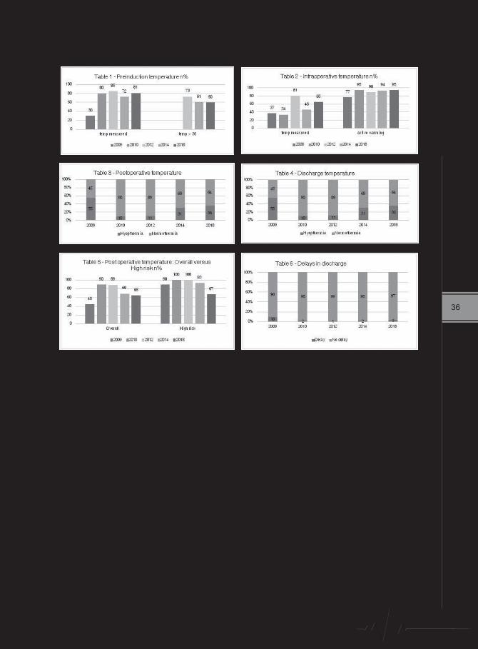
36
20TH ANNUAL CONFERENCE OF SOCIETY OF ANESTHESIOLOGISTS OF NEPAL (SANCON)
Conclusion: Implementing NICE guidelines have shown a consistent improvement in
hypothermia prevention and management with a greater focus on high risk patients. A
third of patients admitted to the recovery had hypothermia however, with no impact on
recovery discharge.

37
ABSTRACT
Sepsis is a common and highly morbid condition with an estimated 1.7 million cases
occurring in the United States each year, resulting in over 270,000 deaths. We don’t
have exact data for Nepal, estimated death due to sepsis is said to be very high. Despite
advances in critical care practices, sepsis remains the most common cause of death in
non-cardiac intensive care units (ICUs) . The traditional thought of organ dysfunction in
sepsis has focused on decreased systemic vascular resistance resulting in decreased
organ perfusion, and ultimately impaired oxygen delivery . Numerous studies, however,
have shown that organ dysfunction can occur during sepsis and septic shock even in
the absence of decreased perfusion. All the guidelines developed so far focuses on
early antibiotics, fluid therapy and vasopressure. Despite following these guidelines
mortality is still very high. This is high time we should change our focus on management
of sepsis and septic shock. In a small, retrospective observational study of septic ICU
patients, the combination of thiamine (200 mg every 12 h), ascorbic acid (1500 mg
every 6 h), and hydrocortisone (50 mg every 6 h) was associated with a dramatic
improvement in organ injury, time to shock reversal, and mortality . This therapy is
based on the concept that a combination of readily available, safe and cheap agents,
which target multiple components of the host’s response to an infectious agent, will
synergistically restore the dysregulated immune response and thereby prevent organ
failure and death. Each component of this combination of therapies has been recently
evaluated individually in septic shock. I have tried to review the pathophysiologic
basis and supporting research for each element of the thiamine, ascorbic acid, and
hydrocortisone drug combination in sepsis and shall be discussing.
Key words : Septic shock , adjunct therapy
Challenges in management of septic shock, do we need to change our focus?
Prof. B.D. Jha, HOD Anesthesia and critical care and Academic director, Phect NEPAL, Kathmandu

38
20TH ANNUAL CONFERENCE OF SOCIETY OF ANESTHESIOLOGISTS OF NEPAL (SANCON)
ABSTRACT
Inadequate organ perfusion is the common clinical finding in critically ill patients and hemodynamic monitoring for adequacy of perfusion is an integral part of critical care management. Hemodynamic monitoring assists in early recognition of organ failure and thus early intervention of hemodynamic support. Assessment of hemodynamic status represents a functional tool that may be used to derive estimates of organ performance and physiological reserve that may in turn guide clinical management.
Hemodynamic monitoring is a combined approach including clinical and laboratory assessment of perfusion, application of basic noninvasive tools and finally use of advanced invasive strategies to assess the organ perfusion. Traditionally, static parameters measuring right and left sided pressure and volume of heart have been used for assessing the hemodynamic status. But, many evidences have shown that the CVP is a poor predictor of fluid responsiveness and may not accurately reflect preload. Dynamic parameters are nowadays widely used for assessing the heamodynamic status. The respiratory variation on stroke volume, pulse pressure and systolic pressure can be used to assess the fluid responsiveness in mechanically ventilated patient. Even dynamic parameters are not free of limitation in view of being highly invasive, costly and not suitable for resource limited settings. In order to overcome the shortcomings of dynamics parameters, passive leg raise test, mini fluid challenge and end expiratory occlusion test can be used at the bedside more effectively that produce more specific result about hemodynamic status of the patients.
The outcome of the patients is not affected by the choice of hemodynamic tools. Moreover, the requirement of monitoring devices may change with the time and depend on the local equipment availability and training. In this regard, the choice of hemodynamics monitoring should be based on clinical assessments and on the patient’s response to the intervention. The interpretation of the hemodynamic monitoring should always be coupled with clinical intervention in order to obtain a better outcome.
Keywords: dynamic parameters, hemodynamic monitoring, perfusion, static parameter,
Hemodynamic monitoring for critically ill patient-an approach
Lalit Kumar RajbanshiMD,FICM, EDIC
Anesthesiologist and IntensivistBirat Medical College and Teaching Hospital, Biratnagar
9852030496

39
ABSTRACT
Chronic pain is a debilitating condition, often affecting 11-40% of the population, and
has serious medical, emotional and economic burden. However, many a times, it is
underreported, under-recognized and undertreated. Trauma to the bones, soft tissues
and even nerves can often progress to chronic pain characterized by persistent pain along
with allodynia and hyperalgesia. Complex regional pain syndrome (CRPS) is a chronic
neurological condition involving the limbs that is characterized by severe pain along with
sensory, autonomic, motor and trophic impairment. Sometimes, this can be maintained
by sympathetic activity and catecholamines, which is termed as sympathetically mediated
pain (SMP), while in some patients, the pain may be sympathetically independent and is
frequently known as sympathetically independent pain (SIP). It is this SMP, which is often
drastically responsive to the sympathetic blocks.
A stellate ganglion block, using local anaesthetic agents is very useful as a diagnostic and
a therapeutic approach to the management of CRPS in the upper extremities. Failure to
alleviate the symptoms with conservative management presents a huge socioeconomic
burden to the patients and they are often depressed as the pain hampers their daily
activities. These blocks can be very useful in such patients, as the pain relief from these
blocks often lasts longer than the action of the drugs we use. And with the availability of
Ultrasound in our daily practice, the safety of these blocks has also dramatically improved.
The role of stellate ganglion block in the management of sympathetically mediated
pain, especially in the complex regional pain syndrome (CRPS) along with few of our
experiences will be discussed.
Keywords: Complex regional pain syndrome, Stellate ganglion block, Sympathetically
mediated pain
Role of stellate ganglion block for the relief of sympathetically maintained pain
Ujma Shrestha1, Baburaja Shrestha2
1 Lecturer 2 ProfessorDepartment of Anaesthesiology and Intensive Care
Kathmandu Medical College Teaching HospitalSinamangal, Kathmandu
Presenter:Ujma Shrestha
Phone: 9851058811E-mail: [email protected]

40
20TH ANNUAL CONFERENCE OF SOCIETY OF ANESTHESIOLOGISTS OF NEPAL (SANCON)
ABSTRACT
Postoperative pain after discectomy involves pre-existing neuropathic and inflammatory
component as well as surgical nociceptive stimuli. In majority of post discectomy pa-
tients, conventional analgesics do not relieve acute surgical pain completely. We planned
this randomized, triple blinded, placebo-controlled trial to study the efficacy of duloxetine
in reducing postoperative pain after lumbar discectomy.
Fifty patients of age between 25-50 years undergoing lumbar discectomy randomized
into two group. Experimental group received oral duloxetine 20 mg BD preoperatively for
2 days, incremented dose of 40 mg BD from day of surgery to 2nd postoperative day
(POD) and 20 mg BD for next 2 days (up to 4th POD). Similar looking placebo capsule
was prepared and given to the patient in placebo group for similar period of 7 days. Post-
operatively, pain scores at rest up to 24 hours and during ambulation up to 48 hours, total
opioid and nonopioid consumption up to 4th POD, time to ambulation and complications
recorded.
Demographic and baseline variables are similar between two groups. Pain scores were
significantly less in experimental group at all times at rest, with movement and during am-
bulation up to 48 hours. There was a significant difference with decrease total analgesic
consumption (both opioid and nonopioid) in the first 24 hours as well as up to 4th POD in
Perioperative duloxetine as part of multimodal analgesia regime reduces postoperative pain in lumbar discectomy: a randomized, triple blind,
placebo-controlled trial
Dr Nishith Govil MBBS MD Associate Professor, Department of Anaesthesiology, AIIMS Rishikesh ,India

41
experimental group. There was no significant difference between the time to ambulation
after surgery and incidence of complication. (Table 1)
Duloxetine significantly reduced postoperative pain scores, analgesic requirement with no
increase incidence of complication.
1. Attia JZ, Mansour HS. Perioperative Duloxetine and Etoricoxib to improve postopera-
tive pain after lumbar Laminectomy: a randomized, double blind, controlled study.
BMC Anesthesiol 2017; 17: 162.
2. Bedin A, Bedin RAC, Vieira JE, Ashmawi HA. Duloxetine as an analgesic reduces
Opioid consumption after spine surgery: a randomized, double blind, controlled study.
Clin J Pain. 2017; 33(10):865–869.
1 Demographics and other
baseline variables ----- ------ -------
2 Pain Scores (VAS) at rest
(up to 24 hr) 4.4 ± 1.2 6.6 ± 1.6 p value = 0.01
95% CI: [-3.00
to -1.39]
3 Pain Scores (VAS) at movement
(up to 24 hr) 5.4 ± 1.1 7.6 ± 1.4 p value = 0.01
95% CI: [ -2.91
to -1.48]
4 Pain scores (VAS) on ambulation
(after 24 hr till 48 hr) 4.2 ± 1.0 6.2 ± 1.4 p value = 0.01
95% CI: [ -2.69
to -1.31]
5 Analgesic Consumption (up to
24 hr) IV Morphine mg 2.48 ± 0.87 4.80 ± 0.86 p value = 0.01
95% CI: [ -2.81
to -1.82]
6 Analgesic Consumption
(after 24 hr till 4th POD)
Oral tramadol mg 224 ± 16.26 424 ± 16.26 p value < 0.01
Oral Paracetamol mg 2900 ±187.06 4800 ± 122.40 p value < 0.01
Table 1 Results showing comparison of VAS and total analgesic consumption
S NoExperimental
groupPlacebogroup
Level ofsignificance
All Values expressed as Mean ± SD; p value < 0.05 is significant

42
20TH ANNUAL CONFERENCE OF SOCIETY OF ANESTHESIOLOGISTS OF NEPAL (SANCON)
ABSTRACT
The use of ultrasound machine in operation theatre leads to change of
anaesthesiologist practice. There practice of pain management change from
landmark guided to real time visualisation of regional block and they start moving
away from central neuraxial block to more safer peripheral nerve blocks. Ultrasound
guided regional anaesthesia techniques are safer and more effective than comparable
landmark based approaches because anaesthesiologist enable to locate target
nerve, plexus or muscular plane easily identified and there is reduced requirement of
local anaesthetics as drug precisely given to target area and less chance of needle
trauma to surrounding structure including nerve , vessels or pleura. Success rate of
blocks increased with use of ultrasound. The most frequently used block techniques
for pain management in thoracic region include the paravertebral block, intercostal
block, pectoralis block (PECI&PECII), serratus anterior block (SAP) recently describe
erector spinae plane (ESP) blocks and mid transverse process to pleura (MTP)
block. Advantage of these ultrasound guided block over central neuraxial block is
better safety profile and less hemodynamic disturbance. In addition to this these
block can be given in patients with coagulopathy while central neuraxial block are
contraindicate in these conditions.
New fronteir of pain management in thoracic region: ultrasound guided newer blocks
Presenter-Dr Rakesh KumarAssistant Professor All India Institute of Medical Science
Jodhpur
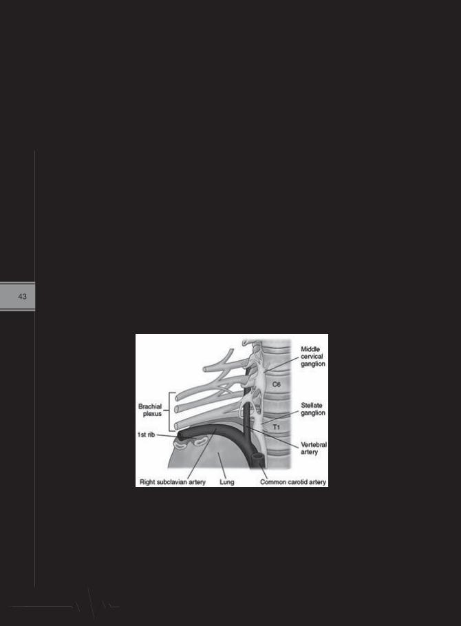
43
ABSTRACT
In the cervical region, the three sympathetic ganglia are superior, middle and inferior.
The inferior ganglia combine with the first thoracic ganglion and this is called the
stellate ganglion which relays the sympathetic signals from the arm and is not involved
with feeling or movement. It is in close proximity of various vessels, brachial plexus and
lung as shown below.
Ultrasound guided stellate ganglion block – is it the holy grail ?
Professor Dr Jyotsna Punj,Department of Anaesthesiology,Pain medicine & Critical care, AIIMS, New Delhi
The stellate ganglion is clinically blocked mainly in condiitons where the vacular supply to
the upper limbs is compromised ( vasculitis ) and in chronic pain conditions where there is
hyperactivity of the sympathetic system in the limb .
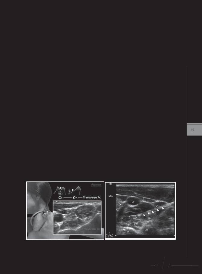
44
20TH ANNUAL CONFERENCE OF SOCIETY OF ANESTHESIOLOGISTS OF NEPAL (SANCON)
Location of the stellate ganglion
It is present anterior to the body of C7 vertebral body between longus colli muscle and
carotid sheath.
Techniques to perform stellate ganglion block
a) Landmark technique
b) fluoroscopic guidance
c) ultrasound guidance
Advantage of the ultrasound technique is visualization of the needle trajectory and
the needle tip along with the spread of the LA makes it useful for correct placement
of the block . Moreover because the needle tip can be placed between the plane of
longus colli muscle and the carotid sheath at the level of C7 rather than C6 , we can
get the effect of a successful block with smaller volumes of drug. Ultrasound guided
block is a real time block with correct idenification of the fascial planes . It avoids
deposition of the drug in the belly of longus colli muscle and also avoids deposition of
drug in carotid sheath both of which cannot be identified on landmark or fluoroscopy
tecnique. Identification of sensitive surrounding vascular structures like Inferior thyroid
/cervical /vertebral/carotid vessels and identification of esophagus are easily done
on ultraound. Ultrasound block is easier in obese patients and safer in patients with
deranged coagulation.

45
ABSTRACT
The key to successful management of neurosurgical cases is early recognition
of complications and its successful management. Brain is a vulnerable
organ and even a seemingly innocuous insult could be devastating. Awake
craniotomies poses special challenges for neuroanesthesiologists and are
not free of complications. Respiratory compromise, seizures, brain bulge
and loss of patient cooperation always remain a concern while managing
these patients. Airway management may be challenging, especially if the
stereotactic head frames are in place. As in any other neurosurgical case,
venous air embolism is always a possibility during awake craniotomy.
Despite utilization of intraoperative cortical and subcortical mapping, there
remains a risk of postoperative neurologic deficits. Agitation, somnolence,
restlessness and intraoperative events may lead to loss of cooperation from
the patient. Postoperative analgesia and treatment of nausea and vomiting
are very important. Carefully selected and well-informed patients are likely to
tolerate awake craniotomy well and this is crucial for a successful outcome.
Near Misses during Awake Craniotomy
Hemanshu Prabhakar, MD, PhDProfessor
Department of Neuroanaesthesiology and Critical CareAIIMS, New Delhi, India

46
20TH ANNUAL CONFERENCE OF SOCIETY OF ANESTHESIOLOGISTS OF NEPAL (SANCON)
ABSTRACT
Introduction: Traditional teaching discouraged the use of ketamine in patients with
intracranial pathologies because of concerns of elevating intracranial pressure (ICP). As
newer evidences emerge, this opinion has been challenged.
Discussion: Ketamine is a widely used anaesthetic with myriad uses. However, some
early studies suggested that ketamine increased regional cerebral blood flow, oxygen
consumption, cerebral blood volume and ICP. Based on these suggestions of adverse
impact on cerebral haemodynamics, its use in neurosurgical anaesthesia was restricted
for several decades. Newer basic and clinical research point at several useful attributes
of the drug. It is believed to offer neuroprotection and some recent studies have found
favourable outcomes when used in ischaemic stroke, refractory status epilepticus and
delayed cerebral ischaemia. There are also beneficial effects when used in neurosurgical
anaesthesia for traumatic brain injury, epilepsy surgery, intraoperative neuromonitoring,
and analgo-sedation regimens in neurocritical care units. Its use has also expanded for
the treatment of central neuropathic and other chronic pain syndromes that are refractory
to conventional treatment modalities.
Conclusion: The use of ketamine in neuroanaesthesia is contentious, but emerging
evidence support using it in different clinical settings. Its usage continues to rise in both
neurosurgical operating rooms and neurointensive care units. Future studies should
provide more concrete evidence both in normal and injured brain, as basic science
research continue to expand our understanding of various excitatory and inhibitory
neural networks, neuro-inflammatory mediators and pharmacologic interactions in these
cascades.
Ketamine: myths, controversies and emerging evidences in neuroanaesthesia
Presenting author: Ritesh Lamsal, MBBS, MD, DMDesignation: Neuroanesthesiologist and Intensivist
Address: 44-Bhimsengola Marga, New Baneshwor, Kathmandu, NepalTelephone: +977-9823553660
Email: [email protected] type: Oral

47
ABSTRACT
Introduction: Improvement in the imaging, computing and surgical instrumentation
have advanced the field of minimal access surgery and in last 10 years a major shift
is focused on minimal access neurosurgical procedures. These subsets of surgeries
present with their unique anaesthetic consideration and specific risks and needs the
modification in anaesthetic technique.
Indications of Neuroendoscopic procedures:
• Diagnostic
1. Ventriculoscopy
2. Spinoscopy
3. Biopsy of intracranial tumors
• Therapeutic
1. Hydrocephalus
2. Removal of intra/periventricular tumors
3. Evacuation of intracranial haematomas
4. Subacute or chronic brain abscess
5. Fenestration of loculated intraventricular cysts
6. Chroid plexus coagulation
7. Endocavitatory syringostomy
8. Retrieval of misplaced shunts
9. Management of CSF rhinorrhea
10. Clipping of aneurysm
Anaesthesia for Neuroendoscopic Procedure
Dr. Sanjay AgrawalDept. of Anaesthesiology, AIIMS Rishikesh

48
20TH ANNUAL CONFERENCE OF SOCIETY OF ANESTHESIOLOGISTS OF NEPAL (SANCON)
Preoperative evaluation and preparation
The age group of patient range from infants to geriatrics. The common problems associated
are:
• ‟Association of other congenital defects, urinary tract infections and impaired renal
function
• ‟Hypovolemia secondary to emesis,use of diuretics decreased intake
• ‟A thorough preanaesthetic assessment be done to rule out decrease cerebral perfusion
and ischemia. Special attention should be placed on state of consciousness, posturing
and papillary signs.
• ‟The use of sedative premedication should be avoided in infants less than 6 months of
age and adults with increased ICP
• ‟Use of anticonvulsants be continued in the pre and perioperative period.
Anaesthetic Considerations:
The patients presenting for endoscopic procedures range from preterm infants to
geriatrics.Endoscopic Third Venticulostomy(ETV) is the common procedure performed for
management of infants /children with aqueduct stenosis or hydrocephalus.
• The anaesthetic management is dictated by factors such as age of patients,neurological
status,underlying disease process,associated medical illness and current health of the
patient.
• The anaesthetic goals are to ensure immobility of patients intraoperatively and to have
an awake neurologically assessable patient after the procedure.
• In infants and neonates due to non fusion of sutures lead to increased head
circumference and fewer sign of increased ICP. The cerebral blood flow in adults is
50ml/100gm/min ,newborn 23-40ml/100gm/min and even less in premature infants.
The range of autoregulation is shifted to the right in infants thus maintaining CBF at
lower arterial pressure.The CMRO2 is high in infants and children to the tuno of 5.2
ml/100 gm/min, therefore the oxygenation requirement mandates careful attention to
avoidance of hypoxia.
• There are chances of increased intracranial pressure during the procedure due to the
use of irrigating fluid either too fast, if outflow tract is occluded or with insufficient space
between the endoscope and introducer sheath. Use of PIN index correlates with ICP.
• Sudden increase in ICP may elicit Cushing reflex ie bradycardia and hypertension that
may be preceded by tachycardia

49
• Brady arrhythmias may be precipitated due to distortion of autonomic nuclei in the
hypothalamus on the floor of IV ventricles. This may even lead to cardiac arrest.
• As a result of ependymal irritation or manipulation of hypothalamus central hyperthermia
may result.Irritation of some areas of brain during ETV may precipitate seizures.
• As the tip of endoscope is near the basilar artery trauma to same leads to torrent
haemorrhage.
• Use of normal saline as the irrigating fluid may precipitate systemic hypertension and in
volumes more than 500 ml may cause CSF acidosis.similarly use of ringer lactate may
precipitate postop hyperkalemia.
Choice of anaesthetic agent /induction:
Eneral anaesthesia is an essential requirement along with endotracheal intubation for securing
airway and controlled ventilation.
Induction of anaesthesia can be achieved with intravenous administration of thiopentone
or propofol and inhalational induction with sevoflurane in children.Non depolarizing muscle
relaxants are used to facilitate endotracheal intubation.Maintainence of anaesthesia is done
with NDMR ,short acting opioids like fentanyl and inhalational agents like sevoflurane/
isoflurane.Use of nitrous oxide is associated with increase in ICP .Use of moderate
hyperventilation should be avoided during ETV as it may decrease the size of ventricle and
make the procedure more difficult for the surgeons.
It is imperative that the patient lie immobile during the procedure .The tip of endoscope is
frequently near the critical structures such as basilar artery .Unexpected movement may
result in severe neurological injury or life threatening haemorrhage.Neuromuscular blocking
agents should therefore be used throughout the procedure and an adequate depth of
anaesthesia be maintained.
Monitoring during neuroendoscopy:
• Standard monitoring such as electrocardiogram, non invasive blood pressure
,pulseoximetry, capnography be applied in all the cases.
• Invsivebloodpressuremonitoringbyarterialcannulationisessentialinsuchcases.
• Measurementof intracranialpressurebuCodmansensorsanduseofPressure inside
Neuroendoscope(PIN) are essential to maintain the cerebral perfusion pressure above 40
mm Hg.

50
20TH ANNUAL CONFERENCE OF SOCIETY OF ANESTHESIOLOGISTS OF NEPAL (SANCON)
Potential Complications during endoscopic procedures:
A. Due to sudden CSF drainage or decompression of ventricles
• Suddenintracranialhypotensionleadingtobradycardiaandevencardiacarrest
• Entrainmentofairleadingtopneumatoventricleandpneumocephalus
• Suddencollapseofventriclesmayleadtodifficultyinvisualization
B. Due to instrumentation
• Majorhaemorrhage
• Inadvertentinjurytobraintissue
• Infection
C. Due to use of irrigation fluid
• Localorsystemichypothermia
• Movementofirrigantintotheperiventriculartissueleadingtrotissuedamage.
• Toxicreaction-fever,headache,neckstiffnessandincreasedcellcountinCSF.
‟ Increase intracranial pressure due to inadequate venting resulting in severe bradycardia
and asystole.
Postoperative Concerns:
• Common problem in immediate postoperative period is delayed emergence,
hyperkalemia, confusion, transient papillary dysfunction, transient hemiplegia and
memory loss
• Transient neurological deficiet is the most common complication.
• Respiratory arrest is common in infants during first hour after neuroendoscopy
• Postoperative monitoring of serum electrolytes is warranted because of diabetes
insipidus and hypothalamic dysfunction are reported.
• Late complications include meningitis and ventriculitis.
Conclusion: Neuroendoscopy surgeries are relatively safe. A careful choice of patient,
scrupulous planning and basics of neuroanaesthesia makes the procedure safe. Close
monitoring of patients and good communication with the surgeon prevents various
complications both perioperatively and postoperatively.

51
ASBTRACT
Introduction: Traumatic brain injury (TBI) has been defined by the Center for
Disease Control and Prevention (CDC) as a disruption in the normal function of
the brain that can be caused by a bump, blow, or jolt to the head, or penetrating
head injury. People of all age groups are at risk of TBI, especially children and
older adults. 1 The disease burden is an even greater problem for resource-
challenged countries like Nepal.
Discussion: Neurotrauma is a critical public health problem. This is a health
community concern that needs serious attention to decrease mortality and
morbidity. Research has also highlighted that TBI usually requires long-term care
with huge economic cost to health systems.2
Guidelines for the management of severe TBI, Fourth Edition, was published
by the Brain Trauma Foundation on September 2016, which consists of 189
publications used for evidences - 5 Class 1, 46 Class II, 136 Class III studies,
and 2 meta-analyses.3 These evidence-based recommendations for treatments
include decompressive craniectomy, prophylactic hypothermia, hyperosmolar
therapy, cerebrospinal fluid drainage, ventilation therapies, anesthetics,
analgesics and sedatives, steroids, nutrition, prophylaxis for infection, deep
vein thrombosis, and seizure. Further, it discusses different types of monitoring
Evidences in Traumatic Brain Injury
Navindra Raj Bista1
1Assistant Professor, Department of Anaesthesiology, Tribhuvan University Teaching Hospital, Maharajgunj, Nepal
Corresponding Author: Dr Navindra Raj Bista, MBBS, MD
Assistant Professor, TUTH Phone no: +9779841461305
Email add: [email protected]

52
20TH ANNUAL CONFERENCE OF SOCIETY OF ANESTHESIOLOGISTS OF NEPAL (SANCON)
that can influence patient outcomes like intracranial pressure (ICP), cerebral
perfusion pressure monitoring (CPP), and advanced cerebral monitoring
(ACM). Threshold values for blood pressure (BP), ICP, CPP, and ACM have
been highlighted to increase the probability of positive outcomes and trigger
a change in treatment.
In pediatric population, the Third Edition Guidelines have been recently
published by the Brain Trauma Foundation in 2019.4
Conclusion: TBI cause enormous losses to individuals, families, and
communities including large number of deaths and impairments leading
to permanent disabilities. Proper preventive measures as well as prompt
management are crucial for good outcomes following TBI.
References: 1. Centers for Disease Control and Prevention. (2019). Traumatic Brain
Injury & Concussion. [online] Available at: https://www.cdc.gov/traumaticbraininjury/index.html [Accessed 2 March. 2019]
2. World Health Organization. (2019). Road Traffic Injuries/Neurotrauma.[online] Available at: https://www.who.int/violence_injury_prevention/road_traffic/activities/neurotrauma/en/ [Accessed 22 April. 2019]
3. Guidelines for the management of severe Traumatic Brain Injury. 4th ed.Brain Trauma Foundation; 2016.
4. Guidelines for the management of Pediatric Severe Traumatic Brain Injury.3rd ed. Trauma Foundation; 2019.

53
ABSTRACT
Heart transplantation is an emergency procedure. Ischemia-reperfusion time of the donor
heart should be less than six hours. A close communication between the harvesting
and recipient teams is essential. Arterial cannulation, central venous cannulation and
pulmonary artery catheterization are done. Aseptic precautions are followed and
Special antibiotics given as these patients are immunosuppressed. Transesophageal
echocardiography is used. External defibrillator patches are applied. Pacemakers or
cardiac defibrillator devices are turned off to avoid electrocautery interference. Patients
on warfarin may have an elevated INR. Previous exposure to aprotinin increases the
chance of anaphylaxis. Redo surgery lengthens the time and increases the risk of
bleeding. Rapid sequence induction is performed. Etomidate, vecuronium/ rocuronium,
fentanyl and sevoflurane are the preferred anaesthetic agents. Hypotension during
induction is treated with alpha agonists or inotropic agents. Antifibrinolytic therapy
(aprotinin or aminocaproic acid) is started. Heparin is administered before vascular
cannulation. Isoprenaline is used to treat bradycardia and Amiodarone for arrhythmias.
Patients may require permanent pacemaker implantation for bradycardia and to over
pace arrhythmias. Protamine is given to reverse heparin. Right ventricular failure is the
most common cause for failure to wean from CPB. Hyperventilation with high FiO2 is
used to reduce PAP. Vasodilators (nitroglycerine, sodium nitroprusside, prostaglandin
E1, prostacyclin) are infused to reduce pulmonary vascular resistance. Inodilators
(milrinone, amrinone) increase contractility and decrease PVR. Noradrenaline or
vasopressin is used to improve systemic arterial pressure and coronary perfusion
when not responsive to catecholamines. Iloprost is more effective than Nitric oxide
in decreasing PVR without decreasing systemic blood pressure. Mechanical assist
devices like Intra-aortic balloon pump, Ventricular assist devices or Extracorporeal
membrane oxygenation may be needed. Patients with post-bypass coagulopathy
require transfusion of platelets, cryoprecipitate, or fresh frozen plasma. Acute rejection
is best diagnosed by endocardial biopsy. Antirejection therapy is continued in the post-
operative period.
Anaesthesia for Cardiac transplantation-an update
Dr. S. Ponnambala Namasivayam. MD.DA.DNB.Professor of Anaesthesiology & Dean
Govt. Stanley Medical College & HospitalChennai, TamilNadu, India

54
20TH ANNUAL CONFERENCE OF SOCIETY OF ANESTHESIOLOGISTS OF NEPAL (SANCON)
Anaesthesia for Liver Transplant: An updateDr. Mritunjay Kumar
Associate Professor, Anaesthesiology and Critical Care,All India Institute of Medical Sciences, Jodhpur, India
ABSTRACT
Liver transplantation is the treatment of choice for end-stage liver disease (ESLD). There
are four types of grafts utilized commonly: donation after brain death, donation after
cardiac death, living related donation and split liver donation where a cadaveric graft is
divided for two recipients.
Two commonly used scoring systems assess the severity of liver dysfunction- Child-
Turcotte-Pugh (CTP) classification and Model for End-stage Liver Disease (MELD)
score. In 2002, the MELD score replaced the CTP score for liver allocation. It is a better
predictor of 3-month waitlist mortality and is less subjective. In 2016, serum sodium was
added to the MELD score for liver allocation, now called the MELD-Sodium.

55

56
20TH ANNUAL CONFERENCE OF SOCIETY OF ANESTHESIOLOGISTS OF NEPAL (SANCON)

57

58
20TH ANNUAL CONFERENCE OF SOCIETY OF ANESTHESIOLOGISTS OF NEPAL (SANCON)

59

60
20TH ANNUAL CONFERENCE OF SOCIETY OF ANESTHESIOLOGISTS OF NEPAL (SANCON)

61
There are 3 basic venous reconstruction techniques for liver transplant:
1. Total occlusion of the vena cava and the portal vein (“full clamp”)- results in a
severe reduction in venous return to the heart during the anhepatic phase
2. “Piggy-back” technique: The inferior vena cava is only partially occluded with a
side-biting clamp
3. Venovenous bypass: Venous blood from the inferior vena cava and femoral
vein is returned into the internal jugular vein using extracorporeal venovenous
cannulas and a centrifugal pump.
Postoperative management
- Consider fast tracking
- Watch for complications:-
Early
1. Bleeding
2. Primary allograft dysfunction
3. Primary allograft non-function
4. Thrombosis of portal vein
5. IVC and hepatic vein thrombosis
6. Biliary tree obstruction
7. Hepatic artery thrombosis
8. Acute kidney injury
9. Sepsis
10. Cardiovascular events
Late
1. Immunosuppressant-related side effects
2. Infection
3. Graft rejection
4. Recurrent primary disease
5. Biliary tree obstruction
Closely monitor for early signs of liver dysfunction, renal dysfunction, and
immunosuppressive drug levels

62
20TH ANNUAL CONFERENCE OF SOCIETY OF ANESTHESIOLOGISTS OF NEPAL (SANCON)
ABSTRACT
I work at an 800-bed university hospital in Ansan, South Korea. Our hospital is not large,
but we are performing liver transplantation and would like to talk about our experience
with living donor liver transplantation.
In the United States, liver biopsy is only about 10-15% of the time, but in Korea, it is a
common procedure for liver transplantation with 85%. Liver transplant surgery requires
several specialist surgeons. For this reason, three doctors in three branch hospitals have
teamed up to divide the donor and the recipient team into one hospital to perform the
operation. And we have an anesthesiologist who specializes in liver transplantation, and
a scrub nurse for liver transplantation.
When choosing a transplant recipient, we consider the blood type, age, body size, and
infection, and consult a psychiatrist. Because of the inconsistent donor and recipient
blood type in the recent liver transplantation, preoperative plasmapheresis and rituximab
were used. In addition, preoperative ICG test was performed to confirm the liver function
and determine the operation method. In the case of a recent liver transplantation, ICG test
result was 14.9%, so we decided to perform left hepatectomy.
Anesthesia was performed with an inhalation anesthesia using desflurane and with
rocuronium and remifentanil during anesthesia. An air warmer and a fluid warmer were
used for heating. During the operation, the Levin tube was performed to reduce the
stomach volume.
We checked the level of fatty liver during the operation and weighed the liver after resection
and confirmed the volume of the remaining liver. After the operation, SugarMadex was
used to reverse the muscle relaxation and to perform tube extubation in the operating
room. PCIA was performed to reduce postoperative pain. Intermittent pneumatic
compression (IPC) devices were used for the prevention of thrombosis. Ambulation was
performed on the 1st postoperative day.
Living Donor Liver Transplantation
Dr. Yoon Ji Choi

63
ABSTRACT
Introduction: Kidney transplantation is the treatment of choice for end-stage
renal disease. Despite the improvements in surgical techniques vascular
complications consists a significant and sometimes life-threatening problem.
According to literature incidence of vascular complication is 10-20% of
patients where as incidence of renal vein and artery thrombosis is ranging from
0.4% to 6%.
Discussion: Renal transplant is regularly done at TUTH for the last 10yrs.
Anastomosis of donor kidney to right external iliac artery in recipient is a routine
practice. So far 5 cases of external iliac artery dissection has been detected
which is a rare vascular complication during renal transplant. Though the
condition is rare if diagnosed early, it is reversible and life saving.
External iliac artery dissection is suspected when there is drastic decrease
urine output, no arterial pulsation in ipsilateral leg, development of severe
acidosis and if abdomen is still open bluish discoloration of transplanted
kidney. Diagnosis is established by colour flow Doppler study. Arteriography
is recommended only when really needed and when it does not delay
revascularization process. Thrombosis due to external iliac artery dissection
extend to arterial anastomosis level compromising donor kidney and ipsilateral
leg.
Case series of external iliac artery dissection in Renal transplant recipient
Dr Renu Gurung, MD Anesthesiology, Fellowship in Pain ManagementAssociate Professor, Department of Anesthesiology,
Maharajgunj Medical Campus, IOM

64
20TH ANNUAL CONFERENCE OF SOCIETY OF ANESTHESIOLOGISTS OF NEPAL (SANCON)
Treatment is high degree of suspicion of external iliac artery dissection,
laparotomy, thrombectomy and ultimately a graft nephrectomy, high dose of
heparin, resection of the affected artery, replaced with an arterial prosthesis.
Reanastomose the kidney to other side internal iliac artery or external iliac
artery or to ipsilateral internal iliac artery.
Immediately after reperfusion, measure potassium as hyperkalaemia is favored
by acidosis or renal failure.
Conclusion: Awareness for early post-operative complications like external
iliac artery dissection, renal vein and artery thrombosis could save allografts
and patients. WE need early clinical suspicion, timely imaging, and prompt
intervention.
References:1) Understanding the Complexities of Kidney Transplantation: Chapter 26
Vascular Complications in Kidney Transplantation: Alexandros Giakoustidis,Nikolaos Antoniadis and Dimitrios Giakoustidis Division of Transplantation,Department of Surgery, Medical School Aristotle University of Thessalonikiand Hippokration General Hospital, Thessaloniki Greece
2) Reminder of important clinical lesson: CASE REPORT External iliac arterydissection causing early renal transplant dysfunction Department of Infection,Immunity and In,ammation: Transplant Group, University of Leicester,Leicester, UK; Leicester General Hospital, University Hospitals of Leicester,Leicester, UK. Gwyn Lee, Adam Barlow, Tahir Doughman, Michael LNicholson. Accepted 5 March 2014
3) Elsevier: Case Report External iliac artery dissection after renal transplant.V.L.N. Murthy Pisapati, Ch Ramreddy, Ramakrishna Pinjala, R.C. Mishra,Department of Urology and Renal Transplantation, Nizam’s Institute ofMedical Sciences, Hyderabad, andhra Pradesh 500082, India

65
ABSTRACT
Perioperative pain management for breast surgeries has traditionally been managed
with multimodal analgesia along with thoracic epidural or paravertebral block.
Although these techniques are indeed effective they are technically challenging
and definitely has a list of some grave complications like sympathectomy &
pneumothorax etc. PECs block a relatively new ultrasound guided intervention for
breast surgeries is equally effective, has a short learning curve and has minimal
complications. Ultrasound being the standard of care in regional anaesthesia
currently, PECs block could be a good alternative for perioperative pain management
in breast surgery.
“ PECs block just some fancy ultrasound guided intervention or a boon for breast
surgery patients ?”
Dr. Prakash Maden LimbuAssistant Prof.
Dept. of Anaesthesiology & Critical care,BPKIHS, Dharan, Nepal

66
20TH ANNUAL CONFERENCE OF SOCIETY OF ANESTHESIOLOGISTS OF NEPAL (SANCON)
ABSTRACT
Thoracic epidural analgesia is the standard and reliable technique of intraoperative and
postoperative pain relief for patients undergoing abdominal and thoracic surgeries. The
conventional method of placement of epidural catheter and spinal is dependent on the use
of surface anatomical landmarks. But in Obstetrics and paediatric patients relies on the
palpation of anatomical landmarks that are not always possible especially at thoracic level,
in obese, kyphoscoliosis patients. Relying on surface anatomy alone can lead to incorrect
identification of the targeted thoracic interspace. In paediatric patients the all epidural is
performed under general anaesthesia so there is always fear of complications that’s why this
technique is not popular in this age group. Due to comparative thinner and softer ligamentum
flavum and narrow space in paediatric age group especially in thoracic region, there are
higher chances of dura puncture in blind landmark technique. With the use of ultrasound the
correct level of insertion, best intervertebral space, angle of insertion and depth of epidural or
intrathecal space can be determined. USG minimizes chances of complications and failure
of the procedure.
Preprocedural imaging, however, still requires blind advancement of the epidural needle and
does not reduce the time or number of needle redirections required to place a thoracic
epidural catheter. Interest in using real-time US visualization during needle advancement to
improve rates of catheter placement at the lumbar spine has recently developed.
Technique
After confirmation of appropriate level with curvilinear or linear probe of ultrasound in
longitudinal plane on the midline of the patient’s back in lateral position of patients, in
paramedian sagittal oblique transverse process view the lamina, ligamentum flavum, anterior
and posterior dura complex is visualized.
The ligamentum flavum appear as shiny structure on USG deep to the shadow of lamina.
Tuohy needle will be advanced until the epidural space will identified with LOR to air or pierces
the ligamentum flavum. On injection of drug in epidural space the dura will move downward
and there will be widening of the epidural space, which will further confirm the space.
Spine: sonoanatomy and real time epidural
Presenter: Dr Manoj KamalAdditional Professor, AIIMS, Jodhpur
Address: 123, Vaishali Avenue, Jhanwar Road, Jodhpur (India)E mail: [email protected]

67
20TH ANNUAL CONFERENCE OF SOCIETY OF ANESTHESIOLOGISTS OF NEPAL (SANCON)
ABSTRACT
Enhanced Recovery after Surgery (ERAS) has become a standard approach to
surgical care as a means of reducing length of stay with similar or improved outcomes
compared to traditional strategies. Multimodal analgesia (MMA) is a mainstay of ERAS
and Regional anesthesia (RA)offers the ability to both reduce the need for general
anesthetics, opioids and improve postoperative pain management, decreased Post-
operative nausea, vomiting and return to both physical and bowel function. In the era
of Ultrasonography, the Regional anesthesia technique is emerging and has become
an integral part of MMA. When the western world is suffering from Opioid crisis, Nepal
is facing opioid scarcity and the role of Regional anesthesia as a part of MMA is ever
increasing. Further studies however is warranted to see the specific measures and
outcome of RA beyond analgesia.
Role of regional anesthesia in enhanced recovery after surgery(eras)
Hemant AdhikariRegistrar Anesthesiologist, Department of
Anesthesiology, Critical care and Pain Medicine, Nepal Mediciti Hospital, Sainbu, Lalitpur.
Email: [email protected]:9851142213Presentation type: Oral

68
ABSTRACT
Introduction: Pre-oxygenation with 100% oxygen results in development of atelectasis within minutes of induction of anesthesia. Preventing the formation of atelectasis during preoxygenation of general anaesthesia may increase the duration of non-hypoxic apnea which in turn will increase the margin of safety during period of endotracheal intubation. Application of continuous positive airway pressure during preoxygenation prevents atelectasis in the basal areas of the lungs and thereby increases the safe apnea time. The aim of the study was to compare the safe duration of apnea with or without application of continuous positive airway pressure during pre-oxygenation.
Materials and methods: Sixty patients scheduled for elective surgeries under general anesthesia with endotracheal intubation were enrolled in this prospective, single-blinded, randomized control interventional study. Patients were randomly allocated into two groups of 30 patients in each. Group A pre-oxygenation with 100% O2 without CPAP and Group B pre-oxygenation with 5cm of H2O positive pressure application for 3 minutes. Tracheal tube was left open in room air following intubation and patient allowed to remain apneic until SPO2 fell to 93%. Time duration from injection of Succinylcholine to fall in SPO2 up to 93% was noted. Monitoring of vitals was done every 2 minutes after initiation of pre-oxygenation till saturation fall to 93%. ETO2 was recorded at the end of pre-oxygenation in both the group.Results: The safe duration of apnea with continuous positive airway pressure of 5 cm of H2O during pre-oxygenation was 248.27±80.43 seconds in Group A and 335.63±68.19 seconds in group B. The p-value is 0.000 which is significantly more in Group B (p<0.05)Conclusion: There is significant increase in safe duration of apnea during pre-oxygenation with continuous positive airway pressure.
Comparison of safe apnea duration of continous positive airway pressure versus normal pre-oxygenation during general
anesthesiaAuthors: Dr Kundu Shrestha, Dr Ravi Ram Shrestha, Dr Anuj Jung Karki
Details of presenting author:Dr Kundu ShresthaThird year resident
NAMS, Bir HospitalAdress: Kuleshwor Awas Chettra-14 , Kathmandu
Mobile no: 9803032890E-mail:[email protected]
For oral presentation

69
20TH ANNUAL CONFERENCE OF SOCIETY OF ANESTHESIOLOGISTS OF NEPAL (SANCON)
ABSTRACT
Background: Post- operative pain is the main obstacle for rapid recovery of patients
undergoing laparoscopic cholecystectomy (LC). Patients undergoing laparoscopic
procedures do experience post-operative pain. Intraperitoneal (IP) injections of local
anaesthetics have been proposed to minimize postoperative pain after laparoscopic
surgery. This study aims to study the post-operative effect of Ropivacaine with and without
Tramadol when instilled IP in patients undergoing LC.
Methodology: A total of 80 patients undergoing elective LC were randomized in 2 groups,
one receiving 18 ml of 0.5% Ropivacaine with 2 ml of normal saline (NS) and other 18 ml
of 0.5% Ropivacaine with 2 ml of Tramadol (100 mg) at the end of surgery intraperitonealy
before the removal of trocar. Pain score was monitored using numerical rating scale (NRS)
scoring system every half hourly for first four hours and then at 6th, 12th and 24th hour. We
observed the time for first rescue analgesia, total analgesic consumption and incidence of
side effects like nausea, vomiting and shoulder tip pain in both the groups.
Comparison of intraperitoneal instillation of ropivacaine with and without tramadol
for post-operative analgesia in laparoscopic cholecystectomy
Kumari Anshu1, Ghimire Bikal2, Acharya Binita2, Shrestha Anil3
1Resident, 2Lecturer, 3Associate Professor, Department of Surgery and Department of Anesthesiology, Maharajgunj Medical Campus, Tribhuvan University, Kathmandu, Nepal.
Correspondence address: Dr Anshu Kumari, Resident, TUTH, Ph: 9862255730, Email: [email protected].

70
Results: A total of 80 patients were evaluated in this study. The severity of pain score
evaluated by NRS score was less in tramadol group in comparison of the normal
saline group and the difference was significant after 2.5 hrs onwards after the surgery
till 24 hrs (p=0.005). Only 17 patients (42.5%) in tramadol group demanded for rescue
analgesia as compared to 30 patients (75%) in normal saline group (p=0.003).The
total analgesic consumption was also significantly reduced in tramadol group (785
mcg) compared to ropivacaine group (1800 mcg) with p value of 0.002. No significant
adverse effects like nausea, vomiting and shoulder pain are found in any group.
Conclusion: From the results obtained in the current study, it is concluded that
intraperitoneal instillation of ropivacaine (90 mg) in combination with tramadol (100
mg) in elective laparoscopic cholecystectomy significantly reduces the post-operative
pain and analgesic requirement in post-operative period as compared to ropivacaine
without tramadol combination.
Key words: Intraperitoneal, laparoscopic cholecystectomy, post-operative pain,
ropivacaine, tramadol.

71
20TH ANNUAL CONFERENCE OF SOCIETY OF ANESTHESIOLOGISTS OF NEPAL (SANCON)
ABSTRACT
Introduction: Shivering after spinal anesthesia is a common complication and can occur in as many as 40%–60% of patients after regional anesthesia and it is very distressing. The aim of this study was to compare the effectiveness of Ketamine with that of Ondansetron and placebo in the prevention of postspinal shivering.
Materials and methods: This was a double blind, interventional comparative study.Patients were randomly divided into three groups of 57 each ; Group A patients received inj. Ketamine 0.25 mg/kg , Group B patients received inj. Ondansetron 4mg and the Group C patients received Normal Saline just after spinal anesthesia. The temperature of operating room was maintained at 24-26˚C. Incidence and severity of shivering, nausea and vomiting, sedation score, axillary and tympanic temperature and hemodynamics were recorded. ANOVA test was used to compare continuous variables. Chi square test was applied to compare ASA grading, shivering score and sedation score. P value of less than 0.05 was considered significant.
Results: Shivering was witnessed in 36 patients (63.1%) in Saline group, 22 patients (38.6%) in Ondansetron group and in 23 patients (40.4%) in Ketamine group (p value=0.001). Post hoc analysis showed that ketamine (p=0.001) and Ondansetron (p= 0.006) were effective in reducing the incidence of shivering compared to Normal saline but there was no statistical significant difference between Ketamine and Ondansetron (p=0.861). On comparing the severity of shivering, we found that four patients in Normal Saline group developed grade 3 shivering. None of the patients in Ketamine and Ondansetron developed shivering above grade 2. Sedation score was significantly higher in Ketamine group (P<0.05) than other groups. All thegroups showed the similar drop in core (p=0.204) and peripheral temperature .
Conclusion: Ketamine and Ondansetron were effective in preventing post spinal shivering.
Comparison of prophylactic low dose ketamine and ondansetron in prevention of
intraoperative shivering after spinal anesthesia
Bista S1, Manandhar S2, Pokharel A3
1 Resident, National Academy of Medical Sciences, Bir Hospital2 Professor, National Trauma centre, NAMS
3 Associate Professor, National Trauma centre, NAMSType of paper- Regional Anesthesia
Presenting Author:Dr. Sachhyatkar [email protected]

72
ABSTRACT
Introduction: For nearly a century, direct laryngoscopy has been the standard technique
for tracheal intubation. Alternatively, indirect techniques for tracheal intubation have been
developed that do not require direct vocal cord visualisation. Lagyngoscopy is mostly
associated with sympathetic stimulation and catecholamines release which causes
changes in hemodynamics. Thus, this study aims to compare the difference between
hemodynamic changes with video and direct laryngoscope devices.
Methods: A prospective clinical study was conducted which involving patients of 100
ASA PS Class I and II, dividing 50 in each group, with group V(Video laryngoscope) and
group D(Direct laryngoscope) undergoing elective surgeries
under general anaesthesia with endotracheal intubation. All patients were included for
airway assessment of Mouth opening, Modified Mallampati Test, Thyromental distance,
Protruded Incisors, Edentulous, Neck mobility, Prognathic ability. The baseline vitals of
these patients were recorded.
Results: There was no significant difference between Video laryngoscopy and direct
laryngoscopy with respect to blood pressure. However, significant differences were
found in heart rate immediately after intubation (110.40 beats per minute for direct
laryngoscopy vs 105.02 beats per minute for video laryngoscopy) and 1 minute (109.30 for direct laryngoscopy vs 106.20 for video laryngoscopy) after intubation
(p<0.001). Also the intubation time was longer for the video laryngoscopy group (22.80 seconds) compared to direct laryngoscopy group (26.54 seconds; p<0.05).
Conclusion: This study concluded that video laryngoscopy causes lesser hemodynamic
changes compared to direct laryngoscopy.
Keywords: Video laryngoscopy, Direct laryngoscopy, Hemodynamic changes
20TH ANNUAL CONFERENCE OF SOCIETY OF ANESTHESIOLOGISTS OF NEPAL (SANCON)
A comparative study of hemodynamic changes during orotracheal intubation using video
laryngoscope and direct laryngoscope.
Upadhyaya Sarobar1 , Pathak Laxmi2
1.Resident ,UCMSTH2Associate Professor,UCMSTH

73
20TH ANNUAL CONFERENCE OF SOCIETY OF ANESTHESIOLOGISTS OF NEPAL (SANCON)
ABSTRACT
INTRODUCTION: Laparoscopic cholecystectomy is a routinely performed surgery these days. The creation of pneumoperitoneum during laparoscopic surgeries may have various systemic effects including rise in intracranial pressure. The sonographic measurement of optic nerve sheath diameter as an indirect measure of raised intracranial pressure has been well described. The purpose of this study was to measure the optic nerve sheath diameter using ultrasound before and after carbon dioxide pneumoperitoneum in laparoscopic cholecystectomy.
MATERIALS AND METHODS: This is a prospective observational study performed using a convenience sampling. The optic nerve sheath diameter was measured by ultrasonography in 55 ASA I–II patients at various time points during laparoscopic cholecystectomy: pre-induction, after intubation, 5, 10 and 20 minutes after carbon dioxide pneumoperitoneum and after the release of pneumoperitoneum. Ultrasound imaging was performed using linear probe of frequency 7.5 MHz. Repeated measures ANOVA or linear mixed model was used to determine the change in the optic nerve sheath diameter, mean arterial pressure and peak inspiratory pressure at various time points.
RESULTS: The baseline mean optic nerve sheath diameter was 5.12 ± 0.54 mm. Twenty minutes after pneumoperitoneum the optic nerve sheath diameter increased to 7.01 ± 0.50 mm. The mean values of the optic nerve sheath diameter at all the time points before the release of pneumoperitoneum were significantly increased compared to that of baseline values. The mean optic nerve sheath diameter after the release of pneumoperitoneum was 5.86 ± 0.50 mm. There was no significant relation between baseline ONSD and age, gender and weight (p > 0.05).
CONCLUSION: There was 26% increase in the ONSD 20 minutes after pneumoperitoneum compared to the baseline values. There was 16% decrease in ONSD after the release of pneumoperitoneum compared to values at 20 minutes of pneumoperitoneum.
KEYWORDS: Laparoscopic cholecystectomy, CO2 pneumoperitoneum, Optic nerve sheath diameter
Sonographic measurement of optic nerve sheath diameter pre and post carbon dioxide pneumoperitoneum in patients undergoing laparoscopic cholecystectomy under general
anaesthesiaManan Karki1, Babu Raja Shrestha2
1.Resident, 2.Professor; Kathmandu Medical College Teaching Hospital

74
ABSTRACT
Introduction: Laryngoscopy and intubation are two of the most consistent maneuvers
that lead to significant increases in blood pressure and heart rate. These responses
may not be significant in otherwise normal individuals but deleterious for patients
with cardiovascular compromise like hypertension, ischemic heart disease and
cerebrovascular diseases.
Materials and Methods: 60 patients of ASA physical status I and II scheduled to
undergo elective surgeries under general anaesthesia with endotracheal intubation were
included in this prospective randomized double blind study and randomly divided 30 in
each group with envelop method. Group D received 0.5 mcg/kg of dexmedetomidine
over 10 minutes and group F received fentanyl 2mcg/kg before induction. Hemodynamic
variables i.e. heart rate , and mean arterial blood pressure were recorded at baseline,
after giving study drug , 1, 3 and 5, 10 minutes of endotracheal intubation.
Comparision of dexmedetomidine and fentanyl for attenuation of the hemodynamic response to
laryngoscopic endotracheal intubation
Dhakal K 1, Sapkota S 2, Shah S 3
1 Resident 3rd year, NAMS, Bir Hospital 2 Professor, Head of Department, Department of Anesthesiology, NAMS, Bir
Hospital3 Assistant Professor. Department of Anesthesiology, NAMS, Bir Hospital
Type of paper: General AnesthesiaPresenting Author:Dr Krishna Dhakal
Resident, MD Anesthesiology, NAMS, Bir HospitalContact number: 9841197874
Email: [email protected]

75
20TH ANNUAL CONFERENCE OF SOCIETY OF ANESTHESIOLOGISTS OF NEPAL (SANCON)
Results: Heart rate and mean arterial pressure at baseline and after giving study
drugs was comparable between the groups. Heart rate (HR) attenuated significantly
in group D (p value <0.001 in 1, 3 and 5 and 10 minutes intervals respectively after
laryngoscopy and intubation) as compared to group F. There was significant decrease
in MAP, 5 and 10 minute after laryngoscopy and intubation in Group D as compared
to Group F (p value of 0.01 and 0.04 respectively) but clinically it was not significant.
The incidence of hypotension and bradycardia was significantly different in between
the groups. Hypotension occurred 10 in group D as compared to 1 in fentanyl group
(p value=0.012) and bradycardia in 9 patients in dexmedetomidine and 1 in group F
(p value=0.002).
Conclusion: The use of dexmedetomidine 0.5μg/kg produces a more favorable
hemodynamic profile while fentanyl 2μg/kg is found to be less effective for the
attenuation of the pressor response to laryngoscopy and endotracheal intubation.
However, further larger studies are required to strengthen these conclusions.

76
ABSTRACTUltrasound as emerged as versatile tool for various specialities of medicine. Ultrasound
has found exit from its radiology suites and from the mastery of radiologist to real bedside
tool for diagnosis and intervention in real time. Its benefits, in general, includes easy
easily available, low running cost, portable, availability of different types of probes and
non-invasive. Ultrasound has emerged truly as point of care tool for the perioperative
care and in also for critically ill patient. Its use has been not only for diagnostic purpose
but also for real time therapeutic intervention tool as well.
Ultrasound has an emerging role in airway armamentarium. It has its role not only for
assessment of the airway but also aids the real time airway management. Ultrasound
has emerged as an important tool for bedside and also real time assessment for the
airway. It has advantages of its repeatability and real time management as well. The
airway assessment may be done with linear array probe usually but at time curvilinear
probe may be required for specific evaluation. Assessment and evaluation of airway
and pre tracheal structure before intubation increases the probability of detection of
difficult airway. Suprahyoid and infrahyoid structures are visible in medial, sagittal or
transverse view. Thyroid cartilage, cricoid, epiglottis, and vocal cord, sub glottis and
tracheal rings are observed Ultrasound detects subglottic stenosis and prediction of
paediatric endotracheal tube and double-lumen tube (DLT) size. Sublingual ultrasound
examination is new in the assessment of airway.
Ultrasound for management of Airway
Authors: Rakesh Garg
MD, DNB, FICA, PGCCHM, MNAMS, CCEPC, FIMSA, Fellowship in Palliative Medicine
Associate Professor, Department of Onco-Anaesthesiology, Pain and Palliative Medicine, Dr BRAIRCH, AIIMS, New Delhi.
Address: Room No. 139, Ist floor, Department of Anaesthesiology, Pain and Palliative Care, Dr BRAIRCH, All India Institute of Medical Sciences,
Ansari Nagar, New Delhi-110029, India.Email: [email protected]
Mobile: +91 9810394950, +91 9868398335.

77
20TH ANNUAL CONFERENCE OF SOCIETY OF ANESTHESIOLOGISTS OF NEPAL (SANCON)
Ultrasound is also useful for surgical airway. It helps in assessing the correct landmark like
cricothyroid membrane, tracheal rings and presence of any aberrant vessels.
Ultrasound can be used for detection of extubation success or failure. Strength of
diaphragm is directly related to the extubation success. Downward displacement of liver
and spleen due to diaphragmatic movement predict the success. Ultrasound image air
column width in vocal cord can be used for predicting post extubation stridor.
Real time dynamic endotracheal intubation can be done. Ultrasound confirm correct
endotracheal and bronchial tube placement easily through direct visualisation of tube
going through vocal cord or in oesophagus, diaphragmatic movement or lung sliding
sign. Direct visualisation of intra tracheal tip is difficult in adult but in neonate it can be
visualised and it can be confirmed. Oesophagus is seen as a cauliflower shape structure
in posterolateral to trachea in the transverse view, deglutition confirm this as peristalsis is
seen in ultrasound image.
Ultrasound has found its role in assessing the correct placement of supraglottic devices
as well.
Percutaneous dilatational tracheostomy and cricothyroidotomy can be performed
under ultrasound guidance with fewer complications compared to blind technique.
Tracheostomy is done between 2nd and 3rd tracheal ring most commonly. With the use
of ultrasound tracheal rings and vessels are identified and high placement of the tube,
vessel puncture avoided by guiding the needle and guide wire in real time. Guide wire
and final tracheal tube positions were immediately verified with bronchoscopy further
enhance the surety of correct level. Patients with morbid obesity, sub-optimal palpable
neck anatomy, previous tracheostomy or cervical spine injury can also tracheostomised
with use of ultrasound successfully.
To conclude, ultrasound is emerging tool for perioperative airway management. Its future
requires more robust evidence and appears it would become an important armamentarium
in the management of airway.

78
ABSTRACT
Introduction: Awake fibreoptic nasotracheal intubation (AFNI) is associated with adverse airway and hemodynamic complications. The aim of this study was to evaluate the role of endotracheal tube (ETT) size on nasal and laryngeal morbidity during awake fibreoptic guided nasotracheal intubation.
Materials and Methods: Eighty patients scheduled to undergo AFNI were randomly allocated to Group C (standard size ETT) and Group S (small size ETT followed by exchange to standard size using Airway Exchange Catheter under general anesthesia) after obtaining institute ethics committee approval and informed patient consent. Olfactory acuity using serial dilution of butanol and mucociliary clearance using saccharin clearance time were assessed pre and postoperatively. Patient discomfort during the AFNI was assessed using grimace score and hemodynamic parameters were recorded. Postoperatively, the incidence of nasal and laryngeal injury was recorded using nasendoscopy and telelaryngoscopy respectively.
Results: Demographic profile between the two groups was comparable. Epistaxis was noted in 47.5 % of patients in group C as compared to 12.5% in group S. Postoperative olfactory acuity was decreased (2 vs 4) and saccharin clearance time was prolonged (314s vs 134s) in Group C than Group S. (p-value < 0.001) Higher grimace score (2 vs 1) and increased hemodynamic response was demonstrated in Group C. (p-value < 0.001) Incidence of nasal injury (2 vs 1) and laryngeal injury (1 vs 0) were also more in Group C as compared to Group S.
Conclusions: AFNI with small size ETT followed by exchange to standard size under general anesthesia reduces nasal, laryngeal and hemodynamic complications.
Role of endotracheal tube size on nasal and laryngeal morbidity during awake nasotracheal
intubation: a randomized controlled trial.
Stalin Vinayagam, Thirumurugan Arikrishnan, Pankaj Kundra, Sunil Kumar Saxena.
Presenting Author:Dr. Stalin Vinayagam
Associate ProfessorDepartment of Anaesthesiology and Critical care,
Jawaharlal Institute of Postgraduate Medical Education and Research (JIPMER), Puducherry. India.
FR4, Sri Anbalaya Apartments, 17th Cross Street, Krishna Nagar,Puducherry. 605008,India
[email protected], +919786397762Type of Presentation: Oral Presentation

79
20TH ANNUAL CONFERENCE OF SOCIETY OF ANESTHESIOLOGISTS OF NEPAL (SANCON)
Summary
In England and Wales, around 30,000 emergency laparotomies are performed every
year. Many of these patients are at high risk of death and complications, and require
highly skilled multidisciplinary team, delivering high quality safe and effective care
throughout their journey.
Following an evidence of a high incidence of death, and a wide variation in the provision
of care and mortality in different NHS hospitals, National Emergency Laparotomy
Audit (NELA) was commissioned in 2013. It looks data at Structure, Process and
Outcome measures for the quality of care, and compares against standards of care
such as: NCEPOD reports, ‘The High- Risk General Surgical Patient: Raising the
standards’ (RCSE 2011and 2018).
On line prospective data are collected from patients over the age of 18 years,
undergoing general surgical emergency laparotomy from 183 NHS hospitals in
England and Wales, and reported on: patient characteristics, key process measures
(risk assessment, consultant input before and during surgery, CT scan, timeliness of
care for patients with peritonitis and sepsis, timeliness of arrival in theatre, critical care
and care of the elderly) and patient outcome.
Two organisational audit reports (May 2014 and Oct 2017) and four patient audit
reports (June 2015, July 2016, Oct 2017 and Nov 2018) have already been published.
So far, this is one of the biggest data collected from patients undergoing emergency
laparotomy (90,000 patients).
Perioperative medicineEmergency laparotomy: improving patient
outcome
Dr Ramesh K Khoju Shrestha, FRCA, FFICM, Consultant Anaesthetist, Bronglais General Hospital, Wales (UK)

80
NELA provides data for each hospital which allow clinical teams to assess and
benchmark their care against national standards, actively encourages teams to use
their own data to drive quality improvement (QI), and also raises awareness of QI
methodology (e.g. by sharing learning resources on website and running a series of
regional workshops for multidisciplinary teams).
Since 2013, national 30- day mortality has fallen from 11.8% to 9.5% and the length of
hospital stay from 19.2 to 16. 6 days.

81
20TH ANNUAL CONFERENCE OF SOCIETY OF ANESTHESIOLOGISTS OF NEPAL (SANCON)
ABSTRACT
Emergency abdominal surgeries especially emergency laparotomy are the
high risk surgical cases. They have high rates of morbidity and mortality
especially if present with co-morbid diseases and old age , which further gets
complicated by sepsis. EWS( Early Warning score) has been incorporated
in the first step in the patient assessment in the ELPQuIC ( Emergency
Laparotomy Pathway Quality improvement Bundle). NEWS is the new early
warning score. This includes the simple six physiological parameters like
respiratory rate, oxygen saturations, Temperature, Systolic blood pressure,
Pulse rate and the level of consciousness. The aggregate NEWS score of 1-4
is low clinical risk. The score of 5-6 or any individual parameter scoring 3 or
Red score is medium clinical risk. The score of 7 or more is high clinical risk.
This helps in “early track and trigger” response. Hence this NEWS scoring
system helps in the initial risk assessment of the emergency cases and in
deciding the appropriate level of care needed by the patients.
Besides, this can be used as a simple peri-operative risk assessment tool for
early detection of the clinical deterioration and the timely response for the
intervention. This can reduce the morbidity and mortality in the emergency
abdominal surgeries.
News scoring system in emergency abdominal surgeries.
Presenting Author: Dr. Ritu PradhanChief Consultant Anaesthesiologist
National Academy of Medical Sciences (NAMS), Bir HospitalMahaboudha, Kathmandu, Nepal
Email: [email protected]

82
Introduction:
•Trauma causes significant health problem in developing countries, involving mainly
the age group of middle age. Trauma causes significant metabolic, endocrine and
inflammatory response. Systemic changes may be aggravated by use of systemic
analgesics in the form of opioids. Opioids have got significant deleterious effects
on the systems. So far trauma patients received OLIGOANALGESIA. As a strategy
to facilitate painless surgery, RA is well validated and continues to retine.
•So far, barriers for regional anaesthesia/peripheral nerve blocks are, multiple or
relative contraindications to RA, competition with resuscitation, analgesia is given
lower priority in acute management. And also lack of appropriate equipment and
training ,improvements in safety profile of GA
•Now, because of increased reliability and safety of neural block, greater recognition
of pain as a disease process, short comings and side-effects of systemic
analgesics and publication of guidelines RA/PNB has taken a front role in trauma
patient management.
•RA/PNB offers many attributes of an ideal analgesic in the form of superior
analgesia, attenuation of stress response, increased alertness, patient cooperation,
facilitating ease of transport and chronic pain protection
Histroy:
•WalterReedArmyMedicalCentre(WRAMC),studiedextensivelyonuseofRA/PNB
in Vietnam war from the year 2003 to 2007. 1287 patients underwent operative
care under RA including single shot, CPNB, Continuous epidural infusions, more
than 900 operations performed before arrival, under RA/PNB
•A Review of Traumatic Injury Patterns and Anesthesia Techniques for Disaster
Relief after Major Earthquakes showed the beneficial effects of RA/PNB in multiple
trauma patients following the earth quake for the last 20 years.
•Manyofthepublishedstudiesshowedthebeneficialeffectslikeantiinflammatory
effects, cardiopulmonary protective effects of PNB in trauma patients.
•RA/PNBhelpsinearlyrecoveryofthepatientsandfulfilsearlyrecoveryprotocols.
Life saving peripheral nerve blocks (PNB) in Trauma
Lt Col (Dr) Krishna PrasadAnaesthesiologist

83
20TH ANNUAL CONFERENCE OF SOCIETY OF ANESTHESIOLOGISTS OF NEPAL (SANCON)
Peripheral Nerve Blocks:
•AmongtheRegionalBlocksthecentralneuraxialblockshavemoresympatheticblockade,
more haemodynamic disturbances and sometimes catastrophe like haematoma or
abscess formation.
•In the lastdecadeorso,PNBbecameverypopularbut there remainedhesitancy to
use. Now lesser use of intravenous opioids in PNB, the incidences of nausea, sedation,
pruritis, respiratory distress, urinary retention is negligible.
•Theuseofnervestimulators,USGandnervecathetershavemadethisamoreacceptable
mode of postop pain reliefmodality either as bolus injection or continous infusion,
prolonging the duration.
For upper extremity:
•Brachialplexusblocks:Interscalene,Supraclavicular,Infraclavicular,Suprascapular,axillary
and individual nerve blocks
Trunkal Blocks:
•Paravertebral,Erectorspinaeplaneblock,TAPblock,QuadratusLumboramBlock,PEC
I & PEC II blocks
For lower extremity:
•LumbarPlexusblock,Sciaticblock,femoralblock,FaciaIliacablock,AdductorCanal
Block, popliteal block
Controversies :
•Withtheadventofbetterinfrastructurethechancesofintraneuralinjection,intravascular
injection or pneumothorax are negligible. But still our surgical colleagues still request not
to use in some cases like compartment syndromes. The recent studies shows PNB is
not a contraindication for that, and lower concentration of drug is used. Another field of
concern is with patients receiving anticoagulants, we have to be careful in such cases.
Conclusion:
•Traumarepresentsaconsiderable,increasing,demandonhealthcareresources.
•Innovationsarechallengingattitudestopainreliefintrauma
•PNBismoreversatileandreliablethaneverbefore
•TheroleofPNBislikelytoexpandfurthertomeetthegrowingburdenoftrauma
•It can be concluded that PNB is a very safe and acceptable mode of anaesthetic
management in trauma patients.

84
ABSTRACT
Introduction: Mobile medical and surgical camps are one of the means of providing medical
and surgical facilities to people living in rural areas of developing country like Nepal.
Methods: An observational cross-sectional retrospective study was done collecting data from
surgical camps done by Nepal army in a five years period from 2013 to 2018 AD. Total number
of surgical cases requiring anaesthesia along with type of anaesthesia, American Society of
Anaesthesiologist physical status classification and anaesthesia related complications were
collected. All surgical cases requiring local anaesthesia were not included in the study.
Results: In a five year period, total number of nine mobile surgical camps was conducted by
Nepali Army. Out of which, one was done in collaboration with Ministry of Health, Government
of Nepal. Total number of 134 surgical cases requiring anaesthesiologist services was
conducted. Out of which, 13 cases were done in general anaesthesia, 84 cases were done
in spinal anaesthesia and 37 cases were done in total intravenous anaesthesia. There was
no intraoperative or postoperative anaesthesia related complications were noted and none
required blood transfusion.
Conclusions: Providing safe anaesthesia in a mobile surgical camp remains a challenge.
Proper screening of camp areas, appropriate selection of surgical cases, transportation of
logistic equipments, utilization of available resources and preparation of operation theatre
as well as postoperative recovery room or wards are some of the considerations before
conducting such camps. Regional anesthesia is the preferred anaesthestic technique for
providing safe anaesthesia.
Keywords: Anaestheisa, surgical camps, safety
Anaesthesia in mobile surgical camps
Bishwo Ram AmatyaLecturer, Nepalese Army Institute of Health Sciences, Sanobharyang,
9841337206, [email protected] presentation

85
20TH ANNUAL CONFERENCE OF SOCIETY OF ANESTHESIOLOGISTS OF NEPAL (SANCON)
ABSTRACT
Patients are kept fasting to reduce the risk of aspiration and post operative
nausea and vomiting .Fasting for 2, 4 and 6 hours is the standard practice for
Paediatric population scheduled for any surgical procedure under GA, regional
anaesthesia or sedation. This guidelines suits for the first case in the list. As
the fasting hour prolongs there is more chances of dehydration, hypoglycemia,
hypotension, irritability in subsequent cases. So in recent guidelines 0 , 4 and 6
hours is recommended but clinical adoption is slow and inconsistent.
Recent NPO guidelines and preoperative hydration in paediatrics
Prof Shanta SapkotaNAMS Bir Hospital

86
ABSTRACT
In 2009, it was reported in Anesthesiology that the incidence of learning disability was greater
in children who had undergone general anaesthesia (GA) more than twice by the age of 3
years than in chdilren who had never undergone GA or had undergone GA only once. Before
that article was published, many animal studies had revealed neurotoxicity of anaesthetic
drugs including volatile anaesthetics in young rats or other species. Furthermore, a large-scale
study that included more than 30,000 children who underwent anaesthesia before 4 years of
age revealed that exposure to anaesthesia has a slight association with later academic and
cognitive performance in adolescence. In another cohort study, it was shown that the hazard
ratios for behavioral disorder, mental retardation, autism, and language/speech problems in
children who had undergone anaesthesia by the age of 3 years were two-times higher than
those in children who had not undergone anaesthesia. Since those studies were retrospective
studies, there were many confounding factors other than GA affecting the incidence of
neurological consequences. Anaesthetic neurotoxicity in the developing brain has various
adverse outcomes. Maladaptive behavior, delirium, sleep disorder, enuresis, adaptation
disorder, and learning disability are all considered to be consequences of GA. However, these
adverse events are also well known to be caused by perioperative anxiety, pain, surgery
itself, and environment. In fact, multiple episodes of early hospital admission are associated
with later behavior and learning disturbance even without surgery and anaesthesia. A GAS
study was conducted to answer to the clinical question “Does general anaesthesia affect
neurological development in infants?” That study included approximately 700 infants who
underwent inguinal hernia repair. The participants were randomly assigned to a GA group
(sevoflurane) or a spinal anaesthesia group (bupivacaine) in a 1:1 manner. Bayley scale was
measured to assess cognitive and language development, and emotional and behavioral
development was also assessed. There were no statistical differences in neurodevelopment at
the age of 2 years. However, possible effects of environmental factors could not be excluded
in that study. The GAS study also cannot answer the question “Is duration of anaesthesia for
3 hours safe for infants?” However, the GAS study will provide information in near future on
neurocognitive function of the participants at 5 years of age.
Anaesthetic Neurotoxicity in Paediatric Patients
Yuichiro Toda M.D., Ph.D.Anesthesiology and Intensive Care Medicine
Kawasaki Medical School

87
20TH ANNUAL CONFERENCE OF SOCIETY OF ANESTHESIOLOGISTS OF NEPAL (SANCON)
Introduction: Anaesthetic management of preterm infants is considered challenging due to
increased prevalence of multiple comorbidities and their increased tendency for apnoea and
bradycardia. General anesthesia (GA) has been shown to be associated with an increased
incidence of postoperative adverse events in this population. After enjoying initial success and
enthusiasm in the paediatric anaesthesia practise, subarachnoid block faded in popularity in
the middle of the 19th century due to various reasons. It was later reintroduced by Abajian et
al for anaesthetizing preterm infants as a safer alternative to general anaesthesia to reduce the
postoperative complications such as apnoea, bradycardia and desaturation which are prevalent
in this population. Several reports followed, all supporting the use of awake spinal anaesthesia
over general anaesthesia (GA) for this group of patients and it has been termed as the ‘gold
standard’ technique of anaesthesia for preterm infants undergoing inguinal herniorrhaphy.
However, there is still a lot of hesitation in the routine use of this technique probably due to lack
of awareness or fear of adverse consequences.
The incidence of postoperative apnoea is inversely proportional to post-conceptual age (PCA) in
premature infants. The incidence of apnoea is maximum till 48 weeks PCA (45%) but elevated
risk is believed to persist till 60 weeks PCA. In addition, other important risk factors are history
of preoperative apnoeic episodes, bronchopulmonary dysplasia, and anaemia. However,
postoperative apnoea can occur without any preceding history of apnoea. Hence, elective
surgical procedures are preferably delayed beyond this period and sole regional anaesthesia
is advocated for these former PT infants within this window to decrease the incidence of
postoperative apnoea, bradycardia and desaturation.
Young infants have a narrow thecal sac (6-8mm) and low CSF pressure. Hence, performing the
subarachnoid block requires greater precision and failure rates are higher.2,9 Both sitting and
lateral positions (with slight head extension to avoid airway obstruction) have been described
to be effective. Sitting and lateral position (with head-up tilt) have been suggested to improve
success rates by increasing hydrostatic pressure of CSF. The recommended size of the spinal
needle in these patients is 25-30mm. EMLA cream is preferred for local anaesthesia of the
lumbar puncture site but it is not licenced for use in preterm infants below 37 weeks. CSF
volume in more in infants and hence the requirement of drug for subarachnoid block is more and
Subarachnoid block as a sole anaesthesia for high risk former preterm infants.
Dr. Anju Gupta

88
the duration of block is shorter in this population. The recommended dose of bupivacaine
for SAB in preterm infants range from 0.6-1mg/kg. Shenkman et al demonstrated 90%
satisfactory SAB in preterm infants using 1mg/kg bupivacaine as compared to 79% in a
study by Frumiento et al who had administered 0.5mg/kg dose for SAB. The use of higher
dose had eliminated the need for sedative supplementation in their study.
Kurth et al studied 47 preterm infants undergoing surgeries under GA before 60
postconceptional weeks and found that 18 of the patients had prolonged (> 15 seconds)
apnoea. They recommended that preterm infants younger than 60 weeks PCA should be
monitored for at least 12 hrs postoperatively using pulse oximetry, respiratory rate and
electrocardiography. Use of these standardized postoperative monitoring is of utmost
importance to prevent life-threatening events from occurring as concluded in a recent
multicentric trial by Davidson et al. Hence, these patients should be kept under full monitoring
(ECG, pulse oximetry and respiratory rate) along with overnight oxygen supplementation.
Any brief periods of apnoea generally resolve spontaneously with no associated bradycardia
or need for any intervention other than occasional tactile stimulation by the caregivers.
Previous large series on use of spinal anaesthesia in preterm babies have reported high
success rates, low incidence of postoperative pulmonary complications, reduced duration
of postoperative fasting and increased OR turnover rates.
However, there are certain limitations to the use of spinal anesthesia for preterm infants.
First, the failure rate is significant i.e., 19% percent in a recent large study. Secondly, the
duration of block is limited and is even shorter for younger infants (60-75 min). Hence, it is
not suitable for longer duration procedures and there is inadequate postoperative analgesia
A paracetamol suppository/peripheral nerve block/ caudal block can be supplemented for
postoperative analgesia. An experienced surgeon should perform the surgery to reduce the
surgical duration. In addition, sedation may be required to avoid patient movement during
the block procedure or during surgery. In former preterm infants, sedation may increase
the incidence of apnoea. However, a recent evidence suggests that incidence of apnoea
after RA with sedation is similar to that of RA alone and is better than GA. Intraoperatively
babies are generally calm, due to the sedation consequent to the deafferentation following
the sensory block.
Hence, to conclude, spinal anaesthesia is a safe and effective anaesthetic modality for
former preterm infants for short lower abdominal surgeries provided vigilant intra and
postoperative monitoring is maintained.

89
20TH ANNUAL CONFERENCE OF SOCIETY OF ANESTHESIOLOGISTS OF NEPAL (SANCON)
ABSTRACT
Introdcution: SA induced hypotension is one of the important complications that
develops after SA.
Several techniques have adapted for prevention of SA induced hypotension. Preloading
with IV fluids and vasopressors has been used.
Main purpose of this study is to find out which solution could be better in order to prevent
spinal induced hypotension in patients undergoing lower limb surgeries.
Materials and Methods: 120 patients belonging to ASAPS I and II, age between 18 to
65 years of either sex, planned for lower limb surgeries, were randomized into two groups
of 60 each. Group A received infusion RL 15ml/ kg over 10 minutes and group B received
infusion HES 5ml/ kg over 10 minutes. The solution was given 10 to 15 minutes prior
Comparative study of crystalloid (ringer’s lactate) and colloid (hydroxy ethyl starch) as preloading fluids in prevention of spinal
hypotension in patients undergoing lower limb surgeries
1Ilyas Abdulla, 2 Renu Gurung, 3Madindra Basnet, 4 Priska Bastola1Resident 2Asso Prof. 3Lecturer
Manmohan Cardiothoracic Vascular and Transplant Center, Department of Anesthesiology, Department of Anesthesiology, Tribhuvan University Teaching Hospital,
Maharajgunj, Kathamadu, NepalCorrespondence Address:
Dr Abdulla Ilyas, 3rd year resident, MD Anesthesiology, IOM, TUTH. Phone no.9810183173, Email: [email protected]

90
to spinal. SBP, DBP, MAP and HR were assessed at 0, 5, 10, 15 and 20 minutes. The
incidence of hypotension after SA and requirement of 250ml of RL as bolus dose, injection
Mephentermine and injection Atropine also recorded
Results: The incidence of hypotension in group B was significantly lower than in group
A 21.67% vs 50%. MAP was significantly higher in group B at 5, 10, 15, and 20 mins
but not significantly different at 0 min. The bolus use of 250ml RL was lesser in group B
than in group A 21.67% vs 50%. The use of injection Mephentermine was lower in group
B compared to group A 15% vs 24%. The use of Atropine in group A was higher when
compared to group B. 11% vs 5% mean HR was increased in group B at 15 and 20 mins
with 86.93bpm and 81.57bpm and in these mins HR of group A was 80.05bpm and
77.80bpm respectively.
Conclusion: Preloading of HES 5ml/ kg is more effective than RL in preventing hypotension
after SA in patients undergoing lower limb surgeries.
Key words: Lower limb surgeries, Hypotension, RL HES, Mephentermine, Atropine, SA

91
20TH ANNUAL CONFERENCE OF SOCIETY OF ANESTHESIOLOGISTS OF NEPAL (SANCON)
Background and Objective: An imaginary line connecting both iliac crests is used to determine the vertebral level for spinal anesthesia. This line crosses the spine at the level of L4or L4-L5space. Though spinal anesthesia is a safe technique for cesarean deliveries but errors in the judgment of puncture mark during spinal anesthesia can leadto significant complications. Palpation methodusing anatomical landmarks is shown to be inaccurate at identifying lumbar vertebrae and their corresponding interspaces.Ultrasound identification of lumbar intervertebral space has been seen to be more specific in many studies. The objective of this study was to determine if the identification of the lumbar intervertebral space by palpationdiffers from that of the corresponding ultrasound image in theparturient.
Methods: Thisobservational study was conducted in 281parturients undergoing elective caesarean sectionunder spinal anesthesia. One anaesthesiologist marked intervertebral space using palpation method and performed spinal anesthesiaand then it was followed by ultrasonographic assessment of intervertebral space by another anaesthesiologist who wasunaware of the level estimated for the mark by the first anaesthesiologist.
Results: The correlation between intervertebral space determined by palpation and that determined by ultrasonography was poor (correlation coefficient r=0.288). The level of intervertebral space determined by palpation wasactually found to be a more cephaladintervertebral space during ultrasonographic examination in60.49%of patients. While in 37.7% of the parturient, the determination of intervertebral space matched between two methods. And the intervertebral space identified by palpation was in fact more caudal during USG examination in 1.81% of patients.
Conclusions:There is significant disagreement between assessment of intervertebral space by palpation and USG.
An ultrasound guided identification of level of lumbar puncture used for subarachnoid block
in elective cesarean deliveryTiwari A.P,1Shrestha. A.B,2Baral B,3Pradhan R.4
1 Resident, 2 Prof. Dr. Amir Babu Shrestha,3 Associate Prof. Dr BidurBaral,4 Associate Prof. Dr. RituPradhan, National Academy of Medical Science
Dr. Arjun Prasad TiwariResident, National Academy of Medical Sciences
Email: [email protected]: 9851184421

92
Introduction: The main objective is to compare the incidence of hypotension after intrathecal
administration of hyperbaric bupivacaine and fentanyl in two different syringes against standard
injection of mixed fentanyl with hyperbaric bupivacaine.
Materials and methods: One hundred and seventy four term pregnant women were enrolled
in the study and were allocated into two groups S and D, each group with 87 participants.
The study is prospective, comparative, randomised, double blinded and was conducted at
Paropakar Maternity and Women’s Hospital in three months period. In the study, group S
was given the spinal anaesthesia with conventional 2.2 ml of mixture of 0.5% hyperbaric
bupivacaine 1.8 ml and fentanyl 0.4 ml in a single syringe whereas group D was given the
spinal anaesthesia with 2.2 ml with 0.5% hyperbaric bupivacaine 1.8 ml in one syringe and
0.4 ml fentanyl in another syringe. Haemodynamic effects and characteristics of block were
monitored and recorded and compared between the two groups.
Results:
The incidence of hypotension was almost similar in the two groups but the drop in SBP and
MAP at 5 minutes and 7.5 minutes after subarachnoid block was significant in group S as
compared to group D (p<0.005 and p<0.005 respectively).
Conclusion:Hyperbaric bupivacaine with fentanyl, which when injected separately without
mixing, is associated with lesser incidence of hypotension following subarachnoid block.
Single versus double syringe technique for intrathecal administration of bupivacaine and
fentanyl to prevent hypotension in patients undergoing elective caesarean section
Shrestha S1, Pokharel JN2, Gurung T31 Resident, National Academy of Medical Sciences, Bir Hospital
2 Professor, SahidGangalal National HeartCenter3 Assistant Professor, Paropakar Maternity and Women’s Hospital
Presenting Author:Dr. Saurav Shrestha
Email: [email protected]

93
20TH ANNUAL CONFERENCE OF SOCIETY OF ANESTHESIOLOGISTS OF NEPAL (SANCON)
ABSTRACT
Background: Spinal anesthesia is one the safest and most widely used anesthetic techniques
performed for Cesarean Section delivery. The most common clinical problem observed with
spinal anesthesia is hypotension. Perfusion index (PI), derived from pulse oximeter, is a non-
invasive measure of peripheral perfusion. The purpose of this study was to see if baseline
perfusion index derived from a pulse oximeter can predict hypotension after spinal anesthesia
for Cesarean delivery.
Methods: After the approval of Institutional Review Board and written consent, this prospective
observational study was performed. 84 parturient were involved in this study. Pulse oximeter
was used to measure the baseline perfusion index on both upper and lower limbs, and at the
interval of every 5 minutes until the end of surgery. Other vital signs were also recorded. The
sensitivity and specificity of both upper as well as lower limb PI was derived and compared
with hypotension, to see its predictive value.
Results: Out of all the parturient, 16 were excluded due to high block. 46 out of 70 developed
hypotension, 40 of them having a baseline lower limb PI >3.5, predicting hypotension with
a sensitivity of 86.9%. Similarly the baseline PI of upper limb had a specificity of 83.3% (p
<0.05).
Conclusion: Baseline PI of upper limb can predict hypotension in a patient undergoing
Cesarean Section under spinal anesthesia with a high specificity, whereas the PI of lower limb
can predict hypotension with a high sensitivity.
To study the perfusion index derived from pulse oximeter in predicting hypotension
during spinal anesthesia for cesarean section
Barahi Sunti, Maharjan Shyam Krishna, KMC, Kathmandu, Nepal
Presenting Author DetailsDr. Sunti Barahi
IIIrd year residentKMC, Sinamangal, Nepal

94
ABSTRACT
Introduction: Both chest radiography and sonography are used for confirmation of tip
position of central venous catheter after cannulation of central vein. Ultrasound has been
found to be as accurate as chest radiography. However sonographic confirmation is
radiation free and it takes less time to allow early and safe use of catheter without delay
as compared to radiographic confirmation. Besides, real time sonography during insertion
of central venous catheter has reduced the number of attempts for insertion and helped
early identification of complications like pneumothorax at the bedside. The purpose of this
study was to determine accuracy and rapidity of ultrasound with reference to chest X-ray
for confirmation of central venous catheter tip position.
Methods: This diagnostic accuracy study was approved and a total of 109 patients
of 15-65 years of age were enrolled for this study. Written informed consent was taken
before enrollment. Bubble study was used for sonographic confirmation of central venous
Ultrasound versus chest x-ray for confirmation of central venous catheter tip position: a
comparative study.
Lokendra Narayan Mandal1, Bashu Dev Parajuli2, Amit Sharma Bhattarai3, Subhash Prasad Acharya4
1Department of Anesthesiology, Maharajgunj Medical Campus, Maharajgunj, Kathmandu, Nepal, MD resident.
2,3Department of Anesthesiology, Maharajgunj Medical Campus, Maharajgunj, Kathmandu, Nepal, Assistant
professor, MD.4Department of Anesthesiology, Maharajgunj Medical Campus, Maharajgunj, Kathmandu, Nepal, Associate
professor, MD, FACC, FCCP.

95
20TH ANNUAL CONFERENCE OF SOCIETY OF ANESTHESIOLOGISTS OF NEPAL (SANCON)
catheter tip position. Chest X-ray was used for radiographic confirmation of its tip
position. Interpretation of tests and time taken by tests were noted. Radiographic
confirmation and sonographic confirmation were compared using sensitivity,
specificity, predictive value, interrater reliability, and percent agreement. Mean time
of confirmation for sonography and radiography were also compared.
Result: Sensitivity, specificity, positive and negative predictive value for ultrasound
were respectively 91.6%, 96.91%, 78.57%, 98.95%. Interrater reliability (kappa, k)
was 0.82. Percent agreement between the results of sonography and radiography
was 96.30%. Mean time of confirmation for ultrasound was 88.29 minutes sooner
than that for chest X-ray.
Conclusion: Sonographic confirmation of central venous catheter tip position by
visualization of saline flush was accurate, and it was sooner than that of radiographic
confirmation by chest X-ray.
Key words: catheter tip position, chest X-ray, ultrasound.

96
ABSTRACT
Introduction: Supraclavicular Brachial Plexus block is a simple and effective technique of providing anesthesia for elbow, forearm and hand surgery. Various adjuncts are added to local anesthetics in brachial plexus block to achieve quick, dense and prolonged block but the results are either inconclusive or associated with side effects. This study investigated the onset of sensory and motor blockade and duration of analgesia after addition of dexamethasone with bupivacaine in supraclavicular brachial plexus block.
Materials and methods: An observational, comparative study was performed at Bir Hospital and National Trauma Center, Kathmandu. Forty patients undergoing surgery of elbow, forearm and hand under Supraclavicular Brachial Plexus Block, with 0.25% bupivacaine with normal saline (n = 20) in I group versus 0.25% bupivacaine with dexamethasone (n = 20) in II group with equal volume of 30 ml in each group. Randomization was done with lottery system using sealed envelope. Onset of sensory and motor blockade and duration of analgesia was compared in both the groups.
Results: The time to onset of sensory block in group I (505.50±72.52) seconds compared to group II (249.75±78.36) seconds was higher and statistically significant (P<0.001). Time to onset of motor block in Group I (792.00±89.94) seconds compared to bupivacaine with Group II (506.25±94.35) seconds was also statistically significant (P<0.001). Time to first rescue analgesia in group I (537.00±162.25) minutes compared to Group II (1371.00±282.95) minutes was faster and also statistically significant (P<0.001).
Conclusion: Time to onset of sensory and motor block was significantly faster and duration of analgesia was significantly prolonged in bupivacaine with dexamethasone group compared to bupivacaine only group in patient undergoing surgery of arm, forearm, and hand surgery under supraclavicular brachial plexus block.
Effectiveness of dexamethasone as an adjuvant to bupivacaine in supraclavicular brachhial
plexus blockJha PK1, Shapkota S2, Shah BL3
1 Resident, 2 Professor, 3Assistant ProfessorNational Academy of Medical Sciences, Bir Hospital
Presenting Author:Dr Prabin Kumar Jha
Resident, MD Anesthesiology, NAMS, Bir HospitalContact number: 9849150432
Email:[email protected]

97
20TH ANNUAL CONFERENCE OF SOCIETY OF ANESTHESIOLOGISTS OF NEPAL (SANCON)
ABSTRACT
Introduction: Advancement of medical therapy and surgical techniques has improved
the survival of patients with complex congenital cardiac abnormalities, resulting in
more adult patients with Fontan physiology presenting for non-cardiac surgery.
Case report: A 34 years old male weighing 71 kg had Blalock-Taussig shunt
performed as a neonate and further at the age of twelve, modified Fontan’s procedure
was accomplished. At the age of thirty, he developed intermittent palpitations with
chest discomfort that was increasing in frequency, occurring a few times a month
despite being on bisoprolol and digoxin. Holter showed atrial tachycardia, and he was
planned to undergo electrophysiological study and catheter ablation.
Anaesthetic management: Our monitoring included arterial line, Central Venous
Catheter, standard monitoring and transesophageal echocardiography. After adequate
preloading, anaesthesia was induced with midazolam, fentanyl and etomidate titrated to
effect. Intubation was facilitated after curarization using rocuronium. Maintenance was
with sevoflurane in oxygen-air mixture & low dose remifentanil infusion. We maintained
Fontan physiology: anaesthetic implications for electrophysiological study and catheter
ablation: a case report.
Presenting Author:Dr K Harikrishnan FANZCA,MClin US(UniMel),FAMS.
Senior ConsultantSingapore General Hospital
Visiting Consultant AnaesthesiologistNational Heart Centre
Adjunct Assistant ProfessorDuke-NUS Medical School
Senior Clinical Lecturer, YLLSoM, Singapore.Email: [email protected]

98
tidal volume of 6ml/kg and respiratory rate of 12-14 to achieve normocapnia.
Transesophageal echo probe was inserted and structure, function of the heart
was assessed. During the procedure intravenous heparin was given to maintain
activated coagulation time between 250- 300 secs. Mapping showed 3 different
foci causing atrial tachycardia.2 foci successfully ablated and while ablating 3
foci patient developed severe tachycardia with hypotension and desaturation and
rise in pulmonary arterial pressure. So, procedure stopped, and patient shifted to
intensive care unit after stabilization.
Discussion: An imbalance between systemic vascular resistance, pulmonary
vascular resistance, preload, cardiac rhythm and left ventricular function may result
in poor haemodynamics in these patients presenting for non-cardiac operations.
Conclusion: Learning points are:1) Importance of having knowledge on Fontan
physiology & peri-procedural complications for such patients presenting for
interventional cardiac & non-cardiac surgery.2) Adequate optimisation of filling
status and maintenance of systemic vascular resistance & pulmonary vascular
resistance.3) Perioperative prophylaxis of infective endocarditis.4)Familiarity with
the electrophysiological study lab set up & issues related to remote location
anaesthesia.5) Good communication amongst the various members of the
healthcare team and prior contingency planning is also paramount in management

99
20TH ANNUAL CONFERENCE OF SOCIETY OF ANESTHESIOLOGISTS OF NEPAL (SANCON)
ABSTRACT
Introduction: Pediatric airways have their own significant challenges. Securing an
airway is a vital task for any anesthesiologist. “Safe anesthesia, Safe surgery, Safe
patients” is the moto of all anesthesiologist.
Subtle anatomical variation, syndrome and path-physiological process affect
the airway which can land routine airway securing to difficulty airway. Goldenher
syndrome, Down syndrome, Post-electric hypertrophied scar, Unilateral facial
hyperplasia,?? Treacher Collins syndrome, Cleft Lip and Palate are routinely
encountered.
Failure to secure pediatrics airway lead to devastating to fetal consequences;
bradycardia, hypoxemia, cardiac arrest, resulting to brain injury to death. Simple
patient positioning airways at adjuvant and surgical airway becomes handy in
such situations.
Conclusion: “Forewarned is Forearmed” so a detail pre anesthetic checkup is a
crucial step for safe anesthesia.
Airway diversity encountered in pediatric ophthalmic anesthesia
Paudyal Kanchan PrakashAnesthesiologist, Sagarmatha Choudhary Eye Hospital, Lahan.

100
ABSTRACT
Introduction: Eclampsia is one of the common causes of maternal mortality in
developing countries like Nepal. Incidence of eclampsia is even higher in high
altitude like Jumla, Kalikot and Dolpa than in low altitude due to hypoxia leading to
vasoconstriction. Aim of this case series is to find out the incidence of eclampsia
in high altitude of Nepal and characterize the present perioperative anesthetic
management in the cases of eclampsia in remote area with limited setup.
Methodology: Retrospective study done after local ethical clearance by collecting
information on 14 parturient that underwent caesarean section for Eclampsia from
13th April 2016 to 13th April 2017. Data was taken from our recorded data in
record section of Karnali Academy of Health Sciences, Jumla.
Results: Most were young primi with incidence of eclampsia 2%. Among total
14 cases four were in status epilepticus other had brief seizure or controlled with
Midazolam and all given MgSO4. All managed with emergency LSCS; six cases
provided General Anesthesia with Endotracheal intubation and eight cases under
spinal anesthesia. No maternal mortality but four prinatal mortality.
Discussion and conclusion: Incidence of Eclampsia is high in high altitude of
Nepal. Even with minimum setup in remote locations if early delivery of fetus and
placenta helps in improving maternal outcome. With proper care of neonate also
can improve the neonatal outcome achieve the national goals.
Key Words: Eclampsia, high altitude, obstetrics anesthesia.
Anesthetic management of eclampsia in remote high altitude: an audit study in 14 cases.
Bhattarai R1, Khapung R2, Mahashet N3, Shah R4, Pyakurel K.

101
20TH ANNUAL CONFERENCE OF SOCIETY OF ANESTHESIOLOGISTS OF NEPAL (SANCON)
ABSTRACT
Introduction: Iatrogenic tracheobronchial injury is rare. Limited data is available on such injuries in infants and management of these is extremely challenging.
Case Report: A seven-month old male child weighing 7 kg, operated case of type C trachea-oesophageal fistula was scheduled for thoracoscopic repair of oesophageal stricture. With standard monitoring, anaesthesia was provided with propofol, fentanyl, atracurium and intubated with 3 mm internal diameter (ID) micro-cuffed endotracheal tube (ETT). Infant was positioned in left semi-prone position and partial lung collapse achieved with CO2 pneumothorax at 5 mmHg. Pressure control ventilation was initiated. During mobilization of oesophagus, sudden drop in ETCO2 was observed with inadequate ventilation. Suspecting there was a tracheal tear, ETT was pushed inside to bypass the rent. Subsequently patient had episodes of desaturation and bradycardia. Right sided Intercostal drain was inserted for pneumothorax. Fibre optic bronchoscopy did not reveal any tracheal rent but there was injury to right main bronchus. Fogarty catheter (5 Fr) was attempted through thoracoscopically and through ETT to isolate the lung but both approaches were unsuccessful. Thoracoscopy was abandoned and repair was done by open thoracotomy. Left sided one lung ventilation was achieved with 2.5 mm uncuffed ETT by pushing it to the left bronchus blindly. After surgical repair of the rent, ET Tube was pulled back in trachea and two lung ventilation was resumed. Patient was shifted to NICU for further management.
Conclusion: Iatrogenic tracheobronchial tear is a life threatening complication of paediatric thoracoscopic surgeries. Intraoperative lung isolation may fail due to narrow airway calibre and airway oedema. Lung isolation from the beginning would have made this injury go unnoticed intraoperatively.
Intraoperative accidental bronchus rupture in infant during oesophageal repair:
anaesthesiologist’s nightmare.Presenting Author:
Name: Dr Lokvendra Singh Budania Designation: Associate Professor
Institution: Kasturba Medical College, Manipal Academy of Higher Education (MAHE), Manipal, Karnataka, India.
Address: Department of Anaesthesiology, Kasturba Medical College (KMC), MAHE, Manipal, Karnataka, India 576104
Telephone: +91 9901628947, Email address: [email protected] Author:
Dr Vamsidhar Chamala, Assistant professor, Department of Anaesthesiology, KMC, MAHE, Manipal, Karnataka, India.

102
ABSTRACT
Introduction: Patients with coronary artery disease for non cardiac surgeries are at
high risk for developing complications like myocardial ischemia, cardiac arrythmias,
conduction abnormalities, heart failure which increases perioperative morbidity and
mortality.
Materials and Methods: A 68 year old female with diagnosis of coronary artery
disease for six months with hypertension and type 2 diabetes mellitus underwent
laparoscopic cholecystectomy under general anesthesia. Thorough preoperative
evaluation and work up was done prior surgery. Coronary angiogram showed triple
vessel disease (left anterior descending artery : 70 % stenosis, Left circumflex artery:
60 % stenosis, right coronary artery : 60 % stenosis). She was medically managed
with antiplatelets, nitrates, beta blockers, antihypertensives, oral hypoglycemic and
insulin.
Results: Laparoscopic cholecystectomy was done under general anesthesia.
Perioperative period remain uneventful.
Conclusion: It is well known that laparoscopic surgeries can compromise the
cardiovascular and respiratory status. But with proper evaluation, adequate
optimization, choice of anesthethic techniques , monitoring, surgical expertise and
postoperative management, successful outcomes can be achieved in patients with
cardiovascular disease.
Keywords: Anaesthesia, Coronary artery disease, Laparoscopic surgery
Laparoscopic surgery in patient with coronary artery disease
Pradhan Rosi1
1Lecturer, Department of anaesthesia, kist medical college teaching hospital, lalitpur
phone no: 9841374533email address : [email protected]

103
20TH ANNUAL CONFERENCE OF SOCIETY OF ANESTHESIOLOGISTS OF NEPAL (SANCON)
ABSTRACT
Introduction: Carotid body tumour (CBT) is a rare neoplasm arising from the bifurcation
of carotid artery. It is a key structure for adaptation to fluctuating concentrations of
oxygen, carbon-dioxide and pH.
Case Report: 55year old female presented with soft, slow-growing swelling on
left side of the neck without compressive symptoms or pain. She had previously
undergone splenectomy: biopsy showed inflammatory pseudotumor. General physical
examination showed HR-86bpm and BP -130/80mmHg. On local examination 5x3
cm soft, compressible, non-pulsatile and non-tender swelling found on left side of the
neck. Systemic and airway assessment was unremarkable.
Preoperative blood and cardiac investigations were normal. CT-Angiography showed
a 4x2.7x6.2 cm lesion at bifurcation of the left common carotid artery, splaying and
partially encasing the left internal and external carotid arteries.
Perioperative management of carotid body tumor: an anaesthetic challenge
Presenting Author:Name: Dr Vamsidhar Chamala
Designation: Assistant ProfessorInstitution: Kasturba Medical College, Manipal Academy of Higher Education (MAHE),
Manipal, Karnataka, India.Address: Department of Anaesthesiology, Kasturba Medical College (KMC), MAHE,
Manipal, Karnataka, India 576104Telephone: +91 8867305588
Email address: [email protected] Author:
Dr Yogesh K Gaude, Associate Professor, Department of Anaesthesiology, KMC, MAHE, Manipal, Karnataka, India.

104
Patient received routine fasting orders and aspiration prophylaxis.
Standard ASA monitors connected. Opening ECG rhythm showed
ventricular bigeminy which subsided spontaneously. Anaesthesia induced
with intravenous midazolam, fentanyl and propofol. Airway secured
after NMB with atracurium. Right IJV cannulated and radial arterial line
secured. Intraoperatively, haemodynamic fluctuations were treated with
IV vasopressors and inotropes. Analgesia was given with IV morphine, IV
paracetamol and local infiltration. Blood loss was approximately 550mL.
Mild hypothermia maintained. MABP was targeted at 60-70 mmHg. At the
end of surgery, NMB was reversed and patient extubated in deep plane. On
video laryngoscopy left VC paucity noted. Post-operative period uneventful.
Conclusion: The successful and safe anaesthetic execution of excision of
CBT requires careful vigilance and monitoring. Haemodynamic monitoring,
monitoring of blood loss and neuroprotective strategies remain the goals of
smooth anaesthetic management.

105
20TH ANNUAL CONFERENCE OF SOCIETY OF ANESTHESIOLOGISTS OF NEPAL (SANCON)
ABSTRACT
Introduction: Congenitally corrected transposition of great arteries (CCTGA) is a complex
congenital heart disease where there is both atrioventricular and ventriculoarterial
discordances, usually accompanied by other cardiovascular malformations. Complete
atrioventricular block occurs in 1/3 of patients.
Case Report: A 24 year old primigravida known case of CCTGA at 37 weeks of
gestation was referred to our hospital for safe delivery. Her ECG showed HR-40 /pm,
ECHO showed Ejection Fraction of 65% and CCTGA. Temporary pacemaker was
inserted by cardiologist in CATH lab, before giving trial of labour in view of complete
heart block. She was planned for normal delivery, but emergency LSCS was planned
due to non-reassuring foetal CTG.
Congenitally corrected transposition of great arteries with complete heart block in a parturient for emergency lscs – anaesthetist
caught off guard.
Presenting Author:Name: Dr Yogesh K Gaude
Designation: Associate ProfessorInstitution: Kasturba Medical College, Manipal Academy of Higher Education
(MAHE), Manipal, Karnataka, India.Address: Department of Anaesthesiology, Kasturba Medical College (KMC), MAHE,
Manipal, Karnataka, India 576104Telephone: +91 8904380406
Email address: [email protected] Author:
Dr Lokvendra Singh Budania, Associate Professor, Department of Anaesthesiology, KMC, MAHE, Manipal, Karnataka, India.

106
Pre – op vitals showed PR-50 bpm with temporary pacemaker, BP- 110/70
mmHg, SpO2 -99% on room air. NPO was inadequate. Patency of two
peripheral IV cannula checked and left radial arterial line secured. Epidural
Anaesthesia was planned and catheter was placed at L2 -L3. Test dose was
given, Anaesthesia was provided with 5ml + 5ml of 2% Lignocaine with 25
mcg Fentanyl and further 2.5Ml of 0.5% Bupivacaine was given to achieve a
sensory level of T6.
LSCS was performed and baby was delivered. Cautery was avoided during
this procedure. IV Oxytocin (15U) was added to 500 mL of RL. Hypotension
was treated with phenylephrine boluses. Patient was shifted to post-operative
care unit and later to HDU for monitoring. Post op vitals remained stable.
Temporary pacemaker was removed after 24 hours of surgery. Her post-
operative period was uneventful and discharged from hospital.
Conclusion: Rare congenital cardiac disorders require detail knowledge of
pathophysiology and associated comorbidities with the disease. Meticulously
planned and carefully titrated neuraxial techniques will be beneficial for such
cases.

107
20TH ANNUAL CONFERENCE OF SOCIETY OF ANESTHESIOLOGISTS OF NEPAL (SANCON)
ABSTRACT
Introduction: Rupture of the uterus is an uncommon yet potentially devastating
complication of pregnancy. The incidence of uterine rupture varies from 0.3 – 1.7%
in women with previous uterine scars and 0.03 – 0.08 % in a previously unscarred
uterus.5 Early diagnosis and prompt treatment plays affects maternal and fetal
outcome. The maternal mortality rates are reported to be 0 -2 %.5 The fetal mortality
varies from 0 – 20%.5
Methods and materials: A 36 year old G5P1 at 38 weeks period of gestation
on regular ANC visits and with no significant antenatal history, was planned for
emergency LSCS for fetal bradycardia (FHS- 60-90bpm). Preoperatively the patient
was well oriented with Blood Pressure of 135/85mm of Hg; Heart Rate in the range
of 110-120bpm with no other significant systemic or airway examination findings.
Modified rapid sequence induction and intubation was done in this patient with inj
Propofol 70mg iv, inj Ketamine 30mg iv, inj Fentanyl 100mg iv (with inj Naloxone on
hand) and inj Succinylcholine 100mg iv. The patient was intubated with 6.5 mm ID
ET tube and anaesthesia was maintained with Isoflurane, oxygen and IPPV. When
the uterus was opened the baby was found lying posterior to the uterus in the
abdominal cavity via a large posterior uterine rupture. The patient’s hemodynamic
parameters started deteriorating with MAP falling <60mm Hg, and heart rate in the
Hemorrhagic shock in an emergency lscs done for fetal bradycardia due to uterine rupture.
Gurung Nikita1, Ravi Rishabh2, Dutta Banashree3, Bista Navindra Raj4
1MD Resident, 2MD Resident, 3MD Resident, 4Assistant Professor, Department of Anaesthesiology, Tribhuvan University Teaching
Hospital, Maharajgunj, Kathmandu, Nepal

108
range of 130-140bpm. Wide bore iv access was obtained in both arms and she
was resuscitated with crystalloid 2 liters, colloid 1 liter, two pints of whole blood
along with inotrope support (inj Noradrenaline) to keep MAP>65mm Hg. A central
venous access was obtained in the right IJV. The surgery lasted for 2 hours during
which she passed 250ml of urine. After surgery she was transported electively
intubated to the ICU where noradrenaline was continued and two more pints
of whole blood were transfused. She was uneventfully extubated after 4 hours.
Her post operative investigations were within normal range without evidence of
coagulopathy. She was transferred out of the ICU the next day vitally stable. The
baby however had to be intubated immediately after birth and he passed away
on the third day of life.
Discussion: Uterine rupture though uncommon can have potentially fatal
complications for both mother and fetus.6 This emergency LSCS was done
for fetal bradycardia which sometimes is the only symptom of uterine rupture.
We conducted the case under GA for fetal bradycardia which was a fortunate
choice since uterine rupture was diagnosed only after laparotomy. GA allowed
us better control over the patient’s hemodynamic parameters. Prompt treatment
of hemorrhagic shock with wide bore iv access and early use of inotrope led to
complete resuscitation of the mother.
Conclusion: Early diagnosis with a high index of suspicion and prompt
resuscitation and treatment of uterine rupture can prevent maternal and fetal
mortality.
Keywords: hemorrhagic shock, LSCS, uterine rupture
References:5. Liwin A.Uterine rupture in a primigravid patient and anesthetic implications.
AANA Journal. 2003;71(5)6. Kumba C, Graignic A, Philippe A, Church A, Kuleva M, Belaroussi I, Maria S,
Roth P, Telion C and Carli P. Complete Uterine Rupture: A Case Report. Journalof Anesthesia and Critical Care. 2017;8(4)1;00316Correspondence address: Dr Nikita Gurung, Resident, TUTH, Phone:9841242133, Email: [email protected]

109
20TH ANNUAL CONFERENCE OF SOCIETY OF ANESTHESIOLOGISTS OF NEPAL (SANCON)
ABSTRACT
Caesarean section in patient with Sickle Cell Anemia and Cardiomyopathy poses major
anesthetic challenges. Different anesthetic techniques have been described among which
Combined Spinal Epidural Anaesthesia (CSEA) is one of preferred technique in relatively stable
hemodynamics.
A 24 year old primigravida at 24 weeks of gestation complained of intense bilateral leg pain. On
examination, she was pale and her hematological investigations revealed sickle cell anaemia.
At 37 weeks of gestation, elective caesarean section for Cephalo-Pelvic Disproprtion (CPD)
was planned under combined spinal epidural anaesthesia. Pre anesthetic check up was normal
except for Echocardiography findings which revealed Dilated Cardiomyopathy with Ejection
Fraction 45%. In addition to basic standard monitoring, invasive blood pressure monitoring
was also done. Surgery was performed successfully without any significant haemodynamic
changes. Postoperatively, her ICU stay was uneventful.
Pulmonary hypertension, arrhythmias and cardiomyopathies are usual findings in longstanding
Sickle Cell Disease. The major concern for anesthesiologist in such patient is to do close
monitoring, to maintain hemodynamic goals and to prevent life threatening events. For this,
CSEA is better alternative as it permits gradual and controlled induction with minimal variation
in hemodynamic parameters with judicious use of intravenous fluids.
Keyword: Sickle Cell Anemia, Dilated Cardiomyopathy, Combined Spinal Epidural Anaesthesia
Anaesthetic management of a patient with sickle cell anaemia with dilated
cardiomyopathy undergoing caesarean section
Sharmila Gurung1, Anand Agrahari2
1 Resident, Department of Anesthesiology, Universal College of Medical Sciences, Bhairahawa2 Lecturer, Department of Anesthesiology, Universal College of Medical Sciences, Bhairahawa
Correspondence:Dr.Sharmila Gurung,Second year resident,
Universal College of Medical Science, BhairahawaMobile no. 9841208391
Email Ad:[email protected]

110
ABSTRACT
Xeroderma Pigmentosa (XP), a rare autosomal recessive disease, characterized
by skin hypersensitivity to ultra violet radiation, presents with multiple skin
tumors and progressive neurological complications. We report the case of
xeroderma pigmentosa, a 13 year old male child with diagnosis of multiple
noduloulcerative BCC with xeroderma pigmentosa planned for wide local
excision with forehead flap was anesthetized with TIVA using dexmedetomidine,
propofol and fentanyl. We want to highlight the perioperative management of
such patients requiring shielding the patient from ultraviolet radiations (such
as OT lights) by using clothing, sunscreen, avoidance of volatile anaesthetic
agents and muscle relaxants. Due to atrophic and neoplastic involvement of
skin, contracture of joints and neuronal dysfunction, we must be prepared
for difficult mask ventilation, difficult intubation along with difficult iv access,
prolongation of neuromuscular blockade effect.
Key Words: basal cell carcinoma, dexmedetomidine, inhalation agents,
muscle relaxants, ultraviolet light, xeroderma pigmentosa
Anesthetic management of a patient with Xeroderma Pigmentosa
1Allen Suwal, 2Bashudev Parajuli11st year Resident, 2Assistant Professor, Department of Anaesthesiology, Maharajgunj Medical Campus, Tribhuvan University Teaching Hospital,
Maharajgunj, Kathmandu, Nepal
Corresponding Author:Dr. Allen Suwal,
Email: [email protected]: +977-9802721953

111
20TH ANNUAL CONFERENCE OF SOCIETY OF ANESTHESIOLOGISTS OF NEPAL (SANCON)
ABSTRACT
Subarachnoid block has been a choice of regional anaesthesia for conducting
caesarean delivery since its advent in around 1900A.D. We performed subarachnoid
block for conducting elective caesarean section in 29 years G3A2 lady at 38+6
weeks of gestation with situs inversus with dextrocardia with congenitally corrected
transposition of great arteries for cephalopelvic disproportion. A total of 2.2
milliliters of 0.5% hyperbaric bupivacaine was injected at L3L4 subarachnoid space
in sitting position, midline approach with 27G Whitacre spinal needle and upper
dermatomal anaesthetic level attained was T4. After delivery, post syntocin infusion
there were three episodes of hypotension treated with injection mephentermine
18milligram and injection phenylephrine 50 microgram intravenously. There were
no other significant intraoperative adverse events and total duration of surgery
was 50 minutes. Hence, caesarean delivery was successfully conducted under
spinal anaesthesia without adverse events. However, there are certain anaesthetic
issues related to situs inversus with dextrocardia.
Spinal Anaesthesia For Caesarean Delivery In A Lady With Situs Inversus With
Dextrocardia
Ashmita Maharjan1, Baburaja Shrestha2
1.Resident, Kathmandu Medical College 2.Professor,Kathmandu Medical College

112
ABSTRACT
Anaphylaxis is a type 1 hypersensitivity reaction involving cardiovascular,
respiratory system with or without skin manifestations. Prevalence of
anaphylactic reactions under general anaesthesia varies from 1 in 1250 to 1
in 13000. Anaphylactic reactions in perioperative period are often serious and
potentially life-threatening condition. History of atopy and presence of drug
or food allergy should be carefully assessed during pre-operative anaesthetic
evaluation. Proper documentation in anaesthesia note and preservation
of records for future reference is necessary. Here we present a case of a
47year old female weighing 56 kg with multilobulated swelling in right parieto-
occipital region with excoriated surface scheduled for excision of mass with
SSG developed bronchospasm, skin rashes, hypotension after induction
and addition of muscle relaxant. We want to highlight the management of
intraoperative anaphylactic shock with adrenaline, hydrocortisone, and fluid.
Keywords: adrenaline, anaphylactic shock
Management of a case of intraoperative anaphylactic shock in a patient scheduled
for excision of mass in right parieto occipital region with split skin graft(ssg)
Dutta Banashree1, Koirala Megha2, Acharya Binita31 Resident, Department of Anaesthesiology, Maharajgunj Medical Campus, Tribhuvan
University Teaching Hospital, Maharajgunj, Kathmandu, Nepal 2Assistant Professor, Department of Anaesthesiology, Tribhuvan University Teaching
Hospital, Maharajgunj, Kathmandu, Nepal3Assistant Professor, Department of Anaesthesiology, Tribhuvan University Teaching
Hospital, Maharajgunj, Kathmandu, NepalCorrespondence address: Dr.Banashree Dutta
Phone: 9816025272Email: [email protected]

113
20TH ANNUAL CONFERENCE OF SOCIETY OF ANESTHESIOLOGISTS OF NEPAL (SANCON)
ABSTRACT
Cut throat injuries are one of the emergency conditions. If not treated in time, they
may lead to death via airway compromise and excess bleeding. Prevention of
these complications depends on immediate resuscitation by securing the airway
by intubation and tracheostomy. Proper examination of extend of injury with airway
examination and proper management plan in necessary for such patient. Here
we present a case of 30 yrs old male weighing 54 kg with self inflicted lacerated
wound of around 4×2 cm present at the level of hyoid bone with breach on
thyrohyoid membrane with air leak and no active bleeding. We want to highlight the
management of airway in patient with cut throat injury, fiberoptic intubation.
Keywords: cut throat injury, fiberoptic intubation
Management of Airway via Fiberoptic intubation in a patient with suicidal cut throat
injury
Adhikari Keshav1, Koirala Megha2, Balla Pujan3, Dahal Semanta4
1Resident, Department of Anaesthesiology, Maharajgunj Medical Campus, Tribhuvan University Teaching Hospital, Maharajgunj, Kathmandu, Nepal
2Assistant Professor, Department of Anaesthesiology, Tribhuvan University Teaching Hospital, Maharajgunj, Kathmandu, Nepal
3Resident, Department of Anaesthesiology, Maharajgunj Medical Campus, Tribhuvan University Teaching Hospital, Maharajgunj, Kathmandu, Nepal
4Resident, Department of Anaesthesiology, Maharajgunj Medical Campus, Tribhuvan University Teaching Hospital, Maharajgunj, Kathmandu, Nepal
Correspondence address: Dr.Keshav AdhikariPhone: 9867134418
Email: [email protected]

114 ABSTRACT
Introduction
Difficult airway has always been challenge for anesthesiologists. Various guidelines
have been proposed and updated. However, we still face difficulty in establishing the
airway now and then.
Case Summary: 15years old male presented with difficulty in mouth opening since
late childhood compromising oral intake of food and was planned for distraction
of mandible. Patient could take fluids and some solid food through a broken tooth
gap. There was no association with congenital syndromes nor any infections in the
past. History of trauma couldnot be ruled out. Airway evaluation revealed restricted
mouth opening (<1cm) with receding mandible and severely deviated mentum to the
left. Bilateral nasal patency and neck movement normal. Mallampati score couldnot
be assessed. Investigations were normal.Patient and patient party counselled about
requirement of awake nasal fiberoptic bronchoscopy. Consent for tracheostomy
taken.
The operating room was prepared for difficult airway. ENT team was ready for
emergency tracheostomy. Patient placed in head up position. Nasal preparation
Unsuccessful nasal intubation with fiberoptic bronchoscope in a patient with severe TMJ
Ankylosis
R.Shakya1, B.Gautam2, M.Karki1
1.Postgraduate Resident,Department of Anaesthesiology and Intensive Care,Kathmandu Medical College and Teaching Hospital.
2.Associate Professor, Department of Anaesthesiology and Intensive Care, KathmanduMedical College and Teaching Hospital.

115
20TH ANNUAL CONFERENCE OF SOCIETY OF ANESTHESIOLOGISTS OF NEPAL (SANCON)
with phenylephrine drops and xylocaine jelly done. Injection glycopyrollate 0.2mg
iv given.Airway reflexes blunted using 1% lignocaine. Injection Dexmedetomidine
and propofol given for sedation. Nasal fiberoptic bronchoscopy proceded but
was difficult to get through epiglottis as it was very floppy. Three attempts were
done, unsuccessful at every attempt. Bag and mask ventilation done in between
with no any hypoxemic events. Tracheostomy was done. After confirming airway
patency, muscle relaxant given. Surgery was commenced which was intended for
lengthening mandible unilaterally on left rather than relieving ankylosis. Hence even
after surgery, mouth opening would not have been improved. Intraoperative period
uneventful. After surgery,reversal given, placed under tracheal mask and shifted to
ICU for observation.
Conclusion: Fiberoptic bronchoscope is not an answer to all cases of difficult
airway. Detailed preoperative assessment, preparation, machine performance and
experience in handling and individualization of every patient is a key to success.

116
ABSTRACT
Introduction: Herniotomy is common procedure in paediatric population for the
management of inguinal hernia. Post operative pain may persists for several days
after the surgery. Various methods such as local anaesthetics used in local infiltration,
nerve blocks, caudal blocks are used. In addition to this, opioids, non-steroidal anti-
inflammatory drugs (NSAIDs), and acetaminophen are used for post operative pain
management. In different studies caudal block was found to be better than nerve block
for post operative pain management. Caudal block with local anaesthetics may be
used in inguinal hernia for post operative analgesia. Bupivacaine is commonly used in
caudal block but it causes motor block so there was a need to study a drug which has
adequate analgesia with less motor block. The objective of the study was to compare
the duration of post operative analgesia and motor block between Bupivacaine and
Ropivacaine group.
Materials and methods: The study was randomized, double blind, prospective,
interventional study. Children between 5 to 16 yrs of ASA PS I and II class undergoing
unilateral or bilateral herniotomy were included in the study. The duration of study
Comparison of bupivacaine and ropivacaine in caudal block for post operative analgesia in paediatric population undergoing herniotomy
Authors’ name: Dr Rishabh Ravi,Designation: Resident,
Institution: Maharajgunj Medical Campus, Institute of Medicine,
Address: Teaching HospitalTelephone: 9808086599
Email: [email protected]

117
20TH ANNUAL CONFERENCE OF SOCIETY OF ANESTHESIOLOGISTS OF NEPAL (SANCON)
was 1 yr. Preoperatively patients weight was taken and children were shown Wong
Baker pain scale to familiarize them with the scale. Herniotomy was performed under
general anaesthesia using laryngeal mask airway. Fentanyl was administered at 2mcg/
kg in both the groups. In addition to general anaesthesia, caudal block was given for
post operative analgesia. Group A received caudal Bupivacaine 0.25 % at 0.75ml/kg
whereas group B received Ropivacaine 0.25% at 0.75ml/kg.
Results: The duration of absolute analgesia is the time taken from arrival of patients in
post operative care unit to administration of 1st rescue analgesia which was 253.51 ±
11.48 minutes in Bupivacaine group and 258.1 ± 12.10 minutes in Ropivacaine group.
The median duration of motor block was 180 minutes in Bupivacaine and 30 minutes
in Ropivacaine group. The grade of motor block was found to be higher in Bupivacaine
group than Ropivacaine group.
Conclusion: Caudal Ropivacaine at concentration of 0.25 % at 0.75ml/kg provides
similar duration of analgesia with less motor block than Bupivacaine at same dose.
Key words: Bupivacaine, Caudal, Post operative analgesia, Ropivacaine

118
ABSTRACT
A 38 years female, a known case of rheumatic heart disease with severe mitral stenosis
with the mitral valve area of 0.7 cm2, with NYHA grade 2 and METS of 3, presented to the
emergency with the history of pain in lower abdomen for 1 day and per vaginal bleeding
for the same duration. She had missed her periods by two (by date 6 weeks) and was
positive results on the urinary ‟hCG dipstick test. She had a positive fast scan with
aspiration of frank blood from the peritoneal cavity. On arrival at ER ,she had unrecordable
BP and feeble pulse .Pt was pale and tachypneic .She was resuscitated with II pint
of RL and I pint of blood was transfused at ER (BP rise to 90/60) and the diagnosis of
ruptured ectopic pregnancy in haemorrhagic shock with severe mitral stenosis was made
and she was posted for emergency exploratory laparotomy. Our goal was to set heart
rate below normal and kept A-line at Right radial artery , started phenylephrine infusion
(advantage of maintaining BP and avoid tachycardia) and induced with etomidate and
blood transfusion was continued, general anaesthesia with endotracheal intubation was
instituted and the surgery conducted. Heart rate was maintained at 70-80bpm and bp
to 110-120/70-80).Bilateral transverse abdominal plane block was administered under
ultrasound guidance at the end of surgery for post operative pain management and
the patient was extubated on table. She was monitored in the cardiac care unit post
operatively and successfully discharged from the hospital. At discharge, she was referred
to the cardiology unit for further management of the mitral stenosis.
Key words: etomidate, mitral stenosis, phenylephrine, ruptured ectopic pregnancy,
transverse abdominal plane block.
Severe mitral stenosis for emergency laparatomy for ruptured ectopic pregnancy- a
case report.
Bidari Prashant 1, Karki Bipin 1 , Parajuli Basudev2
1.Department of Anaesthesiology, Institute of Medicine, TUTH,Kathmandu, Nepal

119
20TH ANNUAL CONFERENCE OF SOCIETY OF ANESTHESIOLOGISTS OF NEPAL (SANCON)
ABSTRACT
Introduction: Pentalogy of fallot is a variant of TOF with coexisting ASD. Patients with
pentalogy of fallot are at risk of cardiovascular complications during pregnancy and delivery.
Methods: 24 years primigravida at 35 weeks of gestation with pentalogy of fallot diagnosed
10 years back had a SPO2 0f 80-85% with pansystolic murmur and had no any signs of
failure. Echo showed cyanotic congenital heart disease with pentalogy of fallot with left to
right shunt. Consultaion with cardiac team was done and CCU bed was arranged. Patient
party was counseled and high risk informed consent was taken. Arterial line was secured in
left radial artery and central venous catheter was secured in right IJV under USG guidance.
Foleys catheter was secured. Preloading was done with 300 ml ringers lactate. Precautions
were taken with no air in IV lines to prevent paradoxical air embolism. Phenylephrine infusion
was started and titrated to maintain SBP OF 100- 110 mm of Hg. Target heart rate was <
100 bpm. Combined spinal epidural anesthesia with test dose of 3 ml of lignocaine with
adrenaline for epidural and 1.2 ml of hyperbaric bupivacaine with 25 mcg of fentanyl for
spinal anesthesia was given. Sensory level of T6 and motor of modified bromage III was
achieved. 1100 grams baby was delivered with APGAR of 5/10 and 7/10 and kept in NICU.
5 IU of oxytocin in 20 ml NS was infused over 5 minutes after baby was delivered. Intra
operative blood pressure was maintained with titration of phenylephrine infusion. 800 ml of
ringers lactate was given intraoperatively with urine output of 250 ml and blood loss of 300
ml. intraoperative and post operative ABG was done. Postoperative she was kept in CCU
with post operative analgesia via epidural at 8 ml/hr of 0.1% ropivacaine was given along
with paracetamol 1 gm QID.
Conclusions: Pentalogy of fallot is a rare variant of TOF. Adverse maternal events are related
to the magnitude of right to left shunting which accentuates arterial hypoxemia. Conditions
which exacerbate right to left shunt include decrease in systemic vascular resistance and
increase in pulmonary vascular resistance. The case was conducted successfully under
combined spinal epidural anesthesia.
Anaesthesia for a case of 24 years Primigravida at 35 weeks of Gestation with Pentalogy of
Fallot Planned for Elective LSCS
Hari Prasad Gyawali1, Navindra Raj Bista2
1Resident 2Assistant Professor Department of Anaesthesiology TUTH, Maharajgunj

120
ABSTRACT
Introduction: Spinal anesthesia is one of the commonly used techniques of regional
anesthesia. An accurate placement of spinal needle is crucial while injecting the drugs. Use of
Ultrasound (USG) has become very popular worldwide. Its use has been known to improve
block success and decrease complications making it popular among anesthetists. Its use for
spinal anesthesia is very less. Pre-procedural ultrasound scanning improves the first needle
attempt success, decreases redirection or further attempts, gives better patient satisfaction.
Materials and methods: This was a prospective, observational study conducted at National
Academy of Medical Sciences, Bir Hospital, Kathmandu among the patients undergoing
elective surgeries under spinal anesthesia. Skin to subarachnoid space depth (SSD) was
measured after performing a lumbar ultrasound using a Sonosite 2-5 MHz curved array probe
after which under all aseptic precaution, subarachnoid block was performed and the length of
spinal needle outside the skin was measured using a low heat sterilized scale and that length
was subtracted from the standard length of needle.
Results: Total patients included in the study was 36 with 28 males and 8 females. The mean
age was 41.6 years (+/-17.22), mean height 158.13 cms (+/-13.85) and weight 58.30 kgs (+/-
9.9). The USG guided SSD was found to be 4.24 cms (+/-0.49) and the SSD using length of
needle was 4.24 cms (+/-0.46). A significant correlation r=.096 (p<0.005) was found between
USG guided SSD and length of needle inserted with significant correlation between these
values and weight of the patient (r=0.46, p value<0.005).
Conclusion: This study supports the idea that the US transversal plane allows the identification
of axial anatomical structures and provides physicians with efficient information to perform
spinal anesthesia.
Ultrasound Guided Estimation of Skin to Subarachnoid Space Depth in Patients
Scheduled for Elective Surgeries under Spinal Anesthesia
Devkota S, Baral B, Paudel PPresenting author: Dr, Sagar Devkota

121
20TH ANNUAL CONFERENCE OF SOCIETY OF ANESTHESIOLOGISTS OF NEPAL (SANCON)
ABSTRACT
Pregnancy is complicated by 1-3% of cardiovascular disease. Rheumatic
Heart Disease (RHD) approximately constitutes 90% of cardiovascular
disease in pregnant women in low income countries. The perioperative
management of pregnant women with RHD poses a unique challenge
to an anaesthesiologist with regard to altered physiology of pregnancy,
pathophysiology of underlying cardiac disease and impact of altered
physiology of pregnancy on hemodynamic response to patient’s cardiac
lesion. We present here a case of 22 years Primigravidae at 39 weeks of
gestation with known case of RHD status post PTMC planned for emergency
Lower Segment Caesarean Section for Thick Meconium Stained Liquor
under combined spinal epidural anaesthesia.
Keywords: Caesarean section, Pregnancy, Rheumatic heart disease,
Combined spinal epidural anaesthesia.
Anaesthesia for Caesarean section in patients with Rheumatic heart disease.
Anup Uprety 1, Bashudev Parajuli2
1MD Resident, Department of Anaesthesiology, Maharajgunj Medical Campus, Maharajgunj, Kathmandu, Nepal
2Assistant Professor, Department of Anaesthesiology, Tribhuvan University Teaching Hospital, Maharajgung, Kathmandu, Nepal
Correspondence address: Dr Anup Uprety, Phone: 9841095632
Email: [email protected]

122
ABSTRACT
Introduction: Meningomyelocele, a type of neural tube defects, is one of the most
common congenital abnormalities requiring definitive surgery in early infancy. Owing
to physiologic and pharmacologic peculiarities and position required for spine surgery,
infants are considered high-risk for anaesthesia.
Summary: A three month male with meningomyelocele with suspected neurologic
deficit was planned for surgery. He had a huge swelling in the lower back since birth
which precluded him to sleep in supine position. Mother had usual antenatal Folic acid
supplement and ultrasound was seemingly normal. Baby had a term delivery at home. No
other illness and syndrome was obvious in preanesthetic evaluation.
In operating room baby was placed in right lateral position. Monitors were attached and
he was wrapped in cotton rolls. Intravenous fluid (Ringer’s lactate 475 ml + 50% Dextrose
25 ml) was started via 26G cannula (100ml for 1st hour). After induction with Ketamine
7mg, Propofol 7mg, Fentanyl 5mcg and Vecuronium 0.5mg he was intubated with 3
mm ID flexometallic tube while being lifted in the air. Anaesthesia was maintained with
Halothane via Jackson-Rees circuit. Surgery was performed in prone position that lasted
for 2.5 hours and was uneventful. After reversal, extubation was done in lateral position.
He was shifted to ICU after being awake and was kept in a heated cot. After three hours
baby started crying inconsolably and had a heart rate of 230/min, respiratory rate of 55/
min and temperature of 103.5 °F. Baby was removed from cot, cold sponged and given
paracetamol. Vitals normalized within next one hour.
Conclusion: The differences in anatomy and physiology of neonates and infants provide
a unique set of anaesthetic challenge. Due to potential difficulties with management of
drug dosing, airway, temperature and fluid every case should be meticulously planned
and closely monitored peri-operatively for a positive outcome.
Spine surgery during early infancy and anaesthetic concern
B. Baral1, B. Gautam2, S. Barahi1
1. Postgraduate Resident, Department of Anaesthesiology and Intensive Care,Kathmandu Medical College and Teaching Hospital
2. Associate Professor, Department of Anaesthesiology and Intensive Care,Kathmandu Medical College and Teaching Hospital

123
20TH ANNUAL CONFERENCE OF SOCIETY OF ANESTHESIOLOGISTS OF NEPAL (SANCON)
ABSTRACT
Juvenile Respiratory papillomatosis is a rare benign condition of the respiratory
tract which is usually characterized by presence of multiple papillomas/growth
over the respiratory tract usually confined to the larynx. Debulking surgeries of the
laryngeal papilomas has always been challenging for both the anesthesiologist
and the surgeons with regards to the encroachment of the same surgical field and
the chances of airway hazards. With this regards, the use of apnoeic oxygenation
during surgery has been a good alternative in providing adequate ventilation and
oxygenation to the patient, taking into consideration the chances of hypercapnea.
Here we present a case of a 5 year old Female child with history hoarseness of
voice since past 5 months associated with noisy breathing and occasional difficulty in
breathing planned to undergo Endoscopic Debulking surgery of the papilloma under
Apnoeic oxygenation technique.
Key words: Apnoeic technique, endoscopic debulking, hypercapnea, Juvenile
respiratory papillomatosis.
Anaesthetic management of a case of juvenile respiratory papillomatosis: an experience
Abinash Dhoj Joshi1, Bashu Dev Parajuli2
1Resident, Department of Anaesthesiology, Maharajgunj Medical Campus, Tribhuvan University Teaching Hospital, Maharajgunj,
Kathmandu, Nepal2Assistant Professor, Department of Anaesthesiology, Maharajgunj
Medical Campus, Tribhuvan University Teaching Hospital, Maharajgunj, Kathmandu, Nepal
Correspondence address: Dr. Abinash Dhoj JoshiPhone: 9841866963
Email: [email protected]
Related Documents