67 Introduction Liver regeneration is an essential component of the repara- tive process following liver injury and surgical resection in man [1]. In its absence, morbidity and mortality rates are often increased. The capacity for hepatic regeneration after hepatectomy is important for allowing surgeons to deter- mine the appropriate extent of resection. Post-hepatectomy liver insufficiency is one of the most serious problems asso- ciated with liver surgery, especially in the cirrhotic liver, which has less function reserve than the normal liver [2]. Since hepatocellular carcinoma (HCC) is often seen in cirrhotic livers, the morbidity and mortality rates after hepatectomy are higher in these patients [3]. Insufficient regeneration and dysfunction of cirrhotic liver following partial hepatectomy (PH) often make the patients vulner- able to postoperative liver failure, which frequently leads to multiple organ failure. In operations for HCC, the degree of underlying liver cirrhosis limits the extent of safe hepatic resection. Experimental hepatic cirrhosis has been induced in rats with chronic administration of hepatotoxins such as carbon tetrachloride (CCI4), dimethylnitrosamine (DMN) or thioacetamide (TAA) [1, 4–6]. These animal models mimic, to various degrees, the pathological processes observed in human hepatic fibrosis. TAA is a well known hepatotoxin and has been used in studying liver pathology. Effect of granulocyte-macrophage colony- stimulating factor on hepatic regeneration after 70% hepatectomy in normal and cirrhotic rats A Erog Ï lu 1 , S Demirci 1 , H Akbulut 2 , N Sever 4 , S Demirer 3 ,AE Ünal 1 1 Department of Surgical Oncology; 2 Department of Medical Oncology; 3 Department of General Surgery, Ankara University Medical School, Ankara,Turkey; and 4 Department of Pathology, Atatürk Thoracic Disease and Surgery Center, Ankara,Turkey Background Post-hepatectomy liver insufficiency is one of the most serious postoperative problems and its prevention is important after major hepatic resection, especially in the cirrhotic liver. Some growth factors and cytokines appear to play important roles in liver regeneration. In the present study we have investigated the effects of granulocyte- macrophage colony-stimulating factor (GM-CSF) on hepatic regeneration after 70% partial hepatectomy (PH) in cirrhotic and non-cirrhotic rats. Methods A rat model of liver cirrhosis was prepared using thioac- etamide (TAA) (a dose of 20 mg/100 g body w, intra- peritoneally) on three days a week for 12 weeks. Adult male rats were divided into four groups:Group 1 (n = 10) no cirrhosis and no GM-CSF; Group 2 (n = 10) no cirrhosis and GM-CSF; Group 3 (n = 10) cirrhosis and no GM-CSF; and Group 4 (n = 10) cirrhosis and GM-CSF. All the rats underwent a 70% hepatectomy, and GM-CSF was admin- istrated immediately after operation in Groups 2 and 4. On postoperative days 2 and 7, fresh samples from the rem- nant liver were obtained to evaluate its regenerative capac- ity.The liver regenerative process was estimated by DNA synthesis, using flow cytometry. Results Proliferation index (PI) of hepatocytes at 48 h was higher in Group 4 rats than Group 3 rats (p < 0.05). On post- operative day 7, PI was elevated in Group 3 rats compared with Group 4 rats, but this difference was not statistically significant. In non-cirrhotic rats given GM-CSF, PI was increased compared with Group 1 rats at day 2 (p < 0.05), but not at day 7. Conclusions The findings suggest that the proliferative capacity of liver cells is impaired and delayed after 70% PH in cirrhotic rat liver. GM-CSF administration might enhance the liver PI in both normal and TAA-induced cirrhotic rats. Keywords cirrhosis, GM-CSF, liver resection, liver regeneration Correspondence to: Dr Aydan Eroglu, Çinar sokak, 149–5,Y.Mahalle 06170, Ankara,Turkey HPB 2002 Volume 4, Number 2 67–73 © 2002 Martin Dunitz Ltd. Martin Dunitz Taylor&Francis healthsciences

Welcome message from author
This document is posted to help you gain knowledge. Please leave a comment to let me know what you think about it! Share it to your friends and learn new things together.
Transcript
67
IntroductionLiver regeneration is an essential component of the repara-tive process following liver injury and surgical resection inman [1]. In its absence, morbidity and mortality rates areoften increased. The capacity for hepatic regeneration afterhepatectomy is important for allowing surgeons to deter-mine the appropriate extent of resection. Post-hepatectomyliver insufficiency is one of the most serious problems asso-ciated with liver surgery, especially in the cirrhotic liver,which has less function reserve than the normal liver [2].
Since hepatocellular carcinoma (HCC) is often seen incirrhotic livers, the morbidity and mortality rates afterhepatectomy are higher in these patients [3]. Insufficient
regeneration and dysfunction of cirrhotic liver followingpartial hepatectomy (PH) often make the patients vulner-able to postoperative liver failure, which frequently leads tomultiple organ failure. In operations for HCC, the degree ofunderlying liver cirrhosis limits the extent of safe hepaticresection.
Experimental hepatic cirrhosis has been induced in ratswith chronic administration of hepatotoxins such as carbontetrachloride (CCI4), dimethylnitrosamine (DMN) orthioacetamide (TAA) [1, 4–6]. These animal modelsmimic, to various degrees, the pathological processesobserved in human hepatic fibrosis. TAA is a well knownhepatotoxin and has been used in studying liver pathology.
Effect of granulocyte-macrophage colony-stimulating factor on hepatic regeneration after 70% hepatectomy in normal and cirrhotic rats
A ErogÏ lu1, S Demirci1, H Akbulut2, N Sever4, S Demirer3,AE Ünal11Department of Surgical Oncology; 2Department of Medical Oncology; 3Department of General Surgery, Ankara University Medical
School, Ankara,Turkey; and 4Department of Pathology, Atatürk Thoracic Disease and Surgery Center, Ankara,Turkey
BackgroundPost-hepatectomy liver insufficiency is one of the most
serious postoperative problems and its prevention is
important after major hepatic resection, especially in the
cirrhotic liver. Some growth factors and cytokines appear
to play important roles in liver regeneration. In the present
study we have investigated the effects of granulocyte-
macrophage colony-stimulating factor (GM-CSF) on
hepatic regeneration after 70% partial hepatectomy (PH)
in cirrhotic and non-cirrhotic rats.
MethodsA rat model of liver cirrhosis was prepared using thioac-
etamide (TAA) (a dose of 20 mg/100 g body w, intra-
peritoneally) on three days a week for 12 weeks. Adult
male rats were divided into four groups:Group 1 (n = 10) no
cirrhosis and no GM-CSF; Group 2 (n = 10) no cirrhosis
and GM-CSF; Group 3 (n = 10) cirrhosis and no GM-CSF;
and Group 4 (n = 10) cirrhosis and GM-CSF. All the rats
underwent a 70% hepatectomy, and GM-CSF was admin-
istrated immediately after operation in Groups 2 and 4. On
postoperative days 2 and 7, fresh samples from the rem-
nant liver were obtained to evaluate its regenerative capac-
ity.The liver regenerative process was estimated by DNA
synthesis, using flow cytometry.
ResultsProliferation index (PI) of hepatocytes at 48 h was higher
in Group 4 rats than Group 3 rats (p < 0.05). On post-
operative day 7, PI was elevated in Group 3 rats compared
with Group 4 rats, but this difference was not statistically
significant. In non-cirrhotic rats given GM-CSF, PI was
increased compared with Group 1 rats at day 2 (p < 0.05),
but not at day 7.
ConclusionsThe findings suggest that the proliferative capacity of liver
cells is impaired and delayed after 70% PH in cirrhotic rat
liver. GM-CSF administration might enhance the liver PI in
both normal and TAA-induced cirrhotic rats.
Keywordscirrhosis, GM-CSF, liver resection, liver regeneration
Correspondence to: Dr Aydan Eroglu, Çinar sokak, 149–5,Y.Mahalle06170, Ankara,Turkey
HPB 2002 Volume 4, Number 2 67–73
© 2002 Martin Dunitz Ltd.
Martin Dunitz Taylor&Francishealthsciences
It causes hepatic centrilobular necrosis after acute adminis-tration, while its chronic administration induces liver cir-rhosis and bile duct carcinoma in a rat model [5–9].
Prevention of postoperative hepatic failure is importantafter liver resection. Hepatic regeneration is a physiologicalmechanism that leads to restoration of remnant hepaticparenchyma after partial hepatectomy (PH). This process ismediated by a variety of cytokines and growth factors[1,2,7,10–15]. Hepatocyte growth factor (HGF) was origi-nally identified as the most potent stimulator of DNAsynthesis in primary hepatocytes [16].
The liver is the target organ for many cytokines, andsome cytokines are known to affect the proliferation ofhepatocytes [1,17,18]. Many growth factors and cytokines,including HGF, epidermal growth factor, transforminggrowth factor- b interleukin-6 (IL-6), tumour necrosisfactor- a (TNF- a ), insulin and noradrenaline (norepineph-rine), appear to play important roles in liver regenerativecapacity in response to loss of hepatic parenchyma.
Granulocyte-macrophage colony-stimulating factor(GM-CSF) is a cytokine able to regulate a number of func-tions. It was first identified as the most potent mitogen forbone marrow [19], but also has been used in local treatmentof impaired wound healing [20]. It not only influences theproliferation and differentiation of stem cells, but also reg-ulates some other cells involved in acute and chronicinflammation [20,21]. We have previously demonstratedthat GM-CSF therapy improves radiation-impaired woundhealing in rats [22]. To our knowledge, there has been noprevious report on the effect of GM-CSF on hepatic regen-eration after PH. We have therefore developed an animalmodel and investigated its effect on hepatic regenerationafter 70% PH in normal and TAA-induced cirrhotic rats.
Material and methodsMale Wistar rats (n = 85) weighing 240–285 g were obtainedfrom the Refik Saydam Central Institute of Hygiene, Drugand Cosmetic Research Department, Ankara, Turkey for usein this study. The study was approved by the local ethicscommittee. The rats were randomly selected and assignedto experimental groups. Animals were kept in an air-conditioned room at 21°C, received humane care and weregiven a standard rat diet and water ad libitum under standardenvironmental conditions.
Liver cirrhosis was induced in 60 rats by administratingTAA (Sigma Chemical Co. St. Louis, MO). Rats in the
cirrhosis groups received intraperitoneal injections of asterile solution of TAA dissolved in 0.15 mol/liter NaCI(40 mg/ml) at a dose of 20 mg/100 g body wt and adminis-tered three times per week for up to 12 weeks. Animalswere then rested for 2 weeks to allow TAA washout beforeoperation. They had no additional treatment during thestudy period. Animals were fasted for 12 h before 70% PH(70% hepatectomy comprised removal of the left lateraland median lobes).
The animals were anaesthetised with an intramuscularinjection of ketamine hydrochloride (100 mg/kg) (Ketalar,Parke-Davis, Morris Plains, NJ, USA). All the normal andTAA-induced cirrhotic rats were subjected to 70% PH,according to the technique of Higgins and Anderson [23].Under anaesthesia, a midline incision was made in the sub-xiphoid area, the abdomen was opened and the liver wasmobilised. The median and lateral lobes were thenremoved.
Rats were divided into four groups: Group 1 had no cir-rhosis and no GM-CSF; Group 2 had no cirrhosis butreceived GM-CSF; Group 3 had cirrhosis but no GM-CSF;and Group 4 had both cirrhosis and GM-CSF.Recombinant human GM-CSF (rh-GM-CSF) was kindlyprovided by Novartis (Ankara, Turkey) in the form ofLeucomax (lyophilized powder with reconstitution fluid).rh-GM-CSF (40 l /ml) was injected subcutaneously imme-diately after hepatectomy in Group 2 and 4 rats.
Hepatic regeneration was documented by determiningDNA synthesis by flow cytometry at various time pointsafter PH. On postoperative day 2 and 7, fresh biopsy speci-mens were obtained from the rats. The specimens werestored at 230°C until flow-cytometric analysis of DNAsynthesis.
Separation of nucleiLiver tissues were mechanically divided into millimetrepieces, which were digested with 1 mg/ml protease (Sigmatype XXIV) at 37°C for 30 minutes with manual shakingevery 5–10 minutes. Following digestion, 2 ml cold PBSwas added to the solution. The solution was filteredthrough a 37- l m nylon mesh. After two washings with coldPBS, cell suspensions (with 5 3 105 to 1 3 106 nuclei perml) were prepared with trypsin buffer and were centrifuged.The nuclei were then incubated with RNAse solution for10 minutes. Finally, the nuclear suspensions were stainedwith propidium iodide and were kept in the dark for at least10 minutes at 4°C. Samples were filtered through a 37- l m
A ErogÏlu et al.
68
nylon mesh before flow cytometric analysis. Since we couldnot purify the liver cells separately, the nuclei obtainedfrom digested liver tissues included Kupffer cells as well ashepatocytes.
Flow cytometryDNA analyses were performed with a FacSort flow cytom-etry (Becton & Dickinson) equipped with a 2 W argon-laser. Excitation of propidium iodid occurred at 488 nm. Atleast 15,000–20,000 nuclei from each specimen wereanalysed.
DNA histograms having only one G0/G1 peak with acoefficient of variation (CV) of less than 5% were definedas diploid [24]. DNA peaks with a CV of more than 9%were not evaluated. The histograms with CV between 5%and 9% were accepted as ‘wide-diploid’ and were then col-lected in the same group with diploid ones. The liver tis-sues were classified as aneuploid if there were at least twodistinct G0/G1 peaks; the latter having at least 10% oftotal counts. Aneuploid tissues with a DNA index (DI)between 1.9 and 2.1 were classified as tetraploid, andthose with a DI >2.1 as hypertetrapooid. Cell cycle analy-ses were carried out with MODFIT software (Becton &Dickinson). DNA histograms having a CV of more than8% were not evaluated for s-phase fraction (SPF) meas-urement [25].
The liver proliferation index (PI) expressed as G2 +SPF/M was compared between the groups. The Wilcoxontest for unpaired measurements was used to analyse thedata. Differences between samples were considered signifi-cant when p value was less than 0.05. The statistical analy-sis was performed by Statistical Package for Social Sciences(SPSS for MS Windows Release 7.0, Chicago, IL, USA).
ResultsTAA was given to 60 rats, 18 of which died during theperiod of administration. Because TAA induces anorexia,pair-fed control rats were given the same amount of labora-tory chow as that consumed by the TAA-treated rats on theprevious day, to equalise the nutritional status betweengroups. After the designated period of TAA injection, ani-mals were kept for two weeks to allow the washout of TAA.During the 12-week period of TAA injection, the meanbody weight of TAA-treated rats was officially lower thanthat of controls.
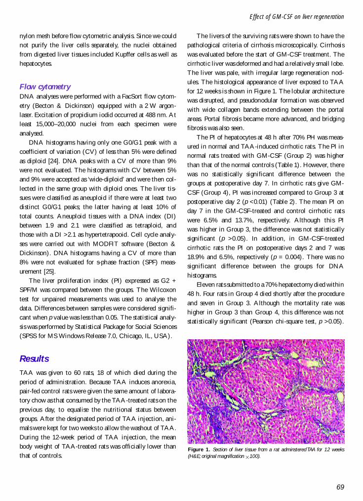
The livers of the surviving rats were shown to have thepathological criteria of cirrhosis microscopically. Cirrhosiswas evaluated before the start of GM-CSF treatment. Thecirrhotic liver was deformed and had a relatively small lobe.The liver was pale, with irregular large regeneration nod-ules. The histological appearance of liver exposed to TAAfor 12 weeks is shown in Figure 1. The lobular architecturewas disrupted, and pseudonodular formation was observedwith wide collagen bands extending between the portalareas. Portal fibrosis became more advanced, and bridgingfibrosis was also seen.
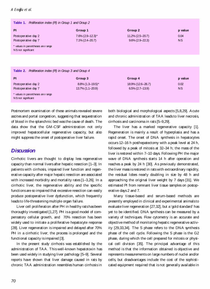
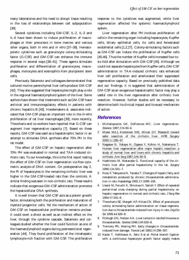
The PI of hepatocytes at 48 h after 70% PH was meas-ured in normal and TAA-induced cirrhotic rats. The PI innormal rats treated with GM-CSF (Group 2) was higherthan that of the normal controls (Table 1). However, therewas no statistically significant difference between thegroups at postoperative day 7. In cirrhotic rats give GM-CSF (Group 4), PI was increased compared to Group 3 atpostoperative day 2 (p <0.01) (Table 2). The mean PI onday 7 in the GM-CSF-treated and control cirrhotic ratswere 6.5% and 13.7%, respectively. Although this PIwas higher in Group 3, the difference was not statisticallysignificant (p >0.05). In addition, in GM-CSF-treatedcirrhotic rats the PI on postoperative days 2 and 7 was18.9% and 6.5%, respectively (p = 0.004). There was nosignificant difference between the groups for DNAhistograms.
Eleven rats submitted to a 70% hepatectomy died within48 h. Four rats in Group 4 died shortly after the procedureand seven in Group 3. Although the mortality rate washigher in Group 3 than Group 4, this difference was notstatistically significant (Pearson chi-square test, p >0.05).
69
Effect of GM-CSF on liver regeneration
Figure 1. Section of liver tissue from a rat adminstered TAA for 12 weeks(H&E; original magnification 3100).
Postmortem examination of these animals revealed severeascites and portal congestion, suggesting that sequestrationof blood in the splanchnic bed was the cause of death. Thedata show that the GM-CSF administration not onlyimproved hepatocellular regenerative capacity, but alsomight suppress the onset of postoperative liver failure.
DiscussionCirrhotic livers are thought to display less regenerativecapacity than normal livers after hepatic resection [1–3]. Inpatients with cirrhosis, impaired liver function and regen-erative capacity after major hepatic resection are associatedwith increased morbidity and mortality rates [1–3,26]. In acirrhotic liver, the regenerative ability and the specificfunctions are so impaired that excessive resection can easilyproduce postoperative liver dysfunction, which frequentlyleads to life-threatening multiple organ failure.
Liver cell proliferation after PH in healthy rats has beenthoroughly investigated [1,27]. PH is a good model of com-pensatory cellular growth, and 70% resection has beenwidely used to initiate a proliferative hepatocyte response[28]. Liver regeneration is impaired and delayed after 70%PH in a cirrhotic liver; the process is prolonged and thefunctional capacity is impaired [3].
In the present study cirrhosis was established by theadministration of TAA. This well-known hepatotoxin hasbeen used widely in studying liver pathology [5–9]. Severalreports have shown that liver damage caused in rats bychronic TAA administration resembles human cirrhosis in
both biological and morphological aspects [5,8,29]. Acuteand chronic administration of TAA leads to liver necrosis,cirrhosis and carcinoma in rats [5–9,29].
The liver has a marked regenerative capacity [1].Regeneration is mainly a result of hyperplasia and has arapid onset. The onset of DNA synthesis in hepatocytesoccurs 12–16 h posthepatectomy with a peak level at 24 h,followed by a peak of mitosis at 32–34 h; the mass of theliver is restored within 7–10 days. Following PH the majorwave of DNA synthesis starts 14 h after operation andreaches a peak by 24 h [30]. As previously demonstrated,the liver mass is restored in rats with extraordinary rapidity,the residual lobes nearly doubling in size by 48 h andapproaching the original liver weight by 7 days [31]. Weestimated PI from remnant liver tissue samples on postop-erative days 2 and 7.
Many tissue-based and serum-based methods arepresently employed in clinical and experimental animals toevaluate liver regeneration [27,32], but a ‘gold standard’ hasyet to be identified. DNA synthesis can be measured by avariety of techniques. Flow cytometry is an accurate andobjective method of monitoring hepatic regenerative activ-ity [29,33,34]. The S phase refers to the DNA synthesisphase of the cell cycle. Following the S phase is the G2phase, during which the cell prepared for mitosis or physi-cal cell division [35]. The principal advantage of thismethod is that the information obtained is objective andrepresents measurements on large numbers of nuclei and/orcells, but disadvantages include the cost of the sophisti-cated equipment required that is not generally available in
A ErogÏlu et al.
70
Table 2. Proliferation index (PI) in Group 3 and Group 4
PI Group 3 Group 4 p value
Postoperative day 2 8.8% (1.3–19.5)* 18.9% (12.6–26.7) 0.02Postoperative day 7 13.7% (1.1–20.9) 6.5% (2.7–13.9) NS
* values in parentheses are rangeNS: not significant
Table 1. Proliferation index (PI) in Group 1 and Group 2
PI Group 1 Group 2 p value
Postoperative day 2 7.6% (2.9–12.3)* 11.2% (2.5–20.7) 0.04Postoperative day 7 7.1% (2.4–20.7) 9.6% (2.9–22.3) NS
* values in parentheses are rangeNS: not significant
many laboratories and the need to disrupt tissue resultingin the loss of relationships between cell subpopulation[36].
Several cytokines including GM-CSF, IL-2, IL-3 andIL-6 have been shown to induce proliferation of macro-phages or their precursors in bone marrow and variousother organs, both in vivo and in vitro [37–39]. Hemato-poietic cytokines such as granulocyte colony-stimulatingfactor (G-CSF) and GM-CSF can enhance the immuneresponse in several ways [38–41]. These agents stimulateproliferation and differentiation of granulocytes, macro-phages, monocytes and eosinophils from pluripotent stemcells.
Previously Sakamoto and colleagues demonstrated thatcultured murine parenchymal liver cells produce GM-CSF[42]. They also suggested that hepatocytes might play a rolein the regional haematolymphoid system of the liver. Someauthors have shown that treatments such as GM-CSF haveantiviral and immunoregulatory effects in patients withchronic hepatitis B [43]. Hoedemakers & associates indi-cated that GM-CSF plays an important role in the in-vitroproliferation of rat liver macrophages [39]; more recently,Theocharis and co-workers have reported that G-CSF canaugment liver regenerative capacity [7]. Based on thesereports, GM-CSF was used as a hepatotrophic factor in anattempt to stimulate DNA synthesis after 70% PH in therat model.
The effect of GM-CSF on hepatic regeneration after70% PH was evaluated in normal and TAA-induced cir-rhotic rats. To our knowledge, this is the first report testingthe effect of GM-CSF on liver regeneration via flow cyto-metric analysis of DNA content. On postoperative day 2,the PI of hepatocytes in the remaining cirrhotic liver washigher in the GM-CSF-treated rats than the controls. Asimilar finding was seen in non-cirrhotic rats. These resultsindicate that exogenous GM-CSF administration promotesthe hepatocellular DNA synthesis.
It is well known that GM-CSF acts as a potent growthfactor, stimulating both the proliferation and maturation ofmyeloid progenitor cells. Yet the mechanism of action ofGM-CSF on hepatocellular proliferation remains unclear.It could exert a direct as well as an indirect effect on theliver, through the cytokine cascade. Sakamoto and col-leagues studied whether the liver could function as one ofthe haematolymphoid organs during postresectional regen-eration [44]. They found proliferation of the intrahepaticlymphocyte-rich fraction with GM-CSF. The proliferative
response to the cytokines was augmented, while liverregeneration affected the systemic haematolymphoidsystem.
Liver regeneration after PH involves proliferation ofcells in the remaining organ including hepatocytes, Kupffercells, biliary epithelial cells, Ito cells and fenestratedendothelial cells [1,2,27]. Colony-stimulating factors suchas GM-CSF can induce the proliferation of Kupffer cells[45,46]. Thus the number of Kupffer cells increased four-to-six-fold after stimulation with GM-CSF [45]. Although wecould not separate hepatocytes from Kupffer cells, GM-CSFadministration in TAA-induced cirrhotic rats enhancedliver cell proliferation and ameliorated their suppressedregenerative capacity. Based on previously published dataand our findings, it is suggested that administration ofGM-CSF as an exogenous hepatotrophic factor may play auseful role in the liver regeneration after cirrhotic liverresection. However, further studies will be necessary todetermine both its clinical impact and its exact mechanismof action.
References1 Michalopoulos GK, DeFrances MC. Liver regeneration.
Science, 1997;276:60–6.2 Moser MAJ, Kneteman NM, Minuk GY. Research toward
safer resection of the cirrhotic liver. HPB Surgery2000;11:285–97.
3 Nagasue N, Yukaya H, Ogawa Y, Kohno H, Nakamura T.Human liver regeneration after major hepatic resection: astudy of normal liver and livers with chronic hepatitis andcirrhosis. Ann Surg 1987;206:30–9.
4 Hashimoto M, Watanabe G. Functional capacity of the cir-rhotic liver after partial hepatectomy in the rat. Surgery1999;126:541–7.
5 Nozu F, Takeyama N, Tanaka T. Changes of hepatic fatty acidmetabolism produced by chronic thioacetamide administra-tion in rats. Hepatology 1992;15:1099–106.
6 Usami M, Furuchi K, Shiroiwa H, Saitoh Y. Effect of repeatedportal-triad cross clamping during partial hepatectomy onhepatic regeneration in normal and cirrhotic rats. J Surg Res1994;57:541–8.
7 Theocharis SE, Margeli AP, Kittas CN. Effect of granulocytecolony stimulating factor administration on tissue regenera-tion due to thioacetamide-induced liver injury in rats. Dig DisSci 1999;44:626–8.
8 Fitzhugh OG, Nelson AA. Liver tumors in rats fed thiourea orthioacetamide. Science 1948;108:626–8.
9 Trennery PN, Waring RH. Early changes in thioacetamide-induced liver damage. Toxicol Lett 1983;19:299–307.
10 Kaido T, Yoshikawa A, Seto S et al. Portal branch ligationwith a continuous hepatocyte growth factor supply makes
71
Effect of GM-CSF on liver regeneration
extensive hepatectomy possible in cirrhotic rats. Hepatology1998;28:756–60.
11 Yamamoto K, Takenaka K, Matsumata T, Shimada M,Sugimachi K. The effect of octreotide on morphologicalhepatic regeneration and hepatic functional recovery after atwo-thirds hepatectomy in rats. Hepatogastroenterology1999;46:1880–4.
12 Theocharis SE, Agapitos EB, Margeli AP et al. Effect of twoforms of granulocyte-colony-stimulating factor on hepaticregeneration after 70% partial hepatectomy in rats. Clin Sci1997;92:315–20.
13 Hashimoto M, Kothary PC, Eckhauser FE, Raper SE.Treatment of cirrhotic rats with epidermal growth factor andinsulin accelerates liver DNA synthesis after partial hepatec-tomy. J Gastroenterol Hepatol 1998;13:1259–65.
14 Shimada M, Matsumata T, Yamamoto K et al. The role ofgrowth hormone, somatostatin and glucagon in hepaticresection. Hepatogastroenterology 1998;45:178–83.
15 Fujiwara K, Nagoshi S, Ohno A et al. Stimulation of livergrowth by exogenous human hepatocyte growth factor innormal and partially hepatectomized rats. Hepatology1993;18:1443–9.
16 Rubin JS, Bottaro DP, Aaronso SA. Hepatocyte growth fac-tor/scatter factor and its receptor, the c-met protooncogenproduct. Biochim Biophy Acta 1993;1155:357–71.
17 Fausto N, Laird AD, Webber EM. Liver regeneration 2. Roleof growth factors and cytokines in hepatic regeneration.FASEB J 1995;9:1527–36.
18 Diehl AM, Rai RM. Liver regeneration 3: Regulation of signaltransduction during liver regeneration. FASEB J 1996;10:215–27.
19 Metcalf D, Burgerss AW. Clonal analysis of progenitor cellcommitment to granulocyte or macrophage production. J CellPhysiol 1982;111:275–83.
20 Arnold F, O’Bnen J, Cherry G. Granulocyte monocyte-colonystimulating factor as an agent or wound healing. J Wound Care1995;4:400–02.
21 da Costa RM, Aniceto C, Jesus FM, Mendes M. Quick heal-ing of leg ulcers after molgramostim. Lancet 1994;344:481–2.
22 ErogÏ lu A, Kurtman C, Ayy õ ld õ z A, Karadayõ K, Demirci S.Effect of granulocyte-macrophage colony-stimulating factoron wound nitrite level in normal and irradiated rats. Med SciRes 1999;27:685–8.
23 Higgins GM, Anderson RM. Experimental pathology of theliver. Restoration of the liver of the white rat following partialsurgical removal. Arch Pathol 1931;12:186–201.
24 Benson NA, Braylan RC. Evaluation of sensitivity in DNAaneuploidy detection using a mathematical model. Cytometry1994;15:53–8.
25 Shankey TV, Rabinovitch PS, Bagwell B. Guidelines forimplementation of clinical DNA cytometry. Cytometry1993;14:472–7.
26 Kawasaki S, Imamura H, Bandai Y, Sanjo K, Idezuki Y. Directevidence for the intact hepatocyte theory in patients withliver cirrhosis. Gastroenterology 1992;102:1351–5.
27 Assay N, Minuk GY. Liver regeneration: methods formonitoring and their applications. J Hepatology 1997;26:945–52.
28 Michalopoulos GK. Liver regeneration: Molecular mecha-nisms of growth control. FASEB J 1990;4:176–87.
29 Zimmermann T, Franke H, Dargel R. Studies on lipid andlipoprotein metabolism in rat liver cirrhosis induced by differ-ent regimens of thioacetamide administration. Exp Pathol1986;30:109–17.
30 Fausto N, Mead J. Regulation of liver growth: protooncogenesand transforming growth factors. Lab Invest 1989;60:4–13.
31 Buchner NLR, Malt RA. Regeneration of Liver and Kidney.Boston Little, Brown & Co., 1971:23–53.
32 Theocharis SE, Skopelitou AS, Margeli AP, Pavlaki KJ, KittasC. Proliferating cell nuclear antigen (PCNA) expression inregenerating rat liver after partial hepatectomy. Dig Dis Sci1994;39:245–52.
33 Garcia RL, Coltrera MD, Gown AM. Analysis of proliferativegrade using anti-PCNA/cyclin monoclonal antibodies infixed, embedded tissues. Comparison with flow cytometricanalysis. Am J Pathol 1989;134:733–9.
34 Braylan RC. Atttibutes and applications of flow cytometry.Ann Clin Lab Sci 1983;13;379–84.
35 Vemuru RP, Aragona E, Gupta S. Analysis of hepatocellularproliferation: study of the archival liver tissue is facilitated byan endogenous marker of DNA replication. Hepatology1992;16:968–73.
36 Larsen JK. Cell proliferation: analysis by flow cytometry. NouvRev Fr Hematol 1992;34:317–35.
37 Metcalf D. The molecular control of cell division, differentia-tion, commitment and maturation in haemopoietic cells.Nature 1989;339:27–30.
38 Chen BD, Clark CR, Chou TH. Granulocyte/macrophagescolony-stimulating factor stimulates monocyte and tissuemacrophage proliferation and enhances their responsivenessto macrophage colony-stimulating factor. Blood 1988;71:997–1002.
39 Hoedemakers RMJ, Scherphof GL, Daemen T. Proliferationof rat liver macrophages in vitro: influence of hemopoieticgrowth factors. Hepatology 1994;19:666–74.
40 Metcalf D, Begley CG, Johnson GR et al. Biologic propertiesin vitro of a recombinant human granulocyte-macrophagecolony-stimulating factor. Blood 1986;67:37–45.
41 Groopman JE, Molina JM, Scadden DT. Hematopoieticgrowth factors. Biology and clinical applications N Engl J Med1989;321:1449–59.
42 Sakamoto T, Mabuchi A, Kuriya S. Production ofgranulocyte-macrophage colony-stimulating factor by adultmurine parenchymal liver cells (hepatocytes). Reg Immunol1990–91;3:260–7.
43 Kountouras J, Boura P, Tsapas G. In vivo effect of granulocyte-macrophage colony-stimulating factor and interferon combi-nation on monocyte-macrophage and T-lymphocyte functionsin chronic hepatitis B leukocytopenic patients. Hepato-gastroenterology 1998;45:2295–302.
A ErogÏlu et al.
72
44 Sakamoto T, Saizawa T, Mabuchi A et al. The liver as apotential hematolyphoid organ examined from modificationsoccurring in the systemic and intrahepatic hematolymphoidsystem during liver regeneration afte partial hepatectomy.Reg Immunol 1992;4:1–11.
45 Hashimoto S, Yamada M, Yanai N et al. Phenotypic changeand proliferation of murine Kupffer cells by colony-
stimulating factors. J Interferon Cytokine Res 1996;16:237–43.
46 Schuurman B, Heuff G, Beelen RH, Meyer S. Enhancedkilling capacity of human Kupffer cells after activation withhuman granulocyte/macrophage-colony-stimulating factorand interferon gamma. Cancer Immunol Immunother1994;39:179–84.
73
Effect of GM-CSF on liver regeneration
Related Documents