Riga, 2021 Comparison of the Results after Arthroscopically Assisted Surgeries of the Articular Distal Radius Fractures with Internal and External Fixation Methods Uldis Krustiņš Summary of the Doctoral Thesis for obtaining a doctoral degree (Ph.D.) Sector – Clinical Medicine Sub-Sector – Orthopaedics doi:10.25143/prom-rsu_2021-19_dts

Welcome message from author
This document is posted to help you gain knowledge. Please leave a comment to let me know what you think about it! Share it to your friends and learn new things together.
Transcript

Riga, 2021
Comparison of the Results after Arthroscopically Assisted Surgeries
of the Articular Distal Radius Fractures with Internal and External
Fixation Methods
Uldis Krustiņš
Summary of the Doctoral Thesis for obtaining a doctoral degree (Ph.D.)
Sector – Clinical MedicineSub-Sector – Orthopaedics
doi:10.25143/prom-rsu_2021-19_dts
Uldis Krustiņš
ORCID 0000-0001-7184-8605
Comparison of the Results
after Arthroscopically Assisted Surgeries
of the Articular Distal Radius Fractures
with Internal and External Fixation Methods
Summary of the Doctoral Thesis
for obtaining a doctoral degree “Doctor of Science (Ph.D.)”
Sector – Clinical Medicine
Sub-Sector – Orthopaedics
Riga, 2021

The Doctoral Thesis was developed at the Department of Traumatology and
Orthopaedics, Rīga Stradiņš University, Latvia, in collaboration with Riga East
Clinical University Hospital “Gaiļezers” and Microsurgery Centre of Latvia
Scientific supervisor:
Dr.med., Associate Professor Andris Jumtiņš,
Rīga Stradiņš University, Latvia
Scientific consultant:
Dr. med., Mārtiņš Kapickis,
Microsurgery Centre of Latvia
Official reviewers:
Dr. med., Associate Professor Pēteris Studers,
Rīga Stradiņš Universitāte, Joint Laboratory of Traumatology
and Orthopaedics, Latvia
Dr. med., Professor Konstantīns Kalnbērzs,
Medical Faculty of University of Latvia
Dr. habil. med., Professor Narunas Porvaneckas,
Vilnius University, Faculty of Medicine, Lithuania
Defence of the Doctoral Thesis will take place at the public session of the
Promotion Council of Clinical Medicine on 15 December 2021 at 12.00 online
via Zoom platform.
The Doctoral Thesis is available in the RSU library and on RSU webpage:
https://www.rsu.lv/en/dissertations
Secretary of the Doctoral Council:
Dr.med., Assistant Professor Ruta Jakušonoka

3
Table of Contents
Abbreviations .................................................................................................... 4 Introduction ...................................................................................................... 6 1 Research section ........................................................................................ 12
1.1 Structure of the research ................................................................... 12 1.2 Surgical protocol for patients treated with VLP ............................... 15 1.3 Surgical protocol for patients treated with EF and K-wires .............. 17 1.4 Post-operative protocols ................................................................... 18 1.5 Primary acquisition methods and secondary data sources ................ 19
2 Results ....................................................................................................... 21 2.1 General data ...................................................................................... 21 2.2 Statistical processing of study data ................................................... 23 2.3 Results of objective measurements ................................................... 23
2.3.1 Wrist flexion .......................................................................... 24 2.3.2 Wrist extension ...................................................................... 27 2.3.3 Wrist radial deviation ............................................................ 29
2.3.4 Wrist ulnar deviation ............................................................. 32
2.3.5 Wrist pronation ...................................................................... 35
2.3.6 Wrist supination .................................................................... 38
2.3.7 Grip force .............................................................................. 41
2.3.8 Key pinch force ..................................................................... 44
2.3.9 Tripod pinch force ................................................................. 47
2.4 Subjective scales: .............................................................................. 50 2.4.1 PRWE .................................................................................... 50 2.4.2 MASS07 ................................................................................ 53 2.4.3 Modified Gartland and Werley scale ..................................... 56
2.5 Associated injuries ............................................................................ 63 2.6 Complications ................................................................................... 63
3 Discussion ................................................................................................. 67 3.1 About external fixator ....................................................................... 68 3.2 About VLP ........................................................................................ 69 3.3 Comparison of both methods ............................................................ 71 3.4 Arthroscopy and distal radius fractures ............................................ 75 3.5 Evaluation of the results ................................................................... 83
Conclusions .................................................................................................... 94 Publications .................................................................................................... 97 References ...................................................................................................... 99 Acknowledgements ....................................................................................... 105

4
Abbreviations
AO Arbeitsgemeinschaft für Osteosynthesefragen
APL m. abductor pollicis longus
CRPS Complex Regional Pain Syndrome
DDRU dorsal distal radio-ulnar portal
DIC dorsal intercarpal ligament
DOA deformative osteoarthrosis
DRCL dorsal radio-carpal ligament
DRF distal radius fracture
DRT dorsal radio-triquetral ligament
DRUJ distal radio-ulnar joint
ECRB m. extensor carpi radialis brevis
ECRL m. extensor carpi radialis longus,
ECU m. extensor carpi ulnaris
EDC m. extensor digitorum communis
EDM m. extensor digiti minimi
EF external fixator
EPL m. extensor pollicis longus
FCR m. flexor carpi radialis
LRL long radio-lunate ligament
LTIL luno-triquetral interosseus ligament
MASS07 Modern Activity Subjective Survey of 2007 score
MC metacarpal bone
MCID minimal clinically important difference
MCR midcarpal radial portal
MCU midcarpal ulnar portal
MRI magnetic resonance imaging

5
N newton (unit)
ORIF Open Reduction and Internal Fixation
PDRU proximal distal radio-ulnar portal
PRUJ proximal radio-ulnar joint
PRWE Patient Related Wrist Evaluation score
PQ m. pronator quadratus
RAKUS Riga East Clinical University hospital
ROM range of motion
RSC radio–scapho–capitate ligament
RSL radio-scapho-lunate ligament
RTG X-ray
RVP radial volar portal
SLAC scapho-lunate advanced collapse
SLIL scapho-lunate interosseus ligament
STT scaphotrapezium-trapezoideum (joint and portal)
TFCC triangular fibrocartilage complex
UVP ulnar volar portal
VLP volar locking plate

6
Introduction
Fractures of the distal end of the radius are the most common skeletal
injuries recorded in emergency rooms. The incidence is from 20 to 30% of all
fractures (Ilyas and Jupiter, 2007; MacIntyre and Dewan, 2016). These
fractures have bimodal age and gender distribution – complicated high energy
distal radius fractures are more common in younger males, but in older
population, even if the fractures are less complicated, they mostly are
experienced in older females (Court-Brown and Caesar, 2006). According to
published data of medical statistics, the incidence of all types of distal radius
fractures per 10.000 inhabitants in different countries shows one particular
trend: women, for various reasons, are more likely to experience injuries. For
example, the women – men ratio in Australia is 17 : 4 (Sanders et al., 1999),
South Korea 66.1 : 16.4 (Park et al., 2011), Netherlands 45.8 : 10 (de Putter
et al., 2013), Canada 49 : 14 (Jaglal et al., 2005), United Kingdom 36.8 : 9
(O'Neill et al., 2001), Norway 75.1 : 18.9 (Diamantopoulos et al., 2012),
Switzerland 63.2 : 17 (Lippuner, 2009). Overall, 15% of women and up to 2%
of men are at risk for the distal radius fracture during the lifetime. (Ruch and
Papadonikolakis, 2006).
Until now, any statistical studies of this kind have not been carried out
in Latvia. Assuming that the incidence of articular DRFs is 20% of all DRFs
treated in emergency departments, then roughly calculating the statistical data
for 2017 available from the Centre for Disease Prevention and Control, and
considering the global calculated percentage trend that AO-C3 fractures
constitute approximately 32% of all DRFs, theoretically there could be around
265 AO-C3 type DRF patients per year in Latvia.
7
The increasing incidence of these injuries may be attributed to an aging
population (osteoporotic fractures) and the growing participation in outdoor
pursuits (higher energy fractures) (Shukla et al., 2014). Nowadays the incidence
of articular distal radius fractures is from 32% (Koo et al., 2013) to 43.3%
(Sander et al., 2018) of all distal radius fractures. Activity of the surgical
treatment is obviously increasing as a result of evolution of the implants and
technical possibilities.
AO (Arbeitsgemeinschaft für Osteosynthesefragen) fracture
classification system, which is used in Latvia since 1998, has also undergone
a number of reviews of treatment criteria and changes of standards
(Walenkamp et al., 2015). Nowadays, the traditional treatment of distal radius
fractures includes 3 or 4 different fixation methods – K-wires and external
fixator (EF), micronails, volar compression plates (VLP), fragment specific
plates and spanning plates. Volar locking plates (VLP) and EF + K-wires have
been generally used in the Microsurgery Centre of Latvia for several years.
Both of these two methods are controversial in the technical meaning (surgical
approach and extra traumatization of soft tissues) as well as in different post-
operative rehabilitation protocols. Despite the numerous comparative studies on
the application of both treatment methods and their results, as well as the
evidence of possibilities of better joint surface reconstruction possibilities in
arthroscopic assisted surgeries, until now, two diametrically opposed surgical
treatment methods have not been compared to arthroscopically assisted study
groups. This study was motivated by H. J. Kreder`s acknowledgment: “It is
neither the fixation nor the implant which dictates the outcome but the ability
of the surgeon to meet the goal of satisfactory reduction and vascular
preservation with the least invasive procedure possible.”

8
During the course of the study, surgical technique was improved,
reducing the timing of surgeries, as well as lessons have been learned about the
benefits of one or other of the surgical method, depending on the type and
configuration of the articular DRF. During this study, different associated soft
tissue injuries were recognized and immediately treated, which would not have
been possible without the arthroscopic assistance. Collection of post-operative
subjective and objective data was performed using PRWE score (Patient Related
Wrist Evaluation), MASSH07 score (Modern Activity Subjective Survey of 2007)
and Gartland & Werley score, which are adapted for interpretation and applied
in international publications (Alexander et al., 2008; Changulani et al., 2008;
MacDermid et al., 2003). An assessment of surgical notes, post-operative
rehabilitation protocols as well as records of subjective and objective outcomes,
justifies the use of both arthroscopic assisted articular DRF which would not have
been possible without treatment methods, evaluating the capabilities of technical
application, equipment as well as the knowledge and skills of the surgeon for the
specific manipulation.
Aim of the study
To compare two arthroscopically assisted surgical treatment methods of
the articular DRFs, according to their early and late clinical, radiological and
functional results, timing of surgery and potential complications. To develop
indications for the application of one or other treatment method depending of the
specific fracture, for a particular group of patients, forecasting potential results
and reducing the risk of potential complications in the future.
9
Tasks of the study
1. To perform a scheduled assessment of post-operative radiological and
functional results of the randomized groups based on assessment of
the patients’ health condition and life quality (Gartland and Werley,
PRWE and MASS07 scores), as well as X-ray controls 1, 3, 6 and 12
months after surgery.
2. To perform a monitoring of post-operative complications and analysis
of compared data in both randomized groups.
3. To determine the usefulness of the arthroscopic part of the surgery in
the treatment of the articular DRFs.
4. To create an algorithm for the uniform selection of the treatment
methods of the articular DRFs in any hospital in Latvia.
5. To create a systematized protocol of post-operative monitoring after
DRFs suitable to further academic studies in Latvia.
Scientific assumptions
Open reduction and internal fixation with plate and screws is mostly
recommended for younger patients with better bone structure, active life patterns
and longer life experience. Gentler fixation with K-wires in addition with joint
distraction in external fixator is recommended in elderly people with a weaker
bone structure with potentially possible migration of implants and lower life
activity. It is considered that arthroscopic assisted surgical treatment of articular
fractures in any localization, provides more accurate reposition of articular
fragments and does not create additional soft tissue injuries resulting from
visualization of joint surfaces by conventional surgical methods.

10
It could be possible, that arthroscopic assisted, less invasive method of
fracture fixation, in patients of any age, may lead to better conditions for
restoring the life quality of patients and wrist functions than open osteosynthesis
with plate, avoiding additional soft tissue injuries during the surgery.
Place of the study
Riga Stradiņš University, Riga Eastern Clinical University Hospital
“Gaiļezers”, Plastic, Reconstructive and Microsurgery centre of Latvia.
Scientific novelty
For the first time a study has been conducted with systemized monitoring
of patients, records of objective and subjective data (Gartland and Werley,
PRWE and MASS07 scores) and resulting analysis of obtained data after
arthroscopic assisted surgical treatment of articular DRFs. Two technically
controversial surgical techniques were compared – minimally invasive
arthroscopic assisted fracture fixation with K-wires and EF as well as standard
open reduction and arthroscopic assisted fixation with VLP.
The monitoring and evaluation system of outcomes used during this
study, is intended to be recommended for more extensive use, and processing
large amounts of data, it can be used not only for scientific publications, but also
for statistical and economic calculations.

11
Practical value of this study
1. The prototype of the surgical instrument “The device for
determination of the exact direction and depth of the K-wire in
arthroscopically assisted surgeries of the articular distal radius
fractures” has been designed and patented.
2. The necessity of the arthroscopic assistance for the exact reposition of
articular DRFs and for the diagnosis of associated soft tissue lesions
as well as for treatment of them has been proved.
3. A practical algorithm for the uniform selection of treatment methods
for articular DRFs has been created to be applied in any trauma and
orthopaedic department or hospital.
4. An algorithm for patient post-operative observation, as well as for the
recording of objective and subjective outcomes, has been introduced,
to be applied in any trauma and orthopaedics department or hospital.

12
1 Research section
1.1 Structure of the research
This is a prospective randomized trial where patients are divided into two
groups using the method of alternative allocation. Target population – persons of
both genders, at least 15 years old with articular distal radius fractures (AO
C1,C2 or C3 type) of the one wrist (the healthy wrist is necessary for functional
comparison). Fractures were classified according the AO classification according
to the primary X-rays, changing the group of classification if different type of
fracture was recognized during the surgery.
The number of participants required for the study and the statistical
capacity of the study was calculated on the basis of the data available in the
literature on hand force measurements (grip strength as the main outcome
measure) and the minimal clinically important difference (MCID) in PRWE
values. MCID for grip strength is 6.5 kg, or 19.5% for percentage grip strength
(Kim et al., 2014). According to these data, only 10 patients in each group would
constitute 80% of the capacity. More meaningful calculation on sample size is
based on the available data on PRWE, where MCID is 11 points and standard
deviation is 14 points (Walenkamp et al., 2015). In capacity calculation it was
determined that required total sample size is 50 patients (25 per group) to confirm
or reject the hypothesis. Assuming the drop-out rate of 30%, at least 71 patients
should be enrolled. Unfortunately no MCID have been established for wrist
ROM as well as for MASS07 and modified Gartland and Werley scores. The
sample size was calculated on the basis of 80% research capacity by the formulas
as follows (Fig. 1.1):
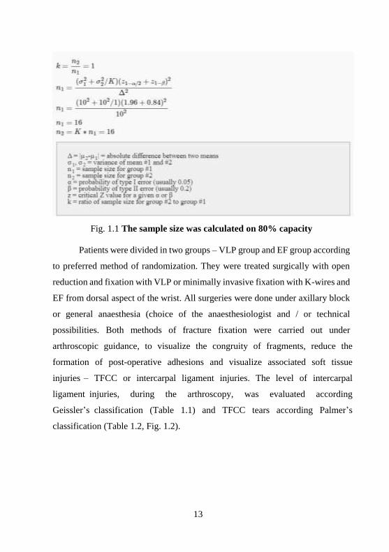
13
Fig. 1.1 The sample size was calculated on 80% capacity
Patients were divided in two groups – VLP group and EF group according
to preferred method of randomization. They were treated surgically with open
reduction and fixation with VLP or minimally invasive fixation with K-wires and
EF from dorsal aspect of the wrist. All surgeries were done under axillary block
or general anaesthesia (choice of the anaesthesiologist and / or technical
possibilities. Both methods of fracture fixation were carried out under
arthroscopic guidance, to visualize the congruity of fragments, reduce the
formation of post-operative adhesions and visualize associated soft tissue
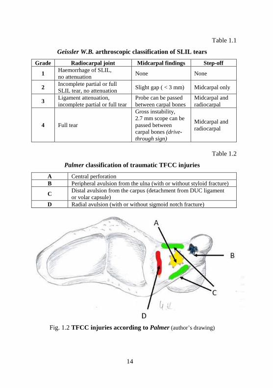
injuries – TFCC or intercarpal ligament injuries. The level of intercarpal
ligament injuries, during the arthroscopy, was evaluated according
Geissler’s classification (Table 1.1) and TFCC tears according Palmer’s
classification (Table 1.2, Fig. 1.2).
14
Table 1.1
Geissler W.B. arthroscopic classification of SLIL tears
Grade Radiocarpal joint Midcarpal findings Step-off
1 Haemorrhage of SLIL,
no attenuation None None
2 Incomplete partial or full
SLIL tear, no attenuation Slight gap ( < 3 mm) Midcarpal only
3 Ligament attenuation,
incomplete partial or full tear
Probe can be passed
between carpal bones
Midcarpal and
radiocarpal
4 Full tear
Gross instability,
2.7 mm scope can be
passed between
carpal bones (drive-
through sign)
Midcarpal and
radiocarpal
Table 1.2
Palmer classification of traumatic TFCC injuries
A Central perforation
B Peripheral avulsion from the ulna (with or without styloid fracture)
C Distal avulsion from the carpus (detachment from DUC ligament
or volar capsule)
D Radial avulsion (with or without sigmoid notch fracture)
Fig. 1.2 TFCC injuries according to Palmer (author’s drawing)

15
The permissible deviations were determined before the initiation of the
study – gap between fragments – less than 1mm, shortening of the radius – less
than 5 mm in comparison with the healthy hand, radial inclination angle > 15°,
volar tilt < −10°. As far as possible, all steps were taken to restore the anatomy
of the distal radius to the highest possible accuracy.
1.2 Surgical protocol for patients treated with VLP
Surgeries were performed under axillary block or general anaesthesia and
with the tourniquet inflated to between 280 and 320 mmHg. The volar locking
plate group (Group VLP) surgeries were performed using the flexor carpi radialis
approach and pronator quadratus muscle elevation. Fracture fixation was
achieved with two different plates: Synthes 2.4 mm LCP distal radius system or
Stryker VariAx plate. Once the fracture was preliminarily fixed with the plate,
the wrist joint was assessed arthroscopically using the 3−4 and 4–5 portals. In
several cases additional portals, 6U and 1–2, were used to remove blood clots
and small articular fragments. If articular step-offs or gaps were present,
additional reposition and fixation with K-wires were performed. Distal screws
were inserted only after arthroscopic inspection of the radiocarpal joint and a
fluoroscopic confirmation of the correct position for the screws. If dorsal, ulnar
or radial fragments, uncontrolled by the plate, were detected, additional K-wires
were inserted. These were cut under the skin and remained indwelling after the
procedure. Associated soft tissue injuries, such as triangular fibrocartilage
complex (TFCC) tear, damage of scapholunate or lunotriquetral ligaments, were
assessed after the fracture had been stabilized. In several cases, debridement of
the injured ligaments or TFCC was performed, as well as trans-articular fixation
of the scapholunate and / or lunotriquetral joints with K-wires, or application of
peripheral sutures for TFCC tears. Bone-grafting was not performed.

16
Fig. 1.3 Standard volar approach, preliminary fixation of VLP
with wires after the primary reposition
Fig. 1.4 Fixation of the wrist in the arthroscopic tower
and the arthroscopic stage of the surgery

17
If the required position of fracture fragments is achieved and position of
implants is optimal, wound is closed starting with suturing of the pronator
quadratus muscle and other soft tissues layer by layer. Active aspiration drainage
was used as well as short arm cast for two weeks after surgery.
1.3 Surgical protocol for patients treated with EF and K-wires
Surgeries were performed under axillary block or general anaesthesia and
with the tourniquet inflated to between 280 and 320 mmHg. The external fixator
and K-wire group (Group EF) surgeries commenced with a primary closed
reduction and fixation with several K-wires, under fluoroscopic guidance.
Following fixation in a traction tower, the articular surfaces were assessed using
the same arthroscopic technique as for Group VLP. Further fragment reductions
were performed, if required, using a probe or K-wires as joysticks through
elongated 3–4, 4–5, 1–2 and in some cases, volar portals. Additional K-wire
fixation was used as required. At this point, associated soft tissue injuries were
assessed and additional procedures were performed as for Group VLP, when
necessary. Once satisfactory reposition was achieved, the bridging external
fixator was applied. Both Synthes Small External Fixator and Stryker Hoffmann
II Compact External Fixator were used. For this purpose, two threaded pins were
inserted in the dorso-radial aspect of the 2nd MC bone and two additional threaded
pins in the dorso-radial aspect of the radius, proximally from fracture site. These
wires were fixated together with connecting rods. The wrist was only then
released from the traction, K-wires were cut under the skin and the wounds were
closed with simple interrupted sutures.

18
All arthroscopically assisted surgeries were performed using the dry
arthroscopy technique recommended by Francisco del Piñal (Del Piñal, 2011).
Fig. 1.5 EF application after arthroscopic fracture
fixation with wires
1.4 Post-operative protocols
For patients treated with VLP:
1 Next day after surgery – removal of the active aspiration drainage and
change of the wound dressing, discharge from the hospital on the same
or following day.
2 Change of the wound dressing in 3 to 4 days.
3 Removal of sutures in 12 to 14 days after surgery.

19
4 Immobilization until removal of sutures.
5 Active movements of shoulder, elbow and finger joints according to
standardized protocol starting the next day after surgery.
6 Workout of the wrist joint active and passive movements under the
guidance of the hand therapist, starting the 3rd week after surgery.
7 X-ray control 4 weeks after the surgery.
8 Scheduled visits 1, 3, 6 and 12 months after surgery.
For patients treated with EF and K-wires:
1 Change of the wound dressing and discharge from the hospital at the
same or following day
2 Change of the wound dressing in 3 to 4 days.
3 Active movements in shoulder, elbow and finger joints according to
standardized protocol starting the next day after surgery.
4 Removal of the sutures in 12 to 14 days after the surgery.
5 X-ray control 4 weeks after the surgery.
6 Removal of the EF and K-wires 4 to 6 weeks after the surgery.
7 Workout of the wrist joint active and passive movements under the
guidance of the hand therapist, after the removal of the implants.
8 Scheduled visits 1, 3, 6 and 12 months after surgery.
1.5 Primary acquisition methods and secondary data sources
The results of treatment were assessed with X ray examinations postero-
anterior position in a 10° tilted-view and lateral position in a 20° tilted-view,
subjective evaluation using the Patient-Rated Wrist Evaluation (PRWE) score
(rating from 0 to 140, with a lower score representing a better result), Modern
Activity Subjective Survey of 2007 (MASS07) score (rating from 0 to 100, with
a lower score representing a better result), and subjective and objective

20
evaluation using the Gartland and Werley score (rating from 17.5 to 100, with
a higher score representing a better result). X-ray assessment was performed by
an independent radiologist as the Gartland and Werley score includes
a radiological assessment of fracture consolidation and ulnar variance.
The results of the treatment were recorded at every visit:
1 Visual evaluation, assessment of the objective data – Grip / pinch /
tripod-pinch strength and range of motion (ROM) were measured.
Wrist mobility was tested using a goniometer, grip strength with
Jamar dynamometer, and pinch and three-point strength with
a pinch gauge as well as X ray examinations postero-anterior position
in a 10° tilted-view and lateral position in a 20° tilted-view were
evaluated by independent radiologist.
2 Subjective / objective evaluation using the Patient-Rated Wrist
Evaluation (PRWE) score (rating from 0 to 140, with a lower score
representing a better result), Modern Activity Subjective Survey of
2007 (MASS07) score (rating from 0 to 100, with a lower score
representing a better result), and subjective and objective evaluation
using the Gartland and Werley score (rating from 17.5 to 100, with
a higher score representing a better result) was also performed.
21
2 Results
2.1 General data
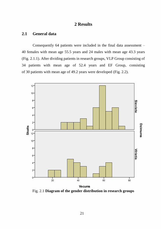
Consequently 64 patients were included in the final data assessment –
40 females with mean age 55.5 years and 24 males with mean age 43.3 years
(Fig. 2.1.1). After dividing patients in research groups, VLP Group consisting of
34 patients with mean age of 52.4 years and EF Group, consisting
of 30 patients with mean age of 49.2 years were developed (Fig. 2.2).
Fig. 2.1 Diagram of the gender distribution in research groups
22
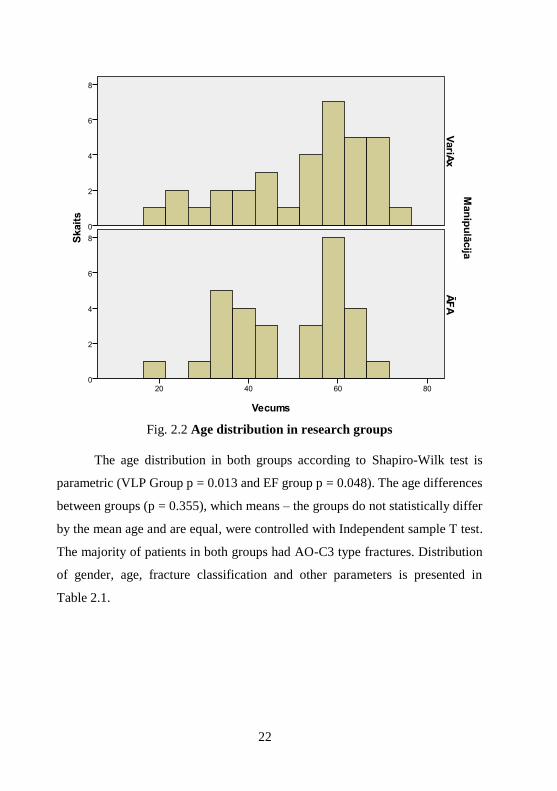
Fig. 2.2 Age distribution in research groups
The age distribution in both groups according to Shapiro-Wilk test is
parametric (VLP Group p = 0.013 and EF group p = 0.048). The age differences
between groups (p = 0.355), which means – the groups do not statistically differ
by the mean age and are equal, were controlled with Independent sample T test.
The majority of patients in both groups had AO-C3 type fractures. Distribution
of gender, age, fracture classification and other parameters is presented in
Table 2.1.

23
Table 2.1
Description of the groups under study
VLP group EF group P-value
Number of patients 34 30 –
Males / Females 11 / 23 13 / 17 0.365
Age (years) 52.4 ± 14.7 49.2 ± 13.0 0.355
Fractured side right / left 9 / 25 12 / 18 0.250
Dominant / not dominant wrist 10 / 24 10 / 20 –
AO – C1 11.8% 6.7% –
AO – C2 26.5% 20.0% –
AO – C3 58.8% 73.3% –
High energy trauma 3 5 0.334
Proc. styloideus ulnae fracture 41.1% 56.7% 0.443
Associated soft tissue injuries
recorded 61.8% 66.7% 0.683
2.2 Statistical processing of study data
Patient population data of both study groups, as well as data derived from
study measurements were classified in Microsoft Excel data processing program.
Statistical processing of clinical data has been carried out using the SPSS 20
(Statistical Package for the Social Sciences) program ‒ forecasting analytic and
statistical analysis software package. Interconnection of data searched with non-
parametric tests and correlation analysis methods. In all cases, a relevance level
has been used to assess statistical hypotheses (p ≤ 0,05 to approve, p > 0,05 to
decline).
2.3 Results of objective measurements
All wrist ROM and grip force measurements show the volume of force or
movements (%) compared to the healthy wrist in 1, 3, 6 and 12 months after
surgery. Since it was impossible to prove that results obtained correspond to
normal distribution (which can also be seen from histograms), only non-
24
parametric tests were used to compile and evaluate the results (Mann-Whitney
and Kolmogorov-Smirnov tests to analyse differences in measurements between
control groups and Wilcoxon Signed Ranks and Sign tests, to show changes in
measurement results depending on the time after the surgery).
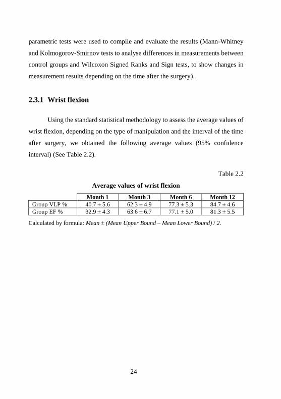
2.3.1 Wrist flexion
Using the standard statistical methodology to assess the average values of
wrist flexion, depending on the type of manipulation and the interval of the time
after surgery, we obtained the following average values (95% confidence
interval) (See Table 2.2).
Table 2.2
Average values of wrist flexion
Month 1 Month 3 Month 6 Month 12
Group VLP % 40.7 ± 5.6 62.3 ± 4.9 77.3 ± 5.3 84.7 ± 4.6
Group EF % 32.9 ± 4.3 63.6 ± 6.7 77.1 ± 5.0 81.3 ± 5.5
Calculated by formula: Mean ± (Mean Upper Bound – Mean Lower Bound) / 2.

25
Visualization of measurement results:
Fig. 2.3 Value of the wrist flexion according to type of manipulation
and time interval after the surgery
Mann-Whitney and Kolmogorov-Smirnov tests were used to analyse
whether wrist flexion measurements have statistically significant differences
between control groups depending on the time after surgery. The following
results were obtained – the statistically significant differences in the values of
wrist flexion parameter depending on a type of manipulation have been found
only at month 1 (p < 0.05) after surgery (See Table 2.3).
26
Table 2.3
Differences in the values of wrist flexion parameter depending
on a type of manipulation
Month 1 Month 3 Month 6 Month 12
There are statistically
significant differences
between Group VLP
and Group EF
Yes No No No
p-value (Mann-Whitney test) 0.048 0.731 0.819 0.325
p-value (Kolmogorov-
Smirnov test) 0.303 0.995 0.999 0.717
P-value match with Asymp. Sig. (2-tailed) in calculation materials. If a single p-value
in a column is less than 0.05, write “Yes”, otherwise write “No”.
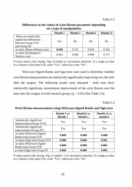
Wilcoxon Signed Ranks and Sign tests were used to determine whether
wrist flexion measurements are statistically significantly improving over the time
after the surgery. The following results were obtained – both tests show
statistically significant, monotonous improvement of the wrist flexion over the
time after the surgery in both research groups (p < 0.01) (See Table 2.4).
Table 2.4
Wrist flexion measurements using Wilcoxon Signed Ranks and Sign tests
Month 3 vs.
Month 1
Month 6 vs.
Month 3
Month 12 vs
month 6
Statistically significant
improvement (Group VLP) Yes Yes Yes
Statistically significant
improvement (Group EF) Yes Yes Yes
p-value (Wilcoxon Signed
Ranks test) Group VLP 0.000 0.000 0.000
p-value (Sign test) Group VLP 0.000 0.000 0.001
p-value (Wilcoxon Signed
Ranks test) Group VLP 0.000 0.000 0.001
p-value (Sign test) Group EF 0.000 0.000 0.003
P-value match with Asymp. Sig. (2-tailed) / 2 in calculation materials. If a single p-value
in a column is less than 0.05, write “Yes”, otherwise write “No”.
27
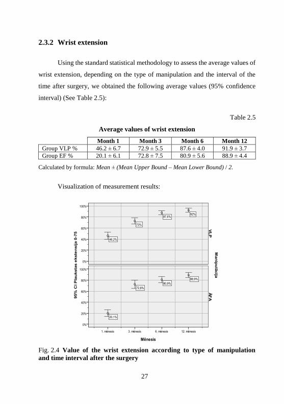
2.3.2 Wrist extension
Using the standard statistical methodology to assess the average values of
wrist extension, depending on the type of manipulation and the interval of the
time after surgery, we obtained the following average values (95% confidence
interval) (See Table 2.5):
Table 2.5
Average values of wrist extension
Month 1 Month 3 Month 6 Month 12
Group VLP % 46.2 ± 6.7 72.9 ± 5.5 87.6 ± 4.0 91.9 ± 3.7
Group EF % 20.1 ± 6.1 72.8 ± 7.5 80.9 ± 5.6 88.9 ± 4.4
Calculated by formula: Mean ± (Mean Upper Bound – Mean Lower Bound) / 2.
Visualization of measurement results:
Fig. 2.4 Value of the wrist extension according to type of manipulation
and time interval after the surgery

28
Mann-Whitney and Kolmogorov-Smirnov tests were used to analyse
whether wrist extension measurements have statistically significant differences
between control groups depending on the time after surgery. The following
results were obtained – the statistically significant differences in the values of
wrist extension parameter depending on a type of manipulation have been found
only at month 1 (p < 0.05) after surgery (See Table 2.6).
Table 2.6
Wrist extension measurements using Mann-Whitney
and Kolmogorov-Smirnov tests
Month 1 Month 3 Month 6 Month 12
There are statistically
significant differences
between Group VLP and
Group EF
Yes No No No
p-value (Mann-Whitney test) 0.000 0.995 0.056 0.324
p-value (Kolmogorov-
Smirnov test) 0.000 0.472 0.181 0.403
P-value match with Asymp. Sig. (2-tailed) in calculation materials. If a single p-value
in a column is less than 0.05, write “Yes”, otherwise write “No”.
Wilcoxon Signed Ranks and Sign tests were used to determine whether
wrist extension measurements are statistically significantly improving over the
time after the surgery. The following results were obtained – both tests show
statistically significant, monotonous improvement of wrist extension over the
time after the surgery in both research groups (p < 0.01) (See Table 2.7).
29
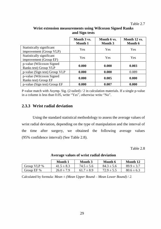
Table 2.7
Wrist extension measurements using Wilcoxon Signed Ranks
and Sign tests
Month 3 vs.
Month 1
Month 6 vs.
Month 3
Month 12 vs.
Month 6
Statistically significant
improvement (Group VLP) Yes Yes Yes
Statistically significant
improvement (Group EF) Yes Yes Yes
p-value (Wilcoxon Signed
Ranks test) Group VLP 0.000 0.000 0.003
p-value (Sign test) Group VLP 0.000 0.000 0.089
p-value (Wilcoxon Signed
Ranks test) Group EF 0.000 0.005 0.000
p-value (Sign test) Group EF 0.000 0.007 0.000
P-value match with Asymp. Sig. (2-tailed) / 2 in calculation materials. If a single p-value
in a column is less than 0.05, write “Yes”, otherwise write “No”.
2.3.3 Wrist radial deviation
Using the standard statistical methodology to assess the average values of
wrist radial deviation, depending on the type of manipulation and the interval of
the time after surgery, we obtained the following average values
(95% confidence interval) (See Table 2.8).
Table 2.8
Average values of wrist radial deviation
Month 1 Month 3 Month 6 Month 12
Group VLP % 41.5 ± 8.3 74.5 ± 5.6 84.3 ± 5.6 89.9 ± 3.7
Group EF % 26.0 ± 7.9 61.7 ± 8.9 72.9 ± 5.5 80.6 ± 6.3
Calculated by formula: Mean ± (Mean Upper Bound – Mean Lower Bound) / 2.

30
Visualization of measurement results:
Fig. 2.5 Value of the wrist radial deviation according to type
of manipulation and time interval after the surgery
Mann-Whitney and Kolmogorov-Smirnov tests were used to analyse
whether wrist radial deviation measurements have statistically significant
differences between control groups depending on the time after surgery. The
following results were obtained – the statistically significant differences in the
values of wrist radial deviation parameter depending on a type of manipulation
have been found in all measurements (p < 0.05) after surgery (See Table 2.9).

31
Table 2.9
Wrist radial deviation measurements using Mann-Whitney
and Kolmogorov-Smirnov tests
Month 1 Month 3 Month 6 Month 12
There are statistically
significant differences
between Group VLP
and Group EF
Yes Yes Yes Yes
p-value (Mann-Whitney test) 0.003 0.014 0.009 0.036
p-value (Kolmogorov-
Smirnov test) 0.012 0.071 0.012 0.098
P-value match with Asymp. Sig. (2-tailed) in calculation materials. If a single p-value
in a column is less than 0.05, write “Yes”, otherwise write “No”.
Wilcoxon Signed Ranks and Sign tests were used to determine whether
wrist radial deviation measurements are statistically significantly improving over
the time after the surgery. The following results were obtained – both tests show
statistically significant, monotonous improvement of wrist radial deviation over
the time after the surgery in both research groups (p < 0.01) (See Table 2.10).
Table 2.10
Wrist radial deviation measurements using
Wilcoxon Signed Ranks and Sign tests
Month 3 vs.
Month 1
Month 6 vs.
Month 3
Month 12
vs. Month 6
Statistically significant
improvement (Group VLP) Yes Yes Yes
Statistically significant
improvement (Group EF) Yes Yes Yes
p-value (Wilcoxon Signed Ranks
test) Group VLP 0.000 0.001 0.058

32
Table 2.10 continued
Month 3 vs.
Month 1
Month 6 vs.
Month 3
Month 12 vs.
Month 6
p-value (Sign test) Group VLP 0.000 0.001 0.043
p-value (Wilcoxon Signed
Ranks test) Group EF 0.000 0.003 0.038
p-value (Sign test) Group EF 0.000 0.011 0.021
P-value match with Asymp. Sig. (2-tailed) / 2 in calculation materials. If a single p-value
in a column is less than 0.05, write “Yes”, otherwise write “No”.
2.3.4 Wrist ulnar deviation
Using the standard statistical methodology to assess the average values of
wrist ulnar deviation, depending on the type of manipulation and the interval of
the time after surgery, we obtained the following average values
(95% confidence interval) (See Table 2.11).
Table 2.11
Average values of wrist ulnar deviation
Month 1 Month 3 Month 6 Month 12
Group VLP % 41.8 ± 8.9 70.1 ± 6.3 79.9 ± 5.5 87.8 ± 3.7
Group EF % 38.5 ± 8.7 64.9 ± 8.1 78.3 ± 6.4 88.0 ± 5.0
Calculated by formula: Mean ± (Mean Upper Bound – Mean Lower Bound) / 2.

33
Visualization of measurement results:
Fig. 2.6 Value of the wrist ulnar deviation according to type
of manipulation and time interval after the surgery
Mann-Whitney and Kolmogorov-Smirnov tests were used to analyse
whether wrist ulnar deviation measurements have statistically significant
differences between control groups depending on the time after surgery. The
following results were obtained – the statistically significant differences in the
values of wrist radial deviation parameter depending on a type of manipulation
have not been found in any measurements (p < 0.05) after surgery
(See Table 2.12).

34
Table 2.12
Wrist ulnar deviation measurements using Mann-Whitney
and Kolmogorov-Smirnov tests
Month 1 Month 3 Month 6 Month 12
There are statistically
significant differences
between Group VLP
and Group EF
No No No No
p-value (Mann-Whitney test) 0.536 0.284 0.655 0.686
p-value (Kolmogorov-
Smirnov test) 0.828 0.252 0.946 0.980
P-value match with Asymp. Sig. (2-tailed) in calculation materials. If a single p-value
in a column is less than 0.05, write “Yes”, otherwise write “No”.
Wilcoxon Signed Ranks and Sign tests were used to determine whether
wrist ulnar deviation measurements are statistically significantly improving over
the time after the surgery. The following results were obtained – both tests show
statistically significant, monotonous improvement of wrist ulnar deviation over
the time after the surgery in both research groups (p < 0.01) (See Table 2.13).
Table 2.13
Wrist ulnar deviation measurements using Wilcoxon Signed Ranks
and Sign tests
Month 3 vs.
Month 1
Month 6 vs.
Month 3
Month 12 vs.
Month 6
Statistically significant
improvement (Group VLP) Yes Yes Yes
Statistically significant
improvement (Group EF) Yes Yes Yes
p-value (Wilcoxon Signed Ranks
test) Group VLP 0.000 0.009 0.006
35
Table 2.13 continued
Month 3 vs.
Month 1
Month 6 vs.
Month 3
Month 12 vs.
Month 6
p-value (Sign test) Group VLP 0.000 0.072 0.045
p-value (Wilcoxon Signed Ranks
test) Group EF 0.000 0.003 0.000
p-value (Sign test) Group EF 0.001 0.063 0.000
P-value match with Asymp. Sig. (2-tailed) / 2 in calculation materials. If a single p-value
in a column is less than 0.05, write “Yes”, otherwise write “No”.
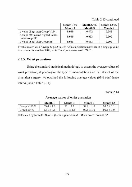
2.3.5. Wrist pronation
Using the standard statistical methodology to assess the average values of
wrist pronation, depending on the type of manipulation and the interval of the
time after surgery, we obtained the following average values (95% confidence
interval) (See Table 2.14).
Table 2.14
Average values of wrist pronation
Month 1 Month 3 Month 6 Month 12
Group VLP % 69.8 ± 7.0 92 ± 3.5 99.2 ± 1.0 99.5 ± 1.1
Group EF % 63.1 ± 7.5 91.1 ± 4.6 97.8 ± 1.6 99.3 ± 1.0
Calculated by formula: Mean ± (Mean Upper Bound – Mean Lower Bound) / 2.
36
Visualization of measurement results:
Fig.2.7 Value of the wrist pronation according to type of manipulation
and time interval after the surgery
Mann-Whitney and Kolmogorov-Smirnov tests were used to analyse
whether wrist pronation measurements have statistically significant differences
between control groups depending on the time after surgery. The following
results were obtained – the statistically significant differences in the values of
wrist pronation parameter depending on a type of manipulation have not been
found in any measurements (p < 0.05) after surgery (See Table 2.15).

37
Table 2.15
Wrist pronation measurements using Mann-Whitney
and Kolmogorov-Smirnov tests
Month 1 Month 3 Month 6 Month 12
There are statistically
significant differences
between Group VLP
and Group EF
No No No No
p-value (Mann-Whitney test) 0.214 0.955 0.117 0.509
p-value (Kolmogorov-
Smirnov test) 0.768 1.000 0.890 1.000
P-value match with Asymp. Sig. (2-tailed) in calculation materials. If a single p-value
in a column is less than 0.05, write “Yes”, otherwise write “No”.
Wilcoxon Signed Ranks and Sign tests were used to determine whether
wrist pronation measurements are statistically significantly improving over the
time after the surgery. The following results were obtained – both tests show
statistically significant, monotonous improvement of wrist pronation over the
time after the surgery in both research groups (p < 0.01), but in Group VLP
the patients’ improvement between month 6 and month 12 is no longer
statistically significant (p > 0.05) (See Table 2.16).
Table 2.16
Wrist pronation measurements using Wilcoxon Signed Ranks
and Sign tests
Month 3 vs.
Month 1
Month 6 vs.
Month 3
Month 12 vs.
Month 6
Statistically significant
improvement (Group VLP) Yes Yes No
Statistically significant
improvement (Group EF) Yes Yes Yes
p-value (Wilcoxon Signed
Ranks test) Group VLP 0.000 0.000 0.713

38
Table 2.16 continued
Month 3 vs.
Month 1
Month 6 vs.
Month 3
Month 12 vs.
Month 6
p-value (Sign test) Group VLP 0.000 0.000 0.625
p-value (Wilcoxon Signed
Ranks test) Group EF 0.000 0.002 0.038
p-value (Sign test) Group EF 0.000 0.007 0.063
P-value match with Asymp. Sig. (2-tailed) / 2 in calculation materials. If a single p-value
in a column is less than 0.05, write “Yes”, otherwise write “No”.
2.3.6. Wrist supination
Using the standard statistical methodology to assess the average values of
wrist supination, depending on the type of manipulation and the interval of the
time after surgery, we obtained the following average values (95% confidence
interval) (See Table 2.17):
Table 2.17
Average values of wrist supination
Month 1 Month 3 Month 6 Month 12
Group VLP % 57.9 ± 8.4 87.4 ± 4.3 93.9 ± 3.0 96.6 ± 2.3
Group EF % 41.0 ± 7.8 78.2 ± 7.0 88.7 ± 3.6 90.7 ± 3.8
Calculated by formula: Mean ± (Mean Upper Bound – Mean Lower Bound) / 2.

39
Visualization of measurement results:
Fig. 2.8 Value of the wrist supination according to type
of manipulation and time interval after the surgery
Mann-Whitney and Kolmogorov-Smirnov tests were used to analyse
whether wrist supination measurements have statistically significant differences
between control groups depending on the time after surgery. The following
results were obtained – the statistically significant differences in the values of
wrist supination parameter depending on a type of manipulation have been found
in all measurements (p < 0.05) after surgery (See Table 2.18).

40
Table 2.18
Wrist supination measurements using Mann-Whitney
and Kolmogorov-Smirnov tests
Month 1 Month 3 Month 6 Month 12
There are statistically
significant differences
between Group VLP
and Group EF
Yes Yes Yes Yes
p-value (Mann-Whitney test) 0.004 0.046 0.019 0.008
p-value (Kolmogorov-
Smirnov test) 0.023 0.017 0.024 0.035
P-value match with Asymp. Sig. (2-tailed) in calculation materials. If a single p-value
in a column is less than 0.05, write “Yes”, otherwise write “No”.
Wilcoxon Signed Ranks and Sign tests were used to determine whether
wrist supination measurements are statistically significantly improving over the
time after the surgery. The following results were obtained – both tests show
statistically significant, monotonous improvement of wrist supination over the
time after the surgery in both research groups (p < 0.01), but in Group EF the
patient’s improvement between month 6 and month 12 is no longer statistically
significant (p > 0.05) (See Table 2.19).
Table 2.19
Wrist supination measurements using Wilcoxon Signed Ranks
and Sign tests
Month 3 vs
month 1.
Month 6 vs.
Month 3
Month 12 vs.
Month 6
Statistically significant
improvement (Group VLP) Yes Yes Yes
Statistically significant
improvement (Group EF) Yes Yes No
p-value (Wilcoxon Signed
Ranks test) Group VLP 0.000 0.000 0.048

41
Table 2.19 continued
Month 3 vs
month 1.
Month 6 vs.
Month 3
Month 12 vs.
Month 6
p-Value (Sign test) Group VLP 0.000 0.001 0.180
p-value (Wilcoxon Signed
Ranks test) Group EF 0.000 0.000 0.191
p-value (Sign test) Group EF 0.000 0.002 0.115
P-value match with Asymp. Sig. (2-tailed) / 2 in calculation materials. If a single p-value
in a column is less than 0.05, write “Yes”, otherwise write “No”.
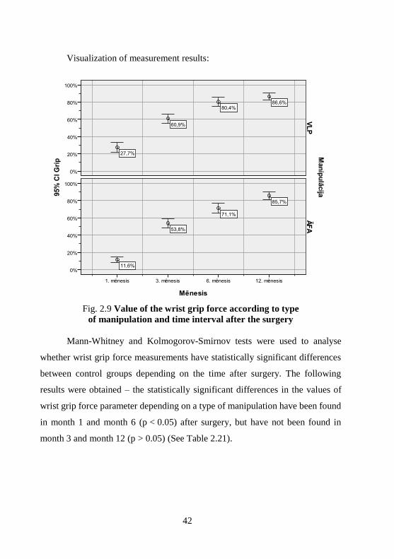
2.3.7. Grip force
Using the standard statistical methodology to assess the average values of
wrist grip force, depending on the type of manipulation and the interval of the
time after surgery, we obtained the following average values (95% confidence
interval) (See Table 2.20):
Table 2.20
Average values of wrist grip force
Month 1 Month 3 Month 6 Month 12
Group VLP % 27.7 ± 5.9 60.9 ± 5.2 80.4 ± 5.3 86.6 ± 4.0
Group EF % 11.6 ± 3.5 53.8 ± 5.8 71.1 ± 5.9 85.7 ± 4.8
Calculated by formula: Mean ± (Mean Upper Bound – Mean Lower Bound) / 2.
42
Visualization of measurement results:
Fig. 2.9 Value of the wrist grip force according to type
of manipulation and time interval after the surgery
Mann-Whitney and Kolmogorov-Smirnov tests were used to analyse
whether wrist grip force measurements have statistically significant differences
between control groups depending on the time after surgery. The following
results were obtained – the statistically significant differences in the values of
wrist grip force parameter depending on a type of manipulation have been found
in month 1 and month 6 (p < 0.05) after surgery, but have not been found in
month 3 and month 12 (p > 0.05) (See Table 2.21).

43
Table 2.21
Wrist grip force measurements using Mann-Whitney
and Kolmogorov-Smirnov tests
Month 1 Month 3 Month 6 Month 12
There are statistically
significant differences
between Group VLP
and Group EF
Yes No Yes No
p-value (Mann-Whitney test) 0.000 0.081 0.009 0.870
p-value (Kolmogorov-
Smirnov test) 0.000 0.351 0.039 0.994
P-value match with Asymp. Sig. (2-tailed) in calculation materials. If a single p-value
in a column is less than 0.05, write “Yes”, otherwise write “No”.
Wilcoxon Signed Ranks and Sign tests were used to determine whether
wrist grip force measurements are statistically significantly improving over the
time after the surgery. The following results were obtained – both tests show
statistically significant, monotonous improvement of wrist grip force over the
time after the surgery in both research groups (p < 0.01) (See Table 2.22).
Table 2.22
Wrist grip force measurements using Wilcoxon Signed Ranks
and Sign tests
Month 3 vs.
Month 1
Month 6 vs.
Month 3
Month 12 vs.
Month 6
Statistically significant
improvement (Group VLP) Yes Yes Yes
Statistically significant
improvement (Group EF) Yes Yes Yes
p-value (Wilcoxon Signed
Ranks test) Group VLP 0.000 0.000 0.017

44
Table 2.22 continued
Month 3 vs.
Month 1
Month 6 vs.
Month 3
Month 12 vs.
Month 6
p-value (Sign test) Group VLP 0.000 0.000 0.026
p-value (Wilcoxon Signed
Ranks test) Group EF 0.000 0.000 0.000
p-value (Sign test) Group EF 0.000 0.000 0.000
P-value match with Asymp. Sig. (2-tailed) / 2 in calculation materials. If a single p-value
in a column is less than 0.05, write “Yes”, otherwise write “No”
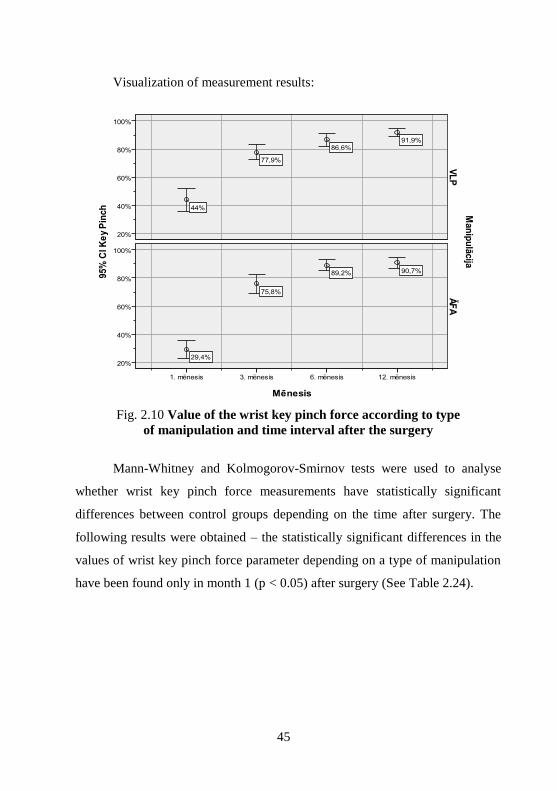
2.3.8 Key pinch force
Using the standard statistical methodology to assess the average values of
key pinch force, depending on the type of manipulation and the interval of the
time after surgery, we obtained the following average values (95% confidence
interval) (See Table 2.23).
Table 2.23
Average values of key pinch force
Month 1 Month 3 Month 6 Month 12
Group VLP % 44.0 ± 8.0 77.9 ± 5.3 86.6 ± 4.4 91.9 ± 2.7
Group EF % 29.4 ± 6.4 75.8 ± 6.8 89.2 ± 4.0 90.7 ± 3.9
Calculated by formula: Mean ± (Mean Upper Bound – Mean Lower Bound) / 2.
45
Visualization of measurement results:
Fig. 2.10 Value of the wrist key pinch force according to type
of manipulation and time interval after the surgery
Mann-Whitney and Kolmogorov-Smirnov tests were used to analyse
whether wrist key pinch force measurements have statistically significant
differences between control groups depending on the time after surgery. The
following results were obtained – the statistically significant differences in the
values of wrist key pinch force parameter depending on a type of manipulation
have been found only in month 1 (p < 0.05) after surgery (See Table 2.24).
46
Table 2.24
Wrist key pinch force measurements using Mann-Whitney
and Kolmogorov-Smirnov tests
Month 1 Month 3 Month 6 Month 12
There are statistically
significant differences
between Group VLP
and Group EF
Yes No No No
p-value (Mann-Whitney test) 0.007 0.594 0.519 0.826
p-value (Kolmogorov-
Smirnov test) 0.003 0.816 0.909 0.958
P-value match with Asymp. Sig. (2-tailed) in calculation materials. If a single p-value
in a column is less than 0.05, write “Yes”, otherwise write “No”.
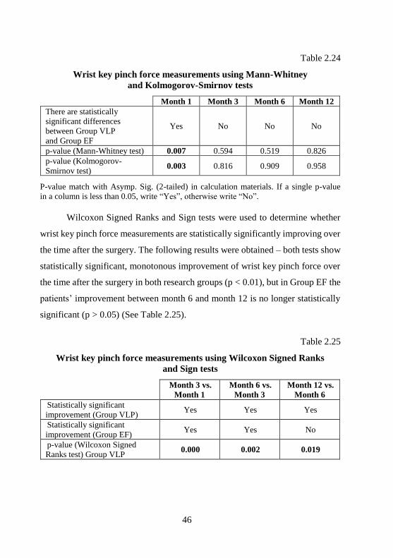
Wilcoxon Signed Ranks and Sign tests were used to determine whether
wrist key pinch force measurements are statistically significantly improving over
the time after the surgery. The following results were obtained – both tests show
statistically significant, monotonous improvement of wrist key pinch force over
the time after the surgery in both research groups (p < 0.01), but in Group EF the
patients’ improvement between month 6 and month 12 is no longer statistically
significant (p > 0.05) (See Table 2.25).
Table 2.25
Wrist key pinch force measurements using Wilcoxon Signed Ranks
and Sign tests
Month 3 vs.
Month 1
Month 6 vs.
Month 3
Month 12 vs.
Month 6
Statistically significant
improvement (Group VLP) Yes Yes Yes
Statistically significant
improvement (Group EF) Yes Yes No
p-value (Wilcoxon Signed
Ranks test) Group VLP 0.000 0.002 0.019

47
Table 2.25 continued
Month 3 vs.
Month 1
Month 6 vs.
Month 3
Month 12 vs.
Month 6
p-value (Sign test) Group VLP 0.000 0.018 0.063
p-Value (Wilcoxon Signed
Ranks test) Group EF 0.000 0.000 0.262
p-value (Sign test) Group EF 0.000 0.000 0.115
P-value match with Asymp. Sig. (2-tailed) / 2 in calculation materials. If a single p-value
in a column is less than 0.05, write “Yes”, otherwise write “No”.
2.3.9. Tripod pinch force
Using the standard statistical methodology to assess the average values of
tripod pinch force, depending on the type of manipulation and the interval of the
time after surgery, we obtained the following average values (95% confidence
interval) (See Table 2.26).
Table 2.26
Average values of tripod pinch force
Month 1 Month 3 Month 6 Month 12
Group VLP
% 39.7 ± 8.0 74.6 ± 5.3 86.6 ± 4.4 92.1 ± 2.7
Group EF % 26.2 ± 6.4 70.4 ± 6.8 86.7 ± 4.0 92.9 ± 3.9
Calculated by formula: Mean ± (Mean Upper Bound – Mean Lower Bound) / 2.

48
Visualization of measurement results:
Fig. 2.11 Value of the wrist tripod pinch force according to type
of manipulation and time interval after the surgery
Mann-Whitney and Kolmogorov-Smirnov tests were used to analyse
whether wrist tripod pinch force measurements have statistically significant
differences between control groups depending on the time after surgery. The
following results were obtained – the statistically significant differences in the
values of wrist tripod pinch force parameter depending on a type of manipulation
have been found only in month 1 (p < 0.05) after surgery (See Table 2.27).

49
Table 2.27
Wrist tripod pinch force measurements using Mann-Whitney
and Kolmogorov-Smirnov tests
Month 1 Month 3 Month 6 Month 12
There are statistically
significant differences
between Group VLP
and Group EF
Yes No No No
p-value (Mann-Whitney test) 0.031 0.306 0.790 0.711
p-value (Kolmogorov-
Smirnov test) 0.027 0.946 0.993 0.995
P-value match with Asymp. Sig. (2-tailed) in calculation materials. If a single p-value
in a column is less than 0.05, write “Yes”, otherwise write “No”.
Wilcoxon Signed Ranks and Sign tests were used to determine whether
wrist tripod pinch force measurements are statistically significantly improving
over the time after the surgery. The following results were obtained – both tests
show statistically significant, monotonous improvement of wrist tripod pinch
force over the time after the surgery in both research groups (p < 0.05)
(See Table 2.28).
Table 2.28
Wrist tripod pinch force measurements Wilcoxon Signed Ranks
and Sign tests
Month 3 vs.
Month 1
Month 6 vs.
Month 3
Month 12 vs.
Month 6
Statistically significant
improvement (Group VLP) Yes Yes Yes
Statistically significant
improvement (Group EF) Yes Yes Yes
p-value (Wilcoxon Signed
Ranks test) Group VLP 0.000 0.000 0.005

50
Table 2.28 continued
Month 3 vs.
Month 1
Month 6 vs.
Month 3
Month 12 vs.
Month 6
p-value (Sign test) Group VLP 0.000 0.000 0.108
p-value (Wilcoxon Signed
Ranks test) Group EF 0.000 0.000 0.006
p-value (Sign test) Group EF 0.000 0.000 0.017
P-value match with Asymp. Sig. (2-tailed) / 2 in calculation materials. If a single p-value
in a column is less than 0.05, write “Yes”, otherwise write “No”.
2.4. Subjective scales:
2.4.1 PRWE
Using the standard statistical methodology to assess the average values of
PRWE scale, depending on the type of manipulation and the interval of the time
after surgery, we obtained the following average values (95% confidence
interval) (See Table 2.29).
Table 2.29
Average values of PRWE scale
Month 1 Month 3 Month 6 Month 12
Group VLP % 50.6 ± 8.0 13.5 ± 5.3 7.2 ± 4.4 2.1 ± 2.7
Group EF % 77.8 ± 6.4 21.2 ± 6.8 9.4 ± 4.0 3.9 ± 3.9
Calculated by formula: Mean ± (Mean Upper Bound – Mean Lower Bound) / 2.
51
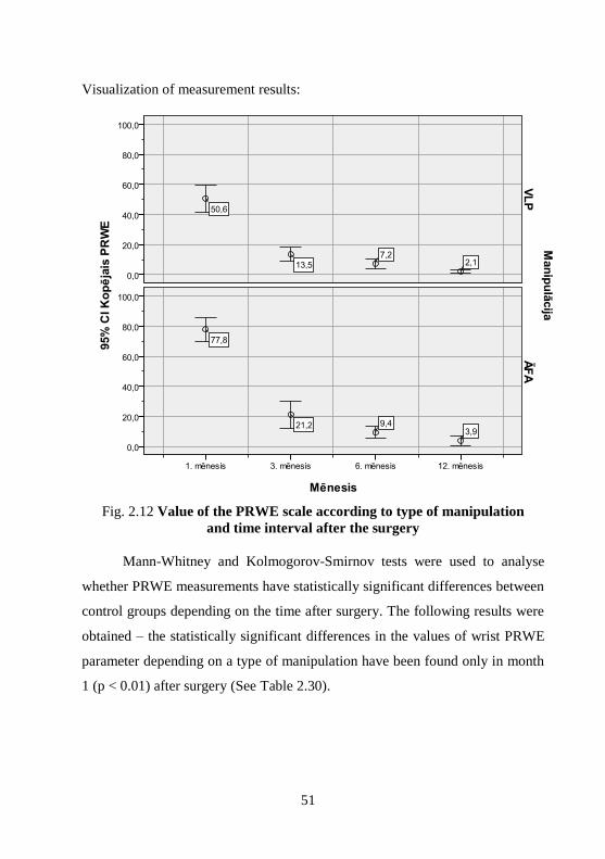
Visualization of measurement results:
Fig. 2.12 Value of the PRWE scale according to type of manipulation
and time interval after the surgery
Mann-Whitney and Kolmogorov-Smirnov tests were used to analyse
whether PRWE measurements have statistically significant differences between
control groups depending on the time after surgery. The following results were
obtained – the statistically significant differences in the values of wrist PRWE
parameter depending on a type of manipulation have been found only in month
1 (p < 0.01) after surgery (See Table 2.30).

52
Table 2.30
PRWE measurements using Mann-Whitney
and Kolmogorov-Smirnov tests
Month 1 Month 3 Month 6 Month 12
There are statistically
significant differences
between Group VLP
and Group EF
Yes No No No
p-value (Mann-Whitney test) 0.000 0.249 0.490 0.320
p-value (Kolmogorov-
Smirnov test) 0.000 0.717 0.717 0.816
P-value match with Asymp. Sig. (2-tailed) in calculation materials. If a single p-value
in a column is less than 0.05, write “Yes”, otherwise write “No”.
Wilcoxon Signed Ranks and Sign tests were used to determine whether
wrist PRWE measurements are statistically significantly improving over the time
after the surgery. The following results were obtained – both tests show
statistically significant, monotonous improvement (reduction) of wrist PRWE
over the time after the surgery in both research groups (p < 0.05)
(See Table 2.31).
Table 2.31
PRWE measurements using Wilcoxon Signed Ranks and Sign tests
Month 3 vs.
Month 1
Month 6 vs.
Month 3
Month 12 vs.
Month 6
Statistically significant
improvement (Group VLP) Yes Yes Yes
Statistically significant
improvement (Group EF) Yes Yes Yes
p-value (Wilcoxon Signed
Ranks test) Group VLP 0.000 0.009 0.000
p-value (Sign test) Group VLP 0.000 0.012 0.000
p-value (Wilcoxon Signed
Ranks tests Group EF 0.000 0.001 0.003
p-value (Sign test) Group EF 0.000 0.009 0.001
P-value match with Asymp. Sig. (2-tailed) / 2 in calculation materials. If a single p-value
in a column is less than 0.05, write “Yes”, otherwise write “No”.
53
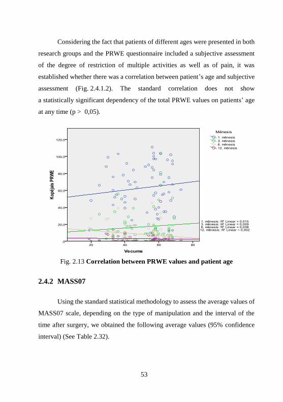
Considering the fact that patients of different ages were presented in both
research groups and the PRWE questionnaire included a subjective assessment
of the degree of restriction of multiple activities as well as of pain, it was
established whether there was a correlation between patient’s age and subjective
assessment (Fig. 2.4.1.2). The standard correlation does not show
a statistically significant dependency of the total PRWE values on patients’ age
at any time (p > 0,05).
Fig. 2.13 Correlation between PRWE values and patient age
2.4.2 MASS07
Using the standard statistical methodology to assess the average values of
MASS07 scale, depending on the type of manipulation and the interval of the
time after surgery, we obtained the following average values (95% confidence
interval) (See Table 2.32).

54
Table 2.32
Average values of MASS07 scale
Month 1 Month 3 Month 6 Month 12
Group VLP % 27.9 ± 9.1 5.1 ± 2.8 1.4 ± 1.4 0,3 ± 0.3
Group EF % 58.1 ± 11.4 8.8 ± 6.6 2.1 ± 1.8 0.9 ± 0.9
Calculated by formula: Mean ± (Mean Upper Bound – Mean Lower Bound) / 2
Visualization of measurement results:
Fig. 2.14 Value of the MASS07 scale according to type
of manipulation and time interval after the surgery
Mann-Whitney and Kolmogorov-Smirnov tests were used to analyse
whether MASS07 measurements have statistically significant differences
between control groups depending on the time after surgery. The following
results were obtained – the statistically significant differences in the values of

55
wrist MASS07 parameter depending on a type of manipulation have been found
only in month 1 (p < 0.01) after surgery (See Table 2.33).
Table 2.33
MASS07 measurements using Mann-Whitney
and Kolmogorov-Smirnov tests
Month 1 Month 3 Month 6 Month 12
There are statistically
significant differences
between Group VLP
and Group EF
Yes No No No
p-value (Mann-Whitney test) 0.000 0.887 0.376 0.967
p-value (Kolmogorov-
Smirnov test) 0.000 0.997 0.993 1.000
P-value match with Asymp. Sig. (2-tailed) in calculation materials. If a single p-value
in a column is less than 0.05, write “Yes”, otherwise write “No”.
Wilcoxon Signed Ranks and Sign tests were used to determine whether
wrist MASS07 measurements are statistically significantly improving over the
time after the surgery. The following results were obtained – both tests show
statistically significant, monotonous improvement (reduction) of wrist MASS07
over the time after the surgery in both research groups (p < 0.05), but in Group
EF the patients’ improvement between month 6 and month 12 is no longer
statistically significant (p > 0.05) (See Table 2.34).
Table 2.34
MASS07 measurements using Wilcoxon Signed Ranks and Sign tests
Month 3 vs.
Month 1
Month 6 vs.
Month 3
Month 12 vs.
Month 6
Statistically significant
improvement (Group VLP) Yes Yes Yes
Statistically significant
improvement (Group EF) Yes Yes No
p-value (Wilcoxon Signed Ranks
test) Group VLP 0.000 0.003 0.031

56
Table 2.34 continued
Month 3 vs.
Month 1
Month 6 vs.
Month 3
Month 12 vs.
Month 6
p-value (Sign test) Group VLP 0.000 0.007 0.227
p-value (Wilcoxon Signed Ranks
test) Group EF 0.000 0.004 0.106
p-values (Sign test) Group EF 0.000 0.007 0.146
P-value match with Asymp. Sig. (2-tailed) / 2 in calculation materials. If a single p-value
in a column is less than 0.05, write “Yes”, otherwise write “No”.
2.4.3 Modified Gartland and Werley scale
Using the standard statistical methodology to assess the average values of
modified Gartland and Werley scale, depending on the type of manipulation and
the interval of the time after surgery, we obtained the following average values
(95% confidence interval) (See Table 2.35):
Table 2.35
Average values of modified Gartland and Werley scale
Month 1 Month 3 Month 6 Month 12
Group VLP % 58.9 ± 3.9 78.1 ± 2.9 85.3 ± 2.9 91.8 ± 2.9
Group EF % 47.9 ± 3.0 72.8 ± 3.6 82.8 ± 3.2 88.5 ± 3.8
Calculated by formula: Mean ± (Mean Upper Bound – Mean Lower Bound) / 2.

57
Visualization of measurement results:
Fig. 2.15 Value of the modified Gartland & Werley scale according to type
of manipulation and time interval after the surgery
Mann-Whitney and Kolmogorov-Smirnov tests were used to analyse
whether modified Gartland & Werley score measurements have statistically
significant differences between control groups depending on the time after
surgery. The following results were obtained – the statistically significant
differences in the values of modified Gartland & Werley score parameter
depending on a type of manipulation have been found in month 1 and month 3
(p < 0.05) after surgery (See Table 2.36).

58
Table 2.36
Modified Gartland & Werley score measurements using Mann-Whitney
and Kolmogorov-Smirnov tests
Month 1 Month 3 Month 6 Month 12
There are statistically
significant differences
between Group VLP
and Group EF
Yes Yes No No
p-value (Mann-Whitney test) 0.000 0.023 0.195 0.189
p-value (Kolmogorov-
Smirnov test) 0.000 0.035 0.425 0.392
P-value match with Asymp. Sig. (2-tailed) in calculation materials. If a single p-value
in a column is less than 0.05, write “Yes”, otherwise write “No”.
Wilcoxon Signed Ranks and Sign tests were used to determine whether
modified Gartland & Werley score measurements are statistically significantly
improving over the time after the surgery. The following results were
obtained – both tests show statistically significant, monotonous improvement
(reduction) of modified Gartland & Werley score over the time after the surgery
in both research groups (p < 0.01) (See Table 2.37).
Table 2.37
Modified Gartland & Werley score measurements
using Wilcoxon Signed Ranks and Sign tests
Month 3 vs.
Month 1
Month 6 vs.
Month 3
Month 12 vs.
Month 6
Statistically significant
improvement (Group VLP) Yes Yes Yes
Statistically significant
improvement (Group EF) Yes Yes Yes
p-value (Wilcoxon Signed
Ranks test) Group VLP 0.000 0.000 0.000
59
Table 2.37 continued
Month 3 vs.
Month 1
Month 6 vs.
Month 3
Month 12 vs.
Month 6
p-value (Sign test) Group VLP 0.000 0.000 0.000
p-value (Wilcoxon Signed
Ranks test) Group EF 0.000 0.000 0.001
p-value (Sign test) Group EF 0.000 0.000 0.000
P-value match with Asymp. Sig. (2-tailed) / 2 in calculation materials. If a single p-value
in a column is less than 0.05, write “Yes”, otherwise write “No”.
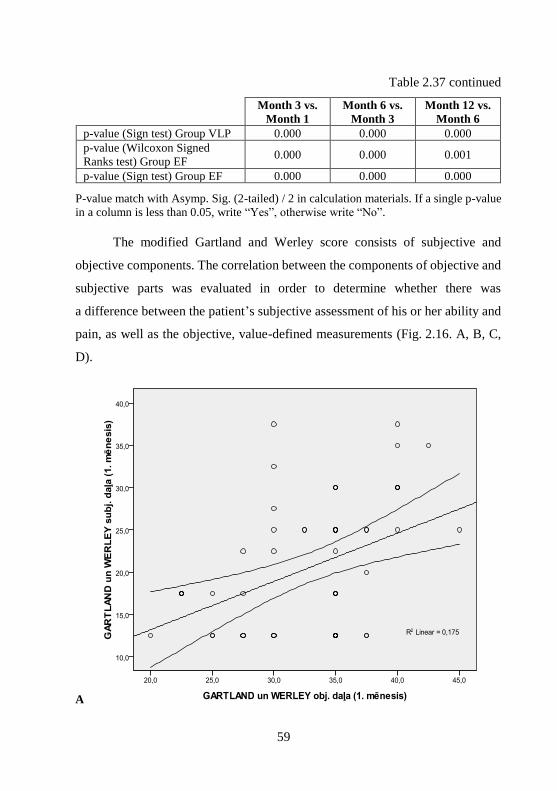
The modified Gartland and Werley score consists of subjective and
objective components. The correlation between the components of objective and
subjective parts was evaluated in order to determine whether there was
a difference between the patient’s subjective assessment of his or her ability and
pain, as well as the objective, value-defined measurements (Fig. 2.16. A, B, C,
D).
A
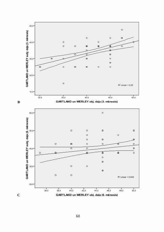
60
B
C

61
D
Fig. 2.16. A, B, C, D Correlation between subjective and objective
components of the modified Gartland & Werley score
The standard correlation analysis shows a statistically significant
correlation between the objective and subjective components at month 1, month
3 and month 12 (p < 0.01).
The standard correlation analysis does not show a statistically significant
correlation between these components at month 6 (p > 0.05).
Evaluating the data from modified Gartland & Werley score for patients
in different age groups, the standard correlation does not show a statistically
significant dependency of the values on patient age at any time of data
registration (p > 0.05) (Fig. 2.17).
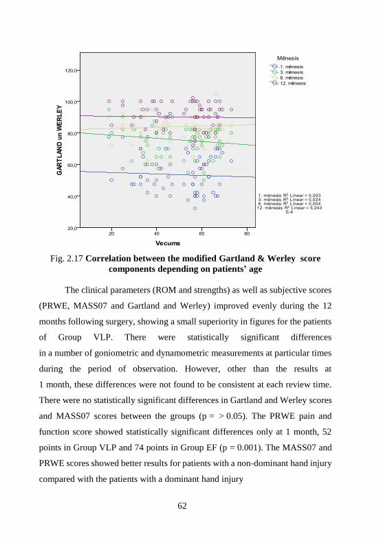
62
Fig. 2.17 Correlation between the modified Gartland & Werley score
components depending on patients’ age
The clinical parameters (ROM and strengths) as well as subjective scores
(PRWE, MASS07 and Gartland and Werley) improved evenly during the 12
months following surgery, showing a small superiority in figures for the patients
of Group VLP. There were statistically significant differences
in a number of goniometric and dynamometric measurements at particular times
during the period of observation. However, other than the results at
1 month, these differences were not found to be consistent at each review time.
There were no statistically significant differences in Gartland and Werley scores
and MASS07 scores between the groups (p = > 0.05). The PRWE pain and
function score showed statistically significant differences only at 1 month, 52
points in Group VLP and 74 points in Group EF (p = 0.001). The MASS07 and
PRWE scores showed better results for patients with a non-dominant hand injury
compared with the patients with a dominant hand injury

63
2.5 Associated injuries
The frequency of ligament co-injury did not statistically significantly
differ between the two groups (p = 0.22). Sixteen patients had scapholunate
ligament tears (Geissler grade II–IV), 11 patients had TFCC tears and eight
patients had both. Scapholunate joint trans-fixation with K-wires with additional
scaphocapitate joint fixation was performed in nine patients with acute Geissler
grade IV scapholunate ligament tears. Seven patients from both groups
underwent ulnar styloid fracture fixation with K-wires and tension bands due to
TFCC and distal radioulnar joint instability. Additional reduction of fracture
fragments at arthroscopy was necessary in 46 cases, 20 patients (59%) in Group
VLP and 26 patients (90%) in Group EF (p = 0.006).
2.6 Complications
There were several complications recorded during the study
(See Table 2.38).
Table 2.38
Rate of complications between research groups
Complication VLP group
(n = 34)
EF group
(n = 30)
All patients
(n = 64)
CRPS 1 (2.9%) 1.6%
Iatrogenic nerve
damage – 2 (6.6%) 3.1%
Secondary deformation
of the joint surface
after K-wire removal
– 2 (6.6%) 3.1%
Migration of K-wires 3 (8.8%) 4.7%
All 4 (11.8%) 4 (13.3%) 8 (12.5%)
CRPS – Complex Regional Pain Syndrome; VLP – volar locking plate; EF – external
fixator.

64
All recorded complications, with the exception of secondary deformation
of the joint surface following the evacuation of K-wires and the prognostically
likely development of osteoarthrosis in two Group EF patients, should be
considered as minor (Fig. 2.6.1.a and Fig. 2.6.1.b), Patient with CRPS in Group
VLP has successfully treated conservatively with physical therapy and appeared
symptom-free within 6 months. The disruption caused by migrated K-wires in
the Group VLP patients disappeared in a short period after the evacuation
of K-wires. Wire-induced damages of the dorsal radial sensory nerve were
treated surgically – one patient require the neurolysis surgery while the other
patient underwent neurinoma resection surgery and microsurgical
reconstruction of the damaged nerve. Neurological symptoms gradually
regressed within 6 to 8 months after the secondary surgeries in both patients.

65
Fig. 2.18. A X-ray immediately after the surgery and at Month 12 –
loss of the radial length
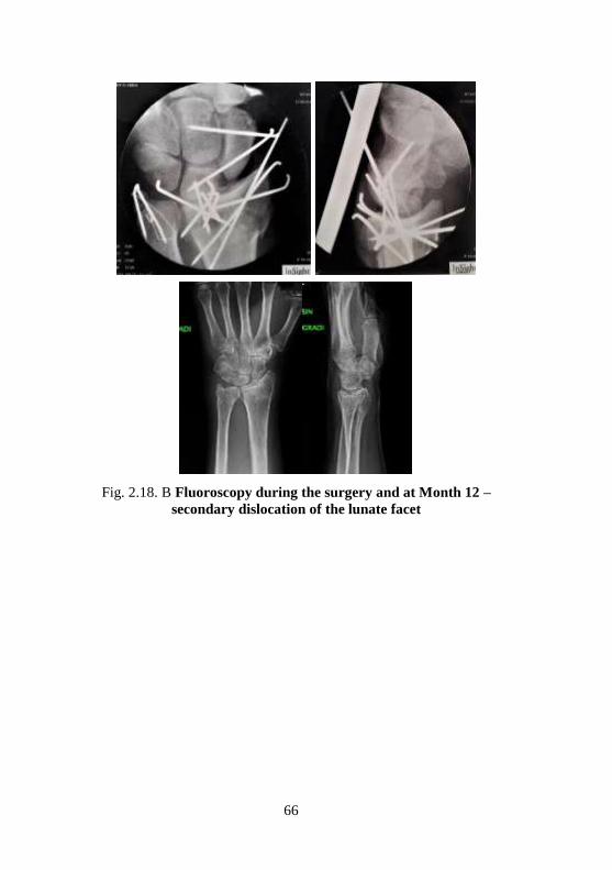
66
Fig. 2.18. B Fluoroscopy during the surgery and at Month 12 –
secondary dislocation of the lunate facet

67
3 Discussion
More than 200 years ago Abraham Colles (1773–1843) described the
“typical” fracture of the distal radius and its treatment (Colles, 1970), concluding
that despite the relatively simple method of closed reduction “the distortion of
the limb instantly returns on the extension being removed” and “… by such
mistakes the patient is doomed to endure for many months considerable lameness
and stiffness of the limb, accompanied by severe pains on attempting to bend the
hand and fingers”.
Discussion on treatment methods and comparison of them have not
subsided during these 200 years. Particularly rapidly, treatment options and
methods have changed since 1929, when Lorenz Böhler introduced the technique
of the closed manual longitudinal forearm traction and elbow contra-traction.
J. J. Gartland & C. W. Werley in 1951 in several sequential trials concluded and
described that good clinical results could be achieved only
in cases when normal anatomy of the wrist has been restored. Authors devised
a demerit point system to score subjective and objective function that continues
to be used today as well as defined the anatomic indices of normal volar tilt,
radial length, and radial inclination to link restoration of anatomy to
restoration of function, a principle that is the basis of modern fracture treatment.
G. Frykman in 1967, in the series of 413 DRFs found that osteoarthritic
changes occurred not only in the radiocarpal joint, but also in the DRUJ
(Frykman, 1967), drawing attention to the fact that in the cases of articular DRFs,
the restoration of the DRUJ surface is also essential for the achievement of the
good late functional results.

68
In 1986, J. L. Knirk and J. B. Jupiter presented results of their study,
which concluded that 91% of DRFs with intraarticular step-off more than 1 mm
and 100% DRFs with intraarticular step-off exceeding 2 mm are complicated by
deforming osteoarthrosis (Knirk and Jupiter, 1986).
H. J. Kreder with co-authors in 2005 published results of a study which
included calculations of the relative risk size (with a confidential interval 95%)
in cases of articular step-off and gap deformations. The results obtained were as
follows – when the step deformity was greater than 2 mm the risk of developing
arthritis was 10.4 times (95% CI 4.1 to 26.6) greater than when it was less than
2 mm. A gap greater than 2 mm was associated with a risk of arthritis which was
eight times (95% CI 2.6 to 24.7) higher than that for patients with a gap less than
2 mm (Kreder et al., 2005).
Surgical treatment methods of DRFs are different, starting from closed
reduction under the fluoroscope control and transcutaneous fixation with K-wires
with or without application of the external fixator and ending with ORIF of all 3
columns as well as fixation with fragment specific plates from both sides.
Given that in this study only two of the possible DRF treatment methods
were used – arthroscopic assisted ORIF and fixation with VLP and arthroscopic
assisted fixation with K-wires and externa fixator, there will be no further
discussion on other methods.
3.1 About external fixator
The use of different external fixation devices started from 40-ties of the
previous century. The popularity of external fixation for the treatment of wrist
fractures was catapulted forward by the concept of “ligamentotaxis” by Jacques
Vidal in 1979, who advocated its use for comminuted articular fractures,
including those of the hip, knee, ankle, spine, and wrist. The principle of

69
ligamentotaxis involved the application of tension “by means of distraction
forces working through capsule-ligamentous structures” to obtain reduction
(Vidal, 1979). Author described the use of this principle for highly comminuted
DRFs that were otherwise suitable for the ORIF. In 80-ties and 90-ties huge
amount of different external fixators and modifications were designed and this
method was widely used especially in the treatment of the articular DRFs.
Approximately 10 years after J. Vidal’s publication different studies started to
report about complications associated with this technique. The most common
complications related to the overdistraction of articular injuries were stiffness of
the fingers, reflex sympathetic dystrophy (CRPS), nerve disfunction as well as
different technical problems – inability to maintain reduction of articular
fragments, radial length with traction alone (Kaempffe et al., 1993; McQueen
et al., 1992; Weber and Szabo, 1986).
Nowadays the technique of external fixation includes “augmentation” of
fixation with supplemental Kirschner wires, mini-open or arthroscopic reduction
(or both), and bone grafting, with a concomitant reduction in traction-related
complications. Setting of the EF in neutral traction allows to start immediate
finger movements, while using additional stabilizing K-wires, removal of the EF
can be carried out within 4 to 6 weeks, leaving the wires into the bone, if
necessary, and allowing to start controlled workout of the wrist movements
(Wolfe, 2017).
3.2 About VLP
Historically open reduction of articular fractures of the distal radius was
indicated in young and active patients with good bone quality. ORIF can be used
in cases when restoration of the joint surface cannot be achieved by closed
manipulation, ligamentotaxis, or percutaneous reduction manoeuvres. It can be

70
used also as an alternative to percutaneous fixation at the preference of the patient
or surgeon.
In 2002, the first results of the application of the volar locking plate in 29
patients and 31 fractures were published (Orbay and Fernandez, 2002). Report
highlighted the positioning of the plate between the PQ muscle and bone which
significantly reduces the risk of the irritation of flexor tendons. It was concluded
that combination of stable internal fixation with the preservation of the dorsal
soft tissues resulted in rapid fracture healing, reduced need for bone grafting, and
low incidence of tendon problems. The greatest benefits of the VLP implant are
that it is strong and it permits stable fixation even in the most comminuted
unstable fractures, therefore immediate wrist ROM is possible with minimal use
of external support (removal wrist splint) (Wright et al., 2005). Osada and co-
authors in the biomechanical model assessed and demonstrated that only the
volarly placed SCS / V plated specimens resisted deformation of 5 degrees or
more at loads up to 250 N, which compares with the physiologic loads expected
with active finger motion, and was significantly stronger and more rigid than the
other plate groups.(Osada et al., 2003).
The management of unstable DRFs with open surgical methods in elderly,
less active patients with osteoporosis traditionally was considered as contra-
indicated. The rate of complications in this group always was higher, including
loss of stability of the fixation, non-union or malunion of the fracture as well as
the risk of the CRPS. The treatment of unstable distal radius fractures in the
elderly patient with a volar fixed-angle plate provided stable internal fixation and
allowed early function. This technique minimized morbidity in the elderly
population by successfully handling osteopenic bone, allowed early return to
function, provided good final results, and was associated with a low complication
rate (Orbay and Fernandez, 2004).
71
Assessing the information provided above, it is logical that there has
always been a desire to compare these different methods of treatment – their
advantages and flaws, and especially the possible outcomes.
3.3 Comparison of both methods
In the next part of the discussion I will present several examples of the
benefits of the VLP or EF method compared to the other method of surgical
treatment. I selected only results of the prospective randomized trials because in
the most of retrospective studies randomisation has not been observed. Only
studies with displaced articular DRFs treated surgically are presented in further
discussion.
In 2000, H. Kapoor et al. reported on study including 90 adult patients
with mean age 39. Patients were randomized in 3 groups – each consisted
of 30 patients. First group was treated with closed reduction and plaster
immobilisation. Treatment results of this group were the worst ones and should
not be presented for further analysis. The follow-up period was 1 year. Results
of the treatment were assessed based on the Sarniento functional score, because,
according to the authors – the DASH score had not yet been introduced when the
study was carried out. Results in the EF group – 80% good and excellent, 20%
average and poor results, results in ORIF group – 63% good and excellent, 26%
average, 11% poor (Kapoor et al., 2000).
In 2005, H. J. Kreder et al. published results of the prospective
randomized trial, including 179 skeletally-mature patients between the ages of
16 and 75 years with displaced intra-articular fractures of the distal radius.
The follow-up period was 2 years. EF group consisted of 88 patients, and ORIF
group – of 91 patients. It should be noted that dorsal or radial column plates also
were also used in ORIF group, making this group heterogeneous. Conclusions

72
made by authors are as follows: Grip and pinch force was better
in EF group within first 6 months. During a two-year observation period patients
from the EF group recovered functionally more rapidly and achieved better
overall results compared to ORIF group patients (Kreder et al., 2005).
In 2014, R. Shukla et al. published results of a study including
110 patients with mean age 39 ± 13, treated surgically because of the articular
DRFs – 68 patients treated with EF and 42 patients treated with VLP. The
assessment of pain, range of motion, grip strength and activity were assessed at
each follow-up visit and scored according to the Green and O'Brien scoring
system. One year after the surgery, 85.5% of EF group patients had good and
excellent results, but only 73.3% of patients had good and excellent results
in VLP group. As additional conclusion on the use of EF in treatment of the
articular DRFs authors mentioned that patients aged < 50 years treated with
external fixation had a better outcome than patients aged > 50 years in all
parameters studied at the end of 1 year. (Shukla et al., 2014).
This conclusion lines with a part of hypothesis of my study – fixation with
EF can be successfully applied in young patients, not only in elderly ones with
osteoporotic bone changes.
In 2016, Chuang Ma et al. presented randomised study with 123 patients
older than 65. EF group enrolled 58 patients and VLP group – 65 patients.
Clinical findings – ROM, Grip, pinch improved evenly in both groups with
a slight majority in favour of the EF group. Both groups significantly differ
in complication rates – 14 different complications were recorded in EF group
with 1 patient who required second surgery because of the complication, however
VLP group had 31 different complications with 6 secondary surgeries (Ma et al.,
2016).
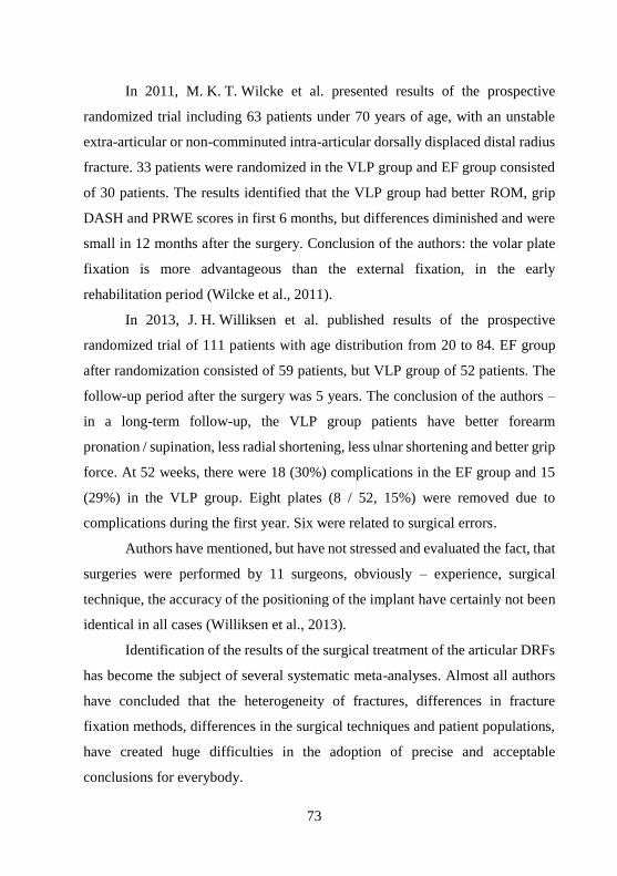
73
In 2011, M. K. T. Wilcke et al. presented results of the prospective
randomized trial including 63 patients under 70 years of age, with an unstable
extra-articular or non-comminuted intra-articular dorsally displaced distal radius
fracture. 33 patients were randomized in the VLP group and EF group consisted
of 30 patients. The results identified that the VLP group had better ROM, grip
DASH and PRWE scores in first 6 months, but differences diminished and were
small in 12 months after the surgery. Conclusion of the authors: the volar plate
fixation is more advantageous than the external fixation, in the early
rehabilitation period (Wilcke et al., 2011).
In 2013, J. H. Williksen et al. published results of the prospective
randomized trial of 111 patients with age distribution from 20 to 84. EF group
after randomization consisted of 59 patients, but VLP group of 52 patients. The
follow-up period after the surgery was 5 years. The conclusion of the authors –
in a long-term follow-up, the VLP group patients have better forearm
pronation / supination, less radial shortening, less ulnar shortening and better grip
force. At 52 weeks, there were 18 (30%) complications in the EF group and 15
(29%) in the VLP group. Eight plates (8 / 52, 15%) were removed due to
complications during the first year. Six were related to surgical errors.
Authors have mentioned, but have not stressed and evaluated the fact, that
surgeries were performed by 11 surgeons, obviously – experience, surgical
technique, the accuracy of the positioning of the implant have certainly not been
identical in all cases (Williksen et al., 2013).
Identification of the results of the surgical treatment of the articular DRFs
has become the subject of several systematic meta-analyses. Almost all authors
have concluded that the heterogeneity of fractures, differences in fracture
fixation methods, differences in the surgical techniques and patient populations,
have created huge difficulties in the adoption of precise and acceptable
conclusions for everybody.
74
M.M.J. Wallenkamp et al. in such a meta-analysis study found that
patients treated with a volar locking plate showed significantly better functional
outcome throughout the entire follow-up. However, this difference was only
clinically relevant during the early postoperative period (Walenkamp et al.,
2013).
In 2013, Chinese authors led by Xuetao Xie published the result of the
meta-analyses study where they concluded that for surgical treatment of distal
radius fractures, internal fixation yields better functional outcomes, forearm
supination, restoration of anatomic volar tilt and radial inclination, and fewer
minor complications. The patients who received ORIF using volar locking plates
for the treatment of distal radius recovered more quickly than did patients who
received EF (Xie et al., 2013).
Meta-analyses study published in 2018 by C. J. C. Gouk et al. concluded,
that the majority of the objective and subjective parameters do not differ
clinically and statistically within 12 months after the treatment of articular DRFs
with VLP or EF. Ulnar variance was better restored by volar locking plates. The
volar locking plates group was associated with higher re-operation rate, and the
external fixation group had a higher infection rate. Current literature suggests
that volar locking plates can provide better subjective scores and radiographic
parameters, especially in the first 3 months, but may be associated with a higher
re-operation rate (Gouk et al., 2018).
The most frequent conclusions are as follows – despite many advantages,
such as stable fixation of fragments or possibility of early active workout of
ROM, there are no evidence of statistically significant advantages for VLP
compared to EF in the treatment of articular DRFs in long time period (Costa et
al., 2014; Roh et al., 2015).

75
It is proved that VLP allows the rigid fixation of the fracture fragments,
but the previous studies do not verify if the fragments fixed by the plate are in
the exact anatomical position. Most of the articles mention different step-off and
gap deformities of the articular surfaces, remained after the surgeries where
fragment visualization and fixation were made only under the control of
fluoroscope. Proceeding fixation with EF and K-wires the anatomical fixation of
the fracture fragments is much more complicated and stability of the fixation is
lesser, resulting in possible secondary dislocations. In both cases, unless the
arthrotomy of the joint for the proper reposition of the articular fragments is
carried out, which causes additional soft tissue injuries and secondary
contractures, precise reposition of AO C2 and C3 fractures is technically
complicated.
3.4 Arthroscopy and distal radius fractures
An arthroscopic assisted method of fracture treatment is proposed to
minimise risks of inaccurate repositioning and for better visualization of the
fracture site.
The place of arthroscopy in this trial deserves a little further consideration.
Our results using wrist arthroscopy to visualize fragment positioning following
preliminary reduction confirm the worth of this method in the treatment of these
fractures.
Over the last two decades, the hand surgeons have been increasingly using
both – diagnostical and therapeutical wrist arthroscopies. Arthroscopic assisted
fixation of DRFs also is becoming more popular. One of the first descriptions
about arthroscopy in the treatment of DRF was published by H.J. Levy et al. in
1993. They were placing the arthroscope in the joint using the open volar
approach for fracture fixation without opening the joint capsule. This technique
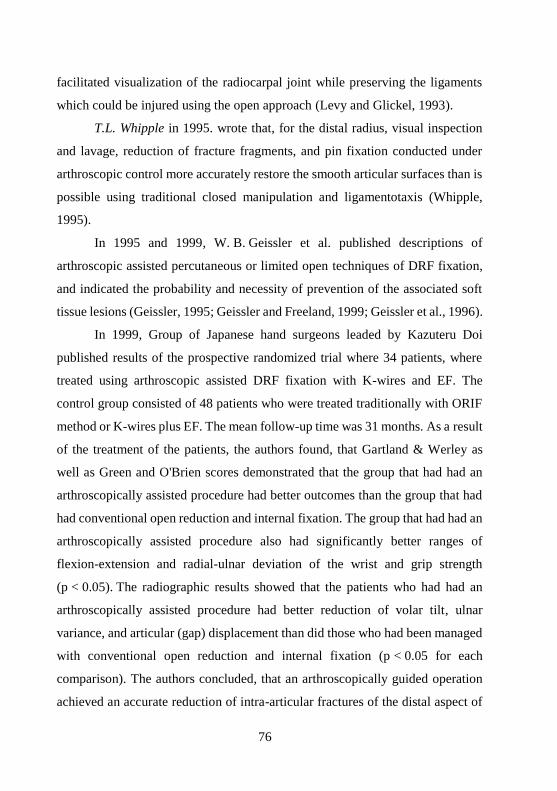
76
facilitated visualization of the radiocarpal joint while preserving the ligaments
which could be injured using the open approach (Levy and Glickel, 1993).
T.L. Whipple in 1995. wrote that, for the distal radius, visual inspection
and lavage, reduction of fracture fragments, and pin fixation conducted under
arthroscopic control more accurately restore the smooth articular surfaces than is
possible using traditional closed manipulation and ligamentotaxis (Whipple,
1995).
In 1995 and 1999, W. B. Geissler et al. published descriptions of
arthroscopic assisted percutaneous or limited open techniques of DRF fixation,
and indicated the probability and necessity of prevention of the associated soft
tissue lesions (Geissler, 1995; Geissler and Freeland, 1999; Geissler et al., 1996).
In 1999, Group of Japanese hand surgeons leaded by Kazuteru Doi
published results of the prospective randomized trial where 34 patients, where
treated using arthroscopic assisted DRF fixation with K-wires and EF. The
control group consisted of 48 patients who were treated traditionally with ORIF
method or K-wires plus EF. The mean follow-up time was 31 months. As a result
of the treatment of the patients, the authors found, that Gartland & Werley as
well as Green and O'Brien scores demonstrated that the group that had had an
arthroscopically assisted procedure had better outcomes than the group that had
had conventional open reduction and internal fixation. The group that had had an
arthroscopically assisted procedure also had significantly better ranges of
flexion-extension and radial-ulnar deviation of the wrist and grip strength
(p < 0.05). The radiographic results showed that the patients who had had an
arthroscopically assisted procedure had better reduction of volar tilt, ulnar
variance, and articular (gap) displacement than did those who had been managed
with conventional open reduction and internal fixation (p < 0.05 for each
comparison). The authors concluded, that an arthroscopically guided operation
achieved an accurate reduction of intra-articular fractures of the distal aspect of

77
the radius. Minimum capsular and adjacent soft-tissue scarring reduced
postoperative contracture, which improved the overall functional results. They
recommended arthroscopically guided reduction and internal fixation not only
for young adults but for all patients who are less than seventy years old and have
an intra-articular fracture of the distal part of the radius with more than one
millimetre of displacement on plain radiographs. (Doi et al., 1999)
The most important advantage of the arthroscopy is the possibility of
visualization of the joint on the screen, in real time, in immersive magnification
and in natural colours that no auxiliary diagnostic methods can provide.
Fluoroscopy alone provides an image that has poorer resolution than that of the
magnified camera used for direct arthroscopic visualization, whereas even a
small degree of displacement is obvious arthroscopically (Lutsky et al., 2008).
In 2001, C. C. Edwards et al. published an article about the study where
articular DRFs after the fluoroscopic guided, “satisfactory” reposition and
fixation with K-wires were examined arthroscopically. Authors discovered that
the optimal reduction obtained using C-arm was found to have an articular
displacement of > 1mm by adjunctive arthroscopy. (Edwards et al., 2001). In
2008, in a similar study K. Lutsky et al. identified that 36% of surgeries required
additional arthroscopic guided fracture repositions.
More severely comminuted and higher energy injuries are more likely to
require readjustment. In AO-C3 fractures the necessity of arthroscopic assisted
additional fracture reposition after initial treatment by indirect reduction
techniques should be necessary up to 71% of patients (Auge and Velazquez,
2000). Necessity for additional fragment reposition during the arthroscopy in my
study was more common than in previously published studies. Despite the large
number of additional arthroscopic manipulations in Group EF patients, the group
VLP patients also needed more additional manipulations in the arthroscopic
stage of the surgery as it was described in the previous studies (Abe and Fujii,

78
2017; Burnier et al., 2018; Khanchandani and Badia, 2013; Ono et al., 2010; Ono
et al., 2012).
In the ensuing part of the discussion I will address to the associated soft
tissue lesions, incidence of them and the recommended treatment methods. It is
possible that in previous comparative trials the unrecognized and misdiagnosed
associated soft tissue lesions did not allow to achieve better long-term functional
results. It is hoped that overall outcomes will be improved once patient-related
and treatment-related factors have been evaluated and previously undetected
associated ligament injuries have been found and treated (Lindau, 2017).
There are three intracarpal soft tissues of particular importance: the
triangular fibrocartilage complex (TFCC), the scapholunate interosseous
ligament (SLIL), and the lunotriquetral interosseous ligament (LTIL). These
structures are invisible on the plain radiograms and intra-operative fluoroscopies,
but nowadays, if there are technical possibilities and an experienced surgical
staff, visualization and prevention of these injuries should be desired. In cases of
the SLIL or LTIL injuries most commonly the Geissler arthroscopic
classification is used, but in cases of TFCC injuries – the Palmer classification
is advised.
In 2013, T. Ogawa et al. described group of 89 patients with arthroscopic
assisted treatment of DRF. They ranged in age from 17 to 92 years (mean, 62.2
years). TFCC injury was present in 59% of cases, SLIL injury
in 54.5% of cases, and LTIL injury in 34.5% of cases. Only 17.1% of patients
(14 / 82 patients) were negative for all three types of injury. In 81% of cases (72
/ 89 patients), some intracarpal soft tissue injury was present in association with
the fracture (Ogawa et al., 2013).
In 2020, S. Roulet et al. in 2020 published study in which the cases of
arthroscopic findings of associated soft tissue lesions in patients with DRF have
been evaluated. The study population consisted of 57 consecutive patients, with

79
mean age 43 years (range, 18–64 years). Among 57 intraarticular radius
fractures, 25 (44%) were associated with arthroscopic SL laxity, 3 (5%) with
arthroscopic LT laxity, and 16 (28%) with traumatic TFCC injury (Roulet et al.,
2020).
The incidence of the interosseus ligament and TFCC injuries varies in
different studies, but on average it ranges at around 50% for TFCC injuries and
30% for SLIL injuries (Ardouin et al., 2018). More detailed analysis of
associated soft tissue injuries could be found in the recent publications, but these
findings do not correlate with clinical findings and long-term functional results.
Clinically it is important to determine the degree of the damage of the stabilizing
structures, in order to avoid the formation of the secondary carpal instability.
This instability can eventually lead to SLAC (Scapho-lunate advance collapse)
deformation and osteoarthritis of the wrist joint.
W. B. Geissler, who introduced the arthroscopic classification of SLIL
and LTIL injuries, recommended to use wrist immobilization in grade I injuries,
arthroscopic reduction and pinning in grade II injuries, arthroscopic or open
reconstruction with additional pinning in grade III injuries and open reduction
and ligament reconstruction with or without tendon graft in grade IV injuries
(Geissler, 2013; Peicha et al., 1999). Such an aggressive surgical approach did
not seem acceptable for many hand surgeons, as well as other studies have been
previously performed, in which more lenient surgical approach also yielded in
good and satisfactory results. In 2007, D. P. Forward et al. published results of
the prospective randomized study where they concluded that grade I and II SLIL
tears treated only with immobilization, are asymptomatic within 1 year after the
surgery (Forward et al., 2007). It is desirable to adjust the immobilization
protocol after the DRF fixation with VLP to the degree of the SLIL injury
detected during the surgery (Ono et al., 2012). Fixation methods and treatment
options of the Geissler III SLIL injuries are still controversial. Several authors
80
agree with the W. B. Geissler’s recommended transfixation in an anatomical
position with K-wires (Kasapinova and Kamiloski, 2015; Khanchandani and
Badia, 2013). On the other hand, in 2015, A. Mrkonjic et al. published long-term
results (13 to 15 years after the primary surgery), where they did not find
statistically significant differences in clinical, subjective, objective and
radiological results between grade I-II and grade III SLIL injuries. (Mrkonjic et
al., 2015). Authors consider that such results could be explained because of the
secondary wrist stabilizers (volarly – RSL, RSC and LRL ligaments, dorsally –
DRT and DIC ligaments) and they recommended to perform further clinical
studies to explain and understand the role of these stabilizers.
Grade IV complete intercarpal injuries can be treated with arthroscopic
reduction and percutaneous Kirschner wire (K-wire) pinning provided there are
no radiological signs of dissociation because this implies that the secondary
stabilizers have been injured as well (Lindau, 2017). In cases of arthroscopic SL
joint transfixation with K-wires, an arthroscopic dorsal SL capsulodesis is also
recommended, which creates additional stability of the joint (DelPinal F, 2010)
Stability of the DRUJ also is one of the key factors of the successful
treatment of the DRFs. The TFCC in combination with a distal part of the
interosseous membrane is the major soft tissue stabilizer of the DRUJ, which
provides unobstructed movements of the forearm. Interaction of the TFCC with
the distal part of the interosseous membrane provides stability of the DRUJ in
any phase of movement (Haugstvedt et al., 2017; Moritomo, 2015).
In 2000, T. Lindau et al. published results of the study with 51 patients
(aged 20 to 57) with results of the treatment of the articular DRFs, highlighting
the functional results of the treatment of the associated TFCC lesions within 1
year after the surgery. Arthroscopy at the time of fracture showed complete or
partial TFCC tears in 43 patients. Ten patients with complete peripheral tear and
7 patients with partial peripheral TFCC tear (totally 39.5%) had instability of the

81
DRUJ 1 year after the DRF showing worse functional and clinical results
comparing to other patients (Lindau et al., 2000). These authors published
repeated results of the same group of patients after 15 years and they found that
only 1 of 17 patients (5.9%) with previously diagnosed DRUJ instability later
was treated surgically. The obtained information leads to the conclusion, that
peripheral TFCC tears less clinical problems as expected (Lindau, 2017).
Discussion about the type of the ulnar styloid fracture and its association
with TFCC tear and instability of the DRUJ is still actual. There is a presumption,
that complete fractures of the ulnar styloid are related to the potential separation
of the proximal part of the TFCC from the head of ulna, causing DRUJ
instability. In 2003, D. S. Ruch et al. describe the results of prevention of 13
symptomatic TFCC tears in a group of 57 DRF fracture patients, treated with
arthroscopic assisted surgeries. Two years after the surgery 12 of 13 patients had
stable DRUJ. This study documented the efficacy and safety of arthroscopically
assisted TFCC repair in conjunction with distal radius fracture fixation. (Ruch et
al., 2003).
In 2018, M. W. M. Fok et al. conducted a study where the occurrence and
nature of the TFCC injuries associated with the surgical treatment of the DRFs
with VLP were analysed. This study enrolled 43 patients with mean age 54
(17 to 75 y.o.) scheduled for implant removal after DRF union, for different
reason except septic complications. Concomitant wrist arthroscopy to
assess the condition of TFCC at the time of plate removal was routinely
offered. Instability of the DRUJ in combination with ulnar wrist pain DRUJ
which is statistically likely attributed to the TFCC tear, was found in 8 patients
(18.6 %) with a total proximal abruption the TFCC from the ulnar head.
Authors concluded that a large majority of TFCC tears remained unhealed
after the union of distal radius fractures. However, not all patients with tear
were symptomatic (Fok et al., 2018).

82
In 2018, A. C-Y. Chen et al. published results of the long-lasting study
where early performed surgery Chen outcomes (1 to 3 months after the accident)
and delayed surgery results (more than 12 months after the accident) of the ulnar
styloid base fracture fixation were compared. The authors concluded that the
fractures of the ulnar styloid base are usually associated with TFCC tears and
creating secondary DRUJ instability. Early detection and surgical repair yielded
better outcomes. Higher complication rates in late-treated fractures show that
surgeons should select surgical candidates and modalities properly (Chen et al.,
2018).
Various associated soft tissue lesions were identified during my study,
and the prevention procedures carried out, when such manipulations were
necessary to reduce the risk of instability of joints and subsequent osteoarthritis.
The frequency of ligament co-injury did not statistically significantly differ
between the two groups (p = 0.22). Sixteen patients (25% of all) had
scapholunate ligament tears (Geissler grade II–IV), 11 patients had TFCC tears
and eight patients had both. Scapholunate joint trans-fixation with K-wires with
additional scaphocapitate joint fixation was performed in nine patients (14% of
all) with acute Geissler grade IV scapholunate ligament tears. LTIL Geissler
grade II tears were found in 6 patients, which coincides with the frequency and
severity of LTIL injuries reported above and described in the literature. In cases
of the acute SLIL grade IV injuries, the fixation of the joint was performed with
K-wires, but in Group EF patients required administration of additional wires
and possibly increased the risk of soft tissue complications. Different isolated
TFCC tears were found in 11 patients (17.1% of all), but
8 patients (12.5%) had combined TFCC and SLIL injuries. Surgical repair of the
TFCC radial tears was not performed because it is known that if the proper
anatomic reposition of the ulnar fragments of the DRF is performed, the radial
tears of the TFCC cannot create instability in the DRUJ (Fok et al

83
., 2018). Central TFCC tears, which could theoretically also be
degenerative in nature, especially in the elder patients, only debridement of the
damaged part was performed. Surgical stabilization of the TFCC was performed
only in patients with DRUJ instability if it remained after the fixation of the DRF.
Three patients (4.7% of all) had arthroscopic dorsal TFCC fixation with
absorbable sutures and 7 patients (10.9% of all) underwent ulnar styloid fracture
fixation with K-wires and tension bands due to TFCC and distal radioulnar joint
instability
3.5 Evaluation of the results
Traditionally functional outcomes of the wrist after the surgical
manipulations are measured by ROM and the grip / pinch force. Both of these
measurements provide good and objective representation of the results; however,
these methods have drawbacks – they do not provide information on other
aspects of the treatment – the patient’s pain, ability to carry out various daily
activities or ability to return to the work, etc. Various functional scales have been
introduced over time to capture and analyse such data.
84
Several common and proven data recording methods were used to record
and subsequently evaluate the results of this study. Considering the fact that a
unified data recording system for the assessment of distal radius fractures has not
yet been introduced in hand surgery, I selected the most popular and
recommended data recording methods for the assessment of hand injuries –
Gartland & Werley modified score and PRWE score, which include both
subjective assessments of pain and function, as well as the recording of certain
objective variables. In addition, I chose to use the MASS07 scale, which includes
functional indicators of the use of modern technological devices, such as
computer keyboards, smart phones, electrical appliances, etc. This short
10-item scale is also recommended by M. Alexander et al., who showed that its
statistical performance is comparable to the DASH and PRWE scales and is
therefore recommended for functional assessment of the wrist joint after the wrist
injuries (Alexander et al., 2008).
S. Gupta et al. published an article comparing the DASH and PRWE
scales, the most commonly used in research. The authors concluded that
statistical analysis of the results of the two scales did not show differences. In
view of the above, and the fact that the PRWE scale is specific to wrist function,
unlike the DASH, which is more focused on shoulder and elbow function, the
authors recommend the PRWE scale as the method of choice for recording of
functional outcomes of the wrist (Gupta et al., 2014).
In 2016, J. F. Waljee et al. published an analytic article on the wrist
treatment outcomes represented of in the medical literature, and recommended a
summary with a preferred minimum of data that could be used in unified data
analysis and meta-analysis studies. This recommended standard includes
measurements of the symmetrical range of motion of both hands, the grip
strength of the hand and fingers (Grip, Pinch, Tripod pinch), visualization of the
subjective pain and function (PRWE, MHQ – Michigan Hand Questionnaire,

85
PROMIS – Patient-Reported Outcomes Measurement Information System
scales), recording of specific complications and the manipulations carried out to
treat them, as well as radiological data – signs of bone healing, characteristics of
joint surfaces, types and sizes of deformities, localisation of metal construction
and possible positional or dimensional errors, standard parameters, i.e. volar tilt,
radial height, radial incline, relationship of the distal ends of forearm bones
(ulna + / −) (Waljee et al., 2016)
Radiological findings in this study were recorded according to the
radiological section of Garland & Werley score, as well as position of the plate
was evaluated according to Soong criteria.
During the follow-up of the patients in this study, statistically significant
differences were found in several objective and subjective parameters, but the
results obtained by comparing the two surgical treatments did not show
significant differences in clinical terms. The Group VLP showed better short-
term functional results, but by 12 months the differences between the two groups
had evened out and there were no longer any clinical differences. The significant
difference in the measurement results 1 month after surgery is explained by the
use of diametrically opposed treatment methods. The Group VLP patients started
rehabilitation in the third post-operative week, which naturally allowed them to
return to normal daily activities much sooner. In the Group EF, the fixation was
removed 4 weeks after surgery or, in some cases, the wires were left in the bone
for a longer period of time. When we look at patients' emotional-subjective
scores, the short-term scores were significantly better in the group VLP, because
they resumed the activities of the injured arm sooner, but no clinically or
subjectively significant differences were found between the groups over the 12-
month period.
86
Subsequent alternating statistically significant differences in some aspects
of the ROM values could be the result of the inconsistency of measurements
performed by three different people. Regardless of these statistically significant
differences, the clinical relevance of these differences is questionable (e.g. 90%
against 97% of normal supination at 12 months). Our conclusion is that we were
unable to demonstrate any clinically relevant differences in any measured
parameter between the two groups, except at 1 month. This refutes the hypothesis
that minimally invasive treatment could lead to better functional outcomes.
The next important topic for discussion is the analysis of complications.
The most common complications of fractures of the distal end of the radius and
their treatment are as follows: contracture or restriction of movements, secondary
dislocation of the fracture and malunion, migration of the implants, irritation or
ruptures of tendons, nerve compressions or injuries, pain or CRPS, as well as
infection.
The overall complication rate for distal end fractures of the radius ranges
from 6% to 80%, with a note - which conditions are defined as complications
(McKay et al., 2001). Fixation of fractures with any metal construction
significantly reduces the chances of secondary dislocation and improper fracture
healing. In general, most of the presented complications can be prevented from
developing after surgery with careful attention to detail during surgery and
modification of surgical techniques on an individualized basis (Rhee et al., 2012).
Performing DRF fixation with VLP and screws, one of the most important
things is to avoid placing the plate too distally, i.e. it is preferable to follow the
Soong criteria and to position the distal edge of the plate so that it does not irritate
the flexor tendons (Soong 0 position).

87
In 2018, C. A. Selles et al. published the results of a study evaluating the
need for plate evacuation in a cohort of 323 patients. Evacuation of the implants
was required in 17% of patients. This group consisted mainly of patients with a
higher Soong classification (I or II). In the cases of Soong II plate position,
evacuation was 6 times more frequent than in the cases of Soong 0 position.
These results suggest that accurate positioning of the plate reduces the incidence
of tendon damage and hardware removal (Selles et al., 2018). Other authors also
note an association of higher grades of Soong classification of plate position with
the risk of flexor tendon complications and the need for plate evacuation (Lutsky
et al., 2015; Soong et al., 2011)
Another major iatrogenic complication is irritation or damage of the
extensor tendons with too long screws, especially in the distal metaphysis. Such
lesions occur in 3% to 5% of patients undergoing osteosynthesis of the DRF
(Soong et al., 2011). Various injuries of the extensor tendons can account up to
57% of all complications (Arora et al., 2007).
In my study, no patient in the Group VLP developed complications related
to tendon irritation or damage, and no plate evacuation was performed. All plates
were placed in Soong 0 or I position – 10 patients (29.4%) in Soong I and 24
patients (70.6%) in Soong 0. K-wires, inserted for additional fragment fixation,
later were removed under local anaesthesia with or without fluoroscope guidance
if the wires were detectable under the skin. No tendon complications related to
wire insertion were recorded.
Three out of 34 patients (9 %) in the Group VLP experienced minimal
migration of K-wires and skin irritation. This minor complication was attributed
to the activation of the patients from the 3rd post-operative week onwards,
resulting in loss of primary stability of some wires. Dislocation of the wires did
not cause the secondary displacement of the fracture fragments, but only irritated

88
surrounding soft tissues, causing discomfort or moderate pain in the patients.
After the evacuation of the wires, all related complaints disappeared.
F. Schuind in clinical trial where EF was used in the treatment of the
articular DRFs described the following complications and their incidence:
Fracture of the 2nd MC bone at the site of the implantation of the threaded rod –
1.6%, carpal tunnel syndrome – 2.6%, neurinoma of the sensory branch of the
radial nerve – 2.1%, secondary dislocation of the fragments from previously
achieved position – 2.1%, pin track infection – 12.8% and CRPS – 0.8%
(Schuind et al., 1989). The possible cause of damage to the dorsal radial nerve in
my study was the extensive use of K-wires during the primary surgery or
application of several K-wires via the 1–2 portal during the arthroscopic assisted
manipulation, reposition and fixation of fragments. This suggests that the more
extensive use of K-wires in reduction and / or fixation during external fixation
and K-wire fixation is more likely to result in nerve damage. In my study, 2
patients (3.1% of all) in the Group EF had secondary bone fragment dislocation
after removal of fixation and initiation of the workout of ROM. This
complication is due to the possible development of incomplete consolidation
between multiple articular fragments, which could be prevented by leaving the
wires in the subchondral bone or between the fragments for a longer period of
time. Unfortunately, follow-up radiographs 4 to 6 weeks after surgery only
visually assess the quality of the callus, which in some cases can be misleading
and lead to deformation of the fracture site later in the period of rehabilitation. It
should be noted that this particular complication was found in patients with AO
– C3 fractures consisting of four or more articular fragments. This leads to the
conclusion that, in cases of especially comminuted DRFs, despite the relatively
accurate arthroscopic assisted repositioning and fixation, secondary
displacement of some fragments is possible during the period of patient's
activation.

89
Both methods of fixation were relatively easy to apply for AO Type C1
fractures. However, surgeries or the more complex AO Type C3 fractures was
much more difficult, regardless of the method of reduction and fixation. The
higher rate of reposition of fragments at arthroscopy suggests that it is more
difficult to obtain optimal reduction in complex fractures with four or more
articular fragments when they are treated with external fixation and K-wires.
Also, primary reduction and fixation with several K-wires does not guarantee the
stability of the extra-articular alignment of the fragments to the same extent as a
volar locking plate. Furthermore, the complication of subsequent loss of position
of fragments also occurred in two of the Group EF patients.
Another possible complication of peripheral nerve involvement in DRFs
is an acute carpal tunnel syndrome, which results from compression caused by
contusion of the median nerve or post-traumatic oedema. In patients with
symptoms of an acute lesion of the median nerve, the dissection of carpal
ligament and neurolysis is recommended if symptoms persist after primary
reposition and fixation of the fracture with a plaster cast or EF. In my study, no
patient showed symptoms of compression or contusion of the median nerve after
the primary repositioning, so routine dissections of the carpal tunnel and
visualisation of the nerve were not performed during surgeries. According to the
literature, the results of prophylactic carpal tunnel release manipulation are
inconsistent – there are authors who argue that this manipulation may increase
the risk of unnecessary complications (Lattmann et al., 2008; Odumala et al.,
2001). The opposite view can be found in publications in which authors describe
a series of operations with standardised carpal ligament dissection in all patients
without complications (Gwathmey et al., 2010; Khanchandani and Badia, 2013).
In the absence of randomised trials of carpal tunnel ligament repair in
asymptomatic patients, undergoing plate fixation of the DRFs, the performance

90
of this manipulation or its abstention is recommended at the discretion of the
surgeon (Seigerman et al., 2019).
One of the most severe and difficult complications is Complex Regional
Pain Syndrome (CRPS). It can develop as a result of both conservative and
surgical treatment. Clinical manifestations of this syndrome include pain and
objective dysfunction of the sympathetic nervous system – oedema, joint
stiffness, discolouration of the involved limb and vasomotor disorders such as
hyperhidrosis and allodynia. The incidence of CRPS ranges from 1 to 37 % in
patients with DRF and it is more common in multifragmentary or high-energy
trauma fractures (Dijkstra et al., 2003; Jellad et al., 2014), and the incidence in
older women is 3 to 4 times higher than in men (Patterson et al., 2011). Complex
therapy is recommended for the treatment of CRPS, including painkillers,
intensive occupational therapy, intravenous steroid injections, antidepressants, in
some cases ganglion stellatum blockades, and psychological and / or psychiatric
help, if necessary. In my study, CRPS was detected in 1 patient (1.6% of all) in
the age group over 60 y.o., who already showed initial signs of CRPS before
surgery – too tight immobilisation, psychological predisposition and relatively
long time (2 weeks after primary injury) to surgery. Following the treatment
algorithm recommended in the literature for CRPS, remission of symptoms was
achieved within 6 months after surgery. The result is in line with the results
published in the medical literature for CRPS, the patient was able to return to her
previous activity level, although she was unable to regain full ROM and grip
strength.
Given that wrist arthroscopy is the main component of this study, in the
next section of the discussion I will focus on the potential complications of this
minimally invasive surgical technique. In 2016, C. Leclercq et al. published
a summary of complications of wrist arthroscopies at 36 hand surgery centres,
including the results of 10107 arthroscopic surgeries. A total of 605 different

91
surgical complications were recorded (5.98%). The most frequent ones were
failure to perform the planned procedure arthroscopically (1.16%), and nerve
lesions (1.17%). Cartilage lesions and complex regional pain syndrome each
occurred in 0.50% cases. Other complications (wrist stiffness, loose bodies,
hematomas, tendon lacerations) were less frequent. Breaking down of the data
according to each surgeon's experience of the technique showed a significant
relationship with the rate of complications, the threshold for a lower complication
rate being approximately 25 arthroscopies a year and / or greater than 5 years of
experience (Leclercq et al., 2016). In my study, no complications were found due
to the arthroscopic technique. No patients in the study groups developed
complications due to infection, which may be associated with standard
perioperative antimicrobial prophylaxis with 2 g of Cefazolin administered
intravenously in the operating theatre just before the surgery.
This study has a number of limitations. Alternate allocation was chosen
as a method of randomization in order to achieve a similar number of patients in
both groups as soon as possible. In some cases, this method of allocation made
it difficult to fix the fragments optimally. It is possible, that that the failure to
choose the best treatment method for the fracture configuration or the most
convenient for the surgeon, created additional technical difficulties and adversely
affected the final outcome of the treatment. Unfortunately, it was not possible to
prove this claim with statistical analysis methods in the context of this study. In
some Group VLP patients, additional K-wire fixation for free fragments was
necessary, although this instability could not be verified fluoroscopically. This
specific information reaffirms the necessity of the arthroscopy in the treatment
of DRF.

92
During the 2-year study period, 74 surgeries were performed (38 VLP and
36 Group EF). Four patients in Group VLP and seven patients in Group EF did
not return for the follow-up and thus were excluded from the analysis.
Consequently, only 34 patients from Group VLP and 29 patients from Group EF
were included in the final data assessment. Of the 63 patients, there were
23 males, mean age 43 year (range 19–66) and 40 females, mean age 56 years
(range 29–73). The demographic and clinical data were comparable between the
groups. In several cases, the follow-up of patients was carried out by the same
surgeon who had performed the surgeries, because of the workload of the hand
therapist.
Performing surgeries on a common framework, prescribing rehabilitation
with the same occupational therapist, equal treatment conditions were provided
to all patients. Both groups were demographically well balanced. We excluded
the possibility of choosing the method of fixation based on patient age or the
comminution of the fracture, so we assessed a mixture of both young and old
patients with a mixture of fracture types, treated with external fixation or VLP
fixation.
The use of EF and K-wires resulted in a higher complication rate than
treatment of fractures with a VLP and required secondary surgical treatment
(nerve damage). The Group EF patients also achieved secondary deformities of
the fracture fragments, which may lead to the post-traumatic osteoarthritis in the
future. The outcomes do not establish absolute evidence for preference of one
fixation method over the other, but confirms that both methods are applicable to
patients of different ages. The hypothesis that EF and wire fixation in
combination with wrist arthroscopy in the treatment of DRF is superior to
fixation with arthroscopic assisted fixation with VLP unfortunately was not
confirmed. The choice in favour of any of these methods must be made

93
considering the geometry of the fracture, the experience of the doctor and the
technical possibilities.
It is possible that statistically significant differences between the two
groups could be obtained by increasing the size of the research groups.

94
Conclusions
1 Evaluating the postoperative radiological and functional outcomes of
the randomised groups, based on the assessment of patients' quality of life and
health (Gartland & Werley, PRWE and MASS07 scales), ROM and strength
measurements, as well as X-ray follow-up at 1, 3, 6 and 12 months after surgery,
the following results were established – at the beginning of the follow-up period,
patients in the Group VLP showed better functional results in several parameters.
At the 12-month follow-up period, no relevant and statistically significant
differences were found between the groups, indicating that both methods are
applicable to the DRF treatment. Less invasive arthroscopic assisted treatment
with EF and K-wires has not demonstrated the predicted superiority over the
arthroscopic assisted ORIF with VLP.
2 Analysis of complications in both groups showed 4 complications in
the Group VLP (11.8% of the group) and 4 complications in the Group EF
(13.3% of the group).
The number of complications was small in both groups and all of them
were resolved during the follow-up period, but complications in the Group EF
were considered to be more significant. Complication related to the surgical
technique were observed only in Group EF patients. The possible cause of
damage to the dorsal radial nerve was the extensive use of K-wires during the
primary surgery or application of several K-wires via the 1–2 portal during the
arthroscopically assisted manipulation, reposition and fixation of fragments. This
suggests that the more extensive use of K-wires in reduction and / or fixation
during external fixation and K-wire fixation is more likely to result in nerve
damage.
95
In the EF group, 2 patients demonstrated secondary dislocation of
articular fragments after the evacuation of K-wires, suggesting that in some cases
prolonged retention of implants (VLP) in bone is essential to ensure stability even
in cases of slower consolidation. Migration of the additional
K-wires used in Group VLP patients has been attributed to the early workout of
the ROM. This minor complication has not caused secondary dislocation of bone
fragments or damage to any soft tissue structures in any patient.
3 The place of arthroscopy in this trial deserves a little further
consideration. Our results using arthroscopy to visualize fragment position
following preliminary reduction confirm the worth of this method
in the treatment of these fractures. Additional fragment reposition was required
in 46 cases – for 20 patients (59%) in Group VLP and 26 patients (87%)
in Group EF, indicating that without arthroscopy, intra-articular dislocation
would not have been prevented, making it absolutely necessary for such complex
fractures.
Several intra-articular soft tissue lesions were also detected during the
arthroscopic stage – injuries of the SLIL, LTIL and TFCC, which were prevented
by various additional manipulations. Without arthroscopy, these lesions would
remain undiagnosed and potentially worsen both objective and subjective long-
term outcomes.
4 The algorithm for the selection of the treatment methods of articular
DRF created during this study, will help the orthopaedic surgeons to choose the
necessity of the use of arthroscopy in the treatment of certain DRFs and to
organise the flow of patients who need this arthroscopically assisted surgical
treatment.
5 The study also resulted in an algorithm for the recommended
monitoring of post-operative follow-up and outcomes, which can be used not
only for assessment of the results of DRF treatment, but also for different wrist

96
injuries or orthopaedic conditions. The recommended package of measures can
be applied in any traumatology and orthopaedics hospital in Latvia without the
use of additional special equipment. The obtained data could be used for unified
clinical and academic studies in the future.
97
Publications 1. Funkcionālo rezultātu salīdzinājums pēc tradicionālās un artroskopiski asistētās
spieķkaula distālā gala artikulārā lūzuma osteosintēzes; Uldis Krustiņš, Andris
Jumtiņš, Diāna Bringina, Kristīne Šitca. https://www.rsu.lv/zinatniskie-
raksti/funkcionalo-rezultati-pec-spiekkaula-distala-gala-artikulara-luzuma-
osteosintezes
2. Krustins U, Krustins J, Bringina D, Laurane K, Jumtins A. Comparison of volar
locking plates with external fixation and k-wires in arthroscopically assisted intra-
articular distal radial fracture fixation. J Hand Surg Eur Vol. 2020, Vol. 45(4)
333-338. DOI: 10.1177/1753193419879567.
3. Comparison of fluoroscopically and arthroscopically assisted volar plating
of articular distal radius fractures. Uldis Krustins, Vadims Nefjodovs, Diana Bringina,
Aija Jaudzema, Andris Jumtins; PROCEEDINGS OF THE LATVIAN ACADEMY
OF SCIENCES. Section B, Vol. 75 (2021), No. 1 (730), pp. 20–30. DOI:
10.2478/prolas-2021-00XX
Presentations on topic
1. ILTOK 2011, Rīga, Latvija – “Plaukstas artroskopija – pirmo 25 gadījumu analīze”.
2. ILTOK 2012, Rīga, Latvija – “Spieķkaula distālo artikulāro lūzumu ārstēšanas
iespējas izmantojot plaukstas artroskopiju”.
3. 2nd Baltic hand Surgery Meeting 2012 Riga, Latvia – “Arthroscopically assisted
treatment of the distal radius fractures”
4. Joint Singapore – Malaysia – Finland – Estonia – Latvia Hand Surgery Societies
Meeting (incorporating the 22nd Comprehensive Hand Review Course), Singapore,
2013, “Arthroscopically Assisted Treatment of Mutilated Distal Radius Fractures”.
5. Joint Singapore – Malaysia – Finland – Estonia – Latvia Hand Surgery Societies
Meeting (incorporating the 22nd Comprehensive Hand Review Course), Singapore,
2013, “Arthroscopical Debridement of Stiff Joints after Distal Radius Fractures”.
6. AOTrauma seminar, faculty national, 2013, Riga, Latvia “Technical Principles
of the Arthroscopic Treatment of the Distal Radius Fractures”.
7. 5th Baltic Congress of Traumatology and Orthopaedics, 2013, Riga, Latvia –
“The Role of Arthroscopic Debridement as a Second Stage Procedure after Articular
Distal Radius Fractures”.
8. 10th Congress of APFSSH, 2014, Kuala Lumpur, Malaysia poster presentation –
“5-Year Follow-up After Bilateral Articular Distal Radius Fractures”

98
9. 26th SSSH Congress, 2016, Levi, Finland – “Functional Outcomes After
Artroscopically Assisted and Traditional Surgical Treatment of Displaced Articular
Distal Radius Fractures”.
10. EWAS wrist arthroscopy course, 2017, St. Petersburg, Russia – “Wrist from Inside –
Basic Principles of the Wrist Arthroscopy”.
11. 11th APFSSH Congress, 2017, Cebu, Phillippines “Arthroscopic vs. Nonarthroscopic
Treatment of Distal Radius Fractures – Evaluation of Outcomes”.
12. 11th APFSSH Congress, 2017, Cebu, Phillippines poster presentations –
“Arthroscopically Assisted Intra-Articular Distal Radius Fracture Surgery with Volar
Locking Plates or External Fixator and K-Wires. Early Results of Ongoing Study”,
“Krukenberg Procedure Completed by Two Microvascular Flaps to Save
Functionality and Length of the Amputation Stem. A Case Report”.
13. Wrist. Practical Arthroscopy course, 2018, St. Petersburg, Russia – “Wrist Central
Column Pathology”.
14. Riga Stradins University International Conference, 2019, “Arthroscopic Treatment
and Bone Grafting of Scaphoid Nonunions”.
15. EWAS wrist arthroscopy course with training on anatomical specimen, 2019,
St.Petersburg, Russia – “Clinical Evaluation of TFCC Injuries”.
16. EWAS wrist arthroscopy course with training on anatomical specimen, 2019,
St.Petersburg, Russia “Ulnocarpal Impingement and TFCC Tears”.
17. EWAS wrist arthroscopy course with training on anatomical specimen , 2019,
St.Petersburg, Russia “SLIL Normal Function, Physiology and Disfunction”.
18. 27th SSSH Congress , 2019, Tallinn, Estonia “Arthroscopy in Distal Radius
Fractures”.
19. 12th APFSSH Congress, 2020, Melbourne, Australia “Comparison of Functional
Outcomes in Limited Carpal Fusions and Proximal Row Carpectomy”.
20. 12th APFSSH Congress, Melbourne, Australia, 2020, “Comparison of Volar Locking
Plates with External Fixation and K-Wires in Arthroscopically Assisted Intra-
Articular Distal Radial Fracture Fixation.”, poster presentation “Long Term Follow-
up after Microsurgical Reconstruction of the Humerus with Two Vascularized Bone
Grafts”.
21. Arthrex Online Webinar, 2020, “Why Is It Important to Eliminate Intra-Articular
Displacement? How Do I Do this with an Acute and Incorrectly Fused Distal Radius
Fracture”.
22. EOF Online Club Webinar, 2021,“Acute scaphoid fractures”
23. 10th Congress of BAS, 2021, Riga, Online, “Arthroscopically Assisted Treatment of
Distal Radius Fractures: 10-Year Experience of the Single Centre”.

99
References
1. Abe Y, Fujii K. Arthroscopic-assisted reduction of intra-articular distal radius
fracture. Hand Clin. 2017, 33: 659–668.
2. Alexander M, Franko OI, Makhni EC, Zurakowski D, Day CS. Validation
of a modern activity hand survey with respect to reliability, construct and criterion
validity. J Hand Surg Eur Vol. 2008, 33: 653–660.
3. Ardouin L, Durand A, Gay A, Leroy M. Why do we use arthroscopy for distal radius
fractures? Eur J Orthop Surg Traumatol. 2018, 28: 1505–1514.
4. Arora R, Lutz M, Hennerbichler A, Krappinger D, Espen D, Gabl M. Complications
following internal fixation of unstable distal radius fracture with a palmar locking-
plate. J Orthop Trauma. 2007, 21: 316–322.
5. Auge WK, 2nd, Velazquez PA. The application of indirect reduction techniques
in the distal radius: The role of adjuvant arthroscopy. Arthroscopy. 2000, 16:
830–835.
6. Burnier M, Le Chatelier Riquier M, Herzberg G. Treatment of intra-articular fracture
of distal radius fractures with fluoroscopic only or combined with arthroscopic
control: A prospective tomodensitometric comparative study
of 40 patients. Orthop Traumatol Surg Res. 2018, 104: 89–93.
7. Changulani M, Okonkwo U, Keswani T, Kalairajah Y. Outcome evaluation measures
for wrist and hand: Which one to choose? Int Orthop. 2008, 32: 1–6.
8. Chen AC, Chiu CH, Weng CJ, Chang SS, Cheng CY. Early and late fixation
of ulnar styloid base fractures yields different outcomes. J Orthop Surg Res. 2018, 13:
193.
9. Colles A. Historical paper on the fracture of the carpal extremity of the radius (1814).
Injury. 1970, 2: 48–50.
10. Costa ML, Achten J, Parsons NR et al. Percutaneous fixation with kirschner wires
versus volar locking plate fixation in adults with dorsally displaced fracture of distal
radius: Randomised controlled trial. BMJ. 2014, 349: g4807.
11. Del Pinal F. MC, Luchetti R., Arthroscopic management of distal radius fractures.
Berlin Springer Verlag, 2010.
12. Dijkstra PU, Groothoff JW, ten Duis HJ, Geertzen JH. Incidence of complex regional
pain syndrome type i after fractures of the distal radius. Eur J Pain. 2003,
7: 457–462.
13. Doi K, Hattori Y, Otsuka K, Abe Y, Yamamoto H. Intra-articular fractures
of the distal aspect of the radius: Arthroscopically assisted reduction compared with
open reduction and internal fixation. J Bone Joint Surg Am. 1999, 81: 1093–1110.

100
14. Edwards CC, 2nd, Haraszti CJ, McGillivary GR, Gutow AP. Intra-articular distal
radius fractures: Arthroscopic assessment of radiographically assisted reduction.
J Hand Surg Am. 2001, 26: 1036–1041.
15. Fok MWM, Fang CX, Lau TW, Fung YKE, Fung BKK, Leung FKL. The status
of triangular fibrocartilage complex after the union of distal radius fractures with
internal plate fixation. Int Orthop. 2018, 42: 1917–1922.
16. Forward DP, Lindau TR, Melsom DS. Intercarpal ligament injuries associated with
fractures of the distal part of the radius. J Bone Joint Surg Am. 2007, 89:
2334–2340.
17. Frykman G. Fracture of the distal radius including sequelae-shoulder-hand-finger
syndrome, disturbance in the distal radio-ulnar joint and impairment of nerve function.
A clinical and experimental study. Acta Orthop Scand. 1967: Suppl 108:3+.
18. Geissler WB. Arthroscopically assisted reduction of intra-articular fractures
of the distal radius. Hand Clin. 1995, 11: 19–29.
19. Geissler WB. Arthroscopic management of scapholunate instability. J Wrist Surg.
2013, 2: 129–135.
20. Geissler WB, Freeland AE. Arthroscopic management of intra-articular distal radius
fractures. Hand Clin. 1999, 15: 455–465, viii.
21. Geissler WB, Freeland AE, Savoie FH, McIntyre LW, Whipple TL. Intracarpal soft-
tissue lesions associated with an intra-articular fracture of the distal end
of the radius. J Bone Joint Surg Am. 1996, 78: 357–365.
22. Gouk CJC, Bindra RR, Tarrant DJ, Thomas MJE. Volar locking plate fixation versus
external fixation of distal radius fractures: A meta-analysis. J Hand Surg Eur Vol.
2018, 43: 954–960.
23. Gupta S, Halai M, Al-Maiyah M, Muller S. Which measure should be used to assess
the patient's functional outcome after distal radius fracture? Acta Orthop Belg. 2014,
80: 116–118.
24. Gwathmey FW, Jr., Brunton LM, Pensy RA, Chhabra AB. Volar plate osteosynthesis
of distal radius fractures with concurrent prophylactic carpal tunnel release using a
hybrid flexor carpi radialis approach. J Hand Surg Am. 2010, 35: 1082–8 e4.
25. Haugstvedt JR, Langer MF, Berger RA. Distal radioulnar joint: Functional anatomy,
including pathomechanics. J Hand Surg Eur Vol. 2017, 42: 338–345.
26. Jellad A, Salah S, Ben Salah Frih Z. Complex regional pain syndrome type i:
Incidence and risk factors in patients with fracture of the distal radius. Arch Phys Med
Rehabil. 2014, 95: 487–492.

101
27. Kaempffe FA, Wheeler DR, Peimer CA, Hvisdak KS, Ceravolo J, Senall J. Severe
fractures of the distal radius: Effect of amount and duration of external fixator
distraction on outcome. J Hand Surg Am. 1993, 18: 33–41.
28. Kapoor H, Agarwal A, Dhaon BK. Displaced intra-articular fractures of distal radius:
A comparative evaluation of results following closed reduction, external fixation and
open reduction with internal fixation. Injury. 2000, 31: 75–79.
29. Kasapinova K, Kamiloski V. Influence of associated lesions of the intrinsic ligaments
on distal radius fractures outcome. Arch Orthop Trauma Surg. 2015, 135: 831–838.
30. Khanchandani P, Badia A. Functional outcome of arthroscopic assisted fixation
of distal radius fractures. Indian J Orthop. 2013, 47: 288–294.
31. Kim JK, Park MG, Shin SJ. What is the minimum clinically important difference
in grip strength? Clin Orthop Relat Res. 2014, 472: 2536–2541.
32. Knirk JL, Jupiter JB. Intra-articular fractures of the distal end of the radius in young
adults. J Bone Joint Surg Am. 1986, 68: 647–659.
33. Kreder HJ, Hanel DP, Agel J et al. Indirect reduction and percutaneous fixation versus
open reduction and internal fixation for displaced intra-articular fractures
of the distal radius: A randomised, controlled trial. J Bone Joint Surg Br. 2005, 87:
829–836.
34. Lattmann T, Dietrich M, Meier C, Kilgus M, Platz A. Comparison of 2 surgical
approaches for volar locking plate osteosynthesis of the distal radius. J Hand Surg
Am. 2008, 33: 1135–1143.
35. Leclercq C, Mathoulin C, Members of E. Complications of wrist arthroscopy:
A multicenter study based on 10,107 arthroscopies. J Wrist Surg. 2016, 5: 320–326.
36. Levy HJ, Glickel SZ. Arthroscopic assisted internal fixation of volar intraarticular
wrist fractures. Arthroscopy. 1993, 9: 122–124.
37. Lindau T. Arthroscopic evaluation of associated soft tissue injuries in distal radius
fractures. Hand Clin. 2017, 33: 651–658.
38. Lindau T, Adlercreutz C, Aspenberg P. Peripheral tears of the triangular fibrocartilage
complex cause distal radioulnar joint instability after distal radial fractures. J Hand
Surg Am. 2000, 25: 464–468.
39. Lutsky K, Boyer MI, Steffen JA, Goldfarb CA. Arthroscopic assessment of intra-
articular distal radius fractures after open reduction and internal fixation from
a volar approach. J Hand Surg Am. 2008, 33: 476–484.
40. Lutsky KF, Beredjiklian PK, Hioe S, Bilello J, Kim N, Matzon JL. Incidence
of hardware removal following volar plate fixation of distal radius fracture. J Hand
Surg Am. 2015, 40: 2410–2415.

102
41. Ma C, Deng Q, Pu H et al. External fixation is more suitable for intra-articular
fractures of the distal radius in elderly patients. Bone Res. 2016, 4: 16017.
42. MacDermid JC, Roth JH, Richards RS. Pain and disability reported in the year
following a distal radius fracture: A cohort study. BMC Musculoskelet Disord. 2003,
4: 24.
43. McKay SD, MacDermid JC, Roth JH, Richards RS. Assessment of complications
of distal radius fractures and development of a complication checklist. J Hand Surg
Am. 2001, 26: 916–922.
44. McQueen mm, Michie M, Court-Brown CM. Hand and wrist function after external
fixation of unstable distal radial fractures. Clin Orthop Relat Res. 1992: 200–204.
45. Moritomo H. The function of the distal interosseous membrane and its relevance to
the stability of the distal radioulnar joint: An anatomical and biomechanical review.
Handchir Mikrochir Plast Chir. 2015, 47: 277–280.
46. Mrkonjic A, Lindau T, Geijer M, Tagil M. Arthroscopically diagnosed scapholunate
ligament injuries associated with distal radial fractures: A 13- to 15-year follow-up. J
Hand Surg Am. 2015, 40: 1077–1082.
47. Odumala O, Ayekoloye C, Packer G. Prophylactic carpal tunnel decompression
during buttress plating of the distal radius – is it justified? Injury. 2001, 32:
577–579.
48. Ogawa T, Tanaka T, Yanai T, Kumagai H, Ochiai N. Analysis of soft tissue injuries
associated with distal radius fractures. BMC Sports Sci Med Rehabil. 2013, 5: 19.
49. Ono H, Furuta K, Fujitani R, Katayama T, Akahane M. Distal radius fracture
arthroscopic intraarticular displacement measurement after open reduction and
internal fixation from a volar approach. J Orthop Sci. 2010, 15: 502–508.
50. Ono H, Katayama T, Furuta K, Suzuki D, Fujitani R, Akahane M. Distal radial
fracture arthroscopic intraarticular gap and step-off measurement after open reduction
and internal fixation with a volar locked plate. J Orthop Sci. 2012, 17: 443–449.
51. Orbay JL, Fernandez DL. Volar fixation for dorsally displaced fractures of the distal
radius: A preliminary report. J Hand Surg Am. 2002, 27: 205–215.
52. Orbay JL, Fernandez DL. Volar fixed-angle plate fixation for unstable distal radius
fractures in the elderly patient. J Hand Surg Am. 2004, 29: 96–102.
53. Osada D, Viegas SF, Shah MA, Morris RP, Patterson RM. Comparison of different
distal radius dorsal and volar fracture fixation plates: A biomechanical study. J Hand
Surg Am. 2003, 28: 94–104.
54. Patterson RW, Li Z, Smith BP, Smith TL, Koman LA. Complex regional pain
syndrome of the upper extremity. J Hand Surg Am. 2011, 36: 1553–1562.

103
55. Peicha G, Seibert F, Fellinger M, Grechenig W. Midterm results of arthroscopic
treatment of scapholunate ligament lesions associated with intra-articular distal radius
fractures. Knee Surg Sports Traumatol Arthrosc. 1999, 7: 327–333.
56. Rhee PC, Dennison DG, Kakar S. Avoiding and treating perioperative complications
of distal radius fractures. Hand Clin. 2012, 28: 185–198.
57. Roh YH, Lee BK, Baek JR, Noh JH, Gong HS, Baek GH. A randomized comparison
of volar plate and external fixation for intra-articular distal radius fractures. J Hand
Surg Am. 2015, 40: 34–41.
58. Roulet S, Ardouin L, Bellemere P, Leroy M. Scapholunate, lunotriquetral and tfcc
ligament injuries associated with intraarticular distal radius fractures: Arthroscopic
assessment and correlation with fracture types. Hand Surg Rehabil. 2020, 39:
102–106.
59. Ruch DS, Yang CC, Smith BP. Results of acute arthroscopically repaired triangular
fibrocartilage complex injuries associated with intra-articular distal radius fractures.
Arthroscopy. 2003, 19: 511–516.
60. Schuind F, Donkerwolcke M, Rasquin C, Burny F. External fixation of fractures
of the distal radius: A study of 225 cases. J Hand Surg Am. 1989, 14: 404–407.
61. Seigerman D, Lutsky K, Fletcher D et al. Complications in the management of distal
radius fractures: How do we avoid them? Curr Rev Musculoskelet Med. 2019, 12:
204–212.
62. Selles CA, Reerds STH, Roukema G, van der Vlies KH, Cleffken BI, Schep NWL.
Relationship between plate removal and soong grading following surgery for
fractured distal radius. J Hand Surg Eur Vol. 2018, 43: 137–141.
63. Shukla R, Jain RK, Sharma NK, Kumar R. External fixation versus volar locking plate
for displaced intra-articular distal radius fractures: A prospective randomized
comparative study of the functional outcomes. J Orthop Traumatol. 2014, 15:
265–270.
64. Soong M, van Leerdam R, Guitton TG, Got C, Katarincic J, Ring D. Fracture
of the distal radius: Risk factors for complications after locked volar plate fixation.
J Hand Surg Am. 2011, 36: 3–9.
65. Vidal J AJ, Connes H, et al. A biomechanical study and clinical application
of the use of hoffman’s external fixator. Baltimore, Williams & Wilkins,, 1979.
66. Walenkamp mm, Bentohami A, Beerekamp MS et al. Functional outcome
in patients with unstable distal radius fractures, volar locking plate versus external
fixation: A meta-analysis. Strategies Trauma Limb Reconstr. 2013, 8: 67–75.
67. Walenkamp mm, de Muinck Keizer RJ, Goslings JC, Vos LM, Rosenwasser MP,
Schep NW. The minimum clinically important difference of the patient-rated wrist
evaluation score for patients with distal radius fractures. Clin Orthop Relat Res. 2015,
473: 3235–3241.
104
68. Waljee JF, Ladd A, MacDermid JC, Rozental TD, Wolfe SW, Distal Radius
Outcomes C. A unified approach to outcomes assessment for distal radius fractures. J
Hand Surg Am. 2016, 41: 565–573.
69. Weber SC, Szabo RM. Severely comminuted distal radial fracture as an unsolved
problem: Complications associated with external fixation and pins and plaster
techniques. J Hand Surg Am. 1986, 11: 157–165.
70. Whipple TL. The role of arthroscopy in the treatment of intra-articular wrist fractures.
Hand Clin. 1995, 11: 13–18.
71. Wilcke MK, Abbaszadegan H, Adolphson PY. Wrist function recovers more rapidly
after volar locked plating than after external fixation but the outcomes are similar after
1 year. Acta Orthop. 2011, 82: 76–81.
72. Williksen JH, Frihagen F, Hellund JC, Kvernmo HD, Husby T. Volar locking plates
versus external fixation and adjuvant pin fixation in unstable distal radius fractures:
A randomized, controlled study. J Hand Surg Am. 2013, 38: 1469–1476.
73. Wolfe SW. Distal radius fractures, 7th edn. Philadelphia, Elsevier, 2017.
74. Wright TW, Horodyski M, Smith DW. Functional outcome of unstable distal radius
fractures: Orif with a volar fixed-angle tine plate versus external fixation. J Hand Surg
Am. 2005, 30: 289–299.
75. Xie X, Xie X, Qin H, Shen L, Zhang C. Comparison of internal and external fixation
of distal radius fractures. Acta Orthop. 2013, 84: 286–291.

105
Acknowledgements
First of all, I`d like to thank my family – my wife Daiga, daughters Laura
and Anna, my son Emīls and his family, who have put up with the 5 or more
years I have had to spend collecting, compiling and writing my doctoral thesis. I
am immensely happy and grateful that they understood the reasons for taking
time away from family and in moments when I was tired of everything, they were
able to inspire and motivate me to keep on going. I thank my
parents – Inta and Bruno, they have dedicated most of their lives to the medical
field and inspired me to become a doctor myself. I am grateful that they still are
beside me, supporting and sharing the joys of this achievement together.
My next gratitude goes to my colleagues and my workplace –
the Microsurgery Centre of Latvia. Without their collegial and financial support,
this project would have remained only as an idea. I am truly pleased to be
working in a team with a spirit of development, academicism and innovation,
and to have my brother Jānis Krustiņš as one of the members of this team. When
we started arthroscopic hand surgery in Latvia, we were essentially investing in
the unknown, but now I am proud to say that we have reached
a level where we are able to keep a similar pace with the leading European hand
surgery clinics.
I am honoured that I am able to bring the name of Latvia into the world
with my academic experience, knowledge and results of the work, which have
been valued by the European and world society of hand surgeons.
I am also grateful to the Rīga Stradiņš University faculty, who educated
me during my PhD studies, and I sincerely respect the deans and professors who
made a doctor out of me during my medical studies (this was a long time ago, as
the institution I graduated from was called the Medical Academy
of Latvia).

106
Finally, I thank all the people who helped me in a technical way and
morally with an advice during the preparation of these thesis, while compiling
the results and by assisting me during surgeries. A special “thank you” to the best
occupational therapist in Latvia – Diāna Bringina, without whose invaluable
efforts and indulgence, a part of the patients I operated on would
not have achieved the desired results.
Related Documents