See discussions, stats, and author profiles for this publication at: https://www.researchgate.net/publication/283565851 Biomarker responses to estrogen and androgen exposure in the brook stickleback (Culaea inconstans): A new bioindicator species for endocrine disrupting compounds ARTICLE in COMPARATIVE BIOCHEMISTRY AND PHYSIOLOGY PART C TOXICOLOGY & PHARMACOLOGY · NOVEMBER 2015 Impact Factor: 2.3 · DOI: 10.1016/j.cbpc.2015.10.013 READS 29 2 AUTHORS: Breda Muldoon University of Saskatchewan 2 PUBLICATIONS 3 CITATIONS SEE PROFILE Natacha S Hogan University of Saskatchewan 33 PUBLICATIONS 360 CITATIONS SEE PROFILE All in-text references underlined in blue are linked to publications on ResearchGate, letting you access and read them immediately. Available from: Natacha S Hogan Retrieved on: 05 January 2016

Welcome message from author
This document is posted to help you gain knowledge. Please leave a comment to let me know what you think about it! Share it to your friends and learn new things together.
Transcript
Seediscussions,stats,andauthorprofilesforthispublicationat:https://www.researchgate.net/publication/283565851
Biomarkerresponsestoestrogenandandrogenexposureinthebrookstickleback(Culaeainconstans):Anewbioindicatorspeciesforendocrinedisruptingcompounds
ARTICLEinCOMPARATIVEBIOCHEMISTRYANDPHYSIOLOGYPARTCTOXICOLOGY&PHARMACOLOGY·NOVEMBER2015
ImpactFactor:2.3·DOI:10.1016/j.cbpc.2015.10.013
READS
29
2AUTHORS:
BredaMuldoon
UniversityofSaskatchewan
2PUBLICATIONS3CITATIONS
SEEPROFILE
NatachaSHogan
UniversityofSaskatchewan
33PUBLICATIONS360CITATIONS
SEEPROFILE
Allin-textreferencesunderlinedinbluearelinkedtopublicationsonResearchGate,
lettingyouaccessandreadthemimmediately.
Availablefrom:NatachaSHogan
Retrievedon:05January2016
Biomarker responses to estrogen and androgen exposure in the brookstickleback (Culaea inconstans): A new bioindicator species forendocrine disrupting compounds
Breda M. Muldoon a,c, Natacha S. Hogan b,c,⁎a Toxicology Graduate Program, University of Saskatchewan, Saskatoon, SK S7N 5B3, Canadab Toxicology Centre, University of Saskatchewan, Saskatoon, SK S7N 5B3, Canadac Department of Animal and Poultry Science, University of Saskatchewan, Saskatoon, SK S7N 5A8, Canada
a b s t r a c ta r t i c l e i n f o
Article history:Received 5 August 2015Received in revised form 1 October 2015Accepted 1 October 2015Available online 6 November 2015
Keywords:Endocrine disruptionBrook sticklebackAndrogenSpigginVitellogeninqPCR
Small-bodied freshwater fish are commonly used in regulatory testing for endocrine disrupting compounds(EDCs) but most lack a sensitive and quantifiable androgen-specific biomarker. Brook stickleback (Culaeainconstans) are a North American freshwater fish whose males produce an androgen-regulated glycoprotein inthe kidney called spiggin. Although spiggin induction in females has been used as an androgen-specific biomark-er of exposure in other stickleback species it has not been characterized in brook stickleback. Therefore, ourobjective was to develop a bioassay using brook stickleback to measure estrogenic and androgenic responsesand establish the sensitivity of traditional and novel biomarkers of exposure. We first developed and optimizeda qPCR assay to measure spiggin and vitellogenin transcript levels in kidney and liver tissue, respectively.Basal levels were differentially expressed in mature wild-caught male and female brook stickleback. Todetermine their sensitivity to EDCs, fish were exposed to nominal concentrations of 1, 10 and 100 ng/L of17α-methyltestosterone (MT) or 17α-ethinylestradiol (EE2) for 21 days (sampled at 7 and 21 days) undersemi-static renewal conditions. MT and EE2 exposure induced spiggin and vitellogenin transcripts in femalekidneys and male livers, respectively. Exposure to EE2 also increased hepatosomatic index in both sexes anddecreased gonadosomatic index in females. Histopathological alterations were observed in the kidney ofEE2-exposed fish and an increase in kidney epithelium cell height occurred in MT-exposed females. Given thesensitivity of these endpoints, the brook stickleback is a promising new freshwater fishmodel for EDC evaluationand a potential bioindicator for EDCs in North American freshwater environments.
© 2015 Elsevier Inc. All rights reserved.
1. Introduction
There is ongoing concern surrounding the release of compounds intothe aquatic environment due to their potential to disrupt the endocrinesystem of many aquatic species. Exposure to endocrine disruptingcompounds (EDCs) can induce changes which span multiple levels ofbiological organization from molecular to whole-organism, including(but not limited to) alterations in transcript and protein levels, changesin concentrations of circulating sex steroids, histopathological changesin target tissues, delayed or altered development, reproductive failureand abnormal behavior in fish (reviewed in Arukwe, 2001; Rotchelland Ostrander, 2003; Hutchinson et al., 2006). More recently, exposureto EDCs has been implicated in population-level effects, including thecollapse and recovery of fish populations (Kidd et al., 2007;Blanchfield et al., 2015). To determine effect thresholds for EDCs several
small fish have been applied as model species in regulatory testingthrough the assessment of biomarker responses which range frommechanistic endpoints to ecologically relevant effects (Ankley andJohnson, 2004; Scholz and Mayer, 2008).
In North America, the fathead minnow (Pimephales promelas) is themost common small model fish species used to assess estrogenic andandrogenic activity of chemicals (Ankley and Johnson, 2004). Severalendpoints in this species are responsive to estrogens, includingincreased expression of vitellogenin, altered sex-steroid production, de-creased nuptial tubercle counts in males, delayed gonadal maturationand decreased fecundity and fertility (Bringolf et al., 2004; Pawlowskiet al., 2004a; Seki et al., 2006). Similarly, assays of several secondarysex characteristics in the fathead minnow have been developed asbiomarkers of exposure to androgenic compounds, including nuptialtubule growth, changes in the size of the dorsal nape pad and alterationsin shape, colouration and breeding behavior (Ankley et al., 2003;Pawlowski et al., 2004b; Martinović et al., 2008). However, these bio-markers are often not responsive to low concentrations of androgeniccompounds (Ankley et al., 2003; Pawlowski et al., 2004b) and are scored
Comparative Biochemistry and Physiology, Part C 180 (2016) 1–10
⁎ Corresponding author at: Department of Animal and Poultry Science, University ofSaskatchewan, Saskatoon, SK S7N 5B3, Canada.
E-mail address: [email protected] (N.S. Hogan).
http://dx.doi.org/10.1016/j.cbpc.2015.10.0131532-0456/© 2015 Elsevier Inc. All rights reserved.
Contents lists available at ScienceDirect
Comparative Biochemistry and Physiology, Part C
j ourna l homepage: www.e lsev ie r .com/ locate /cbpc

using a subjective scale; thus, they may be influenced by measurementbias. Secondary sex characteristics can also be affected by the hierarchi-cal status of the fish (Martinović et al., 2008) making it difficult toattribute a change in appearance to exposure to an EDC. Although thefathead minnow possess unique hormone-responsive traits, there arelimitations to using this species for screening and testing of androgeniccompounds.
The threespine stickleback (Gasterosteus aculeatus) is a small-bodiedmodel fish species that has been more recently used in bioassays toassess the endocrine activity of compounds. Male stickleback producea unique, quantifiable and androgen-responsive glycoprotein in thekidney called spiggin,which is used to construct nests during the breed-ing season (Jakobsson et al., 1999). Although female sticklebacks do notnormally produce spiggin, exposure to exogenous androgens willinduce spiggin production in females, which can be measured in theform of transcript or protein expression (Katsiadaki et al., 2002a,b;Hogan et al., 2008). Spiggin production occurs in the proximal tubulesof the kidney, so increased kidney weight and kidney epithelium cellheight (KEH) have also been used as biomarkers of androgen exposure(Katsiadaki et al., 2002a). Finally, vitellogenin production by the liver inmales can also be assessed as an indicator of estrogen exposure, makingthreespine stickleback one of the few fish species with biochemical andapical endpoints for both (anti-)androgens and (anti-)estrogens.However, the geographical distribution of this species is mostly limitedto coastal and estuarine environments in North America and Europe,which restricts their application as a monitoring species in freshwatersystems.
Brook stickleback (Culaea inconstans) could potentially be used as anadditional small fish model for regulatory testing and ecologicalmonitoring of EDCs. This species has similar reproductive behaviorsand physiology as the threespine (Stewart et al., 2007) and is thereforehypothesized to have similar measurable responses to exogenoushormone exposure. Brook stickleback are widely distributed infreshwater systems in North America (Wootton, 1984) and have beenpreviously studied for reproductive behavior (Reisman and Cade,1967; McKenzie, 1969; McLennan, 1993). Recently, brook sticklebackwere used as a sentinel species to evaluate the effects of municipalwastewater effluents in an effluent-dominated stream in Saskatchewan,Canada (Tetreault et al., 2012). Therefore, the objective of this studywasto develop a bioassay using brook stickleback tomeasure estrogenic andandrogenic responses and determine the sensitivity of varioustraditional and novel biomarkers of exposure. To achieve this, we firstdeveloped and validated a real-time qPCRmethod to assess the expres-sion of spiggin in the kidney and vitellogenin in the liver of brookstickleback. Sex differences in basal transcript abundancewere assessedusing reproductively mature males and females. Short-term exposuresto 17α-methyltestosterone or 17α-ethinylestradiol (ng/L range) wereemployed to determine the hormonal responsiveness of thesetranscripts in addition to apical endpoints such as organosomatic indi-ces, condition factor and histopathological changes in kidney tissues.Sampling was conducted after 7 and 21 days of exposure to determinethe response of these biomarkers over time.
2. Material and methods
2.1. Animals
Brook stickleback used in this study were collected from theCranberry Flats Conservation Area near Saskatoon, Saskatchewan,Canada. Cranberry Flats is a marshy wetland that connects to theSouth Saskatchewan River during the spring snowmelt runoff season.All fish were collected with dip nets. Fish used to assess basal transcriptlevels of spiggin and vitellogenin inmales versus femaleswere collectedin mid-breeding season in July 2014 while stickleback used for thesteroid hormone exposures were collected during September 2013.Fish were transported to the Aquatic Toxicology Research Facility
(Toxicology Centre, University of Saskatchewan, Canada) and placedin an 84″ × 24″ × 14.5″ Min-O-Cool supplied with a constant flow offiltered facility water. Animals were maintained at a temperature of16 ± 1 °C with a photoperiod of 12:12 (light:dark). Fish were fedtwice daily with frozen blood worms (Sally's Bloodworms, SanFrancisco Bay Brand, CA, United States). All methods used in the presentstudy were approved by the University of Saskatchewan's AnimalResearch Ethics Board (AUP #: 20130105) and adhered to the CanadianCouncil on Animal Care guidelines for humane animal use.
2.2. Laboratory exposures and sampling
The synthetic steroids, 17α-methyltestosterone (MT; ≥98% purity)and 17α-ethinylestradiol (EE2; ≥98% purity), used for fish exposureswere purchased from Sigma-Aldrich (Oakville, ON, Canada). Male andfemale brook stickleback were transferred from a holding tank to10-gallon glass tanks one week prior to exposure with a fish loadingrate in compliance with OECD recommendations for the 21 day fishassay at 0.5 g fish/L water (OECD, 2009; MT: 20 fish/tank; EE2: 18fish/tank). Fish were exposed to three nominal concentrations (1, 10and 100 ng/L) of either MT or EE2, or to an acetone-carrier control(0.002%; henceforth referred to as control). Exposures were conductedin a semi-static renewal system with two-third water renewal and re-dose of chemical every 48 h. Previous exposures to MT using a similarsemi-static exposure regime have elicited androgenic responses in thethreespine stickleback andmeasurement ofMT inwater revealed actualconcentrations that were approximately 80% of nominal (Katsiadakiet al., 2002a, 2006). All treatmentswere run in triplicate.Water temper-aturewasmaintained at 16±2 °C, photoperiod at 16:8 (light:dark) andwater quality measurements were measured each week using APIAquarium test strips (pH: 7.5–8.4; ammonia: 0.25–0.5 mg/L).
Sampling was conducted on days 7 and 21 with half of the fishrandomly sampled each day (MT: 30 fish/treatment/day; EE2: 27 fish/treatment/day). Fish were stunned with a blow to the head, weighedand total fork length measured (to the nearest 0.1 mm) prior to beingkilled by spinal severance. The liver, kidney and gonad were excised,weighed (to the nearest 0.01 g), immediately flash frozen on dry iceand stored at −80 °C until RNA extraction. A subset of samples (4–9fish/treatment/day) were collected from one tank in each exposuregroup for kidney histology on days 7 and 21 (MT exposure) and day21 (EE2 exposure). Fish selected for kidney histology were placedwhole (incision made in the abdomen) into 10% neutral-buffered for-malin and later transferred to 70% ethanol for long-term storage priorto processing.
2.3. RNA extraction and reverse transcription
Total RNA was obtained from kidney and liver using E.Z.N.A.MicroElute Total RNA Kit following manufacturer protocols (OmegaBioTek, Norcross, GA, United States). RNA was treated with DNase toeliminate gDNA contamination using Turbo DNA-free kit (Ambion,Burlington, ON, Canada). RNA concentration was quantified on aNanoDrop 2000 Spectrophotometer (Thermo Scientific, Waltham, MA,United States) and quality was verified by gel electrophoresis on a 1%tris-borate EDTA agarose gel. Reverse transcription was performedwith an RNA input of 1 μg for liver and 0.25 μg for kidney using iScriptcDNA synthesis kit according to themanufacturer (Bio-Rad LaboratoriesInc., Hercules, CA, United States). cDNAwas diluted at 10- and 100-foldfor liver and 25-fold for kidney for subsequent gene expression analysis.
2.4. Primer design, optimization and real-time PCR analysis of spiggin andvitellogenin
Spiggin and vitellogenin primers for qPCR were designed from par-tial sequences amplified using degenerate primers. Degenerate primerswere designed against previously identified sequences from other fish
2 B.M. Muldoon, N.S. Hogan / Comparative Biochemistry and Physiology, Part C 180 (2016) 1–10
species obtained from GenBank (http://www.ncbi.nlm.nih.gov/Genbank/index.html) (spiggin: AF323733, DQ018715; vitellogenin:EU399547.1, AB181839.1, DQ0202121). Sequences were aligned usingGeneious Pro 5.6.5 (BioMatters) and primers were designed onGeneious Pro from the consensus sequence for spiggin (sense primer5′-TACGA(CT)CACACCTACCAGCA-3′; antisense primer 5′-TCACAAA(GC)TGGCCTTCAATG-3′) and vitellogenin (sense primer 5′-(AC)AC(CT)TCAGCCCTGGCTGCTC-3′; antisense primer 5′-CCATCTG(AG)GCAGC(AG)CCATTC-3′) and purchased from Invitrogen (LifeTechnologies Inc., Burlington, ON, Canada). PCR reactions of 25 μLwere conductedwith TopTaqDNA Polymerase kit (Qiagen,Mississauga,ON, Canada). The PCR mixture consisted of 1× TopTaq PCR Buffer,0.2 mM of forward and reverse primer, 1 μL of cDNA template and11 μL of RNase-free water. PCR amplification was performed onEppendorf Mastercycler Gradient Thermal Cycler under the followingconditions: initial denaturation for 3 min (94 °C) followed by 40 cyclesof denaturation for 30 s (94 °C), annealing for 30 s (60 °C) and extensionfor 1 min (72 °C). After 40 cycles the reaction ended with a finalextension for 10 min (72 °C). The amplification product was then runon a 1% agarose gel and the DNA product was excised and purifiedusing Qiagen's QIAEX II Gel Extraction Kit. The extracted product wassubmitted for sequencing at Plant Biotechnology Institute of Canada(National Research Council, Saskatoon, Canada). The resulting partialsequence for spiggin (651 bp) was 87–91% similar to that of threespinestickleback spiggin alpha and beta whereas the partial sequence for vi-tellogenin (424 bp) was 91% similar to that of threespine sticklebackvitellogenin.
Basal and steroid hormone-induced transcript levels weremeasuredusing quantitative polymerase chain reaction (qPCR) systemon a CFX96Real-time C1000 Thermal Cycler (Bio-Rad). Partial sequences obtainedas described above were used as the template to design gene specificprimers for amplification of spiggin in the kidney and vitellogenin inthe liverwhereas primers for the two reference genes, 18S and ribosom-al protein L8 (RPL-8), were sourced from the literature (Table 1). Primersets were optimized to determine cDNA input for minimal Ct, optimalannealing temperature and a single sequence-specific peak in the meltcurve using SYBR Green based detection systems (data not shown).PCR reactions were run as 20 μL volume with 1× SsoFast EvaGreenSupermix (Bio-Rad), 0.4 mM each of forward and reverse primers(Invitrogen) and 2 μL of diluted cDNA template. The following condi-tions were used for amplification: initial denaturation for 30 s at 95 °C,followed by 40 cycles of 5 s at 95 °C, 5 s at annealing temperature(Table 1); 95 °C for 1 min; and then melt curve analysis starting at55 °C and increasing in 1 °C increments to 95 °C every 30 s. Each platecontained no-template controls and no-reverse transcriptase controlsas negative controls.
In addition to melt-curve analysis, standard curves for each primerset were constructed using serially diluted cDNA template fromrepresentative samples. Linear curves produced for each primer sethad efficiencies of 100–110% and R2 ≥ 0.98. Standard curves were runon each plate (accepted only if efficiency = 90–110%, R2 ≥ 0.98) andthe relative standard curve method (Applied Biosystems, 1997) wasused to determine the relative transcript abundance (all samplesassayed in duplicate). To correct for minor variations in template
input and transcriptional efficiency between samples, relativeabundance for spiggin in the kidney and vitellogenin in the liver wasnormalized against the expression values of the reference genes, 18Sand RPL-8, in their respective tissues. The geometric mean of thesetwo reference genes was used for normalization.
2.5. Condition factor and organosomatic indices
Condition factor (CF) was calculated using Fulton's K (Ricker, 1975).The organosomatic indices, including hepatosomatic index (HSI),gonadosomatic index (GSI), and nephrosomatic index (NSI) were
Table 1Primers for real-time PCR analysis of gene expression in brook stickleback (Culaea inconstans).
Target Primer Sequence (5′ to 3′) Amplicon length (bp) Annealing temp (°C) Reference
Spiggin Forward TTCGGAAAACCAAGAACTGTCT 227 60 This paperReverse ATGCTGGACCCTTTTTCTCATA
Vitellogenin Forward TGTGTGCAAGACCCACTATCTC 153 60 This paperReverse TCCTCTAGCCTCACACTCAACA
18S Forward GGCGGCGTTATTCCCATGACC 110 62 Esbaugh et al. (2009)Reverse GGTGGTGCCCTTCCGTCAATTC
RPL-8 Forward CGACCCGTACCGCTTCAAGAA 143 60 Shao et al. (2015)Reverse GGACATTGCCAATGTTCAGCTGA
0
5
10
15
20
25
30Fo
ld C
hang
e
**Spiggin
A
Females Males0
25
50
75
100
125
Fold
Cha
nge
***
VitellogeninB
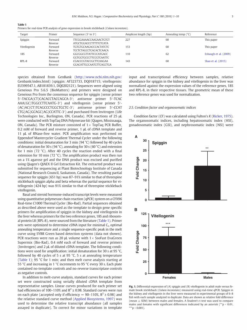
Fig. 1. Differential expression of (A) spiggin and (B) vitellogenin in adult male versus fe-male brook stickleback (Culaea inconstans) measured using real-time qPCR. Spiggin inthe kidney and vitellogenin in the liver were measured in experimental groups of 6–8fish with each sample analyzed in duplicate. Data are shown as relative fold difference(mean ± SEM) between males and females. A Student's t-test was used to comparemales and females with significant differences indicated by an asterisk (**p b 0.01,***p b 0.001).
3B.M. Muldoon, N.S. Hogan / Comparative Biochemistry and Physiology, Part C 180 (2016) 1–10
calculated by dividing liver, gonad or kidney weight (respectively) bythe weight of the fish and multiplying by 100.
2.6. Kidney histology
Preserved fishwere sectioned posterior to the pectoral fin and at theanus prior to tissue processing. Tissues were processed through step-wise dehydration in ethanol, cleared with xylene and embedded inparaffin. Sections were cut at 5 μm thickness with nine transversesections prepared each from posterior, middle and anterior portion ofthe abdomenwhichwere approximately 50 μmapart in order to ensurethe presence of many secondary proximal tubules of the kidney tissue.Sections were stained with hematoxylin–eosin. KEH was measuredusing AxioVision Rel. 4.8 Software (Carl Zeiss Canada, Toronto) onthree serial sections from posterior, middle and anterior portion of thekidney on three tubules per section with three measurements conduct-ed on each tubule. Mean KEH from individual fish was averaged toobtain KEH for each treatment. KEH was analyzed using a masked (orblind) evaluation and the results were verified by a second blindedassessment of a subsample of kidneys with the values verified againstoriginal data.
2.7. Statistical analysis
Data were tested for normality using the Kolmogorov–Smirnov teston residuals and for homogeneity of variance using Levene's test. Datanot meeting the assumptions of parametric statistics were log trans-formed prior to being re-analyzed. The differential expression of basalspiggin and vitellogenin in male versus female wild-caught brookstickleback was analyzed using a Student's t-test. Hormone-induced(EE2 andMT)data for organosomatic indices and CFwere analyzed sep-arately inmales and females using a nested ANOVA, with treatment andtank as factors in the hierarchical order listed, and the nest factor (tank)was removed if p-value was ≥0.250. Data was then re-run as a one-wayANOVA. Hormone-induced (MT and EE2) gene expression data wereanalyzed using a one-way ANOVA with spiggin analyzed in the kidneyof MT-exposed females and vitellogenin in the liver of EE2-exposedmales. The expression of spiggin in the kidney of control males in the
MT trial and vitellogenin expression in the liver of control females inthe EE2 trial were also measured to determine baseline expression inunexposedfish but this datawere not included in the statistical analysis.A post-hoc Dunnett's test for OSI, CF and gene expression data inhormone-treated fish compared each treatment to the solvent-control.All analyses were conducted on IBM SPSS Statistics 22 (Armonk, NY,United States).
3. Results
3.1. Sex difference in basal spiggin and vitellogenin transcript levels
Wild-caught mature brook stickleback exhibited differences inspiggin and vitellogenin transcript levels between sexes (Fig. 1). SpigginmRNA was significantly higher (17-fold) in the kidney of malestickleback relative to the female (p = 0.001, Fig. 1A). Alternately,vitellogenin mRNA was significantly higher (77-fold) in the liver offemale stickleback relative to the male (p = 0.001, Fig. 1B).
3.2. Experimental treatment of brook stickleback with MT or EE2
3.2.1. Effects of MT/EE2 on length, weight, condition factor andorganosomatic indices
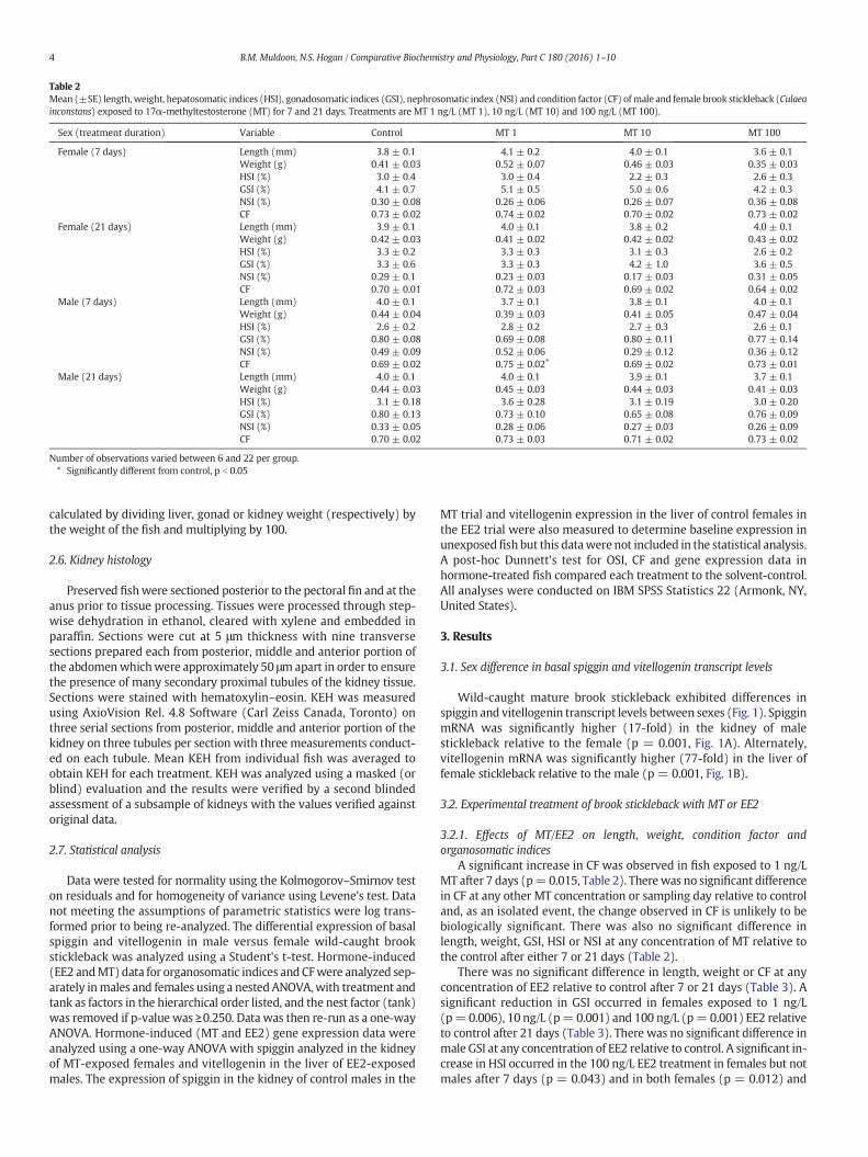
A significant increase in CF was observed in fish exposed to 1 ng/LMT after 7 days (p=0.015, Table 2). Therewas no significant differencein CF at any other MT concentration or sampling day relative to controland, as an isolated event, the change observed in CF is unlikely to bebiologically significant. There was also no significant difference inlength, weight, GSI, HSI or NSI at any concentration of MT relative tothe control after either 7 or 21 days (Table 2).
There was no significant difference in length, weight or CF at anyconcentration of EE2 relative to control after 7 or 21 days (Table 3). Asignificant reduction in GSI occurred in females exposed to 1 ng/L(p= 0.006), 10 ng/L (p= 0.001) and 100 ng/L (p= 0.001) EE2 relativeto control after 21 days (Table 3). There was no significant difference inmale GSI at any concentration of EE2 relative to control. A significant in-crease in HSI occurred in the 100 ng/L EE2 treatment in females but notmales after 7 days (p = 0.043) and in both females (p = 0.012) and
Table 2Mean (±SE) length,weight, hepatosomatic indices (HSI), gonadosomatic indices (GSI), nephrosomatic index (NSI) and condition factor (CF) ofmale and female brook stickleback (Culaeainconstans) exposed to 17α-methyltestosterone (MT) for 7 and 21 days. Treatments are MT 1 ng/L (MT 1), 10 ng/L (MT 10) and 100 ng/L (MT 100).
Sex (treatment duration) Variable Control MT 1 MT 10 MT 100
Female (7 days) Length (mm) 3.8 ± 0.1 4.1 ± 0.2 4.0 ± 0.1 3.6 ± 0.1Weight (g) 0.41 ± 0.03 0.52 ± 0.07 0.46 ± 0.03 0.35 ± 0.03HSI (%) 3.0 ± 0.4 3.0 ± 0.4 2.2 ± 0.3 2.6 ± 0.3GSI (%) 4.1 ± 0.7 5.1 ± 0.5 5.0 ± 0.6 4.2 ± 0.3NSI (%) 0.30 ± 0.08 0.26 ± 0.06 0.26 ± 0.07 0.36 ± 0.08CF 0.73 ± 0.02 0.74 ± 0.02 0.70 ± 0.02 0.73 ± 0.02
Female (21 days) Length (mm) 3.9 ± 0.1 4.0 ± 0.1 3.8 ± 0.2 4.0 ± 0.1Weight (g) 0.42 ± 0.03 0.41 ± 0.02 0.42 ± 0.02 0.43 ± 0.02HSI (%) 3.3 ± 0.2 3.3 ± 0.3 3.1 ± 0.3 2.6 ± 0.2GSI (%) 3.3 ± 0.6 3.3 ± 0.3 4.2 ± 1.0 3.6 ± 0.5NSI (%) 0.29 ± 0.1 0.23 ± 0.03 0.17 ± 0.03 0.31 ± 0.05CF 0.70 ± 0.01 0.72 ± 0.03 0.69 ± 0.02 0.64 ± 0.02
Male (7 days) Length (mm) 4.0 ± 0.1 3.7 ± 0.1 3.8 ± 0.1 4.0 ± 0.1Weight (g) 0.44 ± 0.04 0.39 ± 0.03 0.41 ± 0.05 0.47 ± 0.04HSI (%) 2.6 ± 0.2 2.8 ± 0.2 2.7 ± 0.3 2.6 ± 0.1GSI (%) 0.80 ± 0.08 0.69 ± 0.08 0.80 ± 0.11 0.77 ± 0.14NSI (%) 0.49 ± 0.09 0.52 ± 0.06 0.29 ± 0.12 0.36 ± 0.12CF 0.69 ± 0.02 0.75 ± 0.02⁎ 0.69 ± 0.02 0.73 ± 0.01
Male (21 days) Length (mm) 4.0 ± 0.1 4.0 ± 0.1 3.9 ± 0.1 3.7 ± 0.1Weight (g) 0.44 ± 0.03 0.45 ± 0.03 0.44 ± 0.03 0.41 ± 0.03HSI (%) 3.1 ± 0.18 3.6 ± 0.28 3.1 ± 0.19 3.0 ± 0.20GSI (%) 0.80 ± 0.13 0.73 ± 0.10 0.65 ± 0.08 0.76 ± 0.09NSI (%) 0.33 ± 0.05 0.28 ± 0.06 0.27 ± 0.03 0.26 ± 0.09CF 0.70 ± 0.02 0.73 ± 0.03 0.71 ± 0.02 0.73 ± 0.02
Number of observations varied between 6 and 22 per group.⁎ Significantly different from control, p b 0.05
4 B.M. Muldoon, N.S. Hogan / Comparative Biochemistry and Physiology, Part C 180 (2016) 1–10
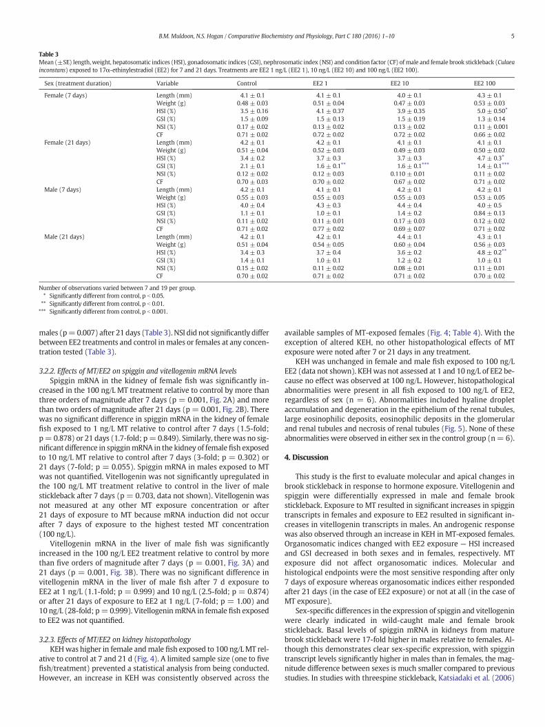
males (p=0.007) after 21 days (Table 3). NSI did not significantly differbetween EE2 treatments and control inmales or females at any concen-tration tested (Table 3).
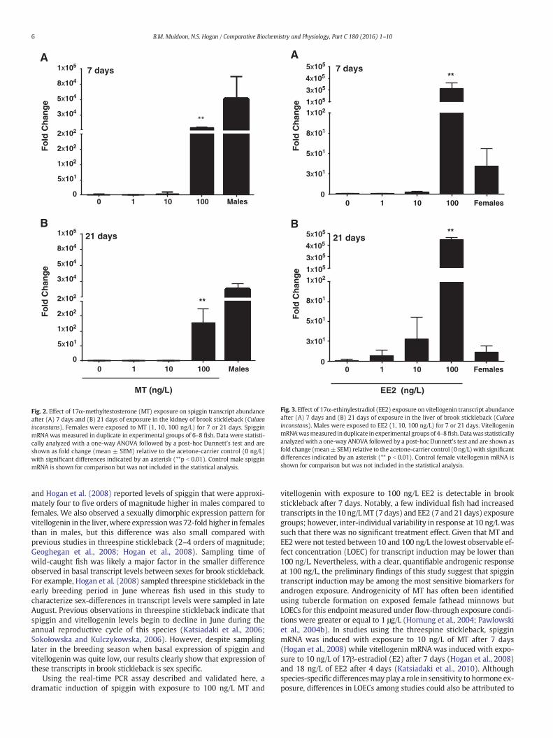
3.2.2. Effects of MT/EE2 on spiggin and vitellogenin mRNA levelsSpiggin mRNA in the kidney of female fish was significantly in-
creased in the 100 ng/L MT treatment relative to control by more thanthree orders of magnitude after 7 days (p = 0.001, Fig. 2A) and morethan two orders of magnitude after 21 days (p = 0.001, Fig. 2B). Therewas no significant difference in spiggin mRNA in the kidney of femalefish exposed to 1 ng/L MT relative to control after 7 days (1.5-fold;p= 0.878) or 21 days (1.7-fold; p= 0.849). Similarly, there was no sig-nificant difference in spigginmRNA in the kidney of female fish exposedto 10 ng/L MT relative to control after 7 days (3-fold; p = 0.302) or21 days (7-fold; p = 0.055). Spiggin mRNA in males exposed to MTwas not quantified. Vitellogenin was not significantly upregulated inthe 100 ng/L MT treatment relative to control in the liver of malestickleback after 7 days (p = 0.703, data not shown). Vitellogenin wasnot measured at any other MT exposure concentration or after21 days of exposure to MT because mRNA induction did not occurafter 7 days of exposure to the highest tested MT concentration(100 ng/L).
Vitellogenin mRNA in the liver of male fish was significantlyincreased in the 100 ng/L EE2 treatment relative to control by morethan five orders of magnitude after 7 days (p = 0.001, Fig. 3A) and21 days (p = 0.001, Fig. 3B). There was no significant difference invitellogenin mRNA in the liver of male fish after 7 d exposure toEE2 at 1 ng/L (1.1-fold; p = 0.999) and 10 ng/L (2.5-fold; p = 0.874)or after 21 days of exposure to EE2 at 1 ng/L (7-fold; p = 1.00) and10 ng/L (28-fold; p=0.999). VitellogeninmRNA in female fish exposedto EE2 was not quantified.
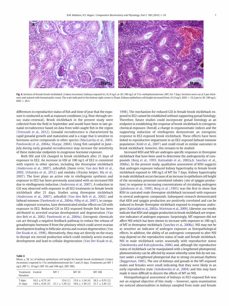
3.2.3. Effects of MT/EE2 on kidney histopathologyKEHwas higher in female andmale fish exposed to 100 ng/LMT rel-
ative to control at 7 and 21 d (Fig. 4). A limited sample size (one to fivefish/treatment) prevented a statistical analysis from being conducted.However, an increase in KEH was consistently observed across the
available samples of MT-exposed females (Fig. 4; Table 4). With theexception of altered KEH, no other histopathological effects of MTexposure were noted after 7 or 21 days in any treatment.
KEH was unchanged in female and male fish exposed to 100 ng/LEE2 (data not shown). KEHwas not assessed at 1 and 10 ng/L of EE2 be-cause no effect was observed at 100 ng/L. However, histopathologicalabnormalities were present in all fish exposed to 100 ng/L of EE2,regardless of sex (n = 6). Abnormalities included hyaline dropletaccumulation and degeneration in the epithelium of the renal tubules,large eosinophilic deposits, eosinophilic deposits in the glomerularand renal tubules and necrosis of renal tubules (Fig. 5). None of theseabnormalities were observed in either sex in the control group (n= 6).
4. Discussion
This study is the first to evaluate molecular and apical changes inbrook stickleback in response to hormone exposure. Vitellogenin andspiggin were differentially expressed in male and female brookstickleback. Exposure to MT resulted in significant increases in spiggintranscripts in females and exposure to EE2 resulted in significant in-creases in vitellogenin transcripts in males. An androgenic responsewas also observed through an increase in KEH in MT-exposed females.Organosomatic indices changed with EE2 exposure — HSI increasedand GSI decreased in both sexes and in females, respectively. MTexposure did not affect organosomatic indices. Molecular andhistological endpoints were the most sensitive responding after only7 days of exposure whereas organosomatic indices either respondedafter 21 days (in the case of EE2 exposure) or not at all (in the case ofMT exposure).
Sex-specific differences in the expression of spiggin and vitellogeninwere clearly indicated in wild-caught male and female brookstickleback. Basal levels of spiggin mRNA in kidneys from maturebrook stickleback were 17-fold higher in males relative to females. Al-though this demonstrates clear sex-specific expression, with spiggintranscript levels significantly higher in males than in females, the mag-nitude difference between sexes is much smaller compared to previousstudies. In studies with threespine stickleback, Katsiadaki et al. (2006)
Table 3Mean (±SE) length,weight, hepatosomatic indices (HSI), gonadosomatic indices (GSI), nephrosomatic index (NSI) and condition factor (CF) of male and female brook stickleback (Culaeainconstans) exposed to 17α-ethinylestradiol (EE2) for 7 and 21 days. Treatments are EE2 1 ng/L (EE2 1), 10 ng/L (EE2 10) and 100 ng/L (EE2 100).
Sex (treatment duration) Variable Control EE2 1 EE2 10 EE2 100
Female (7 days) Length (mm) 4.1 ± 0.1 4.1 ± 0.1 4.0 ± 0.1 4.3 ± 0.1Weight (g) 0.48 ± 0.03 0.51 ± 0.04 0.47 ± 0.03 0.53 ± 0.03HSI (%) 3.5 ± 0.16 4.1 ± 0.37 3.9 ± 0.35 5.0 ± 0.50⁎
GSI (%) 1.5 ± 0.09 1.5 ± 0.13 1.5 ± 0.19 1.3 ± 0.14NSI (%) 0.17 ± 0.02 0.13 ± 0.02 0.13 ± 0.02 0.11 ± 0.001CF 0.71 ± 0.02 0.72 ± 0.02 0.72 ± 0.02 0.66 ± 0.02
Female (21 days) Length (mm) 4.2 ± 0.1 4.2 ± 0.1 4.1 ± 0.1 4.1 ± 0.1Weight (g) 0.51 ± 0.04 0.52 ± 0.03 0.49 ± 0.03 0.50 ± 0.02HSI (%) 3.4 ± 0.2 3.7 ± 0.3 3.7 ± 0.3 4.7 ± 0.3⁎GSI (%) 2.1 ± 0.1 1.6 ± 0.1⁎⁎ 1.6 ± 0.1⁎⁎⁎ 1.4 ± 0.1⁎⁎⁎NSI (%) 0.12 ± 0.02 0.12 ± 0.03 0.110 ± 0.01 0.11 ± 0.02CF 0.70 ± 0.03 0.70 ± 0.02 0.67 ± 0.02 0.71 ± 0.02
Male (7 days) Length (mm) 4.2 ± 0.1 4.1 ± 0.1 4.2 ± 0.1 4.2 ± 0.1Weight (g) 0.55 ± 0.03 0.55 ± 0.03 0.55 ± 0.03 0.53 ± 0.05HSI (%) 4.0 ± 0.4 4.3 ± 0.3 4.4 ± 0.4 4.0 ± 0.5GSI (%) 1.1 ± 0.1 1.0 ± 0.1 1.4 ± 0.2 0.84 ± 0.13NSI (%) 0.11 ± 0.02 0.11 ± 0.01 0.17 ± 0.03 0.12 ± 0.02CF 0.71 ± 0.02 0.77 ± 0.02 0.69 ± 0.07 0.71 ± 0.02
Male (21 days) Length (mm) 4.2 ± 0.1 4.2 ± 0.1 4.4 ± 0.1 4.3 ± 0.1Weight (g) 0.51 ± 0.04 0.54 ± 0.05 0.60 ± 0.04 0.56 ± 0.03HSI (%) 3.4 ± 0.3 3.7 ± 0.4 3.6 ± 0.2 4.8 ± 0.2⁎⁎
GSI (%) 1.4 ± 0.1 1.0 ± 0.1 1.2 ± 0.2 1.0 ± 0.1NSI (%) 0.15 ± 0.02 0.11 ± 0.02 0.08 ± 0.01 0.11 ± 0.01CF 0.70 ± 0.02 0.71 ± 0.02 0.71 ± 0.02 0.70 ± 0.02
Number of observations varied between 7 and 19 per group.⁎ Significantly different from control, p b 0.05.⁎⁎ Significantly different from control, p b 0.01.⁎⁎⁎ Significantly different from control, p b 0.001.
5B.M. Muldoon, N.S. Hogan / Comparative Biochemistry and Physiology, Part C 180 (2016) 1–10
and Hogan et al. (2008) reported levels of spiggin that were approxi-mately four to five orders of magnitude higher in males compared tofemales. We also observed a sexually dimorphic expression pattern forvitellogenin in the liver, where expressionwas 72-fold higher in femalesthan in males, but this difference was also small compared withprevious studies in threespine stickleback (2–4 orders of magnitude;Geoghegan et al., 2008; Hogan et al., 2008). Sampling time ofwild-caught fish was likely a major factor in the smaller differenceobserved in basal transcript levels between sexes for brook stickleback.For example, Hogan et al. (2008) sampled threespine stickleback in theearly breeding period in June whereas fish used in this study tocharacterize sex-differences in transcript levels were sampled in lateAugust. Previous observations in threespine stickleback indicate thatspiggin and vitellogenin levels begin to decline in June during theannual reproductive cycle of this species (Katsiadaki et al., 2006;Sokołowska and Kulczykowska, 2006). However, despite samplinglater in the breeding season when basal expression of spiggin andvitellogenin was quite low, our results clearly show that expression ofthese transcripts in brook stickleback is sex specific.
Using the real-time PCR assay described and validated here, adramatic induction of spiggin with exposure to 100 ng/L MT and
vitellogenin with exposure to 100 ng/L EE2 is detectable in brookstickleback after 7 days. Notably, a few individual fish had increasedtranscripts in the 10 ng/LMT (7 days) and EE2 (7 and 21 days) exposuregroups; however, inter-individual variability in response at 10 ng/L wassuch that there was no significant treatment effect. Given that MT andEE2were not tested between 10 and 100 ng/L the lowest observable ef-fect concentration (LOEC) for transcript induction may be lower than100 ng/L. Nevertheless, with a clear, quantifiable androgenic responseat 100 ng/L, the preliminary findings of this study suggest that spiggintranscript induction may be among the most sensitive biomarkers forandrogen exposure. Androgenicity of MT has often been identifiedusing tubercle formation on exposed female fathead minnows butLOECs for this endpoint measured under flow-through exposure condi-tions were greater or equal to 1 μg/L (Hornung et al., 2004; Pawlowskiet al., 2004b). In studies using the threespine stickleback, spigginmRNA was induced with exposure to 10 ng/L of MT after 7 days(Hogan et al., 2008) while vitellogenin mRNA was induced with expo-sure to 10 ng/L of 17β-estradiol (E2) after 7 days (Hogan et al., 2008)and 18 ng/L of EE2 after 4 days (Katsiadaki et al., 2010). Althoughspecies-specific differencesmay play a role in sensitivity to hormoneex-posure, differences in LOECs among studies could also be attributed to
0 1 10 100 Males0
5x101
1x10²
2x10²
2x10²
3x10⁴
5x10⁴
8x10⁴
1x10⁵
0
5x101
1x10²
2x10²
2x10²
3x10⁴
5x10⁴
8x10⁴
1x10⁵
Fold
Cha
nge
Fold
Cha
nge
**
7 daysA
0 1 10 100 Males
21 daysB
MT (ng/L)
**
Fig. 2. Effect of 17α-methyltestosterone (MT) exposure on spiggin transcript abundanceafter (A) 7 days and (B) 21 days of exposure in the kidney of brook stickleback (Culaeainconstans). Females were exposed to MT (1, 10, 100 ng/L) for 7 or 21 days. SpigginmRNA was measured in duplicate in experimental groups of 6–8 fish. Data were statisti-cally analyzed with a one-way ANOVA followed by a post-hoc Dunnett's test and areshown as fold change (mean ± SEM) relative to the acetone-carrier control (0 ng/L)with significant differences indicated by an asterisk (**p b 0.01). Control male spigginmRNA is shown for comparison but was not included in the statistical analysis.
0 1 10 100 Females0
3x101
5x101
8x101
1x1021x105
3x10⁵
4x10⁵
5x10⁵
0
3x101
5x101
8x101
1x1021x105
3x10⁵
4x10⁵
5x10⁵
**7 days
A
0 1 10 100 Females
EE2 (ng/L)
**21 days
B
Fold
Cha
nge
Fold
Cha
nge
Fig. 3. Effect of 17α-ethinylestradiol (EE2) exposure on vitellogenin transcript abundanceafter (A) 7 days and (B) 21 days of exposure in the liver of brook stickleback (Culaeainconstans). Males were exposed to EE2 (1, 10, 100 ng/L) for 7 or 21 days. VitellogeninmRNAwasmeasured induplicate in experimental groups of 4–8fish. Datawas statisticallyanalyzed with a one-way ANOVA followed by a post-hoc Dunnett's test and are shown asfold change (mean± SEM) relative to the acetone-carrier control (0 ng/L)with significantdifferences indicated by an asterisk (** p b 0.01). Control female vitellogenin mRNA isshown for comparison but was not included in the statistical analysis.
6 B.M. Muldoon, N.S. Hogan / Comparative Biochemistry and Physiology, Part C 180 (2016) 1–10
differences in reproductive status of fish and time of year that the expo-sure is conducted aswell as exposure conditions (e.g. flow-through ver-sus static-renewal). Brook stickleback in the present study werecollected from the field in September and would have been in late go-nadal recrudescence based on data from wild caught fish in the region(Tetreault et al., 2012). Gonadal recrudescence is characterized byrapid gonadal growth and maturation and is a stage that is sensitive tohormone-active compounds in other species (MacLatchy et al., 2003;Pawlowski et al., 2004a; Sharpe, 2004). Using fish sampled in June–July during early gonadal recrudescence may increase the sensitivityof these molecular endpoints to exogenous hormone exposure.
Both HSI and GSI changed in brook stickleback after 21 days ofexposure to EE2. An increase in HSI at 100 ng/L of EE2 is consistentwith reports in other species, including the threespine stickleback(Andersson et al., 2007), zebrafish (Danio rerio; Van den Belt et al.,2002; Urbatzka et al., 2012) and medaka (Oryzias latipes; Ma et al.,2007). The liver plays an active role in vitellogenin synthesis andexposure to EE2 has been previously associated with an increased HSIdue to vitellogenesis induction (Andersson et al., 2007). A reduction inGSI was observed with exposure to all EE2 treatments in female brookstickleback after 21 days. Studies using threespine stickleback(Andersson et al., 2007), zebrafish (Van den Belt et al., 2002) andfatheadminnow (Pawlowski et al., 2004a; Filby et al., 2007), in compa-rable exposure scenarios, have demonstrated similar effects on GSIwithexposure to EE2. Reduced GSI in EE2-exposed female fish has beenattributed to arrested ovarian development and degeneration (Vanden Belt et al., 2002; Pawlowski et al., 2004a). Estrogenic chemicalscan act through a negative feedback pathway to reduce the release ofgonadotropin-hormone II from the pituitary which could arrest ovariandevelopment leading to follicular atresia and ovarian degeneration (VanDer Kraak et al., 1998). Alternatively, theymay act directly on the ovaryto disrupt sex steroid production which could similarly arrest ovariandevelopment and lead to cellular degeneration (Van Der Kraak et al.,
1998). The mechanism for reduced GSI in female brook stickleback ex-posed to EE2 cannot be establishedwithout supporting gonad histology.Therefore, future studies could incorporate gonad histology as anendpoint in establishing the response of brook stickleback to estrogenicchemical exposure. Overall, a change in organosomatic indices and thesupporting induction of vitellogenin demonstrate an estrogenicresponse in EE2-exposed brook stickleback. These effects have beenlinked to reproductive impairment in an EE2-exposed fathead minnowpopulation (Kidd et al., 2007) and could result in similar outcomes inbrook stickleback; however, this remains to be studied.
Increased KEH and NSI are androgen-specific responses in threespinestickleback that have been used to determine the androgenicity of com-pounds (Borg et al., 1993; Katsiadaki et al., 2002a,b; Sanchez et al.,2008a). In the present study, qualitative assessment of KEH suggeststhat androgen exposure induced kidney hypertrophy in female brookstickleback exposed to 100 ng/L of MT for 7 days. Kidney hypertrophyinmale stickleback occurs because of an increase in epitheliumcell heightof the secondary proximal convoluted tubules (site of spiggin produc-tion) in response to increasing concentrations of circulating androgens(Jakobsson et al., 1999). Borg et al. (1993) was the first to show thatKEH in castrated male threespine stickleback increased with exposureto several androgenic compounds. Subsequent research demonstratedthat KEH and spiggin production are positively correlated and can beinduced in female threespine stickleback exposed to exogenous andro-gens (Katsiadaki et al., 2002a;Wartman et al., 2009). Likewise, our resultsindicate that KEHand spiggin production in brook stickleback are respon-sive indicators of androgen exposure. Surprisingly, MT exposure did notaffect NSI which has been shown to increase with exposure to 100 ng/Lof MT in threespine stickleback (Sanchez et al., 2008a). NSI may not beas sensitive an indicator of androgen exposure as histopathologicaleffects. In addition, the ability of an androgenic compound to alter NSImay depend on the reproductive status of male and female stickleback.NSI in male stickleback varies seasonally with reproductive status(Sokołowska and Kulczykowska, 2006) and, although the reproductivestatus of stickleback can be manipulated with a lengthened photoperiod,photosensitivity can be affected by seasonwhereby some fish do notma-ture under a lengthened photoperiod due to strong circannual rhythms(Baggerman, 1985). The size of kidneys and gonads in the MT-exposedmales and females were small indicating that they were likely in anearly reproductive state (Sokołowska et al., 2004) and this may havemade it more difficult to discern the effects of MT on NSI.
Histopathological assessment of kidneys in EE2-exposed fish wasnot an original objective of this study — however, upon examinationwe noticed abnormalities in kidneys sampled from male and female
Fig. 4. Sections of female brook stickleback (Culaea inconstans) kidneys exposed to (A) 0 ng/L or (B) 100 ng/L of 17α-methyltestosterone (MT) for 7 days. Sections were cut at 5 μm thick-ness and stainedwith hematoxylin–eosin. The scale indicated in the bottom right corner is 50 μm.Kidney epitheliumcell height increased from (A) 0 ng/L; KEH=16.2 μmto (B) 100 ng/L;KEH = 26.3.
Table 4Mean ± SE (n) of kidney epithelium cell height for female brook stickleback (Culaeainconstans) exposed to 17α-methyltestosterone for 7 and 21 days. Treatments are MT 1ng/L (MT 1), 10 ng/L (MT 10) and 100 ng/L (MT 100).
TreatmentDuration
Control MT 1 MT 10 MT 100
7 days 16.2 ± 0.75 (4) 13.6 (1) 15.9 ± 1.24 (4) 26.3 ± 0.53 (2)21 days 14.0 ± 0.35 (5) 21.1 ± 1.29 (2) 19.6 ± 1.30 (3) 21.7 ± 2.45 (3)
7B.M. Muldoon, N.S. Hogan / Comparative Biochemistry and Physiology, Part C 180 (2016) 1–10
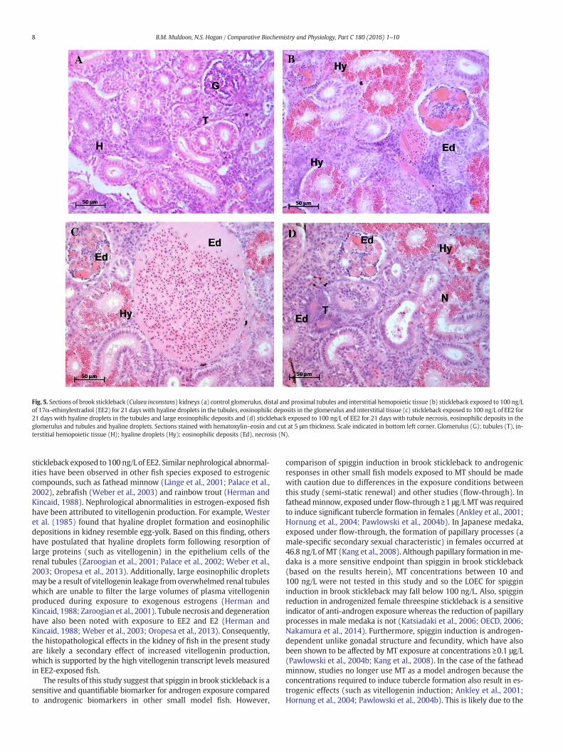
stickleback exposed to 100 ng/L of EE2. Similar nephrological abnormal-ities have been observed in other fish species exposed to estrogeniccompounds, such as fathead minnow (Länge et al., 2001; Palace et al.,2002), zebrafish (Weber et al., 2003) and rainbow trout (Herman andKincaid, 1988). Nephrological abnormalities in estrogen-exposed fishhave been attributed to vitellogenin production. For example, Westeret al. (1985) found that hyaline droplet formation and eosinophilicdepositions in kidney resemble egg-yolk. Based on this finding, othershave postulated that hyaline droplets form following resorption oflarge proteins (such as vitellogenin) in the epithelium cells of therenal tubules (Zaroogian et al., 2001; Palace et al., 2002; Weber et al.,2003; Oropesa et al., 2013). Additionally, large eosinophilic dropletsmay be a result of vitellogenin leakage from overwhelmed renal tubuleswhich are unable to filter the large volumes of plasma vitellogeninproduced during exposure to exogenous estrogens (Herman andKincaid, 1988; Zaroogian et al., 2001). Tubule necrosis and degenerationhave also been noted with exposure to EE2 and E2 (Herman andKincaid, 1988; Weber et al., 2003; Oropesa et al., 2013). Consequently,the histopathological effects in the kidney of fish in the present studyare likely a secondary effect of increased vitellogenin production,which is supported by the high vitellogenin transcript levels measuredin EE2-exposed fish.
The results of this study suggest that spiggin in brook stickleback is asensitive and quantifiable biomarker for androgen exposure comparedto androgenic biomarkers in other small model fish. However,
comparison of spiggin induction in brook stickleback to androgenicresponses in other small fish models exposed to MT should be madewith caution due to differences in the exposure conditions betweenthis study (semi-static renewal) and other studies (flow-through). Infatheadminnow, exposed under flow-through ≥1 μg/LMTwas requiredto induce significant tubercle formation in females (Ankley et al., 2001;Hornung et al., 2004; Pawlowski et al., 2004b). In Japanese medaka,exposed under flow-through, the formation of papillary processes (amale-specific secondary sexual characteristic) in females occurred at46.8 ng/L of MT (Kang et al., 2008). Although papillary formation inme-daka is a more sensitive endpoint than spiggin in brook stickleback(based on the results herein), MT concentrations between 10 and100 ng/L were not tested in this study and so the LOEC for spiggininduction in brook stickleback may fall below 100 ng/L. Also, spigginreduction in androgenized female threespine stickleback is a sensitiveindicator of anti-androgen exposure whereas the reduction of papillaryprocesses in male medaka is not (Katsiadaki et al., 2006; OECD, 2006;Nakamura et al., 2014). Furthermore, spiggin induction is androgen-dependent unlike gonadal structure and fecundity, which have alsobeen shown to be affected by MT exposure at concentrations ≥0.1 μg/L(Pawlowski et al., 2004b; Kang et al., 2008). In the case of the fatheadminnow, studies no longer use MT as a model androgen because theconcentrations required to induce tubercle formation also result in es-trogenic effects (such as vitellogenin induction; Ankley et al., 2001;Hornung et al., 2004; Pawlowski et al., 2004b). This is likely due to the
Fig. 5. Sections of brook stickleback (Culaea inconstans) kidneys (a) control glomerulus, distal and proximal tubules and interstitial hemopoietic tissue (b) stickleback exposed to 100 ng/Lof 17α-ethinylestradiol (EE2) for 21 dayswith hyaline droplets in the tubules, eosinophilic deposits in the glomerulus and interstitial tissue (c) stickleback exposed to 100 ng/L of EE2 for21 days with hyaline droplets in the tubules and large eosinophilic deposits and (d) stickleback exposed to 100 ng/L of EE2 for 21 days with tubule necrosis, eosinophilic deposits in theglomerulus and tubules and hyaline droplets. Sections stained with hematoxylin–eosin and cut at 5 μm thickness. Scale indicated in bottom left corner. Glomerulus (G); tubules (T), in-terstitial hemopoietic tissue (H); hyaline droplets (Hy); eosinophilic deposits (Ed), necrosis (N).
8 B.M. Muldoon, N.S. Hogan / Comparative Biochemistry and Physiology, Part C 180 (2016) 1–10

ability of MT to be aromatized to methyl estradiol, resulting inestrogenicity (Ankley et al., 2001; Hornung et al., 2004). The concentra-tions ofMT employed in the present study induced spiggin but not vitel-logenin. Likewise, MT had no effect on vitellogenin mRNA in threespinestickleback under similar exposure conditions (Hogan et al., 2008). Itappears that in stickleback, MT is not aromatized into methyl estradiolat concentrations high enough to induce vitellogenin in males. Futurestudies should explore the relative potency of other androgeniccompounds (e.g., 5α-dihydrotestosterone, 11-ketotestosterone).Additional research is also required to confirm the sensitivity of brookstickleback to MT and EE2 using comparable exposure scenarios (ie.flow-through) in order to further validate spiggin as an androgenicbiomarker in this species. In terms of estrogen responsiveness, thefathead minnow, medaka and zebrafish may be more suitable testspecies with more established test methods and (in some cases) amore sensitive vitellogenin response than stickleback (reviewed inKatsiadaki et al., 2007). However, the ability to simultaneously assessandrogen and estrogen exposure in a single fish using fully quantitativeendpoints is an advantage held only by the stickleback.
The suitability of brook stickleback as a bioindicator of EDCs isfurther enhanced by their broad distribution and physiology. Brookstickleback are abundant throughout North America and are found ina range of freshwater environments (including lakes, streams andponds; Stewart et al., 2007). They are fairly sedentary and are thereforegood indicators of environmental conditions within a given location(Wootton, 1984). They can survive low oxygen, cold water tempera-tures and repeated handling (Reisman and Cade, 1967; Wootton,1984) and this tolerance may allow them to survive contaminatedenvironments when other fish do not. They can also be bred in the lab-oratory (Reisman and Cade, 1967; McKenzie, 1969; McLennan, 1993)and so could be used in reproductive bioassays that evaluate effects ofEDC exposure on reproductive behavior and fecundity. The definitive,measurable responses to hormone exposure demonstrated here mayextend the use of brook stickleback to regulatory testing and ecologicalmonitoring of endocrine active compounds in aquatic environments.Furthermore, there is potential to develop additional endocrinebioassays for brook stickleback, similar to those used with threespineand other species, such as in vitro gonadal steroidogenesis (MacLatchyet al., 2003; Hogan et al., 2008), gonad histopathology (Allen et al.,2008), behavior (in stickleback related to male nest building andcourting; Sebire et al., 2008; Dzieweczynski and Forrette, 2014) andfecundity (Maunder et al., 2007). Reproductive and behavioralendpoints in brook stickleback could then be utilized in constructingadverse outcome pathways, linking molecular indices to adverseoutcomes that are relevant to risk assessment. There is also significantpotential to assess the anti-androgenic properties of compounds usingin vitro (kidney explants) and in vivo methods (21-day androgenizedfemale stickleback assay) already established in the threespinestickleback (Katsiadaki et al., 2006; Jolly et al., 2009; OECD, 2011). Final-ly, both biomarker and apical endpoints could be evaluated in brookstickleback with exposure to environmental samples in both the fieldand laboratory. The threespine stickleback has been successfullyemployed as a biomonitoring species in contaminated environmentsand to assess the endocrine activity of complex mixtures, includingsewage and pulp and paper mill effluent (Sanchez et al., 2008b;Björkblom et al., 2009, 2013; Wartman et al., 2009; Pottinger et al.,2011; Katsiadaki et al., 2012).
In conclusion,we have developed two sensitive qPCR assays that canbe used to measure either androgen exposure through spiggintranscript induction in female kidneys or estrogen exposure through vi-tellogenin transcript induction in male livers. We have demonstratedthat these endpoints are highly responsive to short term hormoneexposure. Moreover, androgenic and estrogenic responses of brookstickleback were measurable at higher levels of biological organization(e.g. organosomatic indices). As such, we were able to demonstrate aclear, definitive androgenic and estrogenic response, which was
detected not only at themolecular level but also throughmorphologicalendpoints that corroborate an androgenic and estrogenic response. Theability to evaluate whole organism effects and mechanisms of actionpositions the brook stickleback as a promising new freshwater fishmodel for understanding androgenic responses in fish.
Acknowledgments
We thankDr. Jason Raine and the Aquatic Toxicology Research Facil-ity (ARTF) at the Toxicology Centre, University of Saskatchewan for pro-viding space and equipment for the exposure study. We also thank Drs.Steve Wiseman and Tim Jardine for comments on the manuscript andvarious members of the Hogan lab for assistance with field and labwork. This research was funded by a Natural Sciences and EngineeringResearch Council of Canada (NSERC) (358787-2009) Discovery grantto NSH. BM was funded by an NSERC Canada Graduate Scholarshipand Toxicology Devolved Graduate Scholarship.
References
Allen, Y.T., Katsiadaki, I., Pottinger, T.G., Jolly, C., Matthiessen, P., Mayer, I., Smith, A., Scott,A.P., Eccles, P., Sanders, M.B., Pulman, K.G.T., Feist, S., 2008. Intercalibration exerciseusing a stickleback endocrine disrupter screening assay. Environ. Toxicol. Chem. 27,404–412.
Andersson, C., Katsiadaki, I., Lundstedt-Enkel, K., Örberg, J., 2007. Effects of17α-ethynylestradiol on EROD activity, spiggin and vitellogenin in three-spinedstickleback (Gasterosteus aculeatus). Aquat. Toxicol. 83, 33–42.
Ankley, G.T., Johnson, R.D., 2004. Small fish models for identifying and assessing theeffects of endocrine-disrupting chemicals. ILAR J. 45, 469–483.
Ankley, G.T., Jensen, K.M., Kahl, M.D., Korte, J.J., Makynen, E.A., 2001. Description andevaluation of a short-term reproduction test with the fathead minnow (Pimephalespromelas). Environ. Toxicol. Chem. 20, 1276–1290.
Ankley, G.T., Jensen, K.M., Makynen, E.A., Kahl, M.D., Korte, J.J., Hornung, M.W., Henry, T.R.,Denny, J.S., Leino, R.L., Wilson, V.S., Cardon, M.C., Hartig, P.C., Gray, L.E., 2003. Effectsof the androgenic growth promoter 17-β-trenbolone on fecundity and reproductiveendocrinology of the fathead minnow. Environ. Toxicol. Chem. 22, 1350–1360.
Applied Biosystems, 1997. Applied Biosystems, Relative Quantitation of Gene Expression.PE Applied Biosystems, Foster City, CA, p. 36.
Arukwe, A., 2001. Cellular and molecular responses to endocrine-modulators and theimpact on fish reproduction. Mar. Pollut. Bull. 42, 643–655.
Baggerman, B., 1985. The roles of daily and annual biological rhythms in the photoperiod-ic regulation of the breeding season in the stickleback Gasterosteus aculeatus L. Be-haviour 1–7.
Björkblom, C., Högfors, E., Salste, L., Bergelin, E., Olsson, P.E., Katsiadaki, I., Wiklund, T.,2009. Estrogenic and androgenic effects of municipal wastewater effluent onreproductive endpoint biomarkers in three-spined stickleback (Gasterosteusaculeatus). Environ. Toxicol. Chem. 28, 1063–1071.
Björkblom, C., Mustamäki, N., Olsson, P.E., Katsiadaki, I., Wiklund, T., 2013. Assessment ofreproductive biomarkers in three-spined stickleback (Gasterosteus aculeatus) fromsewage effluent recipients. Environ. Toxicol. 28, 229–237.
Blanchfield, P.J., Kidd, K.A., Docker, M.F., Palace, V.P., Park, B.J., Postma, L.D., 2015. Recoveryof a wild fish population from whole-lake additions of a synthetic estrogen. Environ.Sci. Technol. 49, 3136–3144.
Borg, B., Antonopoulou, E., Andersson, E., Carlberg, T., Mayer, I., 1993. Effectiveness ofseveral androgens in stimulating kidney hypertrophy, a secondary sexual character,in castrated male three-spined sticklebacks, Gasterosteus aculeatus. Can. J. Zool. 71,2327–2329.
Bringolf, R.B., Belden, J.B., Summerfelt, R.C., 2004. Effects of atrazine on fatheadminnow ina short-term reproduction assay. Environ. Toxicol. Chem. 23, 1019–1025.
Dzieweczynski, T.L., Forrette, L.M., 2014. Timescale effects of 17α-ethinylestradiol onbehavioral consistency in male threespine stickleback. Acta Ethol. 18, 137–144.
Esbaugh, A.J., Perry, S.F., Gilmour, K.M., 2009. Hypoxia-inducible carbonic anhydrase IXexpression is insufficient to alleviate intracellular metabolic acidosis in the muscleof zebrafish, Danio rerio. Am. J. Physiol. Regul. Integr. Comp. Physiol. 296, R150–R160.
Filby, A., Thorpe, K., Maack, G., Tyler, C., 2007. Gene expression profiles revealing themechanisms of anti-androgen- and estrogen-induced feminization in fish. Aquat.Toxicol. 81, 219–231.
Geoghegan, F., Katsiadaki, I., Williams, T.D., Chipman, J.K., 2008. A cDNA microarray forthe three-spined stickleback, Gasterosteus aculeatus L., and analysis of the interactiveeffects of oestradiol and dibenzanthracene exposures. J. Fish Biol. 72, 2133–2153.
Herman, R.L., Kincaid, H.L., 1988. Pathological effects of orally administered estradiol torainbow trout. Aquaculture 72, 165–172.
Hogan, N.S., Wartman, C.A., Finley, M.A., van der Lee, J.G., van den Heuvel, M.R., 2008.Simultaneous determination of androgenic and estrogenic endpoints in thethreespine stickleback (Gasterosteus aculeatus) using quantitative RT-PCR. Aquat.Toxicol. 90, 269–276.
Hornung, M.W., Jensen, K.M., Korte, J.J., Kahl, M.D., Durhan, E.J., Denny, J.S., Henry, T.R.,Ankley, G.T., 2004. Mechanistic basis for estrogenic effects in fathead minnow(Pimephales promelas) following exposure to the androgen 17α-methyltestosterone:
9B.M. Muldoon, N.S. Hogan / Comparative Biochemistry and Physiology, Part C 180 (2016) 1–10

conversion of 17α-methyltestosterone to 17α-methylestradiol. Aquat. Toxicol. 66,15–23.
Hutchinson, T.H., Ankley, G.T., Segner, H., Tyler, C.R., 2006. Screening and testing forendocrine disruption in fish—biomarkers as “signposts”, not “traffic lights”, in riskassessment. Environ. Health Perspect. 114, 106–114.
Jakobsson, S., Borg, B., Haux, C., Hyllner, S.J., 1999. An 11-ketotestosterone inducedkidney-secreted protein: the nest building glue from male three-spined stickleback,Gasterosteus aculeatus. Fish Physiol. Biochem. 20, 79–85.
Jolly, C., Katsiadaki, I., Morris, S., Le Belle, N., Dufour, S., Mayer, I., Pottinger, T.G., Scott, A.P.,2009. Detection of the anti-androgenic effect of endocrine disrupting environmentalcontaminants using in vivo and in vitro assays in the three-spined stickleback. Aquat.Toxicol. 92, 228–239.
Kang, I.J., Yokota, H., Oshima, Y., Tsuruda, Y., Shimasaki, Y., Honjo, T., 2008. The effects ofmethyltestosterone on the sexual development and reproduction of adult medaka(Oryzias latipes). Aquat. Toxicol. 87, 37–46.
Katsiadaki, I., Scott, A.P., Hurst, M.R., Matthiessen, P., Mayer, I., 2002a. Detection of envi-ronmental androgens: a novel method based on enzyme-linked immunosorbentassay of spiggin, the stickleback (Gasterosteus aculeatus) glue protein. Environ.Toxicol. Chem. 21, 1946–1954.
Katsiadaki, I., Scott, A.P., Mayer, I., 2002b. The potential of the three-spined stickleback(Gasterosteus aculeatus L.) as a combined biomarker for oestrogens and androgensin European waters. Mar. Environ. Res. 54, 725–728.
Katsiadaki, I., Morris, S., Squires, C., Hurst, M.R., James, J.D., Scott, A.P., 2006. Use of thethree-spined stickleback (Gasterosteus aculeatus) as a sensitive in vivo test fordetection of environmental antiandrogens. Environ. Health Perspect. 114, 115–121.
Katsiadaki, I., Sanders, M.B., Sebire, M., Nagae, M., Soyano, K., Scott, A.P., 2007.Three-spined stickleback: an emerging model in environmental endocrinedisruption. Environ. Sci. 14, 263–283.
Katsiadaki, I., Williams, T.D., Ball, J.S., Bean, T.P., Sanders, M.B., Wu, H., Santos, E.M., Brown,M.M., Baker, P., Ortega, F., Falciani, F., Craft, J.A., Tyler, C.R., Viant, M.R., Chipman, J.K.,2010. Hepatic transcriptomic and metabolomic responses in the Stickleback(Gasterosteus aculeatus) exposed to ethinyl-estradiol. Aquat. Toxicol. 97, 174–187.
Katsiadaki, I., Sanders, M.B., Henrys, P.A., Scott, A.P., Matthiessen, P., Pottinger, T.G., 2012.Field surveys reveal the presence of anti-androgens in an effluent-receiving riverusing stickleback-specific biomarkers. Aquat. Toxicol. 122–123, 75–85.
Kidd, K.A., Blanchfield, P.J., Mills, K.H., Palace, V.P., Evans, R.E., Lazorchak, J.M., Flick, R.W.,2007. Collapse of a fish population after exposure to a synthetic estrogen. Proc. Natl.Acad. Sci. 104, 8897–8901.
Länge, R., Hutchinson, T.H., Croudace, C.P., Siegmund, F., Schweinfurth, H., Hampe, P., Panter,G.H., Sumpter, J.P., 2001. Effects of the synthetic estrogen 17α-ethinylestradiol on thelife-cycle of the fathead minnow (Pimephales promelas). Environ. Toxicol. Chem. 20,1216–1227.
Ma, T., Wang, Z., Gong, S., 2007. Comparative sensitivity in Chinese rare minnow(Gobiocypris rarus) and Japanese medaka (Oryzias latipes) exposed toethinylestradiol. J. Environ. Sci. Health A 42, 889–894.
MacLatchy, D.L., Courtenay, S.C., Rice, C.D., Van Der Kraak, G.J., 2003. Development of ashort-term reproductive endocrine bioassay using steroid hormone and vitellogeninend points in the estuarine mummichog (Fundulus heteroclitus). Environ. Toxicol.Chem. 22, 996–1008.
Martinović, D., Blake, L.S., Durhan, E.J., Greene, K.J., Kahl, M.D., Jensen, K.M., Makynen, E.A.,Villeneuve, D.L., Ankley, G.T., 2008. Reproductive toxicity of vinclozolin in the fatheadminnow: confirming an anti-androgenic mode of action. Environ. Toxicol. Chem. 27,478–488.
Maunder, R.J., Matthiessen, P., Sumpter, J.P., Pottinger, T.G., 2007. Impaired reproductionin three-spined sticklebacks exposed to ethinyl estradiol as juveniles. Biol. Reprod.77, 999–1006.
McKenzie, J.A., 1969. The courtship behavior of the male brook stickleback, Culaeainconstans (Kirtland). Can. J. Zool. 47, 1281–1286.
McLennan, D.A., 1993. Changes in female breeding behaviour across the ovulatory cycle inthe brook stickleback, Culaea inconstans (Kirtland). Behaviour 126, 191–218.
Nakamura, A., Takanobu, H., Tamura, I., Yamamuro, M., Iguchi, T., Tatarazako, N., 2014.Verification of responses of Japanese medaka (Oryzias latipes) to anti-androgens,vinclozolin and flutamide, in short-term assays: effects of antiandrogens onJapanese medaka. J. Appl. Toxicol. 34, 545–553.
OECD, 2006. Report of the validation of the 21-day fish screening assay for the detectionof endocrine substances (phase 1b). Series on Testing and AssessmentOECDEnvironment Health and Safety Publications No. 61. Organization for EconomicCo-Operation and Development, Paris, France.
OECD, 2009. Test no. 230: 21-day fish assay. OECD Guidelines for the Testing ofChemicals, Section 2. OECD Publishing.
OECD, 2011. Guidance document on the androgenised female stickleback screen. Serieson Testing and Assessment No. 148. Organization for Economic Co-Operation andDevelopment, Paris, France.
Oropesa, A.L., Jiménez, B., Fallola, C., Pula, H.J., Cuesta, J.M., Gómez, L., 2013. Histologicalalterations on the structure of the excretory renal system in tench (Tinca tinca)after exposure to 17-alpha-ethynylestradiol. Bull. Environ. Contam. Toxicol. 91,623–629.
Palace, V.P., Evans, R.E., Wautier, K., Baron, C., Vandenbyllardt, L., Vandersteen, W., Kidd,K.A., 2002. Induction of vitellogenin and histological effects in wild fathead minnowsfrom a lake experimentally treated with the synthetic estrogen, ethynylestradiol.Water Qual. Res. J. Can. 37, 637–650.
Pawlowski, van Aerle, R., Tyler, C., Braunbeck, T., 2004a. Effects of 17α-ethinylestradiol ina fathead minnow (Pimephales promelas) gonadal recrudescence assay. Ecotoxicol.Environ. Saf. 57, 330–345.
Pawlowski, Sauer, A., Shears, J., Tyler, C., Braunbeck, T., 2004b. Androgenic and estrogeniceffects of the synthetic androgen 17α-methyltestosterone on sexual developmentand reproductive performance in the fathead minnow (Pimephales promelas) deter-mined using the gonadal recrudescence assay. Aquat. Toxicol. 68, 277–291.
Pottinger, T.G., Cook, A., Jürgens, M.D., Rhodes, G., Katsiadaki, I., Balaam, J.L., Smith, A.J.,Matthiessen, P., 2011. Effects of sewage effluent remediation on body size, somaticRNA:DNA ratio, andmarkers of chemical exposure in three-spined sticklebacks. Envi-ron. Int. 37, 158–169.
Reisman, H.M., Cade, T.J., 1967. Physiological and behavioral aspects of reproduction inthe brook stickleback, Culaea inconstans. Am. Midl. Nat. 77, 257.
Ricker, W., 1975. Computation and interpretation of biological statistics of fish popula-tions. Bull. Fish. Res. Board Can. 191, 382.
Rotchell, J., Ostrander, G., 2003. Molecular markers of endocrine disruption in aquatic or-ganisms. J. Toxicol. Environ. Health B 6, 453–496.
Sanchez, W., Goin, C., Brion, F., Olsson, P.E., Goksøyr, A., Porcher, J.M., 2008a. A new ELISAfor the three-spined stickleback (Gasterosteus aculeatus L.) spiggin, using antibodiesagainst synthetic peptide. Comp. Biochem. Physiol. C Toxicol. Pharmacol. 147,129–137.
Sanchez, W., Piccini, B., Ditche, J.M., Porcher, J.M., 2008b. Assessment of seasonal variabil-ity of biomarkers in three-spined stickleback (Gasterosteus aculeatus L.) from a lowcontaminated stream: Implication for environmental biomonitoring. Environ. Int.34, 791–798.
Scholz, S., Mayer, I., 2008. Molecular biomarkers of endocrine disruption in small modelfish. Mol. Cell. Endocrinol. 293, 57–70.
Sebire, M., Allen, Y., Bersuder, P., Katsiadaki, I., 2008. The model anti-androgen flutamidesuppresses the expression of typical male stickleback reproductive behaviour. Aquat.Toxicol. 90, 37–47.
Seki, M., Fujishima, S., Nozaka, T., Maeda, M., Kobayashi, K., 2006. Comparison of responseto 17β-estradiol and 17β-trenbolone among three small fish species. Environ.Toxicol. Chem. 25, 2742–2752.
Shao, Y.T., Tseng, Y.C., Chang, C.H., Yan, H.Y., Hwang, P.P., Borg, B., 2015. GnRH mRNAlevels in male three-spined sticklebacks, Gasterosteus aculeatus, under different re-productive conditions. Comp. Biochem. Physiol., Part A Mol. Integr. Physiol. 180,6–17.
Sharpe, R., 2004. Effects of a model androgen (methyl testosterone) and a model anti-androgen (cyproterone acetate) on reproductive endocrine endpoints in a short-term adult mummichog (Fundulus heteroclitus) bioassay. Aquat. Toxicol. 67, 203–215.
Sokołowska, E., Kulczykowska, E., 2006. Annual reproductive cycle in two free living pop-ulations of three-spined stickleback (Gasterosteus aculeatus L.): patterns of ovarianand testicular development. Oceanologia 48.
Sokołowska, E., Kalamarz, H., Kulczykowska, E., 2004. Seasonal changes in brain melato-nin concentration in the three-spined stickleback (Gasterosteus aculeatus): towardsan endocrine calendar. Comp. Biochem. Physiol. A Mol. Integr. Physiol. 139, 365–369.
Stewart, D.B., Carmichael, T.J., Sawatzky, C.D., Mochnacz, N.J., Reist, J.D., 2007. Fish Dietsand Food Webs in the Northwest Territories: Brook Stickleback (Culaea inconstans).Fisheries and Oceans Canada.
Tetreault, G.R., Bennett, C.J., Cheng, C., Servos, M.R., McMaster, M.E., 2012. Reproductiveand histopathological effects in wild fish inhabiting an effluent-dominated stream,Wascana Creek, SK, Canada. Aquat. Toxicol. 110–111, 149–161.
Urbatzka, R., Rocha, E., Reis, B., Cruzeiro, C., Monteiro, R.A.F., Rocha, M.J., 2012. Effects ofethinylestradiol and of an environmentally relevantmixture of xenoestrogens on ste-roidogenic gene expression and specific transcription factors in zebrafish. Environ.Pollut. 164, 28–35.
Van den Belt, K., Wester, P.W., van der Ven, L.T.M., Verheyen, R., Witters, H., 2002. Effectsof ethynylestradiol on the reproductive physiology in zebrafish (Danio rerio): timedependency and reversibility. Environ. Toxicol. Chem. 21, 767–775.
Van Der Kraak, G.J., Zacharewski, T., Janz, D.M., Sanders, B.M., Gooch, J.W., 1998. Compar-ative endocrinology and mechanisms of endocrine modulation in fish and wildlife.Principles and Processes for Evaluating Endocrine Disruption in WildlifeProceedingsFrom Principles and Processes for Evaluating Endocrine Disruption in Wildlife;March 1996. Society of Environmental Toxicology and Chemistry, Kiawah IslandS.C., Pensacola, Florida, p. 515.
Wartman, C.A., Hogan, N.S., Hewitt, L.M., McMaster, M.E., Landman, M.J., Taylor, S., Kovacs,T.G., van den Heuvel, M.R., 2009. Androgenic effects of a Canadian bleached kraft pulpand paper effluent as assessed using threespine stickleback (Gasterosteus aculeatus).Aquat. Toxicol. 92, 131–139.
Weber, L.P., Hill, R.L., Janz, D.M., 2003. Developmental estrogenic exposure in zebrafish(Danio rerio): II. Histological evaluation of gametogenesis and organ toxicity. Aquat.Toxicol. 63, 431–446.
Wester, P.W., Canton, J.H., Bisschop, A., 1985. Histopathological study of Poecilia reticulata(guppy) after long-term β-hexachlorocyclohexane exposure. Aquat. Toxicol. 6,271–296.
Wootton, R.J., 1984. A Functional Biology of Sticklebacks. University of California Press.Zaroogian, G., Gardner, G., Borsay Horowitz, D., Gutjahr-Gobell, R., Haebler, R., Mills, L.,
2001. Effect of 17β-estradiol, o,p′-DDT, octylphenol and p,p′-DDE on gonadal devel-opment and liver and kidney pathology in juvenile male summer flounder(Paralichthys dentatus). Aquat. Toxicol. 54, 101–112.
10 B.M. Muldoon, N.S. Hogan / Comparative Biochemistry and Physiology, Part C 180 (2016) 1–10
Related Documents