Cell Division

Welcome message from author
This document is posted to help you gain knowledge. Please leave a comment to let me know what you think about it! Share it to your friends and learn new things together.
Transcript

Cell Division

The Basics
Who: Mitosis
What: Cell division
When: New cells are needed
Where: Somatic cells (body cells)
Why: Growth

The BasicsWho: Somatic cells (Normal cells like in your toes)
What: Cellular reproduction for growth, the cell cycle
Where: Happens in somatic cells, regular body cells like in your toes
Why: For growth (baby to adult) and to replace damaged cells (heal a cut)
How: Duplicate cell parts making an exact copy of cell; one "mother" cell becomes two identical "daughter" cells.
Remember: Toes as is mi-toes-is (mitosis)

Mitosis

Mitosis & ChromosomesMitosis is the duplication of chromosomes

Chromosomes are made of
DNA

Genes are part of Chromosomes

Cell Division
• One parent cell becomes two daughter cells.
• Happens in somatic cells, every day cells in the body
• For growth
StartStart
FinishFinish
1 cell
2 cells

Cell Plate (Plants)

Mitosis in an animal cell• 1 cell to 4 cells

Uneven DivisionDivision of cytoplasm is
not always even

The Phases
There are five phases

Cell Cycle
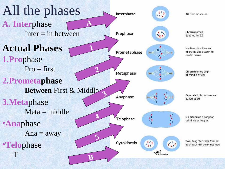
A. InterphaseInter = in between
Actual Phases1.Prophase
Pro = first
2.PrometaphaseBetween First & Middle
3.MetaphaseMeta = middle
•AnaphaseAna = away
•TelophaseT
elo = distant
B. Cytokinesis
All the phases
1
2
3
4
A
5
B

Mitosis with emphasis on metaphase

Mitosis Onion Root Tip

Inte
rpha
se
• Interphase is NOT a part of mitosis.
• Interphase the cell grows before the DNA is duplicated, then DNA is duplicated, and lastly, prepares for division.

Prop
hase
• During prophase, the DNA and proteins start to condense.
• The microtubles are assembled start moving to one of the two centriole pairs toward the opposite end of the cell.
Dark region = condensing chromatinOnion root tip image

Prom
etap
hase
• Sometimes considered part of the prophase.
• Nuclear membrane disintegrates
• Centrioles reach the poles of the cell
• Chromosomes continue to contract.
• Proteins attach to the centromeres.
• The chromosomes begin moving.
Dark region = condensing chromatin, animal cell

Met
apha
se
• During metaphase, the microtubules penetrate nuclear region forming a spindle apparatus.
• They attach to sister chromatids of each chromosome.
• All chromosomes line up at spindle equator.
• Now in their most tightly condensed form.

Ana
phas
e• During
anaphase, attachments between the two sister chromatids of each chromosome break.
• Now separate chromosomes move to opposite spindle poles.
Mid Anaphase Onion root tip

Telo
phas
e• Lastly, in
telophase, the chromosomes decondense, texture of chromatin loosens.
• New patches of membrane fuse to form new nuclear envelopes around them.

Cytokinesis & Daughter CellsAnimal cells. •Pinching of cytoplasm into two cells. Plant cells •Cell plate forms between the two “new” cells. After cytokinesis•Cells now in interphase Called daughter cells. •Cells diploid, two each type of chromosome – same as parent cell's nucleus.

Mitosis on the run

Stages1. Interphase
The cell is engaged in metabolic activity and preparing for mitosis (the next four phases that lead up to and include nuclear division).
Chromosomes are not clearly discerned in the nucleus, although a dark spot called the nucleolus may be visible. The cell may contain a pair of centrioles (or
microtubule organizing centers in plants) both of which are organizational sites for microtubules. This is the longest stage.
2. Prophase Chromatin in the nucleus begins to condense and becomes
visible in the light microscope as chromosomes. The nucleolus disappears. Centrioles begin moving to opposite ends of the cell and fibers extend from the centromeres. Some fibers cross the cell to form the mitotic spindle. 3.

Stages3. Prometaphase Sometimes considered part of the prophase. When the nuclear membrane disintegrates, the centrioles reach the poles of the cell, and the chromosomes continue to contract. Proteins attach to the centromeres. The chromosomes begin moving.
4. MetaphaseSpindle fibers align the chromosomes along the middle of the
cell nucleus. This line is referred to as the metaphase plate. This organization helps to ensure that in the next phase, when the chromosomes are separated, each new nucleus will receive one copy of each chromosome

Stages5. AnaphaseThe paired chromosomes separate at the kinetochores and move to opposite sides of the cell. Motion results from a combination of kinetochore movement along the spindle microtubules and through the physical interaction of polar microtubules.
6. Telophase Chromatids arrive at opposite poles of cell, and new membranes form around the daughter nuclei. The chromosomes disperse and are no longer visible under the light microscope. The spindle fibers disperse, and cytokinesis or the partitioning of the cell may also begin during this stage. 7. CytokinesisIn animal cells, cytokinesis results when a fiber ring composed of a protein called actin around the center of the cell contracts pinching the cell into two daughter cells, each with one nucleus. In plant cells, the rigid wall requires that a cell plate be synthesized between the two daughter cells.

Interphase

Prophase

Prometaphase

Metaphase

Early Anaphase

Late Anaphase

Telophase

Daughter Cells

Mitosis

Can you identify the stages?
1. Are these plant or animal cells?
2. How can you tell?
3
4
5

Can you identify the stages?
6. Are these plant or animal cells?
7. How can you tell?
8
109

Purpose of Mitosis
• Increase the size of an organism.• Replace worn out cells or repair
damaged tissue.• Reproduce identical organisms, or
clones.

Comparing Mitosis & Meiosis• Mitosis
– Happens in somatic cells, every day cells in the body
– For growth– Think: toes toes as is mi-as is mi-
toestoes-is-is
• Meiosis – Happens in sex cells
(pre-embryonic cells)– For sexual
reproduction– Think: e as in sex as
in meiosis

Bibliography•Bio Review Cell Division http://library.thinkquest.org/28751/review/division/4.html•Access Excellence at the National Health Museum About Biotech http://www.accessexcellence.org/AB/GG/meiosis.html•About http://biology.about.com/cs/celldivision/•“The Cell Cycle & Mitosis Tutorial” The Biology Project University of Arizona http://www.biology.arizona.edu/cell_bio/tutorials/cell_cycle/cells3.html• Mitosis http://www.stanford.edu/group/Urchin/mitosis.htm•eMuseum Minnesota State University http://www.anthro.mankato.msus.edu/biology/evolution/genetics/cellsmitosismeosis.html•Molecular Expressions http://micro.magnet.fsu.edu/micro/gallery/mitosis/mitosis.html•Universlity of North Carolina at Charlotte http://www.bioweb.uncc.edu/biol1110/Stages.htm
Related Documents