Reproduced with permission of the copyright owner. Further reproduction prohibited without permission. EFFECTS OF THE RIBONUCLEASE INHIBITOR ON THE BIOLOGICAL ACTIVITY OF PANCREATIC-TYPE RIBONUCLEASES by Kimberly Anne Dickson A dissertation submitted in partial fulfillment of the requirements for the degree of Doctor of Philosophy (Biochemistry) at the UNIVERSITY OF WISCONSIN - MADISON 2006

Welcome message from author
This document is posted to help you gain knowledge. Please leave a comment to let me know what you think about it! Share it to your friends and learn new things together.
Transcript

Reproduced with permission of the copyright owner. Further reproduction prohibited without permission.
EFFECTS OF THE RIBONUCLEASE INHIBITOR ON THE BIOLOGICAL
ACTIVITY OF PANCREATIC-TYPE RIBONUCLEASES
by
Kimberly Anne Dickson
A dissertation submitted in partial fulfillment
of the requirements for the degree of
Doctor of Philosophy
(Biochemistry)
at the
UNIVERSITY OF WISCONSIN - MADISON
2006

Reproduced with permission of the copyright owner. Further reproduction prohibited without permission.
A dissertation entitled
Effect of the Ribonuclease Inhibitor on the Biological Activity of Pancreatic-Type Ribonucleases
submitted to the Graduate School of the University of Wisconsin-Madison
in partial fulfillment of the requirements for the degree of Doctor of Philosophy
by
Kimberly Anne Dickson
Date of Final Oral Examination: March l3, 2006
Month & Year Degree to be awarded: December May August
**************************************************************************************************
Signature, Dean of Graduate School
Reproduced with permission of the copyright owner. Further reproduction prohibited without permission.
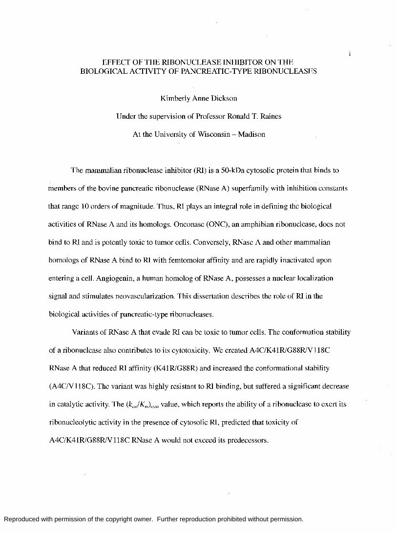
EFFECT OF THE RIBONUCLEASE INHIBITOR ON THE BIOLOGICAL ACTIVITY OF PANCREATIC-TYPE RIBONUCLEASES
Kimberly Anne Dickson
Under the supervision of Professor Ronald T. Raines
At the University of Wisconsin - Madison
The mammalian ribonuclease inhibitor (RI) is a 50-kDa cytosolic protein that binds to
members of the bovine pancreatic ribonuclease (RNase A) superfamily with inhibition constants
that range 10 orders of magnitude. Thus, RI plays an integral role in defining the biological
activities of RNase A and its homologs. Onconase (ONC), an amphibian ribonuclease, does not
bind to RI and is potently toxic to tumor cells. Conversely, RNase A and other mammalian
homologs of RNase A bind to RI with femtomolar affinity and are rapidly inactivated upon
entering a cell. Angiogenin, a human homolog of RNase A, possesses a nuclear localization
signal and stimulates neovascularization. This dissertation describes the role of RI in the
biological activities of pancreatic-type ribonucleases.
Variants of RNase A that evade RI can be toxic to tumor cells. The conformation stability
of a ribonuclease also contributes to its cytotoxicity. We created A4C/K41R1G88R1V118C
RNase A that reduced RI affinity (K41R1G88R) and increased the conformational stability
(A4C1VI18C). The variant was highly resistant to RI binding, but suffered a significant decrease
in catalytic activity. The (kca/Km)cyto value, which reports the ability of a ribonuclease to exert its
ribonucleolytic activity in the presence of cytosolic RI, predicted that toxicity of
A4C1K41R1G88R1V118C RNase A would not exceed its predecessors.
Reproduced with permission of the copyright owner. Further reproduction prohibited without permission.
II
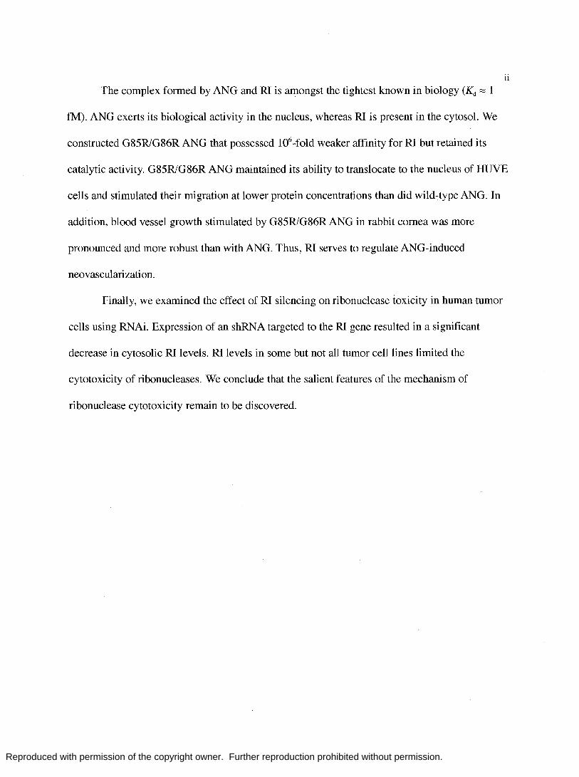
The complex formed by ANG and RI is amongst the tightest known in biology (Kd ~ 1
fM). ANG exerts its biological activity in the nucleus, whereas RI is present in the cytosol. We
constructed G85R1G86R ANG that possessed lO6-fold weaker affinity for RI but retained its
catalytic activity. G85R1G86R ANG maintained its ability to translocate to the nucleus of HUVE
cells and stimulated their migration at lower protein concentrations than did wild-type ANG. In
addition, blood vessel growth stimulated by G85R1G86R ANG in rabbit cornea was more
pronounced and more robust than with ANG. Thus, RI serves to regulate ANG-induced
neovascularization.
Finally, we examined the effect of RI silencing on ribonuclease toxicity in human tumor
cells using RNAi. Expression of an shRNA targeted to the RI gene resulted in a significant
decrease in cytosolic RI levels. RI levels in some but not all tumor cell lines limited the
cytotoxicity of ribonucleases. We conclude that the salient features of the mechanism of
ribonuclease cytotoxicity remain to be discovered.

Reproduced with permission of the copyright owner. Further reproduction prohibited without permission.
Acknowledgements
I am grateful to many people for the support and assistance that they have offered me
during my graduate career. First, I would like to thank my advisor, Ron Raines, who has
provided a vital mix of encouragement, enthusiasm, creativity, and resources with which to
conduct this research. The Raines lab has fostered my intellectual development and helped me
realize my career goals.
III
Many members of the Raines lab, past and present, have contributed greatly to this work.
Pete Leland, Marcia Haigis, and Tony Klink introduced me to many aspects of ribonuclease
biochemistry and have been invaluable resources long after their tenure at UW-Madison. I
would like to thank the chemists of the Raines lab, especially Sunil Chandran, for the
opportunities to collaborate on a variety of projects. While they are not part of this dissertation,
those endeavors have greatly expanded my knowledge of chemistry and my perspectives as a
scientist. My classmate, Steve Fuchs, and the "younger" members of the Raines lab have all
provided valuable advice and support over the years. In particular, I would like to thank Rebecca
Turcotte and Jeremey Johnson, with whom I shared many fascinating discussions, scientific and
otherwise.
I am also very grateful to the members of my thesis committee, who have supported my
research for the past six years and continue to support me as I launch my career at Macalester
College. Their wisdom and thoughtful advice is greatly appreciated.
Finally, I would like to thank my family for their endless support and encouragement. My
graduate career has been long and sometimes difficult. Nevertheless, my parents and my

Reproduced with permission of the copyright owner. Further reproduction prohibited without permission.
husband, Jeff, have offered consistent encouragement and support. This dissertation would
have never been possible without their love.
IV

Reproduced with permission of the copyright owner. Further reproduction prohibited without permission.
v Table of Contents
Abstract ........................................................................................................ .i
Acknowledgements ......................................................................................... .iii
Table of Contents ............................................................................................. v
List of Tables ................................................................................................ vii
List of Figures ............................................................................................... viii
List of Abbreviations ....................................................................................... .ix
Chapter One
Introduction ........................................................................................... 1
Chapter Two
Compensating Effects on the Cytotoxicity of Ribonuclease A Variants ............................ .31
2.1 Abstract .......................................................................................... 32
2.2 Introduction ..................................................................................... 33
2.3 Materials and Methods ......................................................................... 35
2.4 Results ........................................................................................... 38
2.5 Discussion ....................................................................................... 40
Chapter 3
Ribonuclease Inhibitor Regulates Neovascularization by Human Angiogenin ..................... 46
3.1 Abstract ......................................................................................... 47
3.2 Introduction .................................................................................... 48
3.3 Materials and Methods ........................................................................ 50
3.4 Results .......................................................................................... 55

Reproduced with permission of the copyright owner. Further reproduction prohibited without permission.
VI
3.5 Discussion ...................................................................................... 57
Chapter 4
Effects of Ribonuclease Inhibitor Silencing on Ribonuclease Toxicity in Human
Tumor Cells ........................................................................................ 65
4.1 Abstract ......................................................................................... 66
4.2 Introduction .................................................................................... 67
4.3 Materials and Methods ........................................................................ 69
4.4 Results .......................................................................................... 73
4.5 Discussion ...................................................................................... 75
Appendix I ................................................................................................... 82
Appendix II .................................................................................................. 85
References ................................................................................................... 89

Reproduced with permission of the copyright owner. Further reproduction prohibited without permission.
vii
List of Tables
Table 1.1 Kinetic parameters for Rl inhibition of ribonucleases ................................ 24
Table 1.2 Properties of ribonuclease A, its variants, and Onconase® ........................... 25
Table 1.3 Characteristics of LRR protein subfamilies ............................................ 26
Table 2.1 Properties of ribonuclease A, its variants, and Onconase® .......................... .43
Table 3.1 Properties of RNase A, ANG, and variants ............................................. 59
Table 4.1 ICso values of Rl-evasive and non-evasive ribonucleases in tumor cell
lines with and without Rl suppression ................................................... 79

Reproduced with permission of the copyright owner. Further reproduction prohibited without permission.
Figure 1.1
Figure 1.2
Figure 1.3
Figure 1.4
Figure 2.1
Figure 2.2
Figure 3.1
Figure 3.2
Figure 3.3
Figure 3.4
Figure 3.5
Figure 4.1
Figure 4.2
List of Figures
Three-dimensional structures of RI and its complexes with
ribonucleases ............................................................................. 27
Allignment of the amino acid sequence of RI from human, porcine
Vlll
mouse, and rat. ........................................................................... 28
Typical A-type and B-type repeats of RI ............................................... 29
Structures of five representative LRR proteins ..................................... .30
Interactions in the complex of RI and RNase A ............................. " ...... 44
Effect of ribonucleases on the proliferation of K-562 cells ....................... .45
Molecular interactions between human RI andANG ............................... 60
Zymogram electrophoresis of ANG and G85R1G86RANG ....................... 61
Nuclear translocation of ANG and G85R1G86R ANG in HUVE cells .......... 62
Wound healing migration of HUVE cells induced by ANG or
G85R1G86R ANG ....................................................................... 63
Induction of angiogenesis in rabbit cornea in vivo by ANG or
G85R1G86R ANG ....................................................................... 64
Immunoblot analysis of RI suppression in human tumor cell lines .............. 80
Effect of ribonucleases on proliferation of HeLa cells transfected
with pGE-NEG or pGE-DAL. .................... , ................................... 81

Reproduced with permission of the copyright owner. Further reproduction prohibited without permission.
ix
List of Abbreviations
ANG .................................................................................. human angiogenin
BS-RNase ................................................................ bovine seminal ribonuclease
DEPC. ............................................................................. diethylpyrocarbonate
DTT ......................................................................................... dithiolthreitol
EDT A ................................................................. ethylenediaminetetraacetic acid
IPTG ........................................................ .isopropyl-l-thio- -D-galactopyranoside
MES ........................................................... 2-[N -morpholino )ethanesuphonic acid
ONe .............................................................................................. onconase
PBS .............................................................. , ............. phosphate-buffered saline
RI .................................................................................. ribonuclease inhibitor
RNAi ...................................................................................... RNA inhibition
RNase .................................................................. human pancreatic ribonuclease
RNase A ................................................................ bovine pancreatic ribonuclease
shRNA ................................................................................. short hairpin RNA
SDS-PAGE. .......................... sodium dodecyl sulfate-polyacrylamide gel electrophoresis
Tris .................................................................. tris(hydroxymethyl)aminomethane
Tm .................................................. ........ midpoint of the thermal denaturation curve
Uv ................................................................................................ ultraviolet
6-F AM .............................................................................. 6-carboxyfl uorescein
6-T AMRA ...................................................... 6-carboxytetramethylaminorhodamine

Reproduced with permission of the copyright owner. Further reproduction prohibited without permission.
Chapter One
Introduction
Portions of this chapter were published as:
Dickson, K. A., Haigis, M. c., and Raines, R. T. (2005). Ribonuclease Inhibitor: Structure
and Function. Progress in Nucleic Acid Research and Molecular Biology. 80, 349-374.
1

Reproduced with permission of the copyright owner. Further reproduction prohibited without permission.
2 The mammalian ribonuclease inhibitor (RI) is a 50-kDa cytosolic protein that binds
to pancreatic-type ribonucleases with femtomolar affinity and renders them inactive (for
other reviews, see (Roth 1967; Blackburn and Moore 1982; Lee and Vallee 1993; Hofsteenge
1997; Shapiro 2001). Complexes formed by RI and its target ribonucleases are among the
tightest of known biomolecular interactions. The three-dimensional structure of RI is likewise
remarkable, being characterized by alternating units of a-helix and ~-strand that form a
striking horseshoe shape (Fig. 1A) (Kobe and Deisenhofer 1993). The repeating structural
units of RI possess a highly repetitive amino acid sequence that is rich in leucine residues
(Hofsteenge et al. 1988; Lee et al. 1988). These leucine-rich repeats (LRRs) are present in a
large family of proteins that are distinguished by their display of vast surface areas to foster
protein-protein interactions (Janin 1994; Kobe and Deisenhofer 1994; Shapiro et al. 1995;
Kobe and Kajava 2001). The unique structure and function of RI have resulted in its
emergence as the central protein in the study of LRRs, as well as its widespread use as a
laboratory reagent to eliminate ribonucleolytic activity (Pasloske 2001).
The biological role of RI is not known in its entirety. The ribonucleases recognized by
RI are secreted proteins, whereas RI resides exclusively in the cytosol. Nevertheless, RI
affinity has been shown to be the primary determinant of ribonuclease cytotoxicity: only
ribonucleases that evade RI can kill a cell (for reviews, see (Youle and D' Alessio 1997;
Leland and Raines 2001; Matousek 2001; Makarov and Ilinskaya 2003). In addition, the
complex of RI with human angiogenin (ANG), which stimulates neovascularization by
activating transcription in the nucleus (Moroianu and Riordan 1994; Xu et al. 2002), is the
tightest of known RI-ribonuclease complexes. Yet, a role for RI in angiogenesis is not clear.
Also intriguing are the 30-32 cysteine residues of RI, all of which must remain reduced for
Reproduced with permission of the copyright owner. Further reproduction prohibited without permission.
3 the protein to retain activity (Fominaya and Hofsteenge 1992). These observations have lead
researchers to hypothesize multiple biological roles for RI: (1) to protect cells from invading
ribonucleases, (2) to regulate or terminate the activity of ribonucleases with known
intracellular functions, and (3) to monitor the oxidation state of the cell in response to factors
such as aging and oxidative stress. Here, we review the salient features of RI biochemistry
and structure and thereby provide a context for examining the roles of RI in biology.
Biochemical Properties
The inhibitory activity of RI in guinea pig liver extracts was discovered in 1952
(Pirotte and Desreux 1952). This activity was inactivated by proteases, heat, or sulfhydryl
group modification, and was sensitive to changes in pH (for a review, see (Roth 1962). In
addition, the inhibitory activity was isolated in the supernatant fraction during a high-speed
centrifugation, indicative of cytoplasmic localization. In the 1970's, techniques were
developed to purify RI to homogeneity, enabling its biochemical characterization (Blackburn
et al. 1977; Blackburn and Moore 1982). Since then, RI has been isolated from numerous
mammalian sources, including brain (Burton et al. 1980; Cho and Joshi 1989) (Nadano,
1994), liver (Nadano, 1994; Gribnau et al. 1970; Burton and Fucci 1982), testis (Ferreras et
al. 1995), and erythrocytes (Moenner et al. 1998).
Purification. RI is particularly abundant in mammalian placenta and liver, which have
served as the major source of RI for purification. Human placental RI was first purified to
homogeneity using a combination of ion-exchange and ribonuclease-affinity chromatography
(Blackburn et al. 1977). The tight complex formed by RI and bovine pancreatic ribonuclease
(RNase A (Raines 1998); EC 3.1.27.5) has been exploited to achieve a> 1<Y -fold purification
Reproduced with permission of the copyright owner. Further reproduction prohibited without permission.
of RI in a single chromatographic step using immobilized RNase A. Today, most
purification methods rely upon such ribonuclease-affinity chromatography, followed by
anion-exchange chromatography (Garcia and Klebe 1997). Using these purification
techniques, approximately 6 mg RI per kg of wet tissue has been isolated from mammalian
liver (Burton and Fucci 1982) and placenta (Blackburn 1979). Human erythrocytes are also
rich in RI -the erythrocyte fraction of 100 mL of blood has yielded 430 Jig of RI (Moenner
et al. 1998).
Several recombinant systems for the production of RI have been reported, three from
Escherichia coli and one from Saccharomyces cerevisiae (Vescia et al. 1980; Lee and Vallee
1989; Vicentini et al. 1990). Low yields and insolubility have proven to be recurring
problems in producing recombinant RI. To date, the most efficient recombinant system
utilizes the trp promoter from E. coli to drive expression of porcine RI, and yields
approximately 10 mg of RI per liter of culture (Klink et al. 2001).
4
Characterization. RI is an acidic (pI 4.7) cytosolic protein that binds to pancreatic
type ribonucleases with 1: 1 stiochiometry (Blackburn and lailkhan 1979). Members of the
RNase A superfamily of proteins that are inhibited by RI include RNase A, human pancreatic
ribonuclease (RNase 1), ANG, eosinophil-derived neurotoxin (EDN, also known as RNase
2), RNase 4, and monomers of bovine seminal ribonuclease (BS-RNase). When complexed
with RI, these ribonucleases are no longer able to bind or degrade RNA (Lee and Vallee
1993). RI is ineffective against known non-mammalian homologs of RNase A. The amino
acid sequences of human, porcine, mouse, and rat RI share 66% identity (Fig. 2) (Hofsteenge
et al. 1988; Lee et al. 1988; Kawanomoto et al. 1992; Haigis et al. 2002). One third of the
residues that differ are conservative substitutions. To date, RI from human and pigs have
Reproduced with permission of the copyright owner. Further reproduction prohibited without permission.
5 been characterized most thoroughly and exhibit many identical properties (for reviews, see
(Hofsteenge 1997; Shapiro 2001). Thus, the source of RI will be discussed herein only if a
significant divergence occurs with respect to a particular experimental observation.
The affinity of RI for ribonucleases is extraordinary. Accordingly, substantial effort
has been invested in characterizing RI-ribonuclease interactions (for a review, see (Shapiro
2001). Techniques to assess binding rely upon the imposition of physical changes or
inhibition of catalytic activity. A purely physical method is more convenient to use for
ribonucleases with low catalytic activity, such as ANG (Lee et al. 1989). For example,
stopped-flow techniques and the 50% increase in the fluorescence of Trp89 of ANG upon
binding to RI have been used to study the association of RI with ANG. They report a two-
step binding mechanism that involves formation of a loose enzyme· inhibitor complex (E·I)
followed by isomerization to form a tight complex (E-I*), as in Eq. (1):
(1)
ANG and RI rapidly form a loose complex (K1 = kjkl = 0.53 jlM), which converts slowly (k2
= 97 S-I) to a stable complex. The association rate constant, ka = klkAk_l + k2) was found to be
monitoring release of ANG from the RI·ANG complex in the presence of excess RNase A as
a scavenger, and found to be 1.3 x 10⁻⁷ S-1 (Lee and Vallee 1989). This value corresponds to a
half-life of 62 days for the RI·ANG complex. The resulting value of the equilibrium
dissociation constant, Kd = kd1ka = 7.1 x 10⁻¹⁶ M, is exceptionally low, and comparable to the
Kd = 6 X 10-16 M of the avidin· biotin complex (Green 1975). A competition assay based on

Reproduced with permission of the copyright owner. Further reproduction prohibited without permission.
fluorescence changes in ANG has been used to measure Kd = 4.4 X 10⁻¹⁴ M for the
RI·RNase A complex (Lee et al. 1989).
6
RI has only a slight effect on the fluorescence of RNase A, which lacks tryptophan
residues. Enzymatic assays in which the value of Ki is determined by the ability of RI to
compete with RNA are viable alternatives for this and other ribonucleases that possess high
catalytic activity. In general, enzymatic assays require that ribonucleolytic activity can be
performed at low enzyme concentrations-no more than 2 orders-of-magnitude greater than
the Ki (Vicentini et al. 1990). Enzymological methods have been used to assess the affinity of
RI for RNase A, RNase 1, and RNase 4 (Table I) (Vicentini et al. 1990; Zelenko et al. 1994;
Boix et al. 1996; Hofsteenge et al. 1998). For examples, the values of ka = 1.7 X 10⁸ M-1s-1,
kd = 9.8 X 10--6 s-\ and Ki = 5.9 X 10⁻¹⁴ M were determined by measuring the decrease in
ribonuc1eolytic activity upon addition of RI.
The affinity of RNase A and RNase 2 for RI has also been assessed with a
combination of physical and enzymological techniques. The kd value for the RI·RNase A
complex was determined by measuring the release of RNase A in the presence of ANG as a
scavenger (Lee et al. 1989; Lee et al. 1989). The concentration of free RNase A was detected
by high-performance liquid chromatography or by enzymatic activity with RNA substrates
that are not cleaved by ANG. Similar assays have been used to determine the kinetic
parameters for the RI·RNase 2 interaction (Shapiro and Vallee 1991). The kinetic and
thermodynamic parameters determined with a variety of physical and enzymatic methods are
in gratifying agreement (Table I).
A fluorescence-based assay has been developed to facilitate rapid measurement of Kd
for a wide variety of RI·ribonuclease complexes (Abel et al. 2001). This assay employs
Reproduced with permission of the copyright owner. Further reproduction prohibited without permission.
7 fluorescein-labeled G88R RNase A, which has diminished affinity for RI and exhibits an
approximately 20% decrease in fluorescence when bound to RI. Titration of RI with
fluorescein-G88R RNase A yielded Kd = 0.55 X 10⁻⁹ M for the complex. A competition assay
using fluorescein-G88R RNase A was then used to determine the Kd value of unlabeled
ribonucleases (Table II). This assay is limited to measuring complexes with Kd values in the
nanomolar range or higher, as tighter complexes take too long to reach equilibrium.
Nonetheless, this assay has proven to be valuable for determining Kd values of numerous
RNase A variants, some of which possess low catalytic activity (Haigis et al. 2002; Dickson
et al. 2003).
Structure
Three-Dimensional Structure. Leucine is the most abundant residue in RI, comprising
18% of its amino acids (Blackburn et al. 1977; Burton and Fucci 1982). In 1988, the amino
acid sequence of RI from both porcine liver and human placenta was elucidated, revealing
that RI is comprised entirely of leucine-rich repeats (LRR) (Hofsteenge et al. 1988; Lee et al.
1988). Two types of alternating repeats have been described, A-type (which contains 28
residues) and B-type (which contains 29 residues). Porcine RI is built from 8 A-type and 7 B
type repeats, flanked by short terminal segments (Fig. 2) (Kobe and Deisenhofer 1994).
RI was the first LRR protein to be crystallized and have its three-dimensional
structure determined by X-ray diffraction analysis (Kobe and Deisenhofer 1993). Its
horseshoe shape is one of the most captivating of protein structures. The alternating A- and
B-type LRR motifs correspond to structural units, each consisting of an a-helix and ~-strand
connected by loops (Fig. 2A and 2B). The symmetric and non-globular arrangement of LRRs

Reproduced with permission of the copyright owner. Further reproduction prohibited without permission.
8 represents a new protein fold (for reviews, see: (Kobe and Deisenhofer 1995; Kajava 1998;
Kobe and Kajava 2001). The LRR units of RI are arranged so that the a-helices and B-strands
are aligned parallel to a common axis (Fig. lA). An extended B-sheet defines the inner
circumference of the horseshoe and provides a vast surface for interacting with other proteins.
Leucines and other aliphatic residues are essential components of the hydrophobic core of the
protein, and serve to stabilize the interactions Bween the LRR units (Fig. 3). The curvature of
the RI horseshoe is determined by the difference in distance Bween neighboring B-strands
and a-helices (Kajava 1998; Kobe and Kajava 2001). The curvature of RI is quite
pronounced, as the addition of only 5 more LRR units to the native 15 would cause the
termini of RI to collide (Kobe and Deisenhofer 1993).
A Model Leucine-Rich Repeat Protein. The LRR was first described with respect to
the leucine-rich a2-glycoprotein found in human serum (Takahashi et al. 1985). RI was the
first cytosolic protein discovered to possess LRRs (Hofsteenge et al. 1988; Lee et al. 1988).
In the past decade, more than a hundred LRR proteins have been identified; these proteins
have been found to perform remarkably different functions. In most LRR proteins, however,
the LRRs appear to serve as the interface for a protein-protein interaction (for reviews, see
(Kobe and Deisenhofer 1995; Kajava 1998).
LRR proteins have been classified into subfamilies base on the organism of origin,
cellular localization, and LRR consensus sequence (Kobe and Kajava 2001). To date, seven
LRR subfamilies of proteins have been described (Table III), and additional subfamilies
could arise with the discovery of more LRR proteins. Members of the RI-like subfamily are
intracellular proteins found in animals, and are characterized by repeats of 28/29 amino acids
that possess the sequence LXXLXLXX(N/C)XL. Other members of the RI-like subfamily

Reproduced with permission of the copyright owner. Further reproduction prohibited without permission.
include human MHC class II transactivator (P33076), Ran GTPase activating protein from
Saccharomyces pombe (P46060), RNA 1 gene product from Saccharomyces cerevisiae
(X17376), and the mouse homolog of RNA 1 (U208S7).
9
In general, the ~-strand region of the repeat is the most conserved among LRR
proteins (Kobe and Kajava 2001). Subfamilies differ primarily in the secondary structure
displayed in the regions ~ween the ~-strands (Table III, Fig. 4) (Kobe and Kajava 2001).
Short LRR units result in extended conformations in the interstrand region. For example,
members of the bacterial subfamily of LRR proteins are built from repeating units of only 20
amino acid residues. In the SDS22-like family, the a-helix found in RI-like proteins is often
replaced by a 310 helix (Price et al. 1998). In the structure of Y opM, an extracellular protein
that confers bacteria with virulence, the a-helix is replaced with a polyproline type-II (PU)
helix (Table III) (Evdokimov et al. 2001). Structures of representative proteins from five
subfamilies illustrate the diversity in the size and shape of LRR proteins (Fig. 4) (Schulman
et al. 2000; Matteo et al. 2003; Schott et al. 2004).
The structure of RI is repetitive and symmetrical, and its surface area is vast and
largely concave (Fig. IA). These unusual attributes make RI a potential platform for the
creation of new receptors. Towards this goal, a consensus LRR domain determined from the
sequences of rat, pig, and human RI has been used to generate proteins containing 2-12
LRRs (Stumpp et al. 2003). Biophysical analyses ofthe RI-like proteins showed monomeric
behavior and circular dichroism spectra characteristic of wild-type RI, suggesting that RI-like
proteins are viable templates for engineering.
Gene Structure and Evolution. RI homologs have been identified in numerous
mammalian species and have been found in nearly every type of organ, tissue, and gland

Reproduced with permission of the copyright owner. Further reproduction prohibited without permission.
10 investigated to date. Only one copy of the RI gene exists in the human genome (Crawford
et al. 1989), and RIs isolated from different tissues of the same species typically have the
same amino acid sequence. Still, subtle divergences exist. For example, alternative splice-site
forms have been identified in the 5' untranslated region of RI from human placenta
(Crawford et al. 1989). Yet, Northern blot analysis of RI from both placenta and HeLa cells
indicate that RI is expressed as a single transcript (Lee et al. 1988; Schneider et al. 1988).
Proteins from all LRR subfamilies are capable of forming horseshoe-like structures
similar to that of RI (Fig. 4) (Kobe and Kajava 2001). Modeling studies suggest that the
characteristic LRR of a given LRR subfamily cannot be replaced with the LRR from another
subfamily (Kajava and Kobe 2002). Despite similar tertiary structures, the interstrand
segments of LRR proteins exhibit markedly different packing interactions, which are not
compatible. These observations suggest that the LRRs from different subfamilies have
evolved independently, rather than from a single ancestor.
The human RI gene evolved via gene duplication (Haigis et al. 2002). Structural
analysis of the RI gene reveals that the exons of RI correspond directly with the LRR units of
RI: each exon codes for two segments of a-helix and ~-strand (Fig. lA). In addition, the
exons are exactly the same length (171 bases) and exhibit a high degree of identity (50-60%
for the 7 internal exons). Apparently, each module of RI arose from a gene duplication event.
Not all of the modules of RI are necessary for RI to bind RNase A (Lee and Vallee 1990;
Hofsteenge et al. 1991). In fact, as many as two internal modules (113 residues) of RI can be
deleted without abolishing its ability to bind to RNase A or inhibiting its catalytic activity
(Lee and Vallee 1990). Expansion of the RI gene (and protein) to its current size could have
facilitated recognition of additional ribonucleases.

Reproduced with permission of the copyright owner. Further reproduction prohibited without permission.
11 The duplication of RI exons occurred rapidly, perhaps in response to the evolution
and divergence of members of the RNase A superfamily (Haigis et al. 2002). The RI gene has
continued to diverge slowly over a long period of time. Although there is no direct evidence
to support positive selection in the evolution of RI exons, it is probable that RI has co
evolved with its complementary ribonucleases. The binding of RI to members of the
RNase A superfamily is class specific. For example, human RI will bind to mammalian
ribonucleases, but will not inhibit homologous ribonucleases isolated from chicken liver or
frog oocytes (Roth 1962; Kraft and Shortman 1970), consistent with distinct pathways of co
evolution.
Complexes with Ribonucleases
Three-Dimensional Structures. The three-dimensional structures of porcine RI (Kobe
and Deisenhofer 1993) and the porcine RI·RNase A complex (Kobe and Deisenhofer 1995)
were determined in 1993 and 1995 (Fig. IB). Approximately 2900 A2 of surface area is
buried at the RI-RNase A interface, which is 60% more than in a typical antibody· antigen
complex (Kobe and Deisenhofer 1995). The extensive buried surface likely accounts for its
exceptionally high affinity for ribonucleases, producing complexes with a Kd value that is
103 -fold lower than that of a typical antibody·antigen complex. The RI-RNase A interaction
appears to rely on Coulombic forces more than do most protein-protein interactions. The /3-
sheet lining the inner circumference of the horseshoe contributes only 9 of the residues
involved in complex formation. Two contact residues are found in a-helical regions of RI,
and the remaining 17 contacts are found in loops connecting the C-termini of the /3-strands

Reproduced with permission of the copyright owner. Further reproduction prohibited without permission.
with the N-termini of the a-helices. Upon binding to RNase A, the structure of RI flexes
uniformly, and the distance ~ween the N- and C-termini of RI increases by more than 2 A.
12
RNase A is a kidney-shaped molecule (Wlodawer 1985). The active site of the
enzyme is located in a cleft ~ween two lobes of the protein. RI inhibits RNase A by blocking
the active site; many of the amino acid residues of RNase A that are important for RNA
binding and catalysis also interact with RI (Kobe and Deisenhofer 1996). Few of the contacts
provided by RI mimic the RNase A-RNA interaction, though the phenolic ring of Tyr433
does lie in a nucleoside binding site. Thirteen separate patches of residues (28 amino acids)
from dispersed regions of RI interact with 3 clusters of residues (24 amino acids) from
RNase A. The C-terminal module of RI forms extensive contacts with RNase A, accounting
for approximately 30% of the contacts ~ween the two proteins.
The three-dimensional structure of the human RI·ANG complex was determined in
1997 (Papageorgiou et al. 1997). Although the overall docking of ANG with RI is similar to
that of RNase A (Fig. 1C), the flexing of RI in the RI·RNase A complex is not apparent in the
RI·ANG complex. As in the RI·RNase A complex, the active site of ANG is blocked by
numerous contacts with the C-terminus of RI (Papageorgiou et al. 1997). Yet, both
substantial and subtle differences are evident in the two complexes. For example, Lys320 of
human RI contacts Asp41 of ANG, whereas the analogous residue in porcine RI, Lys316,
interacts with Glu86 of RNase A. Using site-directed mutagenesis, the phenyl group of
Tyr434 has been shown to interact with both ANG and RNase A (Chen and Shapiro 1999).
Conversely, the phenolic hydroxyl group of Tyr437 interacts with RNase A, whereas the
phenyl group of that residue contacts ANG. The dissimilar binding interactions of the two

Reproduced with permission of the copyright owner. Further reproduction prohibited without permission.
complexes indicate that the broad specificity of RI for pancreatic-type ribonucleases is
derived from a remarkable ability to recognize specific features of each ribonuclease.
Biomolecular Analyses. The amino acid sequences of RI vary only slightly !)ween
species. Yet, the ribonucleases they inhibit differ significantly, possessing as little as 30%
amino acid sequence identity. In addition, the ribonucleases that form tight complexes with
RI do not exhibit markedly increased sequence identity with each other than with
homologous ribonucleases that do not bind to RI.
13
Prior to the elucidation of its three-dimensional structure, truncated variants of RI
were constructed to examine the requirements of RI binding (Lee and Vallee 1990;
Hofsteenge et al. 1991). For example, a library of RI variants was constructed by the deletion
of one or more LRR modules (one A-type repeat and one B-type repeat) (Lee and Vallee
1990). RI variants missing either modules 3 and 4 or module 6 were found to retain affinity
for RNase A, whereas deletion of other modules disrupted binding completely. In addition,
deletion of module 6 had a substantially greater effect on the affinity of RI for ANG than for
RNase A. In another example, RNase A was found to bind to ,,11-90 RI with only a twofold
increase in the value of Ki (Hofsteenge et al. 1991). These data provided the first evidence of
the modular structure of RI and demonstrated that RI uses disparate regions of its massive
surface area to bind to ribonucleases.
The structure of crystalline RI·RNase A shows Gly88 of RNase A in a hydrophobic
pocket formed by three tryptophan residues of RI. To generate an RI-evasive variant of
RNase A, Gly88 was replaced with an arginine residue (Leland et al. 1998). The steric bulk
of arginine hinders RI binding, and this single substitution increases the Ki value by 1Q4-fold.
A pocket can be created in RI to relieve the steric strain in the RI·RNase A complex imposed

Reproduced with permission of the copyright owner. Further reproduction prohibited without permission.
14 by an arginine residue at position 88 of RNase A. Replacing Trp264 in RI with an alanine
residue allows RI to accommodate Arg88 of G88R RNase A. Although wild-type RI and the
W264A variant inhibit RNase A to a similar extent, only the variant protects 16S- and 23S
rRNA from degradation by G88R RNase A. These data demonstrated that the "knobs-into
holes" concept (Crick 1952) is applicable to an Rhibonuclease complex.
Mutagenesis of key binding residues of RI was found to have varying effects on
binding energy. Replacing some residues that appear to contact RNase A closely (e.g.,
Glu287, Lys320, Glu401, or Arg457) had little effect on binding (Chen and Shapiro 1997).
On the other hand, Tyr434, Asp435, Tyr437, and Ser460 of RI were found to constitute a
"hot spot" of binding energy. Only one of those residues, Asp435, is equally important to the
binding of ANG. Substitution of any two of these residues has a superadditive effect on ANG
binding, but a subadditive effect on RNase A binding (Chen and Shapiro 1999).
Alterations to a second cluster of RI residues, including Trp261, Trp263, Trp318, and
Trp375, have also been shown to display superadditive effects on ANG binding (Shapiro et
al. 2000). Recent studies have reported superadditive effects in the RI·EDN complex (Teufel
et al. 2003); both the C-terminal residues and tryptophan clusters contribute significantly to
binding and demonstrate negative cooperativity, as in ANG binding. To date, no such
negative cooperativity has been demonstrated for binding to RNase A (Chen and Shapiro
1999; Shapiro et al. 2000). These results suggest that the binding energy could be more
widely distributed in the RI·RNase A complex than in the RI·EDN and RI·ANG complexes.
Structural and biochemical studies have provided significant evidence that the
molecular interactions in RI·ribonuclease complexes differ substantially. For example,
residues 408-410 in human RI appear to contact RNase A but not ANG. Remodeling these

Reproduced with permission of the copyright owner. Further reproduction prohibited without permission.
15 residues to yield C408WN409/G41OW RI decreases the Ki value for RNase A and RNase
1 by >108-fold, but increases that value for ANG by only twofold (Kumar et al. 2004). Thus,
the ligand specificity of RI can be altered dramatically by changing only a few residues. It is
noteworthy that the C408W IV 409/G41 OW variant of RI could be a useful tool for future
studies on the biological function of ANG and the RI·ANG complex.
Cysteine Content and Oxidative Instability
LRR proteins commonly have N- and C-terminal domains that are rich in cysteine
residues (Kobe and Kajava 2001). Still, only proteins from the RI-like and cysteine
containing LRR subfamilies contain cysteine residues in their consensus sequence (Kobe and
Kajava 2001). Human RI and porcine RI contain 32 and 30 cysteine residues, respectively,
comprising almost 7% of their amino acid residues (Hofsteenge et al. 1988; Lee et al. 1988).
Sequence analysis of RI from human, pig, mouse and rat shows that 27 of the cysteine
residues are conserved (Fig. 2). Several of the these cysteine residues could play key
structural roles: the sulfhydryl group of the cysteine residue at position 10 of the A-type
repeat appears to donate a hydrogen bond to the main-chain oxygen of residue 8, whereas the
cysteine residue at position 17 of the A-type repeat is part of the hydrophobic core (Kobe and
Deisenhofer 1994) (Fig. 3).
All of its cysteine residues must remain reduced for RI to maintain activity (Fominaya
and Hofsteenge 1992). Oxidation of RI is a highly cooperative process (Fominaya and
Hofsteenge 1992). Reaction of RI with a substoichiometric amount of 5,5-dithiobis(2-
nitrobenzoic acid) (DTNB) yields a mixture of completely oxidized, inactive molecules and
completely reduced, active molecules. Subsequent to oxidation of only a few cysteines, RI

Reproduced with permission of the copyright owner. Further reproduction prohibited without permission.
16 rapidly undergoes a conformational change that results in increasing reactivity of the
remaining thiols (Fominaya and Hofsteenge 1992). Several proximal cysteine residues create
triggers for the oxidation and denaturation of RI. Replacing Cys328 and Cys329 with alanine
residues endows RI with 10- to IS-fold greater resistance to oxidation by hydrogen peroxide
with only a minimal effect on its affinity for RNase A (Kim et al. 1999).
Unlike unbound RI, the RI·RNase A complex can undergo partial oxidation (Ferreras
et al. 1995). Treatment of the RI·RNase A complex with DTNB oxidizes up to 14 of its 30
cysteine residues and allows the enzyme to express up to 15% of its enzymatic activity. Only
after dissociation does RI undergo its typical all-or-none oxidation. Thus, ribonucleases
afford RI with some degree of protection from oxidation.
Degradation of RI correlates to its oxidative inactivation. Inducing oxidative damage
in LLK-PCl cells with hydrogen peroxide and diamide results in the degradation of RI
(Blazuez et al. 1996). Similarly, oxidative stress in human erythrocytes induces decreased
levels of glutathione followed by gradual loss of RI activity in the cytosol (Moenner et al.
1998). In contrast to LLK-PCl cells, inactivated RI is detected in nascent Heinz bodies of
human erythrocytes. Oxidation could be a mechanism by which the activity of RI (and
thereby its cognate ribonucleases) is regulated in the cytosol.
Biological Activities
Expression Levels and Tissue Distribution. RI has been found in the cytosol of many
cell types. Although it inhibits secretory ribonucleases, RI has not been detected in
extracellular fluids, such as plasma, saliva, and urine (Nadano et al. 1994; Futami et al.
1997). The expression patterns of RI have been investigated extensively during the previous
Reproduced with permission of the copyright owner. Further reproduction prohibited without permission.
17 three decades, with the hope of revealing insight into the biological role of RI. Still, the
literature is full of conflicting conclusions. RI biosynthesis seems to correlate positively with
anabolic activity, such as cell proliferation; increased RI levels have been found in rat liver
after treatment with 2-acetamidofluorene to induce tumors (Wojnar and Roth 1965) and in
developing neonatal rats (Suzuki and Takahashi 1970). Yet, RI levels are not elevated in SV-
40-transformed hamster embryo fibroblast cells, stimulated HL-60 cells (Kyner et al. 1979),
or many hepatocyte lines. The labile nature of RI could have compounded the difficulty of
correlating RI levels with physiological relevance. A recent study did, however, find that high
RI levels decreased angiogenesis and tumor formation in mouse xenographs (Botella-Estrada
et al. 2001).
Role in Ribonuclease Cytotoxicity. In 1955, RNase A was found to be toxic to
carcinomas in mice and rats (Ledoux 1955; Ledoux 1955). The antitumor activity of
RNase A showed poor promise as a chemotherapeutic because milligram quantities were
required to achieve a beneficial effect (Roth 1963). In 1973, the antitumor activity of dimeric
BS-RNase towards Crocker tumor transplants in mice was discovered (Matousek 1973).
Further characterization demonstrated, however, that BS-RNase is a poor candidate for
cancer chemotherapy, as it has non-specific toxicity; is antispermatogenic (Matousek 1994),
hinders embryo development (Matousek 1975) and oocyte maturation (Slavik et al. 2000),
and is immunosuppressive (Matousek et al. 1995).
Amphibian ribonucleases from Rana pipiens (Darzynkiewicz et al. 1988), Rana
catesbeiana (Nitta et al. 1987; Nitta et al. 1994), and Ranajaponica (Nitta et al. 1994) were
found to contain antitumor activity. Onconase® (ONC) is an RNase A homolog from Rana

Reproduced with permission of the copyright owner. Further reproduction prohibited without permission.
18 pipiens and is both cytotoxic and cytostatic towards cultured tumor cells (Darzynkiewicz et
ai. 1988; Ardelt et ai. 1991). ONC also causes the regression of xenographs in mice
(Mikulski et ai. 1990). ONC has been successful in the treatment of malignant mesothelioma
in Phase I (Mikulski et ai. 1993; Mikulski et ai. 1995) and Phase II clinical trials (Mikulski et
ai. 2002). Side effects of ONC are reversible and include renal toxicity and proteinuria. Phase
III clinical studies of ONC for the treatment of malignant mesothelioma are in progress.
ONC shares 30% amino acid sequence identity with RNase A (Ardelt et ai. 1991).
Although the key active-site residues of RNase A-HisI2, Lys41, His119-are conserved in
ONC, the amphibian enzyme has ",0.1 % of the ribonucleolytic activity of RNase A (Boix et
ai., 1996; Bretscher et ai., 2000; Leland et ai., 2000). The ribonucleolytic activity of ONC is,
however, essential for its cytotoxicity (Wu et al. 1993; Boix et ai. 1996; Newton et ai. 1997;
Newton et ai. 1998). The structure of crystalline ONC has been determined, and although
ONC is twenty residues shorter than RNase A, the two enzymes share similar secondary and
tertiary structure (Wlodawer 1985; Mosimann et ai. 1994). Deletions within ONC are
positioned within surface loops and at the N-terminus. ONC contains four disulfide bonds,
three of which are present in RNase A. The synapomorphic disulfide bond in ONC secures its
C-terminus, and is responsible for endowing ONC with remarkable conformational stability
(Leland et ai., 2000; Notomista et ai., 2001). For example, the Tm value of ONC is 90°C,
which is 30 °c higher than that of RNase A.
The mechanism by which a ribonuclease is cytotoxic can be dissected into four steps:
(1) cell-surface binding, (2) ribonuclease internalization, (3) translocation into the cytosol,
and (4) evasion of RI and degradation of cellular RNA. ONC has low catalytic activity, but is
a potent cytotoxin, suggesting that it accomplishes these four steps. In contrast, RNase A is
Reproduced with permission of the copyright owner. Further reproduction prohibited without permission.
19 not an efficient toxin. Specifically, RNase A is > 103 -fold less cytotoxic to cells than is
ONC (Wu et al. 1993). Both RNase A and ONC demonstrate nonspecific binding to the cell
surface (K. A. Dickson and R. T. Raines, unpublished results) and no direct measurements of
ribonuclease internalization and translocation to the cytosol have been reported to date. The
distinguishing attribute of an RNase A homolog with cytotoxic activity is its ability to retain
ribonucleolytic activity in the presence of RI. For example, RI does not associate with ONC
but binds RNase A with nearly femtomolar affinity (Wu et ai. 1993; Boix et al. 1996). As a
result, ONC but not RNase A is capable of degrading cellular RNA and causing cell death.
The discovery of ONC in 1988 and its clinical success in subsequent years has
intensified the study of other ribonucleases with biological actions. Current studies are
focusing on understanding the mechanism of ribonuclease-mediated cytotoxicity with hope to
improve potency and specificity. Using the cytotoxicity of ONC as a model, mammalian
pancreatic ribonuclease variants have been endowed with toxic activity (for reviews, see
(Youle and D'Alessio 1997; Leland and Raines 2001; Makarov and Ilinskaya 2003). The
substantial difference in the binding affinities of ONC and RNase A for RI has proven to be a
critical factor in the cytotoxicity of ribonucleases. Variants of pancreatic-type ribonucleases
that have been engineered to evade RI possess cytotoxic activity. RI evasion has been
achieved by covalently linking other proteins, dimerization, and site-directed mutagenesis.
The most common approach used to generate cytotoxic ribonucleases is to engineer
amino acid substitutions that will disrupt contacts in the RI-ribonuclease complex
specifically. For example, G88R RNase A is toxic to human leukemia cells (Leland et ai.
1998). Invoking a similar strategy, RNase 1 has been engineered to contain a G88R-like
surface loop (Leland et al. 1998). This variant evades RI and is also toxic to human leukemia

Reproduced with permission of the copyright owner. Further reproduction prohibited without permission.
20 cells. Enhanced RI evasion can be attained at the expense of lower ribonucleolytic activity,
as in K41RJG88R RNase A and A4C/K41RJG88R1Vl18C RNase A, without compromising
cytotoxicity (Table II) (Bretscher et al. 2000; Dickson et al. 2003).
The ability of a ribonuclease to manifest its catalytic activity in the cytosol is related
to its values of kca/ KM and Kd , and the concentration of RI in the cytosol ([RILyto = 4 JlM
(Haigis et al. 2002). This ability can be described by the parameter (kca/KM)cyto, which is
defined in Eq. (2) (Bretscher et al., 2000; Raines, 1999; Futami et at., 2002):
(2)
The resulting values of (kca/KM)cyto for RNase A, its variants, and ONC are listed in
Table II. The most toxic RNase A variant reported to date has a double substitution in which
Lys7 and Gly88 are replaced with alanine and arginine residues, respectively (Haigis et at.
2002). This variant demonstrates high catalytic activity, evades RI, and is nearly as toxic as
ONC to human leukemia cells.
The role of RI in ribonuclease cytotoxicity has been examined directly by modulating
intracellular levels of RI. Overexpression of RI in K-562 or HeLa cells diminished the
potency of cytotoxic variants of RI without affecting the toxicity of ONC (Haigis et at. 2002).
These findings suggest that ONC has no affinity for RI, such that (kca/KM)cyto = kca/KM; upon
entering a cell, ONC is able to degrade cellular RNA uninhibited. Conversely, the (kca/KM)cyto
values for RNase A variants that maintain affinity for RI are limited by the concentration of
cytosolic RI.
Similar results were obtained using RNAi to suppress levels of cytosolic RI.
Suppression resulted in increased susceptibility to ribonuclease variants that possess
diminished affinity for RI (e.g., G88R RNase A), but did not endow ribonucleases with high

Reproduced with permission of the copyright owner. Further reproduction prohibited without permission.
21 affinity for RI with cytotoxic activity (e.g., wild-type RNase A) (Monti and D'Alessio
2004). The amount of intact exogenous ribonuclease that reaches the cytosol of a cell is
unknown, but likely to be small. Thus, even trace amounts of cytosolic RI could be sufficient
to neutralize an invading ribonuclease with high affinity for RI.
Role in Angiogenesis. ANG is a unique ribonuclease (for reviews, see (Strydom 1998;
Pavlov and Badet 2001; Riordan 2001). ANG acts on endothelial and smooth muscle cells to
induce a wide range of cellular responses including cell proliferation, activation of cell
associated proteases, and cell migration and invasion. ANG binds to a receptor protein and is
transported rapidly to the nucleus, where it activates transcription (Moroianu and Riordan
1994; Moroianu and Riordan 1994; Hu et al. 1997; Xu et al. 2002; Xu et al. 2003).
The role of RI in angiogenesis is controversial. The ribonucleolytic activity of ANG is
weak (l06 -fold less than that of RNase A (Harper and Vallee 1989; Leland et al. 2002) but
essential for its biological activity (Shapiro et al. 1989; Shapiro and Riordan 1989); amino
acid substitutions that abolish ribonucleolytic activity also prevent angiogenesis. RI added
extracellularly also inhibits angiogenesis (Shapiro and Vallee 1987; Polakowski et al. 1993),
most likely by preventing ANG from binding to its receptor. Because the Kd value of the
RI·ANG complex is among the lowest of known biomolecular interactions, RI could serve to
protect cellular RNA from ANG that leaks inadvertently into the cytosol. On the other hand,
RI could serve to control the biological activity of ANG. In one possible scenario, RI
negatively regulates ANG that gains access to the cytosol; inactivation of RI reactivates ANG
that was sequestered in an RI·ANG complex. Finally, the extraordinary affinity of ANG for
RI suggests that the RI·ANG complex itself could have biological activity, though this
hypothesis is contradicted by the known angiogenic activity of ANG in chick embryos, which

Reproduced with permission of the copyright owner. Further reproduction prohibited without permission.
do not possess an RI that binds to mammalian ribonucleases (Kraft and Shortman 1970;
Dijkstra et al. 1978).
22
Alternative Biological Roles. The marked oxidation sensitivity of RI in addition to its
all-or-none mechanism of oxidative inactivation and denaturation is well documented
(Fominaya and Hofsteenge 1992; Kim et al. 1999). Yet, the biological significance of these
properties remains unclear. One hypothesis suggests that RI is an oxidation sensor in the cell.
Overexpression of RI in rat glial cells conferred protection against hydrogen peroxideinduced
stress, as indicated by the increased viability of cells, decreased leakage of lactate
dehydrogenase, and increased content of reduced glutathione (Cui et al. 2003). Injection of
RI into mice also conferred protection from per-oxidative injuries of the liver induced by
exposure to carbon tetrachloride (Cui et al. 2003). These experiments suggest that RI could
protect cells against two distinct onslaughts: invading ribonucleases and oxidative damage.
Surprisingly, significant quantities of RI have been detected in human erythrocytes,
which are essentially devoid of ribonucleases and RNA (Moenner et al. 1998). The presence
of RI in erythrocytes provides additional evidence that RI serves multiple roles in mammalian
cells. Oxidative stress on isolated red blood cells resulted in reduced levels of glutathione
followed by gradual loss of RI activity associated with its aggregation in Heinz bodies
(Moenner et al. 1998). A similar sequence of inactivation and degradation has been noted for
hemoglobin in response to oxidative stress (Allen and Jandl 1961) and other proteins
(Strydom 1998) associated with aging. Decreases in RI activity have been observed in
association with numerous diseases, including cataract formation (Cavalli et al. 1979),
leukemia (Kraft and Shortman 1970), and exposure to ionizing radiation (Kraft et al. 1969).

Reproduced with permission of the copyright owner. Further reproduction prohibited without permission.
Thus, RI in human erythrocytes, as well as nucleated cells, could be a determinant of
cellular lifespan or simply a marker of aging.
Conclusions
23
RI possesses remarkable affinity for pancreatic-type ribonucleases, despite their
limited sequence identity. The resulting noncovalent complexes are some of the tightest
known in biology. Details of the molecular interactions within RI-ribonuclease complexes
have been elucidated from structural and biochemical investigations. Moreover, RI is known
to be a sentry, protecting mammalian cells against invading ribonucleases, which abound in
extracellular fluids. Still, many questions remain regarding the biological activity of RI: Why
have its Ki values evolved to be so low? What is the significance of the oxidation sensitivity
of RI? Does the RI-ribonuclease complex itself have a biological role? In addition, the
potential of the unique tertiary structure of RI to serve as a scaffold for the design of new
receptors is virtually unexplored, but seemingly limitless. Accordingly, future research will
likely be directed at elucidating the biological significance of the remarkable biochemical
properties of RI, and developing RI as a scaffold for protein engineering. We look forward to
learning the results of this effort.
Reproduced with permission of the copyright owner. Further reproduction prohibited without permission.
24 Table 1. Kinetic parameters for RI inhibition of ribonucleases.
RI Ribonuclease ka (M- 1 S-I) Method Ref.
Human ANG 1.8 x 10⁸ 1.3 X 10⁻⁷ 7.1 X 10⁻¹⁶ Physical a, b
ANG 2.0 X 10⁸ 1.1 X 10⁻⁷ 5.4 X 10⁻¹⁶ Physical c
Human RNase A 1.5 X 10⁻⁵ 4.4 X 10⁻¹⁴ Physical/Enzymatic a, b
RNase A 1.2 X 10⁻⁵ 3.5 X 10⁻¹⁴ a, b
RNase 2 1.8 X 10⁻⁷ 9.4 X 10⁻¹⁶ a, b
Porcine RNase A 9.8 X 10⁻⁶ 5.9 X 10⁻¹⁴ Enzymatic d
RNase A 1.5 X 10⁻⁵ 1.13 X 10⁻¹³ e
RNase A 7.4 X 10.14 d
RNase 4 1.3 X 10⁻⁷ 4.0 X 10⁻¹⁵ f
a) From ref (Lee et al., 1989a). b) From ref (Lee et al., 1989b). c) From ref (Papageorgiou et al., 1997). d) From ref (Vicentini et al, 1990.) e) From ref (Zelenka et al., 1994). f) From ref (Hofsteenge et al., 1998).

Reproduced with permission of the copyright owner. Further reproduction prohibited without permission.
Table 2. Properties of ribonuclease A, its variants, and Onconase®
Ribonuclease
Wild-type RNase A
G88R RNase A
A4C/G88RIV 118C RNase A
K41R1G88R RNase A
A4C/K41R1G88R1Vl18C RNase A
K7A/G88R RNase A
ONC
kca/KM (106 M-1s-1)
43 ± 3
14±2
2.6 ± 0.2
0.6 ± 0.06
0.13 ± 0.03
8.8 ± 2.6
0.00035 ± 0.00010
a) From ref (Abel et at., 2001) b) From ref (Haigis et at., 2002) c) From ref (Dickson et at., 2003)
Kd (kca/KM)cyto (nM) (103 M-1s-1)
6.7 x 10⁻⁵ 0.00072
0.57 ± 0.05 2.0
1.3 ± 0.3 0.84
7.5 ± 1.8 1.1
27 ± 3.7 0.87
7.2 ± 0.4 15.8
>0.35
25
IC50 Ref (~M)
>50 a-c
10 ± 1 a-c
4.1 ± 0.6 c
5.2 ± 0.7 a-c
7.6 ± 0.9 c
1.0 ± 0.1 b
0.49 ± 0.06 b

Reproduced with permission of the copyright owner. Further reproduction prohibited without permission.
26 Table 3. Characteristics of LRR protein subfamilies
Cellular Representative Length of 2° Structure
Organism of PDB Subfamily
origin location Protein Function typical LRR
Interstrand code Ref.
(Subfamily) (organism) (range) Region
Typical Animals,
Extracellular TSHR (human) Receptor for
24 (20-27) a-helix
N.A N.A
fungi thyrotropin (model)
RI-like Animals Intracell ular RI (pig) Inhibits 28-29
a-helix IBNH ribonucleases (28-29) a
Cysteine-Animals, Substrate
Containing plants, Intracellular Skp2 (human) binding in 26 (25-27) a-helix IFQV b fungi ubiquitination
Plant-Plants,
Pgip Pathogen Specific
primarily Extracellular (kidney bean) defense
24 (23-25) 310 helix 10GQ c eukaryotes
SD22-like Animals,
Intracellular U2A' (human) Splicing 22 (21-23) 310 helix,
lA9N d fungi a-helix
Gram-YopM Virulence Polyproline
Bacterial negative Extracellular (Y. pestis) factor
20 (20-22) II
lG9U e bacteria
a) From ref (Kobe and Deisenhofer, 1993). b) From ref (Schulman et al., 2000). c) From ref (Matteo et al., 2003). d) From ref (Price et a!., 1998). e) From ref (Evdokimov et al., 2001). f) From ref (Schott et al., 2004).

Reproduced with permission of the copyright owner. Further reproduction prohibited without permission.
27
Figure 1.1 Three-dimensional structures of RI and its complexes with
ribonucleases. (A) Porcine RI with colors corresponding to exon-
encoded modules. (B) Porcine RI·RNase A complex. (C) Human
RI·ANG complex.

Reproduced with permission of the copyright owner. Further reproduction prohibited without permission.
A-Type Repeat B-Typc Repeat
XL:\ X I \ l\.\: c" L 1-": \: x c '\ \ I X X " I :\ X X:\.:\. I X I [ '" I x, 1\ X I I;!):\. li .t "" I (' \ (; I x x I' A X
IIlII:>,.,nl-l1
P"rlHl~· KI :\I,,(j~" 1<1
K.ll RI
JI~l:l1dn RI i'lH"Cilwr{l
:-"lulh\' Rl
R,ll Rl
HlIlll.m RI I',H,·i.w Rl
I\lou,~' 1'1.1
1"::.Lll<1
iluman I{I
l'''llin\' KI M,nh<' I{I
II1Il'1,m Ki i\lrL'llh'RI
I\l,nl'l' KI j{,11 RJ
lIum.\!! RI
!"'Jvirw KI 1\!qlh .... !{! 1..: .• tl<l
ilum.lIlJ-l1 l\wdrH.' KI I\h,l1 ...... \{f
1{,11 RI
11111\',,1\ l-ll P"rlia.: RI M,nl,-.'I{I
KIt ],U
1\loll"~' Rt
K 1
K 1
K 1
Figure 1.2
.MI :-.
() V \' " L DD " (; 1 1 1
1 V V I< 1 J) " ( " 1 1 f L V \' " 1 11 Il , (' 1 (.I V \. I< 1 I> Il , "
1
(J K I. S I. '[T ''> K S 1 ~ I I ,
'-> " S 1 (j I. r I: .... I 1 I
L. AD' "D' 1 '[ill .... ';. \0' '.-r ! !{ I. E ... C (~I r,' S I. K L. L '\j ( {.; I I :\ S I ~ I I. '\j { (, I I ~
1 '~I.D· ,; f[]1. ,\ "[]' (. ,;" I. c W I. \\ t i j) I I .\ ~ ,; C K n \\ [. \\ I: (' D 1 T :\ I: Ci (' K I)
\\ I \\ 1) ( I) \' I ,\ I, (; (' K~I~>..!.;..-,-.!::.....!..-'c.r
V "0'; '-'D' l; ., (1 "J S () I' (j
h: '\ r s () l' [) K,\i,liYPD
Alignment of the amino acid sequence of RI from human, porcine, mouse,
and rat. The consensus sequence for the A-type and B-type repeats is
indicated, along with the corresponding secondary structure. The initiator
methionine residue was not detected in the N-terminal tryptic fragment of
human RI and is shown in parentheses. Conserved residues are in boxes.
Residues of human RI that contact ANG and residues of porcine RI that
contact RNase A are shaded.
28

Reproduced with permission of the copyright owner. Further reproduction prohibited without permission.
29
A B
Figure 1.3. (A) A typical A-type repeat of RI (residues 138-165). (B) Typical B-
type repeat (residues 223-252). The side chains of conserved aliphatic
amino acids are shown.

Reproduced with permission of the copyright owner. Further reproduction prohibited without permission.
30
B
c
D
E
Figure 1.4 Structures of five representative LRR proteins (Table III). (A) Cysteine-
containing protein Skp2. (B) Plant-specific protein Pgip. (C) SDS22-Like
protein U2A'. (D) Bacterial protein YopM. (E) Decorin.

Reproduced with permission of the copyright owner. Further reproduction prohibited without permission.
31
Chapter Two
Compensating effects on the cytotoxicity of ribonuclease A variants
Portions of this chapter were published as
Dickson, K. A., Dahlberg, C. L., and Raines, R. T. (2003) Compensating effects on the
cytotoxicity of ribonuclease A variants. Arch. Biochem. Biophys. 415, 172-177.

Reproduced with permission of the copyright owner. Further reproduction prohibited without permission.
32 2.1 Abstract
Ribonuclease (RNase) A can be endowed with cytotoxic activity by enabling it to evade
the cytosolic ribonuclease inhibitor protein (RI). Enhancing its conformational stability can
increase further its cytotoxicity. The A4C/K41R1G88R1V118C variant of RNase A integrates
four individual changes that decrease RI affinity (K41R1G88R) and increase conformational
stability (A4CIVl18C). Yet, the variant suffers a decrease in ribonucleolytic activity and is
only as potent a cytotoxin as its precursors. Overall, cytotoxicity correlates well with the
maintenance of ribonucleolytic activity in the presence of RI.
Reproduced with permission of the copyright owner. Further reproduction prohibited without permission.
33 2.2 Introduction
Onconase® (ONe (Youle and D' Alessio 1997) is a homologue of bovine pancreatic
ribonuclease (RNase A (Raines 1998). Isolated from the Northern leopard frog (Rana
pipiens), ONC is now in Phase III clinical trials (USA) for the treatment of malignant
mesothelioma (Mikulski et al. 2002). Although ONe is a potent antitumor agent, it has
demonstrated dose-dependent renal toxicity (Mikulski et al. 1993; Mikulski et al. 1995).
RNase A does not possess antitumor activity, but certain variants of RNase A (Leland et al.
1998; Bretscher et al. 2000; Klink and Raines 2000) and its human homologue (Leland et al.
2001) are toxic to tumor cells in vitro. Unlike ONe, mammalian ribonucleases are not
retained in the kidney (Vasandani et al. 1996), and can therefore serve as the basis for new
cancer chemotherapeutics (Leland and Raines 2001).
RNase A and ONe possess 30% amino acid identity (Ardelt et al. 1991) and have similar
tertiary structures (Mosimann et al. 1994; Youle and D' Alessio 1997). Both RNase A and
ONe catalyze the cleavage of the P_05¢ bond of RNA on the 3¢ side of pyrimidine
nucleotides (Messmore et al. 1995). Two biochemical properties of ONC that are known to
contribute to its cytotoxic activity are its conformational stability and its evasion of the
cytosolic ribonuclease inhibitor protein (RI).
Three of the four disulfide bonds in RNase A are conserved in ONe. ONe possesses a
fourth, synapomorphic disulfide bond that tethers the C-terminus to a central j3-strand.
Removal of this disulfide bond compromises the conformational stability as well as the
cytotoxic activity of ONe (Leland et al. 2000). Likewise, incorporating a fifth disulfide that
tethers the N- and C-termini of RNase A (Fig. 1) increases its conformational stability and
cytotoxicity (Klink and Raines 2000).
Reproduced with permission of the copyright owner. Further reproduction prohibited without permission.
34 To date, the known property of secretory ribonucleases that correlates most closely
with cytotoxicity is the ability to evade RI. ONC binds weakly to Rl (estimated K/PP ~ 10-6 M
(Boix et al. 1996), but RNase A binds strongly to the inhibitor (Kd = 6.7 X 10⁻¹⁴ M (Vicentini
et al. 1990). The difference in Rl affinity can be attributed to subtle differences in sequence
and structure. For example, many of the RNase A residues that contact RI are replaced by
dissimilar residues in ONC (Kobe and Deisenhofer 1996; Leland et al. 1998). RNase A
variants have been created that, like ONC, evade RI. For example, Gly88 of RNase A forms a
close contact with Trp257 and Trp259 of RI (Fig. 1). Incorporating the large, hydrophilic
amino acid arginine at position 88 results in a 104-fold decrease in affinity for RI (Leland et
al. 1998). Similarly, Lys41 of RNase A interacts with Tyr430 and Asp431 of RI (Fig. 1).
Replacing Lys41 with arginine results in an additional20-fold decrease in Rl affinity
(Bretscher et al. 2000).
Catalytic activity must be maintained to retain cytotoxicity. Lys41 of RNase A plays an
important role in catalysis by donating a hydrogen bond to a non-bridging phosphoryl oxygen
in the transition state during RNA cleavage (Messmore et al. 1995). The K41R substitution
disrupts the RI·RNase A complex, but also reduces kca/KM by 30-fold relative to G88R
RNase A (Bretscher et al. 2000). Still, the 20-fold increase in its Kd value for binding to RI is
sufficient to produce a more potent ribonuclease. These data imply that cytotoxicity can be
retained in an RNase A variant with decreased catalytic activity if there is a concomitant
decrease in affinity for RI.
Here, we attempt to maximize the cytotoxic potency of RNase A by enhancing both its
ability to evade RI and its conformational stability. Specifically, we combine the K41R and
G88R substitutions intended to disrupt the RI·RNase A complex with a fifth disulfide bond

Reproduced with permission of the copyright owner. Further reproduction prohibited without permission.
that tethers the N- and C-termini. The results reveal that an interplay exists ~ween these
two biochemical properties and provide guidance for the development of new cytotoxic
ribonucleases.
2.3 Materials and methods
35
Materials. E. coli strain BL21(DE3) and the pET22b( +) expression vector were from
Novagen (Madison, WI). E. coli strain TOPP 3 (Rif [F' proAB lacflZb.M15 TnlO (Tetr)
(Kanr)]), which is a non-K-12 strain, was from Stratagene (La Jolla, CA). Enzymes for DNA
manipulations were from Promega (Madison, WI) or New England BioLabs (Beverly, MA).
Oligonucleotides and 6-FAM, .... {dA)rU(dAk,,6-TAMRA, where 6-FAM refers to 6-
carboxyfluorescein and 6-TAMRA refers to 6-carboxytetramethylrhodamine, were from
Integrated DNA Technologies (Coralville, IA). K-562 cells, which were derived from a
human chronic myelogenous leukemia, were from the American Type Culture Collection
(Manassas, VA). [methyPH]Thymidine (6.7 Ci/mmol) was from NEN Life Sciences
(Boston, MA). All other chemicals and biochemicals were of commercial grade or ~ter, and
were used without further purification.

Reproduced with permission of the copyright owner. Further reproduction prohibited without permission.
Instruments. Absorbance measurements were made with a Cary 3 double-beam
spectrophotometer equipped with a Cary temperature controller (Varian, Palo Alto, CA).
Fluorescence measurements were carried out on a QuantaMaster 1 photon-counting
fluorometer equipped with sample stirring (Photon Technology International, South
Brunswick, NJ). Radioactivity was measured with a Beckman model LS 3801 liquid
scintillation counter from Beckman Instruments (Fullerton, CA).
36
Preparation of proteins. Plasmids that direct the production in E. coli of wild-type RNase
A (delCardayre et al. 1995), its G88R (Leland et al. 1998), A4C/G88R1VI18C (Klink and
Raines 2000), and K41R1G88R variants (Bretscher et al. 2000), and ONC (Leland et al.
1998) were described previously. Site-directed mutagenesis of the plasmid encoding
A4C/G88RJV118C RNase A with oligonucleotide
GTGCACAAAGGTGTTAACTGGACGGCA TCT ATCTTTGGT was used to replace the
AAG codon of Lys41 with a codon for arginine (reverse complement in boldface).
Proteins were prepared as described previously (delCardayre et al. 1995; Leland et
al. 1998; Bretscher et al. 2000; Klink and Raines 2000), except that RNase A variants
possessing the G88R substitution were refolded in the presence of 0.5 M arginine instead of
0.1 M NaCl. Ribonucleases were dialyzed extensively against phosphate-buffered saline
(PBS) for use in all cytotoxicity and RI-binding assays.
Ribonuclease concentrations were determined by UV spectroscopy using
E = 0.72 ml·mg-1cm-1 at 277.5 nm for RNase A (Sela et al., 1957) and its variants and
E = 0.87 ml·mg-1cm-1 at 280 nm for ONC (Leland et al. 1998).
Porcine RI was produced as described previously (Klink et al. 2001). The
concentration of active RI was determined by its titration with RNase A.
Reproduced with permission of the copyright owner. Further reproduction prohibited without permission.
37 Assays of conformational stability. The reversible thermal denaturation of
A4C/K41R1G88R1VI18C RNase A was monitored by using UV spectroscopy (Eberhardt et
al. 1996). Specifically, the A287 of a 0.4 mg/mL solution of ribonuclease was monitored as the
temperature of the solution was increased from 25 to 80°C in 1-°C increments. The data were
fitted to a two-state model for denaturation using the program THERMAL (Varian, Palo
Alto, CA). The value of Tm is the temperature at the midpoint of the thermal transition ~ween
the native and unfolded states.
Assays of ribonucleolytic activity. The catalytic activity of ribonucleases was
measured with the fluorogenic substrate 6-FAM~dArUdAdA~6-TAMRA (Kelemen et
al.,999). Cleavage of this substrate results in a ~200-fold increase in fluorescence intensity
(excitation at 492 nm; emission at 515 nm). Assays were performed at 23°C in 2.0 mL of
0.10 M MES-NaOH buffer (pH 6.0) containing NaCI (0.10 M), 6-FAM~dArUdAdA~6-
T AMRA (50 nM), and enzyme (5-500 pM). Data were fitted to the equation: kca/ KM =
(L1I1 L1t)1 {(/rIo)[E]) where MI L1t is the initial velocity of the reaction, 10 is the fluorescence
intensity prior to the addition of enzyme, If is the fluorescence intensity after complete
hydrolysis with excess wild-type enzyme, and [E] is the ribonuclease concentration.
Assays of ribonuclease inhibitor binding. The fluorescence of fluorescein-labeled
A19C/G88R RNase A (fluorescein~G88R RNase A) decreases by nearly 20% upon binding
to RI (Abel et al. 2001). A competition assay exploiting this property was used to determine
the affinity of a each (unlabeled) RNase A variant for RI. Briefly, fluorescein~G88R
RNase A (50 nM (Abel et al. 2001) and an RNase A variant (l nM-2 JlM) were incubated in
2.0 mL of PBS for 30 min at 23°C. The fluorescence intensity (excitation at 491 nm;
emission at 511 nm) was measured before and after the addition of RI (to 50 nM). Values of

Reproduced with permission of the copyright owner. Further reproduction prohibited without permission.
Kd for the complex ~ween RNase A variants and RI were determined as described
previously (Abel et al. 2001).
38
Assays of cytotoxicity. The cytotoxicity of ribonucleases was determined by
monitoring the incorporation of [methyPH]thymidine into the newly synthesized DNA of K-
562 cells (Leland et al. 1998). Briefly, cells were maintained at 37°C in RPMI media
containing FBS (10% v/v), penicillin (100 units/ml), and streptomycin (100 pg/ml).
Ribonucleases were incubated with K-562 cells for 44 h at 37°C. [methyPH]Thymidine
(0.4 pCi/well) was added for 4 h, after which cells were harvested onto a glass fiber filter and
counted. [methyPH]Thymidine incorporation in cells incubated in the absence of
ribonuclease was used to define 100% 3H incorporation. ICso values were calculated by fitting
the data to the equation: S = 100 X IC50/(ICso + [ribonuclease]), where S is the percent of
[methyPH]thymidine incorporated after a 48-h incubation with a ribonuclease (Haigis et al.
2002).
2.4 Results
Conformational stability. Values of Tm for RNase A, its variants, and ONC are listed in
Table 1. The Tm of A4C/K41R1G88R1V118C RNase A was 67.0°C. Thus, the increase in Tm
achieved by installing a fifth disulfide bond in G88R RNase A and K41R1G88R RNase A is
similar.
Ribonucleolytic activity. Values of kca/KM values for RNase A, its variants, and ONC
are listed in Table 1. The kca/KM of A4C/K41R1G88R1V118C RNase A was 1.3 ¥ lOs M-1s-\
which is 330-fold lower than that of wild-type RNase A. The kca/KM values for wild-type
RNase A, its other variants, and ONC are similar to those reported previously (Bretscher et

Reproduced with permission of the copyright owner. Further reproduction prohibited without permission.
39 al. 2000; Klink and Raines 2000).
Ribonuclease inhibitor binding. Values of Kd for complexes of RI with RNase A and
its variants are listed in Table 1. The complex of RI with A4C/K41R1G88RIV 118C RNase A
has Kd = 27 nM. This value is the highest yet reported for a variant of RNase A, and is nearly
106-fold greater than that for wild-type RNase A (Vicentini et al. 1990). The Kd values for
wild-type RNase A, its other variants, and ONC are similar to those reported previously
(Bretscher et al. 2000; Klink and Raines 2000).
The Kd values were used to calculate the change in the free energy of association
(MG) for RI with each of the RNase A variants. These MG values are listed in Table 1.
Cytotoxicity. Data on the cytotoxicity of RNase A, its variants, and ONC are shown in
Fig. 2, and the resulting IC50 values are listed in Table 1. The IC50 values for all four toxic
variants of RNase A vary by only 2.4-fold. Surprisingly, A4C/K41R1G88R1V118C RNase A
was less cytotoxic (IC50 = 7.6 JIM) than either K41R1G88R RNase A or A4C/G88R1V118C
RNase A. The IC50 values for the other RNase A variants and ONC are in gratifying
agreement with those reported previously (Bretscher et al. 2000; Klink and Raines 2000).
(kca/KM)cyto. The ability of a ribonuclease to manifest its catalytic activity in the cytosol is
related to its values of kca/ KM and Kd • This ability can be described by the parameter
(kca/KM)cyto (Raines 1999; Bretscher et al. 2000; Futami et al. 2002; Haigis and Raines 2003):
The value of [RILyto was estimated to be 4 JIM by assuming that RI constitutes 0.08% of
cytosolic protein (Haigis et al. 2002) and that the total concentration of protein in the cytosol
(1)

Reproduced with permission of the copyright owner. Further reproduction prohibited without permission.
40 is 250 mg/ml (Ellis 2001). The resulting values of (kca/KM)cY10 for RNase A, its variants, and
ONC are listed in Table 1.
2.5 Discussion
Ribonucleases exert their cytotoxic activity in the cytosol of the cell. To be cytotoxic,
a ribonuclease must maintain its ribonucleolytic activity (and hence its conformational
stability) so as to degrade cellular RNA, even in the presence of RI. We have created an
RNase A variant (A4C/K41R1G88R1VI18C RNase A) that combines changes that confer
conformational stability and the ability to evade cellular RI.
Many of the residues in RNase A that are most important for substrate binding and
catalysis participate in intermolecular interactions with RI (Kobe and Deisenhofer 1996). Our
data provide direct experimental support for this dichotomy. We find that values of kca/KM for
RNase A variants decrease in the order: G88R > A4C/G88R1VI18C > K41R1G88R >
A4C/K41R1G88R1V118C (Table 1). Values of Kd demonstrate exactly the opposite trend.
Hence for these variants, ribonucleolytic activity is related inversely to the ability to evade
RI. In other words, disrupting the RI·RNase A complex compromises catalytic activity.
The conformational stability afforded by the incorporation of the fifth disulfide bond
in G88R RNase A leads to a more cytotoxic variant (Klink and Raines 2000). Yet, the
addition of the same disulfide bond to K41R1G88R RNase A results in a less cytotoxic
variant (Table 1). The fifth disulfide bond also decreases the value of kca/KM by 5-fold.
Apparently, the benefit of enhanced conformational stability cannot always overcome a
decrease in kca/KM to generate a more cytotoxic ribonuclease.

Reproduced with permission of the copyright owner. Further reproduction prohibited without permission.
41 The value of (kca/KM)cyto reports on the ribonucleolytic activity of a ribonuclease in
the cytosol (Raines 1999; Bretscher et al. 2000; Futami et al. 2002; Haigis and Raines 2003).
Values of (kca/KM)cyto vary by <3-fold for the four cytotoxic RNase A variants (Table 1).
Likewise, values of IC50 vary by <3-fold for these four variants. These small variations are in
marked contrast to the nearly 103 -fold variation in the values of both kca/ KM and Kd • Thus,
(kca/KM\yto correlates much more closely with IC50 than does either kca/KM or Kd • Only the
parameter (kca/KM)cyto, which takes into account both (kca/KM) and Kd, provides a reliable
forecast for the cytotoxicity of an RNase A variant.
What is the limit to the cytotoxicity of an RNase A variant? Suppose a variant could
maintain the kca/KM value of wild-type RNase A and have Kd» 4 }lM. Then according to Eq.
1 and the data in Table 1, its (kca/KM)cyto = 4.3 ¥ 107 M-1s-1. This value is 5 ¥ 104-fold greater
than that of the A4C/G88RJV118C, K41RJG88R, or A4C/K41RJG88RV118C variant. IfIC50
is truly inversely proportional to (kca/KM)cyto, then such an RNase A variant would have an
IC50 = 5 }lM/5 ¥ 104 = 0.1 nM for K-562 cells. This IC50 value is much less than that of ONC
(Table 1). Of course, this analysis is simplistic. Many other factors, including conformational
stability (Klink and Raines 2000), are known to affect cytotoxicity. Still, this analysis
provides both an inspiration for the design of new ribonuclease-based cytotoxins and a
benchmark with which to gauge the success of those designs.
Overcoming inhibition by RI in the cytosol is an even more formidable task than is
apparent from the Kd values listed in Table l.The cytosol is crowded with macromolecules
(Zimmerman and Minton 1991; Ellis 2001). The relatively low concentration of water there
favors the formation of intermolecular complexes, thereby effectively lowering values of Kd•
For example, the Kd value for the dimerization of a typical 40-kDa spherical protein is
Reproduced with permission of the copyright owner. Further reproduction prohibited without permission.
reduced by 4- to 8-fold in the E. coli cytosol (Zimmerman and Minton 1991). Because far
more surface area is buried in the RI·RNase A complex than in a typical protein-protein
interaction (Kobe and Deisenhofer 1995), the RI·RNase A complex is likely to be
significantly more stable in the cytosol than in the dilute solution of in vitro assays.
42
A4C/K41RJG88RJV118C RNase A has an enhanced ability to evade RI and greater
conformational stability than its precursors. These attributes are offset by diminished catalytic
activity. These compensating effects endow A4C/K41RJG88RJV118C RNase A with
cytotoxic activity that differs by <3-fold from that of the G88R, A4C/G88RJV 118C, and
K41RJG88R variants, despite a nearly 103 -fold variation in the values of both kca/ KM and Kd•
Thus, increasing the cytotoxicity of RNase A (or its human homologue) requires diminishing
its affinity for RI without compromising its conformational stability or catalytic activity.

Reproduced with permission of the copyright owner. Further reproduction prohibited without permission.
43 Table 1
Properties of ribonuclease A, its variants, and Onconase®
Ribonuclease T a kc./KMb Kdc MGc
(kca/ KM\Y1O IC50d
m
(OC) (106 M-1s-1) (nM) (kcal/mol) (103 M-1s-1) (flM)
Wild-type RNase A 63.0e 43 ± 3h 6.7 ' 0.0 0.00072 >50 1O-5i
G88RRNaseA 64.0e 14± 2 0.57 ± 0.09 5.3 2.0 10 ± 1 A4C/G88R1V 1I8C
68.8f 2.6 ± 0.2 1.3 ± 0.3 5.8 0.84 4.1 ± 0.6 RNase A
K41R1G88R RNase A 63.0g 0.6 ± 0.06 7.5 ± 1.8 6.8 1.1 5.2± 0.7 A4C1K41R1G88RN1I8C
67.0 0.13 ± 0.03 27 ± 3.7 7.6 0.87 7.6 ± 0.9 RNase A
ONC 90.0 c 0.00035 ± 2!:1 ' 106k >14 >0.35 0.49 ± 0.065 0.0001<Y'
'Value of Tm were determined by UV (RNase A and its variants) or CD (ONC) spectroscopy. bValues of kca/KM (±S.E.M.) are for catalysis of 6-FAM~dArU(dA)2~6-TAMRA cleavage at pH 6.0 and 23°C. "Values of Kd (±S.E.M.) and MG = Rl1n(KdIKdwild-typeRNaseA) are for the complex with porcine RI at 23°C. dValues of IC50 (±S.E.M.) are for cytotoxicity to human chronic myelogenous leukemia line K-562. eRef (Leland et al., 1998). fRef (Klink et al., 2000). gRef (Bretscher et al" 2000. hRef (Haigis et al., 2002. iRef (Vicentini et al., 1990). iRef (Abel et al., 2002). kRef (Boix et al., 1996).

Reproduced with permission of the copyright owner. Further reproduction prohibited without permission.
44
RI·RNase A (front) RI·RNase A (back)
Figure 2.1 Interactions in the complex of RI (blue mainchain; purple sidechains) and
RNase A (green mainchain; yellow sidechains). Images were created by using
the atomic coordinates from Protein Data Bank entry 1DFJ (Kobe and
Deisenhofer, 1995).

Reproduced with permission of the copyright owner. Further reproduction prohibited without permission.
Figure 2.2
45 A
100 Hi e RNase A
c 80 ° " 0
l 80 c .9 ~
40 ~ e c-o; 20
" 0
B 100
:g c
80 ° " 0 t 60 c
° ~ 40 -2 e c-
o; 20
" 0
C 100
g c
80 ° " 0 S- 60 .. c 0
~ 40 ~ e
c-o; 20
" 0 0.01 0.1 1 10 100
[ribonuclease] (11M)
Effect of ribonucleases on the proliferation of K-562 cells. Cell proliferation was
measured by [methyl-3H]thymidine incorporation into cellular DNA after a 44-h
incubation at 37°C with a ribonuclease. Values are the mean (± S.E.M.) of at least
three independent experiments with triplicate samples and are expressed as a
percentage of control cultures lacking an exogenous ribonuclease. For
comparison, data for wild-type RNase A (closed squares), its G88R variant
(closed circles), and Onconase® (closed diamonds) are depicted in each panel.

Reproduced with permission of the copyright owner. Further reproduction prohibited without permission.
Chapter Three
Ribonuclease Inhibitor Regulates N eovascularization by Human
Angiogenin
46
Reproduced with permission of the copyright owner. Further reproduction prohibited without permission.
47 3.1 Abstract
Angiogenin (ANG) is a homolog of bovine pancreatic ribonuclease (RNase A) that
induces neovascularization. Unique to the RNase A superfamily, ANG is the only
ribonuclease with angiogenic activity and the only human angiogenic factor that possesses
ribonucleolytic activity. To stimulate blood vessel growth, ANG must be transported to the
nucleus and must retain its catalytic activity. Like other mammalian members of the RNase A
superfamily, ANG forms an extremely tight complex (Kd;:::: I fM) with the cytoplasmic
ribonuclease inhibitor (RI). To explore whether RI affects ANG-induced angiogenesis, we
created G85R1G86R ANG, which possesses I06-fold weaker affinity for RI yet retains wild
type levels of ribonucleolytic activity. G65R1G86R ANG was translocated to the nucleus of
HUVE cells and stimulated cell migration of HUVE cells at lower protein concentrations
than did wild-type ANG. Moreover, neovascularization of rabbit cornea by G85R1G86R
ANG was more rapid and more pronounced than in eyes implanted with wild-type ANG.
These results indicate that RI serves to regulate the biological activity of ANG.

Reproduced with permission of the copyright owner. Further reproduction prohibited without permission.
48 3.2 Introduction
Angiogenin (AN G) is potent inducer of blood vessel growth (Fett et al. 1985) and has
been implicated in the establishment, growth, and metastasis of tumors (Olson et al. 2001;
Olson et al. 2002). A homolog of bovine pancreatic ribonuclease (RNase A (Raines 1998);
Be 3.1.27.5), ANG is the only human angiogenic factor that possesses ribonucleolytic
activity. ANG was first isolated from the conditioned medium of human adenocarcinoma
cells (Fett et al. 1985) but is present in normal human plasma (Blaser et al. 1993) as well as
numerous other tissues and organs (Moenner et al. 1994). In endothelial and smooth muscle
cells, ANG induces a wide range of cellular responses including transcriptional activation
(Xu et al. 2002), differentiation (Jimi et al. 1995), cell migration and invasion (G.-F. et al.
1994), and tube formation (Jimi et al. 1995). Upon binding to endothelial cells, a nuclear
localization sequence (NLS), RRRGL, directs ANG to the nucleus where it accumulates
rapidly (Moroianu and Riordan 1994). The nuclear localization and ribonucleolytic activity of
ANG are both required for angiogenic activity (Moroianu and Riordan, 1994; Shapiro et aI,
1989).
The ribonuclease inhibitor (RI), a cytosolic protein found in all mammalian tissues
analyzed to date, binds to mammalian ribonucleases with extraordinary affinity (for reviews
see (Roth 1967; Lee and Vallee 1993; Hofsteenge 1997; Shapiro 2001; Dickson et al. 2005).
In particular, the RI·ANG complex (Papageorgiou et al. 1997) (Fig. 1) is the tightest known
RI-ribonuclease complex (Kd::::: 1 fM) (Lee et al. 1989; Lee et al. 1989; Papageorgiou et al.
1997) and one of the tightest non-covalent interactions identified in biology. Binding of RI to
ANG blocks the active site of the enzyme and completely abolishes its ribonucleolytic
activity (Shapiro and Vallee 1987; Papageorgiou et al. 1997).

Reproduced with permission of the copyright owner. Further reproduction prohibited without permission.
49 The role of RI in angiogenesis is controversial. RI added extracellularly inhibits
angiogenesis (Shapiro and Vallee 1987). The ribonucleolytic activity of ANG is weak (106_
fold less than that of RNase A) but essential for its biological activity; amino acid
substitutions that eliminate ribonucleolytic activity also prevent angiogenesis (Shapiro et aI,
1989; Leland et aI, 2002; Shapiro et aI, 1986). RI could serve to protect cellular RNA from
ANG that leaks into the cytosol. Alternatively, RI could control ANG-induced
neovascularization by regulating its catalytic activity.
The route by which ANG is transported from the cell surface to the nucleus is poorly
understood. Homologs of ANG, including RNase A, human pancreatic ribonuclease (RNase
1), bovine seminal ribonuclease (BS-RNase), and Onconase (ONC), do not possess a NLS
and, thus, are not routed to the nucleus. Instead, these ribonucleases are internalized by cells
and gain access to the cytosol, where they encounter RI (Leland et aI, 2001; Matousek et aI,
2001; Rybak et aI, 1999; Haigis et aI, 2003). ONC, which does not bind to RI, can degrade
cellular RNA and kill the cell (Darzynkiewicz et at. 1988). Likewise, variants of RNase A,
RNase 1, and BS-RNase that have been engineered to evade RI demonstrate similar toxicity
(for recent reviews, see (Leland and Raines 2001; Makarov and Ilinskaya 2003; Matou_ek et
al. 2003). For example, Gly 88 of RNase A makes close contacts with three Trp residues of
RI. Replacing Gly 88 with Arg disrupts the RI·RNase A complex and results in a 104-fold
increase in Kd• The resulting G88R RNase A variant displays potent cytotoxic activity
(Leland et at. 1998). Here, we explore the role of RI in ANG-induced angiogenesis by
creating a variant of ANG that evades cytosolic RI.
Reproduced with permission of the copyright owner. Further reproduction prohibited without permission.
50 3.3 Materials and Methods
Materials. Escherichia coli strain BL21(DE3) and the pET22b( +) expression vector
were from Novagen (Madison, WI). E. coli strain TOPP 3 (Rif [F' proAB lacIqZ~M15 TnlO
(Tetf) (Kan f
)]), which is a non-K-12 strain, was from Stratagene (La Jolla, CA). Enzymes for
DNA manipulations were from Promega (Madison, WI) or New England BioLabs (Beverly,
MA). Oligonucleotides and 6-FAM~dArUdAdA~6-TAMRA, where 6-FAM refers to 6-
carboxyfluorescein and 6-TAMRA refers to 6-carboxytetramethylrhodamine, were from
Integrated DNA Technologies (Coralville, IA). Endothelial cell growth medium (EGM) and
endothelial cell basal medium (EBM-2, Mg2+ and Ca2+ free) were purchased from Clonetics
(San Diego, CA). All other chemicals and biochemicals were of commercial grade or ~ter,
and were used without further purifications.
Instruments. Absorbance measurements were made with a Cary model 50
spectrophotometer (Varian, Sugarland, TX). Fluorescence was measured with a
QuantaMasterl photon-counting spectrophotometer from Photon Technology International
(South Brunswick, NJ). Microscopy was performed with a LSM 510 confocal laser scanning
microscopy (Carl Zeiss, Thornwood, NJ).
Preparation of proteins. Plasmids that direct the production in E. coli of wild-type
RNase A, G88R RNase A, and ANG were described previously (Leland et al. 1998; Leland
et al. 2002). Site-directed mutagenesis of the plasmid encoding ANG with the
oligonucleotide AGGCCAGGGAGATCTTCTATGTAGCTT was used to substitute Gly 85
and 86 with Arg (reverse complement in boldface).
Ribonucleases and porcine RI were prepared as described previously (Leland
et al. 1998; Klink et al. 2001; Leland et al. 2002), except that ANG was refolded in the
Reproduced with permission of the copyright owner. Further reproduction prohibited without permission.
51 presence of 0.1 M NaCl instead of 0.5 M arginine. Ribonuclease concentrations were
determined by UV spectroscopy using E = 0.72 mg·mt1cm-1 at 277.5 nm for RNase A (Sela
et al. 1957) and G88R RNase A and E = 0.83 mg·mt1cm-1 at 280 nm for ANG and
G85R1G86R ANG (Leland et al. 2002). Ribonucleases were dialyzed extensively against
phosphate-buffered saline (PBS) prior to use in all RI-binding assays as well as all cell-based
assays.
Enzymatic activity. For measurements of enzymatic activity, trace amounts of
contaminating ribonucleases were separated from ANG and G85R1G86R ANG by
chromatography on a dedicated HiT rap SP cation-exchange column equipped with an LKB
peristaltic pump (Amersham-Pharmacia) as previously described (Leland et al. 2002). In
addition to using DEPC-treated buffers, all tubing and glassware were treated with RNase
Erase (MP Biomedicals, Aurora, OH) and rinsed extensively with DEPC-treated ddH20 prior
to column chromatography. The catalytic activity of ribonucleases was measured with the
fluorogenic substrate 6-FAM,....,dArUdAdA,....,6-TAMRA (Kelemen et al. 1999). Cleavage of
this substrate results in a ,....,200-fold increase in fluorescence intensity (excitation at 492 nm;
emission at 515 nm). Assays were performed at 23°C in 0.1 M MES-NaOH buffer at pH 6.0,
containing NaCI (0.10 M), 6-FAM,....,dArUdAdA,....,6-TAMRA, and enzyme. Data were fitted to
the equation kca/Km = (M/~t)/{(/rIo)[E]), where MIM is the initial velocity of the reaction, 10
is the fluorescence intensity prior to the addition of enzyme, If is the fluorescence intensity
after complete hydrolysis with excess RNase A, and [E] is the ribonuclease concentration.
Zymogram electrophoresis. Zymogram electrophoresis was performed as described
previously to confirm that purified ANG and G85R1G86R were free from contaminating
ribonucleolytic activity (Rib6 et al. 1991; Leland et al. 2002). Briefly, ANG samples were
Reproduced with permission of the copyright owner. Further reproduction prohibited without permission.
52 subjected to standard SDS-PAGE with the following modifications: the reducing agent
was omitted from the sample buffer and the gel was copolymerized with poly(C) (0.5 mg/ml),
which is a substrate for ANG and RNase A. After electrophoresis, SDS was removed from
the gel by washing (2 x 10 min) with 10 mM Tris-HCI buffer at pH 7.5, containing 2-
propanol (20% v/v). Ribonucleases were renatured by washing (2 x 10 min) with 10 mM
Tris-HCI buffer pH 7.5, and then washing (15 min) with 0.1 M Tris-HCI buffer at pH 7.5.
The gel was stained for 1 min with 10 mM Tris-HCI buffer pH 7.5, containing 0.2%
toluidine blue, which stains high-Mr nucleic acids. Finally, the gel was de stained in ddH20
for 10 min. Protein bands possessing ribonucleolytic activity appear clear in a dark purple
background.
Assays of ribonuclease inhibitor binding. The fluorescence of fluorescein-labeled
AI9C/G88R RNase A (fluorescein",RNase A) decreases by ",15% upon binding to porcine RI
{Abel, 2002 #1517}. The affinity of G85R1G86R ANG for RI was determined by a
competition assay in which G85R1G86R ANG was allowed to bind to RI in the presence of
fluorescein",RNase A {Abel, 2002 #1517}. Briefly, G85R1G86R ANG (1 nM - 2 JlM) and
fluorescein",RNase A (50 nM) were incubated in 2.0 ml of PBS for 30 min at 23°C. The
fluorescence intensity (excitation at 491 nm; emission at 511 nm) was measured before and
after addition of porcine RI (50 nM). Values of Kd for the complex ~ween G85R1G86R
RNase A and RI were determined as described previously {Abel, 2002 #1517}.
Cell Culture. Human umbilical vein endothelial (HUVE) cells were isolated from
fresh cords by an adaptation of the method described by Minick and coworkers (Jaffee et ai.
1973). Cells were cultured in endothelial basal medium-2 (EBM-2) containing endothelial
growth medium (EGM) BulletKit supplements (Clonetics, San Diego, CA) and maintained at

Reproduced with permission of the copyright owner. Further reproduction prohibited without permission.
53 37°C in a humidified atmosphere containing 5% CO2(g). Experiments were performed with
HUVE cells from passage 3 to 6.
Nuclear translocation assay. Fluorescein-labeled ANG was prepared by reaction of
the fluorescein succinimidyl ester (Pan Vera, Madison, WI) according to the manufacturer's
protocol. HUVE cells were seeded on coverslips (5 x 10³ cells/cm2) in the wells of a 6-well
culture plate and cultured in the EGM for 24 h. Cells were washed three times with
prewarmed EBM-2 and incubated with fluorescein-labeled ANG or G85R/G86R ANG (1
Jlg/ml) at 37 DC for 1 h. After incubation, cells were washed five times with PBS at 4 DC, and
fixed with absolute methanol at -20 DC for 10 min. Cells were viewed with a confocal laser
scanning microscope (Carl Zeiss LSM 510).
HUVE cell migration assay. The migration of HUVE cells stimulated by ANG or
G85R/G86R ANG was determined by using a scratch wound assay. Briefly, HUVE cells
were seeded in 6-well culture plates (5 x 10³ cells/cm2) and cultured in EGM. After growing
to confluency, cells were washed with prewarmed PBS and cultured in EBM-2 supplemented
with 1 % fetal bovine serum (FBS) for 24 h. The cell monolayer was scraped with a cell
scraper to create a cell-free zone, washed three times with PBS, and incubated with ANG or
G85R/G86R ANG (0-1000 ng/ml) in EBM-2 containing 1 % FBS. After a 24-h incubation,
HUVE cell migration was quantified by measuring the width of the cell free zone (distance
~ween the edges of the injured monolayer) with a Leica DM IRB real-time inverted
mIcroscope.
Rabbit cornea micropocket assay. A hydrogel pellet containing 10 _g of ANG or
G85R/G86R ANG was implanted in the micro pocket located in the transparent corneal
stroma of New Zealand white rabbit eyes. In a blind experiment, the rabbit eyes were

Reproduced with permission of the copyright owner. Further reproduction prohibited without permission.
54 examined daily under slit lamp biomicroscopy by two observers. Pictures of corneal
neovascularization were taken with zoom photographic slit lamp (model SM-50F; Takagi®,
Nakano, Japan). Corneal neovascularization was measured directly from slides using an
image analyzer consisting of a CCD camera (SONY® CCD TR-900, Japan) coupled with a
digital analyzer system (Optima® version 5.1.1) on an IBM compatible computer. Angiogenic
activity was defined as the number of newly developed vessels multiplied by the length of
vessels from the limbus and was measured on postoperative days 3, 7, 10, and 14. Length
values were scored according to the following scale: zero for vessels < 0.3 mm; 1 for 0.3 mm
- 0.6 mm; 2 for 0.7 mm - 0.9 mm; and 3 for> 1.0 mm. In the case of a vessel that branched
into several vessels, the longest vessel was selected as a representative score. The scores of
two observers were summed, and the mean was used as the final score.

Reproduced with permission of the copyright owner. Further reproduction prohibited without permission.
55 3.4 Results
Design of RI-evasive ANG. The 3-dimensional structure of the RI·ANG complex
demonstrates high complementarity f3ween the loop formed by residues 84-89 of ANG and
human RI (Papageorgiou et al. 1997). In particular, Gly 85 and Gly 86 reside in a pocket
formed by Trp 263, Trp 261, Ser 289, and Trp 314. The tight packing of these ANG residues
closely resembles the interaction of Gly 88 in RNase A with porcine RI (Kobe and
Deisenhofer 1995). We introduced Arg residues at positions 85 and 86 of ANG to disrupt RI
binding to ANG. The peptide loop containing residues 85 and 86 is distant from the enzymeic
active site and, therefore, should not affect the catalytic activity of ANG.
Production ofribonucleases and zymogram electrophoresis. Ribonucleases were
produced in E. coli with yields of ~ 40 mg of purified enzyme per liter of culture. Purified
enzymes appeared as a single band after SDS-PAGE (data not shown). ANG and
G85R1G86R ANG migrated as a single band when subjected to zymogram electrophoresis
(Fig. 2). This technique, which can detect as little as 1 pg of RNase A, effectively resolves
RNase A and ANG and is an extremely sensitive assay for detecting low levels of RNase A
contamination in preparations of ANG (Bravo et al. 1994). The presence of a single band for
both ANG and G85R1G86R ANG indicates that the proteins used in this study are free from
contaminating ribonucleolytic activity.
Assays ofribonucleolytic activity. The values of kca/Km for RNase A, G88R RNase A,
ANG, and G85R1G86R ANG are listed in Table 1. The kca/Km values for ANG and
G85R1G86R ANG are indistinguishable, indicating that ribonuc1eolytic activity of ANG is
unaffected by the substitutions.
Assays of ribonuclease inhibitor binding. The values of Kd for complexes of RI with
Reproduced with permission of the copyright owner. Further reproduction prohibited without permission.
56 RNase A, G88R RNase A, ANG, or G85R1G86R ANG are listed in Table 1. The Kd for the
complex of RI with G85R1G86R ANG is 5.0 nM, which is '" 10-fold higher than that of G88R
RNase A (Vicentini et al. 1990) and six orders of magnitude higher that of ANG (Lee et al.
1989). The Kd values were used to calculate the change in the free energy of association
(MG) for RI with each of the ribonucleases. These MG values are listed in Table 1.
Assays of nuclear translocation. HUVE cells incubated with fluorescein-labeled
ANG or G85R1G86R ANG exhibit strong nuclear staining (Fig. 3) and no detectible
cytoplasmic staining, indicating that nuclear translocation is not compromised in G85R1G86R
ANG.
Assays of HUVE cell migrations. Both ANG and G85R1G86R ANG induce HUVE
cell migration in a dose-dependent manner (Fig. 4). The response to both proteins is nearly
identical at high protein concentrations (~500 ng/mL). G85R1G86R ANG, however,
stimulates cell migration more efficiently than does ANG at low protein concentrations (:::;250
ng/mL).
Assays of angiogenesis in rabbit cornea. Neovascularization of rabbit cornea was
stimulated by ANG and G85R1G86R ANG (Fig. 5). Rabbit eyes implanted with a hydrogel
packet containing G85R1G86R ANG not only generated more blood vessels, but also
demonstrated more rapid blood vessel growth than did eyes implanted withANG.

Reproduced with permission of the copyright owner. Further reproduction prohibited without permission.
57 3.5 Discussion
The widely accepted mechanism for ANG-induced neovascularization involves three
primary steps; (1) binding to a protein receptor present on the surface of endothelial cells and
smooth muscle (Hu et aI, 1997), (2) endocytosis and nuclear translocation (Moroianu and
Riordan 1994), and (3) transcriptional activation of rRNA genes (Xu et al. 2002). A role for
RI in ANG-induced angiogenesis has largely been dismissed despite the remarkably tight
complex formed by the two proteins. Numerous studies have identified RI as the primary
determinant of ribonuclease cytotoxicity, acting as an intracellular sentry against invading
ribonucleases (Haigis et al., 2003; Haigis et aI, 2002). Yet, a role for RI in normal biological
activities has yet to be established. We hypothesized that the complex formed by RI and ANG
serves to regulate the biological activity of ANG. Our goals in this study are two-fold: (1) to
identify a biological role for RI, and (2) to shed light on the mechanism of ANG-induced cell
proliferation.
The most widely used tool for investigating angiogenesis is the chick chorioallantoic
membrane (CAM) assay (Riordan 2001). AlthoughANG effectively stimulates
neovascularization in the CAM assay, avian species do not possess a homolog to RI. As a
result, many investigators could have overlooked a role for RI in ANG-induced angiogenesis.
We assayed angiogenic activity in human endothelial cells (in vitro) and rabbit eyes (in vivo)
to search for such a role.
G85R1G86R ANG proved to be an excellent tool for investigating the role of RI in
angiogenesis. Neither its catalytic activity nor its nuclear localization was compromised by
the two substitutions. The drastic increase in Kd exhibited by G85R/G86R ANG is nearly two
orders of magnitude greater than the change in Kd measured for an analogous variant, G88R

Reproduced with permission of the copyright owner. Further reproduction prohibited without permission.
RNase A. Therefore, the observed increase in the biological activity of G85R/G86R ANG
in vitro and in vivo can be attributed to diminished interaction with RI.
Our results are consistent with several mechanisms of ANG regulation by RI.
58
Although RI is not known to be present in the nucleus, its translocation there, as free RI or
complexed with ANG, would provide a tight mode of regulation by inhibiting the
ribonucleolytic activity of ANG. More likely, is a scenario in which ANG enters the cytosol,
where it encounters RI. In this mode of regulation, RI could simply sequester ANG in the
cytosol or target ANG for degradation. In turn, modulation of cytosolic RI levels could have
profound effects on ANG availability and activity.
A major role for RI is to protect cellular RNA from invading ribonucleases (Haigis et
at., 2003). Still, ANG possesses ;dOO-fold lower catalytic efficiency than do its homologs
with cytotoxic activity. Moreover, the IC50 values for cytotoxic ribonucleases are ~102-fold
higher than the concentration of ANG required to induce cell proliferation in vitro. Thus, a
primary role for RI as a cytosolic sentry against invading ANG seems unlikely.
Conclusions. We have revealed a role for RI in ANG-induced angiogenesis. We have
created a variant of ANG that evades RI while maintaining its ribonucleolytic activity and
nuclear localization. This RI-evasive variant of ANG induces endothelial cell proliferation
more efficiently than does ANG both in vitro and in vivo. This finding provides the first direct
evidence thatANG and RI interact in normal biological processes and demonstrates that RI
serves to regulate the activity of ANG.

Reproduced with permission of the copyright owner. Further reproduction prohibited without permission.
Table 1 Properties of RNase A, ANG, and variants
Ribonuclease kca,lKma K/(nM) ,1,,J1,.Gb
(l06M-1s- 1) (kcal/mol) ANG 78 ± 12 0.71 x 1O-7c 0.0
G85R1G86R ANG 73 ±6 5.0 ± 1 8.9
RNase A 2.1 ± 0.4 x 10⁷ 59 X 1O-6d 0.0
G88RRNaseA 0.5 ± 0.3 x 10⁷ 0.57 ± 0.05 5.3
aValues of kca,lKm (±SE) are for catalysis of 6-FAM~dArUdAdA~6-TAMRA cleavage at pH 6.0 and 23°C.
bValues of Kd (±SE) and MG = R71n(K/ariant/Kdwild-type) are for the complex with porcine Rl at 23°C.
cRef (Lee et al. 1989; Lee et al. 1989). dRef (Vicentini et al. 1990).
59

Reproduced with permission of the copyright owner. Further reproduction prohibited without permission.
Figure 3.1
60
Molecular interactions f3ween human RI (blue) and ANG (rose). Images were
created using coordinates from the Protein Data Bank entry lA4Y and the
program PyMOL. (A) Ribbon diagram of the three-dimensional structure of
the RI·ANG complex. (B) Contacts f3ween human RI and residues 85 and 86
ofANG.

Reproduced with permission of the copyright owner. Further reproduction prohibited without permission.
61
ANG RNase A G85R1G86R + ANG
ANG
Figure 3.2 Zymogram electrophoresis of ANG and G85R/G86RANG. Lane 1,
ANG (8 ]lg); lane 2, ANG (8 ]lg) and RNase A (0.25 ng); lane 3,
G85R/G86R ANG (8 ]lg).
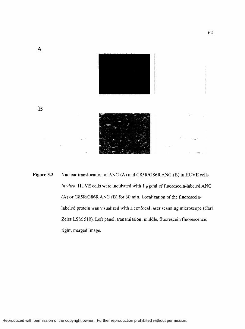
Reproduced with permission of the copyright owner. Further reproduction prohibited without permission.
A
B
Figure 3.3
62
Nuclear translocation of ANG (A) and G85R1G86RANG (B) in HUVE cells
in vitro. HUVE cells were incubated with 1 }tg/ml of fluorescein-labeled ANG
(A) or G85R1G86R ANG (B) for 30 min. Localization of the fluorescein
labeled protein was visualized with a confocal laser scanning microscope (Carl
Zeiss LSM 510). Left panel, transmission; middle, fluorescein fluorescence;
right, merged image.

Reproduced with permission of the copyright owner. Further reproduction prohibited without permission.
63
12
c • wtAng 0 9 +-' mtAng co ~
0>
E \.I-- 6 0
""0 0
LL 3
Figure 3.4 Wound healing migration of HUVE cells induced by ANG (solid bars) or
G85R1G86RANG (shaded bars). HUVE cells were plated in 6-well dish, and
cell migration was quantified by the scratch wound assay.

Reproduced with permission of the copyright owner. Further reproduction prohibited without permission.
A
B
Figure 3.5
Ql
5 u (f)
.\
(iS~[{ (;>i'1({
V..;(,
D .. ·. \ D.I\ -
The Score of Corneal Neovascularization
100
80 T
60 T I 1 T
40 n coL 20 n-n rl II n I r n 0
Can I Wt I Mt Can I Wt I Mt Can I Wt I Mt
Day 3 Day 7 Day 10
1> .. \ 1·1
T
1 .L
n l r Can I Wt I Mt
Day 14
Induction of angiogenesis in rabbit cornea in vivo by ANG or G85R1G86R
64
ANG. (A) Slit lamp photographs of rabbit corneas 3, 7, 10, and 14 days after
implantation withANG or G85R1G86RANG. (B) The score of corneal
neovascularization.
Reproduced with permission of the copyright owner. Further reproduction prohibited without permission.
Chapter Four
Effects of Ribonuclease Inhibitor Silencing on Ribonuclease
Toxicity in Human Tumor Cells
65
Reproduced with permission of the copyright owner. Further reproduction prohibited without permission.
66 4.1 Abstract
The mammalian ribonuclease inhibitor (RI) is an abundant cytoplasmic protein and a
potent inhibitor of ribonucleases from the bovine pancreatic ribonuclease (RNase A)
superfamily. Amphibian homologs of RNase A and engineered variants of mammalian
ribonucleases that evade RI are toxic to tumor cells. The toxicity of a ribonuclease correlates
with the magnitude of its catalytic activity in the presence of RI. Mounting evidence suggests
that ribonuclease toxicity is highly dependent on internalization as determined by the net
positive charge of the protein. Here, we report the effects of RI silencing on the toxicity of
ribonucleases in four tumor-derived cell lines. We demonstrate that native levels of RI are
necessary to protect some cell lines but appear to play little, if any, role in others. The
presence of a protein transduction domain (PfD) loosely correlates with ribonuclease toxicity
in some cell lines and can be sufficient to overwhelm RI regardless of its intracellular
concentration. In conclusion, the cytotoxic activity of ribonucleases is limited by RI in some
but not all human tumor cell lines.

Reproduced with permission of the copyright owner. Further reproduction prohibited without permission.
67
4.2 Introduction
The cytosolic ribonuclease inhibitor (RI) is ubiquitous in mammalian cells, having
been detected in all cell types tested (Lee and Vallee 1993; Hofsteenge 1997; Dickson et al.
2005). RI binds tightly to the active site of bovine pancreatic ribonuclease (RNase A) as well
as other mammalian members of the RNase A superfamily (Lee et al. 1989; Vicentini et al.
1990). Binding to RI inhibits ribonuclease catalytic activity in vitro, though the precise role
of RI in vivo has not been defined.
Onconase (ONC) and other amphibian homologs of RNase A do not bind to RI and,
thus, degrade RNA in its presence. These amphibian ribonucleases demonstrate potent
toxicity towards tumor cells. Engineered variants of RNase A and human pancreatic
ribonuclease (RNase 1) that evade RI also possess cytotoxic activity (Leland et al, 2001;
Leland et al, 1998; Haigis et al, 2002; Rutkoski et al, 2005). The most toxic known variant,
D38RJR39D/N67RJG88R RNase A, is slightly more toxic to a lymphocitic T-cell line than is
ONC (Rutkoski et al. 2005). Overexpression of RI in cultured tumor cell lines confers
protection against G88R RNase A, a toxic variant of RNase A that maintains diminished but
measurable affinity for RI (Haigis et al, 2003). These observations indicate that RI is a
primary determinant of ribonuclease toxicity. The cytotoxicity correlates strongly with the
catalytic activity of a ribonuclease in the presence of RI (Bretscher et al. 2000; Dickson et al.
2003; Rutkoski et al. 2005).
RI evasion is an essential characteristic of a toxic ribonuclease, but it is not the only
property that defines ribonuclease toxicity. The net positive charge of a ribonuclease also
correlates with toxic activity, but the precise contribution this physical characteristic is

Reproduced with permission of the copyright owner. Further reproduction prohibited without permission.
68 difficult to discern. G88R RNase A possessing a cationic N-terminal protein transduction
domain (PTD) composed of 9 arginine residues (R9) demonstrates a 3-fold decrease in its
ICso value (Fuchs and Raines 2005). Cationization of RNase A and human pancreatic
ribonuclease (RNase 1) with ethylenediamine endows both proteins with cytotoxic activity
but also disrupts binding to RI (Futami et al. 2001). Similarly, site-directed mutations
intended to disrupt an RI-ribonuclease complex often have the secondary effect of increasing
the charge of the enzyme (Leland et al. 1998). Nonetheless, increasing the charge of a
ribonuclease has not been demonstrated to be sufficient to generate a toxic protein.
Here, we employ RNA interference (RNAi) to silence cytosolic RI, thus severely
curtailing the protection afforded against an invading ribonuclease. To determine the extent to
which RI evasion is a primary determinant of ribonuclease toxicity, we examined the effects
of RI silencing in four human cell lines. Cells were exposed to both RI-evasive (toxic) and
and non-evasive (non-toxic) ribonucleases under conditions in which cells contained normal
or suppressed levels of RI. The data herein demonstrate that ribonuclease toxicity is
determined as much by cell-specific characteristics that define ribonuclease internalization
and susceptibility to RNA damage as biochemical properties such as RI evasion and net
charge of the protein.

Reproduced with permission of the copyright owner. Further reproduction prohibited without permission.
4.3 Materials and Methods
Materials. K-562, HeLa, Du-145, and Hep-3b cells were from the American Type
Culture Collection (Manasas, VA). Transfection reagents, cell culture medium, and
supplements were from Invitrogen (Carlsbad, CA). GeneEraser™ shRNA Mammalian
Expression Vector System was from Stratagene (La Jolla, CA). Oligonucleotides encoding
shRNAs were synthesized by Integrated DNA Technologies (Coralville, IA). Enzymes for
cloning were from Promega (Madison, WI).
69
Chicken IgY antibodies to human RI and goat anti-chicken secondary antibodies were
produced by Genetel (Madison, WI). Rabbit anti-actin primary antibody and goat anti-rabbit
secondary antibody were from Santa Cruz Biotech (Santa Cruz, CA). Other immunoblotting
reagents including HyBond ECL nitrocellulose membrane, ECL detection reagents, and ECL
film were from Amersham Biosciences (Piscataway, NJ). [methyPH]Thymidine was from
Perkin Elmer Life Sciences (Boston, MA). All other chemicals and biochemicals were of
reagent grade or j3ter and were used without further purification.
Analytical Instruments. Ultraviolet and visible absorption was measured with a Cary
Model 50 spectrophotometer (Varian; Sugarland, TX). For assays of cytotoxicity, cells were
harvested with a PHD Cell Harvester (Cambridge Technology; Watertown, MA).
Radioactivity was measured with a Microj3a Trilux 2 Detector System (Perkin Elmer; Boston,
MA).
Creation of shRNA vectors. Fifteen shRNA sequences designed to target the human RI
gene were cloned into plasmid pGE-l, which directs the expression of shRNAs. Only one of
these shRNA expression vectors, modeled after an shRNA described previously by D' Alessio
and coworkers (Monti and D'Alessio 2004), resulted in significant suppression of RI in the

Reproduced with permission of the copyright owner. Further reproduction prohibited without permission.
70 four cell lines tested. Cloning of this shRNA to create pGE-DAL is described here. Our
shRNA targeted the same sequence in RI but was constructed with a different loop sequence.
Briefly, the oligonucleotides,
5 '-GAT CCC GGT CCT GTC CAG CACACT ACG AAG CTT GGT AGT GTG CTG GAC
AGG ACC TTT TTT-3' and
5'-CTA GAAAAAAAG GTC CTG TCC AGC ACA CTA CCAAGC TTC GTA GTG TGC
TGG ACA GGA CCG GG-3'
were cloned into the BamHI and XbaI sites of pGE-l. Each oligonucleotide (1 Jlg) was
diluted in SO Jll of 10 mM Tris-HCl buffer pH 8.0, containing NaCl (SO mM) and EDTA (0.1
mM). The oligonucleotide mixture was heated to 93°C for 3 min and then cooled slowly to
room temperature. The duplex DNA was ligated into the BamHI and XbaI sites ofpGE-1
using the LigaFast kit (Promega, Madison, WI) as directed by the manufacturer. Clones
containing the shRNA inserts were confirmed by restriction digest and sequence analysis.
Cell Culture. Du-145 and Hep-3b cells were grown in MEM with Earle's salts, L
glutamine (2 mM), non-essential amino acids (0.1 mM), and sodium pyruvate (1 mM). K-562
cells were grown in RPMI medium 1640; HeLa cells were grown in DMEM. All culture
medium was supplemented with fetal bovine serum (10% v/v), penicillin (100 V/ml), and
streptomycin (100 Jlg/ml). Cultures were maintained in a humidified incubator at 37°C
containing 5% COzCg).
Transfection of Human Cells. Transfection ofK-562, HeLa, Du-145, and Hep-3b cells
was carried out in 6-well dishes using Lipofectamine 2000 and Opti-MEM medium
(Invitrogen, Carlsbad, CA). Briefly, cells were seeded at 0.5 x 10⁶ cells/well and incubated at
37°C for 6-12 hours in normal growth medium. DNA-Lipofectamine 2000 complexes were

Reproduced with permission of the copyright owner. Further reproduction prohibited without permission.
formed with 4-7 Jig DNA and 4-10 Jll of Lipofectamine 2000 as directed by the
manufacturer. Cells were washed with warm serum-free Opti-MEM prior to adding
DNA-lipid complexes. Cells were incubated with DNA-lipid complexes for 4-6 h, after
which the medium was replaced with Opti-MEM containing FBS (10% v/v). Cells were
incubated for 44 h before passage to 75 cm2 flasks containing 600 Jlg/ml Geneticin. Stably
transfected cells were maintained and propagated in normal growth medium containing
Geneticin (600 Jlg/ml).
71
Production of Ribonucleases. All ribonucleases used in this study were purified as
describe previously (delCardayre et al. 1995). Protein concentrations were determined by UV
spectroscopy using E = 0.72 mL mg-1 cm-1 at 280 nm for RNase A and its variants and E =
0.87 mL mg-1 cm-1 at 280 nm for ONe. Prior to cytotoxicity assays, ribonucleases were
dialyzed extensively versus phosphate-buffered saline (PBS).
Assays of cytotoxicity. The cytotoxicity of ribonucleases was determined by measuring
the incorporation of [methyPH]thymidine into the DNA of stably transfected human cell
lines. Briefly, cells were seeded in 96-well plate in 100 Jll of normal growth medium at a
density of 5000 cells/well. Ribonucleases in PBS (5 JlL) were incubated with cells for 44 h at
37°C, followed by incubation with [methyPH]thymidine (0.4 JlCi/well) for 4 h. Cells were
harvested onto a glass fiber filter and 3H incorporated into each sample was counted. Cells
incubated with PBS alone were used to determine 100% 3H incorporation. ICso values were
determined by fitting the data to the equation S = 100 X ICso/(lCso + [ribonuclease]), where S
is the percentage of [methyPH]thymidine incorporation after a 48-h incubation with a
ribonuclease.
Immunoblotting. The soluble protein fraction of stably transfected cells was prepared
Reproduced with permission of the copyright owner. Further reproduction prohibited without permission.
72 from a wet cell pellet using MPER lysis solution containing HALT protease inhibitor
cocktail (Pierce Biotech, Rockford, IL). Briefly, cells from a 75-cm2 flask were harvested and
washed 3x with PBS. The cell pellet was resuspended in MPER (10 Jl1I106 cells) and
incubated on ice for 30 min. Cells were passed 5x through a 24--G syringe, and cell debris
was pelleted by centrifugation at 15,000 g for 10 min. Protein extracts were stored at
-80°C until use.
Lysates from stably transfected cells (3 JIg) were subjected to SDS-PAGE on a
4--15% Tris-HCI Ready Gel (BioRad, Hercules, CA) along with recombinant human
RI (1-100 ng) and Precision Plus prestained MW standards (BioRad, Hercules, CA). Proteins
were transferred to HyBond ECLnitrocellulose and then blocked overnight in TBS-T [20
mM Tris-HCI buffer, pH 7.5, containing NaCl (0.137 mM) and Tween 20 (0.2% w/v)]
containing non-fat dry milk (4% w/v). Blots were incubated with anti-hRI IgY (1:3000
dilution in blocking solution) for 1 h and then washed 3x with TBS-T (15 mL). Blots were
then incubated with a horseradish peroxidase (HRP) -conjugated goat anti-chicken antibody
(1:5000 dilution in blocking solution) for 1 h, and then washed (4x) with TBS-T (15 mL). RI
was visualized using ECL-detection reagents and exposure to ECL film.
The results of the two blots were compared directly because they were run
simultaneously and exposed to the same washing and blotting solutions. Bands were analyzed
from a scanned film image using ImageQuantTL (Sunnyvale, CA). Briefly, bands were
selected using ImageQuantTL by drawing a rectangle with a slightly greater area than the
largest band. The intensity of each band in the RI standard curve was calculated using
ImageQuantTL. The values for RI bands in the cell lysate were determined by the same
method and were compared to those obtained from the standard curve.

Reproduced with permission of the copyright owner. Further reproduction prohibited without permission.
73 To insure equal loading of cell lysate samples, the blot was stripped and re-probed
for actin. Briefly, blots were incubated with 30 mL Restore Western Blot Stripping Buffer
(Pierce Biotechnology, Rockford, IL) for 30 min, and then washed briefly with 20 mL TBS-T.
Blots were incubated with primary and secondary antibodies, visualized, and analyzed as
described above. Rabbit anti-actin and HRP-conjugated goat anti rabbit antibodies were
diluted 1:5000 and 1: 10,000, respectively, in blocking solution.
4.4 Results
Suppression of cytosolic RI. We constructed 15 distinct shRNA vectors designed to
target the RI gene. Of the 15 target sequences, 14 were unique target sequences designed
using shRNA algorithms from Invitrogen and Greg Hannon's laboratory (Cold Spring Harbor
Laboratories, NY). One shRNA, pGE-DAL, was modeled after a previously reported shRNA
sequence (Monti and D'Alessio 2004). None of the 14 unique sequences were capable of
significantly reducing RI expression. Thus, RI suppression was achieved using pGE-DAL in
all data described below.
The extent of RI suppression in stably transfected mammalian cells was determined
by immunoblotting. Analysis of the soluble protein fraction of cells transfected with a
negative control vector, pGE-Neg, or pGE-DAL indicated that suppression of RI is not
complete; faint bands corresponding to low levels of RI expression are present in all four
transfected cell lines (Fig IB). After normalizing the intensity of RI bands from lysate
samples according to the intensity of an actin loading control, the percent knockdown was
determined by comparing the intensity of RI bands from celllysates against known amounts
of RI (Fig lA). The extent of suppression ranged from 52 to 63% (Fig lC).

Reproduced with permission of the copyright owner. Further reproduction prohibited without permission.
74 Cytotoxicity. We measured the susceptibility of mammalian cells stably transfected
with pGE-NEG or pGE-DAL to RI-evasive and non-evasive ribonucleases. The RI-evasive
ribonucleases tested included ONC, which demonstrates no measurable affinity for RI, and
two RNase A variants (G88R RNase A and K41R1G88R RNase A), which possess measurable
but weak affinity for RI. Non RI-evasive ribonucleases included RNase A, RNase 1, and
RNase A-R9. In all cases, the ICso values for ONC were the same in cells expressing pGE
Neg or pGE-DAL (Table 1). The cytotoxicity of all other ribonucleases is described below.
The results of cytotoxicity experiments varied significantly ~ween the four
mammalian cell lines examined in this study. Two cell lines, HeLa and Hep-3b, demonstrated
similar trends. Briefly, the ICso values for the RI-evasive variants of RNase A (G88R RNase
A and K41R1G88R RNase A) decreased when cytosolic levels of RI were suppressed (Fig 2,
Table 1). In addition, the ICso values for RNase A-R9, which does not evade RI, decreases by
~4-fold in cells expressing pGE-DAL, whereas the values for RNase A and RNase 1
remained unchanged.
Surprisingly, Du-145 cells reacted similarly to exogenous ribonucleases regardless of
cytosolic RI levels (Table 1). No statistically significant decrease in the ICso values for any
ribonuclease, either RI-evasive or non-evasive, was observed in cells transfected with pGE
DAL. Both wild-type RNase A and RNase A-R9 demonstrated measurable and significant
toxicity towards DU-145 cells; the ICso values for the two proteins differed by only ~2.5-fold.
In addition, G88R RNase A and K41R1G88R RNase A demonstrated similar toxic activity as
wild-type RNase A, possessing ICso values that differed from RNase A by less than 20%.
Unlike RNase A, wild-type RNase 1 demonstrated no detectible toxicity towards Du-145
cells.

Reproduced with permission of the copyright owner. Further reproduction prohibited without permission.
75 In K-562 cells, only G88R RNase A and K41RJG88R RNase A demonstrate
increased cytotoxicity in cells expressing pGE-DAL. All other IC50 values were independent
of cytosolic RI levels.
In general, the magnitude of change ribonuclease cytoxicity induced by RI
suppression was substantial; most ICso values decreased by >75%. These substantial increases
in ribonuclease susceptibility were independent of initial ICso values in cells expressing pGE
Neg. For example, the IC50 values for G88R RNase A and RNase A-R9 differed by lO-fold in
Hep-3b cells expressing pGE-Neg. Yet, both IC50 values decreased by ",75% upon RI knock
down.
4.5 Discussion
RI is horseshoe-shaped protein composed of 16 consecutive leucine-rich repeats
(LRRs) (Kobe and Deisenhofer 1993). The genetic structure of RI mirrors the primary
structure of the protein; 7 exons code for 7 discrete LRR pairs (Haigis et al. 2002). Pairwise
analysis of these exons revealed 50-60% sequence identity (Haigis et al. 2002). Despite its
highly repetitive sequence with significant sequence identity, RI has proved to be a difficult
target for RNAi. In total, we constructed 14 unique shRNA sequences that had little effect on
the cytosolic concentration of RI. Only pGE-DAL was capable of drastically altering RI
levels. Initial observation of immuno blots indicated that RI knock down was substantial,
though not complete. Indeed, quantitative analysis of the blot revealed that RI levels in all
four human cell lines were reduced by only 52-66%. These data demonstrate that
intracellular RI levels in cells harboring pGE-DAL are sufficient to present a formidable line
of defense against invading ribonucleases. IC50 values for ribonucleases will only decrease if

Reproduced with permission of the copyright owner. Further reproduction prohibited without permission.
the amount of ribonuclease internalized is adequate to saturate and thus overwhelm the
remaining RI.
Nonspecific suppression by RNAi has been well documented (Scacheri et al. 2003).
76
To demonstrate that the DAL shRNA specifically targets RI and does not affect other proteins
that enhance ribonuclease susceptibility, we compared the toxicity of ONC in cells
transfected with either pGE-Neg or pGE-DAL. Because ONC does not bind to RI, the IC50
values for ONC should remain unchanged in the presence of the DAL shRNA. Indeed, none
of the four cell lines examined demonstrated increased sensitivity to ONC when RI was
knocked down. Likewise, the IC50 values for ONC, G88R RNase A and K41RJG88R RNase
A incubated with K-562 cells expressing pGE-NEG are similar to values reported previously
for non-transfected K-562 cells (data not shown). Therefore, neither the presence of shRNA
molecules nor the specific suppression of RI in a cell confers additional susceptibility to
ribonucleases.
For a ribonuclease to be toxic to a cell, it must first gain access to the cytosol. This
process involves binding to the cell surface, internalization via an endocytic vesicle, and
translocation through a membrane to reach the cytosol (Haigis and Raines 2003). Most
ribonuclease molecules that enter a cell are degraded in endocytic vesicles; few make it to the
cytosol intact. Thus, internalization is a major determinant of ribonuclease cytotoxicity.
Molecules that are more efficient at reaching the cytosol are more toxic. Presumably, PTDs,
chemical cationization, and increased charge incurred through site-directed mutagenesis
enable ribonucleases to be internalized more efficiently than their wild-type progenitors.
After internalization, a toxic ribonuclease degrades cellular RNA. Presumably,
ribonucleases that bind tightly to RI, such as RNase A and RNase 1, must saturate cytosolic

Reproduced with permission of the copyright owner. Further reproduction prohibited without permission.
77 RI before they can elicit toxic effects. Previous studies in normal and tumor-derived cell
lines indicate that intracellular RI levels do not correlate with the resistance of cells to
ribonucleases (Haigis et al. 2002). Here, immunoblots reveal that the concentration of RI in
pGE-NEG cell extracts differs by less than 30%. Therefore, variations in ribonuclease
resistance !3ween the four cell types examined here cannot be attributed to differences in
cytosolic levels of RI. Rather, ease of internalization or other cell type-specific factors
determine the relative susceptibility or resistance to ribonucleases. The experiments described
herin have allowed us to scrutinize the role of RI evasion in defining ribonuclease toxicity to
provide a clearer picture of the mechanism of ribonuclease cytotoxicity.
HeLa and Hep-3b cells rely on protection by RI, as intracellular levels of RI limit
ribonuclease toxicity. Reduction of RI levels in both of these cell types results in a significant
decrease in the IC50 values of RI-evasive RNase A variants as well as RNase A-R9, which
binds tightly to RI but exhibits extremely efficient internalization due to its PTD. G88R
RNase A and K41R1G88R RNase A become more toxic to the cell because fewer molecules
are hindered by weak interactions with RI. Despite its tight binding to RI, RNase A-R9 is
internalized efficiently and is toxic to cells transfected with pGE-NEG. The IC50 values
decreases in cells possessing pGE-DAL because fewer RNase A-R9 molecules are required
to saturate the remaining RI.
The effect of RI silencing in K-562 cells is similar to that in HeLa and Hep-3b cells.
The IC50 values for G88R RNase A and K41R1G88R RNase A decrease upon suppression of
RI. Surprisingly, K-562 cells are susceptible to RNase A-R9, yet the ICso value for RNase
A-R9 remains unchanged in pGE-DAL K-562 cells. Possibly, the R9 tag affords exceptional
internalization in K-562 cells such that endogenous RI provides little protection at high

Reproduced with permission of the copyright owner. Further reproduction prohibited without permission.
concentrations of RNase A-R9.
Du-145 cells are susceptible to RNase A and its variants, yet are not affected by
suppression of RI. Surprisingly, the ICso values for RNase A are similar to those for RI
evasive variants of RNase A. Possibly, reduction of cytosolic RI levels in Du-145 cells is
insufficient to generate a change in IC50 values. Du-145 cells also exhibit higher IC50 values
for RI-evasive RNase A variants than any other cell line tested. These data indicate that
RNase A and its variants do not gain access to the cytosol of Du-145 cells efficiently.
78
Both RNase 1 and RNase A demonstrated measurable but weak toxicity in some, but
not all cell lines. Tranformation of these cell lines with pGE-DAL had no effect on ICso
values for either protein. RNase 1 and RNase A share 70% sequence identity (Moore and
Stein 1973; Beintema et al. 1984), are both highly cationic proteins (pI = 8.6 and 9.4,
respectively) (Tanford and Hauenstein 1956; Zhang et al. 2003), and are robust catalysts of
RNA degradation (Leland et al. 2001). Despite shared biochemical properties, RNase A and
RNase 1 demonstrate different cytotoxic activities in the cell lines tested.
We have illustrated a complex relationship ~ween ribonucleases and tumor cells in
which internalization, RI evasion, and subsequent RNA degradation are predominantly
dictated by cell type. Although RI evasion correlates directly with cytotoxicity in numerous
cell lines, decreases in intracellular RI levels do not generate consistent decreases in ICso
values. In addition, the net charge of a ribonuclease correlates weakly, if at all, with toxicity.
The data presented here indicate that salient features of the mechanism of ribonuclease
cytotoxicity, including the mode of internalization, remain to be discovered.

Reproduced with permission of the copyright owner. Further reproduction prohibited without permission.
79
Table 4.1. IC50 values of RI-evasive and non-evasive ribonucleases in tumor cell lines with
and without RI suppression. Values in bold-face type decrease significantly upon RI
suppression.
ICso values (JIM)
RI-evasive ribonucleases Non-evasive ribonucleases G88R K41R/G88R ONe RNase RNase A RNase 1
RNase A RNase A A-R9 K-562 NEG 4.5 ± 0.6 9.22 ± 1 0.49 ± 0.07 >200 65 ± 8 29 ± 3
DAL 0.97 ± 0.09 6.28 ± 0.8 0.51 ± 0.06 >200 66 ± 6 27 ± 3 --------------------- -------------------------------------------------- --------------------------------------------HeLa NEG 67 ± 11 >25 0.90 ± 0.22 >200 44 ± 4 14 ± 2
DAL 9.7 ± 2 8.5 ± 2 0.75 ± 0.25 >200 37 ± 5 3.1 ± 0.9 --------------------- -------------------------------------.------------ --------------------------------------------Du-145 NEG 71 :t 8 51 :t 10 0.18 ± 0.02 65 ± 6 >200 11 :t 2
DAL 61:t7 39±5 0.16:t0.02 61:t7 >200 8:t2 --------------------- -------------------------------------------------- --------------------------------------------Hep-3b NEG 13 ± 2 13 ± 2 0.14:t 0.03 >200 >200 112 ± 23
DAL 3.0 ± 0.9 1.9 ± 0.9 0.13 :t 0.03 >200 >200 31 ± 8

Reproduced with permission of the copyright owner. Further reproduction prohibited without permission.
80
a. (ng) 5 10 30 50 100
RI standards ---... K-562 HeLa DU-145 Hep-3b
b. + + + + RI --- - - -
actin -----~--..
c.
K-562
Knock-down 60°'0 63°" 55°0
Figure 4.1 Immunoblot analysis of RI suppression in human tumor cell lines: (a) RI
standards. (b) Cell lysate (30 }lg) transfected with pGE-NEG (-) or pGE-DAL
(+) probed with anti-RI or anti-actin antibodies. (c) Amount of RI present in
transfected cells determined by comparing the intensity of RI bands from cell
lysates against a standard curve generated from RI standards. Analysis was
performed using ImageQuantTL as described in Methods.
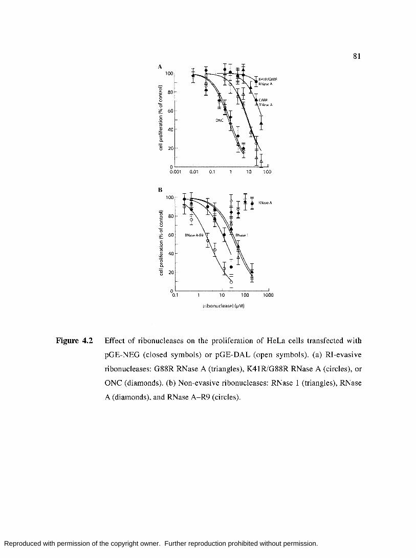
Reproduced with permission of the copyright owner. Further reproduction prohibited without permission.
81 A
100
"2 80 1: 0 u .... 0
~ 60 c 0 .;:; :!:
40 ~ "2 a. Qi 20 u
a 0.001 0.01 0.1 10 100
B 100
RNase A
"2 80 1: 0 u '0 ~ 60 c 0 .;:; :!: 40 ~ "2 a. Qi 20 u
a 0.1 10 100 1000
[ribonuclease] (~M)
Figure 4.2 Effect of ribonucleases on the proliferation of HeLa cells transfected with
pGE-NEG (closed symbols) or pGE-DAL (open symbols). (a) RI-evasive
ribonucleases: G88R RNase A (triangles), K41R1G88R RNase A (circles), or
ONe (diamonds). (b) Non-evasive ribonucleases: RNase 1 (triangles), RNase
A (diamonds), and RNase A-R9 (circles).

Reproduced with permission of the copyright owner. Further reproduction prohibited without permission.
82
Appendix I
Production of a Cysteine-Free Ribonuclease Inhibitor
Reproduced with permission of the copyright owner. Further reproduction prohibited without permission.
83 ALl Introduction
Porcine and human RI contain 30 and 32 cysteines, respectively, which must be
reduced for the protein to be functional (Hofsteenge et al. 1988; Lee et al. 1988). Oxidation
of RI is a highly cooperative process; oxidation of only a few cysteines changes the
conformation of the protein and renders the remaining thiols extremely reactive (Fominaya
and Hofsteeenge, 1992). An oxidation-resistant RI could serve as a useful laboratory reagent
and could be a useful tool in investigating RI-RNase interactions. The goal of this work was
to create a cysteine-free porcine RI (pRI) in which all of the cysteine residues are replaced
with alanine.
AL2 Materials and Methods
Mutagenesis of pRI was performed on the plasmid pTrpRI6.1 (Klink and Raines,
2001). Primers coding for Cys to Ala mutations were produced by IDT (Coralville, IA).
Mutagenesis was performed using the QuickChange Multi kit from Stratagene (La Jolla, CA)
according to the manufacturers instructions. As many as 6 mutations were attempted in a
single round of mutagenesis; 6 rounds of mutagenesis were required to alter all 30 Cys to Ala.
Mutant pRI (pTrpRI6.1-Ala) clones were screened by sequence analysis.
The binding activity Ala-pRI compared to pRI was determined from a lysate of cells
overexpressing RI. Briefly, E. coli TOPP3 cells were transformed with pTrpRI6.1 or
pTrpRI6.1-Ala and grown in LB to mid-log phase (OD600 = 1.0), after which, cells were
harvested and resuspended in M9 minimal media (Klink and Raines, 2001). Cells were grown
at in minimal media at 16°C for 12 hours and then harvested. Cells were lysed by sonication
and cell debris was removed by centrifugation at 40,000 xg. The supernatant of this

Reproduced with permission of the copyright owner. Further reproduction prohibited without permission.
84 procedure is the cell lysate used to assess the activity of Ala-pRI.
AI.3 Results
Cell lysate of E. coli expressing pRIor Ala-pRI was analyzed by SDS-PAGE.
Induction of pRI and Ala-pRI was detectible, and comparable to previously reported levels of
expression (Klink and Raines, 2001). The relative amount of soluble and insoluble protein in
each sample was not determined.
The ability of pRIor Ala-pRI to bind to RNases was determined by its ability to
inhibit the catalytic activty of RNase A. Assays were carried out using the fluorogenic
stubstrate, IDT2, in 100 mM MES buffer, pH 6.0, containing 100 mM NaCl and 1 mM DTT.
Crude cell lysate of cells expressing pRI was diluted 1: 100 in reaction buffer; addition of
only 10 pL of the diluted lysate completely abolished ribonucleolytic activity. Conversely,
addition of 1 - 200 pL of undiluted Ala-pRI lysate did not inhibit RNase A, but slightly
increased the ribonucleolytic activity.
The inability of Ala-pRI to inhibit RNase A could be a result of decreased affinity or
compromised structural stability. No attempt was made to evaluate the ability of Ala-pRI to
fold in vivo or in vitro, and it was concluded that Ala-pRI was not sufficiently functional for
subsequent investigations.

Reproduced with permission of the copyright owner. Further reproduction prohibited without permission.
85
Appendix II
Ribonuclease Binding to the Cell Surface
Reproduced with permission of the copyright owner. Further reproduction prohibited without permission.
86 AII.1 Introduction
Binding of RNase A and its homologs to the suface of mammalian cells facilitates
their entry into the cytosol (Haigis and Raines, 2002). The specific interactions of
ribonucleases with molecules displayed on the surface of cells have not been described. The
purpose of this work was to measure the affinity of RNase A and Onconase ™ for the surface
cultured tumor cells in order to identify specific interactions ~ween ribonucleases and cell
surface residues.
AII.2 Materials and Methods
K-S62, HeLa, and LacZ-9L glioma cells were obtained fromATCC (Manasas, VA)
and were cultured according to ATCC instructions in RPMI (for K-S62) or DMEM (for HeLa
and LacZ-9L). All media contained FBS (10% v/v), penicillin (100 units/mL), and
streptomycin (100 ]lg/mL). A19C RNase A and D16C ONC were produced and labeled with
S-iodoacetamidofluorescein (S-IAF) as described previously (Haigis and Raines, 2002).
Ribonuclease binding to the surface of cells was assayed by flow cytometry. Briefly, cells
were harvested, washed in ice-cold media, and resuspended at a density of 1 x 10⁶ cells/mL.
Cells were maintained on ice for the duration of the experiment. Fluorescently labeled
ribonucleases were added to the suspension of cells and incubated on ice for >30 min. In
some cases, the cells were washed 3x with ice-cold PBS prior to flow cytometry.
AII.3 Results
In all conditions tested, both RNase A and ONC demonstrated non-specific binding
to the surface of cultured mammalian tumor cells. The fluorescence intensity associated with

Reproduced with permission of the copyright owner. Further reproduction prohibited without permission.
87 cells increased linearly with the concentration of protein added to the media (Fig AI). Cells
treated with trypsin or neuraminidase (sialidase) did not demonstrate decreased ribonuclease
binding. In addition, the addition of colominic acid (polysialic acid) at 13.3 mg/ml,
ribonuclease inhibitors (5' -AMP and 3' -UMP at 35 and 400 JIM, respectively), polylysine at
5 mg/ml, heparin sulfate at 3.3 mg/ml, or NaCI at twice the concentration of the media (400
mM total) did not cause a detectible change in ribonuclease binding. Finally, ribonuclease
binding was not affected by the presence of FCS in the media.

Reproduced with permission of the copyright owner. Further reproduction prohibited without permission.
88
108 • • "0 107 • c ::l ~ 0
CO 106 « •
Q) ~ en ctl
105 Z • a:: • en Q)
104 ::l • U Q)
~ 0 :2 103
102
0.001 0.01 0.1 1 10 100
[RNase A] (JiM)
Figure AIL1 Binding of fluorescein-labeled RNase A to K-562 cells. Cells were incubated
with labeled RNase A for 30 min on ice. Fluorescence was measured by flow
cytometry. The number of RNase A molecules bound was determined by
comparing the florescence of cell with that of fluorescein-labeled beads as
described in Methods.

Reproduced with permission of the copyright owner. Further reproduction prohibited without permission.
89
References
Reproduced with permission of the copyright owner. Further reproduction prohibited without permission.
Abel, R A, M. C. Haigis, C. Park and R T. Raines (2001). Fluorescence assay for the binding of ribonuclease A to its cytosolic inhibitor protein. Anal Biochem 360, 100-107.
90
Allen, D. W. and J. H. Jandl (1961). Oxidative hemolysis and precipitation of hemoglobin. II. Role of thiols in oxidant drug action. 1. Clin. Invest. 40, 454-475.
Ardelt, W., S. M. Mikulski and K. Shogen (1991). Amino acid sequence of an anti-tumor protein from Rana pipiens oocytes and early embryos. 1. Bioi. Chem. 266, 245-251.
Beintema, J. J., P. Wietzes, J. L. Weickmann and D. G. Glitz (1984). The amino acid sequence of human pancreatic ribonuclease. Anal. Biochem!. 136,48-64.
Blackburn, P. (1979). Ribonuclease inhibitor from human placenta: Rapid purification and assay. 1. Bioi. Chem. 254, 12484-12487.
Blackburn, P. and B. L. Jailkhan (1979). Ribonuclease inhibitor from human placenta: Interaction with deriviatives of ribonuclease A 1. Bio!. Chem. 254, 12488-12493.
Blackburn, P. and S. Moore (1982). Pancreatic ribonuclease. The Enzymes XV: 317-433.
Blackburn, P., G. Wilson and S. Moore (1977). Ribonuclease inhibitor from human placenta. Purification and properties. 1. Bioi. Chem.!. 252,5904-5910.
Bhlzuez, M., J. M. Fominaya and J. Hofsteenge (1996). Oxidation of sulfhydryl groups of ribonuclease inhibitor in epithelial cells is sufficient for its intracellular degradation. 1. Bioi. Chem. 271, 18638-18642.
Blaser, 1., S. Triebel, C. Kopp and H. Tschesche (1993). A highly sensitive immunoenzymometric assay for the determination of angiogenin. Eur. 1. Clin. Chem. Clin. Biochem. 31, 513-516.
Boix, E., Y. Wu, V. M. Vasandani, S. K. Saxena, W. Ardelt, J. Ladner and R J. Youle (1996). Role of the N terminus in RNase A homologues: Differences in catalytic activity, ribonuclease inhibitor interaction and cytotoxicity. 1. Mol. Bioi. 257, 992-1007.
Botella-Estrada, R, G. Malet, F. Revert, F. Dasi, A Crespo, O. Sanmartin, C. Guillen and S. F. Alina (2001). Antitumor effect of B 16 melanoma cells genetically modified with the angiogenesis inhibitor rnasin. Cancer Gene Ther. 8, 278-284.
Bravo, J., E. Fernandez, M. Rib6, R de Llorens and C. M. Cuchillo (1994). A versatile negative-staining ribonuclease zymogram. Anal. Biochem!. 219, 82-86.

Reproduced with permission of the copyright owner. Further reproduction prohibited without permission.
Bretscher, L. E., R. L. Abel and R. T. Raines (2000). A ribonuclease A variant with low catalytic activity but high cytotoxicity. 1. BioI. Chem. 275,9893-9896.
91
Burton, L. E., P. Blackburn and S. Moore (1980). Ribonuclease inhibitor from bovine brain. Int. 1. Peptide Protein Res,- 16, 359-364.
Burton, L. E. and N. P. Fucci (1982). Ribonuclease inhibitors from the liver of five mammalian species. Int. 1. Peptide Protein Res. 19, 372-379.
Cavalli, L., D. Galaverni, P. Pesando, P. G. Bracchi, G. Campanini and G. Maraini (1979). Control of ribonuclease activity in the human lens during aging and cataract formation. Ophthalmic Res. 11,416-422.
Chen, c.-Z. and R. Shapiro (1997). Site-specific mutagenesis reveals differences in the structural bases for tight binding of RNase inhibitor to angiogenin and RNase A. Proc. Natl. Acad. Sci. USA 94, 1761-1766.
Chen, C. Z. and R. Shapiro (1999). Superadditive and subadditive effects of "hot spot" mutations within the interfaces of placental ribonuclease inhibitor with angiogenin and ribonuclease A. Biochemistry 38, 9273-9285.
Cho, S. and J. G. Joshi (1989). Ribonuclease inhibitor from pig brain: purification, characterization, and direct spectrophotometric assay. Anal. Biochem. 176, 175-181.
Crawford, D., K. Hagerty and B. Beutler (1989). Multiple splice forms of ribonucleaseinhibitor mRNA differ in the 5'- untranslated region. Gene 85, 525-531.
Crick, F. H. C. (1952). Is a-keratin a coiled coil? Nature 170, 882-883.
Cui, X. Y., P. F. Fu, D. N. Pan, Y. Zhao, 1. Zhao and B. C. Zhao (2003). The antioxidant effects of ribonuclease inhibitor. Free Radic. Res. 37, 1079-1085.
Darzynkiewicz, Z., S. P. Carter, S. M. Mikulski, W. J. Ardelt and K. Shogen (1988). Cytostatic and cytotoxic effect of Pannon (P-30 Protein), a novel anticancer agent. Cell Tissue Kinet. 21, 169-182.
delCardayre, S. B., M. Rib6, E. M. Yokel, D. J. Quirk, W. J. Rutter and R. T. Raines (1995). Engineering ribonuclease A: Production, purification, and characterization of wildtype enzyme and mutants at Glnll. Protein Eng. 8, 261-273.
Dickson, K. A., C. L. Dahlberg and R. T. Raines (2003). Compensating effects on the cytotoxicity of ribonuclease A variants. Arch. Biochem. Biophys. 415 172-177.
Dickson, K. A., M. C. Haigis and R. T. Raines (2005). Ribonuclease inhibitor: structure and function. Prog. Nucleic Acid Res. Mol. BioI. 80, 349-374.
Reproduced with permission of the copyright owner. Further reproduction prohibited without permission.
92
Dijkstra, J., J. Touw, I. Halsema, M. Gruber and G. Ab (1978). Estradiol-induced synthesis of vitellogenin. Biochim Biophys Acta. 521, 363-373.
Eberhardt, E. S., P. K. Wittmayer, B. M. Templer and R. T. Raines (1996). Contribution of a tyrosine side chain to ribonuclease A catalysis and stability. Protein Sci. 5, 1697-1703.
Ellis, R. J. (2001). Macromolecular crowding: Obvious but unappreciated. Trends Biochem. Sci. 26, 597 -604.
Evdokimov, A. G., D. E. Anderson, K. M. Routzahn and D. S. Waugh (2001). Unusual molecular architecture of the Yersinia pestis cytotoxin YopM: A leucine-rich repeat protein with the shortest repeating unit. 1. Mol. Bio. 312, 807-821.
Ferreras, M., 1. G. Gavilanes, C. Lopez-Otfn and J. M. Garda-Segura (1995). Thiol-disulfide exchange of ribonuclease inhibitor bound to ribonuclease A. 1. BioI. Chern. 270, 28570-28578.
Fett, J. W., D. 1. Strydom, R. R. Lobb, E. M. Alderman, 1. L. [3hune, J. F. Riordan and B. L. Vallee (1985). Isolation and characterization of angiogenin, an angiogenic protein from human carcinoma cells. Biochemistry 24, 5480-5486.
Fominaya, J. M. and J. Hofsteenge (1992). Inactivation of ribonuclease inhibitor by thiol-disulfide exchange. 1. BioI. Chern. 267,24655-24660.
Fuchs, S. M. and R. T. Raines (2005). Polyarginine as a multifunctional fusion tag. Protein Sci. 14, 1538-1544.
Futami, J., T. Maeda, M. Kitazoe, E. Nukui, H. Tada, M. Seno, M. Kosaka and H. Yamada (2001). Preparation of potent cytotoxic ribonucleases by cationization: Enhanced cellular uptake and decreased interaction with ribonuclease inhibitor by chemical modification of carboxyl groups. Biochemistry 26, 7518-7524.
Futami, J., K. Nukui, T. Maeda, M. Kosaka, H. Tada, M. Seno and H. Yamada (2002). Optimum modification for the highest cytotoxicity of cationized ribonuclease. 1. Biochem. (Tokyo) 132, 223-228.
Futami, J., Y. Tsushima, Y. Murato, H. Tada, J. Sasaki, M. Seno and H. Yamada (1997). Tissue-specific expression of pancreatic-type RNases and RNase inhibitor in humans. DNA Cell. BioI. 16,413-419.
G.-F., H., J. F. Riordan and B. L. Vallee (1994). Angiogenin promotes invasiveness of cultured endothelial cells by stimulation of cell-associated proteolytic activities. Proc. Natl. Acad. Sci. U.S.A. 94, 2204-2209.

Reproduced with permission of the copyright owner. Further reproduction prohibited without permission.
93 Garcia, M. A. and R. J. Klebe (1997). Affinity chromatography of RNase inhibitor. Mol.
Biol. Rep. 24, 231-233.
Green, N. M. (1975). Avidin. Adv. Protein Chem. 29, 85-133.
Gribnau,A. A., J. G. Schoenmakers, M. van Kraaikamp, M. Hilak and H. Bloemendal (1970). Further studies on the ribonuclease inhibitor from rat liver: stability and other properties. Biochim. Biophys. Acta 224, 55-62.
Haigis, M. c., E. S. Haag and R. T. Raines (2002). Evolution of ribonuclease inhibitor by exon duplication. Mol. Biol. Evol. 19,959-963.
Haigis, M. c., E. L. Kurten, R. L. Abel and R. T. Raines (2002). KFERQ sequence in ribonuclease A-mediated cytotoxicity. 1. Biol. Chem. 277,11576-11581.
Haigis, M. c., E. L. Kurten and R. T. Raines (2002). Ribonuclease inhibitor is an intracellular sentry. Nucleic Acids Res. 31, 1024-1032.
Haigis, M. C. and R. T. Raines (2003). Secretory ribonucleases are internalized by a dynamin-independent endocytic pathway. 1. Cell Sci. 116, 313-324.
Harper,1. W. and B. L. Vallee (1989). A covalent angiogenin/ribonuclease hybrid with a fourth disulfide bond generated by regional mutagenesis. Biochemistry 28, 1875-1884.
Hofsteenge, J. (1997). Ribonuclease inhibitor. Ribonucleases: Structures and Functions. G. D'Alessio and J. F. Riordan. New York, Academic Press: 621-658.
Hofsteenge, J., B. Kieffer, R. Matthies, B. A. Hemmings and S. R. Stone (1988). Amino acid sequence of the ribonuclease inhibitor from porcine liver reveals the presence of leucine-rich repeats. Biochemistry 27,8537-8544.
Hofsteenge, J., A. Vicentini and O. Zelenko (1998). Ribonuclease 4, an evolutionarily highly conserved member of the superfamily. Cell. Mol. Life Sci. 54, 804-810.
Hofsteenge, J., A. Vincentini and S. R. Stone (1991). Purification and characterization of truncated ribonuclease inhibitor. Biochem. 1.275,541-543.
Hu, G.-F., J. F. Riordan and B. L. Vallee (1997). A putative angiogenin receptor in angiogenin-responsive human endothelial cells. Proc. Natl. Acad. Sci. USA 94, 2204-2209.
Jaffee, E. A., R. L. Nachman, C. G. Becker and C. R. Minick (1973). Culture of human endothelial cells derived from umbilical veins. Identification by morphologic and immunologic criteria. 1. Clin. Invest. 52, 2745-2756.

Reproduced with permission of the copyright owner. Further reproduction prohibited without permission.
94
Janin, J. (1994). Proteins with a ring. Structure 2, 571-573.
Jimi, S., K. Ito, K. Kohno, M. Ono, M. Kuwano, Y. Itagaki and H. Ishikawa (1995). Modulation by bovine angiogenin of tubular morphogenesis and expression of plasminogen activator in bovine endothelial cells. Biochern. Biophys. Res. Cornrnun. 211,476-483.
Kajava, A. V. (1998). Structural diversity of leucine-rich repeat proteins. J. Mol. BioI. 277, 519-527.
Kajava, A. V. and B. Kobe (2002). Assessment of the ability to model proteins with leucinerich repeats in light of the latest structural information. Prot. Sci. 11, 1082-1090.
Kawanomoto, M., K. Motojima, M. Sasaki, H. Hattori and S. Goto (1992). cDNA cloning and sequence of rat ribonuclease inhibitor, and tissue distribution of mRNA. Biochirn. Biophys. Acta 1129, 335-338.
Kelemen, B. R, T. A. Klink, M. A. Behlke, S. R Eubanks, P. A. Leland and R T. Raines (1999). Hypersensitive substrate for ribonucleases. Nucleic Acids Res. 27, 3696-3701.
Kim, B.-M., L. W. Schultz and R T. Raines (1999). Variants of ribonuclease inhibitor that resist oxidation. Protein Sci. 8, 430-434.
Klink, T. A. and R T. Raines (2000). Conformational stability is a determinant of ribonuclease A cytotoxicity. J. BioI. Chern. 275,17463-17467.
Klink, T. A., A. M. Vicentini, J. Hofsteenge and R T. Raines (2001). High-level soluble production and characterization of porcine ribonuclease inhibitor. Protein Exp. Purif. 22, 174-179.
Kobe, B. and J. Deisenhofer (1993). Crystal structure of porcine ribonuclease inhibitor, a protein with leucine-rich repeats. Nature 366, 751-756.
Kobe, B. and 1. Deisenhofer (1994). The leucine-rich repeat: A versatile binding motif. Trends Biochern. Sci. 19,415-421.
Kobe, B. and 1. Deisenhofer (1995). Proteins with leucine-rich repeats. Curro Opin. Struct. BioI. 5,409-416.
Kobe, B. and 1. Deisenhofer (1995). A structural basis of the interactions ~ween leucine-rich repeats and protein ligands. Nature 374, 183-186.

Reproduced with permission of the copyright owner. Further reproduction prohibited without permission.
95 Kobe, B. and J. Deisenhofer (1996). Mechanism of ribonuclease inhibition by ribonuclease
inhibitor protein based on the crystal structure of its complex with ribonuclease A. 1. Mol. Biol. 264,1028-1043.
Kobe, K and A. V. Kajava (2001). The leucine-rich repeat as a protein recognition motif. Curro Opin. Struct. Biol. 11, 725-732.
Kraft, N. and K Shortman (1970). The phylogeny of the ribonuclease-ribonuclease inhibitor system: Its distribution in tissues and its response during leukaimogenesis and aging. Aust. 1. Biol. Sci. 23: 175-184.
Kraft, N., K Shortman and D. Jamieson (1969). The effect of x-irradiation on the balance ~ween alkaline ribonuclease and the ribonuclease inhibitor of mammalian tissues. Radiation Res. 39, 655-668.
Kumar, K, M. Brady and R. Shapiro (2004). Selective abolition of pancreatic RNase binding to its inhibitor protein. Proc. Natl. Acad. Sci. USA 101, 53-58.
Kyner, D., J. K Christman and G. Acs (1979). The effect of 12-0-tetradecanoyl-phorbol 13-acetate on the ribonuclease activity of circulating human lymphocytes. Eur. 1. Biochem. 99, 395-399.
Ledoux, L. (1955). Action of ribonuclease on certain ascites tumours. Nature 175, 258-259.
Ledoux, L. (1955). Action of ribonuclease on two solid tumours in vivo. Nature 176, 36-37.
Lee, F. S., D. S. Auld and B. L. Vallee (1989). Tryptophan fluorescence as a probe of placental ribonuclease inhibitor binding to angiogenin. Biochemistry 28, 219-224.
Lee, F. S., E. A. Fox, H.-M. Zhou, D. J. Strydom and B. L. Vallee (1988). Primary structure of human placental ribonuclease inhibitor. Biochemistry 27, 8545-8553.
Lee, F. S., R. Shapiro and B. L. Vallee (1989). Tight-binding inhibition of angiogenin and ribonuclease A by placental ribonuclease inhibitor. Biochemistry 28, 225-230.
Lee, F. S. and B. L. Vallee (1989). Expression of human placental ribonuclease inhibitor in Escherichia coli. Biochem. Biophys. Res. Commun. 160, 115-120.
Lee, F. S. and B. L. Vallee (1990). Modular mutagenesis of human placental ribonuclease inhibitor, a protein with leucine-rich repeats. Proc. Natl. Acad. Sci. U.S.A. 87, 1879-1883.
Lee, F. S. and B. L. Vallee (1993). Structure and action of mammalian ribonuclease (angiogenin) inhibitor. Prog. Nucleic Acid Res. Mol. Biol. 44, 1-30.
Reproduced with permission of the copyright owner. Further reproduction prohibited without permission.
Leland, P. A. and R. T. Raines (2001). Cancer chemotherapy-ribonucleases to the rescue. Chem. BioI. 8,405-413.
Leland, P. A., L. W. Schultz, B.-M. Kim and R. T. Raines (1998). Ribonuclease A variants with potent cytotoxic activity. Proc. Natl. Acad. Sci. U.S.A. 98, 10407-10412.
Leland, P. A., K. E. Staniszewski, B.-M. Kim and R. T. Raines (2000). A synapomorphic disulfide bond is critical for the conformational stability and cytotoxicity of an amphibian ribonuclease. FEBS Lett. 477,203-207.
Leland, P. A., K. E. Staniszewski, B. M. Kim and R. T. Raines (2001). Endowing human pancreatic ribonuclease with toxicity for cancer cells. 1. BioI. Chem. 276, 43095-43102.
Leland, P. A., K. E. Staniszewski, C. Park, B. R. Kelemen and R. T. Raines (2002). The ribonucleolytic activity of angiogenin. Biochemistry 41, 1343-1350.
96
Makarov, A. A. and O. N. Ilinskaya (2003). Cytotoxic ribonucleases: Molecular weapons and their targets. FEBS Lett. 540, 15-20.
Matousek, J. (1973). The effect of bovine seminal ribonuclease (AS RNase) on cells of crocker tumour in mice. Experientia 29,858-859.
Matousek, J., J. Soucek, J. Rfha, T. R. Zankel and S. A. Benner (1995). Immunosuppressive activity of angiogenin in comparison with bovine seminal ribonuclease and pancreatic ribonuclease. Compo Biochem. Physiol. 112B, 235-241.
Matousek, J. (1975). Embryotoxic effect of bull seminal ribonuclease and tissue absorption studies in rats. 1. Reprod. Fert. 43, 171-174.
Matousek, J. (1994). Aspermatogenic effect of the bull seminal ribonuclease (BS RNase) in the presence of anti BS RNase antibodies in mice. Animal Genet. 25,45-50.
Matousek, J. (2001). Ribonucleases and their antitumor activity. Compo Biochem. Physiol. 129C, 175-191.
Matousek, J., G. Gotte, P. Pouckova, J. Sou_ek, T. Slavfk, F. Vottariello and M. Libonati (2003). Antitumor activity and other biological actions of oligomers of ribonuclease A. 1. BioI. Chem. 278,23817-23822.
Matteo, A. D., L. Federici, B. Mattei, G. Salvi, K. A. Johnson, C. Savino, G. D. Lorenzo and D. Tsernoglou (2003). The crystal structure of polygalacturonase-inhibiting protein (PGIP), a leucine-rich repeat protein involved in plant defense. Proc. Natl. Acad. Sci. USA 100, 10124-10128.

Reproduced with permission of the copyright owner. Further reproduction prohibited without permission.
Messmore, J. M., D. N. Fuchs and R. T. Raines (1995). Ribonuclease A: Revealing structure-function relationships with semi synthesis. 1. Am. Chem. Soc. 117,8057-8060.
Mikulski, S. M., W. Ardelt, K. Shogen, E. H. Bernstein and H. Menduke (1990). Striking increase of survival of mice bearing M109 Madison carcinoma treated with a novel protein from amphibian embryos. 1. Natl. Cancer Inst. 82, 151-153.
Mikulski, S. M., H. G. Chung, A. Mittelman, T. Panella, C. A. Puccio, K. Shogen and J. J.
97
Costanzi (1995). Relationship ~ween response rate and median survival in patients with advanced non-small cell lung cancer: Comparison of ONCONASE@ with other anticancer agents. Int. J. Oncol. 6, 889-897.
Mikulski, S. M., J. J. Costanzi, N. J. Vogelzang, S. McCachren, R. N. Taub, H. Chun, A. Mittelman, T. Panella, C. Puccio, R. Fine and K. Shogen (2002). Phase II trial of a single weekly intravenous dose of ranpirnase in patients with unresectable malignant mesothelioma. J. Clin. Oncol. 20,274-281.
Mikulski, S. M., A. M. Grossman, P. W. Carter, K. Shogen and 1. J. Costanzi (1993). Phase I human clinical trial of ONCONASE (P-30 Protein) administered intravenously on a weekly schedule in cancer patients with solid tumors. Int. J. Oncol. 3, 57-64.
Moenner, M., M. Gusse, E. Hatzi and J. Badet (1994). The widespread expression of angiogenin in different human cells suggests a biological function not only related to angiogenesis. Eur. J. Biochem. 226, 483-490.
Moenner, M., M. Vosoghi, S. Ryazantsev and D. G. Glitz (1998). Ribonuclease Inhibitor protien of human erythrocytes: Characterization, loss of activity in response to oxidative stress, and association with Heinz bodies. Blood Cells, Molecules, and Diseases 24, 149-164.
Monti, D. M. and G. D'Alessio (2004). Cytosolic RNase inhibitor only affects RNases with intrinsic cytotoxicity. J. Biol. Chem. 279, 39195-39198.
Moore, S. and W. H. Stein (1973). Chemical structures of pancreatic ribonuclease and deoxyribonuclease. Science 180,458-464.
Moroianu, J. and J. F. Riordan (1994). Identification of the nucleolar targeting signal of human angiogenin. Biochem. Biophys. Res. Commun. 203, 1765-1772.
Moroianu, J. and J. F. Riordan (1994). Nuclear translocation of angiogenin in proliferating endothelial cells is essential to its angiogenic activity. Proc. Natl. Acad. Sci. U.S.A. 91, 1677-1681.

Reproduced with permission of the copyright owner. Further reproduction prohibited without permission.
98 Mosimann, S. c., W. Ardelt and M. N. G. James (1994). Refined 1.7 A X-ray
crystallographic structure of P-30 protein, an amphibian ribonuclease with anti-tumor activity. 1. Mol. BioI. 236, 1141-1153.
Nadano, D., T. Yasuda, H. Takeshita, K. Uchide and K Kishi (1994). Purification and characterization of human brain ribonuclease inhibitor. Arch. Biochem. Biophys. 312, 421-428.
Newton, D. L., L. Boque, A. Wlodawer, C. Y. Huang and S. M. Rybak (1998). Single amino acid substitutions at the N-terminus of a recombinant cytotoxic ribonuclease markedly influence biochemical and biological properties. Biochemistry 37,5173-5183.
Newton, D. L., Y. Xue, L. Boque,A. Wlodawer, H. F. Kung and S. M. Rybak (1997). Expression and characterization of a cytotoxic human-frog chimeric ribonuclease: Potential for cancer therapy. Protein Eng. 10,463-470.
Nitta, K, K. Ozaki, M. Ishikawa, S. Furusawa, M. Hosono, H. Kawauchi, K. Sasaki, Y. Takayanagi, S. Tsuiki and S. Hakomori (1994). Inhibition of cell proliferation by Rana catesbeiana and Rana japonica lectins belonging to the ribonuclease superfamily. Cancer Res. 54,920-927.
Nitta, K, G. Takayanagi, H. Kawauchi and S. Hakomori (1987). Isolation and characterization of Rana catesbeiana lectin and demonstration of the lectin-binding glycoprotein of rodent and human tumor cell membranes. Cancer Res. 47,4877-4883.
Olson, K. A., H. R. Byers, M. E. Key and J. W. Fett (2002). Inhibition of prostate carcinoma establishment and metastatic growth in mice by an antiangiogenin monoclonal antibody. Int. 1. Cancer 98, 923-929.
Olson, K. A., J. R. Byers, M. E. Key and J. W. Fett (2001). Prevention of human prostate tumor metastasis in athymic mice by antisense targeting of human angiogenin. Clin. Cancer Res. 7, 3598-3606.
Papageorgiou, A., R. Shapiro and K Acharya (1997). Molecular recognition of human angiogenin by placental ribonuclease inhibitor-an X-ray crystallographic study at 2.0 A resolution. EMBO 1.16,5162-5177.
Pasloske, B. L. (2001). Ribonuclease inhibitors. Nuclease Methods and Protocols. C. H. Schein. Totowa, NJ, Humana Press: 105 - 111.
Pavlov, N. and J. Badet (2001). Angiogenin: Involvement in angiogenisis and tumor growth. Bull. Cancer 88, 725-732.
Pirotte, M. and V. Desreux (1952). Bull. Soc. Chim. Belg. 61, 167.

Reproduced with permission of the copyright owner. Further reproduction prohibited without permission.
99 Polakowski, 1. 1., M. K. Lewis, V. Muthukkaruppan, B. Erdman, L. Kubai and R Auerbach
(1993). A ribonuclease inhibitor expresses anti-angiogenic properties and leads to reduced tumor growth in mice. Am. 1. Pathol. 143,507-517.
Price, S. R, P. R Evans and K. Nagai (1998). Crystal structure of the spliceosomal U2B"U2A' protein complex bound to a fragment of U2 small nuclear RNA. Nature 394, 645-650.
Raines, R T. (1998). Ribonuclease A. Chem. Rev. 98, 1045-1065.
Raines, R T. (1999). Ribonuclease A: from model system to cancer chemotherapeutic. Enzymatic Mechanisms. P. A. Frey and D. B. Northrop. Washington, DC, lOS Press: 235-249.
Rib6, M., E. Fernandez, 1. Bravo, M. asset, M. 1. M. Fallon, R de Llorens and C. M. Cuchillo (1991). Purification of human pancreatic ribonuclease by high performance liquid chromatography. Structure, Mechanism and Function of Ribonucleases. R de Llorens, C. M. Cuchillo, M. v. Nogues and X. Pares. Bellaterra, Spain, Universitat Aut6noma de Barcelona: 157-162.
Riordan, 1. F. (2001). Angiogenin. Methods Enzymol,. 341,263-273.
Roth, 1. S. (1962). Ribonuclease IX. Further studies on ribonuclease inhibitor. Biochim. Biophys. Acta 61, 903-915.
Roth, 1. S. (1963). Ribonuclease activity and cancer: A review. Cancer Res. 23,657-666.
Roth, 1. S. (1967). Some observations on the assay and properties of ribonucleases in normal and tumor tissues. Methods Cancer Res. 3, 153-242.
Rutkoski, T. 1., E. L. Kurten, 1. C. Mitchell and R T. Raines (2005). Disruption of shapecomplementarity markers to create cytotoxic variants of ribonuclease A. 1. Mol. Bioi. 354,41-54.
Scacheri, P. c., O. Rozenblatt-Rosen, N. 1. Caplen, T. G. Wolfsberg, L. Umayam, 1. C. Lee, C. M. Hughes, K. S. Shanmugam, A. Bhattecharjee, M. Meyerson and F. S. Collins (2003). Short interfering RNAs can induce unexpected and divergent changes in the levels of untargeted proteins in mammalian cells. Proc. Nat!. Acad. Sci. U.S.A. 101,1892-1897.
Schneider, R, E. Schneider-Scherzer, M. Thurnher, B. Auer and M. Schweiger (1988). The primary structure of human ribonuclease/angiogenin inhibitor (RAI) discloses a novel highly diversified protein superfamily witha common repetitive module. EMBO 1.7, 4151-4156.
Reproduced with permission of the copyright owner. Further reproduction prohibited without permission.
100 Schott, P. G., P. A. McEwan, C. M. Dodd, E. M. Bergmann, P. N. Bishop and J. Bella
(2004). Crystal structure of the dimeric protein core of decorin, the archetypal small leucine-rich repeat proteoglycan. Proc. Natl. Acad. Sci. USA 101, 15633-15638.
Schulman, B. A, A C. Carrano, P. D. Jeffrey, Z. Bowen, E. R. E. Kinnucan, M. S. Finnin, S. J. Elledge, J. W. Harper, M. Pagano and N. P. Pavletich (2000). Insights into SCF ubiquitin ligases from the structure of the Skpl-Skp2 complex. Nature 408, 381-386.
Sela, M., C. B. Anfinsen and W. F. Harrington (1957). The correlation of ribonuclease activity with specific aspects of tertiary structure. Biochim. Biophys. Acta 26, 502-512.
Shapiro, R (2001). Cytoplasmic ribonuclease inhibitor. Methods Enzymol~ 341,611-628.
Shapiro, R, E. A Fox and J. F. Riordan (1989). Role of lysines in human angiogenin: chemical modification and site-directed mutagenesis. Biochemistry 28, 1726-1732.
Shapiro, Rand J. F. Riordan (1989). Site-directed mutagenesis of histidine-13 and histidine-114 of human angiogenin. Alanine derivatives inhibit angiogenin-induced angiogenesis. Biochemistry 28, 740 1-7408.
Shapiro, R, J. F. Riordan and B. L. Vallee (1995). LRRning the RIte of springs. Nat. Struct. Biol. 2, 350-354.
Shapiro, R., M. Ruiz-Gutierrez and C. Z. Chen (2000). Analysis of the interactions of human ribonuclease inhibitor with angiogenin and ribonuclease A by mutagenesis: Importance of inhibitor residues inside versus outside the C-terminal "hot spot". 1. Mol. Biol. 302,497-519.
Shapiro, Rand B. L. Vallee (1987). Human placental ribonuclease inhibitor abolishes both angiogenic and ribonucleolytic activities of angiogenin. Proc. Natl. Acad. Sci. U.S.A. 84,2238-2241.
Shapiro, Rand B. L. Vallee (1991). Interaction of human placental ribonuclease with placental ribonuclese inhibitor. Biochemistry 30, 2246-2255.
Slavik, T., J. Matou_ek, J. Fulka and R T. Raines (2000). Effect of bovine seminal ribonuclease and bovine pancreatic ribonuclease A on bovine oocyte maturation. 1. Exp. Zool. 287, 394-399.
Strydom, D. J. (1998). The angiogenins. Cell. Mol. Life Sci. 54, 811-824.
Stumpp, M. T., P. Forrer, H. K. Binz and A. Pluckthun (2003). Designing repeat protiens: Modular leucine-rich repeat protein libraries based on mammalian ribonuclease inhibitor family. 1. Mol. Bio. 332,471-487.

Reproduced with permission of the copyright owner. Further reproduction prohibited without permission.
101
Suzuki, Y and Y Takahashi (1970). Developmental and regional variations in ribonuclease inhibitor activity in brain. 1. Neurochem. 17, 1521-1524.
Takahashi, N., Y Takahashi and F. W. Putnam (1985). Periodicity of leucine and tandem repetition of a 24-amino acid segment in the primary structure of leucine-rich aha 2-glycoprotein of human serum. Proc. Nat!. Acad. Sci. USA 82, 1906-1910.
Tanford, C. and 1. D. Hauenstein (1956). Hydrogen Ion Equilibria of Ribonuclease. 1. Am. Chem. Soc. 78, 5287-5291.
Teufel, D. P., R. Y T. Kao, K. R. Acharya and R. Shapiro (2003). Mutational analysis of the complex of human RNase inhibitor and human eosinophil-derived neurotoxin. Biochemistry 42, 1451-1459.
Vasandani, V. M., Y-N. Wu, S. M. Mikulski, R. 1. Youle and C. Sung (1996). Molecular determinants in the plasma clearance and tissue distribution of ribonucleases of the ribonuclease A superfamily. Cancer Res. 56,4180-4186.
Vescia, S., D. Tramontano, G.Augusti-Tocco and G. D'Alessio (1980). In vitro studies on selective inhibition of tumor cell growth by seminal ribonuclease. Cancer Res. 40, 3740-3744.
Vicentini, A. M., B. Kieffer, R. Mathies, B. Meyhack, B. A. Hemmings, S. R. Stone and 1. Hofsteenge (1990). Protein chemical and kinetic characterization of recombinant porcine ribonuclease inhibitor expressed in Saccharomyces cerevisiae. Biochemistry 29, 8827-8834.
Wlodawer, A. (1985). Structure of bovine pancreatic ribonuclease by X-ray and neutron diffraction. Biological Macromolecules and Assemblies, Vol. II, Nucleic Acids and Interactive Proteins. F. A. lumak and A. McPherson. New York, Wiley: 395-439.
Wojnar, R. 1. and 1. S. Roth (1965). Ribonuclease inhibitor and latent ribonuclease in rat liver during feeding of2-acetamidofluorene. Cancer Res. 25,1913-1918.
Wu, Y, S. M. Mikulski, W. Ardelt, S. M. Rybak and R. 1. Youle (1993). A cytotoxic ribonuclease. 1. Bio!. Chem. 268, 10686-10693.
Xu, Z.-p., T. Tsuji, 1. F. Riordan and G.-F. Hu (2003). Identification and characterization of an angiogenin-binding DNA sequence that stimulates luciferase reporter gene expression. Biochemistry 42, 121-128.
Xu, Z. P., T. Tsuji, 1. F. Riordan and G. F. Hu (2002). The nuclear function of angiogenin in endothelial cells is related to rRNA production. Biophys. Res. Commun. 294, 287-292.

Reproduced with permission of the copyright owner. Further reproduction prohibited without permission.
Youle, R. J. and G. D' Alessio (1997). Antitumor RNases. Ribonucleases: Structures and Functions. G. D'Alessio and J. F. Riordan. New York, Academic Press: 491-514.
102
Zelenko, 0., U. Neumann, W. Brill, U. Pieles, H. E. Moser and J. Hofsteenge (1994). A novel fluorogenic substrate for ribonucleases. Synthesis and enzymatic characterization. Nucleic Acids Res. 22,2731-2739.
Zhang, 1., K. D. Dyer and H. F. Rosenberg (2003). Human RNase 7: a new cationic ribonuclease of the RNase A superfamily. Nucleic Acids Res 31,602-607.
Zimmerman, S. B. and A. P. Minton (1991). Estimation of macromolecule concentrations and excluded volume effects for the cytoplasm of Escerichia coli. 1. Mol. Bioi. 222,599-620.
Related Documents