(2Z)-N-(4-Methoxyphenyl)-2-(4- methoxyphenylimino)-2H-1,4- benzoxazin-3-amine Morteza Mehrdad, a * Mohammad Ghanbari, b Khosrow Jadidi, b Amir Salemi a and Hamid Reza Khavasi b a Department of Environmental Pollution, Environmental Sciences Research Institute, Shahid Beheshti University, G.C., Evin, Tehran 1983963113, Iran, and b Department of Chemistry, Shahid Beheshti University, G. C., Evin, Tehran 1983963113, Iran Correspondence e-mail: [email protected] Received 29 November 2010; accepted 1 December 2010 Key indicators: single-crystal X-ray study; T = 298 K; mean (C–C) = 0.004 A ˚ ; R factor = 0.083; wR factor = 0.195; data-to-parameter ratio = 19.3. In the crystal structure of the title compound, C 22 H 19 N 3 O 3 , intermolecular C—HO hydrogen bonds link the molecules into a zigzag chain parallel to the face diagonal of the ac plane. The methoxy phenyl rings make a dihdral angle of 32.38 (7) and form dihedral angles of 0.66 (8) and 24.17 (7) with the fused benzooxazine ring system. Related literature For the Baeyer–Villiger oxidation of 1-alkyl-3-arylimino-2- indolinone with m-chloroperbenzoic acid to afford 1-alkyl-4- (arylimino)-1H benzo[d][1,3]oxazin-2(4H)-one, see: Mehrdad et al. (2011); Azizian et al. (2000); Jadidi et al. (2008). For a related structure, see: Asgari et al. (2011). Experimental Crystal data C 22 H 19 N 3 O 3 M r = 373.40 Monoclinic, P2 1 =n a = 14.4225 (14) A ˚ b = 8.0836 (5) A ˚ c = 16.2749 (14) A ˚ = 107.263 (7) V = 1811.9 (3) A ˚ 3 Z =4 Mo K radiation = 0.09 mm 1 T = 298 K 0.60 0.13 0.04 mm Data collection Stoe IPDS II diffractometer 21467 measured reflections 4893 independent reflections 3190 reflections with I >2(I) R int = 0.111 Refinement R[F 2 >2(F 2 )] = 0.083 wR(F 2 ) = 0.195 S = 1.15 4893 reflections 253 parameters H-atom parameters constrained max = 0.24 e A ˚ 3 min = 0.28 e A ˚ 3 Table 1 Hydrogen-bond geometry (A ˚ , ). D—HA D—H HA DA D—HA C7—H7O3 i 0.93 2.59 3.423 (3) 149 Symmetry code: (i) x 1 2 ; y þ 3 2 ; z þ 1 2 . Data collection: X-AREA (Stoe & Cie, 2005); cell refinement: X- AREA; data reduction: X-AREA; program(s) used to solve structure: SHELXS97 (Sheldrick, 2008); program(s) used to refine structure: SHELXL97 (Sheldrick, 2008); molecular graphics: ORTEP-3 for Windows (Farrugia, 1997); software used to prepare material for publication: WinGX (Farrugia, 1999). The authors thank the Vice President of Research Affairs at Shahid Beheshti University, General Campus, for financial support. Supplementary data and figures for this paper are available from the IUCr electronic archives (Reference: BT5425). References Asgari, D., Mehrdad, M., Ghanbari, M., Jadidi, K., Behzad, S. K. & Khavasi, H. R. (2011). Acta Cryst. E67. Submitted [BT5429] Azizian, J., Mehrdad, M., Jadidi, K. & Sarrafi, Y. (2000). Tetrahedron Lett. 41, 5265–5268. Farrugia, L. J. (1997). J. Appl. Cryst. 30, 565. Farrugia, L. J. (1999). J. Appl. Cryst. 32, 837–838. Jadidi, K., Ghahremanzadeh, R., Mehrdad, M., Ghanbari, M. & Arvin- Nezhad, H. (2008). Monatsh. Chem. 139, 277–280. Mehrdad, M., Ghanbari, M., Jadidi, K., Asgari, D. & Khavasi, H. R. (2011). In preparation. Sheldrick, G. M. (2008). Acta Cryst. A64, 112–122. Stoe & Cie (2005). X-AREA. Stoe & Cie, Darmstadt, Germany. organic compounds Acta Cryst. (2011). E67, o49 doi:10.1107/S1600536810050294 Mehrdad et al. o49 Acta Crystallographica Section E Structure Reports Online ISSN 1600-5368

Welcome message from author
This document is posted to help you gain knowledge. Please leave a comment to let me know what you think about it! Share it to your friends and learn new things together.
Transcript
(2Z)-N-(4-Methoxyphenyl)-2-(4-methoxyphenylimino)-2H-1,4-benzoxazin-3-amine
Morteza Mehrdad,a* Mohammad Ghanbari,b Khosrow
Jadidi,b Amir Salemia and Hamid Reza Khavasib
aDepartment of Environmental Pollution, Environmental Sciences Research Institute,
Shahid Beheshti University, G.C., Evin, Tehran 1983963113, Iran, and bDepartment
of Chemistry, Shahid Beheshti University, G. C., Evin, Tehran 1983963113, Iran
Correspondence e-mail: [email protected]
Received 29 November 2010; accepted 1 December 2010
Key indicators: single-crystal X-ray study; T = 298 K; mean �(C–C) = 0.004 A;
R factor = 0.083; wR factor = 0.195; data-to-parameter ratio = 19.3.
In the crystal structure of the title compound, C22H19N3O3,
intermolecular C—H� � �O hydrogen bonds link the molecules
into a zigzag chain parallel to the face diagonal of the ac plane.
The methoxy phenyl rings make a dihdral angle of 32.38 (7)�
and form dihedral angles of 0.66 (8) and 24.17 (7)� with the
fused benzooxazine ring system.
Related literature
For the Baeyer–Villiger oxidation of 1-alkyl-3-arylimino-2-
indolinone with m-chloroperbenzoic acid to afford 1-alkyl-4-
(arylimino)-1H benzo[d][1,3]oxazin-2(4H)-one, see: Mehrdad
et al. (2011); Azizian et al. (2000); Jadidi et al. (2008). For a
related structure, see: Asgari et al. (2011).
Experimental
Crystal data
C22H19N3O3
Mr = 373.40Monoclinic, P21=na = 14.4225 (14) Ab = 8.0836 (5) Ac = 16.2749 (14) A� = 107.263 (7)�
V = 1811.9 (3) A3
Z = 4Mo K� radiation� = 0.09 mm�1
T = 298 K0.60 � 0.13 � 0.04 mm
Data collection
Stoe IPDS II diffractometer21467 measured reflections4893 independent reflections
3190 reflections with I > 2�(I)Rint = 0.111
Refinement
R[F 2 > 2�(F 2)] = 0.083wR(F 2) = 0.195S = 1.154893 reflections
253 parametersH-atom parameters constrained��max = 0.24 e A�3
��min = �0.28 e A�3
Table 1Hydrogen-bond geometry (A, �).
D—H� � �A D—H H� � �A D� � �A D—H� � �A
C7—H7� � �O3i 0.93 2.59 3.423 (3) 149
Symmetry code: (i) x� 12;�yþ 3
2; zþ 12.
Data collection: X-AREA (Stoe & Cie, 2005); cell refinement: X-
AREA; data reduction: X-AREA; program(s) used to solve structure:
SHELXS97 (Sheldrick, 2008); program(s) used to refine structure:
SHELXL97 (Sheldrick, 2008); molecular graphics: ORTEP-3 for
Windows (Farrugia, 1997); software used to prepare material for
publication: WinGX (Farrugia, 1999).
The authors thank the Vice President of Research Affairs at
Shahid Beheshti University, General Campus, for financial
support.
Supplementary data and figures for this paper are available from theIUCr electronic archives (Reference: BT5425).
References
Asgari, D., Mehrdad, M., Ghanbari, M., Jadidi, K., Behzad, S. K. & Khavasi, H.R. (2011). Acta Cryst. E67. Submitted [BT5429]
Azizian, J., Mehrdad, M., Jadidi, K. & Sarrafi, Y. (2000). Tetrahedron Lett. 41,5265–5268.
Farrugia, L. J. (1997). J. Appl. Cryst. 30, 565.Farrugia, L. J. (1999). J. Appl. Cryst. 32, 837–838.Jadidi, K., Ghahremanzadeh, R., Mehrdad, M., Ghanbari, M. & Arvin-
Nezhad, H. (2008). Monatsh. Chem. 139, 277–280.Mehrdad, M., Ghanbari, M., Jadidi, K., Asgari, D. & Khavasi, H. R. (2011). In
preparation.Sheldrick, G. M. (2008). Acta Cryst. A64, 112–122.Stoe & Cie (2005). X-AREA. Stoe & Cie, Darmstadt, Germany.
organic compounds
Acta Cryst. (2011). E67, o49 doi:10.1107/S1600536810050294 Mehrdad et al. o49
Acta Crystallographica Section E
Structure ReportsOnline
ISSN 1600-5368
supplementary materials
supplementary materials
sup-1
Acta Cryst. (2011). E67, o49 [ doi:10.1107/S1600536810050294 ]
(2Z)-N-(4-Methoxyphenyl)-2-(4-methoxyphenylimino)-2H-1,4-benzoxazin-3-amine
M. Mehrdad, M. Ghanbari, K. Jadidi, A. Salemi and H. R. Khavasi
Comment
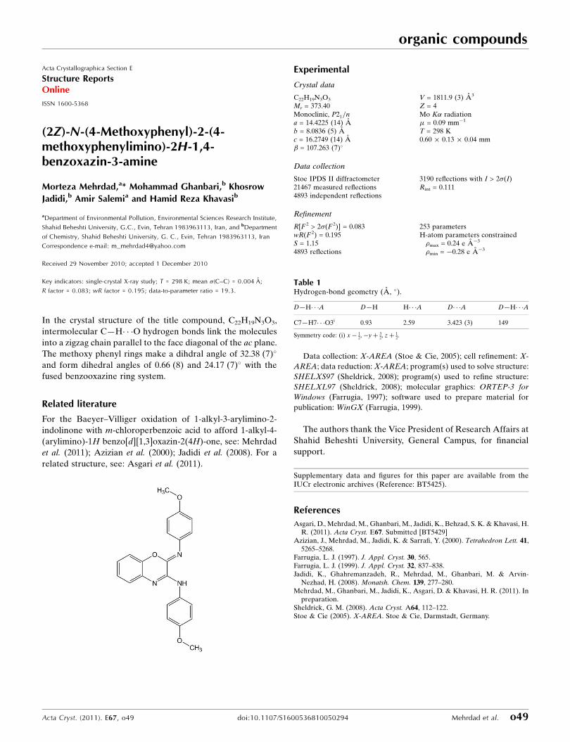
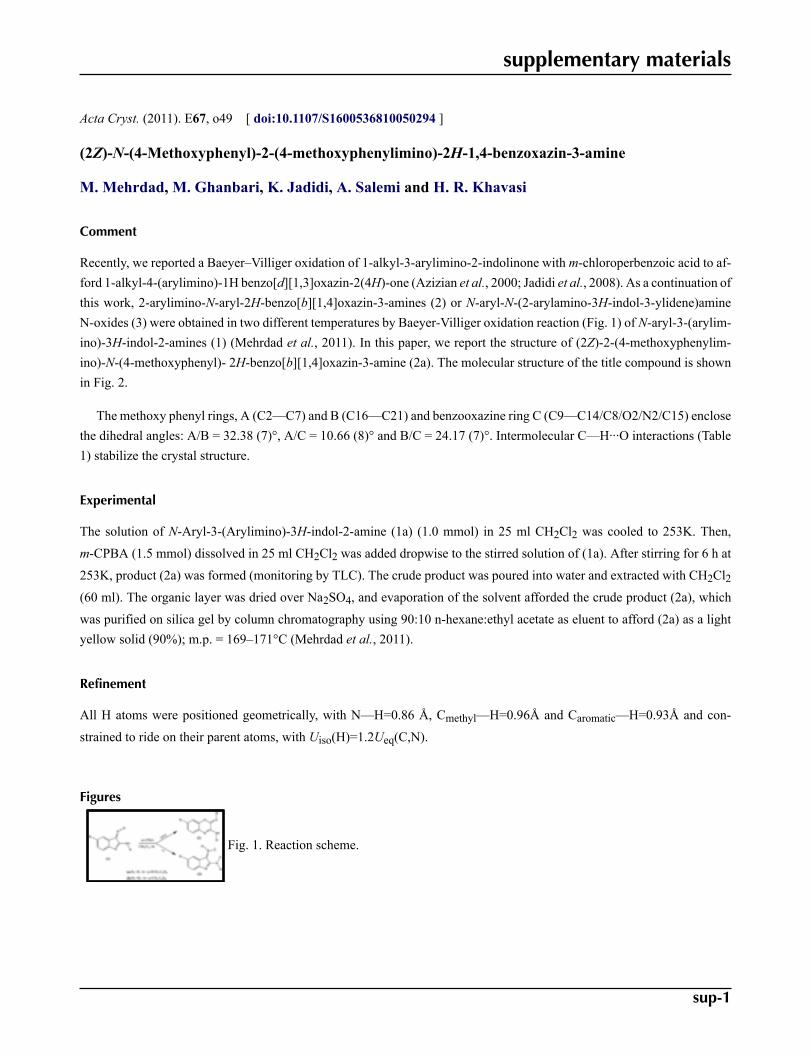
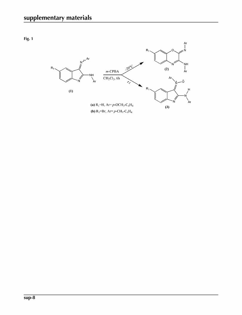
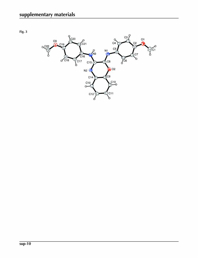
Recently, we reported a Baeyer–Villiger oxidation of 1-alkyl-3-arylimino-2-indolinone with m-chloroperbenzoic acid to af-ford 1-alkyl-4-(arylimino)-1H benzo[d][1,3]oxazin-2(4H)-one (Azizian et al., 2000; Jadidi et al., 2008). As a continuation ofthis work, 2-arylimino-N-aryl-2H-benzo[b][1,4]oxazin-3-amines (2) or N-aryl-N-(2-arylamino-3H-indol-3-ylidene)amineN-oxides (3) were obtained in two different temperatures by Baeyer-Villiger oxidation reaction (Fig. 1) of N-aryl-3-(arylim-ino)-3H-indol-2-amines (1) (Mehrdad et al., 2011). In this paper, we report the structure of (2Z)-2-(4-methoxyphenylim-ino)-N-(4-methoxyphenyl)- 2H-benzo[b][1,4]oxazin-3-amine (2a). The molecular structure of the title compound is shownin Fig. 2.
The methoxy phenyl rings, A (C2—C7) and B (C16—C21) and benzooxazine ring C (C9—C14/C8/O2/N2/C15) enclosethe dihedral angles: A/B = 32.38 (7)°, A/C = 10.66 (8)° and B/C = 24.17 (7)°. Intermolecular C—H···O interactions (Table1) stabilize the crystal structure.
Experimental
The solution of N-Aryl-3-(Arylimino)-3H-indol-2-amine (1a) (1.0 mmol) in 25 ml CH2Cl2 was cooled to 253K. Then,
m-CPBA (1.5 mmol) dissolved in 25 ml CH2Cl2 was added dropwise to the stirred solution of (1a). After stirring for 6 h at
253K, product (2a) was formed (monitoring by TLC). The crude product was poured into water and extracted with CH2Cl2(60 ml). The organic layer was dried over Na2SO4, and evaporation of the solvent afforded the crude product (2a), which
was purified on silica gel by column chromatography using 90:10 n-hexane:ethyl acetate as eluent to afford (2a) as a lightyellow solid (90%); m.p. = 169–171°C (Mehrdad et al., 2011).
Refinement
All H atoms were positioned geometrically, with N—H=0.86 Å, Cmethyl—H=0.96Å and Caromatic—H=0.93Å and con-
strained to ride on their parent atoms, with Uiso(H)=1.2Ueq(C,N).
Figures
Fig. 1. Reaction scheme.
supplementary materials
sup-2
Fig. 2. The molecular structure of the title molecule, with the atom-numbering scheme. Dis-placement ellipsoids are drawn at the 30% probability level.
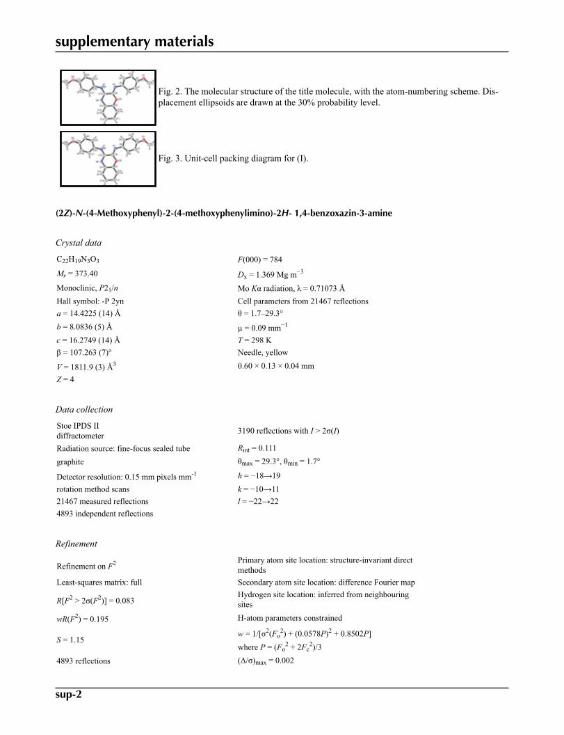
Fig. 3. Unit-cell packing diagram for (I).
(2Z)-N-(4-Methoxyphenyl)-2-(4-methoxyphenylimino)-2H- 1,4-benzoxazin-3-amine
Crystal data
C22H19N3O3 F(000) = 784
Mr = 373.40 Dx = 1.369 Mg m−3
Monoclinic, P21/n Mo Kα radiation, λ = 0.71073 ÅHall symbol: -P 2yn Cell parameters from 21467 reflectionsa = 14.4225 (14) Å θ = 1.7–29.3°b = 8.0836 (5) Å µ = 0.09 mm−1
c = 16.2749 (14) Å T = 298 Kβ = 107.263 (7)° Needle, yellow
V = 1811.9 (3) Å3 0.60 × 0.13 × 0.04 mmZ = 4
Data collection
Stoe IPDS IIdiffractometer 3190 reflections with I > 2σ(I)
Radiation source: fine-focus sealed tube Rint = 0.111
graphite θmax = 29.3°, θmin = 1.7°
Detector resolution: 0.15 mm pixels mm-1 h = −18→19rotation method scans k = −10→1121467 measured reflections l = −22→224893 independent reflections
Refinement
Refinement on F2 Primary atom site location: structure-invariant directmethods
Least-squares matrix: full Secondary atom site location: difference Fourier map
R[F2 > 2σ(F2)] = 0.083Hydrogen site location: inferred from neighbouringsites
wR(F2) = 0.195 H-atom parameters constrained
S = 1.15w = 1/[σ2(Fo
2) + (0.0578P)2 + 0.8502P]where P = (Fo
2 + 2Fc2)/3
4893 reflections (Δ/σ)max = 0.002
supplementary materials
sup-3
253 parameters Δρmax = 0.24 e Å−3
0 restraints Δρmin = −0.28 e Å−3
Special details
Geometry. All e.s.d.'s (except the e.s.d. in the dihedral angle between two l.s. planes) are estimated using the full covariance mat-rix. The cell e.s.d.'s are taken into account individually in the estimation of e.s.d.'s in distances, angles and torsion angles; correlationsbetween e.s.d.'s in cell parameters are only used when they are defined by crystal symmetry. An approximate (isotropic) treatment ofcell e.s.d.'s is used for estimating e.s.d.'s involving l.s. planes.
Refinement. Refinement of F2 against ALL reflections. The weighted R-factor wR and goodness of fit S are based on F2, convention-
al R-factors R are based on F, with F set to zero for negative F2. The threshold expression of F2 > σ(F2) is used only for calculating R-
factors(gt) etc. and is not relevant to the choice of reflections for refinement. R-factors based on F2 are statistically about twice as largeas those based on F, and R- factors based on ALL data will be even larger.
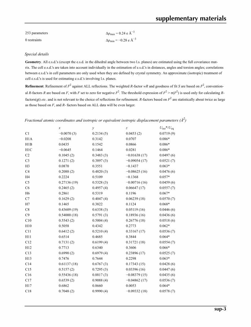
Fractional atomic coordinates and isotropic or equivalent isotropic displacement parameters (Å2)
x y z Uiso*/Ueq
C1 −0.0070 (3) 0.2134 (5) 0.0453 (2) 0.0719 (9)H1A −0.0208 0.3142 0.0707 0.086*H1B 0.0435 0.1542 0.0866 0.086*H1C −0.0645 0.1464 0.0281 0.086*C2 0.1045 (2) 0.3483 (3) −0.01638 (17) 0.0497 (6)C3 0.1271 (2) 0.3897 (3) −0.09054 (17) 0.0521 (7)H3 0.0870 0.3551 −0.1437 0.063*C4 0.2088 (2) 0.4820 (3) −0.08625 (16) 0.0476 (6)H4 0.2224 0.5109 −0.1368 0.057*C5 0.27136 (19) 0.5328 (3) −0.00716 (16) 0.0459 (6)C6 0.2465 (2) 0.4957 (4) 0.06647 (17) 0.0557 (7)H6 0.2861 0.5319 0.1196 0.067*C7 0.1629 (2) 0.4047 (4) 0.06239 (18) 0.0570 (7)H7 0.1465 0.3822 0.1124 0.068*C8 0.43609 (19) 0.6338 (3) 0.05119 (16) 0.0446 (6)C9 0.54000 (18) 0.5791 (3) 0.18936 (16) 0.0436 (6)C10 0.5543 (2) 0.5004 (4) 0.26776 (18) 0.0518 (6)H10 0.5058 0.4342 0.2773 0.062*C11 0.6412 (2) 0.5210 (4) 0.33167 (17) 0.0536 (7)H11 0.6514 0.4685 0.3844 0.064*C12 0.7131 (2) 0.6199 (4) 0.31721 (18) 0.0554 (7)H12 0.7713 0.6340 0.3606 0.066*C13 0.6990 (2) 0.6979 (4) 0.23896 (17) 0.0525 (7)H13 0.7476 0.7644 0.2298 0.063*C14 0.61137 (18) 0.6767 (3) 0.17343 (15) 0.0428 (6)C15 0.5157 (2) 0.7295 (3) 0.03396 (16) 0.0447 (6)C16 0.55436 (18) 0.8817 (3) −0.08379 (15) 0.0435 (6)C17 0.6539 (2) 0.9088 (4) −0.04862 (17) 0.0536 (7)H17 0.6862 0.8660 0.0053 0.064*C18 0.7048 (2) 0.9990 (4) −0.09332 (18) 0.0578 (7)
supplementary materials
sup-4
H18 0.7713 1.0150 −0.0693 0.069*C19 0.6583 (2) 1.0656 (3) −0.17326 (17) 0.0487 (6)C20 0.5592 (2) 1.0388 (4) −0.20832 (17) 0.0548 (7)H20 0.5268 1.0827 −0.2620 0.066*C21 0.5087 (2) 0.9484 (4) −0.16457 (16) 0.0505 (6)H21 0.4425 0.9312 −0.1894 0.061*C22 0.8046 (2) 1.1618 (5) −0.1983 (2) 0.0777 (10)H22A 0.8289 1.0517 −0.1997 0.093*H22B 0.8296 1.2060 −0.1412 0.093*H22C 0.8250 1.2305 −0.2379 0.093*N1 0.35461 (16) 0.6221 (3) −0.01063 (14) 0.0499 (5)N2 0.59911 (15) 0.7521 (3) 0.09392 (12) 0.0413 (5)N3 0.49602 (16) 0.7909 (3) −0.04487 (13) 0.0494 (5)H3A 0.4381 0.7720 −0.0774 0.059*O1 0.02353 (17) 0.2511 (3) −0.02758 (14) 0.0700 (6)O2 0.45078 (13) 0.5596 (3) 0.12731 (12) 0.0556 (5)O3 0.70194 (16) 1.1580 (3) −0.22196 (13) 0.0655 (6)
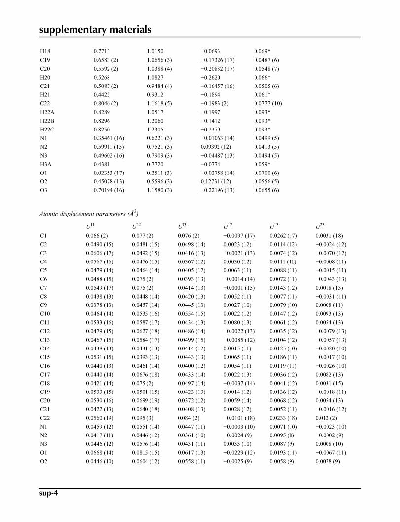
Atomic displacement parameters (Å2)
U11 U22 U33 U12 U13 U23
C1 0.066 (2) 0.077 (2) 0.076 (2) −0.0097 (17) 0.0262 (17) 0.0031 (18)C2 0.0490 (15) 0.0481 (15) 0.0498 (14) 0.0023 (12) 0.0114 (12) −0.0024 (12)C3 0.0606 (17) 0.0492 (15) 0.0416 (13) −0.0021 (13) 0.0074 (12) −0.0070 (12)C4 0.0567 (16) 0.0476 (15) 0.0367 (12) 0.0030 (12) 0.0111 (11) −0.0008 (11)C5 0.0479 (14) 0.0464 (14) 0.0405 (12) 0.0063 (11) 0.0088 (11) −0.0015 (11)C6 0.0488 (15) 0.075 (2) 0.0393 (13) −0.0014 (14) 0.0072 (11) −0.0043 (13)C7 0.0549 (17) 0.075 (2) 0.0414 (13) −0.0001 (15) 0.0143 (12) 0.0018 (13)C8 0.0438 (13) 0.0448 (14) 0.0420 (13) 0.0052 (11) 0.0077 (11) −0.0031 (11)C9 0.0378 (13) 0.0457 (14) 0.0445 (13) 0.0027 (10) 0.0079 (10) 0.0008 (11)C10 0.0464 (14) 0.0535 (16) 0.0554 (15) 0.0022 (12) 0.0147 (12) 0.0093 (13)C11 0.0533 (16) 0.0587 (17) 0.0434 (13) 0.0080 (13) 0.0061 (12) 0.0054 (13)C12 0.0479 (15) 0.0627 (18) 0.0486 (14) −0.0022 (13) 0.0035 (12) −0.0079 (13)C13 0.0467 (15) 0.0584 (17) 0.0499 (15) −0.0085 (12) 0.0104 (12) −0.0057 (13)C14 0.0438 (13) 0.0431 (13) 0.0414 (12) 0.0015 (11) 0.0125 (10) −0.0020 (10)C15 0.0531 (15) 0.0393 (13) 0.0443 (13) 0.0065 (11) 0.0186 (11) −0.0017 (10)C16 0.0440 (13) 0.0461 (14) 0.0400 (12) 0.0054 (11) 0.0119 (11) −0.0026 (10)C17 0.0440 (14) 0.0676 (18) 0.0433 (14) 0.0022 (13) 0.0036 (12) 0.0082 (13)C18 0.0421 (14) 0.075 (2) 0.0497 (14) −0.0037 (14) 0.0041 (12) 0.0031 (15)C19 0.0533 (15) 0.0501 (15) 0.0423 (13) 0.0014 (12) 0.0136 (12) −0.0018 (11)C20 0.0530 (16) 0.0699 (19) 0.0372 (12) 0.0059 (14) 0.0068 (12) 0.0054 (13)C21 0.0422 (13) 0.0640 (18) 0.0408 (13) 0.0028 (12) 0.0052 (11) −0.0016 (12)C22 0.0560 (19) 0.095 (3) 0.084 (2) −0.0101 (18) 0.0233 (18) 0.012 (2)N1 0.0459 (12) 0.0551 (14) 0.0447 (11) −0.0003 (10) 0.0071 (10) −0.0023 (10)N2 0.0417 (11) 0.0446 (12) 0.0361 (10) −0.0024 (9) 0.0095 (8) −0.0002 (9)N3 0.0446 (12) 0.0576 (14) 0.0431 (11) 0.0033 (10) 0.0087 (9) 0.0008 (10)O1 0.0668 (14) 0.0815 (15) 0.0617 (13) −0.0229 (12) 0.0193 (11) −0.0067 (11)O2 0.0446 (10) 0.0604 (12) 0.0558 (11) −0.0025 (9) 0.0058 (9) 0.0078 (9)
supplementary materials
sup-5
O3 0.0595 (13) 0.0817 (15) 0.0544 (11) −0.0078 (11) 0.0157 (10) 0.0101 (11)
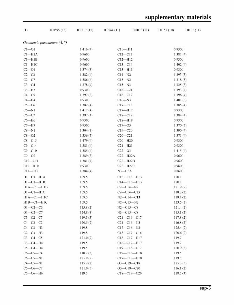
Geometric parameters (Å, °)
C1—O1 1.416 (4) C11—H11 0.9300C1—H1A 0.9600 C12—C13 1.381 (4)C1—H1B 0.9600 C12—H12 0.9300C1—H1C 0.9600 C13—C14 1.402 (4)C2—O1 1.374 (3) C13—H13 0.9300C2—C3 1.382 (4) C14—N2 1.393 (3)C2—C7 1.386 (4) C15—N2 1.318 (3)C3—C4 1.378 (4) C15—N3 1.325 (3)C3—H3 0.9300 C16—C21 1.393 (4)C4—C5 1.397 (3) C16—C17 1.396 (4)C4—H4 0.9300 C16—N3 1.401 (3)C5—C6 1.382 (4) C17—C18 1.385 (4)C5—N1 1.417 (4) C17—H17 0.9300C6—C7 1.397 (4) C18—C19 1.384 (4)C6—H6 0.9300 C18—H18 0.9300C7—H7 0.9300 C19—O3 1.370 (3)C8—N1 1.304 (3) C19—C20 1.390 (4)C8—O2 1.336 (3) C20—C21 1.371 (4)C8—C15 1.479 (4) C20—H20 0.9300C9—C14 1.381 (4) C21—H21 0.9300C9—C10 1.385 (4) C22—O3 1.415 (4)C9—O2 1.389 (3) C22—H22A 0.9600C10—C11 1.381 (4) C22—H22B 0.9600C10—H10 0.9300 C22—H22C 0.9600C11—C12 1.384 (4) N3—H3A 0.8600
O1—C1—H1A 109.5 C12—C13—H13 120.1O1—C1—H1B 109.5 C14—C13—H13 120.1H1A—C1—H1B 109.5 C9—C14—N2 121.9 (2)O1—C1—H1C 109.5 C9—C14—C13 118.8 (2)H1A—C1—H1C 109.5 N2—C14—C13 119.4 (2)H1B—C1—H1C 109.5 N2—C15—N3 123.5 (2)O1—C2—C3 115.8 (2) N2—C15—C8 121.4 (2)O1—C2—C7 124.8 (3) N3—C15—C8 115.1 (2)C3—C2—C7 119.5 (3) C21—C16—C17 117.8 (2)C4—C3—C2 120.5 (2) C21—C16—N3 116.8 (2)C4—C3—H3 119.8 C17—C16—N3 125.4 (2)C2—C3—H3 119.8 C18—C17—C16 120.6 (2)C3—C4—C5 121.0 (2) C18—C17—H17 119.7C3—C4—H4 119.5 C16—C17—H17 119.7C5—C4—H4 119.5 C19—C18—C17 120.9 (3)C6—C5—C4 118.2 (3) C19—C18—H18 119.5C6—C5—N1 125.9 (2) C17—C18—H18 119.5C4—C5—N1 115.9 (2) O3—C19—C18 125.3 (3)C5—C6—C7 121.0 (3) O3—C19—C20 116.1 (2)C5—C6—H6 119.5 C18—C19—C20 118.5 (3)
supplementary materials
sup-6
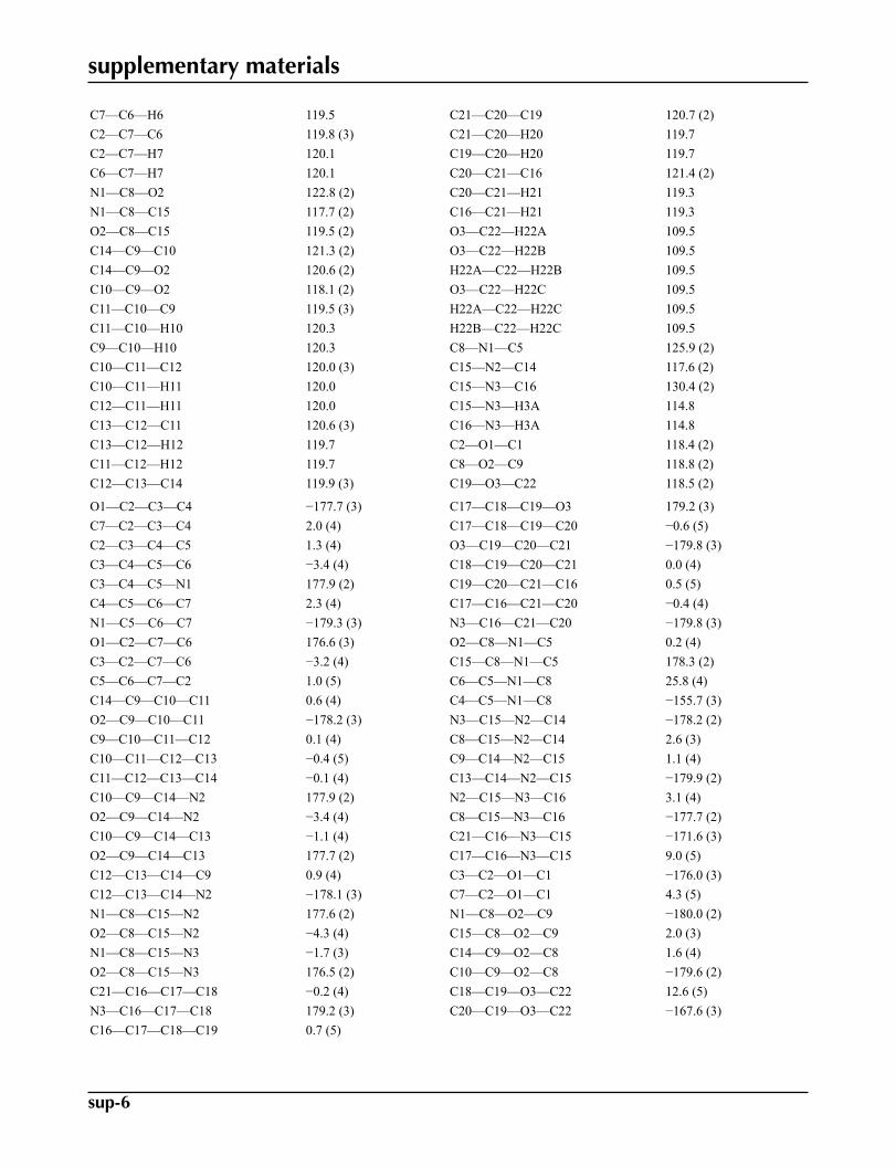
C7—C6—H6 119.5 C21—C20—C19 120.7 (2)C2—C7—C6 119.8 (3) C21—C20—H20 119.7C2—C7—H7 120.1 C19—C20—H20 119.7C6—C7—H7 120.1 C20—C21—C16 121.4 (2)N1—C8—O2 122.8 (2) C20—C21—H21 119.3N1—C8—C15 117.7 (2) C16—C21—H21 119.3O2—C8—C15 119.5 (2) O3—C22—H22A 109.5C14—C9—C10 121.3 (2) O3—C22—H22B 109.5C14—C9—O2 120.6 (2) H22A—C22—H22B 109.5C10—C9—O2 118.1 (2) O3—C22—H22C 109.5C11—C10—C9 119.5 (3) H22A—C22—H22C 109.5C11—C10—H10 120.3 H22B—C22—H22C 109.5C9—C10—H10 120.3 C8—N1—C5 125.9 (2)C10—C11—C12 120.0 (3) C15—N2—C14 117.6 (2)C10—C11—H11 120.0 C15—N3—C16 130.4 (2)C12—C11—H11 120.0 C15—N3—H3A 114.8C13—C12—C11 120.6 (3) C16—N3—H3A 114.8C13—C12—H12 119.7 C2—O1—C1 118.4 (2)C11—C12—H12 119.7 C8—O2—C9 118.8 (2)C12—C13—C14 119.9 (3) C19—O3—C22 118.5 (2)
O1—C2—C3—C4 −177.7 (3) C17—C18—C19—O3 179.2 (3)C7—C2—C3—C4 2.0 (4) C17—C18—C19—C20 −0.6 (5)C2—C3—C4—C5 1.3 (4) O3—C19—C20—C21 −179.8 (3)C3—C4—C5—C6 −3.4 (4) C18—C19—C20—C21 0.0 (4)C3—C4—C5—N1 177.9 (2) C19—C20—C21—C16 0.5 (5)C4—C5—C6—C7 2.3 (4) C17—C16—C21—C20 −0.4 (4)N1—C5—C6—C7 −179.3 (3) N3—C16—C21—C20 −179.8 (3)O1—C2—C7—C6 176.6 (3) O2—C8—N1—C5 0.2 (4)C3—C2—C7—C6 −3.2 (4) C15—C8—N1—C5 178.3 (2)C5—C6—C7—C2 1.0 (5) C6—C5—N1—C8 25.8 (4)C14—C9—C10—C11 0.6 (4) C4—C5—N1—C8 −155.7 (3)O2—C9—C10—C11 −178.2 (3) N3—C15—N2—C14 −178.2 (2)C9—C10—C11—C12 0.1 (4) C8—C15—N2—C14 2.6 (3)C10—C11—C12—C13 −0.4 (5) C9—C14—N2—C15 1.1 (4)C11—C12—C13—C14 −0.1 (4) C13—C14—N2—C15 −179.9 (2)C10—C9—C14—N2 177.9 (2) N2—C15—N3—C16 3.1 (4)O2—C9—C14—N2 −3.4 (4) C8—C15—N3—C16 −177.7 (2)C10—C9—C14—C13 −1.1 (4) C21—C16—N3—C15 −171.6 (3)O2—C9—C14—C13 177.7 (2) C17—C16—N3—C15 9.0 (5)C12—C13—C14—C9 0.9 (4) C3—C2—O1—C1 −176.0 (3)C12—C13—C14—N2 −178.1 (3) C7—C2—O1—C1 4.3 (5)N1—C8—C15—N2 177.6 (2) N1—C8—O2—C9 −180.0 (2)O2—C8—C15—N2 −4.3 (4) C15—C8—O2—C9 2.0 (3)N1—C8—C15—N3 −1.7 (3) C14—C9—O2—C8 1.6 (4)O2—C8—C15—N3 176.5 (2) C10—C9—O2—C8 −179.6 (2)C21—C16—C17—C18 −0.2 (4) C18—C19—O3—C22 12.6 (5)N3—C16—C17—C18 179.2 (3) C20—C19—O3—C22 −167.6 (3)C16—C17—C18—C19 0.7 (5)
supplementary materials
sup-7
Hydrogen-bond geometry (Å, °)
D—H···A D—H H···A D···A D—H···A
C7—H7···O3i 0.93 2.59 3.423 (3) 149Symmetry codes: (i) x−1/2, −y+3/2, z+1/2.
supplementary materials
sup-8
Fig. 1

supplementary materials
sup-9
Fig. 2
supplementary materials
sup-10
Fig. 3
Related Documents


