TOOLS OF MICROBIOLOGY

Welcome message from author
This document is posted to help you gain knowledge. Please leave a comment to let me know what you think about it! Share it to your friends and learn new things together.
Transcript
TOOLS OF MICROBIOLOGY
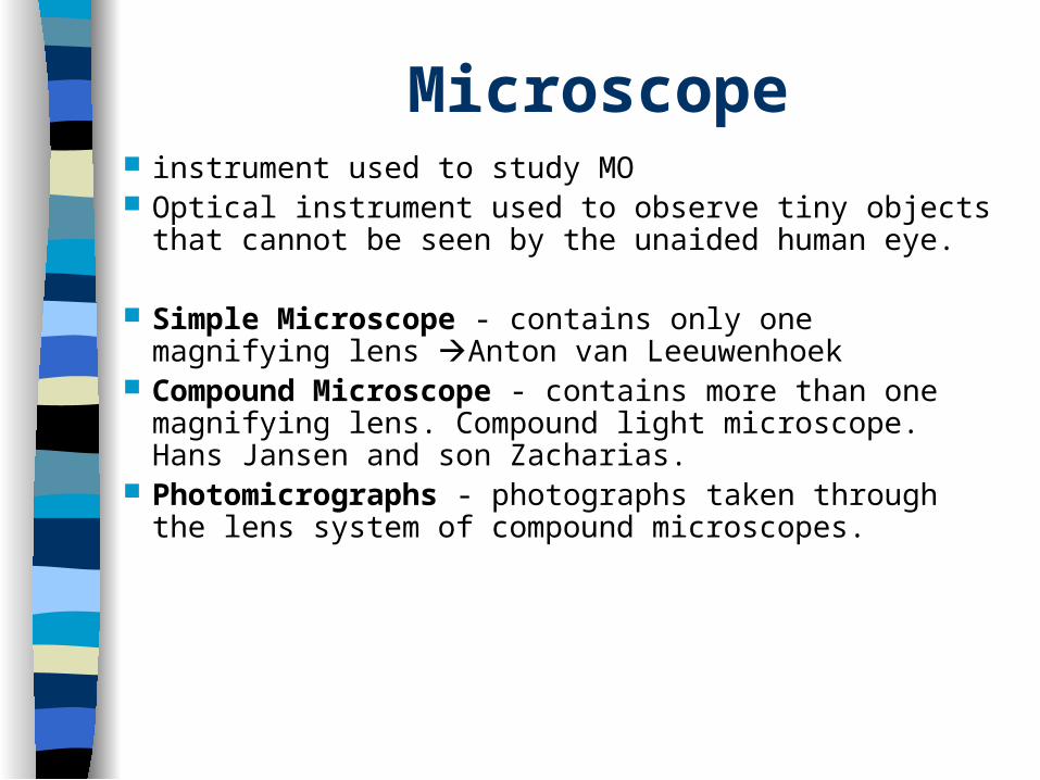
Microscope instrument used to study MO Optical instrument used to observe tiny objects that
cannot be seen by the unaided human eye.
Simple Microscope - contains only one magnifying lens Anton van Leeuwenhoek
Compound Microscope - contains more than one magnifying lens. Compound light microscope. Hans Jansen and son Zacharias.
Photomicrographs - photographs taken through the lens system of compound microscopes.
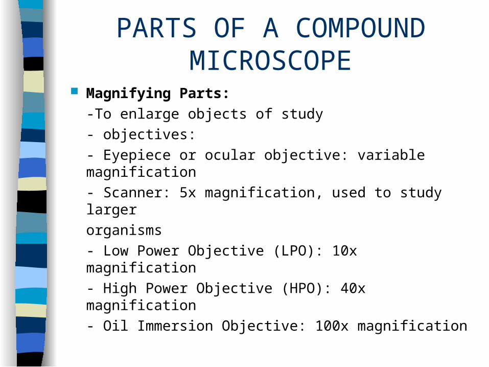
PARTS OF A COMPOUND MICROSCOPE
Magnifying Parts:
-To enlarge objects of study
- objectives:
- Eyepiece or ocular objective: variable magnification
- Scanner: 5x magnification, used to study larger
organisms
- Low Power Objective (LPO): 10x magnification
- High Power Objective (HPO): 40x magnification
- Oil Immersion Objective: 100x magnification
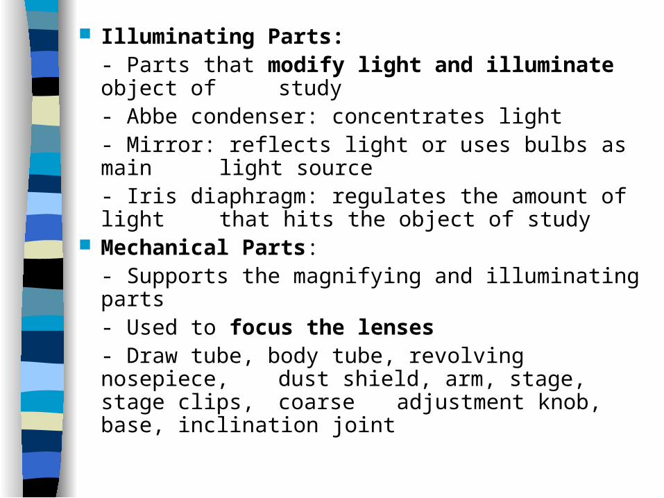
Illuminating Parts:- Parts that modify light and illuminate object of
study- Abbe condenser: concentrates light- Mirror: reflects light or uses bulbs as main
light source- Iris diaphragm: regulates the amount of
light that hits the object of study Mechanical Parts:
- Supports the magnifying and illuminating parts- Used to focus the lenses
- Draw tube, body tube, revolving nosepiece, dust shield, arm, stage, stage clips, coarse adjustment knob, base, inclination joint
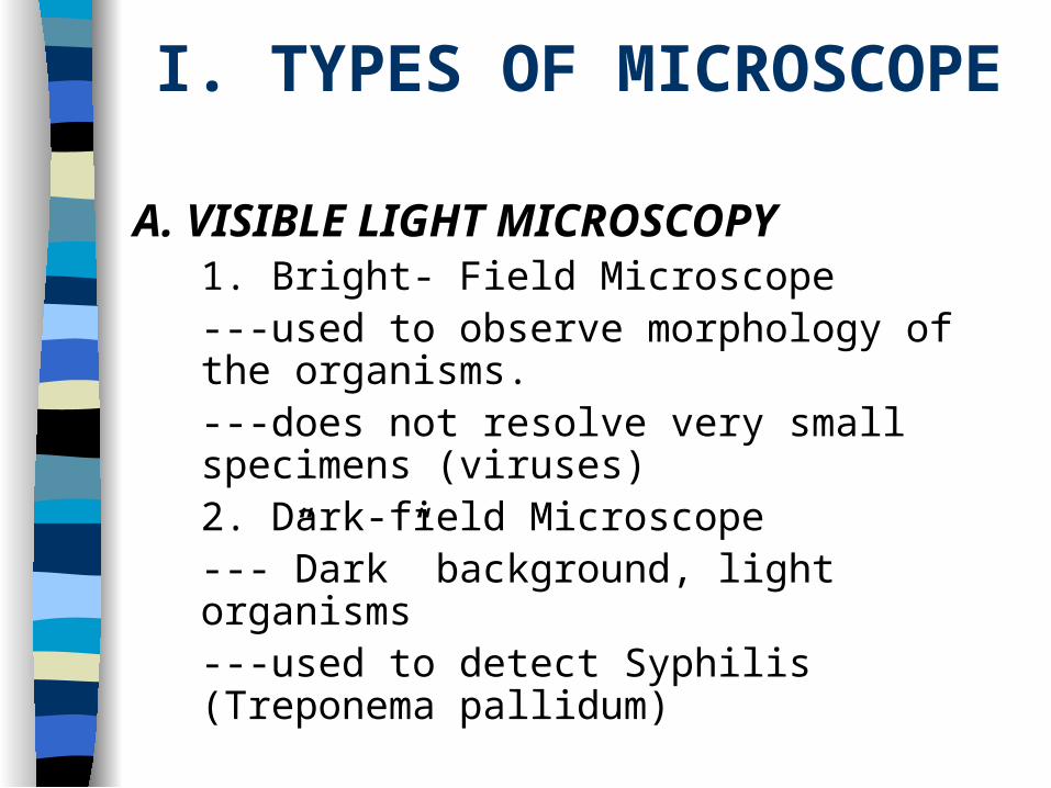
I. TYPES OF MICROSCOPE
A. VISIBLE LIGHT MICROSCOPY1. Bright- Field Microscope
---used to observe morphology of the organisms.
---does not resolve very small specimens (viruses)2. Dark-field Microscope
---”Dark” background, light organisms---used to detect Syphilis (Treponema
pallidum)
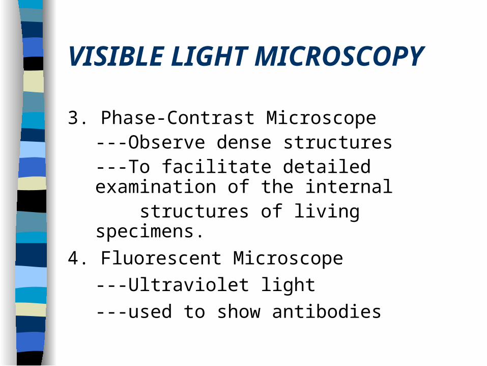
VISIBLE LIGHT MICROSCOPY
3. Phase-Contrast Microscope---Observe dense structures---To facilitate detailed
examination of the internal structures of living specimens.
4. Fluorescent Microscope
---Ultraviolet light
---used to show antibodies
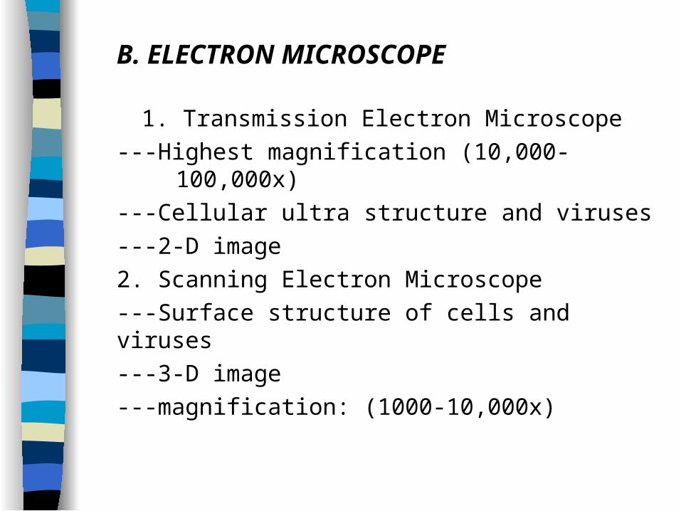
B. ELECTRON MICROSCOPE
1. Transmission Electron Microscope
---Highest magnification (10,000- 100,000x)
---Cellular ultra structure and viruses
---2-D image
2. Scanning Electron Microscope
---Surface structure of cells and viruses
---3-D image
---magnification: (1000-10,000x)
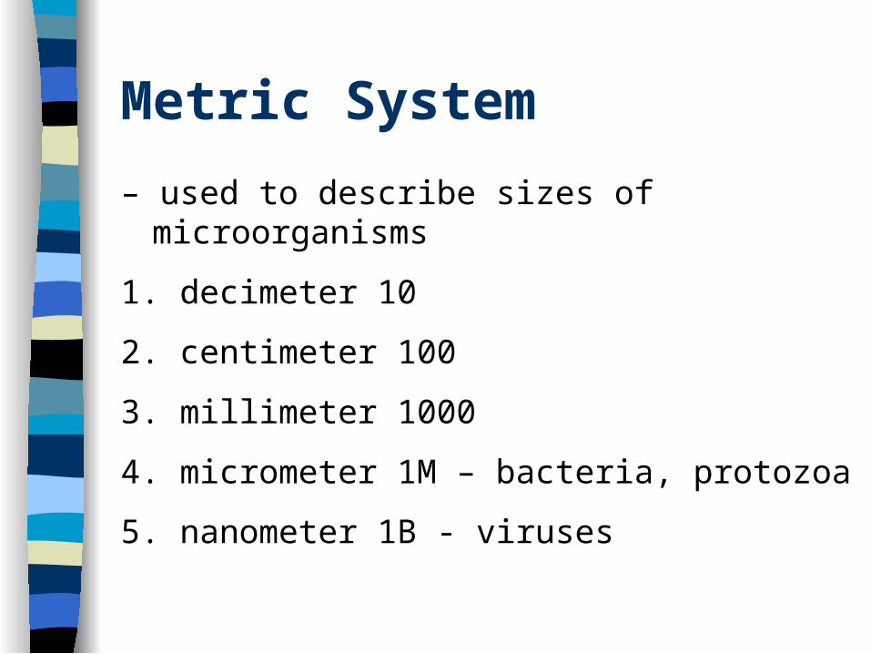
Metric System
– used to describe sizes of microorganisms
1. decimeter 10
2. centimeter 100
3. millimeter 1000
4. micrometer 1M – bacteria, protozoa
5. nanometer 1B - viruses
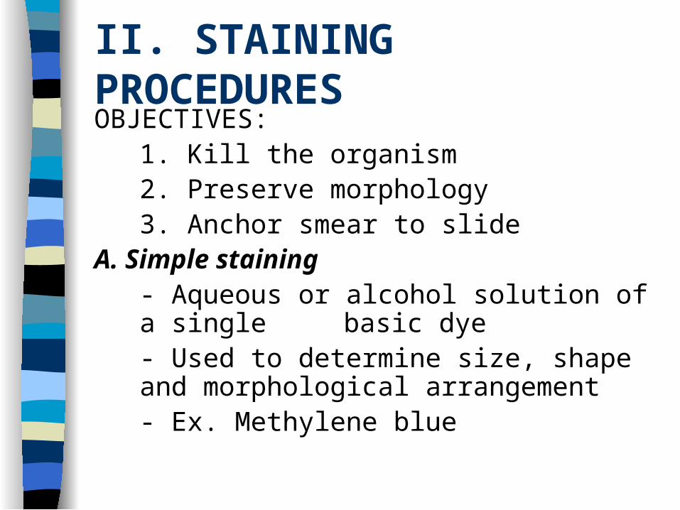
II. STAINING PROCEDURESOBJECTIVES:
1. Kill the organism2. Preserve morphology3. Anchor smear to slide
A. Simple staining- Aqueous or alcohol solution of a single
basic dye- Used to determine size, shape and morphological arrangement- Ex. Methylene blue
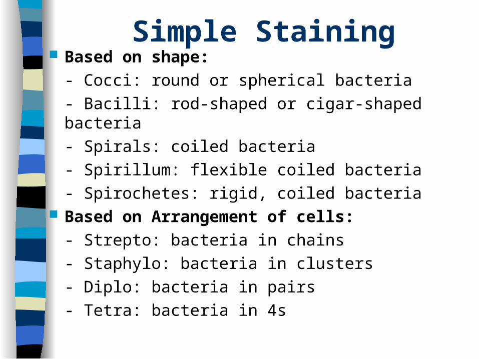
Simple Staining Based on shape:
- Cocci: round or spherical bacteria
- Bacilli: rod-shaped or cigar-shaped bacteria
- Spirals: coiled bacteria
- Spirillum: flexible coiled bacteria
- Spirochetes: rigid, coiled bacteria Based on Arrangement of cells:
- Strepto: bacteria in chains
- Staphylo: bacteria in clusters
- Diplo: bacteria in pairs
- Tetra: bacteria in 4s
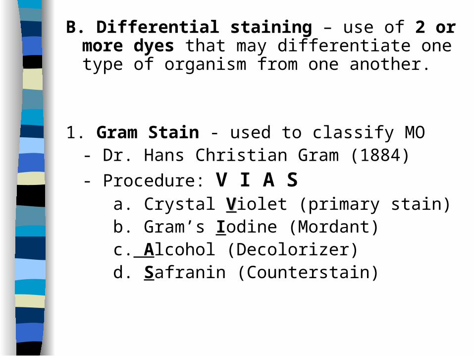
B. Differential staining – use of 2 or more dyes that may differentiate one type of organism from one another.
1. Gram Stain - used to classify MO- Dr. Hans Christian Gram (1884)
- Procedure: V I A S a. Crystal VViolet (primary stain) b. Gram’s Iodine (Mordant) c. Alcohol (Decolorizer) d. Safranin (Counterstain)
Differential staining
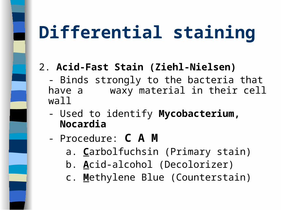
2. Acid-Fast Stain (Ziehl-Nielsen)- Binds strongly to the bacteria that
have a waxy material in their cell wall- Used to identify Mycobacterium, Nocardia
- Procedure: C A M a. Carbolfuchsin (Primary stain) b. Acid-alcohol (Decolorizer) c. Methylene Blue (Counterstain)
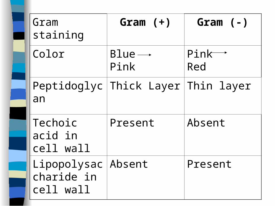
Gram staining Gram (+) Gram (-)
Color Blue Pink Pink Red
Peptidoglycan Thick Layer Thin layer
Techoic acid in cell wall
Present Absent
Lipopolysaccharide in cell wall
Absent Present
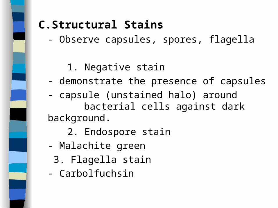
C.Structural Stains- Observe capsules, spores, flagella
1. Negative stain
- demonstrate the presence of capsules
- capsule (unstained halo) around bacterial cells against dark background.
2. Endospore stain
- Malachite green
3. Flagella stain
- Carbolfuchsin
Related Documents