On Two Species of Kallymenia (Rhodophyta: Gigartinales: Kallymeniaceae) from the Hawaiian Islands, Central Pacific 1 Isabella A. Abbott 2 and Karla J. McDermid 3 Abstract: Two species of Kallymenia from the Hawaiian Islands, one rare, K sessilis Okamura, and the other described here for the first time, K thompsonii, n. sp., are examined, compared, and contrasted with other similar Kallymenia spe- cies. Both species are unusual because Kallymenia is generally regarded as a temperate taxon, and tropical or subtropical species are seldom encountered. The two species are alike in that they have a female reproductive apparatus that is monocarpogonial: wherein a single carpogonial filament is associated with a supporting cell also bearing an arrangement of subsidiary cells that is charac- teristic of some of the family Kallymeniaceae. In the genus Kallymenia, vegeta- tive components shown in a cross section are a narrow outer cortex, often only three cells thick, followed inwardly by one to two layers of subcortical cells. In the two species studied here, there appears to be a constant shape and arrange- ment of subcortical cells in each species, whereas the number of medullary fila- ments and their arrangements appear to be less stable in their configuration than the subcortical cells. Branched refractive cells or stellate cells, which often occur in species of Kallymenia, were not seen in K thompsonii and only rarely in K sessilis. Kallymenia thompsonii commonly has perforations in the maturing blades, whereas K. sessilis does not. Kallymenia sessilis OKAMURA, from central Ja- pan, was included in the Hawaiian marine flora recently with a minimum of information because it was not known what the Japanese species looked like beyond its first descrip- tion. A welcome opportunity was provided when a new species of Kallymenia was col- lected from Midway Atoll in the North- western Hawaiian Islands (NWHI), offering material that could be compared in detail with K sessilis. The two species of Kallymenia reported in this paper occur in relatively deep 1 Manuscript accepted 15 August 200l. 2 Correspondence: Deparnnent of Botany, University of Hawai'i at Manoa, 3190 Maile Way, Honolulu, Hawai'i 96822 (telephone, 808-956-8073; fax, 808-956- 3923; E-mail, iabbott®hawaii.edu). 3 Reprint requests: Marine Science Deparnnent, University of Hawai'i at Hilo, 200 West Kawili Street, Hilo, Hawai'i 96720 (telephone, 808-933-3906; fax 808- 974-7693; E-mail, [email protected]). Pacific Science (2002), vol. 56, no. 2:149-162 © 2002 by University of Hawai'i Press. All rights reserved (6 to 10 m) to deeper (12 to 20 m) water depths. One of them, Kallymenia sessilis Oka- mura, 1934, was first reported from the Gulf of Tateyama, central Japan, and remains a rare plant in Japan (T. Yoshida, 1999, pers. comm.). It was described and illustrated by Okamura (1934:20-21, pI. 312, figs. 1-5) and was reported from the NWHI and in the main (southeastern) Hawaiian Islands by Ab- bott (1999). The second species, Kallymenia thompsonii Abbott & McDermid, n. sp., is de- scribed here. The majority of marine algae previously reported from the NWHI, formerly known as the Leeward Hawaiian Islands, are from intertidal to snorkel depths (about 5 m) (Ab- bott 1989), but in recent years, dredging activities and scuba have made algae from greater depths available for study. Such cur- rent collections are obtained from depths of 10 to 100 m and have brought to light rare new records of species in the Hawaiian Is- lands (Abbott 1999), such as Crouania mage- shimensis Itono, Lejolisea pacifica Itono (both Ceramiaceae), and Kallymenia sessilis Okamura from Japan, and Malaconema minimum Hol- 149

Welcome message from author
This document is posted to help you gain knowledge. Please leave a comment to let me know what you think about it! Share it to your friends and learn new things together.
Transcript
On Two Species of Kallymenia (Rhodophyta: Gigartinales:Kallymeniaceae) from the Hawaiian Islands, Central Pacific1
Isabella A. Abbott2 and Karla J. McDermid 3
Abstract: Two species of Kallymenia from the Hawaiian Islands, one rare, Ksessilis Okamura, and the other described here for the first time, K thompsonii, n.sp., are examined, compared, and contrasted with other similar Kallymenia species. Both species are unusual because Kallymenia is generally regarded as atemperate taxon, and tropical or subtropical species are seldom encountered.The two species are alike in that they have a female reproductive apparatus thatis monocarpogonial: wherein a single carpogonial filament is associated with asupporting cell also bearing an arrangement of subsidiary cells that is characteristic of some of the family Kallymeniaceae. In the genus Kallymenia, vegetative components shown in a cross section are a narrow outer cortex, often onlythree cells thick, followed inwardly by one to two layers of subcortical cells. Inthe two species studied here, there appears to be a constant shape and arrangement of subcortical cells in each species, whereas the number of medullary filaments and their arrangements appear to be less stable in their configuration thanthe subcortical cells. Branched refractive cells or stellate cells, which often occurin species of Kallymenia, were not seen in K thompsonii and only rarely in Ksessilis. Kallymenia thompsonii commonly has perforations in the maturing blades,whereas K. sessilis does not.
Kallymenia sessilis OKAMURA, from central Japan, was included in the Hawaiian marineflora recently with a minimum of informationbecause it was not known what the Japanesespecies looked like beyond its first description. A welcome opportunity was providedwhen a new species of Kallymenia was collected from Midway Atoll in the Northwestern Hawaiian Islands (NWHI), offeringmaterial that could be compared in detailwith K sessilis. The two species of Kallymeniareported in this paper occur in relatively deep
1 Manuscript accepted 15 August 200l.2 Correspondence: Deparnnent of Botany, University
of Hawai'i at Manoa, 3190 Maile Way, Honolulu,Hawai'i 96822 (telephone, 808-956-8073; fax, 808-9563923; E-mail, iabbott®hawaii.edu).
3 Reprint requests: Marine Science Deparnnent,University of Hawai'i at Hilo, 200 West Kawili Street,Hilo, Hawai'i 96720 (telephone, 808-933-3906; fax 808974-7693; E-mail, [email protected]).
Pacific Science (2002), vol. 56, no. 2:149-162© 2002 by University of Hawai'i Press.All rights reserved
(6 to 10 m) to deeper (12 to 20 m) waterdepths. One of them, Kallymenia sessilis Okamura, 1934, was first reported from the Gulfof Tateyama, central Japan, and remains arare plant in Japan (T. Yoshida, 1999, pers.comm.). It was described and illustrated byOkamura (1934:20-21, pI. 312, figs. 1-5) andwas reported from the NWHI and in themain (southeastern) Hawaiian Islands by Abbott (1999). The second species, Kallymeniathompsonii Abbott & McDermid, n. sp., is described here.
The majority of marine algae previouslyreported from the NWHI, formerly knownas the Leeward Hawaiian Islands, are fromintertidal to snorkel depths (about 5 m) (Abbott 1989), but in recent years, dredgingactivities and scuba have made algae fromgreater depths available for study. Such current collections are obtained from depths of10 to 100 m and have brought to light rarenew records of species in the Hawaiian Islands (Abbott 1999), such as Crouania mageshimensis Itono, Lejolisea pacifica Itono (bothCeramiaceae), and Kallymenia sessilis Okamurafrom Japan, and Malaconema minimum Hol-
149
150 PACIFIC SCIENCE· April 2002
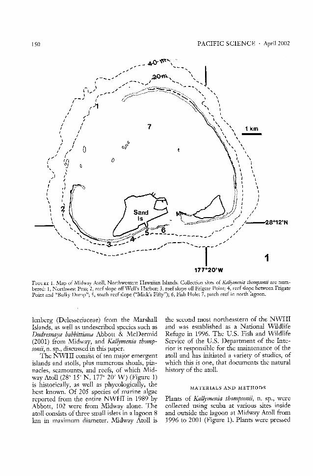
FIGURE 1. Map of Midway Atoll, Northwestern Hawaiian Islands. Collection sites of Kallymenia thompsonii are numbered: 1, Northwest Pass; 2, reef slope off Well's Harbor; 3, reef slope off Frigate Point; 4, reef slope between FrigatePoint and "Bulky Dump"; 5, south reef slope ("Mick's Fifty"); 6, Fish Hole; 7, patch reef in north lagoon.
lenberg (Delesseriaceae) from the MarshallIslands, as well as undescribed species such asDudresnaya babbittiana Abbott & McDermid(2001) from Midway, and Kallymenia thompsonii, n. sp., discussed in this paper.
The NWHI consist of ten major emergentislands and atolls, plus numerous shoals, pinnacles, seamounts, and reefs, of which Midway Atoll (28 0 15' N, 17JO 20' W) (Figure 1)is historically, as well as phycologically, thebest known. Of 205 species of marine algaereported from the entire NWHI in 1989 byAbbott, 102 were from Midway alone. Theatoll consists of three small islets in a lagoon 8km in maximum diameter. Midway Atoll is
the second most northeastern of the NWHIand was established as a National WildlifeRefuge in 1996. The u.s. Fish and WildlifeService of the U.S. Department of the Interior is responsible for the maintenance of theatoll and has initiated a variety of studies, ofwhich this is one, that documents the naturalhistory of the atoll.
MATERIALS AND METHODS
Plants of Kallymenia thompsonii, n. sp., werecollected using scuba at various sites insideand outside the lagoon at Midway Atoll from1996 to 2001 (Figure 1). Plants were pressed
Two Species of Kallymenia from the Hawaiian Islands . Abbott and McDermid 151
as herbarium specimens and a few portionswere preserved in 5% formalin and seawater.Specimens were later sectioned and preparedfor examination following the methods outlined by Tsuda and Abbott (1985), using0.75% aniline blue, intensified with a drop of1% HCI. Slides were mounted in a 35% glucose syrup. The type specimen will be deposited in the Herbarium of the BishopMuseum (BISH) in Honolulu (as BISH 654689),with duplicates distributed to the U.S. National Herbarium, Smithsonian Institution(us), and the University of California, Berkeley (uc). Microscope slides will be retained atBISH. Herbarium specimens of K sessilis collected on Blonde Reef, Hilo Bay, Hawai'i,were sectioned and examined microscopically.Japanese specimens of K sessilis loaned fromthe Okamura herbarium were also examined.
RESULTS
Kallymenia thompsonii Abbott & McDermid,n. sp.Figures 2-11
LATIN DIAGNOSIS: Plantae foliosae, paginis lubricis ubi vivae, compositae ex rosulis2-3 stratorurn laminarurn communi hapterocentrali. Lamina basalis grandissirna et expansa ad 10(-18) cm diam.j laminae superaerosulae parviores, plus irregulares in circumferentia et diametroj lamina basalis adsubstratum affixa per discum incrassatum velcrassitudes ventrales hic illic. Laminae atroroseae ad vinacerasj ubi maturae et reproducti, laminas secundarias, ut pote laminulassecus margines distales edentes, vel marginibus erosis laceratisque librisquej aliter laminae marginibus non profunde crenatis vel latedentatis. Paginae laminarum aliquando foraminis amplitudine variabilia. In sectione transversa, laminae 140-250 J.lm latae, 2-3 stratisvalde pigmentosarurn cellularurn corticalium,et 2-3 stratis grandium, irregulatim ovaliumvel plerurnque trapezialium cellularurn subcorticalium. Ornnis cellula subcorticalis ad 36 fila medullosa conjuncta. Cellulae externicorticis minutae, ovalis, minus quam 4 J.lmdiam., cellulae ferenti circa 5 J.lm lata longaque et parurn irregulari in forma. Medulla
centralis partem majorem sectionis formans,filis periclinatis et anticlinatis ad cellulas subcorticales et inter se trans medullam connexisjfila 3-13 J.lill lata, conspicuis plastis taeniforrnibus. Plantae eystocarpiferae numerosisfilis periclinatis et parurn paucioribus filisanticlinatis, autem plantae terasporangiferaefilis anticlinatis periclinatisque sirnilaribusnumeris. Nec stellatae nec giganto-refractivaecellulae observatae. Gametophyta et tetrasporophyta isomorpha. T etrasporangia in corticeaggregata, numerosaj ubi matura, cruciatimdivisa, 13-20 J.lm lata, 18-22 J.lm longa.Spermatangia non visa. Procarpia monocarpogonialia, uno filo carpogoniali inter 6 velplus cellulas subsidiarias. Cystocarpia oculonudo visibilia, circa 160-200 J.lm diametro,pustulas parvas manifestis carpostomatibusin pagina laminae formantes. Carposporangiasphaerica aspectu polari, 13-20 J.lm aspectilaterali. Fere OInnes cellulae gonirnoblasti incarposporangia transformantes, cellulis sterilis interrnixtis.
ENGLISH TRANSLATION: Plants leafy, withslippery surfaces when fresh, consisting ofrosettes of 2-3 layers of blades with a common central holdfast. The lower blade thelargest and expanded, up to 10 (in one specimen 18) cm in diameterj the upper blades ofthe rosette smaller, more irregular in bothcircumference and diameterj the lowest bladeattached to the substratum by a thickeneddisk or by ventral thickenings here and there.Blades dark rose to wine redj when matureand reproductive, putting forth secondaryblades along the distal margins as bladelets,or with eroded and torn and free marginsjotherwise blades with shallowly scalloped orbroadly dentate margins. Surfaces of bladessometimes with variously sized perforations.In cross section, blades 140-250 J.lm wide,with 2, rarely 3, layers of strongly pigmentedcortical cells, and 2-3 layers of large irregularly oval or commonly trapezoidal subcortical cells. Each subcortical cell connected to3-6 medullary filaments. Outer cortical cellstiny, oval, less than 4 J.lm in diameter, withtheir bearing cell about 5 J.lm wide by 5 J.lmlong and slightly irregular in shape. Thecentral medulla forming the major part of thecross section, with periclinal and anticlinal
152 PACIFIC SCIENCE· April 2002
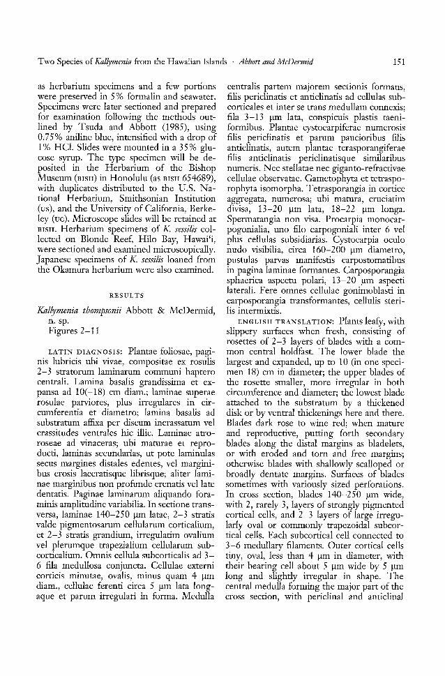
FIGURES 2-3. Kallymenia thompsonii Abbott & McDermid. (2) Holotype, KM 4025a (BISH 654689), with cystocarpsseen among holes in the thallus at upper left, and dark patch seen at the middle right indicating several layers of bladeslying on each other, originating from a central region. The holes are not present in young and developing blades.Scale = 1.5 cm. (3) Paratype (KM 4025b) collected at the same place and date by the same collector as the holotype.Tetrasporangial. Scale = 1.5 cm.
filaments connected to subcortical cells and toeach other across the medulla; filaments 3-13llm wide, with conspicuous ribbonlike plastids. Cystocarpic plants with numerous periclinal filaments and slightly fewer anticlinalfilaments, whereas tetrasporangial plants withsimilar numbers of anticlinal and periclinalfilaments. Neither stellate nor giant refractivecells observed. Gametophytes and tetrasporophytes isomorphic. Tetrasporangia numerous, aggregated in the cortex; when mature,cruciately divided, 13-20 llm wide and 18-22llm long. Spermatangia were not seen. Procarps monocarpogonial, with a single carpogonial filament among 6 or more subsidiarycells. Cystocarps visible with the unaidedeye, about 160-200 llm in diameter, formingsmall pustules with obvious carpostomes onthe blade's surface. Carposporangia sphericalin polar view, 13-20 llm in lateral view.Nearly all gominoblast cells becoming carposporangia, intermixed with sterile cells.
TYPE MATERIAL: Holotype: KM 4025a(eystocarpic) (BISH 654689); paratype: KM4025b (tetrasporangial) (BISH, us).
TYPE LOCALITY: At 12-19 m depths,Northwest Pass, Midway Atoll, collected byKarla J. McDermid, 19 September 1996, oncoral rubble.
OTHER SPECIMENS EXAMINED: Collectedat Midway Atoll (see Figure 1) by KarlaJ. McDermid, 28 June 1997, 13.5 to 20 mdepths off Frigate Point, KM 4341; 2 August1997, 20 m depth between Frigate Point and"Bulky Dump," KM 4372; 2 August 1997, 20m depth in Well's Harbor, KM 4383; 14June1999, 3-5 m depth in crevices of patch reef innorth lagoon, KM 4673; 17 June 1999, 12 mdeep at Fish Hole, KM 4697; 17 June 1999,12 m deep on south reef slope at "Mick'sFifty," KM 4698; 18 May 2001, 14 m deep atFish Hole, IA 28447.
ETYMOLOGY: Named for Mark Thompson, whose vision for the opening of MidwayAtoll for controlled ecotourism has made itpossible for many to explore and appreciatethe exciting natural resources of the MidwayAtoll National Wildlife Refuge.
VEGETATIVE STRUCTURES: Plants areerect, in isolated clumps as rosettes composedof 2-3 tiers of blades (Figures 2, 3), a winered, the largest blades 10(-18) cm in widthbelow, with smaller-sized blades above,fastened ventrally by thickened portions ofblades or a discoid holdfast. As the plantsmature, the margins become torn and erodedin places so that the remaining outer blades(Figure 2) appear to be stipitate from the tat-
Two Species of Kallymenia from the Hawaiian Islands . Abbott and McDermid 153
tered margin proximally. The lower bladesare undulate along their circumferences andwhen dried appear folded upon themselves(Figure 3). Perforations sometimes occur onblades.
In cross sections, the blades show a twolayered (Figure 4) cortex, 13 Ilm tall (n = 20;range, 12.5-13.5 Ilm), in which the tiny outercortical cells are only 3-4 Ilm when measuredin the cross section or measured superficially.When examined from the surface, theyappear to be paired above their bearing cells,never occurring in triplets. The inner subcortical cells (Figures 4-6, sub) are five ormore times larger than the distally adjacentbearing cells of the outer cortex, and in thisspecies inner subcortical cells are mostly trapezoidal (Figures 5, 6), although some cylindrical or rounded cells occur toward thecentral medulla (Figure 4) from which medullary filaments and individual cells areformed. The subcortical cells may be connected to one to several irregular layers ofmedullary filaments (Figure 6) that cross themedulla and are especially conspicuous insections of the blade margins where the medullary filaments appear to have as many as4-6 pit-connections with these large cells thathave thickened walls (Figure 5). Irregularlyshaped elongate cells with arms and highlyrefractive contents are not seen in this species, nor are stellate cells, which are generallystar-shaped with pulled-out angular connections either to other similar cells or to filaments.
REPRODUCTIVE STRUCTURES: Tetrasporangia form small, rounded patches over thethallus (Figure 7). The tetrasporangia arenearly spherical and are slightly longer thanbroad. No spermatangia were observed. Carpogonial branch apparatuses and cystocarpsare present in plants distinct from those thatbear tetrasporangia. The carpogonial apparatus has a single 3-celled carpogonial (monocarpogonial) filament (Figures 8-10) amonglarge, bone-shaped subsidiary cells (Figure 9,sbc). No connecting filaments were seenleaving this group of cells, nor were any subsidiary or associated cells seen with connecting filaments near them, which would beevidence of the transfer of a fertilized nucleus
from the carpogonial apparatus to an auxiliary cell. The conspicuous and deeply staining subsidiary cells belonging to the monocarpogonial apparatus were never seen at thebase of a developing cystocarp, which wouldhave perhaps indicated fusion of the carpogonium with the adjacent supporting cell.
The mature cystocarp bears many smallcarposporangia about 10 Ilm in diameter
. (Figure 11) before release. They are borneamong sterile filaments (Figure 11, sf), whichis characteristic of taxa in this family. On release, they are somewhat spherical, 13 Ilm indiameter in polar view, and 20 Ilm in lateralview or longer than broad. The cystocarpsmake minute blisters on the surface; each hasa clear carpostome.
Kallymenia sessilis OkamuraKallymenia sessilis Okamura, 1934, leones of
Japanese Algae, 7 (3): 20, pI. 312.
Algae dark bluish red, or brownish red,forming flat blades in clusters, or singly, ormembranous and broad, sometimes filmy,with shallow or deep lobes (Figure 12), attached by thickened portions of lower margins, or by several pegs; blades with entiremargins; outer cortex 20 Ilm tall, the tinyoutermost cells 2-3 Ilm in diameter, innermost (subcortical) cells 50 to 150 Ilm wide,rounded (Figure 13), attached by elongatedpit-connections to similar adjacent cells andto medullary filaments (Figures 14, 15); medullary filaments consisting of cells 8-13 Ilmin diameter, of various lengths, mostly periclinally directed, not thickly dispersed, andrelatively short. Refractive cells linear andvery rare.
Spermatangia superficial, 2-3 attached onouter cortical cells in patches. Procarps withone carpogonial branch (monocarpogonial)and 5-6 subsidiary cells per supporting cell.Immediate postfertilization events not observed. Cystocarp (Figure 16) 150 to 200 Ilmin diameter, slightly elevating the corticallayers, forming carposporangia amidst sterilefilaments that extend beyond the spore mass;in surface view, cystocarps each appearinglike small blisters, each with a carpostome.
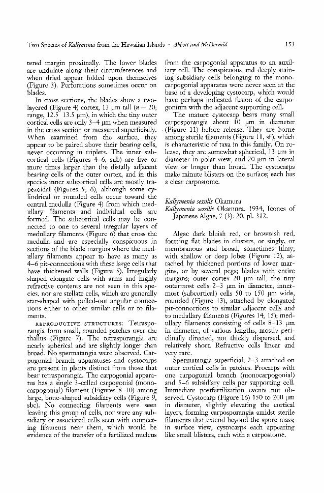
FIGURES 4-7. Kallymenia thompsonii. (4) Cross sectiollshowing two-layered cortex (co) (top arrow) with largesubcortical (sub) cells below, and formation of secondaryfilaments that will become medullary filaments (med fil).The section is from the center of the blade. med, medulla. Scale = 40 J.lI11. (5) Cross section to show triangularand trapezoidal subcortical cells (sub) attached to medullary filaments. Phase contrast micrograph. Scale = 40 )lm.(6) Oblique to median section showing several layers ofsquashed cortical cells at top, and trapezoidal subcorticallayer (double arrows) beneath a cortex, with medullaryfilaments (med fil) (single arrow) atrached to subcorticalcells (sub) and to each other. Scale = 40 )lm. (7) Surfaceview of scattered tetrasporangia (larger dark objects)among paired cortical cells (arrowheads). Scale = 40 )lm.
Two Species of Kallymenia from the Hawaiian Islands . Abbott and McDermid 155
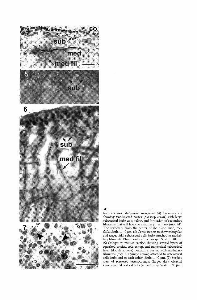
FIGURES 8-11. Knllymenia thompsonii. (8) Portion of young monocarpogonial arrangement showing parts of youngcarpogonium (cp) with trichogyne (tr) reaching the cortex. Scale = 40 J.UIl. (9). Squashed monocarpogonial apparatus,showing trichogyne (tr) at top, part of carpogonial filament (cp) hidden by subsidiary cell, and several lobed subsidiarycells (sbc). Scale = 30 f.lm. (10) Squashed subsidiary cells showing relationship to supporting cell (sue), and carpogonium (cp) with displaced trichogyne (tr). Scale = 30 f.lm. (11) Portion of mature eystocarp with carposporangia (ca)mixed with sterile filaments (sf) in the medulla. Scale = 40 J.UIl.
Tetrasporangia (Figure 17) are in scatteredpatches in the cortex, nearly spherical toslightly elongated, 18-26 J..lm in diameter by26-39 J..lm long.
DISTRIBUTION: Gulf of Tateyama, centralJapan (type locality); Hawaiian Islands fromKure Atoll, French Frigate Shoals, and NihoaIsland (NWHI), and Kaua'i, O'ahu, Maui,
156 PACIFIC SCIENCE· April 2002
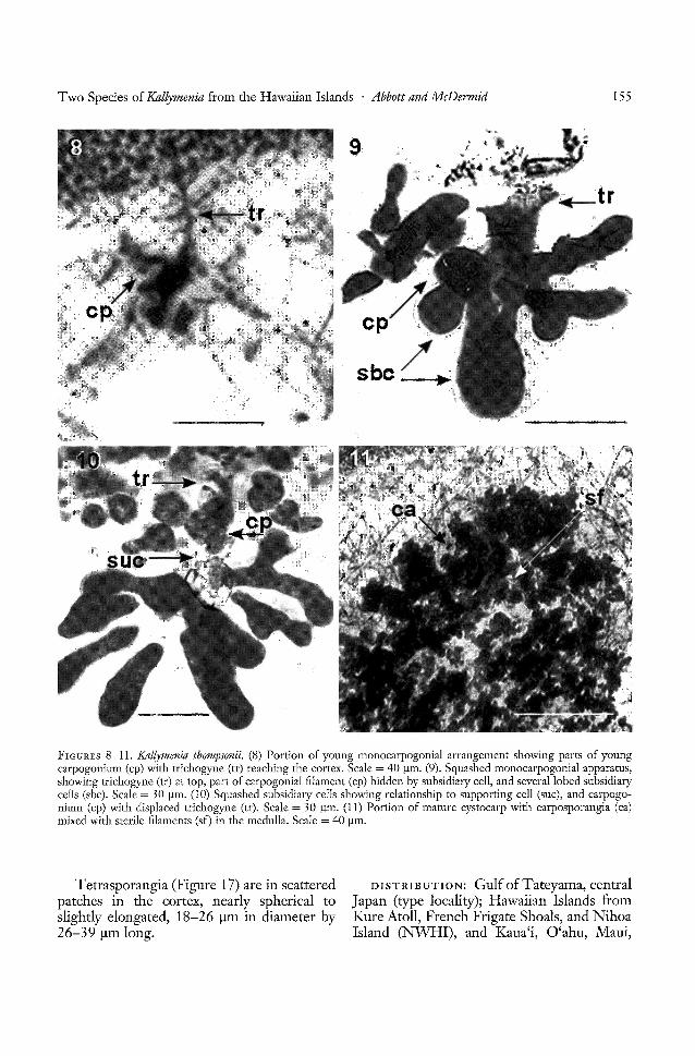
FIGURE 12. Hawaiian specimen of Kallymenia sessilis (KM 5050), a species from]apan, showing broadly rounded lobes,no layers of blades, and thicker blades than the thallus of K. tbompsonii, collected at Blonde Reef, Hilo, Hawai'i Island,6.5 m depth, 18 August 1995. Scale = 3 em.
and Hawai'i Islands among the main southeastern Hawaiian Islands. All collections weremade at depths of 1 to 10 m, often growingamong living coral.
This description is largely based upon thatof Abbott (1999), with additional informationfrom specimens that were collected subsequently and not included in that publication.The greatest similarity between this description and that of Okamura (1934) is in theshapes of the thick-walled, rounded subcortical cells (Figures 13-14 in this paper andOkamura's pI. 312, nos. 3, 4). Indeed, inmany sections of the type specimen of Kallymenia sessilis from the Okamura herbarium(SAP), the only feature found that could usefully be compared with the more recentlycollected Hawaiian material was the round,thick-walled cells. The Japanese material hadcell walls that had polymerized so much fromage that they shattered, rather than yieldingto a sharp razor blade, and the material"gelatinized" completely on application ofwater or detergent.
The greatest differences are shown in Figure 15, in which the relationship of the filamentous medullary cells to the subcortical
cells is detailed; in Okamura's plate the medullary filaments are not shown at all. In mostmodern studies of the Kallymeniaceae (e.g.,Womersley 1994), the structure of the medulla is shown in most taxa. A further difference is the thickness of the blades of theJapanese specimens, 130-160 Ilm, comparedwith 350-400 Ilm widths in the Hawaiianplants.
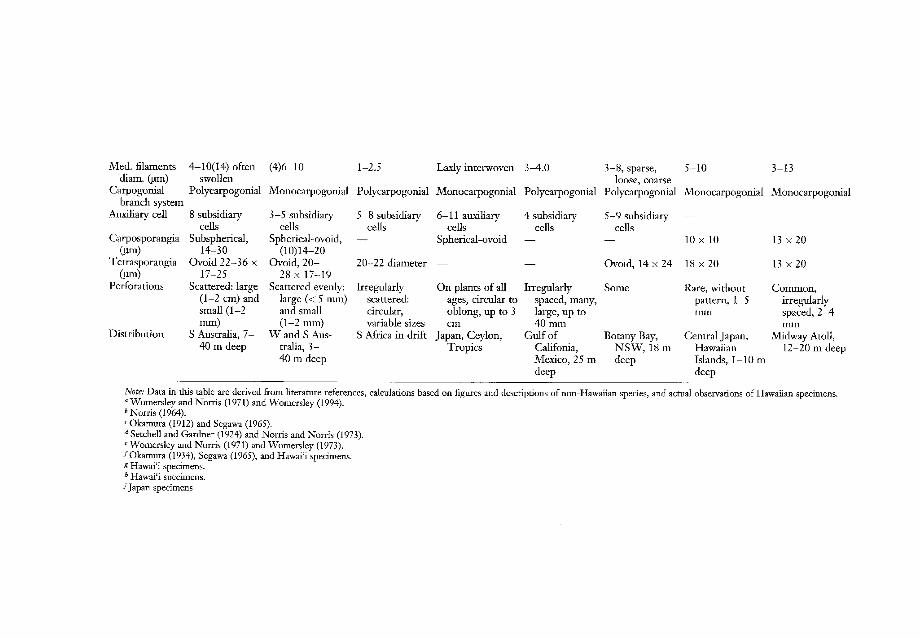
Table 1 compares and contrasts variousmorphological, anatomical, and reproductivecharacteristics of several Kallymenia species.The Hawaiian species, K. sessilis and K. thompsonii, are juxtaposed with other Pacific Basinand one South African species that sharesimilar general foliose blade morphology, including the presence of perforations. Thesespecies were also chosen for comparisonbecause reproductive material has been described and well illustrated. This collection oftaxa immediately can be divided into twogroups: species with polycarpogonial carpogonial branch systems (K. cribrogloea W 0
mersley & Norris, K. papenfussii Norris, K.pertusa Setchell & Gardner, and K. roseaW omersley & Norris), and species with monocarpogonial carpogonial branch systems (K.
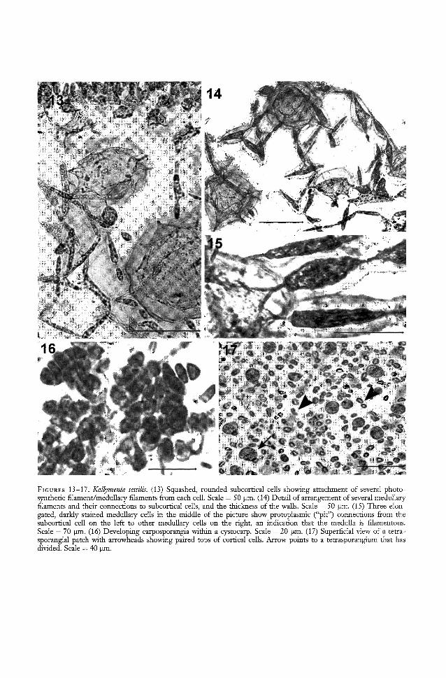
FIGURES 13-17. Kallymenia sessilis. (13) Squashed, rounded subcortical cells showing attachment of several photosynthetic filament/medullary filaments from each cell. Scale = 50 11m. (14) Detail of arrangement of several medullaryfilaments and their connections to subcortical cells, and the thickness of the walls. Scale = 50 11m. (15) Three elongated, darkly stained medullary cells in the middle of the picture show protoplasmic ("pit") connections from thesubcortical cell on the left to other medullary cells on the right, an indication that the medulla is filamentous.Scale = 70 11m. (16) Developing carposporangia within a cystocarp. Scale = 20 11m. (17) Superficial view of a tettasporangial patch with arrowheads showing paired tops of cortical cells. Arrow points to a tettasporangium that hasdivided. Scale = 40 IJID.

TABLE 1
Comparison of Perforate Knllymenia Species with K sessilis and K thompsonii from Hawai'i
Parameter K cribrogloeaa K cribrosaa K papenfussiib K perforataC K pertusad K roseae K. sessilisJ K thrympsoniig
Habit Foliose, Foliose, Cuneate thallus, Undulato-plaited, Irregular Foliose, lobed Clusters of large Rosettes, tiers ofundivided or undivided, lacinate at somewhat outline, with leaflets flat blades, 2-3 blades,lobed, margins broad end, umbilicated, crenate to on surface, lobed, entire undulategelatinous, smooth, thin waved, entire serrate soft, rubbery, margins marginsand firm gelatinous, and margin, lobed, margin, thin thin
firm stronglyadherent
Color Rose to Rose red Light rose red Deep crimson Rose red Rose red Rose red to Dark rose orbrownish red lake to rose to brownish red wine red
dirty redAttachment Short stipe with Subsessile or Single stipe and Minute scutate Attached to Short stipe Thickened Disk or
discoid short stipe with holdfast basal disks or Sargassum portions, pegs thickenedholdfast 1-3 holdfast 1-3 wherever blade edgesmmdiam. mm touches
Thallus size 50 x 50 cm 30 x 30 cm 18 x 11 cm 30-60 cm x 5-35 cm 30 x 30 cm 9-18(30) cm Up to 10 cm30-60 cm length x length
14 cm wideCross section 250-700 200-500(1,000) 122 at margins, 1,140 300-370 Dried 350-400h 140-250
(11m) 1,143 at base <100(150), 130-160jfresh(150)250-300
No. of cortex 2 to 4 3 to 4 1 1 to 2 2 to 4 2 to 3 2, width = 2, width =layers 20 11m 13 11m
Outer cortical 5-9(12) angular (3)4-6(7) rounded - rounded - 4-6, 5-9, compact, 2-3, ovoid 3-4, roundedcells (I!ffi) palisadelike round to
ovoidInner cortical Oval to polygon Ovoid to polygon Angular, Roundish Large angular Ovoid, with Rounded-obovate, Trapezoidal 15-
cells (11m) or spherical elongate large 52 x 52 to 78- 39 x 30-80,intercellular 92x no thickenedspaces 97-115, inner wall
wall thickenedStellate cells Light staining Light staining, Yes, deep - Yes, filled with Light staining, Refractive cells None observed
50-100 11m (40)90-160 11m, staining dense about 50 11m rare, linear,with arms 1- arms rarely material 30 diameter 5 x 130 11m2 x as long as longer than cell I!ffi diametercell body body
Med. filaments 4-10(14) often (4)6-10 1-2.5 Laxly interwoven 3-4.0 3-8, sparse, 5-10 3-13diam. (JlIIl) swollen loose, coarse
Carpogonial Polycarpogonial Monocarpogonial Polycarpogonial Monocarpogonial Polycarpogonial Polycarpogonial Monocarpogonial Monocarpogonialbranch system
Auxiliary cell 8 subsidiary 3-5 subsidiary 5-8 subsidiary 6-11 auxiliary 4 subsidiary 5-9 subsidiarycells cells cells cells cells cells
Carposporangia Subspherical, Spherical-ovoid, - Spherical-ovoid - - 10 x 10 13 x 20(Ilm) 14-30 (10)14-20
Tetrasporangia Ovoid 22-36 x Ovoid, 20- 20-22 diameter - - Ovoid, 14 x 24 18 x 20 13 x 20(Ilm) 17-25 28x17-19
Perforations Scattered: large Scatrered evenly: Irregularly On plants of all Irregularly Some Rare, without Common,(1-2 cm) and large « 5 mm) scattered: ages, circular to spaced, many, pattern, 1-5 irregularlysmall (1-2 and small circular, oblong, up to 3 large, up to mm spaced, 2-4mm) (1-2 mm) variable sizes em 40mm mm
Distribution S Australia, 7- Wand S Aus- S Africa in drift Japan, Ceylon, Gulf of Botany Bay, Central Japan, Midway Atoll,40 m deep tralia, 3- Tropics Califonia, NSW,18m Hawaiian 12-20 m deep
40 m deep Mexico, 25 m deep Islands, 1-10 mdeep deep
Note: Data in this table are derived from literature references, calculations based on figures and descriptions of non-Hawaiian species, and actual observations of Hawaiian specimens.a Womersley and Norris (1971) and Womersley (1994).b Norris (1964).'Okamura (1912) and Segawa (1965).d Setchell and Gardner (1924) and Norris and Norris (1973).'Womersley and Norris (1971) and Womersley (1973).f Okamura (1934), Segawa (1965), and Hawai'i specimens.g Hawai'i specimens.h Hawai'i specimens.jjapan specimens.
160
cribrosa Harvey, K peiforata J. Agardh, K sessilis Okamura, and K thompsonii Abbott &McDermid, n. sp.). The latter four monocarpogonial species are similar in the displayof their subsidiary cells in the carpogonialbranch system and sporangial measurements(when reported). However, these four monocarpogonial species differ among themselvesin shapes and sizes of blades, attachment,blade thickness, inner cortical cell shapes, andin the position of their medullary filaments(periclinal versus anticlinal). The two Hawaiian species are similar in that they aremonocarpogonial with conspicuous bonelikesubsidiary cells, and they have large subcortical cells that contribute to the formationof the medullary filaments directly throughnumerous secondary pit-connections. However, thanks to abundant material for study, wewere able to document that these two speciesconsistently differ in other characteristics aswell, including blade habit, size of cross sections, shapes of large subcortical cells, andcarposporangial and tetrasporangial sizes. Ofthe monocarpogonial, foliose, tropical speciescompared in Table 1, K thompsonii is the onlyone that forms low-growing rosettes, has thesmallest blade dimensions, has the thinnestcross section, shows trapezoid-shaped innercortical cells, lacks refractive cells, and commonly has irregularly scattered small perforations. Blade perforations are rare andrandomly positioned in K sessilis (Segawa1965); large and small perforations are scattered evenly throughout the blades in K cribrosa (Womersley and Norris 1971), and in Kpeiforata the large holes dominate the blade,creating an Ulva reticulata-like appearance(Okamura 1912, pI. 86)-all very differentfrom K thompsonii.
DISCUSSION
When faced with the identification of folioseRhodophyta, several features can be appliedgenerally to various species of Halymeniaand Cryptonentia (Halymeniaceae) and sharedwith species of Kallymenia (Kallymeniaceae),such as attachment, size of the blade, widthof the cross section, anticlinal medullary filaments, and the number of cortical layers.
PACIFIC SCIENCE· Apri12002
Slender, elongate, higWy refractive cells maybe found in species of Cryptonemia and Halymenia, as well as other species of Kallymenia(Norris 1957, Chiang 1970), and may beabundant or few in various parts of the samethallus in K oblongifructa (Setchell) Setchell(Abbott 1968), for example. The anatomy ofmost Kallymenia species seen in Norris(1964), Womersley and Norris (1971), andW omersley (1994) shares a preponderance ofpericlinal filaments in the medulla, whereasanticlinal filaments are common in the twoHawaiian Kallymenia species. Anticlinal medullary filaments are also one of the important features of Halymenia. The twoKallymenia species discussed in this paper differ from Halymenia in terms of the sizes andshapes of the inner cortical cells (Kraft andAbbott 1997) (in general smaller in Halymenia, with a few more subcortical layers). Moreimportant, members of the genus Kallymeniashow a cystocarp in which carposporangia aredeveloped among many sterile filaments, nottrue of any species of Halymenia or Cryptonemia. Among these three "red blade" genera,only Kallymenia shows the characteristicallylarge subsidiary cells that are the hallmark ofKallymenia and other genera of the Kallymeniaceae. These last two features are thereforepivotal, and are required when identifyingspecies within the genus and the family. Weemphasize these reproductive features because the bulk of tropical red, bladelike algaeare sterile when examined and not wellenough known to identify in that state.
The principal reproductive difference between the two Hawaiian species and thetype species of the genus, Kallymenia reniformis (Turner) J. Agardh (1851), as well assome morphologically similar species (seeTable 1), is that the Hawaiian species haveone carpogonial branch per supporting cell(monocarpogonial); whereas K reniformis(Hommersand & Ott 1970), K cribrogloea, Kpapenfussii, K pertusa, and K rosea have twoor more carpogonial branches per supportingcell (polycarpogonial). The bulk of Kallymenia species and genera in the Kallymeniaceae in Australia (Womersley 1994) in whichthe carpogonial apparatus is known showthe monocarpogonial arrangement associated
Two Species of Kallymenia from the Hawaiian Islands· Abbott and McDermid 161
with the supporting cell. Four foliose speciesthat show a monocarpogonial carpogonialbranch system and conspicuously lobed or"bone-shaped" subsidiary cells of the carpogonial branch apparatus are now reportedfrom the Indo-Pacific Ocean: Kallymenia cribrosa, K peiforata, K sessilis, and K thompsonii.The monocarpogonial condition is thoughtto be more advanced evolutionarily than thepolycarpogonial one (Norris 1957: fig. 22).Assuming that fertilization had taken place,we did not observe a connecting filamentleaving the monocarpogonial apparatus andits subsidiary cells, nor did we observe a developing auxiliary cell and its subsidiary cellsthat might serve as the site of initiation of thegonimoblast. Should these connecting structures not be found, the implication is that inthese species of Kallymenia it is possible thatthis step in development has been eliminatedand the supporting cell of the single carpogonial branch is serving as the site of gonimoblast initiation, as postulated by Norris(1957) for Euthora and Callocolax.
To distinguish species in Kallymenia requires a shift of emphasis to vegetative structure and assessment of enough specimens tounderstand the variation in the selected features. In a survey of foliose, perforate speciesof Kallymenia, we found that the sizes andshapes of subcortical (inner cortical) cellsvaried among these species (Table 1), so thatexamination of many sections of each mightshow nearly all types of cells. However, thereseemed to be a preponderance of one or another kind in each species. Together with thepresence or absence of stellate cells and/orrefractive cells, these cellular differences seemto contribute one of several adequate characteristics by which species may be distinguished. At least for the material we have ofK sessilis, the large, rounded to obovate cellsof the subcortex that lie just below the cortical cells are contrasted with the numeroustrapezoidal cells in a similar position in Kthompsonii. It is possible that more material,perhaps collected in a different season, mayshow that these shapes and sizes are not species-specific, but at this time we find them tobe fairly constant in both reproductive andsterile specimens. The rounded subcortical
cells are found in the same specimens thatbear slender, higWy refractive, elongate cellsamong the subcortical cells in K sessilis; theseare lacking in K thompsonii. In addition, bladeperforation frequency, pattern on the blade,and size varied substantially among the species, enough to distinguish K thompsonii fromother monocarpogonial species. Finally, Kthompsonii with its tiers of blades that formrosettes is distinctive for reported species ofKallymenia.
ACKNOWLEDGMENTS
We thank the U.S. Fish and Wildlife Service(U.S. Department of the Interior, Pacific Islands Region) for permission to collect marine algae in the Midway National WildlifeRefuge and for boat and diving help. Theisolation of Midway Atoll from the main Hawaiian Islands made it impossible for us toconduct this research without their interestand help. We acknowledge the help of TeenaMichael in photographically interpreting theanatomical intricacies shown by these species.We also thank Professor M. Masuda for theloan of specimens of Kallymenia sessilis fromthe Okamura herbarium. We greatly appreciate the help of the referees, particularly MaxHommersand, for suggestions that helped toexpand the interpretations of our observations.
Literature Cited
Abbott, 1. A. 1968. Studies in some foliose redalgae of the Pacific coast: III. Dumontiaceae, Weeksiaceae, Kallymeniaceae. J.Phycol. 4:180-198, 38 figs.
---. 1989. Marine algae of the NorthwestHawaiian Islands. Pac. Sci. 43:223-233.
---. 1999. Marine red algae of the Hawaiian Islands. Bishop Museum Press,Honolulu, Hawai'i.
Abbott, 1. A., and K. J. McDermid. 2001.Dudresnaya babbittiana (Dumontiaceae,Gigartinales), a new red algal species fromMidway Atoll, North Central Pacific.Cryptogam. Algol. 22:249-261.
162
Agardh, J. G. 1851. Species, genera et ordinesalgarum. 2: Algas florideas complectens.Pt. 1. Lund.
Chiang, Y. M. 1970. Morphological studies ofred algae of the family Cryptonemiaceae.Univ. Calif. Publ. Bot. 58:1-92, 10 pIs.
Hommersand, M. H., and D. W. Ott. 1970.Development of the carposporophyte ofKallymenia reniformis (Turner) J. Agardh.J. Phycol. 6:322-331.
Kraft, G. T., and I. A. Abbott. 1997. Platomaardreanum (Schizymeniaceae, Gigartinales)and Halymenia chiangiana (Halymeniaceae,Halymeniales), two new species of proliferous, foliose red algae from the Hawaiian Islands. Cryptogam. Algol. 18:97-116,27 figs.
Norris, R. K 1957. Morphological studies onthe Kallymeniaceae. Univ. Calif. Publ.Bot. 28:251-334, pIs. 28-40.
--. 1964. The morphology and taxonomy of South African Kallymeniaceae.Bot. Mar. 7:90-129.
Norris, R. K, and J. N. Norris. 1973. Kallymenia pertusa (Rhodophyceae, Cryptonemiales) from the Gulf of California.Phycologia 12 (1/2): 71-74.
Okamura, K. 1912. leones of Japanese algae.Vol. 2, pt. 8, pp. 127-141, pIs. 86-90.Tokyo.
PACIFIC SCIENCE· April 2002
---. 1934. leones ofJapanese algae. Vol.7, pp. 19-48 (English), 17-43 (Japanese),pIs. 311-325. Tokyo.
Segawa, S. 1965. Coloured illustrations ofthe seaweeds of Japan. Rev. ed. Hoikusha,Osaka.
Setchell, W. A., and N. L. Gardner. 1924.Expedition of the California Academy ofSciences to the Gulf of California in 1921.The marine algae. Proc. Calif. Acad. Sci.,4th series, 12:695-949, pIs. 12-28, map.
Tsuda, R. T., and I. A. Abbott. 1985. Collection, handling, preservation, and logistics.Pages 67-86 in M. M. Littler and D. S.Littler, eds. Handbook of phycologicalmethods: Ecological methods for macroalgae. Cambridge University Press, NewYork.
Womersley, H. B. S. 1973. Further studies onAustralian Kallymeniaceae (Rhodophyta).Trans. R. Soc. S. Aust. 97:253-256.
---. 1994. The marine benthic floraof Southern Australia, Rhodophyta. PartIlIA. Flora of Australia SupplementarySeries No. 1. Australian Biological Resources Study, Canberra.
Womersley, H. B. S., and R. E. Norris.1971. The morphology and taxonomy ofAustralian Kallymeniaceae (Rhodophyta).Aust. J. Bot. Suppl. Ser. 2: 1-62.
Related Documents