1.Name the layers of the blood vessels. 2.How many cells thick are the capillaries and why? 3.Compare and contrast the structure of veins and arteries.

1.Name the layers of the blood vessels. 2.How many cells thick are the capillaries and why? 3.Compare and contrast the structure of veins and arteries.
Dec 26, 2015
Welcome message from author
This document is posted to help you gain knowledge. Please leave a comment to let me know what you think about it! Share it to your friends and learn new things together.
Transcript

1. Name the layers of the blood vessels.
2. How many cells thick are the capillaries and why?
3. Compare and contrast the structure of veins and arteries.

1. Name the major arteries of the following areas: head, arms, legs.
2. How are the precapillay sphincter muscles controlled?
3. What is the difference between hydrostatic pressure & osmotic pressure?
4. If someone’s blood pressure is 140/80, what is their pulse pressure?

• What do the two numbers represent in your blood pressure?
• What is an example of a normal blood pressure?
• Which blood vessels have the highest and lowest blood pressure? Why?
• Explain how baroreceptors and chemoreceptors help maintain normal blood pressure.

Blood Vessel and Circulation
Functions1. Carry Blood2. Exchange of nutrients3. Transport4. Regulate blood pressure5. Direct blood flow

I Blood Vessels - Forms a closed circulatory system (Arteries ->Capillaries ->Veins)
Made up of three layers
a. Lumen - space for the flow of blood.
b. Tunica Intima - inner lining.
c. Tunica Media - smooth muscle, contractibility.
d. Tunica Adventitia – anchoring

A. Arteries - Carries blood away from the heart
Strong & Elastic
1. Elastic Arteries – Largest in diameter. Mainly elastic
2. Muscular arteries – medium sized and small diameter.
Mainly smooth muscle
3. Distributing arteries – vasoconstriction -Vasomotor fibers of the sympathetic
(autonomic).
vasodilatation - nerve impulse is inhibited - muscle relaxes & elastic fibers recoils.
4. Small arteries
5. Arterioles - 0.5 mm in diameter

B. Capillaries - Thin wall blood vessels that permit exchanges of material.
1. Connect arteries to veins
2. 0.01 mm in diameter - lumen
3. Can only fit 1 RBC at a time.
4. Form capillary beds or networks.
• Thoroughfare channels - connect arterioles directly to veins
• True Capillaries - 10-100 per bed
Precapillary Sphincter - valve that regulates flow of blood into those capillaries.
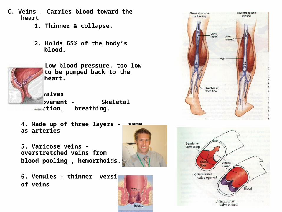
C. Veins - Carries blood toward the heart1. Thinner & collapse.
2. Holds 65% of the body’s blood.
3. Low blood pressure, too low to be pumped back to the heart.
1 way valvesBody movement - Skeletal contraction, breathing.
4. Made up of three layers - same as arteries
5. Varicose veins - overstretched veins from blood pooling , hemorrhoids.
6. Venules – thinner versions of veins

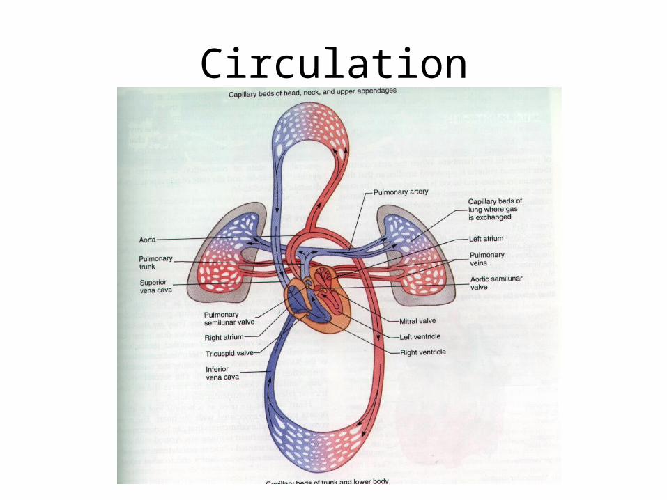
Blood flow through
the circulatory
system

Arteries

Major arteries of the body
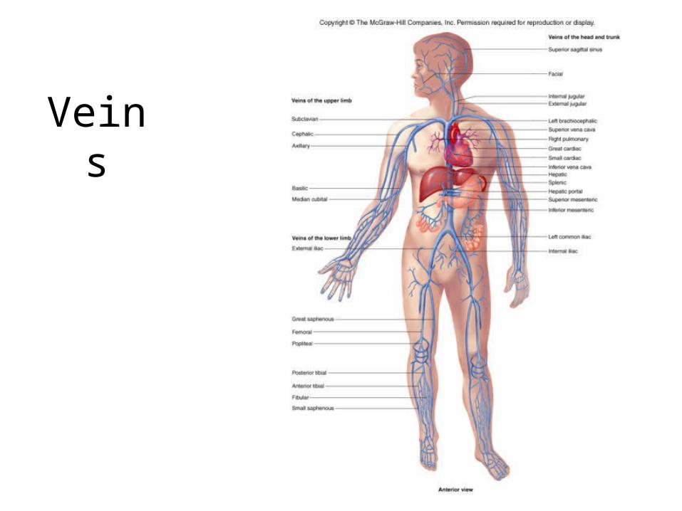
Veins
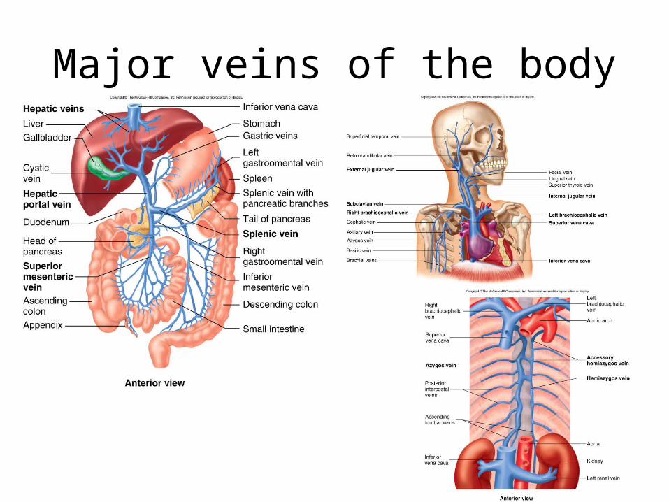
Major veins of the body

II. Blood Pressure - force exerted by blood against the inner walls of vesselsA. Influenced by:
Cardiac OutputBlood VolumePeripheral Resistance which is
regulated by nerve, kidneys, hormones.
B. Moves from regions of higher pressure to lower pressure
Systolic - Peak pressure - 120 mmHg
Diastolic - Resting - 70 - 80 mmHg
Sphygmomanometer - measures blood pressure.
Stethoscope – Hearing
Korotkoff - tapping sound sounds

C. Pulse - rhythmic expanding & recoiling of an arterial wallAverage
Adult 70 - 90 bpmChild 80 - 140 bpm
tachycardia - heart rate is above 100 bpm at restbradycardia - heart rate is lower than 60 bpm at rest
Auscultatory MethodSphygmomanometerStethoscopeKorotkoff sounds

D. Blood Pressure

Pulse PressurePulse pressure – the difference between
systolic & diastolic pressure.
120/80 – 40 pulse pressure.
Factors effecting pulse pressure.Activity - increaseArteriosclerosis- decrease

III Capillary Exchange
10 Billion capillaries
Materials (gases, nutrients) move across capillary walls by diffusion, osmosis, facilitated diffusion & active transport.
Small amount of fluid also moves across the capillary walls:
Hydrostatic pressure - blood pressure within the capillaries
Osmotic pressure - movement of fluid from cells to plasma
90% reabsorbed back into the blood
10% returned back by the lymphatic system

IV. Regulation of blood pressure - regulation of steady pressure is important.
A. Nervous Control - Adjusts cardiac output & peripheral resistant by autonomic fibers to the SA node & reflex center (vasomotor center in the medulla oblongata & pons).
1. Peripheral resistance - control by activity of vasomotor between the smooth muscle & reflex center of Medulla (Vasomotor tone, increases = constriction).
2. Control of vasomotor center (MAP)Map = CO X PR
a. Baroreceptors – located in major vessels above the heart & detect changes in the blood pressure.
Moment to moment control

b. Chemoreceptors - Sensitive to changes in oxygen levels or hydrogen ions (pH) level.
carotid bodies – carotid sinusaorta bodies - aorta

B. Hormone Control1. Epinephrine & norepinephrine
a. Activated by the sympathetic division of the autonomic division
b. Fight or flight response - times of stressc. Epinephrine - increases cardiac outputd. Norepinephrine - increases peripheral
resistance
2. Atrial natriuretic factor - secreted by the atria of the hearta. Reduces blood volumeb. Stimulates the kidney to secrete more sodium
& waterc. Less water
d. Drop in blood volume

3. Antidiuretic hormone (vasopressin)
a. Produced by the hypothalamusb. Stimulates the kidneys to increase water absorbtionc. Increase in blood volume
C. Kidney Control - Main mechanism of long term control
1 High blood pressure - excretes more waterLow blood pressure - conserve more water
2. Release of special chemicals
a. Reduced blood flow causes renin to be released by the kidney and causes the formation of Angiotensin I.
Angiotensin I + angiotensin-converting enzyme makes angiotension II = vasoconstrictor.
b. Angiotensin II - Aldosterone release by the adrenal glands to stimulate the reabsorbtion of sodium & water.
V. Cardiovascular diseases
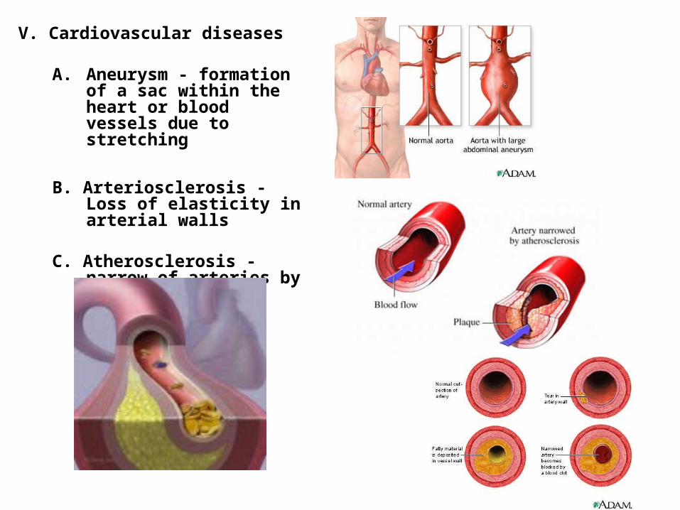
A. Aneurysm - formation of a sac within the heart or blood vessels due to stretching
B. Arteriosclerosis - Loss of elasticity in arterial walls
C. Atherosclerosis - narrow of arteries by plaque build up

Cardiovascular diseases

Cardiovascular diseases

D. Congestive heart failure - failure of the heart to pump blood to the body tissues
E. Heart Block - Failure of the SA or AV. to generate impulses
F. Heart fibrillation - Heart beats at a irregular paceG. Heart flutter - heart race up to 300 bpmH. Hypertension - elevated blood pressureI. Murmur - Leaking of blood through a closed valveJ. Myocarditis - infection of the heart muscle
K. Pericarditis - Infection of the pericardial sac which results in thicken or scarring
Circulation
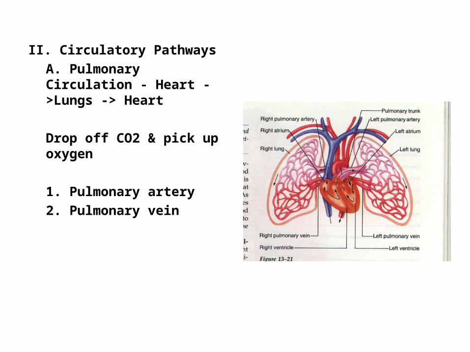
II. Circulatory PathwaysA. Pulmonary Circulation - Heart ->Lungs -> Heart
Drop off CO2 & pick up oxygen
1. Pulmonary artery2. Pulmonary vein

B. Systemic Circulation - Heart ->Body ->Heart
Nutrients to cells & remove metabolic wastes
Transports: gases, nutrients, wastes, blood cells, proteins (enzymes), hormones
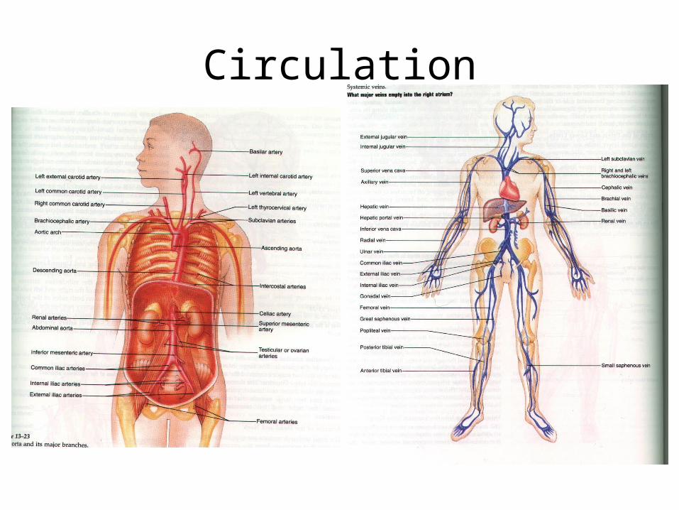
1. Major Arteriesa. Carotid Artery (internal & external)b. Brachiocephalic artery (arms)c. Subclavian (shoulder)d. Aortae. Common iliac arteryf. renal artery kidneyg. Axillary (armpit)h. Brachail arteryi. Radial arteryJ. ulnar arteryk. femorall. posterior/anterior tibial artertm. fibular artery

2. Major Veinsa. Jugular veins (internal & external)b. Right & Left Brachiocephalic veinsc. Inferior & superior vena cavad. Common Iliac veine. Subclavin veinf. renal vein kidneyg. Axillary (armpit)h. Brachail veini. Radial veinJ. ulnar veink. femoral veinl. posterior/anterior tibial veinm. fibular veinn. hepatic portal vein
Circulation
Related Documents


