Gonioscopy Dr.Gowri J Murthy, Glaucoma Service, Vittala International Institute of Ophthalmology, Prabha Eye Clinic and Research Centre Bangalore.

Welcome message from author
This document is posted to help you gain knowledge. Please leave a comment to let me know what you think about it! Share it to your friends and learn new things together.
Transcript
Gonioscopy
Dr.Gowri J Murthy,Glaucoma Service,
Vittala International Institute of Ophthalmology,Prabha Eye Clinic and Research Centre
Bangalore.
Gonioscopy
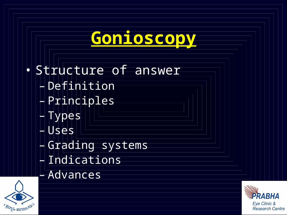
• Structure of answer– Definition– Principles– Types– Uses– Grading systems– Indications– Advances
• Use line diagrams
• Flow charts
• Underline key points
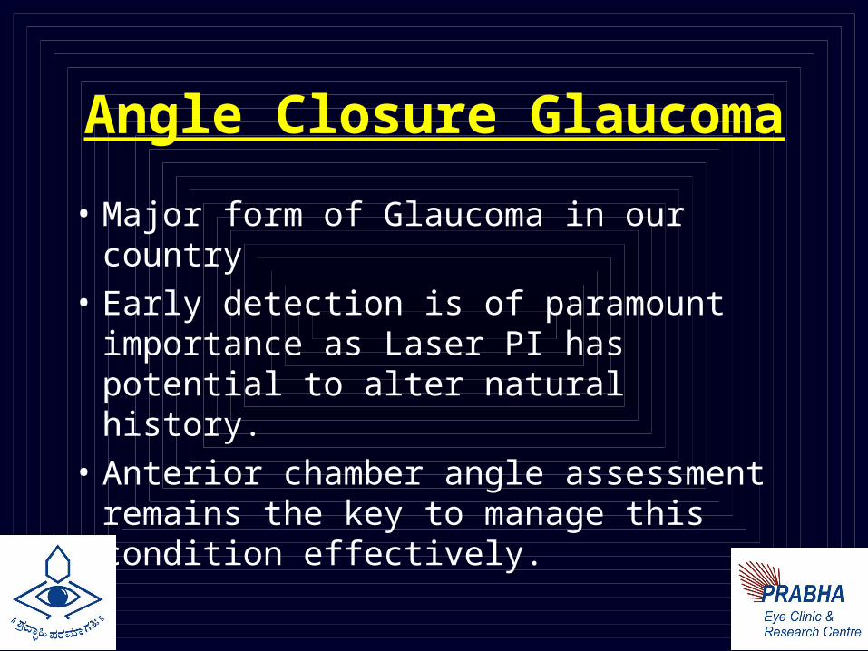
Angle Closure Glaucoma
• Major form of Glaucoma in our country
• Early detection is of paramount importance as Laser PI has potential to alter natural history.
• Anterior chamber angle assessment remains the key to manage this condition effectively.
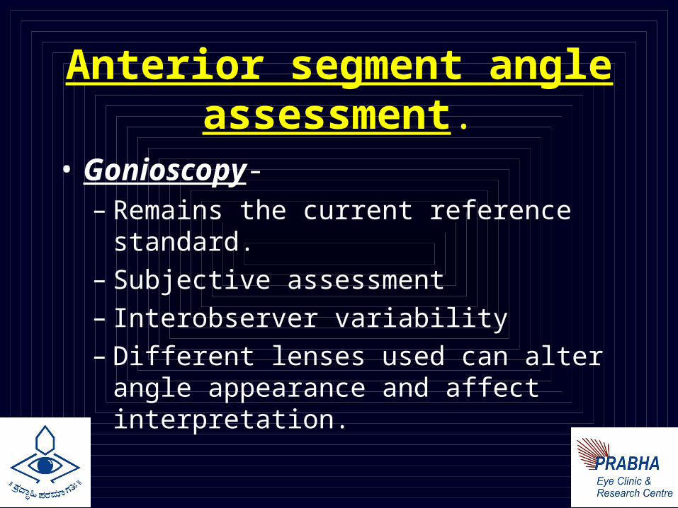
Anterior segment angle assessment.
• Gonioscopy- – Remains the current reference standard.– Subjective assessment– Interobserver variability– Different lenses used can alter angle appearance
and affect interpretation.
Objective Angle Assessment
• Ultrasound Biomicroscopy.
• AS OCT
• Scheimpflug photography– Objective methods.– Are yet to replace Gonioscopy for angle
assessment.
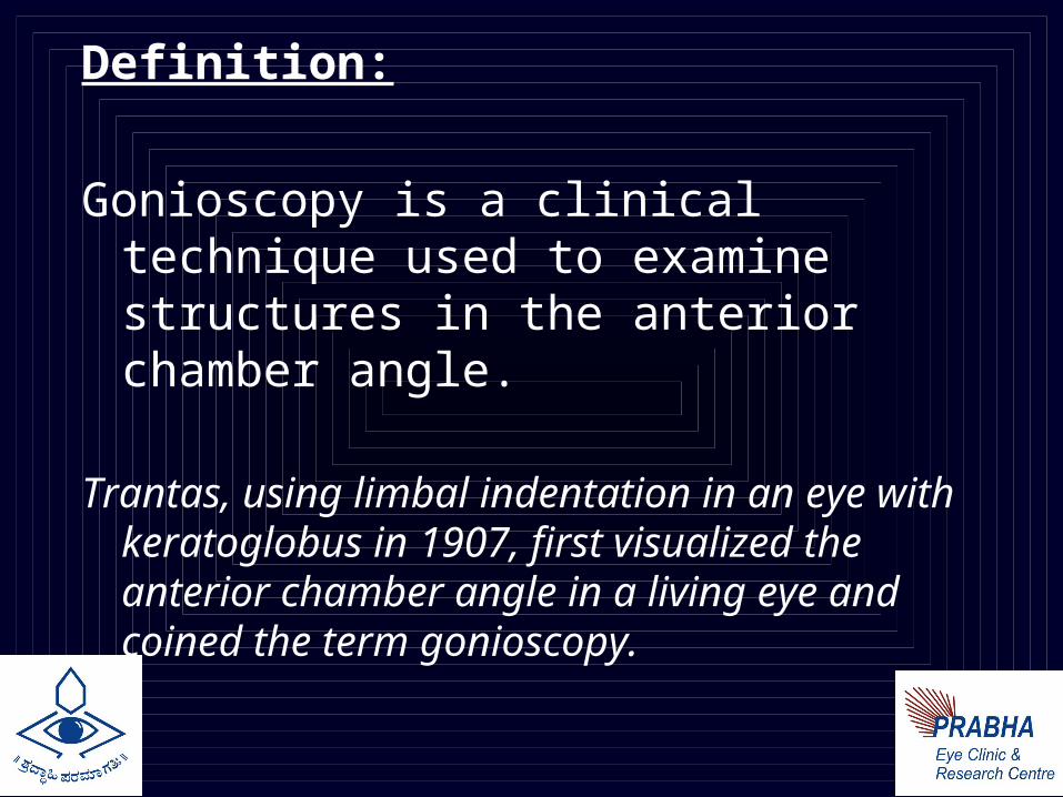
Definition: Gonioscopy is a clinical technique
used to examine structures in the anterior chamber angle.
Trantas, using limbal indentation in an eye with keratoglobus in 1907, first visualized the anterior chamber angle in a living eye and coined the term gonioscopy.
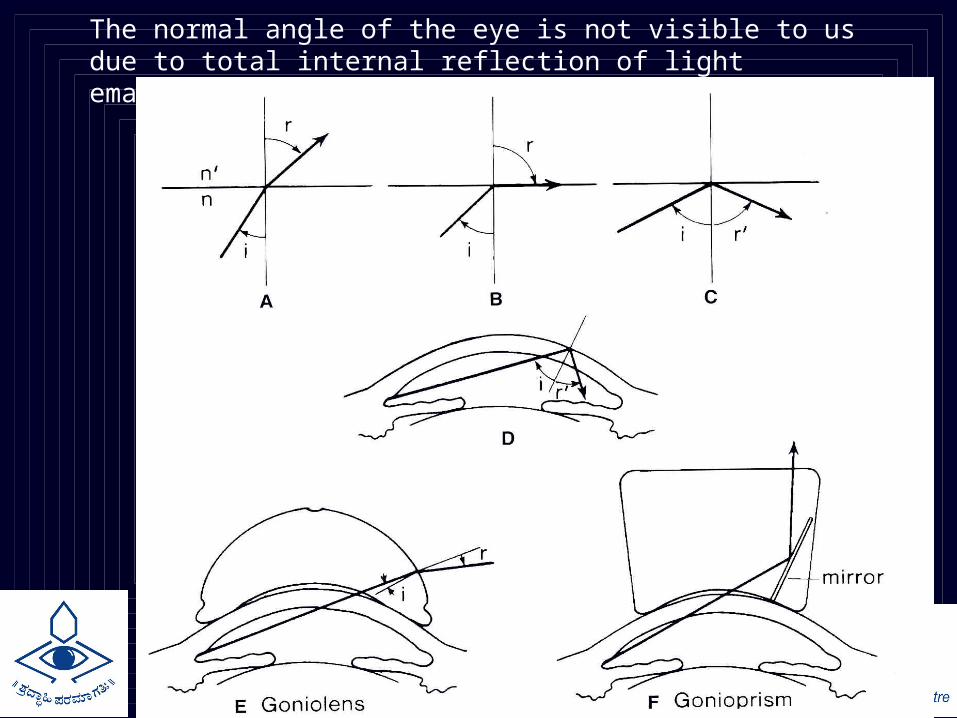
The normal angle of the eye is not visible to us due to total internal reflection of light emanating from the angle.
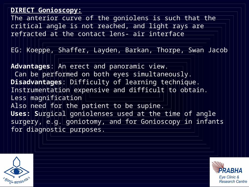
DIRECT Gonioscopy:The anterior curve of the goniolens is such that the critical angle is not reached, and light rays are refracted at the contact lens- air interface
EG: Koeppe, Shaffer, Layden, Barkan, Thorpe, Swan Jacob
Advantages: An erect and panoramic view. Can be performed on both eyes simultaneously.Disadvantages: Difficulty of learning technique. Instrumentation expensive and difficult to obtain.Less magnification Also need for the patient to be supine.Uses: Surgical goniolenses used at the time of angle surgery, e.g. goniotomy, and for Gonioscopy in infants for diagnostic purposes.
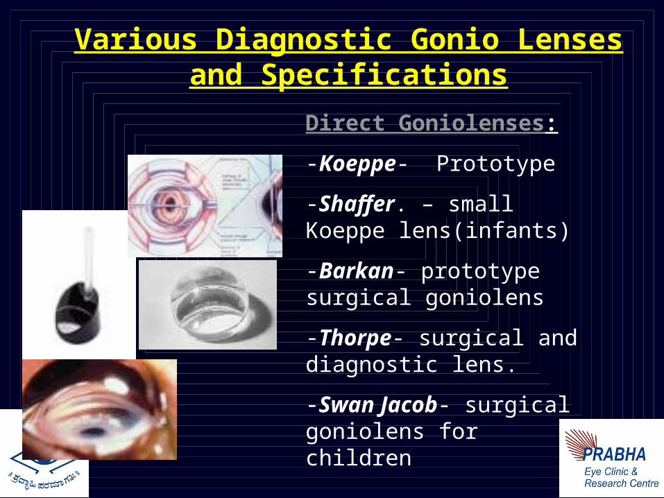
Various Diagnostic Gonio Lenses and Specifications
Direct Goniolenses:
-Koeppe- Prototype
-Shaffer. – small Koeppe lens(infants)
-Barkan- prototype surgical goniolens
-Thorpe- surgical and diagnostic lens.
-Swan Jacob- surgical goniolens for children
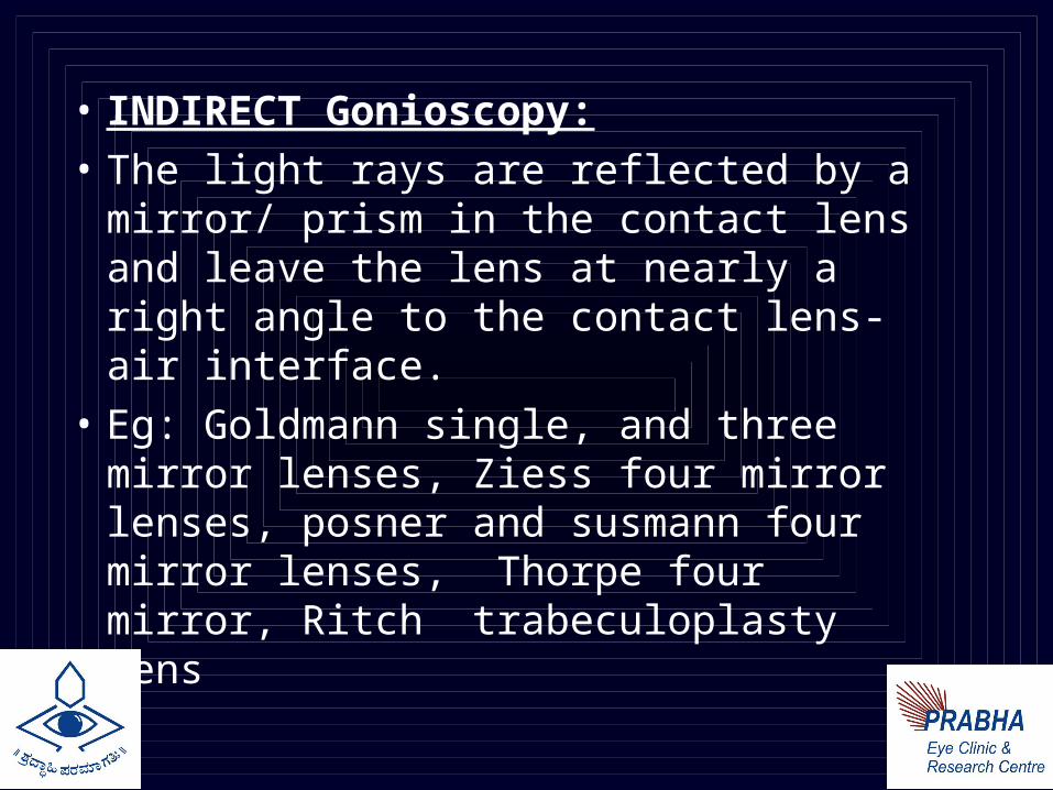
• INDIRECT Gonioscopy:• The light rays are reflected by a
mirror/ prism in the contact lens and leave the lens at nearly a right angle to the contact lens- air interface.
• Eg: Goldmann single, and three mirror lenses, Ziess four mirror lenses, posner and susmann four mirror lenses, Thorpe four mirror, Ritch trabeculoplasty lens
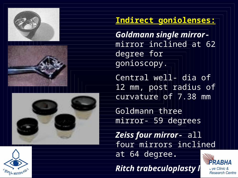
Indirect goniolenses:
Goldmann single mirror- mirror inclined at 62 degree for gonioscopy.
Central well- dia of 12 mm, post radius of curvature of 7.38 mm
Goldmann three mirror- 59 degrees
Zeiss four mirror- all four mirrors inclined at 64 degree.
Ritch trabeculoplasty lens.
Goldmann type lenses:
•Ease in learning technique and less expensive. • Greater visibility of detail than with the Koeppe technique because of higher magnification. •Therefore, it is better for detection of details such as subtle neovascularization in the angle.• Stability of lens over cornea better.
Disadvantages: Cannot perform dynamic, or indentation Gonioscopy.
Four mirror lenses- Ziess type: Allows quick evaluation of angle structures. • No coupling solution necessary.• Enables differentiation between appositional (reversible) and synechial angle closureDisadvantages:• Mastery of proper technique requires skill and practice.• Tendency to underestimate the narrowness of the angle; it is difficult to avoid inadvertently applyingpressure to the central cornea,thus artificially widening the angle.
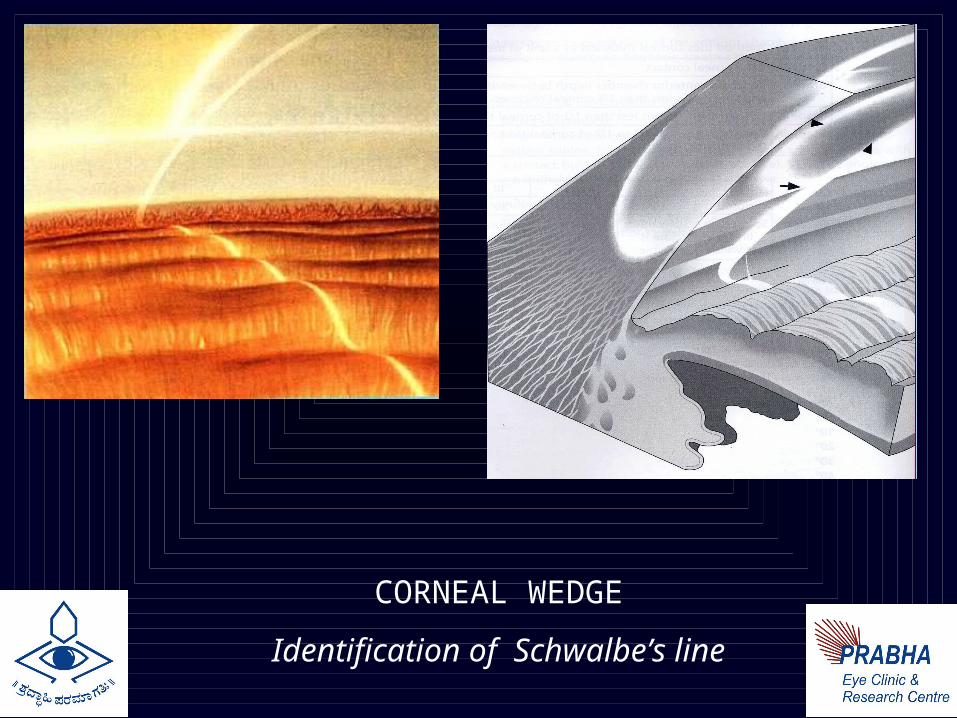
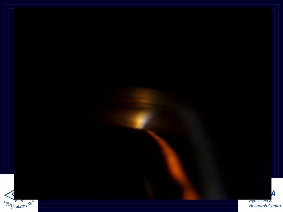
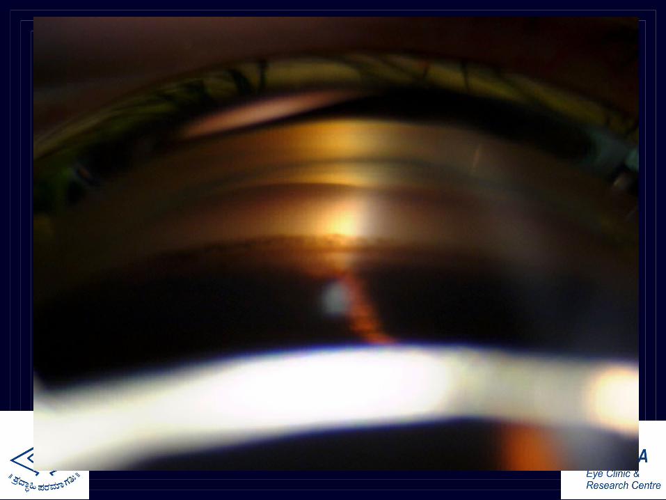
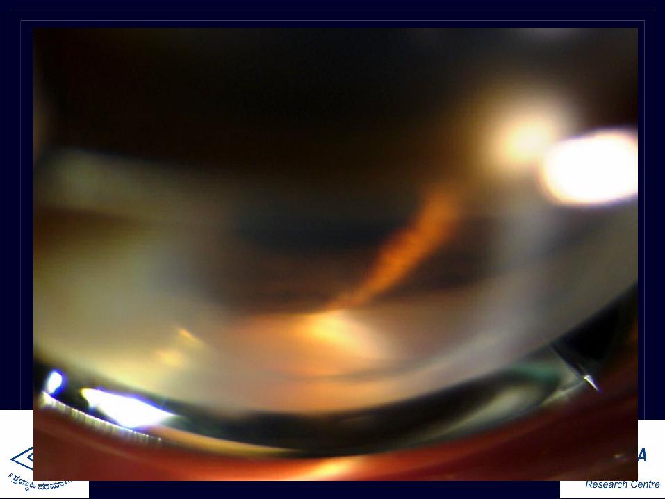
CORNEAL WEDGE
Identification of Schwalbe’s line
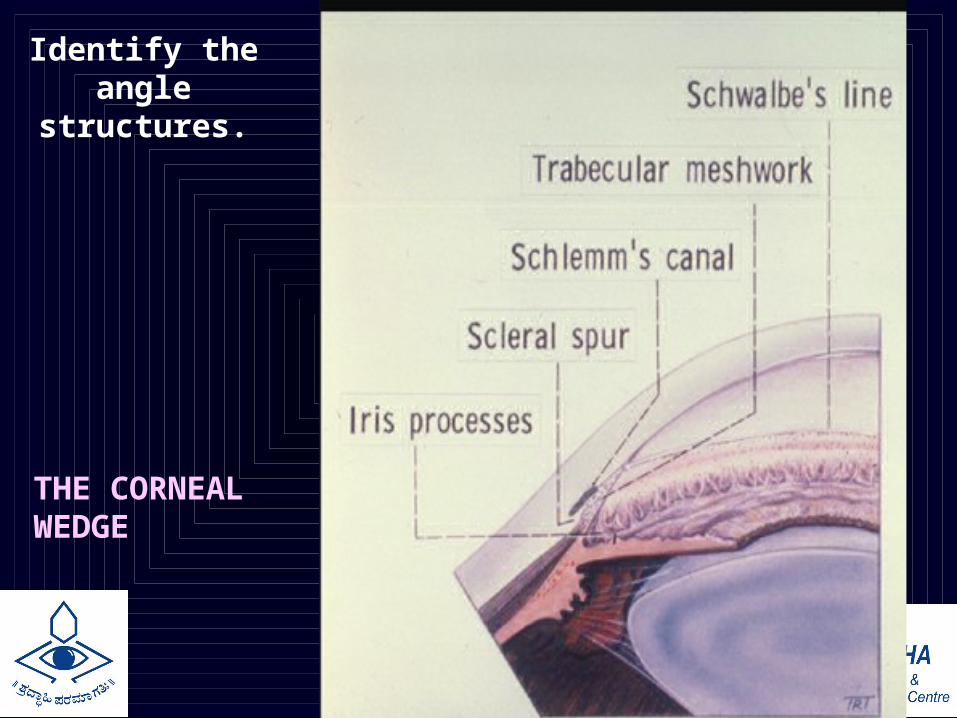
Identify the angle
structures.
THE CORNEAL WEDGE
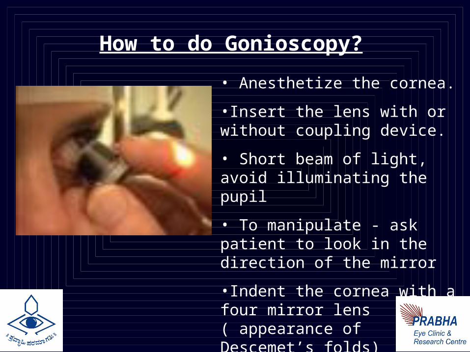
How to do Gonioscopy?
• Anesthetize the cornea.
•Insert the lens with or without coupling device.
• Short beam of light, avoid illuminating the pupil
• To manipulate - ask patient to look in the direction of the mirror
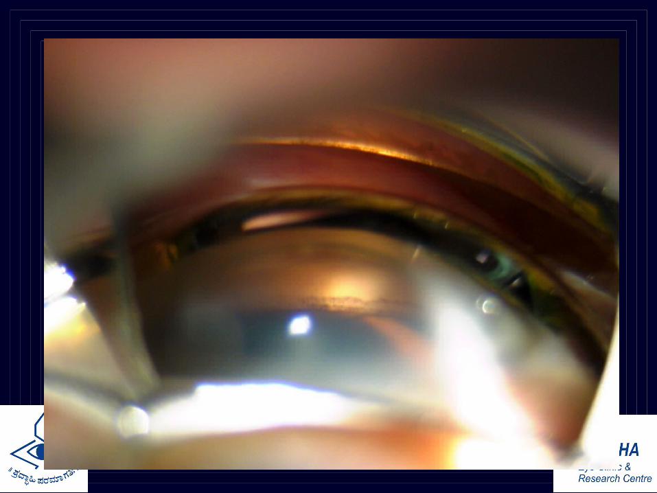
•Indent the cornea with a four mirror lens ( appearance of Descemet’s folds)
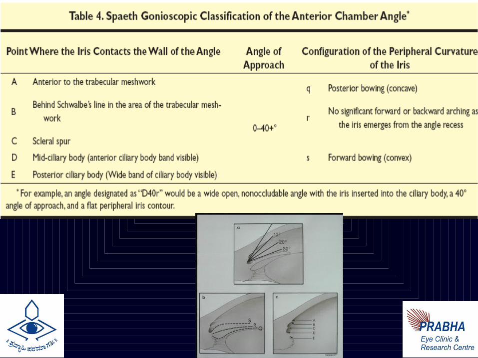
Angle Grading systems for Gonioscopy: Several grading systems
Shaffer, Scheie, and Spaeth devised the three most commonly used systems
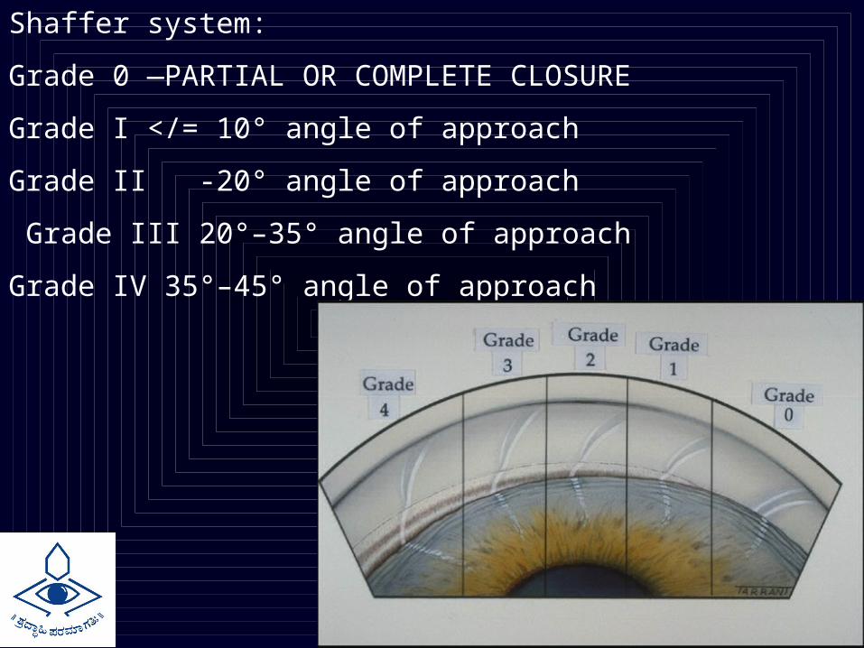
Shaffer system:
Grade 0 —PARTIAL OR COMPLETE CLOSURE
Grade I </= 10° angle of approach
Grade II -20° angle of approach
Grade III 20°–35° angle of approach
Grade IV 35°–45° angle of approach
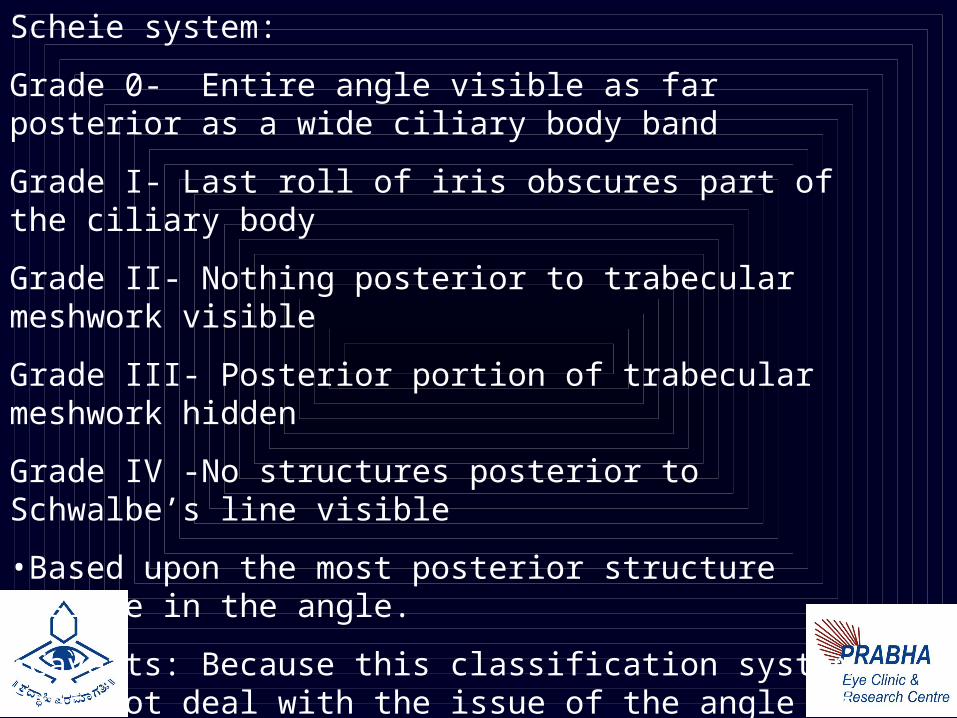
Scheie system:
Grade 0- Entire angle visible as far posterior as a wide ciliary body band
Grade I- Last roll of iris obscures part of the ciliary body
Grade II- Nothing posterior to trabecular meshwork visible
Grade III- Posterior portion of trabecular meshwork hidden
Grade IV -No structures posterior to Schwalbe’s line visible
•Based upon the most posterior structure visible in the angle.
•Caveats: Because this classification system does not deal with the issue of the angle of approach and, hence, occludability, the scleral spur could be visible for its entire circumference in an eye with an occludable angle.
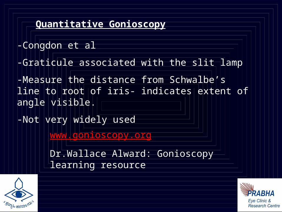
Quantitative Gonioscopy
-Congdon et al
-Graticule associated with the slit lamp
-Measure the distance from Schwalbe’s line to root of iris- indicates extent of angle visible.
-Not very widely used
www.gonioscopy.org
Dr.Wallace Alward: Gonioscopy learning resource
Applications of Gonioscopy:
•Diagnostic•Therapeutic
o Lasero Surgical
Finally, in addition to diagnosis and treatment of glaucoma, gonioscopy is often necessary in the diagnosis and management of
•ocular trauma, •intraocular foreign bodies, •complications of intraocular surgery
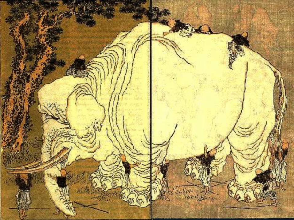
Blind men and the Elephant
Elephant:
Burden of angle closure glaucoma in our population.
Ophthalmologists without Gonioscopy:
Blind Men/Women

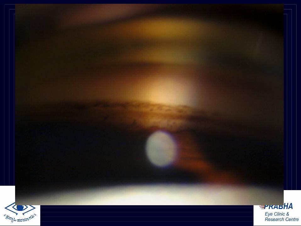
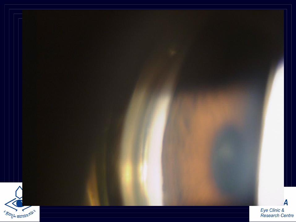
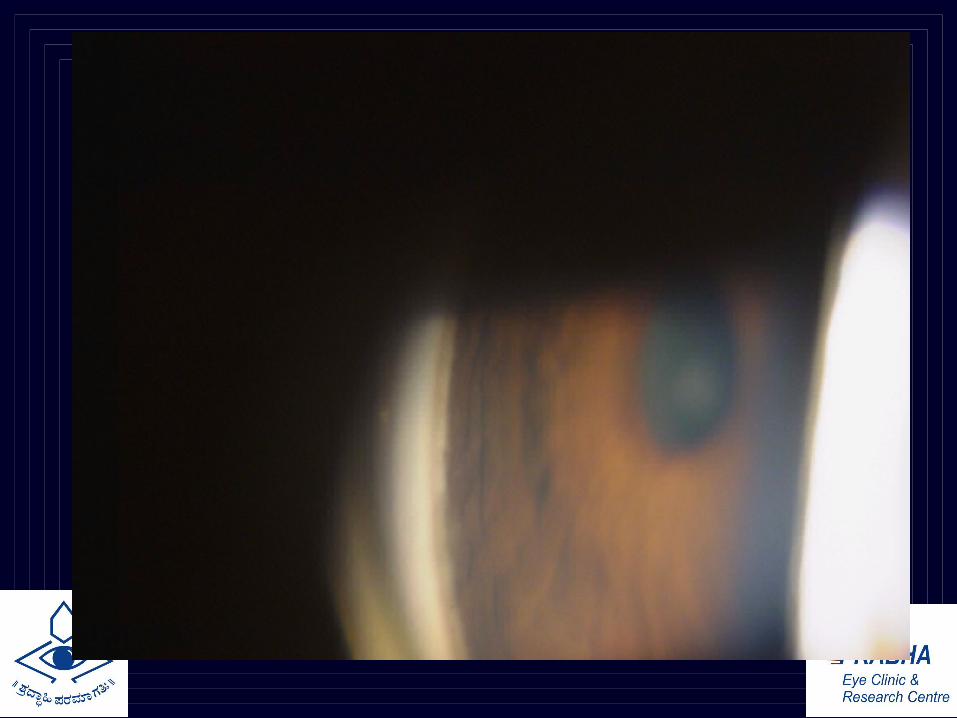
Example open angle
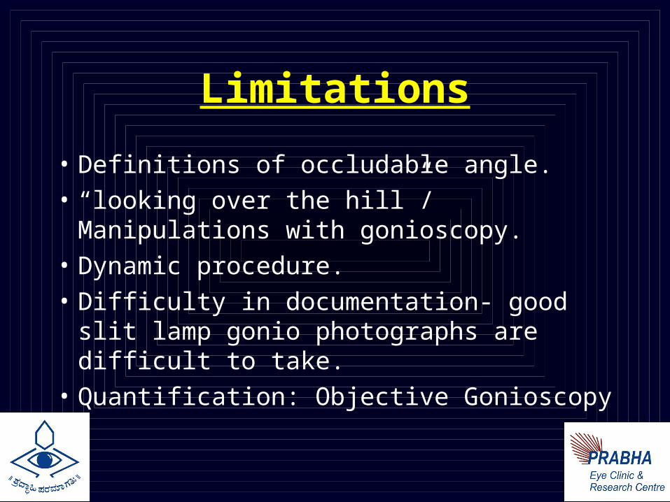
Limitations
• Definitions of occludable angle.
• “looking over the hill”/ Manipulations with gonioscopy.
• Dynamic procedure.
• Difficulty in documentation- good slit lamp gonio photographs are difficult to take.
• Quantification: Objective Gonioscopy
Related Documents