SAGE-Hindawi Access to Research Enzyme Research Volume 2011, Article ID 250842, 10 pages doi:10.4061/2011/250842 Research Article 16S rRNA-Based Identification of a Glucan-Hyperproducing Weissella confusa Shraddha Shukla and Arun Goyal Department of Biotechnology, Indian Institute of Technology Guwahati, Guwahati 781039, Assam, India Correspondence should be addressed to Arun Goyal, [email protected] Received 31 December 2010; Accepted 21 January 2011 Academic Editor: Alane Beatriz Vermelho Copyright © 2011 S. Shukla and A. Goyal. This is an open access article distributed under the Creative Commons Attribution License, which permits unrestricted use, distribution, and reproduction in any medium, provided the original work is properly cited. A gram-positive, nonmotile, irregular, short, rod-shaped new strain of Weissella confusa bacterium was isolated from fermented cabbage. The isolate was physiologically and biochemically characterised. The 16S rDNA was amplified by polymerase chain reaction (PCR). The isolate was identified as Weissella confusa (GenBank accession number: GU138518.1) based on nucleotide homology and phylogenetic analysis. The isolate produces glucansucrase when grown in sucrose-supplemented culture medium which catalyses glucan formation. This novel isolate possesses high capacity of industrial use due to its high productivity of glucan (34 mg/mL) as compared to other strains reported. The optimum temperature for glucansucrase production was 25 ◦ C. The shaking condition gave an enzyme activity of 6.1 U/mL which was 1.5 times higher than that given by static condition (4.1 U/mL). The temperature 35 ◦ C, pH 5.4, and ionic strength 10–20mM were optimum for enzyme assay. This investigation unraveled the abundance of industrially valuable microflora of the north east India. 1. Introduction The Weissella species are gram-positive, non-spore-forming, heterofermentative, nonmotile, and irregular or coccoid rod- shaped organisms. Members of the genus Weissella have been isolated from a variety of sources, such as fresh vegetables, fermented silage, meat, or meat products. Several lactic acid bacteria belonging to the genera Weissella have been introduced to wheat sourdough baking for in situ production of exopolysaccharides [1]. Weissella kimchii PL9023 was selected as a prebiotic as it mostly produced hydrogen per- oxide which inhibited the growth and adherence of vaginal isolates of Candida albicans, Escherichia coli, Staphylococcus aureus, and Streptococcus agalactiae [2]. Lactic acid bacteria (LAB) that produce dextrans include various Leuconostoc, Streptococcus, and Lactobacillus species [3]. Smitinont et al. [4] had emphasised on dextran-synthesizing ability of the Pediococcus genus. Patel et al. [5] reported the dextran- production ability of Pediococcus pentosaceus for the first time. Additionally, dextran production has typically served as a phenotypic test in the identification of bacteria classified in the genus Weissella [6]. Maina et al. [7] reported industrially useful dextran-producing Weissella confusa E392 which could be a suitable alternative to the widely used L. mesen- teroides B512F in the production of linear dextran. First proposed by Collins et al. [6] on the basis of the results of 16S rDNA phylogenetic analyses, the genus Weissella encompasses a phylogenetically coherent group of lactic acid bacteria and includes twelve validated Leu- conostoc-like species, currently, including W. confusa (for- merly Lactobacillus confusus), W. minor (formerly Lacto- bacillus minor), W. kandleri (formerly Lactobacillus kand- leri), W. halotolerans (formerly Lactobacillus halotolerans), W. viridescens (formerly Lactobacillus viridescens), Weissella paramesenteroides (formerly Leuconostoc paramesenteroides), W. hellenica, W. thailandensis, W. cibaria, Weissella kimchii, Weissella soil, and Weissella koreensis. Alternative molecular biological approaches have been developed in place of clas- sical phenotypic methods for the identification of Weissella species. Such methods include soluble whole-cell protein pattern analyses [8], restriction pattern of ribosomal DNA [8] fatty acid analyses [9], random amplified polymor- phic DNA-PCR (RAPD-PCR) [10], denaturing gradient gel electrophoresis (DGGE) [11], and PCR targeting 16S/23S

Welcome message from author
This document is posted to help you gain knowledge. Please leave a comment to let me know what you think about it! Share it to your friends and learn new things together.
Transcript
SAGE-Hindawi Access to ResearchEnzyme ResearchVolume 2011, Article ID 250842, 10 pagesdoi:10.4061/2011/250842
Research Article
16S rRNA-Based Identification of a Glucan-HyperproducingWeissella confusa
Shraddha Shukla and Arun Goyal
Department of Biotechnology, Indian Institute of Technology Guwahati, Guwahati 781039, Assam, India
Correspondence should be addressed to Arun Goyal, [email protected]
Received 31 December 2010; Accepted 21 January 2011
Academic Editor: Alane Beatriz Vermelho
Copyright © 2011 S. Shukla and A. Goyal. This is an open access article distributed under the Creative Commons AttributionLicense, which permits unrestricted use, distribution, and reproduction in any medium, provided the original work is properlycited.
A gram-positive, nonmotile, irregular, short, rod-shaped new strain of Weissella confusa bacterium was isolated from fermentedcabbage. The isolate was physiologically and biochemically characterised. The 16S rDNA was amplified by polymerase chainreaction (PCR). The isolate was identified as Weissella confusa (GenBank accession number: GU138518.1) based on nucleotidehomology and phylogenetic analysis. The isolate produces glucansucrase when grown in sucrose-supplemented culture mediumwhich catalyses glucan formation. This novel isolate possesses high capacity of industrial use due to its high productivity ofglucan (34 mg/mL) as compared to other strains reported. The optimum temperature for glucansucrase production was 25◦C. Theshaking condition gave an enzyme activity of 6.1 U/mL which was 1.5 times higher than that given by static condition (4.1 U/mL).The temperature 35◦C, pH 5.4, and ionic strength 10–20 mM were optimum for enzyme assay. This investigation unraveled theabundance of industrially valuable microflora of the north east India.
1. Introduction
The Weissella species are gram-positive, non-spore-forming,heterofermentative, nonmotile, and irregular or coccoid rod-shaped organisms. Members of the genus Weissella have beenisolated from a variety of sources, such as fresh vegetables,fermented silage, meat, or meat products. Several lacticacid bacteria belonging to the genera Weissella have beenintroduced to wheat sourdough baking for in situ productionof exopolysaccharides [1]. Weissella kimchii PL9023 wasselected as a prebiotic as it mostly produced hydrogen per-oxide which inhibited the growth and adherence of vaginalisolates of Candida albicans, Escherichia coli, Staphylococcusaureus, and Streptococcus agalactiae [2]. Lactic acid bacteria(LAB) that produce dextrans include various Leuconostoc,Streptococcus, and Lactobacillus species [3]. Smitinont et al.[4] had emphasised on dextran-synthesizing ability of thePediococcus genus. Patel et al. [5] reported the dextran-production ability of Pediococcus pentosaceus for the firsttime. Additionally, dextran production has typically served asa phenotypic test in the identification of bacteria classified inthe genus Weissella [6]. Maina et al. [7] reported industrially
useful dextran-producing Weissella confusa E392 whichcould be a suitable alternative to the widely used L. mesen-teroides B512F in the production of linear dextran.
First proposed by Collins et al. [6] on the basis ofthe results of 16S rDNA phylogenetic analyses, the genusWeissella encompasses a phylogenetically coherent groupof lactic acid bacteria and includes twelve validated Leu-conostoc-like species, currently, including W. confusa (for-merly Lactobacillus confusus), W. minor (formerly Lacto-bacillus minor), W. kandleri (formerly Lactobacillus kand-leri), W. halotolerans (formerly Lactobacillus halotolerans),W. viridescens (formerly Lactobacillus viridescens), Weissellaparamesenteroides (formerly Leuconostoc paramesenteroides),W. hellenica, W. thailandensis, W. cibaria, Weissella kimchii,Weissella soil, and Weissella koreensis. Alternative molecularbiological approaches have been developed in place of clas-sical phenotypic methods for the identification of Weissellaspecies. Such methods include soluble whole-cell proteinpattern analyses [8], restriction pattern of ribosomal DNA[8] fatty acid analyses [9], random amplified polymor-phic DNA-PCR (RAPD-PCR) [10], denaturing gradient gelelectrophoresis (DGGE) [11], and PCR targeting 16S/23S
2 Enzyme Research
rRNA gene spacer region [12]. In the present study, anovel Weissella species isolated from fermented cabbage wascharacterised up to genus and species level using 16S rDNA-based sequence analyses. Its glucan formation capacityhas been explored in detail and compared with that ofLeuconostoc mesenteroides NRRL B-512F.
2. Materials and Methods
2.1. Isolation and Maintenance of Microorganism. Freshchopped cabbage was subjected to fermentation in 2.5%(w/v) NaCl solution. One gram of fermented cabbage wasground to paste and mixed in 10 mL of saline (0.9% w/v)homogeneously in test tubes. The serial dilutions were made,till the dilution factor 10−5. One hundred microliter from allthe dilutions of cabbage from 10−0 to 10−5 was taken andspread plated on petri plates of 1.7% (w/v) MRS agar withglucose as carbon source [13]. Petri plates were incubatedat 28◦C for 24 h. Based on the higher-glucansucrase activityand glucan concentration determined (as described later), amicrobe Cab3 (named after its colony number) was isolated.The isolated bacterium was propagated as stab in MRS agarsupplemented with sucrose as carbon source [14] at 25◦Cand stored at 4◦C. For long-term preservation, the isolate waskept in 20% (v/v) glycerol at −80◦C.
2.2. Morphological, Physiological, and Biochemical Character-ization. The colony morphology of isolates grown on MRSmedium for overnight at 25◦C was observed directly andby light microscopy. Cell mobility and gliding movementwere assessed by phase-contrast microscopy (1000x) usingcells of MRS broth. The Gram reaction was performed usingthe KOH method of Gregesen [15]. The cell morphologyof the selected isolate was examined by scanning electronmicroscopy (Leo1430 VP, Leo Electron Microscopy Ltd.,Cambridge, UK) operated at 10.0 kV. The catalase activitywas determined by transferring fresh colonies from MRSagar to a drop of 5% (v/v) H2O2 on a glass slide. For thetemperature tolerance of the microorganism, the growth ofselected isolate was carried out using MRS broth at differenttemperatures, that is, 4◦C, 10◦C, 15◦C, 20◦C, 30◦C, 37◦C,and 42◦C for 2 days. For salt tolerance of the isolate, thegrowth was observed using MRS broth in the presence of4.0% and 6.5% (w/v) of NaCl incubated at 25◦C for 2 d.The production of glucan (slimy layer) by isolate Cab3 fromsucrose was observed on MRS agar in which glucose wasreplaced by sucrose [14].
The strain Cab3 was tested for its ability to fermentvarious carbohydrates using the method of Kandler andWeiss [21]. From the overnight-grown MRS broth contain-ing 2% (w/v) glucose as carbohydrate source, 50 µL wasinoculated in 5.0 mL liquid MRS medium lacking glucosebut containing phenol red and other test carbohydrates togive a final inoculum to medium ratio of 1% (v/v). The testmedia were incubated for 2 days at 30◦C without shaking.The acid production was recorded between 24 and 48 h. Theacid production was indicated by a change in the colour ofthe phenol red indicator dye from red to yellow. The isolated
strain cab3 was tested for susceptibility to 30 antibioticsusing a standardized filter-paper disc-agar diffusion assay, todetermine the drug susceptibility of microorganisms [22].
The antibiotic tests were performed using commer-cially available antibiotic octadiscs containing Amoxyclav(Ac), cephalexin (Cp), ciprofloxacin (Cf), clindamycin(Cd), cloxacillin (Cx), erythromycin (E), tetracycline (T),ampicillin (A), carbenicillin (Cb), cefotaxime (Ce), chlo-ramphenicol (C), co-Trimazine (Cm), gentamicin (G),norfloxacin (Nx), oxacillin (Ox), amikacin (Ak), amoxy-cillin (Am), bacitracin (B), cephalothin (Ch), novobiocin(Nv), oxytetracycline (O), vancomycin (V), penicillin-G(P), tobramycin (Tb), cephaloridine (Cr), kanamycin (K),lincomycin (L), methicillin (M), norfloxacin (Nf), andoleandomycin (Ol) from Hi-media Pvt. Ltd. India.
2.3. 16S rDNA-Based Sequence Analyses
2.3.1. Extraction of Genomic DNA. The bacterial cell pelletwas lysed using a solution containing guanidium thiocyanate(a chaotropic agent) and SDS (a detergent), to extractDNA [23]. This lysis solution was used to disrupt the cell,remove proteins and polysaccharides, and partial hydrolysisof RNA. The genomic DNA was extracted by GeNeiTMGenomic DNA Extraction kit (Bangalore Genei Pvt. Ltd.).DNA was then precipitated using alcohol and washed with70% (v/v) alcohol to remove contaminants. DNA pelletwas solubilized in sodium acetate buffer at pH 5.5 addedto a final concentration of 0.3 M in isopropanol at highertemperature (50–55◦C), to increase the solubility of genomicDNA. The extracted DNA was purified using Ultrapureprep kit (KT83B, Bangalore Genei, India). Genomic DNAwas purified in a gravity flow column using elution buffer,followed by treatment with lysozyme, proteinase K, andRNase A supplied with the kit. DNA concentrations weredetermined in duplicate using a spectrophotometer. Thequality of purified DNA was evaluated on 1.2% Agarosegel, a single band of high-molecular-weight DNA wasobserved.
2.3.2. PCR Amplification. The genomic DNA of isolate Cab3was used for amplification of 16S rRNA gene. The univer-sal 16S rDNA primers, forward primer 8F (5′AGTTGA-TCCTGGCTCAG3′), and reverse primer 1492R (5′ACC-TTGTTACGACTT3′) were used for the polymerase chainreaction (PCR). The PCR amplification was carried outin a reaction mixture containing ∼10 ng genomic DNA astemplate, 1 µL dNTP mix (2.5 mM each), 100 ng/µL eachof forward and reverse primer, 1X Taq DNA polymeraseassay buffer (10X), 3U Taq DNA polymerase enzyme(Bangalore Genei Ltd.,Bangalore, India), and distilled waterenough to make up the volume to 50 µL reaction mixture.Amplification conditions were 5 min initial denaturation at94◦C, 30 s denaturation at 94◦C, 30 s of primer annealingat 54◦C, 1 min elongation at 72◦C for 35 cycles, and a finalextension of 10 min at 72◦C. The reactions were carriedout in a Thermal Cycler (Applied Biosystems, model ABI2720).
Enzyme Research 3
2.3.3. Electrophoresis and Sequencing of 16S rRNA Gene.Fifteen µL of PCR amplified product was electrophoresed on1.2% (w/v) low-EEO agarose gel in 1X TBE buffer (45 mMTris-borate, pH 8.3, and 1 mM Na 2 EDTA) at 100 V for2 h. The gel was stained with ethidium bromide in a finalconcentration of 0.5 µg/mL, visualized, and photographedunder UV light. The amplified 16S rRNA gene from theisolate was eluted and purified from the gel slice using theGeneiPure TM Gel Extraction Kit (QIAGEN). Forward andreverse DNA sequencing reaction of PCR amplicon wascarried out with 8F and 1492R primers using BDT v3.1 Cyclesequencing kit on ABI 3730xl Genetic Analyzer. Consensussequence of 1288 bp of 16S rDNA gene was generated fromforward and reverse sequence data using aligner software.
2.3.4. Sequence Alignment of 16S rRNA Gene. The 16S rDNAgene sequence of the isolate cab3 was used to carry outBLAST with the nr database of NCBI GenBank database.Based on maximum identity score, first ten sequences wereselected and aligned using multiple alignment softwareprogram ClustalW. Distance matrix was generated usingRDP database, and the phylogenetic tree was constructedusing MEGA 4 [28].
2.4. Production of Glucansucrase and Its Assay
2.4.1. Enzyme Activity Assay. The enzyme assay was carriedout in 1 mL reaction mixture containing 5% (w/v) sucrose,20 mM sodium acetate buffer (pH 5.4), and 20 µL cell-freesupernatant. The enzymatic reaction was performed at 30◦Cfor 15 min. 100 µL aliquot from the reaction mixture wastaken for reducing sugar estimation. The enzyme activitywas determined by estimating the released reducing sugar byNelson [29] and Somogyi [30] method. The absorbance ofthe colour developed was measured by spectrophotometer at500 nm. Fructose was used to plot the standard graph.
2.4.2. Effect of Temperature on Enzyme Production. The glu-cansucrase was produced in the enzyme production mediumas described by Tsuchiya et al. [31]. Fermentations werecarried out in triplicate sets of 60 mL enzyme productionmedium in a 250 mL Erlenmeyer flask incubated at 25◦Cunder shaking condition at 180 rpm. The samples (1 mL)were withdrawn at indicated time intervals and centrifugedat 8000 g for 10 min at 4◦C to separate the cells. The cell-free supernatant was analyzed for enzyme activity. Weissellaconfusa was grown at various temperatures: 22◦C, 25◦C,28◦C, 30◦C, 32◦C, 37◦C, and 40◦C in the flasks as describedpreviously in an orbital shaking incubator at 180 rpm todetermine the optimum incubation temperature for themaximum enzyme production. The culture (1.0 mL) waswithdrawn at the indicated time intervals and centrifuged at8000 g for 10 min at 4◦C to pellet out the cells. The cell-freesupernatant was analyzed for enzyme activity.
2.4.3. Effect of Shaken-Flask Culture on Enzyme Production.The production glucansucrase was compared under shaken-flask condition with the static flask culture at 25◦C in
triplicate sets of 60 mL enzyme production medium in250 mL Erlenmeyer flasks. The shaking was carried out in anorbital shaking incubator at 180 rpm. The samples (1.0 mL)were withdrawn at indicated time intervals and centrifugedat 8000 g for 10 min at 4◦C to pellet out the cells. The cell-free extract was analysed for enzyme activity and proteinconcentration as described earlier.
2.4.4. Analysis of Glucan Content of the Isolated Strain. Thepolysaccharide content of the isolated strain (Cab3) wasdetermined by phenol-sulphuric acid method [32] in amicrotitre plate [33]. The isolated strain was grown in 60 mLliquid medium described by Tsuchiya et al. [31] at 25◦C and180 rpm up to 32 h. The samples (1 mL) were withdrawnat regular intervals. To 200 µL of the culture supernatant,three volumes of the prechilled ethanol were added andcentrifuged at 12,000 g. The supernatant was discarded, andthe precipitate was resuspended in 200 µL distilled water. Theprocess was repeated two more times. The final precipitatewas air dried and dissolved in 200 µL distilled water. DextranT-40 was used to plot the standard.
2.5. Purification and Identification of Glucansucrase by Activ-ity Staining. The crude 50 mL cell-free supernatant withenzyme activity 6 U/mL and specific activity 1 U/mg was sub-jected to purification by fractionation using PEG-400 (33%,w/v). The enzyme activity was determined as describedearlier. The protein concentration was determined by themethod of Lowry et al. [34]. To confirm the existence ofglucansucrase, the PAS staining of the sucrose- and raffinose-incubated gel was carried out on 7% nondenaturing SDS-PAGE [35]. The electrophoresis was performed on a mini gelunit (BioRad) using 1.5 mm thick gels, following the methodof [36]. After run, the gel was treated thrice with a solutioncontaining 20 mM sodium acetate buffer, pH 5.4, 0.1% (w/v)Triton X-100, and 0.005% (w/v) calcium chloride for 20 minto remove SDS. Then, the gel was incubated with 5% (w/v)sucrose/raffinose solution in 20 mM sodium acetate buffer,pH 5.4 for 10–12 h. Following incubation, the gel was washedtwice with 75% (v/v) ethanol for 20 min and incubated insolution with 0.7% (w/v) periodic acid in 5% (v/v) acetic acidfor 20 min at room temperature. The gel was then washedthrice with 0.2% (w/v) sodium bisulfate in 5% (v/v) aceticacid solution and finally stained with Schiff ’s reagent (0.5%w/v basic fuchsin, 1% (w/v) sodium bisulfate, and 0.1 N HCl)until the discrete magenta bands appeared within the gel.
2.6. Effect of Temperature, pH, and Ionic Strength on EnzymeAssay. The enzyme assay conditions were optimized to findmaximum enzyme activity. The purified enzyme having10.4 U/mg specific activity (1.0 mg/mL) was used for furtheroptimization of assay conditions. The ionic strength of thebuffer was varied from 10 mM to 400 mM. The assay wascarried out in 1 mL reaction mixture containing 5% (w/v)final sucrose concentration, in sodium acetate buffer (pH5.4) at 30◦C for 15 min. To study the effect of varied pHon enzyme activity, the pH of buffer was varied from 4to 7 in 1 mL reaction mixture containing 5% (w/v) final
4 Enzyme Research
Table 1: Differential characteristics of Weissella confusa and other lactic acid bacteria.
Characteristics 1 2 [24] 3 [21] 4 [21] 5 [6] 6 [25] 7 [26] 8 [18] 9 [27] 10 [27]
Acid from:
Ribose + + + + − − + NT NT NT
D-Xylose + + − + − + − + + +
Galactose + − + + − + + + + +
D-Fructose + − + + + NT − + + +
Cellobiose + − − + − + − + − +
Melibiose − + − − − + + + + +
Sucrose + + − + + + D + + +
Trehalose − + − − + − D + + +
D-Raffinose − + − − − − + + + −Maltose + NT − + + + + − + +
Dextran formation + − + + − + − + + +
Cell morphologyShortrods
Shortrods, oftenthickenedto one end
Irregularrods
Short rods,thickenedat one end
Largespherical orlenticularcells
NT Spherical
Cocci-shapedand randomarrangement ingroups or chains
NT NT
1, W. confusa (Cab3) 2, W. soli; 3, W. kandleri; 4, W. confusa; 5, W. hellenica; 6, W. kimchii; 7, W. thailandensis; 8, P. pentosaceus; 9, L. mesenteroides B-512F;10, L. mesenteroides B-640. Symbols: +, 90% or more strains were positive; −, 90% or more strains were negative; D, 11–89% of strains were positive; NT, nottested.
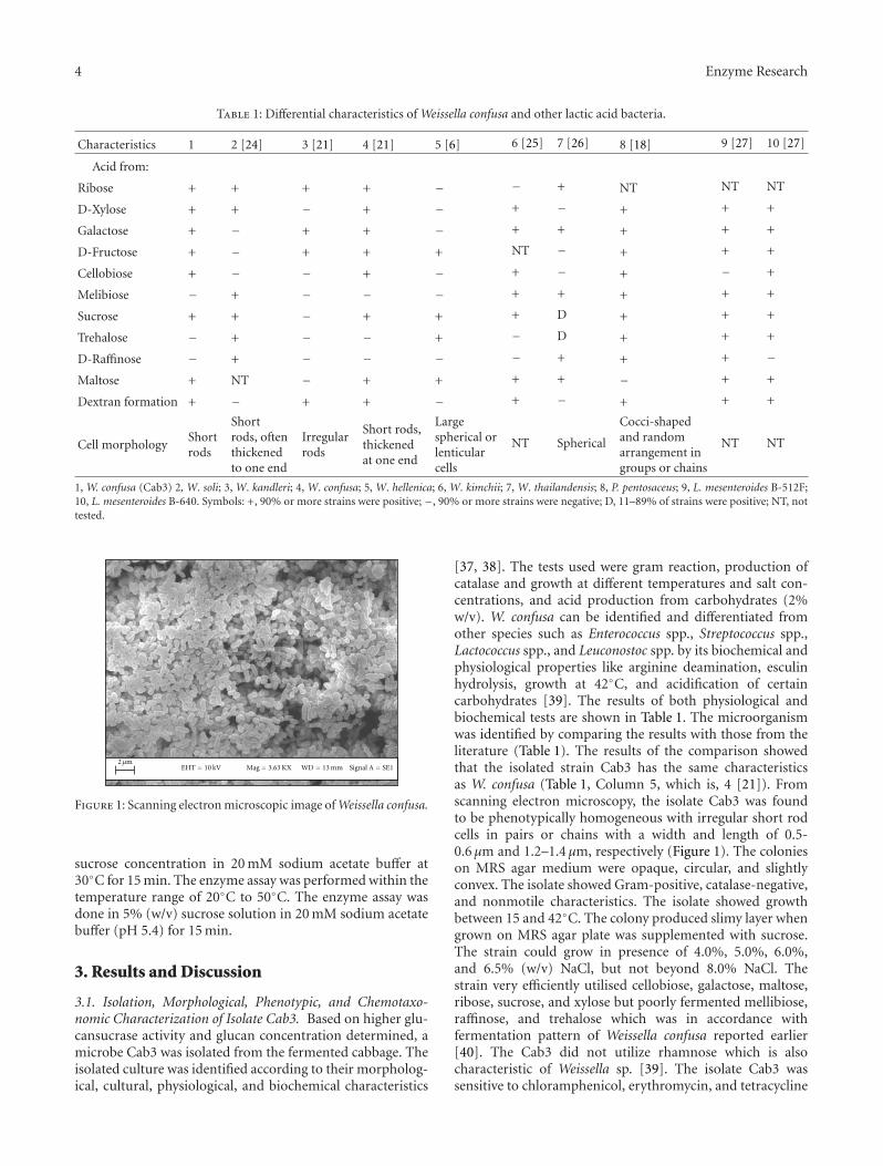
2 µmEHT = 10 kV Mag = 3.63 KX WD = 13 mm Signal A = SE1
Figure 1: Scanning electron microscopic image of Weissella confusa.
sucrose concentration in 20 mM sodium acetate buffer at30◦C for 15 min. The enzyme assay was performed within thetemperature range of 20◦C to 50◦C. The enzyme assay wasdone in 5% (w/v) sucrose solution in 20 mM sodium acetatebuffer (pH 5.4) for 15 min.
3. Results and Discussion
3.1. Isolation, Morphological, Phenotypic, and Chemotaxo-nomic Characterization of Isolate Cab3. Based on higher glu-cansucrase activity and glucan concentration determined, amicrobe Cab3 was isolated from the fermented cabbage. Theisolated culture was identified according to their morpholog-ical, cultural, physiological, and biochemical characteristics
[37, 38]. The tests used were gram reaction, production ofcatalase and growth at different temperatures and salt con-centrations, and acid production from carbohydrates (2%w/v). W. confusa can be identified and differentiated fromother species such as Enterococcus spp., Streptococcus spp.,Lactococcus spp., and Leuconostoc spp. by its biochemical andphysiological properties like arginine deamination, esculinhydrolysis, growth at 42◦C, and acidification of certaincarbohydrates [39]. The results of both physiological andbiochemical tests are shown in Table 1. The microorganismwas identified by comparing the results with those from theliterature (Table 1). The results of the comparison showedthat the isolated strain Cab3 has the same characteristicsas W. confusa (Table 1, Column 5, which is, 4 [21]). Fromscanning electron microscopy, the isolate Cab3 was foundto be phenotypically homogeneous with irregular short rodcells in pairs or chains with a width and length of 0.5-0.6 µm and 1.2–1.4 µm, respectively (Figure 1). The colonieson MRS agar medium were opaque, circular, and slightlyconvex. The isolate showed Gram-positive, catalase-negative,and nonmotile characteristics. The isolate showed growthbetween 15 and 42◦C. The colony produced slimy layer whengrown on MRS agar plate was supplemented with sucrose.The strain could grow in presence of 4.0%, 5.0%, 6.0%,and 6.5% (w/v) NaCl, but not beyond 8.0% NaCl. Thestrain very efficiently utilised cellobiose, galactose, maltose,ribose, sucrose, and xylose but poorly fermented mellibiose,raffinose, and trehalose which was in accordance withfermentation pattern of Weissella confusa reported earlier[40]. The Cab3 did not utilize rhamnose which is alsocharacteristic of Weissella sp. [39]. The isolate Cab3 wassensitive to chloramphenicol, erythromycin, and tetracycline
Enzyme Research 5
(kbp)
0.2
0.4
0.7
1
1.5
2.5
47
20
1.5 kbp
1 2
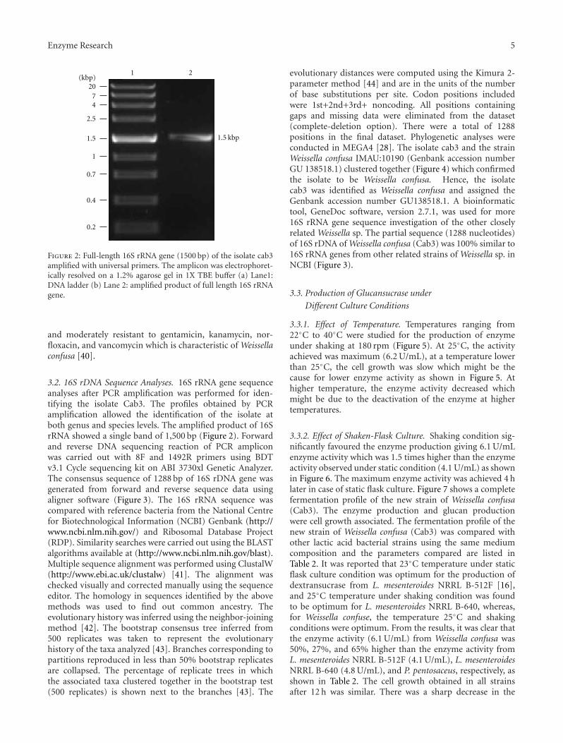
Figure 2: Full-length 16S rRNA gene (1500 bp) of the isolate cab3amplified with universal primers. The amplicon was electrophoret-ically resolved on a 1.2% agarose gel in 1X TBE buffer (a) Lane1:DNA ladder (b) Lane 2: amplified product of full length 16S rRNAgene.
and moderately resistant to gentamicin, kanamycin, nor-floxacin, and vancomycin which is characteristic of Weissellaconfusa [40].
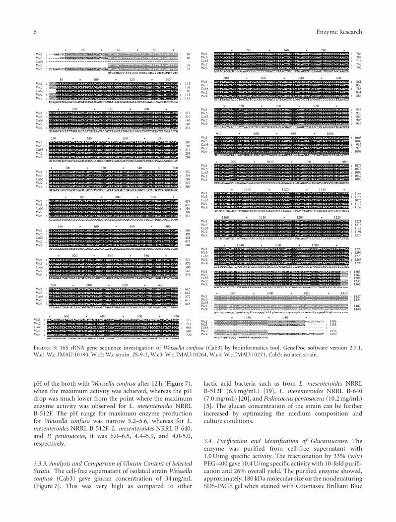
3.2. 16S rDNA Sequence Analyses. 16S rRNA gene sequenceanalyses after PCR amplification was performed for iden-tifying the isolate Cab3. The profiles obtained by PCRamplification allowed the identification of the isolate atboth genus and species levels. The amplified product of 16SrRNA showed a single band of 1,500 bp (Figure 2). Forwardand reverse DNA sequencing reaction of PCR ampliconwas carried out with 8F and 1492R primers using BDTv3.1 Cycle sequencing kit on ABI 3730xl Genetic Analyzer.The consensus sequence of 1288 bp of 16S rDNA gene wasgenerated from forward and reverse sequence data usingaligner software (Figure 3). The 16S rRNA sequence wascompared with reference bacteria from the National Centrefor Biotechnological Information (NCBI) Genbank (http://www.ncbi.nlm.nih.gov/) and Ribosomal Database Project(RDP). Similarity searches were carried out using the BLASTalgorithms available at (http://www.ncbi.nlm.nih.gov/blast).Multiple sequence alignment was performed using ClustalW(http://www.ebi.ac.uk/clustalw) [41]. The alignment waschecked visually and corrected manually using the sequenceeditor. The homology in sequences identified by the abovemethods was used to find out common ancestry. Theevolutionary history was inferred using the neighbor-joiningmethod [42]. The bootstrap consensus tree inferred from500 replicates was taken to represent the evolutionaryhistory of the taxa analyzed [43]. Branches corresponding topartitions reproduced in less than 50% bootstrap replicatesare collapsed. The percentage of replicate trees in whichthe associated taxa clustered together in the bootstrap test(500 replicates) is shown next to the branches [43]. The
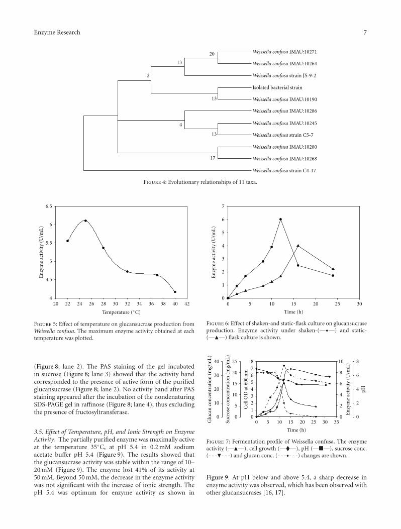
evolutionary distances were computed using the Kimura 2-parameter method [44] and are in the units of the numberof base substitutions per site. Codon positions includedwere 1st+2nd+3rd+ noncoding. All positions containinggaps and missing data were eliminated from the dataset(complete-deletion option). There were a total of 1288positions in the final dataset. Phylogenetic analyses wereconducted in MEGA4 [28]. The isolate cab3 and the strainWeissella confusa IMAU:10190 (Genbank accession numberGU 138518.1) clustered together (Figure 4) which confirmedthe isolate to be Weissella confusa. Hence, the isolatecab3 was identified as Weissella confusa and assigned theGenbank accession number GU138518.1. A bioinformatictool, GeneDoc software, version 2.7.1, was used for more16S rRNA gene sequence investigation of the other closelyrelated Weissella sp. The partial sequence (1288 nucleotides)of 16S rDNA of Weissella confusa (Cab3) was 100% similar to16S rRNA genes from other related strains of Weissella sp. inNCBI (Figure 3).
3.3. Production of Glucansucrase under
Different Culture Conditions
3.3.1. Effect of Temperature. Temperatures ranging from22◦C to 40◦C were studied for the production of enzymeunder shaking at 180 rpm (Figure 5). At 25◦C, the activityachieved was maximum (6.2 U/mL), at a temperature lowerthan 25◦C, the cell growth was slow which might be thecause for lower enzyme activity as shown in Figure 5. Athigher temperature, the enzyme activity decreased whichmight be due to the deactivation of the enzyme at highertemperatures.
3.3.2. Effect of Shaken-Flask Culture. Shaking condition sig-nificantly favoured the enzyme production giving 6.1 U/mLenzyme activity which was 1.5 times higher than the enzymeactivity observed under static condition (4.1 U/mL) as shownin Figure 6. The maximum enzyme activity was achieved 4 hlater in case of static flask culture. Figure 7 shows a completefermentation profile of the new strain of Weissella confusa(Cab3). The enzyme production and glucan productionwere cell growth associated. The fermentation profile of thenew strain of Weissella confusa (Cab3) was compared withother lactic acid bacterial strains using the same mediumcomposition and the parameters compared are listed inTable 2. It was reported that 23◦C temperature under staticflask culture condition was optimum for the production ofdextransucrase from L. mesenteroides NRRL B-512F [16],and 25◦C temperature under shaking condition was foundto be optimum for L. mesenteroides NRRL B-640, whereas,for Weissella confuse, the temperature 25◦C and shakingconditions were optimum. From the results, it was clear thatthe enzyme activity (6.1 U/mL) from Weissella confusa was50%, 27%, and 65% higher than the enzyme activity fromL. mesenteroides NRRL B-512F (4.1 U/mL), L. mesenteroidesNRRL B-640 (4.8 U/mL), and P. pentosaceus, respectively, asshown in Table 2. The cell growth obtained in all strainsafter 12 h was similar. There was a sharp decrease in the
6 Enzyme Research
Wc1 :Wc3 :
Wc2 :Wc4 :
Cab3 :
Wc1 :Wc3 :
Wc2 :Wc4 :
Cab3 :
Wc1 :Wc3 :
Wc2 :Wc4 :
Cab3 :
Wc1 :Wc3 :
Wc2 :Wc4 :
Cab3 :
Wc1 :Wc3 :
Wc2 :Wc4 :
Cab3 :
Wc1 :Wc3 :
Wc2 :Wc4 :
Cab3 :
Wc1 :Wc3 :
Wc2 :Wc4 :
Cab3 :
Wc1 :Wc3 :
Wc2 :Wc4 :
Cab3 :
Wc1 :Wc3 :
Wc2 :Wc4 :
Cab3 :
Wc1 :Wc3 :
Wc2 :Wc4 :
Cab3 :
Wc1 :Wc3 :
Wc2 :Wc4 :
Cab3 :
Wc1 :Wc3 :
Wc2 :Wc4 :
Cab3 :
Wc1 :Wc3 :
Wc2 :Wc4 :
Cab3 :
Wc1 :Wc3 :
Wc2 :Wc4 :
Cab3 :
Wc1 :Wc3 :
Wc2 :Wc4 :
Cab3 :
Wc1 :Wc3 :
Wc2 :Wc4 :
Cab3 :
Wc1 :Wc3 :
Wc2 :Wc4 :
Cab3 :
Wc1 :Wc3 :
Wc2 :Wc4 :
Cab3 :
Wc1 :Wc3 :
Wc2 :Wc4 :
Cab3 :
Wc1 :Wc3 :
Wc2 :Wc4 :
Cab3 :
Wc1 :Wc3 :
Wc2 :Wc4 :
Cab3 :
20 40 60: 69: 66: -: 39: 72
: 141: 138: 68: 111: 144
: 213: 210: 140: 183: 216
: 285: 282: 212: 255: 288
: 357: 354: 284: 327: 360
: 429: 426: 356: 399: 432
: 501: 498: 428: 471: 504
: 573: 570: 500: 543: 576
: 645: 642: 572: 615: 648
: 717: 714: 644: 687: 720
: 789: 786: 716: 759: 792
: 861: 858: 788: 831: 864
: 933: 930: 860: 903: 936
: 1005: 1002: 932: 975: 1008
: 1077: 1074: 1004: 1047: 1080
: 1149: 1146: 1076: 1119: 1152
: 1221: 1218: 1148: 1191: 1224
: 1293: 1290: 1220: 1263: 1296
: 1365: 1362: 1288: 1335: 1368
: 1437: 1434: -
: -
: 1407: 1440
: 1496: 1483
: 1436: 1499
140
740 760 780
860840820800
880 900 920
1000980960940
1020 1040 1060 1080
114011201100
1160 1180 1200 1220
128012601240
1380
1460 1480
1400 1420 1440
12010080
160 180 200
280260240220
300 320 340 360
420400380
440 460 480 500
560540520
580 600 620 640
720700680660
� � � �
���
� � � �
���
� � � �
����
� � �
����
� � �
� � � �
����
� � � �
���
� � � �
����
� � �
����
� � �
����
���
Figure 3: 16S rRNA gene sequence investigation of Weissella confusa (Cab3) by bioinformatics tool, GeneDoc software version 2.7.1.W.c1:W.c IMAU:10190, W.c2: W.c strain JS-9-2, W.c3: W.c IMAU:10264, W.c4: W.c IMAU:10271, Cab3: isolated strain.
pH of the broth with Weissella confusa after 12 h (Figure 7),when the maximum activity was achieved, whereas the pHdrop was much lower from the point where the maximumenzyme activity was observed for L. mesenteroides NRRLB-512F. The pH range for maximum enzyme productionfor Weissella confusa was narrow 5.2–5.6, whereas for L.mesenteroides NRRL B-512F, L. mesenteroides NRRL B-640,and P. pentosaceus, it was 6.0–6.5, 4.4–5.9, and 4.0-5.0,respectively.
3.3.3. Analysis and Comparison of Glucan Content of SelectedStrain. The cell-free supernatant of isolated strain Weissellaconfusa (Cab3) gave glucan concentration of 34 mg/mL(Figure 7). This was very high as compared to other
lactic acid bacteria such as from L. mesenteroides NRRLB-512F (6.9 mg/mL) [19], L. mesenteroides NRRL B-640(7.0 mg/mL) [20], and Pediococcus pentosaceus (10.2 mg/mL)[5]. The glucan concentration of the strain can be furtherincreased by optimizing the medium composition andculture conditions.
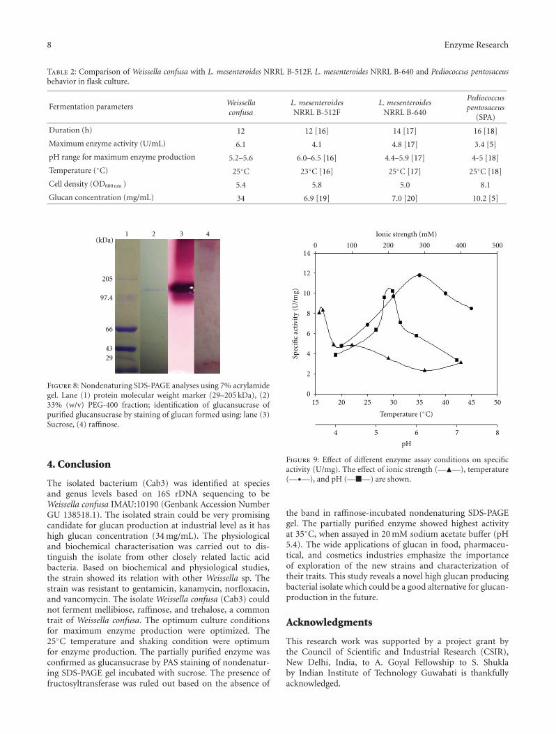
3.4. Purification and Identification of Glucansucrase. Theenzyme was purified from cell-free supernatant with1.0 U/mg specific activity. The fractionation by 33% (w/v)PEG-400 gave 10.4 U/mg specific activity with 10-fold purifi-cation and 26% overall yield. The purified enzyme showed,approximately, 180 kDa molecular size on the nondenaturingSDS-PAGE gel when stained with Coomassie Brilliant Blue
Enzyme Research 7
Weissella confusa IMAU:10271
Weissella confusa IMAU:10264
Weissella confusa strain JS-9-2
Isolated bacterial strain
Weissella confusa IMAU:10190
Weissella confusa IMAU:10286
Weissella confusa IMAU:10245
Weissella confusa IMAU:10280
Weissella confusa IMAU:10268
2
13
13
13
20
4
17
Weissella confusa strain C5-7
Weissella confusa strain C4-17
Figure 4: Evolutionary relationships of 11 taxa.
Temperature (◦C)
20 22 24 26 28 30 32 34 36 38 40 42
En
zym
eac
tivi
ty(U
/mL
)
4
4.5
5
5.5
6
6.5
Figure 5: Effect of temperature on glucansucrase production fromWeissella confusa. The maximum enzyme activity obtained at eachtemperature was plotted.
(Figure 8; lane 2). The PAS staining of the gel incubatedin sucrose (Figure 8; lane 3) showed that the activity bandcorresponded to the presence of active form of the purifiedglucansucrase (Figure 8; lane 2). No activity band after PASstaining appeared after the incubation of the nondenaturingSDS-PAGE gel in raffinose (Figure 8; lane 4), thus excludingthe presence of fructosyltransferase.
3.5. Effect of Temperature, pH, and Ionic Strength on EnzymeActivity. The partially purified enzyme was maximally activeat the temperature 35◦C, at pH 5.4 in 0.2 mM sodiumacetate buffer pH 5.4 (Figure 9). The results showed thatthe glucansucrase activity was stable within the range of 10–20 mM (Figure 9). The enzyme lost 41% of its activity at50 mM. Beyond 50 mM, the decrease in the enzyme activitywas not significant with the increase of ionic strength. ThepH 5.4 was optimum for enzyme activity as shown in
Time (h)
0 5 10 15 20 25 30
En
zym
eac
tivi
ty(U
/mL
)
0
1
2
3
4
5
6
7
Figure 6: Effect of shaken-and static-flask culture on glucansucraseproduction. Enzyme activity under shaken-(—•—) and static-(—�—) flask culture is shown.
Time (h)
0 5 10 15 20 25 30 35
En
zym
eac
tivi
ty(U
/mL
)
0
2
4
6
pH
0
2
4
6
8
8
Sucr
ose
con
cen
trat
ion
(mg/
mL)
0
5
10
15
20
25 10
Glu
can
con
cen
trat
ion
(mg/
mL)
0
10
20
30
40
Cel
lOD
at60
0n
m
012345678
Figure 7: Fermentation profile of Weissella confusa. The enzymeactivity (—�—), cell growth (—�—), pH (—�—), sucrose conc.(- - -�- - -) and glucan conc. (- - -•- - -) changes are shown.
Figure 9. At pH below and above 5.4, a sharp decrease inenzyme activity was observed, which has been observed withother glucansucrases [16, 17].
8 Enzyme Research
Table 2: Comparison of Weissella confusa with L. mesenteroides NRRL B-512F, L. mesenteroides NRRL B-640 and Pediococcus pentosaceusbehavior in flask culture.
Fermentation parameters Weissellaconfusa
L. mesenteroidesNRRL B-512F
L. mesenteroidesNRRL B-640
Pediococcuspentosaceus
(SPA)
Duration (h) 12 12 [16] 14 [17] 16 [18]
Maximum enzyme activity (U/mL) 6.1 4.1 4.8 [17] 3.4 [5]
pH range for maximum enzyme production 5.2–5.6 6.0–6.5 [16] 4.4–5.9 [17] 4-5 [18]
Temperature (◦C) 25◦C 23◦C [16] 25◦C [17] 25◦C [18]
Cell density (OD600 nm ) 5.4 5.8 5.0 8.1
Glucan concentration (mg/mL) 34 6.9 [19] 7.0 [20] 10.2 [5]
2943
66
97.4
205
1 2 3 4(kDa)
Figure 8: Nondenaturing SDS-PAGE analyses using 7% acrylamidegel. Lane (1) protein molecular weight marker (29–205 kDa), (2)33% (w/v) PEG-400 fraction; identification of glucansucrase ofpurified glucansucrase by staining of glucan formed using: lane (3)Sucrose, (4) raffinose.
4. Conclusion
The isolated bacterium (Cab3) was identified at speciesand genus levels based on 16S rDNA sequencing to beWeissella confusa IMAU:10190 (Genbank Accession NumberGU 138518.1). The isolated strain could be very promisingcandidate for glucan production at industrial level as it hashigh glucan concentration (34 mg/mL). The physiologicaland biochemical characterisation was carried out to dis-tinguish the isolate from other closely related lactic acidbacteria. Based on biochemical and physiological studies,the strain showed its relation with other Weissella sp. Thestrain was resistant to gentamicin, kanamycin, norfloxacin,and vancomycin. The isolate Weissella confusa (Cab3) couldnot ferment mellibiose, raffinose, and trehalose, a commontrait of Weissella confusa. The optimum culture conditionsfor maximum enzyme production were optimized. The25◦C temperature and shaking condition were optimumfor enzyme production. The partially purified enzyme wasconfirmed as glucansucrase by PAS staining of nondenatur-ing SDS-PAGE gel incubated with sucrose. The presence offructosyltransferase was ruled out based on the absence of
Spec
ific
acti
vity
(U/m
g)
0
2
4
6
8
10
12
14
pH
4 5 6 7 8
Ionic strength (mM)
0 100 200 300 400 500
15 20 25 30 35 40 45 50
Temperature (◦C)
Figure 9: Effect of different enzyme assay conditions on specificactivity (U/mg). The effect of ionic strength (—�—), temperature(—•—), and pH (—�—) are shown.
the band in raffinose-incubated nondenaturing SDS-PAGEgel. The partially purified enzyme showed highest activityat 35◦C, when assayed in 20 mM sodium acetate buffer (pH5.4). The wide applications of glucan in food, pharmaceu-tical, and cosmetics industries emphasize the importanceof exploration of the new strains and characterization oftheir traits. This study reveals a novel high glucan producingbacterial isolate which could be a good alternative for glucan-production in the future.
Acknowledgments
This research work was supported by a project grant bythe Council of Scientific and Industrial Research (CSIR),New Delhi, India, to A. Goyal Fellowship to S. Shuklaby Indian Institute of Technology Guwahati is thankfullyacknowledged.
Enzyme Research 9
References
[1] K. Katina, N. H. Maina, R. Juvonen et al., “In situ productionand analysis of Weissella confusa dextran in wheat sourdough,”Food Microbiology, vol. 26, no. 7, pp. 734–743, 2009.
[2] Y. Lee, “Characterization of Weissella kimchii PL9023 as apotential probiotic for women,” FEMS Microbiology Letters,vol. 250, no. 1, pp. 157–162, 2005.
[3] R. L. Sidebotham, “Dextrans,” Advances in CarbohydrateChemistry and Biochemistry, vol. 30, pp. 371–444, 1974.
[4] T. Smitinont, C. Tansakul, S. Tanasupawat et al.,“Exopolysaccharide-producing lactic acid bacteria strainsfrom traditional thai fermented foods: isolation, identificationand exopolysaccharide characterization,” International Journalof Food Microbiology, vol. 51, no. 2-3, pp. 105–111, 1999.
[5] S. Patel, N. Kasoju, U. Bora, and A. Goyal, “Structural analysisand biomedical applications of dextran produced by a newisolate Pediococcus pentosaceus screened from biodiversityhot spot Assam,” Bioresource Technology, vol. 101, no. 17, pp.6852–6855, 2010.
[6] M. D. Collins, J. Samelis, J. Metaxopoulos, and S. Wallbanks,“Taxonomic studies on some leuconostoc-like organisms fromfermented sausages: description of a new genus Weissella forthe Leuconostoc paramesenteroides group of species,” Journalof Applied Bacteriology, vol. 75, no. 6, pp. 595–603, 1993.
[7] N. H. Maina, M. Tenkanen, H. Maaheimo, R. Juvonen, andL. Virkki, “NMR spectroscopic analysis of exopolysaccharidesproduced by Leuconostoc citreum and Weissella confusa,”Carbohydrate Research, vol. 343, no. 9, pp. 1446–1455, 2008.
[8] F. Villani, G. Moschetti, G. Blaiotta, and S. Coppola, “Char-acterisation of strains of Leuconostoc mesenteroides by analysisof soluble whole-cell protein pattern, DNA fingerprinting andrestriction of ribosomal DNA,” Journal of Applied Microbiol-ogy, vol. 82, no. 5, pp. 578–588, 1997.
[9] J. Samelis, J. Rementzis, E. Tsakalidou, and J. Metaxopoulos,“Usefulness of rapid GC analysis of cellular fatty acidsfor distinguishing Weissella viridescens, Weissella parame-senteroides, Weissella hellenica and some non-identifiable,arginine-negative Weissella strains of meat origin,” Systematicand Applied Microbiology, vol. 21, no. 2, pp. 260–265, 1998.
[10] P. Kurzak, M. A. Ehrmann, and R. F. Vogel, “Diversity of lacticacid bacteria associated with ducks,” Systematic and AppliedMicrobiology, vol. 21, no. 4, pp. 588–592, 1998.
[11] F. Ampe, N. ben Omar, C. Moizan, C. Wacher, and J.P. Guyot, “Polyphasic study of the spatial distribution ofmicroorganisms in Mexican pozol, a fermented maize dough,demonstrates the need for cultivation-independent methodsto investigate traditional fermentations,” Applied and Environ-mental Microbiology, vol. 65, no. 12, pp. 5464–5473, 1999.
[12] P. Kurzak, M. A. Ehrmann, and R. F. Vogel, “Diversity of lacticacid bacteria associated with ducks,” Systematic and AppliedMicrobiology, vol. 21, no. 4, pp. 588–592, 1998.
[13] J. C. De Man, M. Rogosa, and M. E. Sharpe, “A medium forthe cultivation of lactobacilli,” Journal of Applied Bacteriology,vol. 23, pp. 130–135, 1960.
[14] A. Goyal and S. S. Katiyar, “Regulation of dextransucraseproductivity of Leuconostoc mesenteroides NRRL B-512F bythe maintenance media,” Journal of General and AppliedMicrobiology, vol. 42, no. 1, pp. 81–85, 1996.
[15] T. Gregersen, “Rapid method for distinction of gram negativefrom gram positive bacteria,” European Journal of AppliedMicrobiology, vol. 5, no. 2, pp. 123–127, 1978.
[16] A. Goyal, M. Nigam, and S. S. Katiyar, “Optimal conditions forproduction of dextransucrase from Leuconostoc mesenteroidesNRLL B-512F and its properties,” Journal of Basic Microbiol-ogy, vol. 35, no. 6, pp. 375–384, 1995.
[17] R. K. Purama and A. Goyal, “Optimization of conditions ofLeuconostoc mesenteroides NRRL b-640 for production of adextransucrase and its assay,” Journal of Food Biochemistry, vol.33, no. 2, pp. 218–231, 2009.
[18] S. Patel and A. Goyal, “Isolation, characterization and muta-genesis of exopolysaccharide synthesizing new strains of lacticacid bacteria,” Internet Journal of Microbiology, vol. 8, no. 1,pp. 3–4, 2010.
[19] M. Santos, J. Teixeira, and A. Rodrigues, “Production ofdextransucrase, dextran and fructose from sucrose usingLeuconostoc mesenteroides NRRL B512(f),” Biochemical Engi-neering Journal, vol. 4, no. 3, pp. 177–188, 2000.
[20] A. Majumder, S. Bhandari, R. K. Purama, S. Patel, andA. Goyal, “Enhanced production of a novel dextran fromLeuconostoc mesenteroides NRRL B-640 by response surfacemethodology,” Annals of Microbiology, vol. 59, no. 2, pp. 309–315, 2009.
[21] O. Kandler and N. Weiss, “Genus Lactobacillus beijerinck1901, 212AL,” in Bergey’s Manual of Systematic Bacteriology,P. H. A. Sneath, N. S. Mair, M. E. Sharpe, and J. G. Holt, Eds.,vol. 2, pp. 1209–1234, Williams and Wilkins, Baltimore, Md,USA, 1986.
[22] A. L. Barry and C. Thornsberry, “Susceptibility testing:diffusion procedures,” in Manual of Clinical Microbiology, E.H. Lennette, A. Balows, W. J. Hausler Jr., and J. P. Truant, Eds.,pp. 463–474, American Society for Microbiology, Washington,DC, USA, 1980.
[23] D. G. Pitcher, N. A. Saunders, and R. J. Owen, “Rapid extrac-tion of bacterial genomic DNA with guanidium thiocyanate,”Letters in Applied Microbiology, vol. 8, no. 4, pp. 151–156, 1989.
[24] J. Magnusson, H. Jonsson, J. Schnurer, and S. Roos, “Weissellasoli sp. nov., a lactic acid bacterium isolated from soil,” Inter-national Journal of Systematic and Evolutionary Microbiology,vol. 52, no. 3, pp. 831–834, 2002.
[25] H. J. Choi, C. I. Cheigh, S. B. Kim et al., “Weissella kimchii sp.nov., a novel lactic acid bacterium from kimchi,” InternationalJournal of Systematic and Evolutionary Microbiology, vol. 52,no. 2, pp. 507–511, 2002.
[26] S. Tanasupawat, O. Shida, S. Okada, and K. Komagata,“Lactobacillus acidipiscis sp. nov. and Weissella thailandensis sp.nov., isolated from fermented fish in Thailand,” InternationalJournal of Systematic and Evolutionary Microbiology, vol. 50,no. 4, pp. 1479–1485, 2000.
[27] R. K. Purama, M. Agrawal, A. Majumder, S. Ahmed, andA. Goyal, “Antibiotic sensitivity, carbohydrate fermentationcharacteristics, and plasmid profiles of glucansucrase produc-ing four Leuconostoc strains,” Journal of Pure and AppliedMicrobiology, vol. 2, no. 1, pp. 139–146, 2008.
[28] K. Tamura, J. Dudley, M. Nei, and S. Kumar, “MEGA4:Molecular Evolutionary Genetics Analysis (MEGA) softwareversion 4.0,” Molecular Biology and Evolution, vol. 24, no. 8,pp. 1596–1599, 2007.
[29] N. Nelson, “A photometric adaptation of the Somoyogimethod for the determination of glucose,” The Journal ofBiological Chemistry, vol. 153, pp. 375–380, 1944.
[30] M. Somogyi, “A new reagent for the determination of sugars,”The Journal of Biological Chemistry, vol. 160, pp. 61–68, 1945.
10 Enzyme Research
[31] H. M. Tsuchiya, H. J. Koepsell, J. Corman et al., “The effectof certain cultural factors on production of dextransucrase byLeuconostoc mesenteroides,” Journal of Bacteriology, vol. 64, no.4, pp. 521–526, 1952.
[32] M. Dubois, K. A. Gilles, J. K. Hamilton, P. A. Rebers, and F.Smith, “Colorimetric method for determination of sugars andrelated substances,” Analytical Chemistry, vol. 28, no. 3, pp.350–356, 1956.
[33] J. D. Fox and J. F. Robyt, “Miniaturization of three carbohy-drate analyses using a microsample plate reader,” AnalyticalBiochemistry, vol. 195, no. 1, pp. 93–96, 1991.
[34] O. H. Lowry, N. J. Rosebrough, A. L. Farr, and R. J. Randall,“Protein measurement with the Folin phenol reagent,” TheJournal of Biological Chemistry, vol. 193, no. 1, pp. 265–275,1951.
[35] T. Vasileva, A. Kirilov, V. Bivolarski,, M. S. Bounaix, andV. Gabriel, “Characterisation of glucansucrase activities fromLeuconostoc mesenteroides Lm17 and Ure13,” Biotechnology andBiotechnological Equipment, vol. 23, pp. 698–701, 2009.
[36] U. K. Laemmli, “Cleavage of structural proteins during theassembly of the head of bacteriophage T4,” Nature, vol. 227,no. 5259, pp. 680–685, 1970.
[37] R. Patel, F. R. Cockerill, M. K. Porayko, D. R. Osmon, D. M.Ilstrup, and M. R. Keating, “Lactobacillemia in liver transplantpatients,” Clinical Infectious Diseases, vol. 18, no. 2, pp. 207–212, 1994.
[38] M. Green, K. Barbadora, and M. Michaels, “Recovery ofvancomycin-resistant gram-positive cocci from pediatric livertransplant recipients,” Journal of Clinical Microbiology, vol. 29,no. 11, pp. 2503–2506, 1991.
[39] A. Olano, J. Chua, S. Schroeder, A. Minari, M. La Salvia, andG. Hall, “Weissella confusa (Basonym: Lactobacillus confusus)bacteremia: a case report,” Journal of Clinical Microbiology, vol.39, no. 4, pp. 1604–1607, 2001.
[40] J. Y. Liu, A. H. Li, C. Ji, and W. M. Yang, “First descriptionof a novel Weissella species as an opportunistic pathogen forrainbow trout Oncorhynchus mykiss (Walbaum) in China,”Veterinary Microbiology, vol. 136, no. 3-4, pp. 314–320, 2009.
[41] M. Casanellas and J. Fernandez-Sanchez, “Geometry of theKimura 3-parameter model,” Advances in Applied Mathemat-ics, vol. 41, no. 3, pp. 265–292, 2008.
[42] N. Saitou and M. Nei, “The neighbor-joining method: anew method for reconstructing phylogenetic trees,” MolecularBiology and Evolution, vol. 4, no. 4, pp. 406–425, 1987.
[43] J. Felsenstein, “Confidence limits on phylogenies: an approachusing the bootstrap,” Evolution, vol. 39, pp. 783–791, 1985.
[44] M. Kimura, “A simple method for estimating evolutionaryrates of base substitutions through comparative studies ofnucleotide sequences,” Journal of Molecular Evolution, vol. 16,no. 2, pp. 111–120, 1980.

Submit your manuscripts athttp://www.hindawi.com
Hindawi Publishing Corporationhttp://www.hindawi.com Volume 2014
Anatomy Research International
PeptidesInternational Journal of
Hindawi Publishing Corporationhttp://www.hindawi.com Volume 2014
Hindawi Publishing Corporation http://www.hindawi.com
International Journal of
Volume 2014
Zoology
Hindawi Publishing Corporationhttp://www.hindawi.com Volume 2014
Molecular Biology International
GenomicsInternational Journal of
Hindawi Publishing Corporationhttp://www.hindawi.com Volume 2014
The Scientific World JournalHindawi Publishing Corporation http://www.hindawi.com Volume 2014
Hindawi Publishing Corporationhttp://www.hindawi.com Volume 2014
BioinformaticsAdvances in
Marine BiologyJournal of
Hindawi Publishing Corporationhttp://www.hindawi.com Volume 2014
Hindawi Publishing Corporationhttp://www.hindawi.com Volume 2014
Signal TransductionJournal of
Hindawi Publishing Corporationhttp://www.hindawi.com Volume 2014
BioMed Research International
Evolutionary BiologyInternational Journal of
Hindawi Publishing Corporationhttp://www.hindawi.com Volume 2014
Hindawi Publishing Corporationhttp://www.hindawi.com Volume 2014
Biochemistry Research International
ArchaeaHindawi Publishing Corporationhttp://www.hindawi.com Volume 2014
Hindawi Publishing Corporationhttp://www.hindawi.com Volume 2014
Genetics Research International
Hindawi Publishing Corporationhttp://www.hindawi.com Volume 2014
Advances in
Virolog y
Hindawi Publishing Corporationhttp://www.hindawi.com
Nucleic AcidsJournal of
Volume 2014
Stem CellsInternational
Hindawi Publishing Corporationhttp://www.hindawi.com Volume 2014
Hindawi Publishing Corporationhttp://www.hindawi.com Volume 2014
Enzyme Research
Hindawi Publishing Corporationhttp://www.hindawi.com Volume 2014
International Journal of
Microbiology
Related Documents


![confusa confusus confusa: confusadownloads.hindawi.com/journals/psyche/1981/063509.pdf · 1981] Nimmo Walker Types ofvdropsyche 261 While not absolutely identical (the differences](https://static.cupdf.com/doc/110x72/5f31024dec9b1b1708249e8e/confusa-confusus-confusa-1981-nimmo-walker-types-ofvdropsyche-261-while-not-absolutely.jpg)

