11-1 Anatomy and Physiology, Seventh Edition Rod R. Seeley Idaho State University Trent D. Stephens Idaho State University Philip Tate Phoenix College Copyright © The McGraw-Hill Companies, Inc. Permission required for reproduction or display. *See PowerPoint Image Slides for all figures and tables pre-inserted into PowerPoint without notes. Chapter 11 Chapter 11 Lecture Lecture Outline Outline *

Welcome message from author
This document is posted to help you gain knowledge. Please leave a comment to let me know what you think about it! Share it to your friends and learn new things together.
Transcript
11-1
Anatomy and Physiology, Seventh Edition
Rod R. SeeleyIdaho State UniversityTrent D. StephensIdaho State UniversityPhilip TatePhoenix College
Copyright © The McGraw-Hill Companies, Inc. Permission required for reproduction or display.
*See PowerPoint Image Slides for all figures and tables pre-inserted into PowerPoint without notes.
Chapter 11Chapter 11
Lecture OutlineLecture Outline**

11-2
Functions of the Nervous System
1. Sensory input: Monitor internal and external stimuli
2. Integration. Brain and spinal cord process sensory input and initiate responses
3. Motor output: Controls of muscles and glands
4. Homeostasis. Regulate and coordinate physiology
5. Mental activity. Consciousness, thinking, memory, emotion

11-3
The Nervous System
•Components–Brain, spinal cord, nerves, and sensory receptors
•Subdivisions–Central nervous system (CNS): brain and spinal cord
–Peripheral nervous system (PNS)•Nerves: Sensory and Motor
11-4
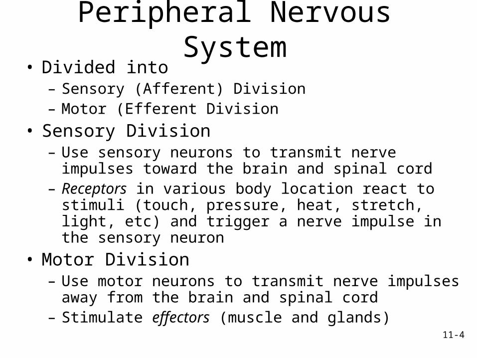
Peripheral Nervous System
• Divided into– Sensory (Afferent) Division– Motor (Efferent Division
• Sensory Division– Use sensory neurons to transmit nerve impulses
toward the brain and spinal cord– Receptors in various body location react to
stimuli (touch, pressure, heat, stretch, light, etc) and trigger a nerve impulse in the sensory neuron
• Motor Division– Use motor neurons to transmit nerve impulses
away from the brain and spinal cord– Stimulate effectors (muscle and glands)
11-5
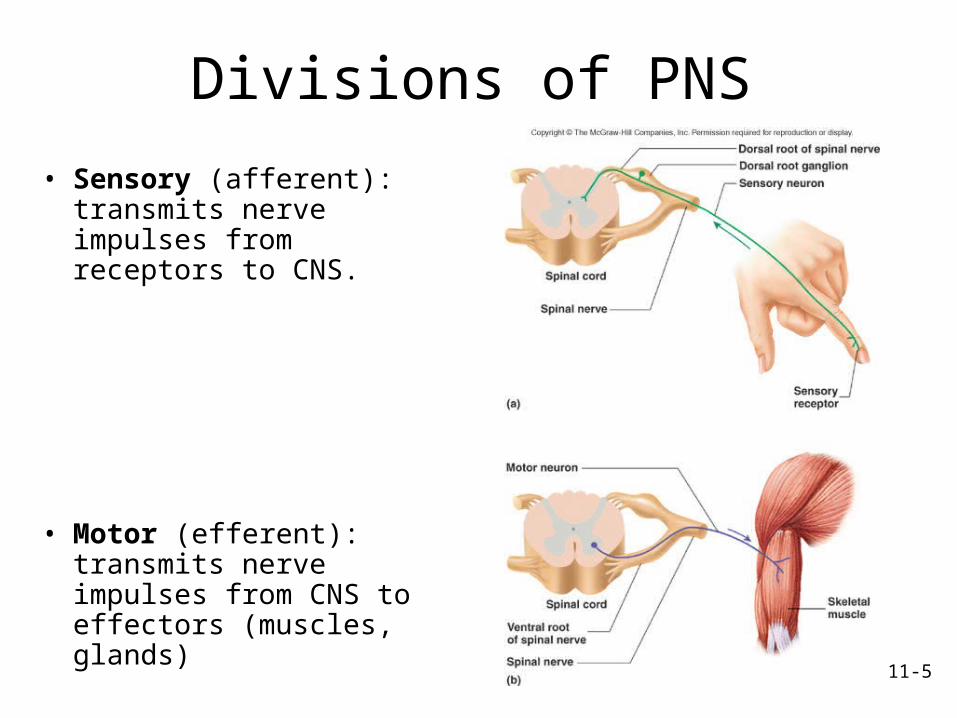
Divisions of PNS
• Sensory (afferent): transmits nerve impulses from receptors to CNS.
• Motor (efferent): transmits nerve impulses from CNS to effectors (muscles, glands)
11-6
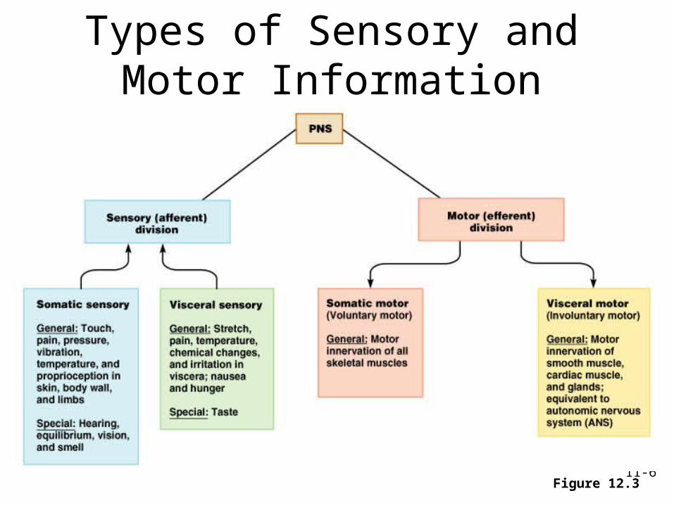
Types of Sensory and Motor Information
Figure 12.3
11-7
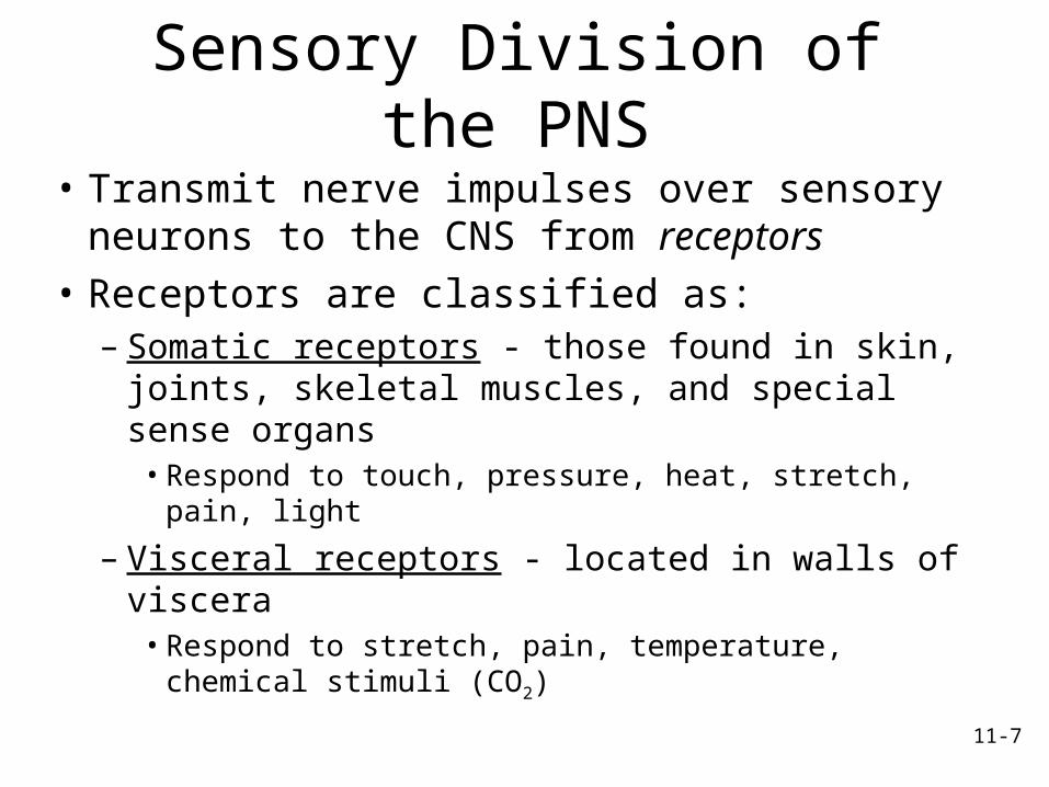
Sensory Division of the PNS
• Transmit nerve impulses over sensory neurons to the CNS from receptors
• Receptors are classified as:– Somatic receptors - those found in skin,
joints, skeletal muscles, and special sense organs• Respond to touch, pressure, heat, stretch, pain,
light
– Visceral receptors - located in walls of viscera• Respond to stretch, pain, temperature, chemical
stimuli (CO2)

11-8
Motor Division of PNS• Transmits impulses away from the CNS to effectors• Somatic nervous system: from CNS to skeletal
muscles. – Voluntary. – Single neuron system. Use anterior horn motor neurons to
stimulate the skeletal muscle
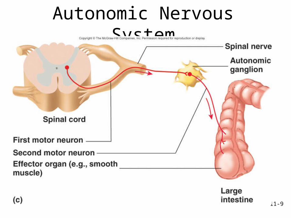
• Autonomic nervous system (ANS): from CNS to smooth muscle, cardiac muscle and certain glands. – Subconscious or involuntary control. – Two neuron system: first from CNS to ganglion; second
from ganglion to effector. – Divisions of the ANS
• Sympathetic. Prepares body for physical activity.• Parasympathetic. Regulates resting or vegetative
functions such as digesting food or emptying of the urinary bladder.
11-9
Autonomic Nervous System

11-10
Cells of Nervous System• Neurons or nerve
cells receive stimuli and transmit action potentials to other neurons or effectors– Organization
• Cell body or soma• Dendrites: input• Axons: output
• Neuroglia or glial cells– Support and protect
neurons
11-11
The Neuron• The human body contains billions of neurons
– Basic functional unit of the nervous system• Specialized cells conduct electrical impulses along the
plasma membrane – Nerve impulse: a series of action potentials
• Characteristics– Longevity – can live and function for a lifetime– Do not divide – fetal neurons lose their ability to
undergo mitosis; neural stem cells are an exception
– High metabolic rate – require abundant oxygen and glucose
11-12
Parts of the Neuron
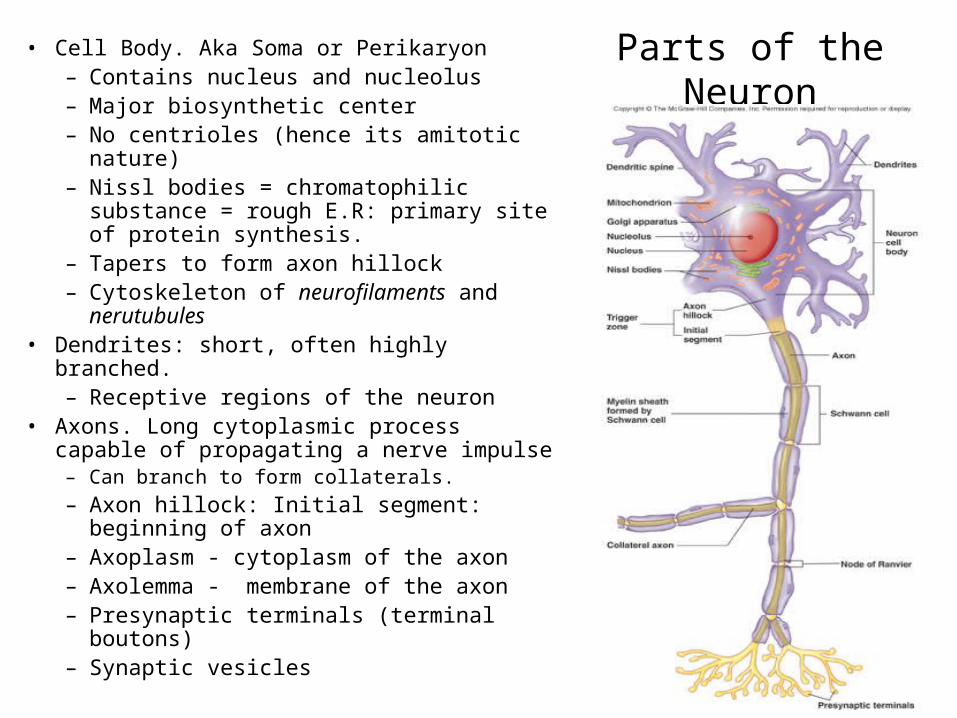
• Cell Body. Aka Soma or Perikaryon– Contains nucleus and nucleolus– Major biosynthetic center– No centrioles (hence its amitotic
nature)– Nissl bodies = chromatophilic
substance = rough E.R: primary site of protein synthesis.
– Tapers to form axon hillock– Cytoskeleton of neurofilaments and
nerutubules• Dendrites: short, often highly branched.
– Receptive regions of the neuron• Axons. Long cytoplasmic process capable
of propagating a nerve impulse– Can branch to form collaterals. – Axon hillock: Initial segment:
beginning of axon– Axoplasm - cytoplasm of the axon– Axolemma - membrane of the axon– Presynaptic terminals (terminal
boutons)– Synaptic vesicles

11-13
Structure of a Neuron• Dendrites: short, often highly
branched.– Receptive regions of the neuron
• Axons. Long cytoplasmic process capable of propagating a nerve impulse– Axoplasm - cytoplasm of the axon– Axolemma - membrane of the axon– Axon hillock: Initial segment:
beginning of axon – Can branch to form collaterals. – Presynaptic terminals (terminal
boutons)– Telodendria - the fine terminal
branches of the main axon; end at– Synaptic terminals; aka synaptic
knob• Contain vesicles filled with neuro-
transmitter (NT)

11-14
Axoplasmic Transport• Anterograde:
– Axoplasm moved from cell body toward terminals. – Supply materials for growth, repair, renewal. – Can move cytoskeletal proteins, organelles away
from cell body toward axon terminals.
• Retrograde– Away from axonal terminal toward the cell body– Damaged organelles, recycled plasma membrane,
and substances taken in by endocytosis can be transported up axon to cell body.
– Rabies and herpes virus can enter axons in damaged skin and be transported to CNS. Would include toxins such as heavy metals (the chemical, not the noise)

11-15
Types of Neurons• Functional classification
– Sensory or afferent: action potentials toward CNS
– Motor or efferent: action potentials away from CNS
– Interneurons or association neurons: within CNS from one neuron to another
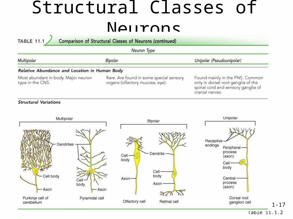
• Structural classification– Multipolar: most neurons in
CNS; motor neurons– Bipolar: sensory in retina of
the eye and nose– Unipolar: single process that
divides into two branches. Part that extends to the periphery has dendrite-like sensory receptors

11-16
Structural Classes of Neurons
Table 11.1.1
11-17
Structural Classes of Neurons
Table 11.1.2

11-18
Structural Classes of Neurons
Table 11.1.3

11-19
Neurons Classified by Function
Figure 12.11

11-20
Neuroglia of CNS: Astrocytes• Largest and most numerous
• Functions include:1. Form the blood-brain barrier
– Processes form feet that cover the surfaces of neurons and blood vessels and the pia mater. Regulate what substances reach the CNS from the blood
2. Repair damaged neural tissue3. Guide neuron development4. Control the interstitial environ-
ment.– Regulate concentration of Na, K,
and CO2
– Control the volume of blood flow through cerebral capillaries
– Absorb and recycle some NTs

11-21
Neuroglia of CNS: Ependymal Cells
• Line brain ventricles and spinal cord central canal. Specialized versions of ependymal form choroid plexuses.
• Choroid plexus – Secrete
cerebrospinal fluid. Cilia help move fluid thru the cavities of the brain.

11-22
Neuroglia of CNS: Microglia and
Oligodendrocytes
• Microglia: specialized macrophages. Respond to inflammation, phagocytize necrotic tissue, microorganisms, and foreign substances that invade the CNS.
• Oligodendrocytes: form myelin sheaths if surround axon. Single oligodendrocytes can form myelin sheaths around portions of several axons.

11-23
Neuroglia of PNS
• Schwann cells or neurolemmocytes: – Wrap around portion of only one axon to form myelin
sheath.– Wrap around many times. – As cells grow around axon, cytoplasm is squeezed out
and multiple layers of cell membrane wrap the axon. Cell membrane primarily phospholipid.
– Outer surface of Schwann cell called the neurilemma
• Satellite cells: surround neuron cell bodies in ganglia, provide support and nutrients
11-24
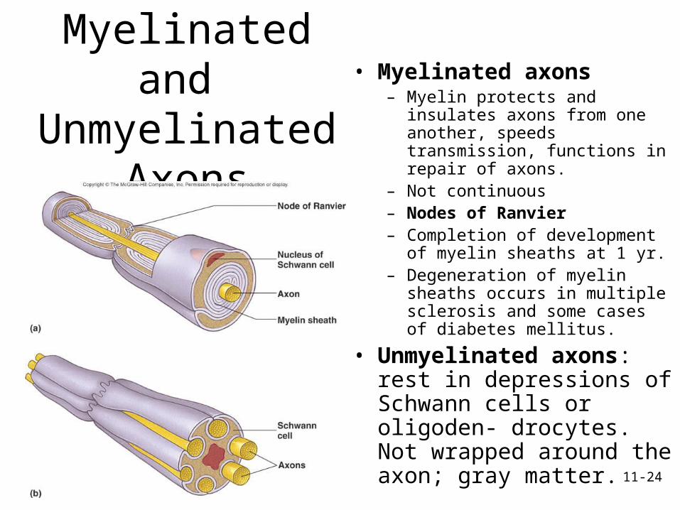
Myelinated and
Unmyelinated Axons
• Myelinated axons– Myelin protects and insulates
axons from one another, speeds transmission, functions in repair of axons.
– Not continuous– Nodes of Ranvier– Completion of development of
myelin sheaths at 1 yr.– Degeneration of myelin
sheaths occurs in multiple sclerosis and some cases of diabetes mellitus.
• Unmyelinated axons: rest in depressions of Schwann cells or oligoden- drocytes. Not wrapped around the axon; gray matter.
11-25
Regions of the Brain & SCWhite vs. Gray Matter
• White matter– Dense collections of myelinated axons. – Only axons are myelinated!!
• Gray matter– Unmyelinated axons, cell bodies, dendrites,
neuroglia. – Integrative functions
• In brain– Gray matter forms the outer layer (cortex) as
well as inner nuclei; – White matter is deeper.
• In spinal cord– White matter is outer layer,– Gray matter is deeper.

11-26
Nerve Fiber Types
• Type A: large-diameter, myelinated. Conduct at 15-120 m/s. Motor neurons supplying skeletal and most sensory neurons
• Type B: medium-diameter, lightly myelinated. Conduct at 3-15 m/s. Part of ANS
• Type C: small-diameter, unmyelinated. Conduct at 2 m/s or less. Part of ANS

11-27
The Synapse• Junction between two neurons• Site where nerve impulse in one cell cause
excitation in the next cell• Types of cells in synapse
– Presynaptic neuron - conducts impulse toward the synapse– Postsynaptic neuron - conducts impulse away from the
synapse
• Two major types of synapses– Electrical synapses - rare in the CNS– Chemical synapses - most common type in CNS
• Synapses between a neuron and its effector:– Neuromuscular junction - between a neuron and a muscle– Neuroglandular synapse - between a neuron and a gland
11-28
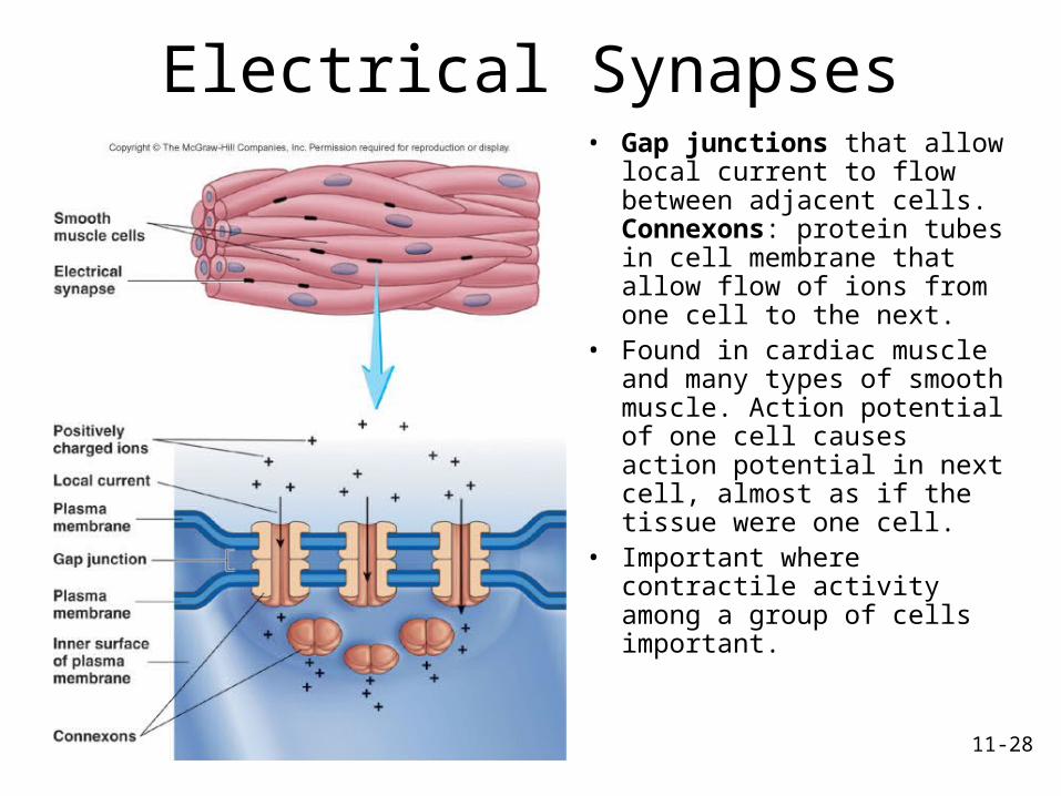
Electrical Synapses• Gap junctions that allow
local current to flow between adjacent cells. Connexons: protein tubes in cell membrane that allow flow of ions from one cell to the next.
• Found in cardiac muscle and many types of smooth muscle. Action potential of one cell causes action potential in next cell, almost as if the tissue were one cell.
• Important where contractile activity among a group of cells important.

11-29
Types of Neural Synapses

11-30
Chemical Synapse• Presynaptic bulb has secretory vesicles that contain neurotrans- mitter chemical (NT)• NT must pass across the synaptic cleft, space that separates pre- and postsynaptic membranes• Postsynaptic membrane contains receptors specific for each type of NT• Binding of NT to its receptor causes ion channels to open or close• Postsynaptic membrane is thus either stimulated or inhibited

11-31
Nerves
• Nerves - bundles of axons– If only sensory axons, called sensory nerves– If only motor axons, called motor nerves– If both sensory and motor axons, called mixed
nerves
• Connective Tissue Coverings– Endoneurium – layer of delicate connective tissue
surrounding the axon• Nerve fascicles – groups of axons bound into bundles
– Perineurium – connective tissue wrapping surrounding a nerve fascicle
– Epineurium – whole nerve is surrounded by tough fibrous sheath

11-32
Structure of a Nerve
Figure 12.16a
A. Note the similarity of a nerve to a muscle
1. Just as a muscle is a collection of muscle fibers, a nerve is a collection of nerve fibers (axons).
2. Each is broken up in smaller units known as fascicles
3. Each is covered by connective tissue:• Epimysium vs. Epineurium• Perimysium vs.
Perineurium• Endomysium vs.
Endoneurium

11-33
Neuronal Pathways and Circuits
• Organization of neurons in CNS varies in complexity– Convergent pathways: many neurons converge and synapse
with smaller number of neurons. E.g., synthesis of data in brain.
– Divergent pathways: small number of presynaptic neurons synapse with large number of postsynaptic neurons. E.g., important information can be transmitted to many parts of the brain.
Related Documents