Welcome message from author
This document is posted to help you gain knowledge. Please leave a comment to let me know what you think about it! Share it to your friends and learn new things together.
Transcript


Universiti Putra Malaysia PressSerdang • 2019
EDITORSRasedee Abdullah
Siti Suri ArshadWan Mastura Shaik Mossadeq
Arifah Abdul KadirKhor Kuan Hua
Mark Hiew Wen HanNur Indah Ahmad
Nor Yasmin Abd RahamanRozaihan MansorMazlina Mazlan
Mohd Hezmee Mohd NoorIntan Nur Fatiha Shafie
Intan Shameha Abdul RazakChen Hui Cheng
Gayathri Thevi SelvarajahMohd Sharom Salisi
Tengku Rinalfi Putra Tengku Azizan

© Universiti Putra Malaysia press 2019First Print 2019
All right reserved. No part if this publication may be reproduced or transmitted in any form or by any means, electronic or mechanical including photocopy, recording, or any information storage and retrieval system, without permission in writing from Universiti Putra Malaysia Press.
UPM Press is a member of the Malaysian Book Publishers Association (MABOPA) and a member of Majlis Penerbitan Ilmiah Malaysia (MAPIM)
Perpustakaan Negara Malaysia Cataloguing-in-Publication Data
Seminar on Veterinary Sciences (14th : 2019 : Serdang, Selangor) 14th Proceeding of the Seminar on Veterinary Sciences, Faculty of Veterinary Medicine UPM, 18-20th September 2019 / Editors: Rasedee Abdullah, Siti Suri Arshad, Wan Mastura Shaik Mossadeq, Arifah Abdul Kadir, Khor Kuan Hua, Mark Hiew Wen Han, Nur Indah Ahmad, Nor Yasmin Abd Rahaman, Rozaihan Mansor, Mazlina Mazlan, Mohd Hezmee Mohd Nor, Intan Nur Fatiha Shafie, Intan Shameha Abdul Razak, Chen Hui Cheng, Gayathri Thevi Selvarajah, Mohd Sharom Salisi, Tengku Rinalfi Putra Tengku Azizan, ISBN 978-967-344-997-2 1. Veterinary medicine--Congresses. 2. Animal health--Congresses. 3. Livestock--Diseases--Congresses 4. Government publications--Malaysia I. Rasedee Abdullah. II. Siti Suri Arshad. III. Wan Mastura Shaik Mossadeq. IV. Arifah Abdul Kadir. V. Khor, Kuan Hua. VI. Hiew, Mark Wen Han. VII. Nur Indah Ahmad. VIII. Nor Yasmin Abd. Rahaman. IX. Rozaihan Mansor. X. Mazlina Mazlan. XI. Mohd. Hezmee Mohd Nor. XII. Intan Nur Fatiha Shafie. XIII. Intan Shameha Abdul Razak. XIV. Mohd. Sharom Salisi. XVII. Tengku Rinalfi Putra Tengku Azizan. XVIII. Universiti Putra Malaysia. Faculty of Veterinary Medicine. XIX. Title. 636.089
Cover Design : Muhammad Arif Sambudin
Printed by

i
Content
Preface xiii
1 Isolation of veterinary and public health importance fungi from protected wildlife enclosures at National Zoo, Malaysia Jacqueline Meikwei Yee, Sharina Omar, Azlan Che’ Amat, Kavitha Jayaseelan & Mat Naim Ramli
1
2 Effect of halal and non-halal slaughter on bacterial contamination of poultry meat Shahira Mohd Tahir & Lokman Hakim Idris
6
3 Seroprevalence of brucellosis in cattle after the massive 2014 flood in Kelantan, Malaysia Foo Yen Ping & Siti Khairani Bejo
13
4 Effect of omega-3 fatty aicd-enriched diet on chicken serum lipid concentration, egg chemical composition, and egg yolk colour Lee Wei Zheng, Rasedee Abdullah, Goh Yong Meng & Hasliza Abu Hassim
18
5 Computed tomography characteristics of cervical and thoracolumbar intervertebral disc herniations and their association with neurological severity in dogs Chai Shu Wan, Intan Nur Fatiha Shafie & Lau Seng Fong
22
6 Accuracy of genomic prediction in swamp buffalo using deregressed breeding value estimated from purebred and crossbred offspring phenotypes Lyeonna Amber Garcia De Chavez, Mohd Shahrom Salisi, Mark Hiew Wen Han, Jonny Engkias & Azizan Maruf
30
7 Antibacterial properties of apple cider vinegar against Staphylococcus aureus and Staphylococcus pseudintermedius Nurul Zulaikha Norizal, Mazlina Mazlan & Sharina Omar
36
8 Gastrointestinal and blood parasites in African pygmy hedgehog (Atelerix albiventris) Hoe Kai Thong & Mohd Hezmee Mohd Noor
42
ii
9 Owner awareness on potentially hazardous substances to dogs in foods and household items Choong Yee Ph’ng, Goh Yong Meng & Noordin Mohamed Mustapha
45
10 Meat and eating qualities of different quail breeds Myzatul Hanis Zahiyah Yusof & Goh Yong Meng
50
11 Rectal carriage of extended-spectrum β-lactamase Escherichia coli and Klebsiella pneumoniae in shelter cats and dogs in Klang Valley, Malaysia Nur Lyana Sabri, Nur Indah Ahmad, Puteri Azaziah Megat Abdul Rani & Sharina Omar
55
12 Comparison of antimicrobial resistance between selected bacteria isolated from eggs in conventional and organic production Fatin Nabilah Idrus, Latiffah Hassan & Saleha Abdul Aziz
60
13 Antifungal properties of apple cider vinegar on Candida albicans, Candida krusei and Candida parapsilosis Afiqah Shahirah Anwar Mirza, Mazlina Mazlan & Sharina Omar
66
14 Clinical, laboratory, and histological investigation in cats naturally infected with leptospira sp. Zher Min Tan, Lau Seng Fong, Siti Khairani Bejo, Annas Salleh, & Rozanaliza Radzi
71
15 Retrospective study on aquatic animal diseases case reports and canvassing a business model using cases submitted to Aquatic Animal Health Unit, Faculty of Veterinary Medicine, Universiti Putra Malaysia Hidayatu Husna Selahuddeen, Norhariani Mohd Nor & Hassan Haji Mohd Daud
76
16 Clinical, laboratory, and histological investigation in dogs naturally infected with leptospirosis Joanne Tan Sze Yinn, Lau Seng Fong, Siti Khairani Bejo,
Khor Kuan Hua & Annas Salleh
83
17 Effect of lime juice exposure time on bacterial activity in umai (Sarawak raw fish salad) Daryl Ian Raja & Latiffah Hassan
89
18 Clinicopathologic and radiographic features in cats diagnosed with pneumonia associated with Rhodococcus equi infection Chelly Chin Sze Lee, Lau Seng Fong, Nur Indah Ahmad, Puteri Azaziah Megat Abdul Rani & Rozanaliza Radzi
95

iii
19 Detection of interdigital bacteria and fungi in cattle that used SanctuaryTM Veterinary Hoof Cover Siti Jazmina Shaik Husseinudin, Siti Zubaidah Ramanoon, Siti Khairani Bejo & Arifah Abdul Kadir
100
20 Molecular detection and risk factor analyses of enteric protozoa infection among Bornean orangutans (Pongo pygmaeus) in Sabah, Malaysia Adeline Tsen, Reuben Sunil Kumar Sharma & Norhadila Zulkifli
105
21 Bacteria contamination in boar semen following semen collection via glove hand technique Aaron Michael Anthony, Mark Hiew Wen Han, Ooi Peck Toung & Siti Khairani Bejo
106
22 Determinants of dog aggression among pet dogs in Klang Valley, Malaysia Cherilyn Mok Jia Ying, Khor Kuan Hua, Mark Hiew Wen Han & Lynn Walker
107
23 Morphology of the gastrointestinal tract of water monitor lizard (Varanus salvator) Chong Chiu Nie, Intan Shameha Abdul Razak & Azlan Che’ Amat
108
24 Occurrence of Salmonella spp. and Campylobacter spp. in ducks at Pusat Pembiakan Itik Paya Jaras, Selangor, Malaysia Wong Chin Wooi, Jalila Abu & Saleha Abdul Aziz
109
25 Histopathological evaluation of brain, kidney and gills of Red Hybrid tilapia fish (Oreochromis sp.) following Aeromonas hydrophila infection Nurul Ashila Mustapha & Md Sabri Mohd Yusoff
110
26 Morphological and molecular characterisation of Fasciola sp. in ruminants slaughtered at Shah Alam and Banting abattoirs, Selangor, Malaysia Nurhanim Rohaizad, Nur Mahiza Md Isa, Lokman Hakim Idris, & Nor Azlina Abdul Aziz
111
27 Effects of pretreatments on gelatine extracted from rabbit (Oryctolagus cuniculus) bone Tengku Syaiza Izzati Tengku Shaiful Bahril, Lokman Hakim Idris & Mohd Adha P. Rameli
112

iv
28 A retrospective study on chronic kidney disease in cats presented to University Veterinary Hospital, Universiti Putra Malaysia from 2015-2017 Tiu Kian Siang & Puteri Azaziah Megat Abdul Rani
113
29 Effects of water change on the behavior of Asian small-clawed otter (Amblonyx cinereus) in captivity at the Melaka Zoo & Night Safari, Malacca, Malaysia Ivy Ang Sye Roo & Hafandi Ahmad
114
30 Isolation and characterisation of antimicrobial resistant Escherichia coli and Enterococcus species from four village chicken farms in Hulu Langat, Selangor, Malaysia Muhammad Syazani Japri, Nur Indah Ahmad, Siti Khairani Bejo & Nik Mohd Faiz Nik Mohd Azmi
115
31 Nutritional composition of cow and goat milk kefirs Sim Juin Jia, Hasliza Abu Hassim & Mohd Hezmee Mohd Noor
116
32 Histopathological evaluation on gills of Juvenile Hybrid groupers exposed to non-ionised ammonia at different temperatures Lakshmipriya Thaigarajan & Annas Salleh
117
33 Estimated breeding value and phenotypic correlation for selected weight traits of Murrah-cross buffaloes Kimberly Jane Hugh, Mohd Shahrom Salisi, Mark Hiew Wen Han, Jonny Engkias & Azizan Mohd Maruf
118
34 Intestinal and skin microflora of Leopard gecko (Eublepharis macularius) Mohd Asrul Syafiq, Zunita Zakaria & Saleha Abdul Aziz
119
35 Efficacy of ionized water of various pHs against common bacteria present on horse wounds Afiqah Zafirah Abdul Rahman, Noraniza Mohd Adzahan & Zunita Zakaria
120
36 Correlation between ultrasonographic and morphometric testicular measurements with semen quality in bucks Banumathy Gunasegaran, Mark Hiew Wen Han & Nurhusien Yimer Degu
121
v
37 Evaluation of BCL2/BAX ratio in liver of steatotic rats supplemented with Moringa oleifera leaf extract Gan Hwee Yee, Hazilawati Hamzah, Mazlina Mazlan, Lau Seng Fong, Abdullah Misron, Mohd Rosly Shaari, Mohd Farhan Hanif Reduan & Nurul Syahirah Ahmad Sayuti
122
38 External and internal parasites of wild reticulated python Nurul Atiqah Mohd Khairun Kiang, Azlan Che’ Amat, Shaik Mohamed Amin Babjee & Reuben Sunil Kumar Sharma
123
39 Prevalence of canine filariasis in shelter dogs in Kedah, Malaysia Kartiyayini Sinathurai, Malaika Watanabe, Puteri Azaziah Megat Abdul Rani & Lau Seng Fong
124
40 Retrospective study on classical swine fever and Aujeszky’s disease serological status of blood samples submitted to University Veterinary Hospital, Faculty of Veterinary Medicine, University Putra Malaysia Ang Dian Wen, Ooi Peck Toung & Low Suet Ee
125
41 Brucellosis seroprevalence among goats in Universiti Putra Malaysia foster farms Syazwani Ahmad, Abd Wahid Haron & Siti Khairani Bejo
126
42 Comparative economic performance between swamp and cross-bred buffaloes in the Buffalo Breeding and Research Centre, Telupid, Sabah, Malaysia Nurain Syahida Mohd Dali, Norhariani Mohd Nor & Mohd Zamri Saad
127
43 Molecular detection and nucleotide sequence analysis of Porcine Circovirus Type 3 in post-weaned pigs in Peninsular Malaysia Keerati Opaskornkul, Siti Suri Arshad, Ooi Peck Toung, Tan Chew Yee & Lee Chee Yien
128
44 Pathogenicity and immunogenicity of infectious bursal disease virus attenuated in BGM-70 cell line in commercial broiler chickens Lim Yee Ning & Mohd Hair Bejo
129
45 Seroprevalence of Toxoplasma gondii in cattle of farms in Hulu Langat, Selangor, Malaysia Iqmal Syahmi Adam, Sharifah Salmah Syed Hussain
Siti Zubaidah Ramanoon & Juriah Kamaludeen
131

vi
46 Determination of nutritional composition of sago worm (Rhynchophorus schach) and mealworm (Tenebrio molitor) larvae Mary Loria Kong Ming, Hafandi Ahmad & Hasliza Abu Hassim
132
47 Evaluation of Newcastle disease virus strain AF2240 as an oncolytic agent for canine osteosarcoma cells in vitro Nagaswitra Manukaran, Gayathri Thevi Selvarajah, Chia Suet Lin, Leong Sze Wei & Ng Shing Wei
133
48 Molecular detection of West Nile virus in bats Selvi Viji, Nor Yasmin Abd Rahaman, Siti Suri Arshad & Nur Ain Najwa Mohd Yuseri
134
49 Economic analysis on rearing interventions at the Buffalo Breeding and Research Centre, Sabah, Malaysia Norafiza Roslan, Norhariani Mohd Nor & Mohd Zamri Saad
135
50 Scrotal circumference and semen evaluation of breeding and non-breeding Damara rams Intan Nur Ain Sarwan, Abd Wahid Haron, Mark Hiew Wen Han & Nurhusien Yimer Degu
136
51 Ultrastructure and functional significance of swiftlet podocytes Lim Su Xian & Tengku Azmi Tengku Ibrahim
137
52 Morphology and functional histology of the reticulated python (Malayopython reticulatus) respiratory system Joash Shane Benedict, Intan Shameha Abdul Razak & Azlan Che’ Amat
138
53 Association between udder morphology, teat-end lesions and intramammary infections in dairy cows of University Putra Malaysia foster farms Kesavan Sivagiganesan, Rozaihan Mansor & Sharina Omar
139
54 Retrospective study on neoplasia in golden retrievers presented to the university veterinary hospital, Universiti Putra Malaysia Chong Hui Min, Gayathri Thevi Selvarajah, Goh Yong Meng
140
55 Comparison of helminths and ectoparasites infestation in ICR mice from two animal facilities Losheni Subramaniam, Nur Fazila Saulol Hamid & Nur Mahiza Md Isa
141
vii
56 Molecular serotyping and phylogenetic analysis of Haemophilus parasuis in porcine samples from Penang, Selangor, and Johore, Malaysia Tan Yi Jing & Ooi Peck Toung
142
57 Pathogenicity and immunogenicity of fowl adenovirus attenuated in SPF chicken embryonated eggs in commercial broiler chickens Teoh Kah Ying & Mohd Hair Bejo
143
58 Occurrence of Salmonella and Campylobacter in pigeons in selected areas of Selangor, Malaysia Siti Farahani Mohd Sederi, Jalila Abu & Saleha Abdul Aziz
144
59 Phytochemical and nutritional composition analysis of Malaysian stingless bee propolis Nadiah Syakirah Abu Shukor, Abdul Aziz Saharee & Hasliza Abu Hassim
145
60 Fish quality and nutritional properties of Indian mackerel (Rastrelliger spp.) and tilapia (Oreochromis spp.) sold in wet markets and supermarkets Nur Marini Awanis Kamaruddin, Hasliza Abu Hassim, Hassan Haji Mohd Daud & Mohd Fuad Matori
146
61 Molecular and pathogenicity study of infectious bronchitis virus (Gammacoronavirus) in Japanese quail (Coturnix coturnix japonica) Nur Fadhilah Abd Shukor, Mohd Hezmee Mohd Noor, Lokman Hakim Idris & Nor Yasmin Abd Rahaman
147
62 Screening for West Nile virus in mosquitoes from Kuala Gula Bird Sanctuary, Perak, Malaysia Maizatul Amira Janil, Nor Yasmin Abd Rahaman, Natasha Jaafar Ali & Nur Mahiza Md Isa
148
63 Pathological evaluation of Oreochromis sp. challenged with Aeromonas hydrophilla following application of effective microorganisms Muhammad Afnan Muhamad Munim & Md Sabri Mohd Yusoff
149
64 Effect of environmental enrichment on locomotion level of captive white-handed gibbons Nor Liyana Mohtar, Tengku Rinalfi Putra Tengku Azizan & Hafandi Ahmad
150

viii
65 Rapid and sensitive droplet digital polymerase chain reaction method in the quantification of Orf virus from clinical specimens Cassandra Alexius, Mohd Azmi Mohd Lila, Jamilu Abubakar Bala, Krishnan Nair Balakrishnan & Noordin Mohamed Mustapha
151
66 Retrospective study on common health problems and pathological changes in ruminants presented to Post-Mortem Laboratory, Faculty of Veterinary Medicine, Universiti Putra Malaysia Maisarah Zakaria & Annas Salleh
152
67 Molecular characterisation and phylogenetic analysis of porcine group a rotavirus from selected swine farm in Selangor, Malaysia Yong Ee-Leen, Ooi Peck Toung & Kenny Voon Gah Leong
153
68 Retrospective study on clinical management involving post-partum diseases in ruminants at selected farms in Klang Valley, Selangor, Malaysia Nuriza Tukiran, Faez Firdaus Jesse Abdullah & Mohd Azmi Mohd Lila
154
69 Effect of storage duration on physical and nutritional composition of soy waste Amiera Mohd Halimi, Hasliza Abu Hassim,
Ahmad Afifi Abdul Ghani & Hafandi Ahmad
155
70 Viability of commercial Newcastle disease live vaccines using various water preparation methods Yim Yan Nei, Nik Mohd Faiz Nik Mohd Azmi, Nor Yasmin Abd Rahaman & Yong Chiun Khang
156
71 In vitro evaluation of antibacterial activities of betel (Piper betle) against Streptococcus agalactiae and Enterococcus faecium Siti Fatima Az Zahra Abdul Rahim, Hassan Haji Mohd Daud, Hasliza Abu Hassim & Mohd Fuad Matori
157
72 Helminths and ectoparasite infestations in Sprague-Dawley rats from two animal facilities Nur Kuain Hamka, Nur Fazila Saulol Hamid & Nur Mahiza Md Isa
158
73 Seroprevalance of Besnoitia besnoiti in cattle of farms in Hulu Langat, Selangor, Malaysia Mohamad Hafizuddin Mohd Hamzah, Nur Azlina Abdul Aziz & Sharifah Salmah Syed Hussain
159

ix
74 Effect of diarylpentanoid analogues of curcumin on a canine prostate carcinoma cell line Chrisann Po Wanxin, Gayathri Thevi Selvarajah, Leong Sze Wei, Chia Suet Lin & Ng Shing Wei
160
75 Assessing biosecurity practices in small-scale Universiti Putra Malaysia ruminant foster farms Nur Fariza Abdul Aziz & Abdul Aziz Saharee
161
76 Retrospective study on canine babesiosis at University Veterinary Hospital, Universiti Putra Malaysia for 2010 - 2017 Muhammad Imran Mohd Ramdzan, Puteri Azaziah Megat Abdul Rani & Malaika Watanabe
162
77 Isolation of fungi from animal enclosures at the National Wildlife Rescue Centre, Sungkai, Perak, Malaysia Fathiah ‘Aqilah Jalaludin, Azlan Che’ Amat & Sharina Omar
163
78 Serum and plasma cardiac troponin I concentrations in cats with and without heart disease Lean Chyng Mun, Khor Kuan Hua & Rasedee Abdullah
164
79 Prevalence of helminths and coccidial infections in selected turkey farms in Johore, Malaysia Nor Afifah Idris, Lokman Hakim Idris, Nur Mahiza Md Isa & Shaik Mohamed Amin Babjee
165
80 Seroprevalence of Neospora caninum among cattle in Hulu Langat, Selangor, Malaysia Muhamad Hafizuddin Abdul Kadir, Sharifah Salmah Syed Hussain, Rozaihan Mansor & Siti Zubaidah Ramanoon
166
81 Influence of olfactory environmental enrichment on temporal measurement of behaviour of captive Malayan tigers (Panthera tigiris jacksoni) Norfakhrina Hanim Badruddin, Tengku Rinalfi Putra Tengku Azizan & Azlan Che' Amat
167
82 Morphological changes in liver of suckermouth catfish (Hypostomus plecostomus) as a bioindicator of pollution in Langat river, Kajang, Selangor, Malaysia Fakhri Izzat Zainudin, Intan Shameha Abdul Razak & Mohd Fuad Matori
168
x
83 Molecular prevalence of babesiosis and ehrlichiosis in shelter dogs in Selangor, Malaysia Zarith Nabilla Zulkeflle, Nor Azlina Abdul Aziz & Puteri Azaziah Megat Abdul Rani
169
84 Microbiological quality of raw sushi from sushi bars and sushi retailers Nur Yasirah Che Alias, Saleha Abdul Aziz & Siti Khairani Bejo
170
85 Udder health management practices and bulk milk somatic cell count in dairy cattle of Universiti Putra Malaysia foster farms Mariam Nadhirah Azlan, Rozaihan Mansor, & Sharifah Salmah Syed Hussain
171
86 Occurrence of external and gastrointestinal parasites in three commercial meat-farmed rabbits in Selangor, Malaysia Nurul Nadiah Mohamad Radzi, Azlan Che’ Amat & Shaik Mohamed Amin Babjee
172
87 Association between teat-end condition, udder cleanliness, and bovine subclinical mastitis Ili Liyana Kalam & Rozaihan Mansor
173
88 Pathogenicity of Aspergillus fumigatus isolate from a Malaysian outbreak experimentally inoculated in commercial chicken Siti Nor Aishah Baharon, Nik Mohd Faiz Nik Mohd Azmi, Mohd Hair Bejo, Sharina Omar & Mazlina Mazlan
174
89 Molecular prevalence of Babesia spp. and Ehrlichia canis in shelter dogs of Northern region, Peninsular Malaysia Ain Atiffah Jefri, Malaika Watanabe & Nor Azlina Abdul Aziz
175
90 Isolation and identification of bacteria from the gut and hepatopancreas of Asian green mussels (Perna viridis, Linnaeus, 1758) from wet markets in Selangor, Malaysia Muhammad Amir Syahir Dollah, Hassan Haji Mohd Daud, Saleha Abdul Aziz, & Sharina Omar
176
91 Moringa oleifera leaf extract enhances BCL-2 protein expression in rats fed with high cholesterol diet and alcohol Quek Jia Le & Hazilawati Hamzah
177
92 Pathogenicity and immunogenicity of fowl adenovirus, attenuated in chicken embryo liver cells, in commercial broiler chickens Tan Goh Jia Ying & Mohd Hair Bejo
178

xi
93 Prevalence of liver fluke in buffaloes of a farm in Taiping, Perak, Malaysia ‘Iffah Laila Fadhlul Hadi, Nur Mahiza Md Isa, Lokman Hakim Idris & Nor Azlina Abdul Aziz
179
94 Dog population dynamics in the rabies-free area of Seri Serdang, Selangor, Malaysia Wan Nur Shaqeena Wan Abdul Razak, Noordin Mohamed Mustapha & Mazlina Mazlan
180
95 Dog population dynamics in rabies immune belt area of Tumpat, Kelantan, Malaysia Daruni Eh Win, Noordin Mohamed Mustapha & Mazlina Mazlan
181
96 Molecular detection of Bartonella spp. in blood and fleas (Ctenocephalides felis) of shelter cats Tengku Syed Muhammad Syahmi Tengku Syed Mansor, Farina Mustaffa Kamal, Malaika Watanabe & Nur Indah Ahmad
182
97 Retrospective study on clinical management of respiratory diseases in ruminants from selected farms in Klang Valley, Malaysia Nur Hanim Abdul Mubin & Faez Firdaus Jesse Abdullah
183
98 Prophylactic effects of garlic essential oil on Aeromonas hydrophila infection in Red Hybrid tilapia under heat stress Nesea Janoh & Md Sabri Mohd Yusoff
184
99 Stress and encephalographic changes in cats exposed to dogs in a clinical setting Delna Mazda & Goh Yong Meng
185
100 Preliminary study on nutrition and digestibility of wild Asian elephants at Royal Belum State Park, Perak, Malaysia Hannah Hayati Mohd Sharifuddin, Tengku Rinalfi Putra Tengku Azizan & Hasliza Abu Hassim
186
101 Molecular detection of zoonotic enteric protozoa infection in captive carnivores in Peninsular Malaysia Phoebe Simon, Reuben Sunil Kumar Sharma, & Norhadila Zulkifli
187
102 Molecular detection of Bartonella spp. in blood and saliva of shelter cats Raja Aiman Hakim Raja Mahmood, Farina Mustaffa Kamal, Malaika Watanabe & Nur Indah Ahmad
188

xii
103 Retrospective study on clinical management of mastitis in ruminants from selected farms in Klang Valley, Malaysia Sim Ee Ling & Faez Firdaus Jesse Abdullah
189
Author Index 191
xiii
Preface Although most veterinary curriculums are now practicing student-centred learning, dissemination of information in the curriculum still relies heavily on lectures and laboratory instructions. Even in rounds, rotations, and hospital and field case management and practices, the teacher often takes centre stage. Although there are serious attempts to implement self-learning, it will take time before it will be the main mode of education in the Faculty.
Classroom instructions are most often based on accepted theories and concepts, and ideal situations. Unfortunately, the practice of veterinary medicine is far from ideal, and frequently requires innovations. Independent research in veterinary science and medicine is one of the surest ways for students to experience the uncertainties of real-life situations. The experience in the conduct of research in priceless, and an important contributor to the development of knowledgeable and competent veterinarians. We are fortunate to have a curriculum that allows for students to conduct independent research. We also are fortunate to have a means to publicise findings from these research projects through our annual proceedings. With the grace of God, this is our 14th Proceedings of the Seminar on Veterinary Sciences.
The editors wish to congratulate all students and supervisors for preparing the abstracts, and the Faculty for full support to the publication of the Proceedings. Editors Rasedee Abdullah Siti Suri Arshad Wan Mastura Shaik Mossadeq Arifah Abdul Kadir Khor Kuan Hua Mark Hiew Wen Han Nur Indah Ahmad Nor Yasmin Abd Rahaman Rozaihan Mansor Mazlina Mazlan Mohd Hezmee Mohd Noor Intan Nur Fatiha Shafie Intan Shameha Abdul Razak Chen Hui Cheng Gayathri Thevi Selvarajah Mohd Sharom Salisi Tengku Rinalfi Putra Tengku Azizan

xiv

1
13th Proceedings of the Seminar on Veterinary Sciences, 18 – 20 September 2018
ISOLATION OF VETERINARY AND PUBLIC HEALTH IMPORTANCE FUNGI FROM PROTECTED WILDLIFE
ENCLOSURES AT NATIONAL ZOO, MALAYSIA
Jacqueline Meikwei Yee, 1*Sharina Omar, 2,3Azlan Che’ Amat, 4Kavitha Jayaseelan & 4Mat Naim Ramli
1Department of Veterinary Pathology and Microbiology 2Department of Veterinary Clinical Studies
3Research Centre for Wildlife Faculty of Veterinary Medicine
Universiti Putra Malaysia, 43400 UPM Serdang, Selangor, Malaysia 4Zoo Negara, Ulu Kelang, 68000 Ampang, Selangor, Malaysia
*Correspondence: [email protected]
ABSTRACT
Fungi are the least considered in the general list of pathogens affecting both animals and humans. However, the establishment of baseline fungal composition in captive wildlife is necessary for the understanding of their overall role in wildlife health. This study isolated and identified fungi of veterinary and public health importance from the enclosures of Orangutan, Malayan tiger, and Malayan sun bear at the National Zoo, Malaysia. The all night quarters, exercise yards, and enrichments of these wildlife species were subjected to cross-sectional study sampling method. Soil (n=6) and swab (n=25) samples were collected and plated onto Sabouraud dextrose agar (SDA). Air samples (n=25) from the night quarters and exercise yards were collected directly on exposed SDA plates. Nine species of fungi were identified and confirmed as Aspergillus niger, Aspergillus flavus, Penicillium spp., Trichophyton spp., Paecilomyces sp., Conidiobolus coronatus, Cladosporium spp., Fonsecea spp., and yeast. Penicillium and Trichophyton spp. were the most frequent fungi isolated at 60 and 27% of all samples, respectively. Fungi were mostly isolated from air (55%), followed by enrichments (43%), and soil (2%). Most of these isolates were reported to be of public health significance and can cause diseases in animals such as localised skin lesions, ear infections, allergic responses, and respiratory lesions. The study shows that fungi of both veterinary and public health importance were present in the wildlife enclosures and environments of the National Zoo, Malaysia. Keywords: fungi, environmental, wildlife, public health, zoonotic
2
INTRODUCTION Fungi are often overlooked as a potential cause of diseases. However, fungal infections are on the rise and becoming an important cause of emerging diseases in wildlife (Rothenburger, 2017). Zoonotic fungi can be naturally transmitted between animals and humans, and these infections are of public health concerns. Fungi are ubiquitous and reproduce by means of spores that can be inhaled or transmitted via direct contact, especially the skin. Fungal infections usually first occur in the lungs and on the skin (Revankar, 2018). These pathogenic fungi cause allergic responses, and skin and mucosal infections. In certain instances, the infections are fatal. Fungi are extremely persistent in the environment, and therefore isolating and identifying those of veterinary and public health importance from protected wildlife enclosures could for the formulation of management plans to minimise the increase and spread of fungal diseases (Kolbert, 2016). Thus, this study was carried out to determine the type of fungi present in the enclosures and the environments of selected endangered wildlife species. MATERIALS AND METHODS Location and subjects This study was conducted at the National Zoo, Malaysia, in enclosures of the Malayan tiger, Malayan sun bear, and Orangutan. The air, soil, and swab samples were obtained from the exercise yards and their environment were subjected to sampling by the cross-sectional study method. Isolation and identification Air samples (n=25) were collected by directly exposing sterile Sabouraud dextrose agar (SDA) plates in the night quarters and exercise yards for 15 min. Swab samples (n=25) from the wall, floor and enrichments were taken from each night enclosure and plated on SDA plates. Soil (n=6) and swab samples were collected from the exercise yards, diluted 10-fold and 0.1 µL from the third dilution was inoculated onto SDA medium. All plates were incubated for up to 4 weeks at 20 and 25°C. Isolation and microscopic identification of fungi were done by conventional methods based on colony morphological characteristics such as colour, texture, special features, and reverse plate characteristics. A clear cellophane tape of a direct impression of the fungal colony was placed on a glass slide with a drop of Lacto Phenol Blue (LCB). Macroscopic and microscopic evaluation of the fungal culture was performed using the key taxonomy reference (Ellis et al., 2007) and references from International Society for Human and Animal Mycology database (ISHAM).
3
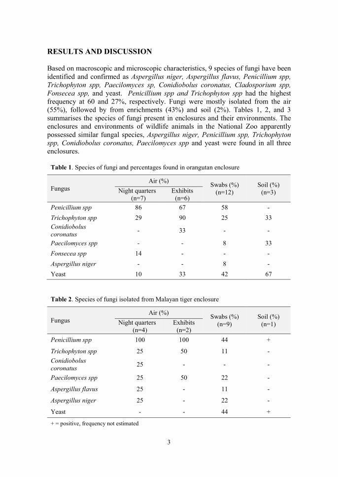
RESULTS AND DISCUSSION Based on macroscopic and microscopic characteristics, 9 species of fungi have been identified and confirmed as Aspergillus niger, Aspergillus flavus, Penicillium spp, Trichophyton spp, Paecilomyces sp, Conidiobolus coronatus, Cladosporium spp, Fonsecea spp, and yeast. Penicillium spp and Trichophyton spp had the highest frequency at 60 and 27%, respectively. Fungi were mostly isolated from the air (55%), followed by from enrichments (43%) and soil (2%). Tables 1, 2, and 3 summarises the species of fungi present in enclosures and their environments. The enclosures and environments of wildlife animals in the National Zoo apparently possessed similar fungal species, Aspergillus niger, Penicillium spp, Trichophyton spp, Conidiobolus coronatus, Paecilomyces spp and yeast were found in all three enclosures.
Table 1. Species of fungi and percentages found in orangutan enclosure
Fungus Air (%)
Swabs (%) (n=12)
Soil (%) (n=3) Night quarters
(n=7) Exhibits
(n=6)
Penicillium spp 86 67 58 -
Trichophyton spp 29 90 25 33 Conidiobolus coronatus
- 33 - -
Paecilomyces spp - - 8 33
Fonsecea spp 14 - - -
Aspergillus niger - - 8 -
Yeast 10 33 42 67
Table 2. Species of fungi isolated from Malayan tiger enclosure
Fungus Air (%)
Swabs (%) (n=9)
Soil (%) (n=1) Night quarters
(n=4) Exhibits
(n=2)
Penicillium spp 100 100 44 +
Trichophyton spp 25 50 11 -
Conidiobolus coronatus
25 - - -
Paecilomyces spp 25 50 22 -
Aspergillus flavus 25 - 11 -
Aspergillus niger 25 - 22 -
Yeast - - 44 +
+ = positive, frequency not estimated

4
Table 3. Species of fungi isolated from Malayan sun bear enclosure
Fungus Air (%)
Swabs (%) (n=12)
Soil (%) (n=3) Night quarters
(n=7) Exhibits
(n=6)
Penicillium spp 50 100 25 -
Trichophyton spp - 50 25 -
Conidiobolus coronatus
- 25 - -
Paecilomyces spp - 25 - -
Aspergillus flavus - - 25 -
Aspergillus niger - 25 25 -
Yeast - - 25 100
Wildlife populations worldwide are under increasing threat from the environmental changes including climate and loss of habitat loss that could cause stress and threaten their survival (Hing et al, 2016). Animals under stress are immunocompromised and prone to infections, such as from fungi, that can be fatal. Thus, it is imperative that zoos are mindful of good management practices to minimise stress to safeguard the welfare of captive wildlife animals. CONCLUSION Fungi of both veterinary and public health importance were present in enclosures and and environment of captive endangered wildlife animals. The infections threaten the health of animals, zoo personnel, and the public. Thus, all animal enclosures must be regularly monitored and precautions taken to ensure that animals and people are infected by emerging fungal diseases. REFERENCES Ellis D, Davis S, Alexiou H, Handke R, Bartley R (2007). Descriptions of medical
fungi, 2nd Edition, Adelaide Medical Centre for Women and Children, North Adelaide.
Hing S, Edward J, Naraya EJ, Thompson RCA, Godfrey SS (2016). The relationship between physiological stress and wildlife disease: consequences for health and conservation. Wildlife Research, 43: 51-60.
ISHAM (International Society for Human and Animal Mycology). Mycological links. https://www.isham.org/mycology-resources/mycological-links (Accessed on 24 August 2019).
5
Kolbert E (2016). What’s Causing Deadly Outbreaks of Fungal Diseases in World’s Wildlife. https://e360.yale.edu/features/whats_causing_deadly_outbreaks_of_fungal_diseases_in_worlds_wildlife (Accessed on 24 August 2019).
Revankar SG (2018). Overview of Fungal Infections. Online Merck Manual https://www.msdmanuals.com/professional/infectious-diseases/fungi/overview-of-fungal-infections (Accessed on 24 August 2019).
Rothenburger J (2017). Emerging fungal diseases threaten wildlife. The Western producer. https://www.producer.com/2017/06/emerging-fungal-diseases-threaten-wildlife/ (Accessed on 26 August 2019).
6
14th Proceedings of the Seminar on Veterinary Sciences, 18 – 20 September 2018
EFFECT OF HALAL AND NON-HALAL SLAUGHTER ON BACTERIAL CONTAMINATION OF POULTRY MEAT
Shahira Mohd Tahir & 1*Lokman Hakim Idris
1Department of Veterinary Preclinical Sciences Faculty of Veterinary Medicine
Universiti Putra Malaysia, 43400 UPM Serdang, Selangor, Malaysia *Correspondence: [email protected]
ABSTRACT
The minimal amount of residual blood in the meat from chickens that were slaughtered using the halal method may lead to a longer shelf life due to the presence of low number of microorganisms in the meat. However, the non-halal slaughter method could lead to high residual blood that can lower the wholesomeness of meat. Thus, this study aims to determine the effect of halal and non-halal slaughter method on bacterial contamination of poultry meat. Ten village chickens of the same age weighing 0.9 to 1 kg were selected for the study. Five chickens were slaughtered using the halal method and 5 slaughtered using the non-halal method by cutting only one side of the jugular vein and carotid artery. The bleeding time, death time, volume of blood loss, microbial count from standard plate count (SPC) and coliform plate count (CPC) were determined immediately at slaughter and post-slaughter. There was no significant difference (p>0.05) in CPC among slaughter methods. The bleeding time, death time, and SPC for non-halal-slaughtered were significantly (p<0.05) longer and higher than the halal-slaughtered chicken. The volume of blood loss was significantly (p<0.05) lower from non-halal-slaughtered chickens. Cumulatively, the data suggest that non-halal-slaughtered chicken meat contain more residual blood that can cause increase in bacterial count. Keywords: village chicken, halal slaughter, microbial count, residual blood, chicken meat
INTRODUCTION The poultry industry continues to grow at a rapid rate of 10 to 15% annually with an estimated 37% attributed to the sale of chicken meat (Darshana et al., 2014).
Chicken meat generally refers to either the whole carcass or parts of the carcass or boned out meat of the Gallus gallus species. Slaughtering is the most crucial stage in the transformation of an animal into meat fit for human consumption (Nakyinsige et al., 2013). Evacuation of a significant amount of blood from the carcass can be achieved through proper halal slaughtering. Post-slaughter residual blood is often associated with the meaty flavour and decreased shelf life due to bacterial
7
contamination (Alvarado et al., 2007). Meat and blood are perfect mediums for bacterial growth because of the high moisture content and presence of minerals, vitamins, nitrogenous compounds such as essential amino acids and proteins as well as other growth factors (Darshana et al., 2014). Common pathogenic microorganisms that can contaminate poultry meat are Salmonella, Campylobacter, Staphylococcus aureus, Eschericia coli and Listeria spp.
Halal slaughtering involves severing of trachea, oesophagus, carotid arteries, and jugular veins (JAKIM, 2011). A minimal amount of residual blood in the meat of chickens that were slaughtered using the halal method may lead to a longer shelf life as there is a low number of microorganisms in the flesh. In contrast, the non-halal slaughter method could lead to high residual blood that can lower the wholesomeness of chicken meat.
This study aims to determine the effect of halal to non-halal slaughter method on bacterial contamination in Malaysian village chicken (Gallus gallus domesticus) meat. The chickens used in this study were a crossbred between the Red Jungle fowl and mixed exotic domestic breeds (Lokman et al., 2011). MATERIALS AND METHODS Malaysian Village Chicken Ten village chickens of about 60 days of age were randomly selected from a farm. The chickens were fed commercial poultry pellet but fasted, but free access to water, for 12 h prior to experimentation. Sampling The chickens were weighed prior and divided into the halal slaughter (Group A, n=5) and non-halal slaughter (Group B, n=5) groups. The halal slaughter method was carried out by severing the jugular vein, carotid artery, and trachea (Department of Islamic Development Malaysia). The non-halal method was conducted by cutting only one side of jugular vein and carotid artery. The bleeding time (min), death time (min) and volume of blood loss (mL) during the slaughtering process of each chicken were recorded. The duration of time taken by each chicken to die after slaughter was determined by performing the pupillary and corneal reflex using a feather. Absence of these reflexes and movement at post slaughter were taken as indicators of death. De-feathering and evisceration process on these chickens were performed manually. Three samples (25 g each) of the breast meat (pectoralis major) were used for analyses. The breast muscle was aseptically sampled every 2 h at 0, 2 and 4 h post-slaughter. The 2 and 4 h samples were placed in sterile petri dishes and left exposed to room temperature. Bacterial contamination of the meat samples was determined using the standard plate count and the coliform plate count. Statistical analysis Independent t-test was used to analyse the bleeding time, death time, blood loss and SPC results. The CPC result was analysed using Mann-Whitney U test. The number

8
of microbes (cfu/g) was converted to log cfu/g prior to statistical analysis. Data are presented as mean ± SEM. Significance level was set at p<0.05. All data were analysed using IBM SPSS version 23. RESULTS AND DISCUSSION Bleeding time The bleeding time for the halal method was significantly (P<0.05) faster than the non- halal method (Table 1). The shorter bleeding and death time achieved by cutting both carotid arteries and jugular veins in the halal method were consistent with an earlier study featured in the Compassion in World Farming Trust (2005).
Table 1: Bleeding time in chicken after halal and non halal slaughter.
Slaughter method Bleeding time (min) p-value
Halal 1.40 ± 0 .55 0.000
Non-halal 3.60 ± 0 .55
Values (n=10) are mean ±SEM; independent t-test; statistically significant when p<0.05
Blood Loss Results in Table 2 showed that the volume of blood loss in non-halal-slaughtered were lesser than in halal-slaughtered chickens (p<0.05). Chickens that have an average live weight of 1 kg lost around 25% of blood when slaughtered using the halal method. In contrast, the average blood loss for the non-halal slaughtering method was lesser (7%), which in agreement with Sayda et al. (2011).
Table 2: Blood loss from chickens after halal and non-halal slaughter.
Slaughter method Blood loss (mL) p-value
Halal 24.90 ± 5.57
0.000 Non-halal 6.86 ± 2.59
Values (n=10) are mean± SEM; independent t-test; statistically significant when p<0.05
Death time Compared to the non-halal slaughtered chicken, the death time was significantly (p<0.05) shorter for halal-slaughtered chickens (Table 3), which is in agreement with Gregory and Wotton (1986). It is clear the quickest method for bleeding and death is by cutting both the carotid arteries and jugular veins. According to an article by the Meat Research Institute 1984 (as cited in Compassion in World Farming Trust, 2005),
9
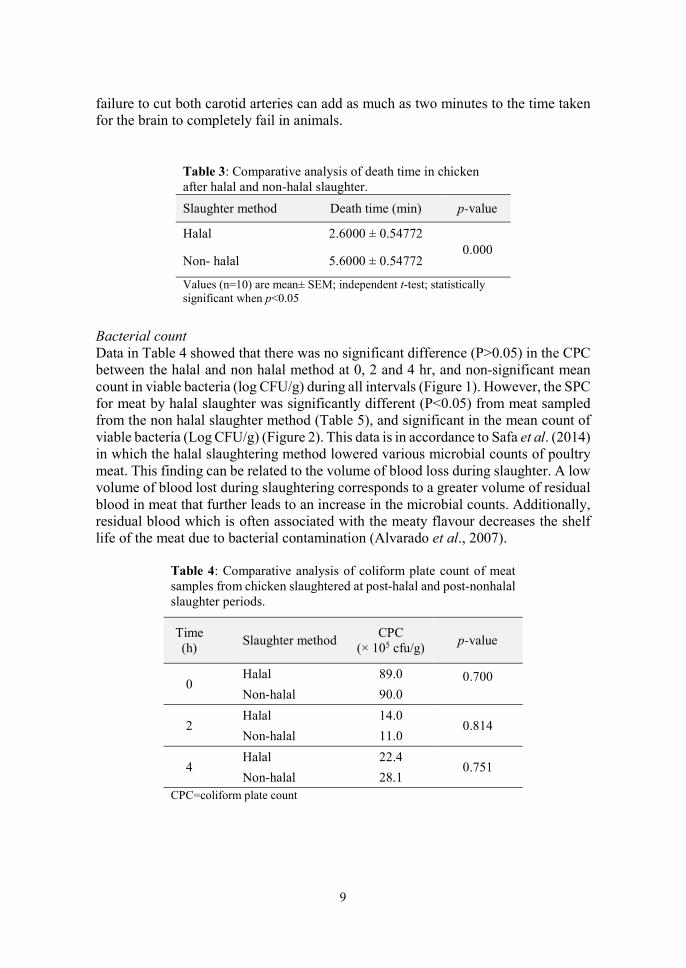
failure to cut both carotid arteries can add as much as two minutes to the time taken for the brain to completely fail in animals.
Table 3: Comparative analysis of death time in chicken after halal and non-halal slaughter.
Slaughter method Death time (min) p-value
Halal 2.6000 ± 0.54772 0.000
Non- halal 5.6000 ± 0.54772
Values (n=10) are mean± SEM; independent t-test; statistically significant when p<0.05
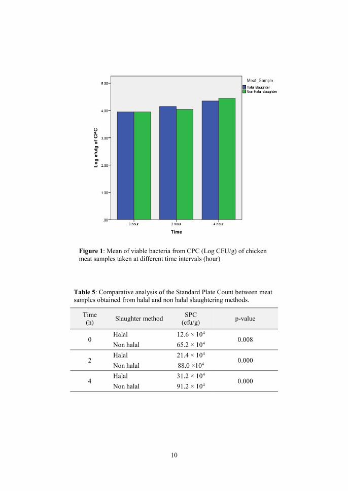
Bacterial count Data in Table 4 showed that there was no significant difference (P>0.05) in the CPC between the halal and non halal method at 0, 2 and 4 hr, and non-significant mean count in viable bacteria (log CFU/g) during all intervals (Figure 1). However, the SPC for meat by halal slaughter was significantly different (P<0.05) from meat sampled from the non halal slaughter method (Table 5), and significant in the mean count of viable bacteria (Log CFU/g) (Figure 2). This data is in accordance to Safa et al. (2014) in which the halal slaughtering method lowered various microbial counts of poultry meat. This finding can be related to the volume of blood loss during slaughter. A low volume of blood lost during slaughtering corresponds to a greater volume of residual blood in meat that further leads to an increase in the microbial counts. Additionally, residual blood which is often associated with the meaty flavour decreases the shelf life of the meat due to bacterial contamination (Alvarado et al., 2007).
Table 4: Comparative analysis of coliform plate count of meat samples from chicken slaughtered at post-halal and post-nonhalal slaughter periods.
Time (h)
Slaughter method CPC
(× 105 cfu/g) p-value
0
Halal 89.0 0.700 Non-halal 90.0
2
Halal 14.0 0.814
Non-halal 11.0
4
Halal 22.4 0.751
Non-halal 28.1 CPC=coliform plate count
10
Figure 1: Mean of viable bacteria from CPC (Log CFU/g) of chicken meat samples taken at different time intervals (hour)
Table 5: Comparative analysis of the Standard Plate Count between meat samples obtained from halal and non halal slaughtering methods.
Time (h)
Slaughter method SPC
(cfu/g) p-value
0 Halal 12.6 × 10⁴
0.008 Non halal 65.2 × 10⁴
2 Halal 21.4 × 10⁴
0.000 Non halal 88.0 ×10⁴
4 Halal 31.2 × 10⁴
0.000 Non halal 91.2 × 10⁴

11
Figure 2: Mean of viable bacteria from SPC (Log CFU/g) of chicken meat sampled at different time intervals.
CONCLUSION The results from this study indicate that the halal and non halal slaughtering method influenced the bleeding time, volume of blood loss, death time and SPC. However, there is no direct effect of different slaughtering methods on the CPC. These findings suggest that meat from non halal-slaughtered chickens may have higher bacterial counts that can lead to a shorter shelf-life. A higher bacterial count may be attributed to the presence of residual blood left in the muscle post slaughter. REFERENCES Alvarado CZ, Richards MP, O’Keefe SF, Wang H, (2007). The effect of blood
removal on oxidation and shelf life of broiler breast meat. Poultry Science, 86:156–161.
Compassion in World Farming Trust (2005). The Welfare of Broiler Chickens in the European Union. Hampshire, England. https://www.ciwf.org.uk/media/3818904/welfare-of-broilers-in-the-eu.pdf (Acesssed on 6 September 2019).
Darshana B, Bhaisare D, Thyagarajan R, Richard C, Punniamurthy N, (2014). Bacterial pathogens in chicken meat: Review. International Journal of Life Sciences Research, 2(3):1-7.

12
Department of Islamic Development Malaysia (JAKIM) (2011). Malaysian protocolfor the halal meat and poultry productions. www.halal.gov.my (Accessed on 18 March 2018).
Gregory N, Wotton SB, (1986). Effect of slaughter on the spontaneous and evoked activity of the brain. British Poultry Science, 27(2):195-205.
Department of Islamic Development Malaysia. Malaysian Protocol for the Halal Meat and Poultry Productions. http://www.halal.gov.my/v4/images/pdf/protocol%20halal%20meat%20poultry.pdf (Accessed on 6 September 2019).
Lokman IH, Zuki ABZ, Goh YM, Sazili AQ, Noordin MM, (2011). Carcasscompositions in three different breeds of chicken and their correlation with growth performance. Pertanika Journal of Tropical Agricultural Science, 34(2):247-252.
Nakyinsige K, Che Man YB, Aghwan ZA, Zulkifli I, Goh YM, Abu Bakar F, Al-Kahtanih A, Sazili AQ, (2013). Stunning and animal welfare from Islamic and scientific perspectives. Meat Science, 95(2):352-361.
Safa MI, Mutaman AA, Abdel Moneim ES, (2014). Impact of halal and non-halal slaughtering on the microbiological characteristics of broiler chicken meat and sausages. Food and Public Health, 4(5):223-228.
Sayda Ali AM, Hyder Abdalla O, Ibrahim Mahgoub M, (2011). Effect of slaughtering method on the keeping quality of broiler chickens' meat. Egyptian Poultry Science, 31(4): 727-736.

13
13th Proceedings of the Seminar on Veterinary Sciences, 18 – 20 September 2018
SEROPREVALENCE OF BRUCELLOSIS IN CATTLE AFTER THE MASSIVE 2014 FLOOD IN
KELANTAN, MALAYSIA
Foo Yen Ping & 1*Siti Khairani Bejo 1Department of Veterinary Pathology and Microbiology
Faculty of Veterinary Medicine Universiti Putra Malaysia, 43400 UPM Serdang, Selangor, Malaysia
*Correspondence: [email protected]
ABSTRACT
Brucellosis is a zoonotic disease caused by the bacterial genus Brucella. The bacteria is transmitted from animals to humans mainly by direct contact with an infected animal or environment contaminated with discharges from infected animals. This study was conducted to determine the seroprevalence of brucellosis after a massive flood and the association between flood and seroprevalence of brucellosis in cattle. Serum samples were obtained from 1031 cattle, three months after the 2014 massive flood in Kelantan, Malaysia. Rose Bengal plate test was performed for the detection of antibodies against Brucella abortus. The seroprevalence of brucellosis in cattle was 4.75% (49/1031). The seroprevalence of brucellosis was higher in districts not affected (52%) than in districts affected (3.88%). by flood. The study showed that flood is not epidemiologically important in the transmission of brucellosis in cattle. Keywords: brucellosis, cattle, flood, Brucella abortus
INTRODUCTION Brucellosis is a zoonotic disease-causing abortion, orchitis, and epididymitis in cattle, and undulant fever in humans. Rose Bengal Plate Test (RBPT) is a suitable screening test for brucellosis control programme (OIE, 2016). Screening test using RBPT is to identify infected farm or herd while complement fixation test (CFT) for confirmation of the infection (Zamri-Saad and Kamarudin, 2016). According to the Malaysia Veterinary Protocol for Brucellosis (DVS, 2008), screening using RBPT should be done on 15% of all serum samples sent to the diagnostic laboratories, irrespective of suspected disease. At the abattoir, 50% of cattle, irrespective of breed, should be randomly selected for screening of brucellosis using RBPT (DVS, 2008). In Malaysia, animals tested positive with brucellosis by CFT are culled with compensation (Zamri-Saad and Kamarudin, 2016). In year 2013, the prevalence of brucellosis in Malaysia was 4.85 and 0.80% for B. abortus and B. mellitensis, respectively (Hamid, 2014).

14
The monsoon flood in Kelantan from December 2014 to January 2015 and was regarded as the worst flood in Kelantan since 1927 (Ismail and Haghroosta, 2018). The flood involves all districts except Bachok and Pasir Puteh (Hussin et al, 2015). Brucella can remain viable for long periods in in damp soil for up to 4 months (Diaz Aparicio, 2013). Thus, the aim of this study is to determine the seroprevalence of brucellosis after a massive flood and association between flood and seroprevalence of brucellosis in cattle in Kelantan. MATERIALS AND METHODS Sample collection Serum samples (n=1031) were collected from cattle in all districts of Kelantan by random sampling 3 months after the massive flood in Kelantan in year 2014. Rose Bengal Plate Test All serum samples were subjected to the RBPT (VLA®) antigen test. Briefly, equal volumes (30 uL) of serum samples and antigen were mixed well using toothpick and the mixture agitated gently for 4 min. Positive results are mixtures with visible agglutination. Positive and negative serum samples were used as controls. Data Analysis Percentage of serum samples tested positive was calculated as seroprevalence of brucellosis and the association between flood and seroprevalence of brucellosis was calculated using Chi-Square Test. RESULTS AND DISCUSSION Out of 1031 serum samples, 49 samples tested positive. The seroprevalence of brucellosis in cattle was calculated to be 4.75%. Serum sample from Pasir Putih, a district not affected by flood showed the highest prevalence of brucellosis at 86.6% (13/15). Overall, the prevalence of brucellosis in district affected with flood (Pasir Mas, Jeli, Kuala Krai, Machang, Kota Bahru, Gua Musang, Tumpat and Tanah Merah) was 3.8% (20/515). Table 1 shows the result of the RBPT tests on all serum samples.
In total, 31 out of 515 samples from flooded districts tested positive by RBPT while only 2 out of 25 samples from non-flood districts were positive for brucellosis. There is an association (p<0.05) between flood and seroprevalence of brucellosis in the cattle. Serum samples from districts not affected by flood were 26.8 times more likely to be positive for brucellosis.
Previous studies showed that the survival period of the bacteria depends largely on the environmental conditions; with survival time of 4.5 h under direct sunlight (MacMillan, 1990), < 4 days in dry soil at approximately 20°C (Corbel,2006), and 2

15
days in dry manure (MacMillan, 1990). These studies showed that B. abortus can only persist in the Malaysian environment for a short period.
Table 1. Detection of brucellosis-positive in serum samples collected after a massive flood in Kelantan.
Districts Total Positive sample
Non-flooded
Bachok
Pasir Puteh
Bachok
10
15
10
0
13
0
Flooded
Pasir Mas
Jeli
Kuala Krai
Machang
Kota Bahru
Gua Musang
Tumpat
Tanah Merah
Total
3
27
15
41
119
126
166
18
540
1
0
1
1
2
8
7
0
33
B. abortus is transmitted primarily through mucosal contact with bacteria-contaminated fluids or tissues. The infection is associated with birth or abortion of infected foetuses (Olsen, 2010). Brucellosis can also be transmitted via artificial insemination, in utero to foetuses, perinatally to calves (Robinson, 2003), sharing of teat cups during milking of cows (Diaz Aparicio, 2013), and feeding pooled colostrum to calves (Corbel, 2006).Another factor contributing to the spread of infection is active animal movement, for example, during importation of cattle (Zamri-Saad and Kamarudin, 2016).
Manifestation of brucellosis depends on the level of exposure to the bacteria. It was suggested that a minimum dose of 15.6 × 10⁶ live B. abortus is required to infect 100% of cattle (Cheville, 1998). The massive flood occurred in Kelantan in December 2014, equivalent to more than 60 days of rainfall (Ismail and Haghroosta, 2018), had reduced the concentration of Brucellae in the soil, thus, reducing the possibility of bacteria concentration reaching a level high enough to cause infection in cattle.

16
CONCLUSION The seroprevalence of brucellosis (Brucella abortus) determined by the RBPT was 4.75% in cattle population after a massive flood in Kelantan. The seroprevalence of brucellosis is higher in districts not affected by the massive flood than those affected. It is suggested that flood is not epidemiologically important in the transmission of brucellosis in cattle. REFERENCES Diaz Aparicio E (2013). Epidemiology of brucellosis in domestic animals caused by
Brucella melitensis, Brucella suis and Brucella abortus. Revue scientifique et technique (International Office of Epizootics), 32(1):53-60.
Cheville NF, McCullough DR, Paulson LR (1998). The Disease and Transmission. In: Brucellosis in the Greater Yellowstone Area., The National Academies Press., Washington. Pp16-41.
Corbel MJ (2006). Diagnosis. In: Brucellosis in humans and animals., World Health Organization., Switzerland. Pp. 22-35. https://www.who.int/csr/resources/publications/Brucellosis.pdf (Accessed on 10 October 2019).
DVS (Department of Veterinary Services Malaysia) (2008). Malaysia veterinary protocol for brucellosis (Brucella abortus). http://www.dvs.gov.my/dvs/resources/auto%20download%20images/560cadf87b4c0.pdf (Assessed on 21 August 2018).
Hamid NA (2014). Country Report of Malaysia. In: 4th FAO-APHCA/OIE Regional workshop on brucellosis diagnosis and control in Asia and Pacific Region, Thailand. http://www.fao.org/fileadmin/templates/rap/files/meetings/2014/140318-cr.malaysia.pdf (Accessed on 10 October 2019)
Hussin WNTW, Zakaria NH, Ahmad MN (2015). Knowledge sharing and lesson learned from flood disaster: A Case In Kelantan. Journal of Information System Research and Innovation, 9(2): 1-10.
Ismail, W.R. and Haghroosta, T. (2018). Extreme weather and floods in Kelantan state, Malaysia in December 2014. Research in Marine Sciences, 3(1): 231-244.
MacMillan A. (1990). Conventional Serological Tests. In: Animal Brucellosis., Nielsen K and Duncan JR (Editors) CRC Press. Pp.153 -189.
OIE (2016). Brucellosis. World assembly of delegates of the OIE, May 2016. https://www.oie.int/en/animal-health-in-the-world/animal-diseases/Brucellosis/ (Accessed on 10 October 2019).
Olsen, S. (2010). Bovine Brucellosis. Veterinary Clinics of North America: Food Animal Practice, 26(1): 15-27.
Robinson, A. (2003). Review of the epidemiology of brucellosis. In: Guidelines for coordinated human and animal brucellosis surveillance, FAO., Rome. Pp. 3-4. http://www.fao.org/3/a-y4723e.pdf (Accessed on 10 Oct 2019)

17
Zamri-Saad, M. and Kamarudin, M. I. (2016). Control of animal brucellosis: The Malaysian experience. Asian Pacific Journal of Tropical Medicine: 1136-1140.
18
14th Proceedings of the Seminar on Veterinary Sciences, 18-20 September, 2018
EFFECT OF OMEGA-3 FATTY AICD-ENRICHED DIET ON CHICKEN SERUM LIPID CONCENTRATION, EGG CHEMICAL
COMPOSITION, AND EGG YOLK COLOUR
Lee Wei Zheng, 1*Rasedee Abdullah, 2,3Goh Yong Meng
& 2,3Hasliza Abu Hassim 1Department of Veterinary Laboratory Diagnosis
2Department of Veterinary Preclinical Sciences 3Research Centre for Ruminant Diseases
Faculty of Veterinary Medicine, Universiti Putra Malaysia, 43400 UPM Serdang, Selangor, Malaysia
*Correspondence: [email protected]
ABSTRACT
Omega-3 (ω-3) fatty acids are highly beneficial for the maintenance of health. To fulfill nutritional requirements of consumers, ω-3 fatty acid-rich eggs are obtained by enriching the diet of layer hens with ω-3 fatty acids. The objective of this study was to determine the effect of dietary ω-3 fatty acids on chicken serum lipid, egg chemical composition, and egg yolk colour. Twenty-five egg and eight blood samples each from control (normal diet) and treatment (Equi Balance, ω-3 fatty acid enriched diet) groups of layer chickens were obtained immediately after the conditioning period (BS) and at week 2 and 4 of the experimental period. Eight eggs per group were subjected to proximate analysis while 12 eggs per group were used for the determination of egg yolk colour using the DSM Yolk Colour Fan. The remaining 5 eggs per group were pooled for fatty acid and cholesterol analyses using the gas and high-performance liquid chromatography, respectively. The serum cholesterol and triglyceride concentrations were determined using the chemistry analyser. There was no significant (p>0.05) difference in serum triglyceride, egg yolk proximate parameters, cholesterol, and ω-6 fatty acid contents among groups. However, the serum cholesterol concentration in chicken on ω-3 fatty acid enriched diet decreased significantly (p<0.05) from BS to week 2 of the treatment period. The egg yolk ω-3 fatty acid level increased by week 2 and decreased again by week 4 while the ω-6:ω-3 fatty acid ratio decreased by week 2 and increased again by week 4 on ω-3 fatty acid enriched diet. The egg yolk mean colour score increased from BS to 2 weeks into the diet, followed by a slight drop at week 4. In conclusion, the study showed that ω-3 fatty acid enriched diet improved egg quality by increasing egg yolk ω-3 fatty acid level, ω-6:ω-3 fatty acid ratio, and egg yolk colour as early as 2 weeks after with feeding ω-3 fatty acid-rich diet, without affecting the proximate composition of egg. Keywords: omega-3 fatty acids, eggs, serum lipid, egg yolk colour, proximate analysis
19
INTRODUCTION Omega-3 or ω-3 fatty acids are highly beneficial to health and they are the nutrients most researched among fatty acids (Yashodhara et al., 2009). Omega-3 fatty acids are polyunsaturated fatty acids with the first double bond located between the third and fourth carbon atom counting from the methyl end of the fatty acid chain. The ω-3 fatty acids are beneficial to human health because they decrease risk of cardiovascular diseases, prevent hypertension, diabetes mellitus, inflammatory diseases, autoimmune disorders, and cancers (Gogus and Smith, 2010; Calder, 2014). Eggs are high in ω-3 fatty acids amino acids, vitamins contents (Shapira, 2010). The chicken egg yolk can be further enriched with ω-3 fatty acids through the diet. Among ways to enrich poultry eggs with ω-3 fatty acid is by providing chickens with diet containing oils from seeds or terrestrial and marine sources. Because of their high metabolisable energy (>2000 kcal/kg), protein (>22%), fat (>38%), and ALA (>50%) contents, flaxseed is one of mostly commonly used seeds to enrich egg with ω-3 fatty acid.
To meet the current recommendation for ω-3 fatty acid intake, there is increasing interest in the development of ω-3 fatty acid enriched foods, particularly ω-3-enriched eggs for human consumption. The study investigated the effect of dietary ω-3 fatty acid diet on chicken serum lipids, egg chemical composition, and egg yolk colour. MATERIAL AND METHOD The study was conducted in a farm in Malacca, Malaysia. Fifty ISA Brown hens aged 24 weeks were used in this study. The hens were divided into two groups, control and treatment groups, each comprising of 25 birds, to provide five replicate groups. The control and treatment groups were placed randomly in separate closed houses under the same environmental condition. The experiment was conducted for duration of 6 weeks; 2 weeks of pre-experimental dietary conditioning and 4 weeks of experimentation. The chickens were either given normal diet (control) or diet enriched with treated with Equi Balance (APSN Biotech, Malaysia), an ω-3 fatty acid enriched diet containing 1.5% flaxseed. Twenty-five eggs and eight blood samples each from the control and treatment groups, thrice; just after conditioning period (BS), and at weeks 2 and 4 of experimentation. Eight eggs per group were used for proximate analysis to determine crude fat, crude protein, ash and moisture contents (AOAC, 2011). Another five eggs per group were pooled for the fatty acid and cholesterol analysis using gas and high-performance liquid chromatography, respectively (Beyer et al., 1989; Zhang et al., 1998)). The remaining twelve eggs per group were used for egg yolk colour determination using the DSM Yolk Colour Fan (DSM YolkFan™, DSM Holland). Blood samples per group were obtained from 8 chickens to determine the serum cholesterol and triglyceride concentrations using the chemistry analyser (Siemens Dimension, Xpand Plus System). The proximate

20
parameters and serum lipid concentration were subjected to statistical analysis using the descriptive statistic and univariate general linear model (GLM). RESULTS AND DISCUSSION Equi Balance ω-3 fatty acid diet caused significant decreases in chicken cholesterol concentrations by week 2 of treatment. However, the treatment diet did not significantly affect serum triglyceride concentration, a finding similar to that reported by Shafey et al. (2015) and Neijat et al. (2016). The study also showed that the egg proximate parameters and yolk cholesterol were not affected by the inclusion of Equi Balance in the chicken diet. This suggest that the diets of control and treatment hens were equally isonitrogenous and isocaloric.
The egg yolk cholesterol was not affected by the treatment diets. According to Hargis (1988), layers have a physiological control mechanism that causes egg production to cease when the yolk cholesterol content is inadequate for embryo survival. This indicates that the egg yolk cholesterol cannot be easily manipulated by the chicken diet. In our study, there were moderate increases in egg yolk ω-3 fatty acid content after 2 weeks of feeding ω-3 enriched diet; however, the ω-3 fatty acid content decreased after 4 weeks. The drop in ω-3 fatty acid level is probably due to some confounding effects such as reduction in egg yolk weight (Lemahieu et al., 2015) and feed intake (Neijat et al., 2016).
The egg yolk ω-6:ω-3 fatty acid ratio decreased moderately after 2 weeks and increased again after 4 weeks on the ω-3 fatty acid enriched diet, a finding similar to that shown by Neijat et al. (2016). It is suggested the increase in egg yolk ω-6:ω-3 fatty acid ratio is due to the stress of the peak laying period of the hen or abrupt weather changes affecting feed intake by the chickens. Like the ω-6:ω-3 fatty acid ratio, the egg yolk mean colour score increased after 2 weeks on the diet and then decreased slightly after 4 weeks. It is postulated that the colour changes in egg yolk from chickens on diet supplemented with Equi Balance is due to the high carotenoid content of the diet. CONCLUSION From this study, it can be concluded that the ω-3 fatty acid-enriched diet improved egg quality by increasing egg yolk ω-3 fatty acid, ω-6:ω-3 fatty acid ratio, and egg yolk colour as early as 2 weeks after feeding the diet, without affecting the proximate composition of eggs. REFERENCES AOAC. (2010). Official methods of analytical chemist, 18th Edition, Horwtiz W and
Latimer Jr G. (Editors).
21
Beyer JD, Milani FX, Dutelle MJ, Bradley RL. (1989). Gas chromatographic determination of cholesterol in egg products. Association of Official Analytical Chemists, 72(5): 746-748.
Calder PC. (2014). Very long chain omega-3 (n−3) fatty acids and human health. European Journal of Lipid Science and Technology, 116: 1280-1300.
Gogus U and Smith C. (2010). n−3 Omega fatty acids: a review of current knowledge. International Journal of Food Science and Technology, 45: 417-436.
Hargis PS. (1988). Modifying egg yolk cholesterol in the domestic fowl - a review. World's Poultry Science Journal, 44: 17-29.
Lemahieu C, Bruneel C, Termote-Verhalle R, Muylaert K, Foubert I, Buyse J. (2015). Dynamics of omega-3 long chain polyunsaturated fatty acid incorporation in egg yolk by autotrophic microalgal supplementation, European Journal of Lipid Science and Technology, 117: 1391-1397.
Neijat M, Ojekudo O, House JD. (2016). Effect of flaxseed oil and microalgae DHA on the production performance, fatty acids and total lipids of egg yolk and plasma in laying hens. Prostaglandins, Leukotrienes and Essential Fatty Acids, 115: 77-88.
Shafey TM, Al-Batshan HA, Farhan AMS. (2015). The effect of dietary flaxseed meal on liver and egg yolk fatty acid profiles, immune response and antioxidant status of laying hens. Italian Journal of Animal Science, 14(3): 428-435.
Shapira N. (2010). Every egg may have a targeted purpose: toward a differential approach to egg according to composition and functional effect. World’s Poultry Science Journal, 66:271-284.
Yashodhara BM, Umakanth S, Pappachan JM, Bhat SK, Kamath R, Choo B. (2009). Omega–3 fatty acids: a comprehensive review of their role in health and disease. Postgraduate Medical Journal, 85: 84-90.
Zhang R, Li L, Chen R, Rao P. (1998). An improved method of cholesterol determination in egg yolk by high performance liquid Chromatography. Chinese Journal of Chromatography, 16(2): 91-94.
22
14th Proceedings of the Seminar on Veterinary Sciences, 18 -20 September, 2018
COMPUTED TOMOGRAPHY CHARACTERISTICS OF CERVICAL AND THORACOLUMBAR INTERVERTEBRAL DISC
HERNIATIONS AND THEIR ASSOCIATION WITH NEUROLOGICAL SEVERITY IN DOGS
Chai Shu Wan, 1*Intan Nur Fatiha Shafie & 1 Lau Seng Fong
1Department of Veterinary Clinical Studies Faculty of Veterinary Medicine
Universiti Putra Malaysia, 43400 UPM Serdang, Selangor, Malaysia *Correspondence: [email protected]
ABSTRACT Intervertebral disc disease (IVDD) is a significant clinical entity in small animal practice and described as the most common reason for spinal surgery in dogs. However, there is lack of information on the degree of spinal cord compression and how it relates to neurological severity in dogs. Hence, this study aimed to document clinical signs of patients presented with IVDD, characterise computed tomographic features of disc herniations and evaluate the relationship between the degree of spinal compression and neurological severity in dogs. Descriptive statistics on basic signalment and clinical features of 28 dogs with IVDD were documented. Dogs at age >1 year (100.0%, n=28) equally represented by chondrodystrophic (53.6%, n=15) and non-chondrodystrophic breeds (46.4%, n=13) are most commonly diagnosed with disc disease. Twenty-four dogs (85.7%) were presented with acute clinical signs. Fifty percent of the patients with thoracolumbar IVDD recorded grade III severity. Patients with cervical IVDD tended to have more severe clinical signs (grade V; 5.3%). Lesion at the caudal cervical region; C5-C6 (n=9) and C6-C7 (n=7) were overrepresented, while thoracolumbar lesion at T12-T13 (n=5) and T13-L1 (n=3) IVD spaces were most commonly observed in the study. More than 50% of dogs had a single lesion and the common disc distribution pattern is at the ventral aspect of the spinal cord (58.8%, n=30). There a positive correlation (rs=0.435, p=0.021) between the degree of spinal cord compression and neurological grade. The study showed that the degree of spinal cord compression moderately influence grade of neurological severity in both cervical and thoracolumbar IVDD patients. Keywords: IVDD, dogs, computed tomography, grading, spinal cord compression
INTRODUCTION Intervertebral disc disease (IVDD) is a significant clinical entity in small animal practice as well as the most common reason for neurosurgery in dogs. This disease involves degeneration of the intervertebral disc (Jeffery et al., 2013; Reynolds et al.,
23
2013). Clinical signs of the intervertebral disc disease can either be classified as acute or chronic, progressive or non-progressive and most often patients are presented with difficulty in walking, loss of coordination or balance, partial or total paralysis, and/or pain. IVDD in dogs has to be assessed, diagnosed and treated quickly to increase the chances for recovery. Diagnosis of IVDD requires advanced imaging modalities such as computed tomography (CT) with myelography or magnetic resonance imaging (MRI), which will determine the degree of the spinal cord compression (Nelson and Couto, 2014).
There is lack investigation on the relationship between spinal cord compression and neurological severity in dogs in Malaysia. In addition, previous computed tomographic studies were only performed in patients with thoracolumbar disc extrusion but with different method of measurements (Lim et al., 2010) and only one study had described cervical disc extrusion using MRI (Ryan et al., 2008). Understanding the association between the severity of the lesion and clinical signs may assist in decision-making and delineating prognosis particularly in financial-constrained owners and patients presented with severe clinical signs (Seo et al., 2014; Purdoiu et al., 2018). Therefore, the objectives of this study were, 1) to document basic signalment and clinical features of dogs presented with cervical and/or thoracolumbar IVD herniations; 2) to describe CT characteristics of cervical and thoracolumbar IVD herniations using cone beam CT; 3) to determine the relationship between the degree of spinal cord compression and the neurological severity in dogs with IVDD. MATERIALS AND METHODS Medical records of canine patients that underwent spinal computed tomography (CT) at University Veterinary Hospital, Faculty of Veterinary Medicine, Universiti Putra Malaysia (UVH-FPV, UPM), between November 2014 and August 2018 were reviewed. Dogs included in the study were those that underwent CT myelography and with diagnosis of IVDD and without concurrent neurological or other serious medical conditions. Data collection Eighteen dogs with cervical IVDD and 10 with thoracolumbar IVDD were recruited in the study and their ages and breeds recorded. The dogs were divided into 3 major life stages; puppy (≤ 2 years old), adult (2 - 7 years old), and senior (≥8 years old). Based on the history, the onset of neurological signs of the patients was categorised into acute (≤24 h) and chronic (>24 h). Neurological examination and grading system The severity of neurological disorder of each dog at the first neurological examination was assessed using a grading system for IVDD. The grade of IVDD were determined according to the criteria list in Table 1 (Ryan et al., 2008). Dogs with thoracolumbar IVDD were also classified as grade I to V (Table 2) (Griffiths, 1982; Itoh et al., 2008) with severity increasing from Grade I to V.

24
Table 1: Cervical IVDD grade Table 2: Thoracolumbar IVDD grade
Computed tomography (CT) imaging Computed tomographic and myelographic images were reviewed using the Animage’s Fidex workstation software based on multiplanar reconstruction (MPR) view and slice thickness. A standardised window width of 2404 and a window level of 1023 HU was used to visualise the spinal cord outline.
The site of spinal cord compression was examined on the sagittal images and the distribution pattern of disc herniation was classified according to the following pattern; single - disc herniation at only one intervertebral disc space, continuous - disc herniation at >2 adjacent intervertebral disc spaces, multiple - disc herniation at >2 sites.
On transverse images, the location of disc herniation was described as follows; dorsal - herniated disc material was in the dorsal part of spinal cord, lateral - apex of the herniated disc material was close to the lateral recess, ventral - apex at ventral region of the spinal cord (Lim et al., 2010). The transverse images of normal spinal cord and spinal cord under maximal compression were selected for measurement. The image of normal spinal cord was identified at the region closest cranially to the compression site.
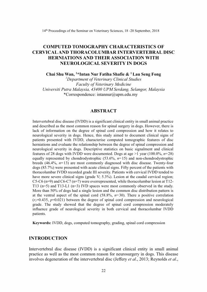
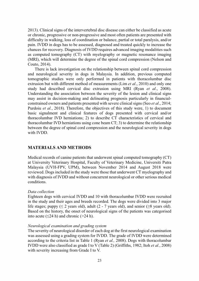
ImageJ software was used to measure the cross-sectional area of the spinal cord. The images were magnified manually for optimum visualisation. Standard calibration for each image was done by converting the pixels to millimeters. The cross-sectional area of the spinal cord was measured by tracing the outline of the spinal cord (Figures 1 and 2). The measurement of each image was repeated thrice and averaged. Standard deviation was calculated for intra-observer agreement. Then the degree of spinal cord compression was calculated as a percentage of the cross-sectional area (CSA) of the normal spinal cord using the following formula (Ryan et al., 2008):
x 100
25
Figure 1. Transverse computed tomography myelography image of the cervical spine of a dog, with the compressed spinal cord at C5-C6 IVD space highlighted in red.
Figure 2. Transverse computed tomography myelography image of the cervical spine of a dog, with the uncompressed spinal cord at C5 level highlighted in red.
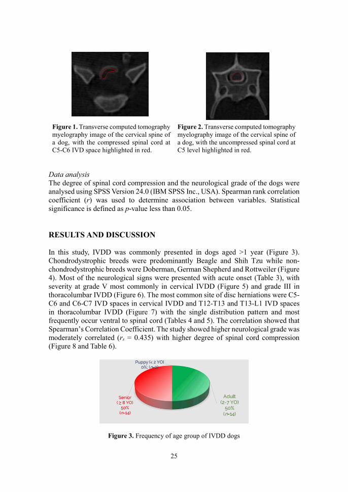
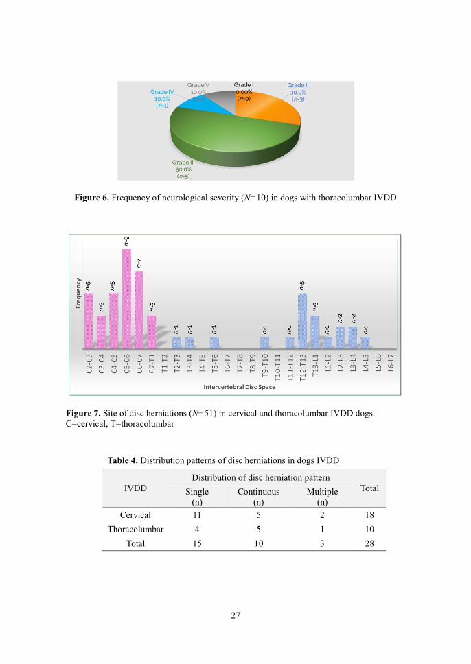
Data analysis The degree of spinal cord compression and the neurological grade of the dogs were analysed using SPSS Version 24.0 (IBM SPSS Inc., USA). Spearman rank correlation coefficient (r) was used to determine association between variables. Statistical significance is defined as p-value less than 0.05. RESULTS AND DISCUSSION In this study, IVDD was commonly presented in dogs aged >1 year (Figure 3). Chondrodystrophic breeds were predominantly Beagle and Shih Tzu while non-chondrodystrophic breeds were Doberman, German Shepherd and Rottweiler (Figure 4). Most of the neurological signs were presented with acute onset (Table 3), with severity at grade V most commonly in cervical IVDD (Figure 5) and grade III in thoracolumbar IVDD (Figure 6). The most common site of disc herniations were C5-C6 and C6-C7 IVD spaces in cervical IVDD and T12-T13 and T13-L1 IVD spaces in thoracolumbar IVDD (Figure 7) with the single distribution pattern and most frequently occur ventral to spinal cord (Tables 4 and 5). The correlation showed that Spearman’s Correlation Coefficient. The study showed higher neurological grade was moderately correlated (rs = 0.435) with higher degree of spinal cord compression (Figure 8 and Table 6).
Figure 3. Frequency of age group of IVDD dogs

26
Figure 4. Breed frequency of dogs with IVDD
Table 3. Frequency of onset of neurological signs in dogs with IVDD.
IVDD Onset of neurological signs
Total Acute (n)
Chronic (n)
Cervical 16 2 18
Thoracolumbar 8 2 10
Total 24 4 28
Figure 5. Frequency of neurological severity in dogs with cervical IVDD
27
Figure 6. Frequency of neurological severity (N=10) in dogs with thoracolumbar IVDD
Figure 7. Site of disc herniations (N=51) in cervical and thoracolumbar IVDD dogs. C=cervical, T=thoracolumbar
Table 4. Distribution patterns of disc herniations in dogs IVDD
IVDD Distribution of disc herniation pattern
Total Single (n)
Continuous (n)
Multiple (n)
Cervical 11 5 2 18
Thoracolumbar 4 5 1 10
Total 15 10 3 28

28
Table 5. Location of disc herniation in dogs with IVDD
IVDD
Location of disc herniation
Total Ventral
(n) Left lateral
(n) Right lateral
(n) Dorsal
(n)
Cervical 9 4 6 1 20
Thoracolumbar 12 5 5 0 31
Total 30 9 11 1 51
Figure 8. Relationship between the degree of spinal cord compression and the neurological grade in dogs (N=28) with cervical and thoracolumbar IVDD.
CONCLUSION The study showed that the degree of spinal cord compression has moderate influences on the grade of neurological severity in cervical and/or thoracolumbar IVDD dogs. It can be concluded that dogs with IVDD presenting with severe clinical signs would have higher degree of spinal cord compression, leading to poor prognosis. REFERENCES Griffiths I (1982). Spinal disease in the dog. In Practice, 4(2):44-52. Itoh H, Hara Y, Yoshimi N, Harada Y, Nezu Y, Yogo T, Tagawa M (2008). A
retrospective study of intervertebral disc herniation in dogs in Japan: 297 cases. Journal of Veterinary Medical Science, 70(7):701-706.
Jeffery ND, Levine JM, Olby NJ, Stein VM (2013). Intervertebral disk degeneration in dogs: Consequences, diagnosis, Treatment, and Future Directions. Journal of Veterinary Internal Medicine, 27(6):1318–1333.
Lim C, Kweon O-K, Choi M-C, Choi J, Yoon J (2010). Computed tomographic

29
characteristics of acute thoracolumbar intervertebral disc disease in dogs. Journal of Veterinary Science, 11(1):73.
Nelson R and Couto C (2014). Small Animal Internal Medicine. 5th ed. Canada: Elsevier Inc.
Purdoiu RC, Ashur R, Condor L, Lacatus R (2018). Computed Tomography Findings in Spinal Compression in 196 Dogs. Bulletin of University of Agricultural Sciences and Veterinary Medicine Cluj-Napoca. Veterinary Medicine, 75(1):46.
Reynolds D, Brisson BA, Nykamp SG (2013). Agreement between magnetic resonance imaging, myelography, and surgery for detecting recurrent, thoracolumbar intervertebral disc extrusion in dogs. Veterinary and Comparative Orthopaedics and Traumatology, 26(1): 12-8.
Ryan TM, Platt SR, Llabres-Diaz FJ, McConnell JF, Adams VJ (2008). Detection of spinal cord compression in dogs with cervical intervertebral disc disease by magnetic resonance imaging. Veterinary Record, 163(1):11–15.
Seo E, Choi J, Choi M, Yoon J (2014). Computed tomographic evaluation of cervical vertebral canal and spinal cord morphometry in normal dogs. Journal of Veterinary Science, 15(2):187-193.

30
14th Proceedings of the Seminar on Veterinary Sciences, 18-20 September, 2018
ACCURACY OF GENOMIC PREDICTION IN SWAMP BUFFALO USING DEREGRESSED BREEDING VALUE ESTIMATED FROM
PUREBRED AND CROSSBRED OFFSPRING PHENOTYPES
Lyeonna Amber Garcia De Chavez, 1*Mohd Shahrom Salisi, 2Mark Hiew Wen Han, 3Jonny Engkias & 3Azizan Maruf
1Department of Veterinary Preclinical Sciences
2Department of Veterinary Clinical Studies Faculty of Veterinary Medicine
Universiti Putra Malaysia, 43400 UPM Serdang, Selangor, Malaysia 3Sabah Department of Veterinary Service
Wisma Pertanian Sabah, 88999 Kota Kinabalu, Sabah, Malaysia *Correspondence: [email protected]
ABSTRACT
Poor reproductive performance is one of the factors contributing to the decline in buffalo population. This study assessed the accuracy of breeding value prediction for purebred swamp buffalo sire based on phenotype of purebred offspring (BWPB) and purebred and crossbred offspring (BWPBCB). Birth weight trait records of 149 buffaloes offsprings born between 2013 and 2017 from 1 sire obtained from the Buffalo Breeding and Research Centre Farm, Telupid, Sabah, Malaysia, were used for prediction of breeding values for birth weight (BW) trait. The heritability required for deregression estimated using the parent-offspring regression method for BWPB and BWPBCB were 0.28 and -0.14, respectively. The breeding value (BV) for BW of sire using estimated breeding value (EBV) equation were 34.32 and 33.42 for BWPB and BWPBCB, respectively. The estimation of BV using EBV equation on BWPBCB (0.79) is more accurate than on BWPB (0.16). There was a weak, positive correlation between purebred and crossbred performance (r=0.314, n=15, p=0.254), indicating that selection based on only purebred offspring data will not maximise genetic improvement. In conclusion, the low purebred-crossbred correlation coefficient combined higher relative accuracy of EBV based on BWPBCB is a better estimate of the true breeding value than that based on BWPB data. Keywords: accuracy, breeding value, birth weight, Pearson’s purebred-crossbred correlation, swamp buffalo
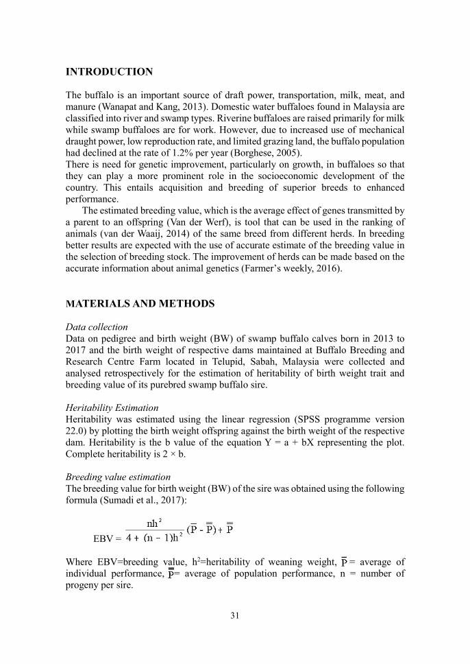
31
INTRODUCTION The buffalo is an important source of draft power, transportation, milk, meat, and manure (Wanapat and Kang, 2013). Domestic water buffaloes found in Malaysia are classified into river and swamp types. Riverine buffaloes are raised primarily for milk while swamp buffaloes are for work. However, due to increased use of mechanical draught power, low reproduction rate, and limited grazing land, the buffalo population had declined at the rate of 1.2% per year (Borghese, 2005). There is need for genetic improvement, particularly on growth, in buffaloes so that they can play a more prominent role in the socioeconomic development of the country. This entails acquisition and breeding of superior breeds to enhanced performance.
The estimated breeding value, which is the average effect of genes transmitted by a parent to an offspring (Van der Werf), is tool that can be used in the ranking of animals (van der Waaij, 2014) of the same breed from different herds. In breeding better results are expected with the use of accurate estimate of the breeding value in the selection of breeding stock. The improvement of herds can be made based on the accurate information about animal genetics (Farmer’s weekly, 2016). MATERIALS AND METHODS Data collection Data on pedigree and birth weight (BW) of swamp buffalo calves born in 2013 to 2017 and the birth weight of respective dams maintained at Buffalo Breeding and Research Centre Farm located in Telupid, Sabah, Malaysia were collected and analysed retrospectively for the estimation of heritability of birth weight trait and breeding value of its purebred swamp buffalo sire. Heritability Estimation Heritability was estimated using the linear regression (SPSS programme version 22.0) by plotting the birth weight offspring against the birth weight of the respective dam. Heritability is the b value of the equation Y = a + bX representing the plot. Complete heritability is 2 × b. Breeding value estimation The breeding value for birth weight (BW) of the sire was obtained using the following formula (Sumadi et al., 2017):
EBV = Where EBV=breeding value, h2=heritability of weaning weight, , = average of individual performance, = average of population performance, n = number of progeny per sire.

32
Accuracy of Estimated Breeding Value The relative accuracy (RA) of the deregressed estimated breeding value (DEBV) was estimated using the following formula (Sumadi et al., 2017): RA = 0.5h × where t = 0.25 h2.
Purebred-crossbred Correlation The Pearson’s purebred-crossbred correlation coefficient was estimated by Pearson’s correlation using SPSS programme version 22.0.
RESULTS AND DISCUSSION The regression line equation obtained from using only purebred offspring information (BWPB) was y=0.02x+30.59 (Figure 1). Therefore, the heritability estimated from BWPB is 0.04, which is low.
Figure 1. Scatter plot of progeny phenotype (birth weight of purebred swamp buffalo offspring only) and the parental phenotype (birth weight of purebred swamp buffalo dam).
The regression line equation obtained from using purebred offspring and crossbred offspring (BWPBCB) information was y=0.32x+21.34 (Figure 2). Hence, the heritability estimate is 0.64, which was moderate.
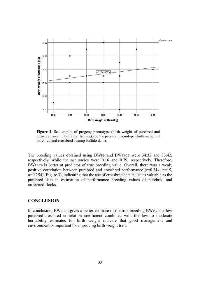
33
Figure 2. Scatter plot of progeny phenotype (birth weight of purebred and crossbred swamp buffalo offspring) and the parental phenotype (birth weight of purebred and crossbred swamp buffalo dam).
The breeding values obtained using BWPB and BWPBCB were 34.32 and 33.42, respectively, while the accuracies were 0.16 and 0.79, respectively. Therefore, BWPBCB is better at predictor of true breeding value. Overall, there was a weak, positive correlation between purebred and crossbred performance (r=0.314, n=15, p=0.254) (Figure 3), indicating that the use of crossbred data is just as valuable as the purebred data in estimation of performance breeding values of purebred and crossbred flocks. CONCLUSION In conclusion, BWPBCB gives a better estimate of the true breeding BWPB.The low purebred-crossbred correlation coefficient combined with the low to moderate heritability estimates for birth weight indicate that good management and environment is important for improving birth weight trait.
34
Figure 3. Scatter plot to assess the relationship between the performance of purebred swamp buffalo and swamp-murrah cross buffalo offsprings. REFERENCES Borghese A (2005). Buffalo population and strategies in the world. In Buffalo
Production and Research, Reu Technical Series 67. http://www.fao.org/3/ah847e/ah847e00.pdf (Accessed on 28 August 2019).
Farmer’s weekly (2016). Understanding estimated breeding values. (2016). Retrieved from https://www.farmersweekly.co.za/farm-basics/how-to-livestock/understanding-estimated-breeding-values/
(Accessed on 27 August 2019). Sumadi, Fathoni A, Maharani D, Ngadiyono N, Widayati DT, Noviandi CT,
Khusnudin M (2017). Breeding values of sires based on offspring weaning weight as a recommendation for selecting kebumen Ongole Grade cattle. Journal of the Indonesian Tropical Animal Agriculture, 42(3): 160-166.
van der Waaij KOL (2014). Textbook animal breeding: animal breeding and genetics for BSc students. Centre for Genetic Resources and Animal Breeding and Genomics Group, Wageningen University and Research Centre. https://www.wur.nl/upload_mm/d/b/b/614bcc19-036f-434e-9d40-609364ab26da_Textbook%20Animal%20Breeding%20and%20Genetics-v17-20151122_1057.pdf (Accessed on 28 August 2019).
Van der Werf J. Chapter 10: Principles of estimation of breeding values. https://jvanderw.une.edu.au/Chapter10.pdf (Accessed on 28 August 2019).

35
Wanapat M and Kang S (2013). World buffalo production: Challenges in meat and milk production, and mitigation of methane emission. Buffalo Bulletin, 32 (Special issue 1): 1-21.
file:///C:/Users/Win%2010/Downloads/IBBUSI201301001.pdf (Accessed on 28 August 2019).

36
13th Proceedings of the Seminar on Veterinary Sciences, 18 – 20 September 2019
ANTIBACTERIAL PROPERTIES OF APPLE CIDER VINEGAR AGAINST STAPHYLOCOCCUS AUREUS AND STAPHYLOCOCCUS
PSEUDINTERMEDIUS
Nurul Zulaikha Norizal, 1*Mazlina Mazlan & 1Sharina Omar 1Department of Veterinary Pathology and Microbiology
Faculty of Veterinary Medicine Universiti Putra Malaysia, 43400 UPM Serdang, Selangor, Malaysia
*Correspondence: [email protected]
ABSTRACT
The issue of antimicrobial resistance (AMR) has garnered attention due to the increase of superbugs known for their resistance to antibiotics. Previous studies have shown that apple cider vinegar (ACV) possesses an antimicrobial affect against bacteria infecting skin wounds. Hence, this study was conducted to determine the antimicrobial effect of ACV on Staphylococcus aureus and Staphylococcus pseudintermedius, two bacteria commonly isolated from wounds in companion animals. The efficacy of ACV with acidity of 1.2, 2.5, 3.75, and 5% was tested using disk diffusion method, agar diffusion method, and time-kill assay. The findings from this study revealed no clear zone of inhibition for the first two methods. However, results of time-kill assay showed that all concentrations of ACV had bactericidal effect on both bacteria. ACV showed complete inhibition of S. pseudintermedius growth within 2 h post-exposure to 2.5 and 3.75% acidity, while S. aureus was completely killed within a 2 h post-exposure to 5% acidity. This study revealed that ACV showed significant (p<0.05) antimicrobial properties with the most effective ACV acidity of 2.5% for S. pseudintermedius and 5% for S. aureus. In conclusion, ACV shows evidence of antimicrobial activity and therefore has the potential as an alternative antimicrobial agent. Keywords: apple cider vinegar, Staphylococcus aureus, Staphylococcus pseudintermedius, disk diffusion method, agar diffusion method, time-kill assay
INTRODUCTION Methicillin-resistant Staphylococcus aureus (MRSA) and methicillin-resistant Staphylococcus pseudintermedius (MRSP) are not foreign anymore to the medical world. These species are also capable of being multidrug-resistant (Stryjewski and Corey, 2014). This shows that Staphylococcus is versatile and constantly evolves to develop resistance against new antibiotics.
Previous studies showed that apple cider vinegar (ACV) has the ability to inhibit the growth of several bacteria such as Escherichia coli, S. aureus and Salmonella paratyphi (Saqib, 2017), and the effect was concentration-dependent (Yagnik et al.,
37
2018). In this particular study, S. aureus and S. pseudintermedius were chosen to determine antibacterial effect of ACV. MATERIALS & METHODS Four concentrations of ACV, 1.25, 2.5, 3.75, and 5%, were used to determine its antibacteria effect on the clinical strain S. aureus (ATCC 25923) and field strain S. pseudintermedius.. Biochemical tests were performed to reconfirm the bacterial strains. The bacteria were then sub-cultured on nutrient agar for 24 h prior to the experiment. Isolated colonies were taken from the plate using inoculating loop and suspended in normal saline. The turbidity was then adjusted to 0.5 McFarland standard. The S. aureus and S. pseudintermedius suspensions were subjected to the disk diffusion and well diffusion methods and time-kill assay. Disk Diffusion Method This method was performed as described by Ortez (2005) and Vineetha et al. (2015). Mueller-Hinton agar was inoculated with bacterial suspensions. The entire plate was streaked, ensuring the inoculum was evenly distributed. Blank disks impregnated with 25 L of various concentrations of apple cider vinegar (ACV) were placed on the agar surface. Cephalexin was used as the positive control. The disks were gently tapped using sterile forceps to ensure that they stick to the surface of the agar before inverting the plate and incubating at 37 °C for 24 h. The diameter of zone of inhibition (ZDI) was measured using Vernier callipers (mm). Well Diffusion Method Well diffusion was performed based on the modified method from Bonev et al. (2008). Five 6 mm holes were punched on nutrient agar using a sterile glass spreader. The agar surface was then inoculated, as in the previous method. Each of the holes were filled with 25 L of cephalexin and different concentrations of ACV. The agar was allowed to dry for 1 h before the plate was inverted and incubated at 37 °C for 24 h. The ZDI was measured using Vernier callipers (in mm). Time-Kill Assay A time-kill assay was performed according to the methods described previously with modification (Bamberger et al., 2012; Kumar et al., 2017; Marliyana et al., 2017;). The bacterial suspensions containing approximately 1.5 x 10⁸ CFU/mL were adjusted to 0.5 McFarland One millilitre bacterial suspension was pipetted into test tubes containing ACV at various concentrations. Control test tube contained bacteria suspended in 0.9% NaCl. At 0, 2, 4, 6, 8, 10 and 24 h, an aliquot from the test tube was diluted in NaCl 0.9% and 100 L spread on nutrient agar using sterile glass spreader. The agar plate was inverted and incubated at 37 °C for 24 h. After incubation, viable single colony was counted using a colony counter and the result expressed as CFU/mL. Time-kill curve was then drawn by plotting log10 CFU/mL against exposure time.

38
Statistical Analysis Statistical Package for the Social Science (SPSS version 22) and two-way ANOVA were used to analyse data. RESULTS AND DISCUSSION Only cephalexin produced a clear zone of inhibition for S. aureus, while no clear zone of inhibition was observed.S. pseudintermedius (Figure 1). A faint zone of inhibition was observed with ACV treatment, especially at 5% acidity. Methicillin-resistant staphylococci are resistant to all β-lactam antimicrobials (Weese and van Duijkeren, 2010). The S. pseudintermedius strain in this study may be of the MRSP strain, since it was resistant to β-lactam antibiotic, cephalexin.
Figure 1: Zone diameter of inhibition of (a) Staphylococcus aureus and (b) Staphylococcus pseudintermedius treated with cephalexin and apple cider vinegar (ACV) determined by the disk diffusion method. C=cephalexin. 25, 50, 75, and 100 are % concentrations of ACV. Using the time-kill assay, S. aureus was completely inhibited after 2 h of exposure to 5% ACV [Figure 2(a)]. However, the bacteria could still proliferate after 6 h and only became inhibited from 8 h onwards. The results for time-kill assay for S. pseudintermedius is shown in Figure 2(b). The study showed that the growth of S. pseudintermedius began to be inhibited upon exposure 5% ACV. However, bacteria proliferation was still evident at 2 h post-exposure. Nagoba et al. (2015) suggested that acidic environment is conducive to wound healing; however, the effect of acidity only lasted a few hours after exposure (Morsi et al., 2016).
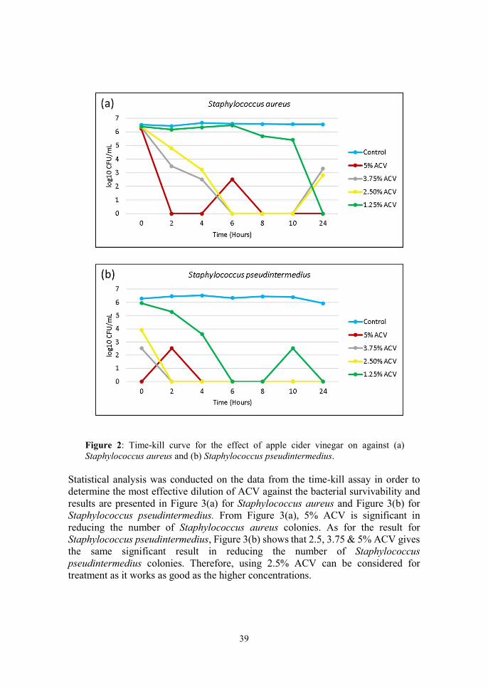
39
Figure 2: Time-kill curve for the effect of apple cider vinegar on against (a) Staphylococcus aureus and (b) Staphylococcus pseudintermedius.
Statistical analysis was conducted on the data from the time-kill assay in order to determine the most effective dilution of ACV against the bacterial survivability and results are presented in Figure 3(a) for Staphylococcus aureus and Figure 3(b) for Staphylococcus pseudintermedius. From Figure 3(a), 5% ACV is significant in reducing the number of Staphylococcus aureus colonies. As for the result for Staphylococcus pseudintermedius, Figure 3(b) shows that 2.5, 3.75 & 5% ACV gives the same significant result in reducing the number of Staphylococcus pseudintermedius colonies. Therefore, using 2.5% ACV can be considered for treatment as it works as good as the higher concentrations.
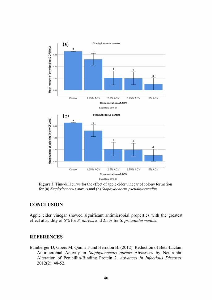
40
Figure 3. Time-kill curve for the effect of apple cider vinegar of colony formation for (a) Staphylococcus aureus and (b) Staphylococcus pseudintermedius.
CONCLUSION Apple cider vinegar showed significant antimicrobial properties with the greatest effect at acidity of 5% for S. aureus and 2.5% for S. pseudintermedius. REFERENCES Bamberger D, Goers M, Quinn T and Herndon B. (2012). Reduction of Beta-Lactam
Antimicrobial Activity in Staphylococcus aureus Abscesses by Neutrophil Alteration of Penicillin-Binding Protein 2. Advances in Infectious Diseases, 2012(2): 48-52.
41
Bonev B, Hooper J and Parisot J. (2008). Principles of Assessing Bacterial Susceptibility to Antibiotics Using the Agar Diffusion Method. Journal of Antimicrobial Chemotherapy, (2008)61: 1295-1301.
Kumar S, Bandyopadhyay M, Sk M, Mukhopadhyay K, Kumar Bandyopadhyay M, Chatterjee A and Chatterjee M. (2017). Antimicrobial Synergy Testing By Time-Kill Methods For Extensively Drug-Resistant Acinetobacter Baumannii Isolates. IOSR Journal of Dental and Medical Sciences, 16(12): 79-84.
Marliyana S, Mujahidin D, Syah Y and Rukayadi Y. (2017). Time-Kill Assay of 4-Hydroxypanduratin A Isolated from Kaempferia Pandurata Against Foodborne Pathogens. Molekul, 12(2): 166-173.
Morsi AEF, Mustafa FM and Tokhy AE. (2016). Vinegar Simple Method in Dressing of Pseudomonas Infected Wound. International Invention Journal of Medicine and Medical Sciences, 3(8): 143-146.
Nagoba BS, Suryawanshi NM, Wadher B and Selkar S. (2015). Acidic Environment and Wound Healing: A Review. Wounds, 27(1): 5-11.
Ortez, JH. (2005). Disk Diffusion Testing. In: Manual of Antimicrobial Susceptibility Testing. Coyle, MB (Ed.). American Society for Microbiology. Pp. 39-52.
Saqib A. (2017). Antimicrobial Activity of Apple Cider, Mapana Journal of Sciences, 16(2): 11–15.
Stryjewski ME and Corey GR. (2014). Methicillin-resistant Staphylococcus aureus: an evolving pathogen. Clinical Infectious Diseases, 58(S1): 10-19.
Vineetha N, Sridhar D and Vignesh R. (2015). Preparation, Standardization of Antibiotic Discs and Study of Resistance Pattern for First-Line Antibiotics in Isolates from Clinical Samples. International Journal of Applied Research, 1(11): 624-631.
Weese JS and van Duijkeren E. (2010). Methicillin-resistant Staphylococcus aureus and Staphylococcus pseudintermedius in veterinary medicine. Veterinary Microbiology, 140(3-4): 418-429.
Yagnik D, Serafin V and Shah AJ. (2018). Antimicrobial activity of apple cider vinegar against Escherichia coli, Staphylococcus aureus and Candida albicans; downregulating cytokine and microbial protein expression. Scientific Reports, 8(1): 1-12.

42
14th Proceedings of the Seminar on Veterinary Sciences, 18 – 20 September 2018
GASTROINTESTINAL AND BLOOD PARASITES IN AFRICAN PYGMY HEDGEHOG (ATELERIX ALBIVENTRIS)
Hoe Kai Thong & 1*Mohd Hezmee Mohd Noor
1 Department of Veterinary Preclinical Sciences Faculty of Veterinary Medicine
Universiti Putra Malaysia, 43400 UPM Serdang, Selangor, Malaysia *Correspondence: [email protected]
ABSTRACT
African pygmy hedgehogs (Atelerix albiventris) is an Old World nocturnal insectivorous mammal under the order Eulipotyphla, family Erinaceidae, and subfamily of Erinaceinae. The African pygmy hedgehogs were imported to Malaysia and have gained popularity as pets because of their colour, low maintenance cost, and independent nature. This study investigated the presence of gastrointestinal and blood parasites in African pygmy hedgehogs. Fresh faecal samples were collected from 30 hedgehogs owned by pet owners and breeders in Selangor, Malaysia. Simple floatation and sedimentation tests were performed on the faecal samples to determine presence of helminth and protozoan eggs. Giemsa-stained thin blood smears of 4 blood samples were examined for parasites. The study showed that faecal and blood samples were negative for parasite, suggesting that African pygmy hedgehogs in Selangor, Malaysia were free of parasite infestation. Keywords: African pygmy hedgehog, gastrointestinal parasites, blood parasites.
INTRODUCTION In Malaysia, the African pygmy hedgehog, Atelerix albiventris, has recently gained popularity as pet. Since, contact with this mammal is a potential risk for infections, there is need to determine the susceptibility of these hedgehogs towards gastrointestinal and blood parasite infestation and their zoonotic and epizootic risk as vectors of transmissible diseases in human and animals. Among these transmissible diseases are cryptosporidiosis and toxoplasmosis (Santana, et al., 2010). Wild hedgehogs harbour several species of parasites including Brachylaemus spp. and Capillaria spp., and occasionally coccidian and Cryptosporidium spp. (Beck, 2007). Ricketssia massiliae, a type of blood parasite causing spotted fever disease in human (Bitam et al., 2006) and Anaplasma spp. and Theileria spp. that can infect ruminants were detected in the hedgehogs (Skuballa et al., 2010; Chen et al., 2014). These infections may pose a risk to the ruminant industry in Malaysia, the livestock animals are exposed to infected or carrier wild hedgehogs.

43
MATERIALS AND METHODS Convenient sampling method was used to obtain hedgehogs from owners or breeder in Serdang, Selangor, Malaysia. Faecal samples were obtained from 30 hedgehogs for detection of gastrointestinal parasites.
Blood samples were obtained from 4 hedgehogs belonging to consenting owners. The hedgehogs were held by the rump and head with one of the hind legs pulled downwards. The leg and paw were wiped with dry tissue paper to remove dirt. Blood samples collected from the toe pad using the prick method and 27 G hypodermic needles, were used to make thin smears. The hedgehogs were observed to determine general appearance, mentation, body condition score, and skin condition. Faecal samples were subjected to simple floatation and sedimentation tests according to the method described by Hansen and Perry (1994). RESULTS AND DISCUSSION Among the 30 hedgehogs, 4 had body condition score of 4, while 26 had an ideal body condition score of 3. The skin condition of all hedgehogs were good, although skin flakes was observed in 10 hedgehogs. Four out of 30 had a history of mite infestation. All hedgehogs appeared bright, alert, and responsive. The results of faecal sample and blood examinations are summarised in Tables 1 and 2, respectively.
On first sampling, only one positive faecal sample was positive for parasites with the detection of 8 coccidian oocysts. The floatation test was repeated on a second set of samples and this time the results showed all 30 hedgehogs were negative for parasite. In the first sampling, among the 8 coccidian oocysts detected, 3 were sporulated oocysts. Oocysts are present only in 24 to 48 hour faecal samples (Matuschka, 1984). Since the faecal samples collected in this study were fresh, it suggests the oocyst-positive findings were actually false positive results.
No gastrointestinal or blood parasite was not detected in hedgehogs in this study. Since most gastrointestinal parasites only infect hedgehogs through ingestion of intermediate host, while the hedgehogs in this study were primarily on the Kibbles diet, the absence of parasites in the animals was expected. The hedgehogs were reared indoors and in individual container or cages. This is decreased the probability of the hedgehogs of ingesting on or coming in contact with insects.
There is no hedgehog species that is native to Malaysia and there is no reservoir for host-specific coccidian and cryptosporidium that could infect pet hedgehogs. This could explain the absence of parasites in the hedgehogs in this study. CONCLUSION This study showed that African pygmy hedgehog in Selangor, Malaysia are free of parasites and apparently safe to be kept as companion pets.

44
REFERENCES Santana EM, Jantz HE, Best TL (2010). Atelerix albiventris (Erinaceomorpha:
Erinaceidae). Mammalian Species, 42(857): 99-110. Beck W (2007). Endoparasitem beim Igel. Wien Klin Wochenschr 119 (Suppl 3):40-
44. Bitam I, Parola P, De La Cruz KD, Matsumoto K, Baziz B, Rolain JM (2006). First
molecular detection of Rickettsia felis in fleas from Algeria. American Journal of Tropical Medicine and Hygiene, 74(4): 532-535.
Hansen J and Perry B (1994). Techniques for parasite assays and identification in faecal samples In: The epidemiology, diagnosis and control of helminth parasites in ruminants. International Laboratory for Research on Animal Disease, Nairobi, Kenya. https://pdfs.semanticscholar.org/306d/f1bf88e876d54943c726772f7d3acb6dc3fc.pdf (Accessed on 16 October 2019)
Skuballa J, Petney T, Pfäffle M, Taraschewski H (2010). Molecular detection of Anaplasmaphagocytophilum in the European hedgehog (Erinaceuseuropaeus) and its ticks. Vector Borne and Zoonotic Diseases, 10(10):1055–1057.
Chen Z, Liu Q, Jiao F, Xu B, Zhou X (2014). Detection of piroplasms infection in sheep, dogs and hedgehogs in Central China. Infectious Diseases of Poverty, 3(1):18. doi: 10.1186/2049-9957-3-18. eCollection 2014
Matuschka F (1984). Endodyogeny in Isosporarastegaievae from the Eurasian hedgehog (Erinaceuseuropaeus L.). Parasitology, 88(1): 9.-12
45
14th Proceedings of the Seminar on Veterinary Sciences, 18 – 20 September 2018
OWNER AWARENESS ON POTENTIALLY HAZARDOUS SUBSTANCES TO DOGS IN FOODS AND HOUSEHOLD ITEMS
Choong Yee Ph’ng, 1,3*Goh Yong Meng & 2,3Noordin Mohamed Mustapha
1Department of Veterinary Preclinical Sciences 2Department of Veterinary Pathology and Microbiology
3Research Centre for Ruminant Diseases Faculty of Veterinary Medicine
Universiti Putra Malaysia, 43400 UPM Serdang, Selangor, Malaysia *Correspondence: [email protected]
ABSTRACT
Toxicosis or poisoning is common in pets. These incidences are mainly attributed to lack of awareness of owners or caretakers of the presence of hazardous substances around their pets. The objective of this study was to determine the level of awareness among owners and caretakers of potential hazardous household items and foods to dogs. Fifty pet owners were surveyed using questionnaires distributed via a private veterinary clinic in Cheras, Selangor, Malaysia while 50 veterinary students from University Putra Malaysia were included as the control group. In general, the average owner or caretaker were less aware of potential hazardous household items that are exposed to their dogs. The study indicated that there is urgent need to educate owners and caretakers on potential toxicants in the environment of their pets. Client education should also focus on improving the ability of owners to prevent and mitigate toxicosis in their pet dogs. Keywords: toxicosis, toxicant, hazardous, awareness
INTRODUCTION
Cats and dogs are easily to be exposed to various poisonous substances (Pet Poison Helpline) Manifestation of intoxication dependents on the type of agents, the number of agents, and the sensitivity of the animals towards to the agents.
Pets are at risk to toxicosis even in the relatively safety of the owners’ home. There are many common foodstuff and household items that are health hazards to cats and dogs. Currently, there is no published data on the epidemiology of pet intoxication in Malaysia. Thus, the objective of this study was to determine whether the average pet owners or caretakers in Malaysia are aware of the type of food and household substances that are potentially hazardous to their pets. This information is crucial for the planning of client education on toxicosis in pet animals.
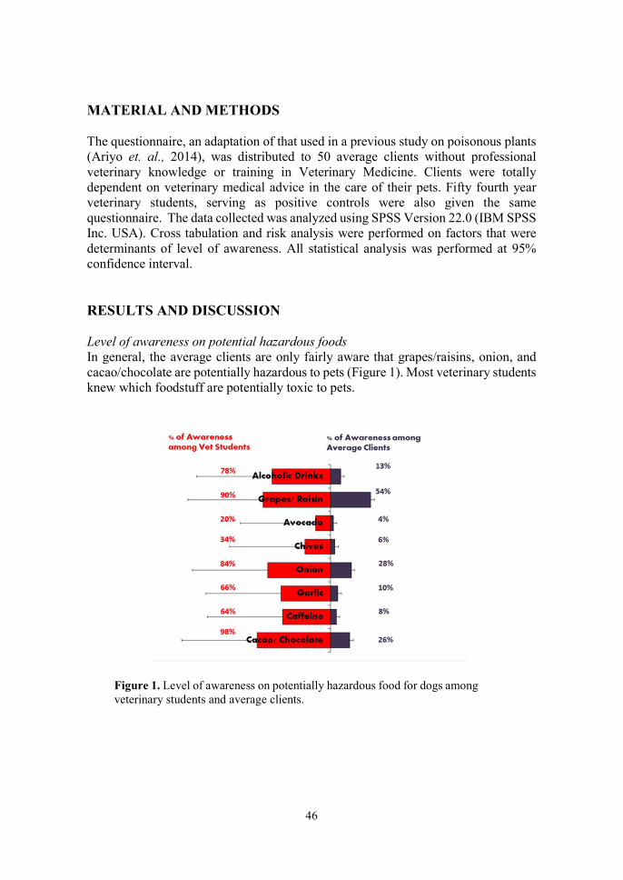
46
MATERIAL AND METHODS The questionnaire, an adaptation of that used in a previous study on poisonous plants (Ariyo et. al., 2014), was distributed to 50 average clients without professional veterinary knowledge or training in Veterinary Medicine. Clients were totally dependent on veterinary medical advice in the care of their pets. Fifty fourth year veterinary students, serving as positive controls were also given the same questionnaire. The data collected was analyzed using SPSS Version 22.0 (IBM SPSS Inc. USA). Cross tabulation and risk analysis were performed on factors that were determinants of level of awareness. All statistical analysis was performed at 95% confidence interval. RESULTS AND DISCUSSION Level of awareness on potential hazardous foods In general, the average clients are only fairly aware that grapes/raisins, onion, and cacao/chocolate are potentially hazardous to pets (Figure 1). Most veterinary students knew which foodstuff are potentially toxic to pets.
Figure 1. Level of awareness on potentially hazardous food for dogs among veterinary students and average clients.

47
Unlike foodstuff most average clients know than many household items can potentially cause harm to their dogs. Average clients are only not that aware that paints (Figure 2), tea tree oil, ethylene glycol, and lily flowers are potentially toxic to dogs.
Figure 2. Level of awareness of potentially hazardous household items for dogs among veterinary students and average clients.
Figure 3. Level of awareness on potentially hazardous and rarely encountered items for among veterinary students and average clients.
48
DISCUSSION
The study highlighted the failure of average clients to recognise common household items as potential toxicants to pets. The study also showed the importance of client education in the prevention of pet toxicosis and safety of pets in the care of owners. This is obvious because veterinary students, in their curriculum, were provided information on issues of toxicities in pets. However, in spite of education, many veterinary students were not aware that chives and avocado too can be toxic to pets. Chives, for example, contains compounds such as organosulfoxides and Di-propul-disulfide and allyl propylisulfide, which are similarly found in onion, a foodstuff that can cause toxicity in cats and dogs (Salgado et al., 2011).
Avocado toxicity is due its persin (“avocado toxin”) content and the large pit that can gastrointestinal obstruction (Nicholas, 2015). However, persin in avocado only poses a mild risk of gastrointestinal upset in dogs. Although the extent of toxicities due to avocado and chives in dogs is still not certain, pet owners should still be aware of the potential toxicities from ingestion of these foodstuff. Unlike veterinary students, most average clients were not aware the rarely encountered items like tea tree oil, ethylene glycol, and lily flowers are toxic non-food substances. To understand the dangers of these substances for pets, owners and pet carers require adequate education.
Average client often uses human tooth paste to clean teeth of their pets. Human tooth paste contains xylitol, a chemical that promotes insulin release by the pancreas, which subsequently cause rapid and significant decrease in blood glucose level (Piscitelli et al., 2010). Pets that suffer from xylitol toxicity are depressed, ataxic, and could go into seizure. In serious conditions, xylitol toxicity can lead to liver necrosis, which could be fatal to pets (Schmid and Hovda, 2016).
Due to lack of knowledge, average clients may practice home remediation by giving paracetamol to their pets to alleviate symptoms of sickness. Paracetamol or acetaminophen commonly causes toxicity in pets (Thompson, 2014). CONCLUSION
The study showed average clients generally lack knowledge on the item accessible to pet that can cause toxicities. It is imperative that average clients of the veterinary hospital and clinics be educated on the dangers of household items and foodstuff that could potentially cause toxicities in their pets.
REFERENCES
Ariyo AL, Peter AO, Muyideen K, Ramota LI (2014). Botanical survey of poisonous
plants within the Federal Capital Territory, Abuja, Nigeria. Journal of Biology, Agriculture and Healthcare, 4(20): 196-214.
49
Nicholas J (2015). The truth about avocado for dogs & cats. Preventive Vet. https://www.preventivevet.com/dogs/the-truth-about-avocado-for-dogs-and-cats (Accessed on 4 September 2019).
Pet Poison Helpline. What did your pet ingest? https://www.petpoisonhelpline.com/poisons/ (Accessed on 4 September 2019).
Piscitelli CM, Dunayer EK, Aumann M (2010). Xylitol toxicity in dogs. Compendium: Continuing Education for Veterinarians, 32(2): E2-E4.
Salgado, B. S., Monteiro, L. N. & Rocha, N. S. (2011). Allium species poisoning in dogs and cats. The Journal of Venomous Animals and Toxins including Tropical Diseases, 17, 4-11.
Schmid RD and Hovda LR (2016). Acute hepatic failure in a dog after xylitol ingestion. Journal of Medical Toxicity, 12: 201-205.
Thompson A (2014). Canine toxicity in the home enviroment. Veterinary Nursing Journal, 27(10): 380-382.
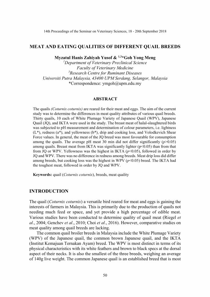
50
14th Proceedings of the Seminar on Veterinary Sciences, 18 - 20th September 2018
MEAT AND EATING QUALITIES OF DIFFERENT QUAIL BREEDS
Myzatul Hanis Zahiyah Yusof & 1,3*Goh Yong Meng 1Department of Veterinary Preclinical Science
Faculty of Veterinary Medicine 3Research Centre for Ruminant Diseases
Universiti Putra Malaysia, 43400 UPM Serdang, Selangor, Malaysia *Correspondence: [email protected]
ABSTRACT
The quails (Coturnix coturnix) are reared for their meat and eggs. The aim of the current study was to determine the differences in meat quality attributes of various quail breeds. Thirty quails, 10 each of White Plumage Variety of Japanese Quail (WPV), Japanese Quail (JQ), and IKTA were used in the study. The breast meat of halal-slaughtered birds was subjected to pH measurement and determination of colour parameters, i.e. lightness (L*), redness (a*), and yellowness (b*), drip and cooking loss, and Volodkevich Shear Force values. In general, the meat of the JQ breed was most favourable for consumption among the quails. The average pH meat 30 min did not differ significantly (p>0.05) among quails. Breast meat from IKTA was significantly lighter (p<0.05) than from that from JQ or WPV. Yellowness was the highest in IKTA (p<0.05), followed in order by JQ and WPV. There was no difference in redness among breeds. Meat drip loss did differ among breeds, but cooking loss was the highest in WPV (p<0.05) breed. The IKTA had the toughest meat, followed in order by JQ and WPV. Keywords: quail (Coturnix coturnix), breeds, meat quality
INTRODUCTION The quail (Coturnix coturnix) a versatile bird reared for meat and eggs is gaining the interests of farmers in Malaysia. This is primarily due to the production of quails not needing much feed or space, and yet provide a high percentage of edible meat. Various studies have been conducted to determine quality of quail meat (Riegel et al., 2004; Genchev et al., 2010; Choi et al., 2016). However, comparative studies on meat quality among quail breeds are lacking.
The common quail broiler breeds in Malaysia include the White Plumage Variety (WPV) of the Japanese quail, the common brown Japanese quail, and the IKTA (Institut Kemajuan Ternakan Ayam) breed. The WPV is most distinct in terms of its physical characteristics with its white feathers and brown to black specs at the dorsal aspect of their necks. It is also the smallest of the three breeds, weighing an average of 140g live weight. The common Japanese quail is an established breed that is most

51
commonly reared by large scale commercial farms and traditional backyard farmers in Malaysia.
Quails display distinct sexual dimorphism. The females have scattered spots on the pectoral region that is absent in the males. The average live weight of the common brown Japanese quail is approximately 190g. The IKTA breed is a hybrid developed by the Institut Kemajuan Ternakan Ayam, Malaysia. It is a product of selective breeding for higher meat yield between the French and Japanese quails. The IKTA is distinctively heavier than the common Japanese quail with live weight of approximately 290g.
This study was conducted to determine the quality of quail meat for human consumption and the difference between meats from breeds of quail. MATERIALS AND METHODS The quails were sourced from commercial slaughter houses at the AF Kendong Farm in Rembau, Negeri Sembilan and Puyumas Farm in Jasin, Malacca, Malaysia. Meat quality analyses were performed at the Meat Science Laboratory, Department of Animal Science, Faculty of Agriculture, Universiti Putra Malaysia. Thirty live quails 10 each from the White Plumage Variety of Japanese quail (WPV), Japanese quail (JQ), and IKTA were purchased for this study. The birds were subjected to Halal slaughter and processing upon arrival in UPM, and fresh carcasses obtained. pH The pH of meat samples was taken at 60 min post-slaughter. An incision was made into the musculus pectoralis major, a probe inserted to simultaneously measure the pH and temperature of the meat samples. Colour The fundamental measurement was based on the lightness (L*), redness (a*), and yellowness (b*) by using the Hunter Lab System (ColorFlex™, USA). The meat samples from the chiller were allowed to bloom for 75 min. The surface exposed to air was placed face-down in the ColorFlex™ cup and on the reader. The Chroma (C*) and hue angle (H*) were calculated from the readings. Drip Loss The breast muscles were weighed individually and recorded as initial weight (W1). The samples were then stored at 4 °C for 24 h. The samples from the chiller were gently blotted dry using paper towels. The samples were re-weighed and recorded as the final weight (W2). The drip loss was calculated as % W2 of W1 values. Cooking Loss The same samples were placed in a water-impermeable polyethylene plastic bag and cooked in a preheated water bath set at 80 °C for 20 min. The samples were cooled,

52
removed from the bag, gently blotted dry with paper towels without pressure, and re-weighed to record the final weight (W3). Cooking loss is % W3 of W2 values. Tenderness The cooked samples were cut up into 2 × 1 × 1 cm cubes. The cuts were made parallel to the long axis of the fibres. The Texture Analyser (TA.HD plus® Stable Micro System, Surrey, UK), equipped with a Volodkevich bite jaw, was used to determine meat tenderness. Each sample cube was placed on the base plate of the texture analyser and sheared once in the centre of the cube. The bite jaw was closed perpendicularly against the long axis of the muscle fibres. The shear force values of each sample are averaged and reported in g/m2. Statistical Analysis Datasets obtained were subjected to One-Way Analysis of Variance (ANOVA) to compare for differences across breeds at 95 % confidence level. Significantly different means were then elucidated using the Tukey’s multiple comparison test. RESULTS AND DISCUSSION Table 1 shows the average pH of 30 min post-slaughter samples. There was no significant difference (p>0.05) in pH of samples across breeds. Samples from JQ and IKTA showed higher (p<0.05) L* values than WPV. There no difference in a* values among breeds of quail. Meat from IKTA quails had significantly (p<0.05) higher b* values than those from either WPV or JQ. Both the C* and H* did differ among meat from the three breeds of quail.
Table 1. Colour and physical characteristics of quail meat.
Parameter Breed of quail
p-value WPV JQ IKTA
pHns 5.84 ± 0.04 5.91 ± 0.04 5.80 ± 0.03 0.179
L 39.58a ± 1.00 42.17ab ± 0.59 44.15b ± 0.69 0.001
ans 9.23 ± 0.36 8.25 ± 0.34 8.83 ± 0.36 0.161
b 15.14a ± 0.48 15.34a ± 0.39 16.54b ± 0.34 0.046
Cns 17.78 ± 0.41 17.58 ± 0.31 18.75 ± 0.39 0.077
Hns 58.51 ± 1.45 61.65 ± 1.38 61.91 ± 1.07 0.145
Values are mean±std deviation. ns not significantly different at p< 0.05. a,b,c Means within row with different superscripts differed significantly at p< 0.05. WPV=white plumage variety of Japanese quail, JQ=Japanese Quail (JQ), and IKTA=Institut Kemajuan Ternak Ayam quail breed. pH = acidity, L=lightness, a=redness, b=yellowness, C=chroma, H=hue angle

53
The drip loss values of meat did not differ between breeds. However, of the three breeds, cooking loss was highest in samples from WPV and lowest from JQ (p<0.05). Finally, meat samples from WPV breed were the most tender and those from IKTA breed the toughest.
Table 2. Drip loss, cooking loss, and tenderness of quail meat.
Parameter Breed of quail
p-value WPV JQ IKTA
Drip Lossns (%)
2.73±0.51 2.43±0.79 1.83±0.27 0.526
Cooking Loss (%)
17.07 a±1.08 12.46 b±1.10 14.96ab±0.80 0.012
Tenderness (g/cm2)
594.01a±14.28 645.55a± 35.80 754.61b±23.33 0.001
Values are mean±std deviation. ns not significantly different at p< 0.05. a,b,c Means within row with different superscripts differed significantly at p< 0.05. WPV=white plumage variety of Japanese quail, JQ=Japanese Quail (JQ), and IKTA=Institut Kemajuan Ternak Ayam quail breed.
In general, the results showed that the IKTA breed, despite having its breed origins in both JQ and European breeds, did not produce meat of desirable traits for consumption. CONCLUSION The overall study showed that there were differences between the meat quality and palatability among quail breeds. Overall, among the three breeds of quail investigated in this study, the Japanese quail showed best meat in terms quality and palatability. REFERENCES Choi YM, Hwang S, Lee K, (2016). Comparison of muscle fibre and meat quality
characteristics in different Japanese quail lines. Asian-Australasian Journal of Animal Sciences, 29(9):1331-1337.
Genchev A, Ribaraski S, Zhelyazkov G, (2010). Physicochemical and technological properties of Japanese quail meat. Trakia Journal of Sciences, 8(4): 86-94.

54
Riegel J, Rosner F, Schmidt R, Schüler L, Wicke M, (2003). Investigation of meat quality of Musculus pectoralis superficialis in male and female Japanese quails (Coturnix japonica). Archiv fur Geflugelkunde, 68(4):170-175.
55
13th Proceedings of the Seminar on Veterinary Sciences, 18 – 20 September 2018
RECTAL CARRIAGE OF EXTENDED-SPECTRUM β-LACTAMASE ESCHERICHIA COLI AND KLEBSIELLA PNEUMONIAE IN SHELTER CATS
AND DOGS IN KLANG VALLEY, MALAYSIA
Nur Lyana Sabri, 1*Nur Indah Ahmad, 2Puteri Azaziah Megat Abdul Rani & 1Sharina Omar 1Department of Veterinary Pathology and Microbiology,
2Department of Companion Animal Medicine and Surgery, Faculty of Veterinary Medicine
Universiti Putra Malaysia, 43400 UPM Serdang, Selangor, Malaysia *Correspondence: [email protected]
ABSTRACT
Emergence of extended-spectrum β-lactamase (ESBL) enterobacteriaceae has become a concern in human and veterinary medicine. The aim of this study were to isolate ESBL producing Escherichia coli and Klebsiella pneumoniae from shelter cats and dogs and to characterize the antibiogram profiles of the isolates. Rectal swabs were taken from 30 cats and 30 dogs by which 51 samples produced colonies with metallic green sheen on eosin methylene blue (EMB) agar suspected to be E. coli, while only 7 samples produced pink colony with mucoid appearance suspected of K. pneumoniae. Among the presumptive colonies, 51 and 4 isolates were identified as E. coli and K. pneumoniae, respectively, following confirmation by biochemical test. Double disc sensitivity method revealed that none of the isolated E. coli and K. pneumoniae were ESBL producer. However, multiple drug resistant (MDR) E. coli were detected from 3 cats and 4 dogs while another dog harboured the only MDR K. Pneumoniae isolated in this study. Although ESBL producers were not detected, the presence of multi-drug resistant isolates in the samples should not be taken lightly. Thus, the contribution of these shelter animals in transmitting resistant bacterial pathogens or resistance genes to humans and the environment should be investigated further. Keywords: extended-spectrum β-lactamase, Escherichia coli, Klebsiella pneumoniae, shelter cats and dog, multiple drug resistance
INTRODUCTION The advent of antimicrobial drugs has tremendously improved human and animal health. However, indiscriminate use of antimicrobial drugs has resulted in the emergence of bacteria resistant to multiple antibiotics. One example is the extended-spectrum β-lactamases (ESBLs)-producing enterobacteriaceae, particularly Escherichia coli and Klebsiella pneuomoniae which confers resistance to three drug classes; penicillin, cephalosporins, and monobactams.
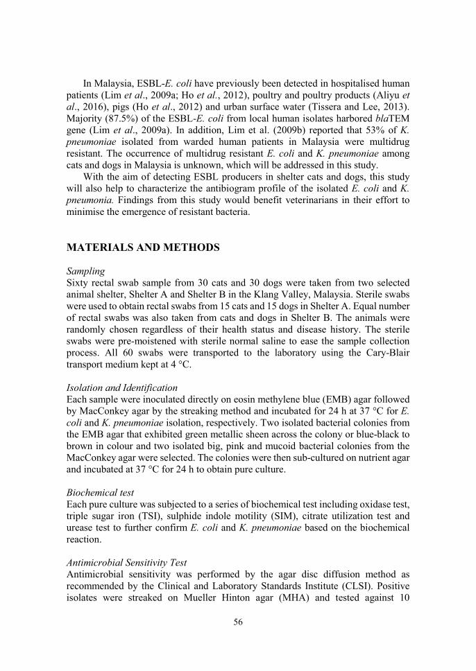
56
In Malaysia, ESBL-E. coli have previously been detected in hospitalised human
patients (Lim et al., 2009a; Ho et al., 2012), poultry and poultry products (Aliyu et al., 2016), pigs (Ho et al., 2012) and urban surface water (Tissera and Lee, 2013). Majority (87.5%) of the ESBL-E. coli from local human isolates harbored blaTEM gene (Lim et al., 2009a). In addition, Lim et al. (2009b) reported that 53% of K. pneumoniae isolated from warded human patients in Malaysia were multidrug resistant. The occurrence of multidrug resistant E. coli and K. pneumoniae among cats and dogs in Malaysia is unknown, which will be addressed in this study.
With the aim of detecting ESBL producers in shelter cats and dogs, this study will also help to characterize the antibiogram profile of the isolated E. coli and K. pneumonia. Findings from this study would benefit veterinarians in their effort to minimise the emergence of resistant bacteria. MATERIALS AND METHODS Sampling Sixty rectal swab sample from 30 cats and 30 dogs were taken from two selected animal shelter, Shelter A and Shelter B in the Klang Valley, Malaysia. Sterile swabs were used to obtain rectal swabs from 15 cats and 15 dogs in Shelter A. Equal number of rectal swabs was also taken from cats and dogs in Shelter B. The animals were randomly chosen regardless of their health status and disease history. The sterile swabs were pre-moistened with sterile normal saline to ease the sample collection process. All 60 swabs were transported to the laboratory using the Cary-Blair transport medium kept at 4 °C. Isolation and Identification Each sample were inoculated directly on eosin methylene blue (EMB) agar followed by MacConkey agar by the streaking method and incubated for 24 h at 37 °C for E. coli and K. pneumoniae isolation, respectively. Two isolated bacterial colonies from the EMB agar that exhibited green metallic sheen across the colony or blue-black to brown in colour and two isolated big, pink and mucoid bacterial colonies from the MacConkey agar were selected. The colonies were then sub-cultured on nutrient agar and incubated at 37 °C for 24 h to obtain pure culture. Biochemical test Each pure culture was subjected to a series of biochemical test including oxidase test, triple sugar iron (TSI), sulphide indole motility (SIM), citrate utilization test and urease test to further confirm E. coli and K. pneumoniae based on the biochemical reaction. Antimicrobial Sensitivity Test Antimicrobial sensitivity was performed by the agar disc diffusion method as recommended by the Clinical and Laboratory Standards Institute (CLSI). Positive isolates were streaked on Mueller Hinton agar (MHA) and tested against 10

57
antimicrobial agents: cephalexin (30µg), meropenem (10µg), aztreonam (30µg), chloramphenicol (30µg), streptomycin (10µg), ampicillin (10µg), amoxicillin (10µg), enrofloxacin (5µg/ml), gentamicin (10µg) and tetracycline (30µg). ESBL Screening and Confirmatory Test Isolates that were categorised as intermediate and resistant to first-generation cephalosporin, cephalexin was selected for ESBL screening test. Disc screening test was performed on the selected isolates using cefotaxime (30µg) and ceftazidime (30µg) on MHA to screen for ESBL producers and measured according to CLSI guideline.
For confirmatory test, isolates presumed to be ESBL producers were further tested with double disc synergy test (DDST) using ceftazidime (30μg), cefotaxime (30μg), and amoxicillin-clavulanate (20 + 10μg) disc. Any distortion or increase in the zone towards the disc of amoxicillin-clavulanate was considered as positive for the ESBL production. RESULTS AND DISCUSSION Positive rectal carrier of ESBL producing E. coli and K. pneumoniae was not detected in the study. The failure to detect ESBL producers might be due to some factors including small sample size, type of sample, and absence of pre-enrichment step.
The level of rectal carriage of E. coli was 85 %, while only 2.4 % of all samples were positive for K. pneumoniae (Figure 1). Four cats and 3 dogs were identified to harbour multiple drug resistant (MDR) E.coli whereas one dog was detected with MDR K. pneumoniae. By the AST, resistance was prevalent to tetracycline, amoxicillin, ampicillin and streptomycin, chloramphenicol, enrofloxacin and gentamicin. The bacteria showed highest resistant to tetracycline at <35 % for E. coli while K. pneumoniae isolates from 4 dogs were 100 % resistant to amoxicillin and ampicillin. These drugs are common antibiotics used in small animal medicine and therefore might contribute to antimicrobial resistance. However, one dog with MDR K. pneumoniae was also resistant to meropenem, an antibotic not commonly used in veterinary medicine. This find may be attributed to the feeding nature of the stray dogs where they are more likely to consume food scraps from the hospital waste or drinking sewage contaminated with resistant bacteria (Holmes et al., 2016). This implies that animals may develop antibiotic resistance without actually consuming the antibiotics. CONCLUSION ESBL producing E. coli and K. pneumoniae was absent from the rectal swabs of cats and dogs from two shelters however a small proportion of samples had MDR-E. coli and MDR-K. pneumoniae. All MDR bacteria strains were resistant to ampicillin, amoxicillin and tetracycline. In contrast, all E. coli were 100% susceptible to cephalexin, aztreonam and meropenem while all K. pneumoniae isolates were
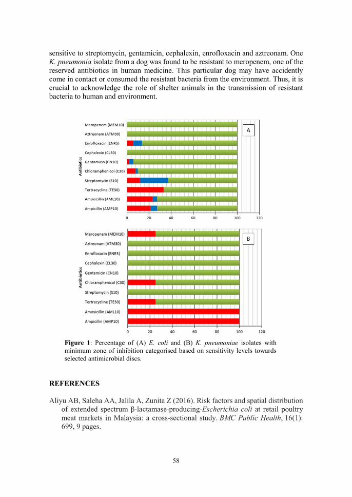
58
sensitive to streptomycin, gentamicin, cephalexin, enrofloxacin and aztreonam. One K. pneumonia isolate from a dog was found to be resistant to meropenem, one of the reserved antibiotics in human medicine. This particular dog may have accidently come in contact or consumed the resistant bacteria from the environment. Thus, it is crucial to acknowledge the role of shelter animals in the transmission of resistant bacteria to human and environment.
Figure 1: Percentage of (A) E. coli and (B) K. pneumoniae isolates with minimum zone of inhibition categorised based on sensitivity levels towards selected antimicrobial discs.
REFERENCES Aliyu AB, Saleha AA, Jalila A, Zunita Z (2016). Risk factors and spatial distribution
of extended spectrum β-lactamase-producing-Escherichia coli at retail poultry meat markets in Malaysia: a cross-sectional study. BMC Public Health, 16(1): 699, 9 pages.

59
Ho WS, Balan G, Puthucheary S, Kong BH, Lim KT, Tan LK, Koh XP, Yeo CC, Thong KL (2012). Prevalence and characterization of multidrug-resistant and extended-spectrum beta-lactamase-producing Escherichia coli from pediatric wards of a Malaysian hospital. Microbial Drug Resistance, 18(4):408-416.
Lim KT, Yasin R, Yeo CC, Puthucheary S, Thong KL (2009a). Characterization of multidrug resistant ESBL-producing Escherichia coli isolates from hospitals in Malaysia. BioMed Research International, Article ID 165637, 10 pages.
Lim K, Yeo C, Md Yasin R, Balan G, Thong K. (2009b). Characterization of multidrug-resistant and extended-spectrum β-lactamase-producing Klebsiella pneumoniae strains from Malaysian hospitals. Journal of Medical Microbiology. 58(11): 1463-1469.
Tissera, S. and Lee, S.M., 2013. Isolation of extended spectrum β-lactamase (ESBL) producing bacteria from urban surface waters in Malaysia. The Malaysian Journal of Medical Sciences, 20(3): 14-22.

60
14th Proceedings of the Seminar on Veterinary Sciences, 18 – 20 September 2018
COMPARISON OF ANTIMICROBIAL RESISTANCE BETWEEN SELECTED BACTERIA ISOLATED FROM EGGS IN
CONVENTIONAL AND ORGANIC PRODUCTION
Fatin Nabilah Idrus, 1*Latiffah Hassan & 2Saleha Abdul Aziz Department of Veterinary Laboratory Diagnosis
Department of Veterinary Pathology and Microbiology Faculty of Veterinary Medicine
Universiti Putra Malaysia, 43400 UPM Serdang, Selangor, Malaysia *Correspondence: [email protected]
ABSTRACT
Organic animal products are increasing in demand due to consumer awareness on food hygiene and safety. Consumers assume that the organic products have better quality and safety therefore are willing to pay more for the products. This study determined the microbiological quality of eggs produced in conventional and organic production and compared the level of antimicrobial resistance of selected bacteria; Salmonella spp., Escherichia coli and Staphylococcus spp. from the eggs. Thirty samples from 6 brands of table eggs were purchased from retail stores and swabs of egg shell and egg content were cultured to isolate bacteria and to perform the standard plate count (SPC) and coliform plate count (CPC) on the samples. The study showed that 93% of eggs were positive for Staphylococcus spp. Disc diffusion assay determined the isolates susceptibility patterns to Penicillin G (10 µg), Erythromycin (15 µg), Tetracycline (30 µg), Ceftriaxone (30 µg), Gentamicin (10 µg) and Clindamycin (2 µg). No significant differences (p> 0.05) was observed in level of antimicrobial resistance between the two types of production. The SPC of egg shells produced in conventional and organic production were 25.84 and 47.06 × 10³ cfu/cm2 respectively, while the SPC of egg content from conventional and organic production were 0.25 and 1.18 × 10³ cfu/g respectively. The CPC of the egg shells from the conventional and organic production were 0.61 and 3.89 × 10³ cfu/cm2 respectively, whereas the egg content was negative for coliform. Microbiological quality was not significantly different (p>0.05) between the two types of production. A total of 93% of eggs were positive for Staphylococcus spp. isolates, indicating that the eggs have been contaminated, either during handling or by the environment. Based on the UNECE Standards 1986 and the Food Regulations 1985, the coliform count in these eggs are beyond safety level, therefore, must heat-treated before consumption. Keywords: antimicrobial resistance, microbiological quality, egg shell, egg content, conventional production, organic production, standard plate count, coliform plate count.

61
INTRODUCTION According to the Department of Veterinary Services Malaysia (2014), organic is a labelling term which indicates a product was produced according to the standards of organic production that is certified by a duly constituted certification body or authority. In Malaysia, there is a growing trend of consumer acceptance and preferance for organic food products with the popular belief that organic food is safer, higher in nutrition value and more delicious (Hassan et al., 2015). However, Engvall and Sundrum described higher potential of microbiological safety risks in organic production because slow-growing breeds of animals are raised outdoors and prohibition of using antimicrobial in the production (cited in Shenghui et al., 2005).
The emergence of antimicrobial resistance (AMR) is a global threat and a great public health concern. Poultry production and husbandry has been linked with high level of antimicrobial usage especially in the intensive or commercial farms (Spitzer, 2016). Organically raised chickens should have minimum exposure to antibiotics. Therefore, in theory the bacteria found in chickens or by-products of chickens raised in this type of production system should show lower level of resistance. As there is demand for organic products, these eggs are sold at higher prices than other commercial eggs. Consumers assume that the organic products have better quality and safer, therefore, are willing to pay the higher price for these eggs. This study compared microbiological quality between organic and conventional eggs and the level of antimicrobial resistance (AMR) of bacteria isolated from these eggs. MATERIALS AND METHODS Thirty table eggs of 6 brands from conventional and organic production systems were purchased from retail stores in Selangor, Malaysia. The conventional production method involves rearing of large numbers birds in temperature controlled and open floor houses (Bartlett et al., 2015), while the organic production system by definition were free from conventionally-produced feedstuffs, genetically modified organisms, animal origin by-products, and synthetic additives (Cobanoglu et al., 2014). Table eggs from organic production were selected based on brands from organic production or free-range farming, and labelled as antibiotic-free. Isolation and Identification A sterile cotton swab was dipped into the rinse solution of the egg shell and placed into a bottle containing 10 mL of buffered peptone water (BPW; OXOID, Basingstoke, England) for isolation of Salmonella spp. Then, the solution was spread onto Chromocult® coliform agar (Merck KGaA, Germany) for isolation of Escherichia coli, Mannitol Salt Agar (MSA; OXOID, Basingstoke, England) for isolation of Staphylococcus spp. Similar method was repeated for isolation of bacteria from the homogenised egg content for each egg sample. Identification of Salmonella spp. and E. coli were done with biochemical tests according to the method described

62
in the Bacteriological Analytical Manual (BAM, 2001). The biochemical tests used in identification of Staphylococcus spp. were based on the principles and methods described by Jang et al., (2005). Antibiotic Sensitivity Test Preparation of inoculum for disk diffusing test was conducted according to the method described in Performance Standards for Antimicrobial Disk Susceptibility Tests developed by Clinical and Laboratory Standards Institute (CLSI, 2015). Standard disc diffusion method is used to test the susceptibility of the organism towards antimicrobial agents. The antimicrobial agents chosen for this study were Penicillin G (10 µg), Erythromycin (15 µg), Tetracycline (30 µg), Ceftriaxone (30 µg), Gentamicin (10 µg) and Clindamycin (2 µg). The susceptibility of the organism towards the antimicrobial agents for each antibiotic disc were determined by measuring the zone of inhibition according to the Disc Diffusion Supplemental Tables adapted from the Performance Standards for Antimicrobial Disk Susceptibility Tests (CSLI, 2015). Standard Plate Count (SPC) and Coliform Plate Count (CPC) The bacterial counts were performed for egg shell and egg content of each egg sample according to the method used in BAM (2001) for standard plate count and coliform plate count. The microbiological standard used as reference was according to the United Nations Economic Commission for Europe standards and Food Regulations 1985 (UNECE, 2009). Statistical Analysis All plates with colonies presenct were counted and the results were computed and calculated by referring the rules of selecting plates and counting colonies (BAM, 2001). Comparison of antimicrobial resistance level and microbiological quality between the conventional and organic of production was done using the non-parametric Mann-Whitney U test at 95% confidence interval. All the calculations were performed using IBM Statistical Package for the Social Sciences (SPSS) version 25 (IBM Corp., Armonk, NY, U.S.A.). RESULTS AND DISCUSSION None of the table eggs from this study showed presence of Salmonella spp. Absence of Salmonella spp. in the egg samples may be attributed by the effective Salmonella spp. control programmes practiced in poultry farms.
The isolation of E. coli from egg shells and contents was performed by using differential Chromocult® coliform agar. Very few or no light-blue colonies were present. According to the Merck Microbiology Manual (2005), other gram-negative bacteria would produce colourless or light-blue to turquoise colonies on the Chromocult® coliform agar plate. However, the Gram-staining performed on the light-blue colonies showed cellular morphologies of Gram-positive cocci in clusters.
63
It is possible that a laboratory accident may have occurred, since even the positive control of E. coli ATCC 25922 on the Chromocult® coliform agar did not produce growth.
Twenty-nine (93%) egg samples were positive for Staphylococcus spp. (Table 1), but no Staphylococcus aureus was recovered. The antimicrobial resistance level of the isolates recovered from the two types of production (Table 2) showed a similar trend in antimicrobial resistance, the highest resistance recorded, in order, was for tetracycline, clindamycin, and erythromycin. This is suggested to be due to the rampant use of the antimicrobial agents in chickens and feeds (Ali et al., 2017). It is also possible that the resistance was acquired from exposure to environmental bacteria in the farms. There was no significant difference (p>0.05) in antimicrobial resistance level between isolates of eggs from conventional and organic farming systems (Table 3). The eggs from organic production showed a non-significant higher (p>0.05) bacterial count than those for conventional production (Table 4).
It should be noted that antimicrobial resistance in bacteria is not from abuse of antimicrobials alone, in fact, antimicrobial resistance may also be disseminated from a complex of ecologic and genetic factors. Among these factors are age, type of production, and environmental pressures (Marshall and Levy, 2011).
Table 1. Isolation of Staphylococcus spp. from eggs of conventional and organic production.
Type of Production
Sample Staphylococcus spp.
Egg site Number Positive Samples
Coagulase- negative
Coagulase-positive
Conventional Shell 15 14 6 14
Content 15 3 0 3
Organic Shell 15 14 2 13 Content 15 2 0 2
Total 29 8 32
Table 2. The antibiotic resistance profiles (%) of the Staphylococcus spp. isolates from egg shells and egg contents of conventional and organic production
Type of Production
No. of Isolates
No. of antibiotic resistance isolates
P10 E15 T30 CRO30 CN10 DA2
Conventional 23 3 (13) 10 (43) 16 (69) 2(8) 0 14 (60)
Organic 17 5 (29) 10 (58) 14 (82) 1 (5) 0 13 (76)
Number in brackets = %; P10=Penicillin G (10 µg), E15=Erythromycin (15 µg), T30=Tetracycline (30 µg), CRO30=Ceftriaxone (30 µg), CN10= Gentamicin (10 µg), DA2=Clindamycin (2 µg).

64
CONCLUSION The antimicrobial resistance level and microbiological quality of eggs from conventional and organic production methods were similar. Therefore, based on these findings, there is no clear benefit from consumption of organically produced eggs. REFERENCES Ali Y, Islam MA, Muzahid NH, Faruk Sikder MO, Hossain MA, Marzan LW, (2017).
Characterization, prevalence and antibiogram study of Staphylococcus aureus in poultry. Asian Pacific Journal of Tropical Biomedicine, 7(3):253-256
BAM (Bacteriological analytical manual). (2001). https://www.fda.gov/food/foodscienceresearch/laboratorymethods/ucm2006949.htm. (Accessed on 24 August 2018)
Table 3. Antimicrobial resistance level of Staphylococcus spp. isolates from conventional and organic production.
Antibiotic
Type of Production
p-value Conventional
(n=17) Organic (n=16)
Antimicrobial resistance (%)
Penicillin G 13 29 0.281 Erythromycin 43 58 0.399
Tetracycline 69 82 0.085
Ceftriaxone 8 5 0.353
Gentamicin 0 0 0.836
Clindamycin 60 76 0.160
Table 4. Microbial count of eggs from conventional and organic product.
Type of Production
Egg site No of
Samples SPC (× 103)
(cfu/g) CPC (× 103)
(cfu/g)
Conventional Shell 15 25.84 0.61
Content 15 0.25 NA
Organic Shell 15 47.06
3.89
Content 15 1.18 NA
SPC=Standard plate count; CPC=coliform plate count; NA=Not available

65
Bartlett JR, Liles KM, Beckford RC, (2015). Comparing the Effects of Conventional and Pastured Poultry Production Systems on Broiler Performance and Meat Quality. Journal of Agriculture and Life Sciences, 2:2375-4222
CLSI (Clinical and Laboratory Standards Institute) (2015). Performance Standards for Antimicrobial Disk Susceptibility Tests. Retrieved from https://www.researchgate.net/file.PostFileLoader.html?id=581d9d8fcbd5c2f99c04d4b1&assetKey=AS%3A424985668919296%401478335887189 (Accessed on 24 August 2018)
Cobanoglu F, Kucukyilmaz K, Cinar M, Bozkurt M, Catli AU, Bintas E, (2014). Comparing the Profitability of Organic and Conventional Broiler Production. Brazilian Journal of Poultry Science, 16: 89-96
Hassan SH, Loh WY, Kok JR, (2015). Purchasing intention towards organic food among generation Y in Malaysia. Journal of Agribusiness Marketing, 7:16-32.
Jang SS, Hirsh DC, Biberstein, EL, (2005). Diagnostic Manual of Veterinary Clinical Bacteriology and Mycology, revised edn. Davis, CA: University of California Press, 2005.
Marshall BM and Levy, SB, (2011). Food Animals and Antimicrobials: Impacts on Human Health. Clinical Microbiology Reviews, 24 (4):718-733
Merck Microbiology Manual (12th Edition). (2005). Darmstadt, Germany: Merck. Shenghui C, Beilei G, Jie Z, Jianghong, M, (2005). Prevalence and antimicrobial
resistance of Campylobacter spp. and Salmonella serovars in organic chickens from Maryland retail stores. Applied and Environmental Microbiology, 71(7):4108-4111.
Spitzer H (2016). An analysis of bacterial contamination of chicken eggs and antimicrobial resistance. All College Thesis Program. Paper 27. http://digitalcommons.csbsju.edu/honors_thesis/27 (Accessed on 24 August 2018)
UNECE (United Nations Economic Commission for Europe). (2009). UNECE standard for hen egg products for use in the food industry. Retrieved from https://www.unece.org/fileadmin/DAM/trade/agr/standard/eggs/e/63produc.pdf. (Accessed on 24 August 2018)

66
14th Proceedings of the Seminar on Veterinary Sciences, 18 - 20th September 2018
ANTIFUNGAL PROPERTIES OF APPLE CIDER VINEGAR ON CANDIDA ALBICANS, CANDIDA KRUSEI AND CANDIDA PARAPSILOSIS
Afiqah Shahirah Anwar Mirza, 1*Mazlina Mazlan & 1Sharina Omar
1Department of Veterinary Pathology and Microbiology Faculty of Veterinary Medicine
Universiti Putra Malaysia, 43400 UPM Serdang, Selangor, Malaysia *Correspondence: [email protected]
ABSTRACT
Apple cider vinegar (ACV) is a cheap, easily obtainable, fermented solution commonly prescribed in folk medicine for health benefit including as an antifungal agent. Candidiasis can be caused by many species of Candida, particularly Candida albicans and Candida parapsilosis. There is concern on increase of occurrence of antimicrobial resistance of Candida towards the first- and second-line antifungal drugs. Moreover, treatment of candidiasis can be costly with prolonged treatment duration. Thus, an alternative treatment to combat fungal diseases is highly warranted. Therefore, this study aimed to evaluate the effects of ACV on C. albicans, C. parapsilosis and C. krusei. In this study, commercial ACV with 5% acidity was used to determine the minimum inhibitory concentration (MIC) and minimum fungicidal concentration (MFC) on the fungi, using the 96-well plate microdilution technique. Itraconazole was used as the control. The study revealed that 50% ACV inhibited growth of C. albicans and C. krusei while 25% ACV was sufficient to inhibit growth and kill C. parapsilosis. The results suggest that ACV can be used in the treatment of candidiasis. Keywords: apple cider vinegar, antifungal, MIC, MFC, Candida
INTRODUCTION Antimicrobial resistance is an increasingly serious problem in medicine. Among microbes, several species of Candida have been reported to develop resistance towards the first and second line of antifungal drugs. For example, among fungal isolates from canine cystitis, 59% Candida albicans isolates were resistant to fluconazole and 43% to amphotericin B (Bhanderi et al., 2009). Another study (Santhanam et al., 2013) showed that 22% of Candida isolates were resistant to at least one antifungal drug, two isolates had multi-drug resistance, and one was resistant to seven antifungal drugs.
Conventional treatments of fungal disease are few, and current available antifungal drugs have limited spectrum, are expensive, and require prolonged application (Silva Cde et al., 2008). In addition, prolonged use of antifungal drugs
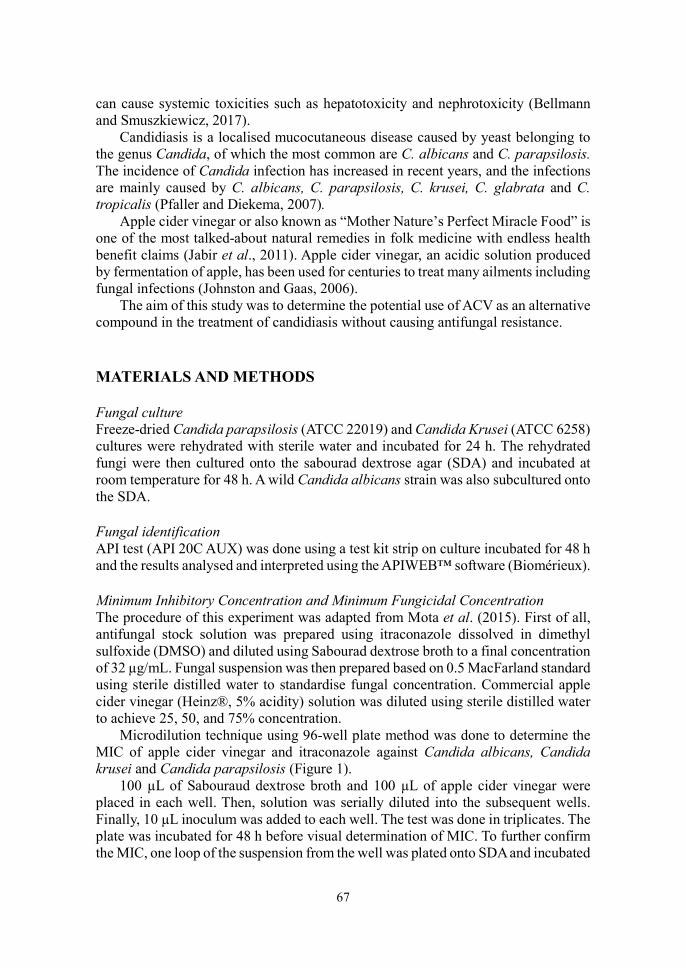
67
can cause systemic toxicities such as hepatotoxicity and nephrotoxicity (Bellmann and Smuszkiewicz, 2017).
Candidiasis is a localised mucocutaneous disease caused by yeast belonging to the genus Candida, of which the most common are C. albicans and C. parapsilosis. The incidence of Candida infection has increased in recent years, and the infections are mainly caused by C. albicans, C. parapsilosis, C. krusei, C. glabrata and C. tropicalis (Pfaller and Diekema, 2007).
Apple cider vinegar or also known as “Mother Nature’s Perfect Miracle Food” is one of the most talked-about natural remedies in folk medicine with endless health benefit claims (Jabir et al., 2011). Apple cider vinegar, an acidic solution produced by fermentation of apple, has been used for centuries to treat many ailments including fungal infections (Johnston and Gaas, 2006).
The aim of this study was to determine the potential use of ACV as an alternative compound in the treatment of candidiasis without causing antifungal resistance. MATERIALS AND METHODS Fungal culture Freeze-dried Candida parapsilosis (ATCC 22019) and Candida Krusei (ATCC 6258) cultures were rehydrated with sterile water and incubated for 24 h. The rehydrated fungi were then cultured onto the sabourad dextrose agar (SDA) and incubated at room temperature for 48 h. A wild Candida albicans strain was also subcultured onto the SDA. Fungal identification API test (API 20C AUX) was done using a test kit strip on culture incubated for 48 h and the results analysed and interpreted using the APIWEB™ software (Biomérieux). Minimum Inhibitory Concentration and Minimum Fungicidal Concentration The procedure of this experiment was adapted from Mota et al. (2015). First of all, antifungal stock solution was prepared using itraconazole dissolved in dimethyl sulfoxide (DMSO) and diluted using Sabourad dextrose broth to a final concentration of 32 µg/mL. Fungal suspension was then prepared based on 0.5 MacFarland standard using sterile distilled water to standardise fungal concentration. Commercial apple cider vinegar (Heinz®, 5% acidity) solution was diluted using sterile distilled water to achieve 25, 50, and 75% concentration.
Microdilution technique using 96-well plate method was done to determine the MIC of apple cider vinegar and itraconazole against Candida albicans, Candida krusei and Candida parapsilosis (Figure 1).
100 µL of Sabouraud dextrose broth and 100 µL of apple cider vinegar were placed in each well. Then, solution was serially diluted into the subsequent wells. Finally, 10 µL inoculum was added to each well. The test was done in triplicates. The plate was incubated for 48 h before visual determination of MIC. To further confirm the MIC, one loop of the suspension from the well was plated onto SDA and incubated
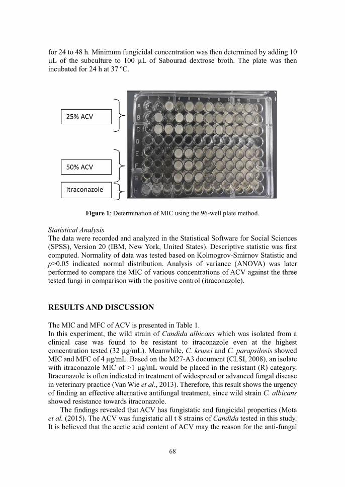
68
for 24 to 48 h. Minimum fungicidal concentration was then determined by adding 10 µL of the subculture to 100 µL of Sabourad dextrose broth. The plate was then incubated for 24 h at 37 ºC.
Figure 1: Determination of MIC using the 96-well plate method.
Statistical Analysis The data were recorded and analyzed in the Statistical Software for Social Sciences (SPSS), Version 20 (IBM, New York, United States). Descriptive statistic was first computed. Normality of data was tested based on Kolmogrov-Smirnov Statistic and p>0.05 indicated normal distribution. Analysis of variance (ANOVA) was later performed to compare the MIC of various concentrations of ACV against the three tested fungi in comparison with the positive control (itraconazole). RESULTS AND DISCUSSION The MIC and MFC of ACV is presented in Table 1. In this experiment, the wild strain of Candida albicans which was isolated from a clinical case was found to be resistant to itraconazole even at the highest concentration tested (32 µg/mL). Meanwhile, C. krusei and C. parapsilosis showed MIC and MFC of 4 µg/mL. Based on the M27-A3 document (CLSI, 2008), an isolate with itraconazole MIC of >1 μg/mL would be placed in the resistant (R) category. Itraconazole is often indicated in treatment of widespread or advanced fungal disease in veterinary practice (Van Wie et al., 2013). Therefore, this result shows the urgency of finding an effective alternative antifungal treatment, since wild strain C. albicans showed resistance towards itraconazole.
The findings revealed that ACV has fungistatic and fungicidal properties (Mota et al. (2015). The ACV was fungistatic all t 8 strains of Candida tested in this study. It is believed that the acetic acid content of ACV may the reason for the anti-fungal
25% ACV
50% ACV
Itraconazole
69
properties. Increasing the dilution of ACV results in increase in pH. It has been proposed that acetic acid in ACV inhibits fungal growth by causing loss of cell integrity (León Peláez et al, 2012).
Table 1: Summary of result of MIC and MFC of selected Candida spp. against apple cider vinegar.
Fungus Apple cider vinegar (%)
Itraconazole 25 50 75 100
Candida albicans R S S S R Candida krusei R S S S R Candida parapsilosis S S S S R R=resistant, S=susceptible
The fact that ACV with antifungal properties is cheap, safe, easily obtainable makes the solution a good safer alternative to standard antifungal drugs for the treatment of localised infection such dermatitis, otomycosis and stomatitis caused by the Candida spp. CONCLUSION The findings from this study suggest that ACV has antifungal property against C. albicans, C. krusei, and C. parapsilosis and thus, can be used as therapeutic alternative for candidiasis. Apple cider vinegar at 50% dilution can inhibit the growth and can kill C. albicans and C. krusei while 25% ACV can kill and inhibit the growth of C. parapsilosis. REFERENCES Bellmann R and Smuszkiewicz P, (2017). Pharmacokinetics of antifungal drugs:
practical implications for optimized treatment of patients. Infection, 45(6):737-779.
Bhanderi BB, Yadav MM, Roy A, (2009). Antifungal drug resistance - concerns for veterinarians. Veterinary World, 2(5):204-207.
Jabir HB, Abbas FN, Khalaf RM, (2011). In vitro assessment of antifungal potential of apple cider vinegar and acetic acid versus fluconazole in clinical isolates of otomycosis. Thi-Qar Medical Journal, 5(1):126-133.
Johnston CS and Gaas CA, (2006). Vinegar: medicinal uses and antiglycemic effect. Medscape General Medicine, 8(2):61-68.

70
León Peláez AM, Serna Cataño CA, Quintero Yepes EA, Gamba Villarroel RR, De Antoni GL, Giannuzzi L, (2012). Inhibitory activity of lactic and acetic acid on Aspergillus flavus growth for food preservation. Food control, 24(1-2):177-183.
Mota AC, de Castro RD, de Araújo Oliveira J, de Oliveira Lima E, (2015). Antifungal activity of apple cider vinegar on Candida species involved in denture stomatitis. Journal of Prosthodontists, 24(4):296-302.
CLSI (Clinical and Laboratory Standards Institute) (2008). M27-A3. Reference method for broth dilution antifungal susceptibilit testing of yeast; approved standard – Third Edition. ISBN 1-56238-666-2, ISSN 0273-3099. https://clsi.org/media/1461/m27a3_sample.pdf (Accessed on 15 October 2019)
Pfaller MA and Diekema DJ, (2007). Epidemiology of invasive candidiasis: a persistent public health problem. Clinical Microbiology Reviews, 20(1):133-163.
Santhanam J, Nazmiah N, Aziz MN, (2013). Species distribution and antifungal susceptibility patterns of Candida species: Is low susceptibility to itraconazole a trend in Malaysia? Medical Journal of Malaysia, 68(4):343-347.
Silva Cde B, Guterres SS, Weisheimer V, Schapoval EE, (2008). Antifungal activity of the lemongrass oil and citral against Candida spp. Brazilian Journal of Infectious Diseases, 12(1):63-66.
Van Wie E, Chen AV, Thomovsky SA, Tucker RL, (2013). Successful long-term use of itraconazole for the treatment of Aspergillus diskospondylitis in a dog. Case Reports in Veterinary Medicine, 2013, Article ID 907276, 4 pages.
71
13th Proceedings of the Seminar on Veterinary Sciences, 18 – 20 September 2018
CLINICAL, LABORATORY, AND HISTOLOGICAL INVESTIGATION IN CATS NATURALLY INFECTED WITH
LEPTOSPIRA SP.
Zher Min Tan, 1*Lau Seng Fong, 2Siti Khairani Bejo, 2,3Annas Salleh, & 1Rozanaliza Radzi
1Department of Veterinary Clinical Studies 2Department of Veterinary Pathology and Microbiology
3Research Centre for Ruminant Diseases Faculty of Veterinary Medicine
Universiti Putra Malaysia, 43400 UPM Serdang, Selangor, Malaysia *Correspondence: [email protected]
ABSTRACT
Leptospirosis is a zoonotic disease that can infect cattle, dogs, swine, horses, and humans. This study described the histopathological changes in the kidney, liver, and lungs of cats naturally infected with Leptospira sp. and correlated the changes with their kidney and liver functions. Kidney, liver, and lung samples obtained from cats (n=12) diagnosed with leptospirosis were fixed in 10% neutral-buffered formalin. The samples were sectioned and stained with Hematoxylin-Eosin (H&E) and Warthin-Starry (WS) staina. Among the kidney samples, one (8%) showed mild lesions and 11 (92%) showed moderate lesions. The predominant lesions were glomerulitis and renal tubular degeneration. Eight infected cats (67%) showed livers with mild lesions, while 4 (33%) with moderate lesions. The predominant findings in the livers were congestion and Kupffer cell hyperplasia. Generally, the BUN, creatinine, and ALT were normal, and these findings did not correlate with observed histopathological changes in the kidneys and liver. In the lung, the major lesions were emphysema, atelectasis, interalveolar septal thickening, and congestion. Two (17%) of the kidneys were positive for spirochaetes. From our results, natural infection with the Leptospira sp. produced have lesions in the kidneys, livers, and lungs of cats. The presence of spirochaetes in two cats showed that the organism is shed in urine. Keywords: cat, Leptospira, histopathology, Hematoxylin and Eosin, Warthin-Starry stain
INTRODUCTION Leptospirosis is now an emerging zoonotic disease in Malaysia (Thayaparan et al., 2013). Leptospirosis is an occupational hazard for high risk groups, such as workers in agricultural sectors, search and rescue workers in high risk environments, sewerage

72
workers, and livestock handlers (Garba et al., 2017). The infection can transmit from animals, such cats, to humans. However, in Malaysia, there is little information on the association between cats and leptospirosis. Histopathology is a diagnostic aid for leptospirosis that has been used in many species, including dogs, rodents, cattle, and swine but in cats. Hence, the objectives of this study were to determine the histopathology and function of the kidneys, liver, and lungs of cats diagnosed with leptospirosis. MATERIALS AND METHODS Samples Stray cats from two animal shelters to be euthanised were recruited for the study. Blood samples were collected in plain venoject tubes (Terumo™) and serum used to determine urea, creatinine, and alanine transaminase (ALT) concentrations.microscopic agglutination test (MAT) and polymerase chain reaction (PCR). Serum samples were also tested for antibodies titre against 20 Leptospira serovars such as Hardjobovis, Hebdomadis, Celledoni, Malaysia, Pomona, Tarassovi, Pyrogenes, Australis, Grippothyposa, Cynopteri, Canicola, Lai, Icterohaemorrhagiae, Bataviae, Javanica, Autumnalis, Ballum, Djasiman, Copenhageni, and Patoc for microscopic agglutination test (MAT). Polymerase chain reaction (PCR) was also conducted to detect the DNA of pathogenic Leptospira species in serum and urine samples. Cats positive for both MAT and PCR were euthanised by administering an overdose (60 mg/kg) of sodium pentobarbital and their kidneys, liver and lungs fixed in formalin, sectioned, stained and examined histopathologically. Staining Tissues sections were either stained with Haematoxylin and Eosin (H&E) (Fischer et al., 2008) or Warthin-Starry Silver stain (Warthin and Starry, 1922). Lesion scoring Kidney sections stained with H&E examined microscopically on 5 randomly selected views. These lesions were graded based on a quantitative histopathological scoring scheme described in AbuBakar and Zamri-Saad (2011) and Annas et. al., (2014) where 0=normal or no significant lesion, 1=mild lesion, 2=moderate lesion, and 3: severe lesion. The lesions were interstitial nephritis, tubular dilatation, tubular degeneration, necrosis, glomerulitis, thickened Bowman’s capsule, interstitial oedema, and renal congestion.
Liver sections were also scored microscopically on 5 random views based on the following: sinusoid dilatation, congestion, fibrosis, Kupffer cell hyperplasia, hepatocytes degeneration, hepatocytes necrosis, fatty infiltration, and inflammatory infiltrates.
The lesions in lungs sections were not scored, instead recorded descriptively. Warthin-Starry Silver-stained kidney and liver sections were viewed to detect spirochaetes. Stained sections had pale yellow to brown background while the
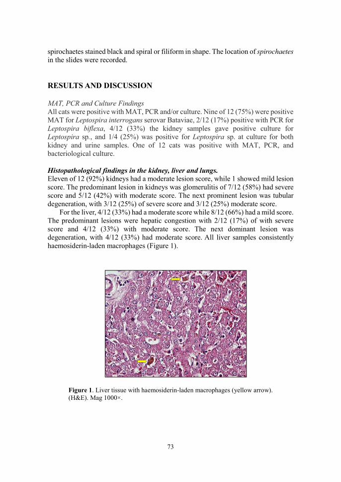
73
spirochaetes stained black and spiral or filiform in shape. The location of spirochaetes in the slides were recorded. RESULTS AND DISCUSSION MAT, PCR and Culture Findings All cats were positive with MAT, PCR and/or culture. Nine of 12 (75%) were positive MAT for Leptospira interrogans serovar Bataviae, 2/12 (17%) positive with PCR for Leptospira biflexa, 4/12 (33%) the kidney samples gave positive culture for Leptospira sp., and 1/4 (25%) was positive for Leptospira sp. at culture for both kidney and urine samples. One of 12 cats was positive with MAT, PCR, and bacteriological culture. Histopathological findings in the kidney, liver and lungs. Eleven of 12 (92%) kidneys had a moderate lesion score, while 1 showed mild lesion score. The predominant lesion in kidneys was glomerulitis of 7/12 (58%) had severe score and 5/12 (42%) with moderate score. The next prominent lesion was tubular degeneration, with 3/12 (25%) of severe score and 3/12 (25%) moderate score.
For the liver, 4/12 (33%) had a moderate score while 8/12 (66%) had a mild score. The predominant lesions were hepatic congestion with 2/12 (17%) of with severe score and 4/12 (33%) with moderate score. The next dominant lesion was degeneration, with 4/12 (33%) had moderate score. All liver samples consistently haemosiderin-laden macrophages (Figure 1).
Figure 1. Liver tissue with haemosiderin-laden macrophages (yellow arrow). (H&E). Mag 1000×.
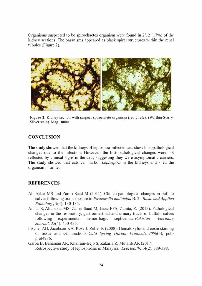
74
Organisms suspected to be spirochaetes organism were found in 2/12 (17%) of the kidney sections. The organisms appeared as black spiral structures within the renal tubules (Figure 2).
Figure 2. Kidney section with suspect spirochaete organism (red circle). (Warthin-Starry Silver stain). Mag 1000×.
CONCLUSION The study showed that the kidneys of leptospira-infected cats show histopathological changes due to the infection. However, the histopathological changes were not reflected by clinical signs in the cats, suggesting they were asymptomatic carriers. The study showed that cats can harbor Leptospira in the kidneys and shed the organism in urine.
REFERENCES Abubakar MS and Zamri‐Saad M (2011). Clinico‐pathological changes in buffalo
calves following oral exposure to Pasteurella multocida B: 2. Basic and Applied Pathology, 4(4), 130-135.
Annas S, Abubakar MS, Zamri-Saad M, Jesse FFA, Zunita, Z. (2015). Pathological changes in the respiratory, gastrointestinal and urinary tracts of buffalo calves following experimental hemorrhagic septicemia. Pakistan Veterinary Journal, 35(4): 430-435.
Fischer AH, Jacobson KA, Rose J, Zeller R (2008). Hematoxylin and eosin staining of tissue and cell sections. Cold Spring Harbor Protocols, 2008(5), pdb-prot4986.
Garba B, Bahaman AR, Khairani-Bejo S, Zakaria Z, Mutalib AR (2017). Retrospective study of leptospirosis in Malaysia. EcoHealth, 14(2), 389-398.

75
Klarenbeek A, and Schuffner WAP (1933). Appearance in Holland of leptospira differing from Weil strain. Nederlands Tijdschrift voor Geneeskunde 77: 4271- 4276
Sutherland A, Simmons G, Kenny G (1949). Bovine leptospirosis. Three outbreaks in calves in Queensland. Australian Veterinary Journal, 25(9), 197-202.
Thayaparan S, Robertson ID, Fairuz A, Suut L, Abdullah MT (2013). Leptospirosis, an emerging zoonotic disease in Malaysia. Malaysian Journal of Pathology, 35(2):123-132.
Warthin AS and Starry AC (1922). The staining of spirochetes in cover-glass smears by the silver-agar method. The Journal of Infectious Diseases, 30(6):592-600.
76
14th Proceedings of the Seminar on Veterinary Sciences, 18 – 20 September 2018
RETROSPECTIVE STUDY ON AQUATIC ANIMAL DISEASES CASE REPORTS AND CANVASSING A BUSINESS MODEL USING
CASES SUBMITTED TO AQUATIC ANIMAL HEALTH UNIT, FACULTY OF VETERINARY MEDICINE,
UNIVERSITI PUTRA MALAYSIA
Hidayatu Husna Selahuddeen, 1*Norhariani Mohd Nor & 1Hassan Haji Mohd Daud
1Department of Veterinary Preclinical Sciences 2Department of Veterinary Clinical Studies
Faculty of Veterinary Medicine, Universiti Putra Malaysia, 43400 UPM Serdang, Selangor, Malaysia
*Correspondence: [email protected]
ABSTRACT
Aquatic Animal Health Unit (AAHU) has been receiving samples for disease diagnosis and screening. However, case reports over the past 10 years were not analysed leading to lack of information on trends of disease cases and revenue. Thus, this study was carried out to identify the trends of disease, diagnosis, revenue and canvas out the business model for AAHU. The case reports (851 cases, 14 variables) from January 2008 to December 2017 were collected and edited using Microsoft Excel® and analysed using IBM® SPSS Statistic 25. Results showed over the 10 years, the average total number of cases handled were 85 and average total profit margin was RM198,107.65 with Polymerase Chain Reaction (PCR) assay charges being the largest contributor (50%). The common disease agents found were Streptococcus agalactiae (12%), Aeromonas hydrophila type ii (12%), Dactylogyrus sp (35%), Saprolegnia sp. (5%), and Iridovirus (13%). These cases decreased over the years as the number of cases decreased by 21%. The trend of these diseases was not an indicator of prevalence of aquatic animal diseases in Malaysia, since the cases were by chance. A business model canvas (BMC) was made comprising customer segment (focus more on individuals owning aquarium fishes), value proposition (proposing annual screening of common agents), channel (word of mouth and website) and improving the revenue stream (aquaculture herd health), key activities (clear standard of procedures (SOP) and key partners (form cooperation with private labs and rekindle ties with Department of Fisheries). To conclude, the revised BMC would be able to help AAHU to sustain its business abilities. Keywords: Business Model Canvas (BMC), retrospective, aquaculture, disease agents
77
INTRODUCTION Aquatic Animal Health Unit (AAHU) is a part of Veterinary Laboratory Service Unit in Faculty of Veterinary Medicine, Universiti Putra Malaysia. It provides disease diagnosis and screening services for aquatic industry and teaching cases for DVM students. Case reports coming in to AAHU vary by species and purpose as samples came both from fish hobbyist and aquaculture entrepreneurs. The cases are categorised into fish, crustaceans, and others (OIE, 2003).
Business model on how a business creates, delivers and captures value has been proposed (Osterwalder and Pigneur, 2010). Business model canvas (BMC) is a tool that is easy and quick to used and can be changed once the ideas has been tested. Using this tool, the managers can seek for ways to improve a business organisation such as AAHU profitability.
Thus, this study was conducted to identify trends of disease, diagnosis, revenue and canvas out the business model for Aquatic Animal Health Unit (AAHU), Universiti Putra Malaysia. MATERIALS AND METHOD Case reports from January 2008 to December 2017 were collected from AAHU records. A total of 851 cases were submitted to AAHU within the study period. The variables in case reports include sample origin, date and year of sampling, type and species of samples, examination requested (e.g. disease diagnosis, screening, post mortem, histopathology and bacteriology) results, and total cost charged. There were a total of 27 raw variables and new variables were added amounting to a total of 228 variables. The new variables included species of bacteria, parasite and virus diagnosed, and diagnostic tests requested (e.g. PCR, bacterial culture, API20E, BBL Crystal and parasite indentification). Data was collected and edited using Microsoft Excel and were then analysed using IBM SPSS Statistic version 25. Business model for AAHU was constructed using information from data analysis on type of customer, revenue, type of samples and species sent and type of diagnosis. An alternative business model for AAHU was constructed. RESULTS AND DISCUSSION Figure 1 shows the type of aquatic animals submitted to AAHU by year over the period of 2008 to 2017. Fish are classified as vertebrates with gills and fins while crustaceans are shrimp and those with chitinous exoskeleton. Others in this study refer to those other than fish and crustaceans, which were turtle, bivalves and artemia. Generally, there was increasing trend of cases with fish cases topping the chart. Fish cases began to decrease in 2010 and crustacean cases increasing in 2013. However, the scenario did not reflect disease trend because cases submitted were by chance.

78
This was shown when Malaysia’s shrimp industry was affected by early mortality syndrome (EMS) in 2010 (Lee and Wee, 2014).
Figure 1. Type of aquatic animal sent to AAHU by year Figure 2 reflects the species of fish submitted to AAHU where groupers (114 cases) and koi (99 cases) form the species most often received by the unit. In August 2016, a rich in omega-3 jade perch commercial project was launched Terengganu and Malacca. During that period, AAHU received increased number of cases for disease diagnosis.
Figure 2. Fish species sent to AAHU from 2008 to 2017.
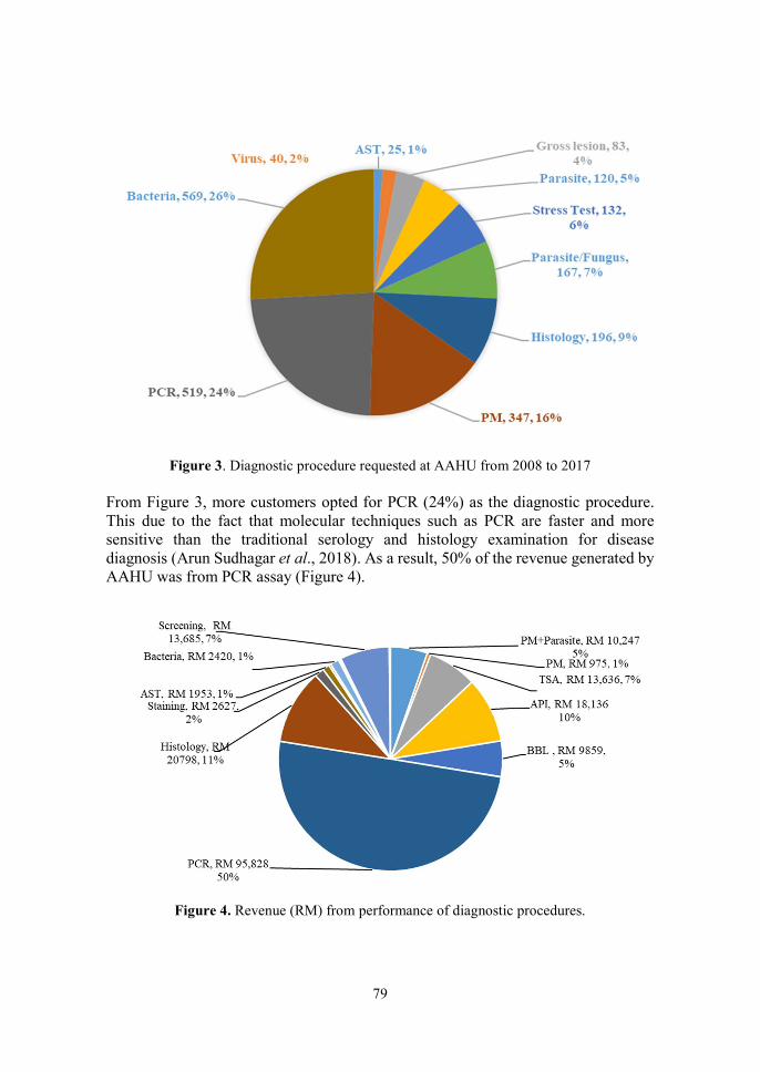
79
Figure 3. Diagnostic procedure requested at AAHU from 2008 to 2017
From Figure 3, more customers opted for PCR (24%) as the diagnostic procedure. This due to the fact that molecular techniques such as PCR are faster and more sensitive than the traditional serology and histology examination for disease diagnosis (Arun Sudhagar et al., 2018). As a result, 50% of the revenue generated by AAHU was from PCR assay (Figure 4).
Figure 4. Revenue (RM) from performance of diagnostic procedures.

80
Table 1. Type of cases submitted to Aquatic Animal Health Unit, Faculty of Veterinary Medicine, Universiti Putra Malaysia and total revenue
Year
Type of cases (Number) Revenue
(RM) Bacterial Viral Parasitic Fungal
2008
2009
2010
2011
2012
2013
2014
2015
2016
2017
17
76
125
108
111
48
60
36
45
42
55
79
55
43
40
15
31
23
37
15
32
64
62
50
35
16
29
24
36
41
1
0
5
11
20
12
28
23
34
33
22,015.00
27465.00
28712.00
29513.00
22488.00
19168.10
10813.00
10240.50
14719.22
11973.86
The bacteria identified most commonly identified in the cases at AAHU were A. hydrophila type II and S. agalactiae. The number of cases began to decline after 2010 and the number of cases decreased by 21% over the 10-year period (Table 1).
The same goes for the total revenue generated by AAHU, which began to decrease after 2011 and was lowest in 2014 and 2015. The revenue decreases 12% over the 10-year period.
In the proposed Business Model using the Ostenwalder and Pigneur Business Model Canvas (Figure 5), the plan was to focus more on individuals and aquaculture companies for new businesses for AAHU. Ornamental fish are exported to Singapore, thus, the Agri-Food and Veterinary Authority of Singapore (2015) stipulates that they must be free from diseases. The value we are proposing for AAHU services is the ISO 17025 accreditation for laboratory services. This will attract customers to use the services of AAHU. Increase in cases referred to the Unit will also benefit veterinary students by way of providing opportunity for have hands-on experience on the diagnosis of aquatic cases.
To deliver the values of diagnostic services at AAHU to the customers, the channels must be created as a means of promotion through the social media profiles. Currently, the only online information available on AAHU is via the Veterinary Laboratory Services Unit (VLSU), UPM website. The website need upgrading for easy access while providing relevant information on AAHU. The Unit can also market its services at agriculture exhibitions. The Unit must also provide good pre-and after sale services to attract customers.
To increase revenue AAHU should introduce subscription membership to improve clientele. The unit need to be upgraded with up-to-date facilities, techniques and methods of diagnosis. This entails training of technicians to acquire new skills

81
and expertise. The Unit can initiate ‘Partnership Schemes’ and cooperative work with other government and non-governmental organisations. with relevant organisations the management of AAHU can also be improved to reduce time and material wastage.
CONCLUSION The AAHU received a total of 851 diagnostic cases from January 2008 to December 2017 of which 42% bacterial, 24% viral and parasitic, and 10% fungal cases. The number of cases began to decline after 2010 and and fluctuated thereon. The number of cases decreased by 21% while the by 12% over the 10-year period. The Business Model Canvas proposed in this study is expected to improve the business sustainability of AAHU by upgrading value proposition through the ISO certification and improving the key activities to improve visibility and marketability of the Unit.
Key Partner -Establish cooperation with private labs in Malaysia -Department of Fisheries
Key Activities -Marketing -Staff training -Foster farms for teaching purposes -Standard Operating Procedures
Value Proposition -ISO/IEC 17025 -Annual screening for subscribed customers
Customer Relationship -After sales service -Word of mouth
Customer Segments -Individuals and companies owning aquarium fishes
Key Resources -Lab technician -Upgrade the building and facilities
Channels -Social media -Website upgrade -Agriculture exposition
Cost Structure -Salary for lab technicians -Raw materials for annual screening -Building and facilities
Revenue Streams -Membership of subscribed customers -From ISO certification -New customers
Figure 5. Proposed Business Model using the Business Model Canvas (Ostenwalder and Pigneur, 2010)

82
REFERENCES Agri-Food and Veterinary Authority of Singapore. (2015). https://www.ava.gov.sg/ (Accessed on 1 October 2018). Arun Sudhagar S, Ezhil Nilavan S, Linga Prabu D, Rathi Bhuvaneswari G,
Chandrasekar S and Rajesh kumar R. Diagnostic Tools Used in Fish Disease Diagnosis. Aquafind. http://aquafind.com/articles/FishDiseaseDiagnosis.php
Accessed on 30 August 2018). Lee SW and Wee W (2014). Diseases in Aquaculture. Research Journal of Animal
and Veterinary Sciences, 7(1): 1-6. Osterwalder A and Pigneur Y (2010). Busines Model Generation. John Wiley and
Sons Ltd, United Kingdom. OIE (Office International Des Epizooties - World Organisation for Animal Health)
(2003). Aquatic Animal Health Code. Paris, France. https://rr-africa.oie.int/docspdf/en/Codes/en_csaa.pdf (Accessed on 16 October 2019) Osterwalder A and Pigneur Y (Editors) (2010). Business Model Generation: A
handbook for visionaries, game changers, and challengers. John Wiley and Sons Ltd, United Kingdom.
83
14th Proceedings of the Seminar on Veterinary Sciences, 18 – 20 September 2018
CLINICAL, LABORATORY, AND HISTOLOGICAL INVESTIGATION IN DOGS NATURALLY INFECTED WITH
LEPTOSPIROSIS
Joanne Tan Sze Yinn, 1 *Lau Seng Fong, 2 Siti-Khairani Bejo, 1 Khor Kuan Hua & 3,4Annas Salleh
1Department of Veterinary Clinical Studies 2Department of Veterinary Pathology and Microbiology
3Department of Veterinary Laboratory Diagnosis 4Research Centre forRuminant Diseases
Faculty of Veterinary Medicine Universiti Putra Malaysia, 43400 UPM Serdang, Selangor, Malaysia
*Correspondence: [email protected]
ABSTRACT
The most common pathogenic Leptospira serovars affect dog are icterohaemorrhagiae, canicola, pomona, brastilava, grippotyphosa, and autumnalis. Typical clinical signs of leptospirosis in dogs are depression, anorexia, vomiting, diarrhea, myalgia, and arthralgia. The objectives of this study were to describe histopathological changes in livers and kidneys of dogs naturally infected with leptospirosis and to relate the changes with the clinical signs and laboratory findings. Five deceased dogs due to leptospirosis were recruited in this study. All of the dogs displayed clinical signs of vomiting and inappetance while 4 exhibited jaundice and anuria. Most dogs in the study had biochemical profiles consistent with hepatorenal dysfunction. Using the microscopic agglutination test (MAT), 2 dogs tested positive for Leptospira bataviae while polymerase chain reaction (PCR) assay tested 3 dogs positive for pathogenic Leptospira. IDEXX SNAP Lepto test kit tested 4 dogs positive for leptospirosis. Cultural isolation of leptospires showed blood from 1 and urine from 3 dogs were positive for the organism. All dogs showed moderate histological changes in kidneys with lesions including tubular necrosis and lymphoplasmacytic and mononuclear interstitial nephritis. Hepatic lesions included hepatocellular dissociation, hepatocyte vacuolation, necrosis, and degeneration. Based on DNA sequencing, 3 dogs were positive for L. bataviae and 2 for L. canicola. Keywords: Leptospirosis, clinical, laboratory, histology, dogs
INTRODUCTION Leptospirosis is an important worldwide re-emerging zoonosis and is an endemic disease in Malaysia. This disease is caused by the pathogenic Gram-negative obligate aerobic spirochaete Leptospira genus, characteristically thin, highly motile, flexible with question mark-shaped hooks at both ends (Faine et al., 1999). The genus

84
Leptospira consists of 20 species, which includes 9 pathogenic, 5 intermediate and 6 saprophytic species (Picardeau, 2013). Currently, more than 250 pathogenic serovars have been identified worldwide. In Malaysia, 37 Leptospira serovars have been isolated from wildlife animals and human. The first reported isolation of Leptospira in Malaysia was from black rats (Rattus rattus) in 1928 while serovar hebdomadis was the first Leptospira to be isolated from dogs in 1928 by Fletcher (Bahaman and Ibrahim, 1988). To date, the most common serovars found in dogs in Malaysia are canicola, bataviae, and icterohaemorrhagiae (Lau et al., 2016, 2017; Khor et al., 2016).
This study described histopathological changes in livers and kidneys of dogs naturally infected with leptospirosis and related the changes with clinical signs and laboratory findings. MATERIALS AND METHODS Dogs suspected of leptospirosis were treated at the University Veterinary Hospital, Universiti Putra Malaysia, and their blood urea nitrogen (BUN) and serum creatinine alanine transaminase (ALT), and alkaline phosphatase (ALP) concentrations determined. Five dogs that died naturally from leptospirosis were recruited for the study. The kidneys and livers of these dogs were dissected and collected aseptically. The samples were immediately fixed in 10% neutral-buffered formalin for at least 24 h. Samples were prepared according to standard histological procedures. Briefly, the tissue samples were fixed, trimmed, processed, embedded, sectioned to 4 μm thickess and placed on glass slides. The slides were stained with Hematoxylin and Eosin (H&E) stain for histopathological examination and Warthin-Starry Silver Stain for detection of Leptospira. Histopathology examination and lesion scoring of H&E-stained tissue sections Microscopic observations (200×) described and graded the severity of histopathological lesions of at cortex of both kidneys and liver samples for each dog. These lesions were graded based on a semi-quantitative histopathological scoring scheme, using ordinal scores as follows; where, 0=normal or no significant lesion, 1=mild lesion, 2=moderate lesion, and 3=severe lesion (Abubakar and Zamri-Saad, 2011; Annas et al., 2014). The scores of five random views were average. Detection of Leptospira using Warthin-Starry Silver staining Stained tissue slides were viewed microscopically (1000×) to detect Leptospira. The spirochaetes stained black while background tissues stained from pale yellow to brown.

85
RESULTS AND DISCUSSION The signalment, clinical, biochemical and laboratory findings from 5 dogs are summarised in Table 1. The dogs were confirmed positive for leptospirosis based on MAT, PCR, culture, DNA sequencing. The common clinical signs identified in this study were inappetance, vomiting, anuria and jaundice. All dogs have elevated BUN. Dogs that exhibited clinical signs of anuria had increased creatinine while those with jaundice showed increased ALP, both by 5-fold above normal (Table 1). The common histopathological features in kidneys of dogs with anuria were moderate to severe tubular necrosis and interstitial nephritis (Figure 1). Interstitial nephritis was characterised by inflammatory cell infiltration including mononuclear and lymphoplasmatic cells and interstitial edema (Figure 2). One dog with anuria had mild tubular necrosis. According to Schuller et al. (2015) and Sykes et al. (2011), the typical histopathological features in canine leptospirosis are acute interstitial nephritis and tubular necrosis.
In the liver, the main histopathological features in dogs with jaundice were moderate to severe hepatocytes dissociation where the hepatocytes were not in contact with adjacent cells and not arranged in the hepatic cord, and with mild to severe vacuolation (Figure 3). The livers also showed severe hepatocytes degeneration and necrosis (Figure 4). The hallmarks of leptospiral infection in dogs are hepatocytes dissociation and hepatocytes necrosis (Greene, 1998). One dog without clinical sign of jaundice did not show hepatocyte dissociation.
Using DNA sequencing, two Leptospira serovars were identified in the samples sent. Six dogs were infected with Leptospira serovar bataviae while 4 with serovar canicola (Table 1). To date, there are no reports that correlates infection with L. bataviae with clinical disease in animals. According to case report in humans (Sitprija and Evans, 1970), kidney of patients infected with L. bataviae displayed clinical severity from febrile illness with normal function to renal failure and death. In a study conducted by Benacer et al. (2013), it was shown that urban rats in Malaysia harbour two serovars, bataviae and javanica. The dogs infected with L. bataviae were suspected to have been exposed to infected rat urine. Out of three dogs infected with L. bataviae, only two displayed anuria while all three had jaundice. According to Sessions and Greene (2004), L. canicola can cause acute kidney failure but lesser hepatic involvement in dogs. In our study, both dogs infected with L. canicola had showed anuria while only one had jaundice. Therefore, manifestation of leptospirosis in dogs depends on the virulence of serovar and stage of the disease. CONCLUSION Both bataviae and canicola serovars can cause kidney and liver failure in dogs. This was evident histopathologically where infected dogs with anuria were showed interstitial nephritis and tubular necrosis whereas in dogs with jaundice showed hepatocytes dissociation. In conclusion, in Malaysia, dogs should be vaccinated against Leptospira serovars, bataviae, canicola, icterohaemorrhagiae, pomona and grippotyphosa.

86

87
Figure 1. Severe tubular dilatation and tubular necrosis with severe infiltration of inflammatory cells (red circles). (H&E stain, 200x)
Figure 2. Infiltration of mononuclear cells (arrows) into the interstitial space with severe interstitial edema and tubular dilatation (H&E stain, 400x)
Figure 3. Hepatocytes vacuolation (arrows) and hepatocytes dissociation of liver (arrow heads). (H&E stain, 200x)
Figure 4. Scattered individual hypereosinophilic cells with pyknotic nuclei (arrows) and hepatocytes dissociation (arrow heads). (H&E stain, 400x)
88
REFERENCES Bahaman AR and Ibrahim AL (1988). A review of leptospirosis in Malaysia.
Veterinary Research Communications, 12(2-3): 179-189. Benacer D, Zain SN, Amran F, Galloway RL, Thong, KL (2013). Isolation and
molecular characterisation of Leptospira interrogans and Leptospira borgpetersenii isolates from the urban rat populations of Kuala Lumpur, Malaysia. The American Journal of Tropical Medicine and Hygiene, 88(4): 704-709.
Faine S, Adler B, Bolin C, Perolat P (1999). Leptospira and leptospirosis (2nd Edition). Melbourne: Medisci Press.
Greene C (1998). Leptospirosis. In C. Greene, M. Miller, C. Brown, Infectious Diseases of the dog and cat Philadelphia: WB Saunders. Pp 273-281.
Khor KH, Tan WX, Lau SF, Roslan MA, Radzi R, Bejo S-K, Bahaman AR (2016). Seroprevalence and molecular detection of leptospirosis from a dog shelter. Tropical Biomedicine, 33(2): 276-284.
Lau SF, Low KN, Khor KH, Roslan MA, Bejo S-K, Radzi R, Bahaman AR (2016). Prevalence of leptospirosis in healthy dogs and dogs with kidney disease in Klang Valley, Malaysia. Tropical Biomedicine, 33(3): 469-475.
Lau SF, Wong JY, Khor KH, Roslan MA, Rahman MA, Bejo S-K, Bahaman AR (2017). Seroprevalence of Leptospirosis in working dogs. Topics in Companion Animal Medicine, 32(4):121-125.
Picardeau M (2013). Diagnosis and epidemiology of leptospirosis. Médecine et Maladies Infectieuses, 43(1):1-9.
Schuller S, Francey T, Hartmann K, Hugonnard M, Kohn B, Nally JE, Sykes J (2015). European consensus statement on leptospirosis in dogs and cats. Journal of Small Animal Practice, 56(3): 159-179.
Sessions JK and Greene CE (2004). Canine leptospirosis: epidemiology, pathogenesis, and diagnosis. Compendium on Continuing Education for the Practising Veterinarian-North American Edition, 26(8): 606-624.
Sitprija V and Evans H. (1970). The kidney in human leptospirosis. The American Journal of Medicine, 49(6): 780-788.
Sykes JE, Hartmann K, Lunn KF, Moore GE, Stoddard RA, Goldstein RE (2011). 2010 ACVIM small animal consensus statement on leptospirosis: diagnosis, epidemiology, treatment, and prevention. Journal of Veterinary Internal Medicine, 25(1):1-13.
89
14th Proceedings of the Seminar on Veterinary Sciences, 18 – 20 September 2018
EFFECT OF LIME JUICE EXPOSURE TIME ON BACTERIAL ACTIVITY IN UMAI (SARAWAK RAW FISH SALAD)
Daryl Ian Raja & 1Latiffah Hassan
1Department of Veterinary Laboratory Diagnosis Faculty of Veterinary Medicine
Universiti Putra Malaysia, 43400 UPM Serdang, Selangor, Malaysia *Correspondence: [email protected]
ABSTRACT
The consumption of umai (Sarawak raw fish salad) is common practice among Sarawakians. Since the dish uses raw fish, lemon or lime juice is a key ingredient in the preparation of this dish, as a form of flavouring and spoilage microorganism growth inhibitor. In this study, the effects of lime juice on the bacterial activity of raw fish in umai was investigated. Five black pomfret and 5 king mackerel samples were divided into two groups, control and treated, with the treated group exposed to 20 mL fresh lime juice for 0, 15, 30 or 60 mins. Microbiological enumeration was carried out on both treated and control samples over the fixed time using the standard plate count (SPC) and coliform plate count (CPC). The study showed that condition as an independent variable had a statistically significant effect (p<0.05) on the colony forming unit count. On the other hand, time had no statistically significant effect (p>0.05) on the colony forming unit (CFU) count. However, there was a statistically significant interaction effect (p<0.05) between condition and time on the CFU count for both CPC and SPC in the treated group, beginning at 15 and 60 min, respectively with an initial mean of log 2.975±1.17 CFU/g at 0 min and decreasing to log 2.676±1.03 CFU/g at 15 min, for CPC and an initial mean of log 4.037±0.88 CFU/g at 0 min and decreasing to log 3.562±1.05 CFU/g at 60 min, for SPC. The study showed that lime juice has significant effect in reducing the bacterial colony over time and based from the interaction effect, 15 min of marination of umai in lime juice is the minimal time required to allow for the lime juice to have its antimicrobial effect. Keywords: Lime juice, raw fish, seafood, bacterial activity.
INTRODUCTION Fish is a widely available and highly nutritious and healthy food option worldwide (IOM, 2007; Maciel et al., 2016). Fish is the primary dietary source for docosahexaenoic acid (DHA) and eicosapentaenoic acid (EPA) (Oken et al., 2012), which possesses anti-inflammatory effect (Wall et al., 2010) and protective properties against chronic diseases (Cole et al., 2010).
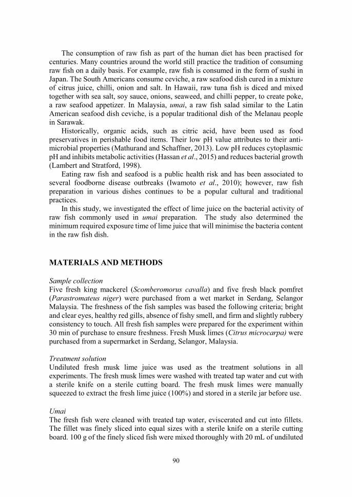
90
The consumption of raw fish as part of the human diet has been practised for centuries. Many countries around the world still practice the tradition of consuming raw fish on a daily basis. For example, raw fish is consumed in the form of sushi in Japan. The South Americans consume ceviche, a raw seafood dish cured in a mixture of citrus juice, chilli, onion and salt. In Hawaii, raw tuna fish is diced and mixed together with sea salt, soy sauce, onions, seaweed, and chilli pepper, to create poke, a raw seafood appetizer. In Malaysia, umai, a raw fish salad similar to the Latin American seafood dish ceviche, is a popular traditional dish of the Melanau people in Sarawak.
Historically, organic acids, such as citric acid, have been used as food preservatives in perishable food items. Their low pH value attributes to their anti-microbial properties (Mathurand and Schaffner, 2013). Low pH reduces cytoplasmic pH and inhibits metabolic activities (Hassan et al., 2015) and reduces bacterial growth (Lambert and Stratford, 1998).
Eating raw fish and seafood is a public health risk and has been associated to several foodborne disease outbreaks (Iwamoto et al., 2010); however, raw fish preparation in various dishes continues to be a popular cultural and traditional practices.
In this study, we investigated the effect of lime juice on the bacterial activity of raw fish commonly used in umai preparation. The study also determined the minimum required exposure time of lime juice that will minimise the bacteria content in the raw fish dish. MATERIALS AND METHODS Sample collection Five fresh king mackerel (Scomberomorus cavalla) and five fresh black pomfret (Parastromateus niger) were purchased from a wet market in Serdang, Selangor Malaysia. The freshness of the fish samples was based the following criteria; bright and clear eyes, healthy red gills, absence of fishy smell, and firm and slightly rubbery consistency to touch. All fresh fish samples were prepared for the experiment within 30 min of purchase to ensure freshness. Fresh Musk limes (Citrus microcarpa) were purchased from a supermarket in Serdang, Selangor, Malaysia. Treatment solution Undiluted fresh musk lime juice was used as the treatment solutions in all experiments. The fresh musk limes were washed with treated tap water and cut with a sterile knife on a sterile cutting board. The fresh musk limes were manually squeezed to extract the fresh lime juice (100%) and stored in a sterile jar before use. Umai The fresh fish were cleaned with treated tap water, eviscerated and cut into fillets. The fillet was finely sliced into equal sizes with a sterile knife on a sterile cutting board. 100 g of the finely sliced fish were mixed thoroughly with 20 mL of undiluted
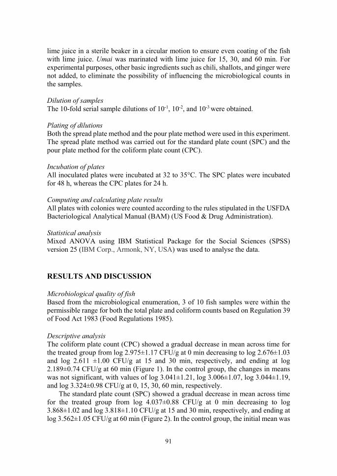
91
lime juice in a sterile beaker in a circular motion to ensure even coating of the fish with lime juice. Umai was marinated with lime juice for 15, 30, and 60 min. For experimental purposes, other basic ingredients such as chili, shallots, and ginger were not added, to eliminate the possibility of influencing the microbiological counts in the samples. Dilution of samples The 10-fold serial sample dilutions of 10-1, 10-2, and 10-3 were obtained. Plating of dilutions Both the spread plate method and the pour plate method were used in this experiment. The spread plate method was carried out for the standard plate count (SPC) and the pour plate method for the coliform plate count (CPC). Incubation of plates All inoculated plates were incubated at 32 to 35°C. The SPC plates were incubated for 48 h, whereas the CPC plates for 24 h. Computing and calculating plate results All plates with colonies were counted according to the rules stipulated in the USFDA Bacteriological Analytical Manual (BAM) (US Food & Drug Administration). Statistical analysis Mixed ANOVA using IBM Statistical Package for the Social Sciences (SPSS) version 25 (IBM Corp., Armonk, NY, USA) was used to analyse the data. RESULTS AND DISCUSSION Microbiological quality of fish Based from the microbiological enumeration, 3 of 10 fish samples were within the permissible range for both the total plate and coliform counts based on Regulation 39 of Food Act 1983 (Food Regulations 1985). Descriptive analysis The coliform plate count (CPC) showed a gradual decrease in mean across time for the treated group from log 2.975±1.17 CFU/g at 0 min decreasing to log 2.676±1.03 and log 2.611 ±1.00 CFU/g at 15 and 30 min, respectively, and ending at log 2.189±0.74 CFU/g at 60 min (Figure 1). In the control group, the changes in means was not significant, with values of log 3.041±1.21, log 3.006±1.07, log 3.044±1.19, and log 3.324±0.98 CFU/g at 0, 15, 30, 60 min, respectively.
The standard plate count (SPC) showed a gradual decrease in mean across time for the treated group from log 4.037±0.88 CFU/g at 0 min decreasing to log 3.868±1.02 and log 3.818±1.10 CFU/g at 15 and 30 min, respectively, and ending at log 3.562±1.05 CFU/g at 60 min (Figure 2). In the control group, the initial mean was
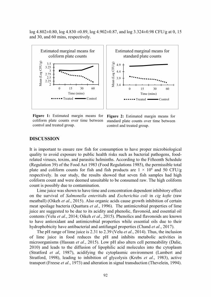
92
log 4.802±0.80, log 4.830 ±0.89, log 4.902±0.87, and log 3.324±0.98 CFU/g at 0, 15 and 30, and 60 mins, respectively.
DISCUSSION It is important to ensure raw fish for consumption to have proper microbiological quality to avoid exposure to public health risks such as bacterial pathogens, food-related viruses, toxins, and parasitic helminths. According to the Fifteenth Schedule (Regulation 39) of the Food Act 1983 (Food Regulations 1985), the permissible total plate and coliform counts for fish and fish products are 1 × 10⁶ and 50 CFU/g respectively. In our study, the results showed that seven fish samples had high coliform count and were deemed unsuitable to be consumed raw. The high coliform count is possibly due to contamination.
Lime juice was shown to have time and concentration dependent inhibitory effect on the survival of Salmonella enteritidis and Escherichia coli in cig kofte (raw meatball) (Oikeh et al., 2015). Also organic acids cause growth inhibition of certain meat spoilage bacteria (Quattara et al., 1996). The antimicrobial properties of lime juice are suggested to be due to its acidity and phenolic, flavonoid, and essential oil contents (Velu et al., 2014; Oikeh et al., 2015). Phenolics and flavonoids are known to have antioxidant and antimicrobial properties while essential oils due to their hydrophobicity have antibacterial and antifungal properties (Chand et al., 2017).
The pH range of lime juice is 2.31 to 2.39 (Velu et al., 2014). Thus, the inclusion of lime juice in food reduces the pH and inhibits metabolic activities in microorganisms (Hassan et al., 2015). Low pH also alters cell permeability (Dalie, 2010) and leads to the diffusion of lipophilic acid molecules into the cytoplasm (Stratford et al, 1987), acidifying the cytoplasmic environment (Lambert and Stratford, 1998), leading to inhibition of glycolysis (Krebs et al., 1983), active transport (Freese et al., 1973) and alteration in signal transduction (Thevelein, 1994).
Figure 2: Estimated margin means for standard plate counts over time between control and treated group.
Figure 1: Estimated margin means for coliform plate counts over time between control and treated group.
22.252.5
2.753
3.253.5
0 15 30 60
Mea
n (L
og C
FU/g
)
Time (mins)
Estimated marginal means for coliform plate counts
Treated Control
3.4
3.9
4.4
4.9
0 15 30 60
Mea
n (L
og C
FU/g
)
Time (mins)
Estimated marginal means for standard plate counts
Treated Control
93
The net effect is disruption of homeostasis, causing growth inhibition or death of microorganisms. CONCLUSION Lime juice is effective in reducing bacterial count in umai. The study showed that 15 min marination of raw fish in lime juice is the minimum time required for the juice to significantly reduce the bacterial count in umai. REFERENCES Chand RR, Jokhan AD, Gopalan RD, Osborne T (2017). Antibacterial and antifungal
activities of essential oils from medicinal plants found in the South Pacific. The South Pacific Journal of Natural and Applied Sciences, 35(1): 10-19.
Cole GM, Ma QL, Frautschy, SA, (2010). Dietary fatty acids and the aging brain. Nutrition Reviews, 68(2):102-111.
Dalie DKD, Deschamps AM, Forget FR, (2010). Lactic acid bacteria – potential for control of mold growth and mycotoxins: A review. Food Control, 21:370-380.
Food Regulations 1985. https://extranet.who.int/nutrition/gina/sites/default/files/MYS%201985%20Food%20Regulations_0.pdf (Accessed on 16 October 2019)
Freese E, Sheu CW, Galliers E, (1973). Function of lipophilic acids as antimicrobial food additives. Nature, 241:321-325.
Hassan R, El-Kadi S, Sand M, (2015). Effect of some organic acids on some fungal growth and their toxins production. International Journal of Advances in Biology (IJAB), 2(1):1-11.
IOM (2007). Seafood choices: balancing benefits and risks. National Academy Press, P722.
Iwamoto M, Ayers T, Mahon BE, Swedlow DL, (2010). Epidemiology of seafood-associated infections in the United States. Clinical Microbiology Reviews, 23:399-411.
Krebs HA, Wiggins D, Stubs M, Sols A, Bedoya F, (1983). Studies on the mechanism of the antifungal action of benzoate. Biochemical Journal, 214: 657-663.
Lambert RJ, Stratford M, (1998). Weak-acid preservatives: modelling microbial inhibition and response. Journal of Applied Microbiology, 86:157-164.
Maciel ES, Sonati JG, Lima LKF, Savay-da-Silva LK, Galvao JA, Oetterer M, (2016). Similarities and distinctions of fish consumption in Brazil and Portugal measured through electronic survey. International Food Research Journal, 23(1):395-402.
Mathurand P and Schaffner DW (2013). The effect of lime juice on Vibrio parahaemolyticus and Salmonella enterica inactivation during the preparation of raw fish dish ceviche. Journal of Food Protection, 76(6): 1027-1030.
94
Oikeh EI, Omoregie, ES, Oviasogie, FE, Oriakhi, K (2015). Phytochemical, antimicrobial, and antioxidant activities of different citrus juice concentrates. Food Science & Nutrition 2016, 4(1):103-109.
Oken E, Choi, AL Karagas, MR, Marien, K Rheinberger CM, Schoeny R, Sunderland, E, Korrick, S (2012). Which should I eat? Perspectives influencing fish consumption choices. Environmental Health Perspectives, 120(6):790-798.
Quattara B, Simard RE, Holley RA, Piette GJP, Begin A (1996). Inhibitory effect of organic acids upon meat spoilage bacteria. Journal of Food Protection, 60(3):246-253.
Stratford, M, Morgan, P, Rose, AH, (1987). Sulphur dioxide resistance in Sacharomyces cerevisiae and Saccharomycodes ludwigii. Journal of General Microbiology, 133:2173 – 2179.
Thevelein, JM, (1994). Signal transduction in yeast. Yeast, 10:1753-1790. US Food & Drug Administration. Bacteriological laboratory manual (BAM).
https://www.fda.gov/food/laboratory-methods-food/bacteriological-analytical-manual-bam (Accessed on 5 September 2019).
Velu S, Abu Bakar F, Mahyuddin NA, Saari N, Zaman MZ (2014). In vitro antimicrobial activity of musk lime, key lime and lemon extracts against food related pathogenic and spoilage bacteria. International Food Research Journal, 21(1):379-386.
Wall R, Ross RP, Fitzgerald GF, Stanton C, (2010). Fatty acids from fish: the anti-inflammatory potential of long-chain omega-3 fatty acids. Nutrition Review, 68(5):280-289.
95
14th Proceedings of the Seminar on Veterinary Sciences, 18 - 20th September 2018
CLINICOPATHOLOGIC AND RADIOGRAPHIC FEATURES IN CATS DIAGNOSED WITH PNEUMONIA ASSOCIATED WITH
RHODOCOCCUS EQUI INFECTION
Chelly Chin Sze Lee, 1*Lau Seng Fong, 2Nur Indah Ahmad, 1Puteri Azaziah Megat Abdul Rani & 1Rozanaliza Radzi
1Department of Veterinary Clinical Studies 2Department of Veterinary Pathology and Microbiology
Faculty of Veterinary Medicine Universiti Putra Malaysia, 43400 UPM Serdang, Selangor, Malaysia
*Correspondence: [email protected]
ABSTRACT
Rhodococcosis a zoonotic disease that typically affects immunocompromised patients. It is a common pathogen in equine but it has been reported in other domestic animals. The clinicopathologic and radiographic features in cats infected with Rhodococcus have not been well described. The objectives of this study were to describe the clinical, diagnostic and radiographic findings in cats diagnosed with Rhodococcosis at University Veterinary Hospital, Universiti Putra Malaysia during the period of January 2012 to June 2018. Cats infected with Rhodococcus with radiographic investigation were included. Clinical data including signalment, clinical signs and blood results were retrieved and radiographic findings evaluated. Forty cats met the inclusion criteria, with 36 cats had pulmonary lesion, and 4 cats had subcutaneous lesion. In this study, 77.5% of cats were less than one-year-old and were predominantly intact male (50%). 64% of cats were kept in indoor with 90% of them were from multi-cat household. Cytological results showed numerous of phagocytosed coccal bacteria, with neutrophils and macrophages. Haematological result revealed neutrophilia with left shift, monocytosis, thrombocytopenia, hyperglobulinemia and hypoalbuminemia. Radiological findings revealed pleural effusion, hepatomegaly and lymphadenopathy. Atelectasis/ consolidation were showed after post-thoracocentesis. Radiography features in cats infected with Rhodococcus were general and could resemble other infectious disease such as feline infectious peritonitis. Therefore, bacteria culture is still crucial in diagnosis of Rhodococcosis. Keywords: cat, Rhodococcus equi, clinical presentation, diagnostic, radiographic findings

96
INTRODUCTION Rhodococcus equi was formerly known as Corynebacterium equi and Mycobacterium equi (Hines, 2014). It is a ubiquitous Gram-positive coccobacillus, intracellular bacterium with the capability to infect macrophages by intervening with the phagolysosomal fusion. In human, there have been many reports on Rhodococcus equi pulmonary infection in immunocompromised patient. In 1983, this infection became more recognised in humans with immunosuppressive disease such as human immunodeficiency virus/acquired immune deficiency syndrome (HIV/AIDs) (Yamshchikov et al., 2010).
Rhodococcosis is very rare in dogs and cats, however there are more cases of Rhodococcus equi infection in cats than dogs. There were few reports regarding pulmonary R. equi infection in cats and the infection appears to be confined to the skin and the subcutaneous tissues. The first report of feline pulmonary Rhodococus equi infection with detailed findings on the pathological, microbiological and the virulence features associated with an acute necrosuppurative pneumonia in cat occurred in Italy. Three virulence levels of Rhodococcus equi have been identified; virulent virulence-associated antigens A (Vap A), intermediate (Vap B), and avirulent. The Vap A gene has been isolated in a previous study (Passamonti, 2011). Although, there have been sporadic reports of cats infected with Rhodococcus equi infection, the clinicopathologic and radiographic features in cats are not well described. Hence, the purpose of this study was to describe the clinicopathologic and radiographic features in cats diagnosed with pneumonia associated with Rhodococcus equi infection. MATERIAL AND METHODS Data collection A retrospective study was performed on 40 cases of cats diagnosed with Rhodococcus equi infection which were confirmed by bacterial culture and supported with radiography in University Veterinary Hospital (UVH), Universiti Putra Malaysia from 2012-2018. Records were retrieved to obtain the data on patient signalment which included age, breed, sex, and types of household management (multi-cat/single or indoor/ourdoor). The history and clinical signs of the affected cats were recorded. The diagnostic reports which included result from bacterial culture, complete blood count and serum biochemistry, cytology, thoracic radiography and pathology findings were also obtained. RESULTS AND DISCUSSION In this study, 77.5% of the cats were less than one-year-old, 75% were Domestic Shorthair (DSH) breed, and they were predominantly males (65%) where 50% were
97
intact males. A total of 80% of the intact toms were managed in multi-cat households while 64% kept indoor.
Young animal is more susceptible to disease because their immune immaturity and the serum immunoglobulin concentration as not reached full adult levels (Day, 2007). Since the DSH is the most popular breed of cats in Malaysia, it cannot be concluded from the results that they are the most susceptible breed to R. equi infection. Cats usually reach their puberty at the age of 5 to 12 months. Intact males will start mating and are frequently involved in territorial fights, especially in multi-cat households (Waddell et al., 2002). This can lead to stress and subsequently causing immunosuppression and susceptibility to infections.
Clinical signs often displayed in cats in this study were inappetence (60%), dyspnoea (42.5%), fever (37%), lethargic (23%) and hypothermia (14%). Dyspnoea, inappetence and lethargy are common findings in cats with rhodococcosis (Barrs and Beatty, 2009). Hypothermia in cats with pyothorax could possibly due to severe sepsis. Exogenous pyrogens are microbes that induce the release of endogenous pyrogens, interleukin-1, tumour necrosis factor-α and other cytokines, which then lead to fever (Bush, 2018). However, only 37% of cats in this study had fever and this could be due to the chronic stage of infection where microbes could have already been cleared by the monocyte-phagocytic system. In general, neutrophilia with left shift was a common finding in all cases of Rhodococcus infection in cats (59%). Monocytosis is the second most common findings in cats with chronic infection.
The serum biochemical profile showed findings were hyperglobulinemia (64%), which is indicative of infection or inflammation. Mild hypoalbuminemia was the third most common abnormalities in this study. However, the blood profile for this study was general and similar to other infectious disease. In cytology findings, phagocytosed coccal bacteria (86%, n=24) were the most prevalent finding in this study. Presence of neutrophils (79%, n=22) and macrophages (71%, n=20) were also very common in rhodococcal infection in cats (Fairley and Fairley, 1999).
Thoracic radiographs were performed on 36 cases of rhodococcosis. A total of 32 cats showed evidence of pleural effusions most probably due to pyothorax. The occurrence of pyothorax in this study could be due to pyogranulomatous formation in the lungs which led to compromised lung and bacterial entry into the pleural spaces and triggering inflammatory responses (Barrs and Beatty, 2009). A total of 81% of the cats showed hepatomegaly, which is suggested to be due to haematogenous dissemination of the Rhodococcus that had spread to the liver.
At post-thoracocentesis, 13 (39%) cats showed lymphadenopathy and 11 with sternal lymphadenopathy. Sternal lymph nodes are mostly affected in most of the cases as they received drainage from a broad area of the thoraco-abdominal region and haematogenous dissemination in rhodococcosis (Iwasaki et al., 2016). In this study, 17 cats (52%) showed atelectasis/consolidation at post-thoracocentesis. Increase in number of cases with post-thoracocentesis findings could be due to the pneumothorax. A total of 14% of the cases involved collapsed right middle lung lobe. The clinical presentations of these cat were sneezing, coughing, and concurrent with

98
feline upper respiratory disease (FURD). Therefore, atelectasis in these cases could be due to underlying problem of the cats.
A total of 8 cats (23%) showed cavitary formation at post-thoracocentesis. In human cases, it was shown that 69% showed cavitary formation (Torres-Tortosa et al., 2003). Cavitary formation can be contributed by pyogenic lung abscess in rhodococcosis. Suppurative necrosis will occur leading to gas-filled areas in the lung (Gadkowski et al., 2008). In our study, alveolar pattern was the most common finding most probably because of infectious pneumonia.
Only 4 cats developed subcutaneous lesions in this study. A total of 75% of the cats showed soft tissue swellings at the limbs in the extrapulmonary cases. This finding was similar as previously reported by Passamonti et al. (2011). Fifty percent of the cats showed radiographic evidence of gas pocket at the swelling, which may be caused by open wounds. In pulmonary cases, the prognosis was fair as there were systemic sign involvement such as fever and inappetence. Haematogenous dissemination can occur in pulmonary cases of R. equi. However, if the disease is diagnosed early and sensitive antibiotic given to cast, the prognosis can be good (Barrs and Beatty, 2009).
Post-mortem was performed only on 3 cats with rhodococcosis. The necropsy findings were pyogranulomatous lesions in lungs in all 3 cases, 2 of which had pulmonary atelectasis. Congestion of liver and kidney were observed in all cats. In a previous (Passamonti et al., 2011) it was reported that the disease caused slight enlargement of the liver and kidneys. This manifestation could indicate haematogenous dissemination of the disease.
Histopathologically, inflammatory cells such as macrophages, neutrophils and plasma cells were found in the lungs of all necropsied cats. Intra-histiocytic coccobacillary bacteria were present in 2 cases and only one case presented with thickened inter-alveolar septum. These findings were consistent with findings reported by Passamonti et. al. (2011). Hepatocellular necrosis and renal tubular necrosis occurred in 2 cases in this study, suggesting haematogenous dissemination of the disease to the liver and kidney (Yamshchikov et al., 2010). CONCLUSION The blood profile and radiographic features in cats infected with Rhodococcus equi were non-specific and could resemble any infectious disease. Therefore, bacteria culture is still the most certain method in the diagnosis of rhodococcosis. REFERENCES Barrs VR, Beatty JA, (2009). Feline pyothorax – new insights into an old problem:
part 1. Aetiopathogenesis and diagnostic investigation. The Veterinary Journal, 179(2):163-170.
99
Bush LM, Schmidt CE, (2018). Fever infectious Diseases: MSD Manual Professional Edition.https://www.msdmanuals.com/professional/infectious-diseases/biologyof-infectious-disease/fever (Accessed on 18 July 2018).
Day MJ, (2007). Immune system development in the dog and cat. Journal of Comparative Pathology, 137(1):10-15.
Fairley RA, Fairley NM, (1999). Rhodococcus equi infection of cats. Veterinary Dermatology, 10(1):43-46.
Gadkowski LB, Stout JE, (2008). Cavitary pulmonary disease. Clinical Microbiology Reviews, 21(2):305-333.
Hines MT, (2014). Rhodococcus equi. In: Equine Infectious Diseases, Sellon, DC, Long MT (Editors), 2nd Edition, Elsevier. Pp.287-301.
Iwasaki R, Mori T, Ito Y, Kawabe M, Murakmi M, Maruo K., (2016). Computed tomographic evaluation of presumptively normal canine sternal lymph nodes. Journal of the American Animal Hospital Association, 52(6):371–377.
Passamonti F, Lepri E, Coppola G, Sforna M, Proietti PC, Chiodetti, I. Coletti M, Marenzoni ML, (2011). Pulmonary rhodococcosis in a cat. Journal Feline Medicine and Surgery, 13:283-285.
Torres-Tortosa M, Arrizabalaga J, Villanueva JL, Gálvez J, Leyes M, Valencia ME, Flores J, Peña JM, Pére-Cecilia E, Quereda C, (2003). Prognosis and clinical evaluation of infection caused by Rhodococcus equi in HIV-Infected patients. Chest, 123(6):1970-1976.
Waddell LS, Brady CA, Drobatz, KJ, (2002). Risk factors, prognostic indicators, and outcome of pyothorax in cats: 80 cases (1986-1999). Journal of the American Veterinary Medical Association, 221(6):819–824.
Yamshchikov AV, Schuetz A, Lyon GM, (2010). Rhodococcus equi infection. The Lancet Infectious Diseases, 10(5):350-359.

100
14th Proceedings of the Seminar on Veterinary Sciences, 18 – 20 September 2018
DETECTION OF INTERDIGITAL BACTERIA AND FUNGI IN CATTLE THAT USED
SANCTUARYTM VETERINARY HOOF COVER
Siti Jazmina Shaik Husseinudin, 1*Siti Zubaidah Ramanoon, 2Siti Khairani Bejo & 3Arifah Abdul Kadir
1Department of Farm Animal and Exotic Medicine and Surgery 2Department of Veterinary Pathology and Microbiology
3Department of Veterinary Preclinical Sciences Faculty of Veterinary Medicine
Universiti Putra Malaysia, 43400 UPM Serdang, Selangor, Malaysia *Correspondence: [email protected]
ABSTRACT
Treatment of interdigital lesions comprise of topical treatment, and manual bandaging that is applied to the hoof, creating an environment suitable for healing. The SanctuaryTM
Veterinary Cow Hoof Cover is an invention devised to enclose bandages as manual bandaging. The objective of this pilot study was to detect the presence of interdigital bacterial and fungal growth in cattle using the Hoof Cover. Twelve hooves of cattle at the Large Animal Ward, Universiti Putra Malaysia (UPM), Serdang, Selangor, Malaysia, were used for the study. The right front and hindlimbs were the control group (n=6), and the left forelimbs and hindlimbs were the Hoof Cover (HC) group (n=6). Individual aseptic interdigital swabs were taken from both groups before and after HC application. All swab samples were subjected to bacteriological and fungal culture, isolation, identification at the Veterinary Bacteriology Laboratory, Faculty of Veterinary Medicine, UPM. In the control group, all bacterial cultures were positive for Staphylococcus sp.: Staphylococcus aureus (55.56%) and Staphylococcus hyicus (22.22%). In the HC group, bacteria isolated were Staphylococcus aures (36.17%) and Proteus mirabilis (14.89%). Three yeast species, Candida sp. (8.33%), Cryptococcus sp. (8.33%), and Curvularia sp. (2.78%) were identified in the HC group. There was no fungal isolation in the control group. The shows that the interdigital bacteria identified cows with hoof cover were of the normal skin microflora, whereas the fungal isolates were of soil or plant origin acquired during grazing at the grass area of the Large Animal Ward. The study shows that the Sanctuary™ Veterinary Cow Hoof Cover can be used to aid treatment of interdigital lesions in cattle. Keywords: cattle, Hoof Cover, interdigital, bacteria, fungus

101
INTRODUCTION Interdigital lesions in cattle, such as digital dermatitis, is the result of factors including loose housing, slatted flooring, wet, and unhygienic conditions amongst others were listed as associated elements (Clegg et al., 2015). Treatment of interdigital lesions is generally initiated with topical treatment, and standard bandaging with cotton wool and self-adherent wraps that are applied to the affected hoof. This is to ensure cleanliness of affected hooves and to maintain contact between the infected tissues and topical antibiotics. Since the conventional method of bandaging interdigital lesions in cows using cotton and self-adhesive wraps such as Coban is unsanitary, Sanctuary Health Sdn. Bhd., a Malaysian-based company had devised the Cow Hoof Cover (SanctuaryTM Veterinary) to be used to over the bandages. The Cow Hoof Cover is made of latex and although water-resistant may create an environment suitable for bacterial and fungal growth. Hence, the objective of the study is to determine the presence of interdigital bacterial and fungal growths in cows using the SanctuaryTM Veterinary Cow Hoof Cover. MATERIALS AND METHODS Sample Collection The hooves (n=16) of 4 resident cows at the Large Animal Ward, University Veterinary Hospital, Universiti Putra Malaysia were used in this study. A swab sample of the interdigital space of each cattle hoof was taken using sterile swabs. Then, a gauze was wrapped around the hoof, which was then covered with the SanctuaryTM Cow Hoof Cover and secured with Leukoplast® (Figure 1). Swab samples were obtained on 3rd, 6th, and 9th day post-hoof cover application. Hoof Cover and gauze were removed and replaced at each sampling. The swab samples were analysed at the Bacteriology Laboratory, Faculty of Veterinary Medicine, Universiti Putra Malaysia, within 2 hours of collection.
Figure 5. Cow hoof with Hoof Cover secured with Leukoplast®
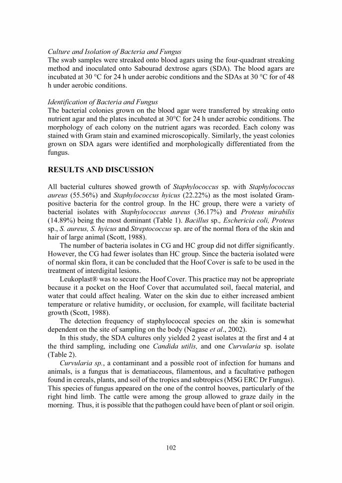
102
Culture and Isolation of Bacteria and Fungus The swab samples were streaked onto blood agars using the four-quadrant streaking method and inoculated onto Sabourad dextrose agars (SDA). The blood agars are incubated at 30 °C for 24 h under aerobic conditions and the SDAs at 30 °C for of 48 h under aerobic conditions. Identification of Bacteria and Fungus The bacterial colonies grown on the blood agar were transferred by streaking onto nutrient agar and the plates incubated at 30°C for 24 h under aerobic conditions. The morphology of each colony on the nutrient agars was recorded. Each colony was stained with Gram stain and examined microscopically. Similarly, the yeast colonies grown on SDA agars were identified and morphologically differentiated from the fungus. RESULTS AND DISCUSSION All bacterial cultures showed growth of Staphylococcus sp. with Staphylococcus aureus (55.56%) and Staphylococcus hyicus (22.22%) as the most isolated Gram-positive bacteria for the control group. In the HC group, there were a variety of bacterial isolates with Staphylococcus aureus (36.17%) and Proteus mirabilis (14.89%) being the most dominant (Table 1). Bacillus sp., Eschericia coli, Proteus sp., S. aureus, S. hyicus and Streptococcus sp. are of the normal flora of the skin and hair of large animal (Scott, 1988).
The number of bacteria isolates in CG and HC group did not differ significantly. However, the CG had fewer isolates than HC group. Since the bacteria isolated were of normal skin flora, it can be concluded that the Hoof Cover is safe to be used in the treatment of interdigital lesions.
Leukoplast® was to secure the Hoof Cover. This practice may not be appropriate because it a pocket on the Hoof Cover that accumulated soil, faecal material, and water that could affect healing. Water on the skin due to either increased ambient temperature or relative humidity, or occlusion, for example, will facilitate bacterial growth (Scott, 1988).
The detection frequency of staphylococcal species on the skin is somewhat dependent on the site of sampling on the body (Nagase et al., 2002).
In this study, the SDA cultures only yielded 2 yeast isolates at the first and 4 at the third sampling, including one Candida utilis, and one Curvularia sp. isolate (Table 2).
Curvularia sp., a contaminant and a possible root of infection for humans and animals, is a fungus that is dematiaceous, filamentous, and a facultative pathogen found in cereals, plants, and soil of the tropics and subtropics (MSG ERC Dr Fungus). This species of fungus appeared on the one of the control hooves, particularly of the right hind limb. The cattle were among the group allowed to graze daily in the morning. Thus, it is possible that the pathogen could have been of plant or soil origin.

103
Table 1. Interdigital bacterial isolates in cattle using hoof cover.
Bacterial isolate
Control group Hoof cover Group
Sampling number Total
isolate (%)
Sampling number Total
isolate (%)
1 2 3 1 2 3
No of isolates No of isolates
Acinetobacter baumanni - - - 0.00 - 2 1 6.38
Alcaligenes faecalis - - - 0.00 - 1 - 2.13
Bacillus sp. 1 - 1 4.44 - - - 0.00
Chromobacterium sp - - - 0.00 - 1 1 4.26
Citrobacter freundii - - - 0.00 - 1 5 12.77
Enterobacter sp. - - 2 4.44 - - - 0.00
Enterococcus faecalis - - 1 2.22 - 1 - 2.13
Escherichia coli - - - 0.00 - - 2 4.26
Pantoea agglomerans - - 1 2.22 - - - 0.00
Proteus mirabilis - - 1 2.22 - 4 3 14.89
Salmonella enterica - - - 0.00 - - 1 2.13
Serratia sp. - - - 0.00 - - 1 2.13
Staphylocccus intermedius - 1 1 4.44 - 3 - 6.38
Staphyloccus aureus 13 6 6 55.56 11 4 2 36.17
Staphyloccus hyicus 1 8 1 22.22 - - - 0.00
Staphylococcus pseudintermedius - - - 0.00 1 1 1 6.38
Streptococcus bovis - - 1 2.22 - - - 0.00
Grand Total 15 15 15 100.00 12 18 17 100.00
Sampling number: 1=3rd Aug 2018; 2=6th Aug 2018; 3=9th Aug 2018
Table 1. Interdigital fungal isolates in cattle using hoof cover.
Fungal isolate
Control group Hoof cover Group
Sampling number Total
isolate (%)
Sampling number Total
isolate (%)
1 2 3 1 2 3
No of isolates No of isolates
Candida sp. - - - 0.00 - - 3 8.33%
Cryptococcus sp - - - 0.00 - 2 1 8.33%
Curvularia sp. - - - 0.00 - - 1 2.78%
No growth 12 12 12 100.00 12 10 7 80.56
Grand Total 12 12 12 100.00 12 12 12 100.00
Sampling number: 1=3rd Aug 2018; 2=6th Aug 2018; 3=9th Aug 2018
104
CONCLUSION In conclusion, interdigital bacterial isolates in this study were of the normal skin microflora, whereas the fungal isolates may have been acquired during daily grazing in the grass area of the Large Animal Ward. However, the study shows that Sanctuary™ Veterinary Cow Hoof Cover has a potential to be used to aid treatment of interdigital lesions in cattle. REFERENCES Clegg SR, Mansfield KG, Newbrook K, Sullivan LE, Blowey RW, Carter SD, Evans
NJ (2015). Isolation of digital dermatitis treponemes from hoof lesions in wild North American elk (Cervus elaphus) in Washington State, USA. Journal of Clinical Microbiology, 53(1): 88-94.
MSG ERC Dr Fungus. Mycoses Study Group Education and Research Consortium. (2018). Curvularia Species. https://drfungus.org/knowledge-base/curvularia-species/ (Accessed on 6 September 2019O).
Nagase N, Sasaki A, Yamashita K, Shimizu A, Wakita Y, Kitai S, Kawano J (2002). Isolation and Species Distribution of Staphylococci from Animal and Human Skin. The Journal of Veterinary Medical Science, 64(3), 245-250.
Sanctuary™ Veterinary. http://sanctuaryvet.com/our-products/ (Accessed on 16 Oct 2019).
Scott DW (1988). Structure and function of the skin. In:, Large Animal Dermatology, Scott DW (Editor), New York: WB Saunders. Pp1-19.

105
14th Proceedings of the Seminar on Veterinary Sciences, 18 – 20 September 2018
MOLECULAR DETECTION AND RISK FACTOR ANALYSES OF ENTERIC PROTOZOA INFECTION AMONG BORNEAN
ORANGUTANS (PONGO PYGMAEUS) IN SABAH, MALAYSIA
Adeline Tsen, 1,2*Reuben Sunil Kumar Sharma & Norhadila Zulkifli 1Department of Veterinary Laboratory Diagnostics
2Research Centre for Wildlife Faculty of Veterinary Medicine
Universiti Putra Malaysia, 43400 UPM Serdang, Selangor, Malaysia *Correspondence: [email protected]
ABSTRACT
Orangutans are the only great apes in Asia and are considered to be critically endangered throughout their natural home range. In spite of this, there is a paucity of information on the health status and pathogen diversity in both captive and free-ranging Orangutans. The present study was undertaken to determine the diversity and prevalence of enteric protozoa among captive and free-ranging Bornean Orangutans (Pongo pygmaeus) by using conventional microscopy and molecular detection methods. Faecal samples were collected from 25 individuals representing the different management systems, age and gender, from two zoological facilities in Sabah, Malaysia. Microscopy examination revealed the presence of three genera of enteric protozoa, namely Cryptosporidium, Blastocystis and Balantidium. Polymerase chain reaction (PCR) amplification using genus-specific primers revealed that the molecular prevalence was highest for Cryptosporidium (76%) followed by Blastocystis (72%), Balantidium (68%), Buxtonella (54%), and Giardia (20%). Pearson Chi-square analysis revealed that there was significant effect of the management types and the risk of getting Giardia infection, whereby the odds of infection was 5.9 times higher among the animals that were managed fully in captivity. Binary logistic regression analysis revealed that there is no significant difference in the prevalence of the enteric protozoa among Orangutans in relation to the various risk factors (management, age and gender) examined. The high prevalence of zoonotic enteric protozoa among the Orangutans warrants biosecurity measures to be implemented in these zoological facilities in order to prevent human infection. Keywords: Bornean orangutans, Pongo pygmaeus, zoonotic enteric protozoa, Sabah

106
14th Proceedings of the Seminar on Veterinary Sciences, 18 – 20 September 2018
BACTERIA CONTAMINATION IN BOAR SEMEN FOLLOWING SEMEN COLLECTION VIA GLOVE HAND TECHNIQUE
Aaron Michael Anthony, 1*Mark Hiew Wen Han,
1Ooi Peck Toung & 2Siti Khairani Bejo 1Department of Veterinary Clinical Studies
2Department of Veterinary Pathology and Microbiology Faculty of Veterinary Medicine
Universiti Putra Malaysia, 43400 UPM Serdang, Selangor, Malaysia *Correspondence: [email protected]
ABSTRACT
Artificial insemination (AI) is a technique that is widely used in the swine industry in Malaysia for breeding sows and gilts. Collection of semen from boars via the gloved hand technique has been known to lead to bacteriospermia which in turn can cause many reproductive problems to the sow. Hence, this study was conducted to evaluate and determine the qualitative and quantitative aspects of bacterial contamination in semen collected via the gloved hand technique in Malaysia. Semen samples were collected from 29 boars from 14 farms on the west coast of Peninsular Malaysia. The method of semen collection was recorded as possible risk factor. Routine bacterial culture, isolation and identification was performed as part of qualitative analysis together with total plate counts (TPC) and coliform plate counts (CPC) for quantitative analysis. Among samples, 76% (22/29) had at least one type of bacterial growth. Most of the bacterial isolates obtained were Gram-positive bacteria of which 46% were Staphylococcus sp., 26% Gram-positive rods and 2% Streptococcus sp. The remaining 26% were Gram-negative bacteria of various genus and species. The most common isolates were Staphylococcus pseudintermedius, Staphylococcus intermedius, Corynebacterium sp., Bacillus cereus and Micrococcus sp., each at 10%. Risk analysis revealed that there is no statistically significant risk factor (p < 0.05) associated with semen contamination. In conclusion, the semen collected in this study had bacterial contamination, but the associated risk factors could not be determined. Keywords: boar semen, bacteria, semen collection, bacteriospermia

107
14th Proceedings of the Seminar on Veterinary Sciences, 18 – 20 September 2018
DETERMINANTS OF DOG AGGRESSION AMONG PET DOGS IN KLANG VALLEY, MALAYSIA
Cherilyn Mok Jia Ying, 1*Khor Kuan Hua,
1Mark Hiew Wen Han & 2Lynn Walker 1Department of Veterinary Clinical Studies
Faculty of Veterinary Medicine Universiti Putra Malaysia, 43400 UPM Serdang, Selangor, Malaysia
Human Research Protection Program 2Office of the Executive Vice President for Research and Partnership
Purdue University West Lafayette, Indiana 47907, United States of America
*Correspondence: [email protected]
ABSTRACT
Aggression in dogs has been one of the leading causes on relinquishment of pet dogs. This study investigated the determinant of aggression among pet dogs from Klang Valley, Malaysia using a survey modified from the Canine Behavioural Assessment and Research Questionnaire (C-BARQ). A total of 165 dog owners completed the survey. Information of owners, their dogs, interactions with their dog, living environment and dog’s training methods were obtained. The most prominent aggression was dog-directed aggression which correlated with other types of aggression. The significant determinants of dog-directed aggression were owners’ ethnicity (p=0.002), dogs’ age (p=0.025), dogs living in a house with yard (p=0.017), and >4 household members (p=0.031). Mixed-breed (p=0.045) and adopted (p=0.020) dogs, and those that spend ≤ 3 hours (p=0.037) with their owners were the determinants for stranger-directed aggression. Additionally, male (p= 0.010) and young (p= 0.024) dogs and those that were trained (p=0.030) were more likely to develop owner-directed aggression, whereas constantly walking the dogs was the only determinant (p=0.010) for dog rivalry. The results of this study showed that proper management of dogs will improve human-dog interactions. Keywords: canine aggression, C-BARQ, questionnaire, behavioural study

108
13th Proceedings of the Seminar on Veterinary Sciences, 18 – 20 September 2019
MORPHOLOGY OF THE GASTROINTESTINAL TRACT OF
WATER MONITOR LIZARD (VARANUS SALVATOR)
Chong Chiu Nie, 1*Intan Shameha Abdul Razak & 1,2Azlan Che’ Amat
1Department of Veterinary Preclinical Sciences 3Department of Veterinary Clinical Studies
2Research Centre for Wildlife Faculty of Veterinary Medicine
Universiti Putra Malaysia, 43400 UPM Serdang, Selangor, Malaysia *Correspondence: [email protected]
ABSTRACT
Currently, reptiles such as snakes and monitor lizards are recognised as economically profitable animals and are being heavily exploited. In order to meet the demands, captive breeding is necessary. Thus, the anatomical study of the digestive system is essential for better understandings of the feeding and diet management of this species. To date, data on the morphology of the gastrointestinal tract (GIT) of Varanus salvator in Malaysia is still lacking. A total of 8 (4 females, 4 males) opportunistic carcasses of monitor lizards was used in this study. Prior to post-mortem, the body parameters and GIT organs were measured and weighed while the stomach contents examined and recorded. Samples were then taken for routine histological examination. In general, male monitors had higher body weights and organ sizes than females. The monitors had a slender forked tongue, a long and muscular oesophagus that connects to a muscular stomach, a thick coiled small intestine and soft and straight large intestine with the absence of caecum, a three-lobed liver and a small pancreas. Histologically, the GIT layers were similar to higher vertebrates and are mainly composed of mucosa, submucosa, tunica muscularis, and serosa layers. However, a few differences were observed, namely the presence of folded oesophageal mucosa lined with ciliated columnar epithelium with goblet cells and neck cells in the fundic mucosa. The latter findings are similar to that of boas but differ from Green Iguanas and other lizards. Keywords: water monitor lizard (Varanus salvator), gastrointestinal tract, morphology, histology
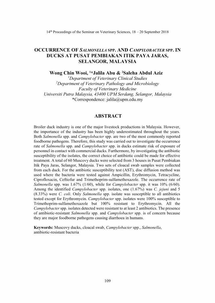
109
14th Proceedings of the Seminar on Veterinary Sciences, 18 – 20 September 2018
OCCURRENCE OF SALMONELLA SPP. AND CAMPYLOBACTER SPP. IN DUCKS AT PUSAT PEMBIAKAN ITIK PAYA JARAS,
SELANGOR, MALAYSIA
Wong Chin Wooi, 1*Jalila Abu & 2Saleha Abdul Aziz 1Department of Veterinary Clinical Studies
2Department of Veterinary Pathology and Microbiology Faculty of Veterinary Medicine
Universiti Putra Malaysia, 43400 UPM Serdang, Selangor, Malaysia *Correspondence: [email protected]
ABSTRACT
Broiler duck industry is one of the major livestock productions in Malaysia. However, the importance of the industry has been highly underestimated throughout the years. Both Salmonella spp. and Campylobacter spp. are two of the most commonly reported foodborne pathogens. Therefore, this study was carried out to investigate the occurrence rate of Salmonella spp. and Campylobacter spp. in ducks estimate risk of exposure of personnel in contact with commercial ducks. Furthermore, by investigating the antibiotic susceptibility of the isolates, the correct choice of antibiotic could be made for effective treatment. A total of 60 Muscovy ducks were selected from 3 houses in Pusat Pembiakan Itik Paya Jaras, Selangor, Malaysia. Two sets of cloacal swab samples were collected from each duck. For the antibiotic susceptibility test (AST), disc diffusion method was used where the bacteria were tested against Ampicillin, Erythromycin, Tetracycline, Ciprofloxacin, Ceftiofur and Trimethoprim-sulfamethoxazole. The occurrence rate of Salmonella spp. was 1.67% (1/60), while for Campylobacter spp. it was 10% (6/60). Among the identified Campylobacter spp. isolates, one (1.67%) was C. jejuni and 5 (8.33%) were C. coli. Only Salmonella spp. isolate was susceptible to all antibiotics tested except for Erythromycin. Campylobacter spp. isolates were 100% susceptible to Trimethoprim-sulfamethoxazole but 100% resistant to Erythromycin. All the Campylobacter spp. isolates detected were resistant to at least 2 antibiotics. The presence of antibiotic-resistant Salmonella spp. and Campylobacter spp. is of concern because they are major foodborne pathogens causing diarrhoea in humans. Keywords: Muscovy ducks, cloacal swab, Campylobacter spp., Salmonella, antibiotic-resistant bacteria
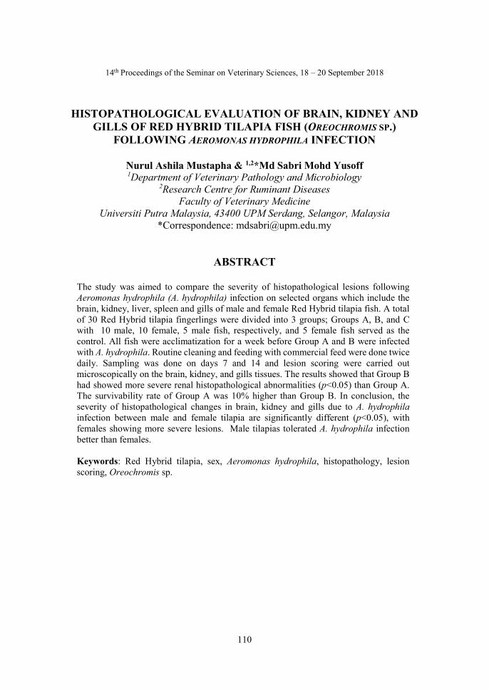
110
14th Proceedings of the Seminar on Veterinary Sciences, 18 – 20 September 2018
HISTOPATHOLOGICAL EVALUATION OF BRAIN, KIDNEY AND GILLS OF RED HYBRID TILAPIA FISH (OREOCHROMIS SP.)
FOLLOWING AEROMONAS HYDROPHILA INFECTION
Nurul Ashila Mustapha & 1,2*Md Sabri Mohd Yusoff 1Department of Veterinary Pathology and Microbiology
2Research Centre for Ruminant Diseases Faculty of Veterinary Medicine
Universiti Putra Malaysia, 43400 UPM Serdang, Selangor, Malaysia *Correspondence: [email protected]
ABSTRACT
The study was aimed to compare the severity of histopathological lesions following Aeromonas hydrophila (A. hydrophila) infection on selected organs which include the brain, kidney, liver, spleen and gills of male and female Red Hybrid tilapia fish. A total of 30 Red Hybrid tilapia fingerlings were divided into 3 groups; Groups A, B, and C with 10 male, 10 female, 5 male fish, respectively, and 5 female fish served as the control. All fish were acclimatization for a week before Group A and B were infected with A. hydrophila. Routine cleaning and feeding with commercial feed were done twice daily. Sampling was done on days 7 and 14 and lesion scoring were carried out microscopically on the brain, kidney, and gills tissues. The results showed that Group B had showed more severe renal histopathological abnormalities (p<0.05) than Group A. The survivability rate of Group A was 10% higher than Group B. In conclusion, the severity of histopathological changes in brain, kidney and gills due to A. hydrophila infection between male and female tilapia are significantly different (p<0.05), with females showing more severe lesions. Male tilapias tolerated A. hydrophila infection better than females. Keywords: Red Hybrid tilapia, sex, Aeromonas hydrophila, histopathology, lesion scoring, Oreochromis sp.

111
14th Proceedings of the Seminar on Veterinary Sciences, 18 – 20 September 2018
MORPHOLOGICAL AND MOLECULAR CHARACTERISATION OF FASCIOLA SP. IN RUMINANTS SLAUGHTERED AT
SHAH ALAM AND BANTING ABATTOIRS, SELANGOR, MALAYSIA
Nurhanim Rohaizad, 1*Nur Mahiza Md Isa,
2Lokman Hakim Idris, & 1Nor Azlina Abdul Aziz 1Department of Veterinary Pathology and Microbiology
2Department of Veterinary Preclinical Sciences Faculty of Veterinary Medicine
Universiti Putra Malaysia, 43400 UPM Serdang, Selangor, Malaysia *Correspondence: [email protected]
ABSTRACT
Fascioliasis or liver fluke infection is a common parasitic disease in ruminant. Fasciola hepatica and Fasciola gigantica are two main species of Fasciola infecting both human and animals. While F. gigantica is occurs mainly in tropical and F. hepatica in temperate areas, the presence of these species overlaps in subtropical zones. These two species of liver flukes have been traditionally classified based on their morphological features. Several molecular studies have been used to for genotype analysis of Fasciola spp. isolated from ruminants. However, similar study has not been done in Malaysia. Thus, this study attempts to identify Fasciola sp. in the liver of ruminant slaughtered at Shah Alam and Banting abattoirs, Selangor, Malaysia and to characterise the Fasciola sp. by using morphological and molecular methods. Adult Fasciola spp. were collected from eight animals (4 cattle, 4 buffaloes) from the abattoirs. The biliary tract of the liver was dissected to observe for the presence of adult fluke. Flukes positively identified were further examined based on eight parameters; body length (BL), maximum body width (BW), conical length (CL), conical width (CW), distance between the ventral sucker and the posterior end of the body (VS-P), ratio between BL and VS-P, ratio between BL and BW and body area (BA). Subsequently, genomic DNA was extracted from the flukes and PCR was used to characterise the isolates. The cytochrome c oxidase subunit I from individual liver flukes were amplified and the amplicons sequenced. Morphologically, all the parameters from eight animals observed were overlapping for cattle and buffaloes, although the mean value of parameters for cattle was higher than that for buffaloes. Based on morphological and molecular analyses, the adult liver flukes were suggestive of Fasciola gigantica. This findings from this study provide new insights into ruminant fascioliasis in Malaysia. Keywords: fascioliasis, liver fluke, molecular morphology, ruminants

112
14th Proceedings of the Seminar on Veterinary Sciences, 18 – 20 September 2018
EFFECTS OF PRETREATMENTS ON GELATINE EXTRACTED FROM RABBIT (ORYCTOLAGUS CUNICULUS) BONE
Tengku Syaiza Izzati Tengku Shaiful Bahril,
1*Lokman Hakim Idris & 1Mohd Adha P. Rameli 1Department of Veterinary Preclinical Sciences
Faculty of Veterinary Medicine Universiti Putra Malaysia, 43400 UPM Serdang, Selangor, Malaysia
*Correspondence: [email protected]
ABSTRACT
Gelatine is derived through partial hydrolysis of collagen from animal skin, muscle tissues and bones. Apart from porcine and bovine sources, gelatine can be extracted from chicken, fish, and rabbits. This study aimed to determine the effects of pretreatments on the yield, purity, gelling, and melting quality of gelatine extracted from rabbit bones. The muscles from 9 rabbit carcasses were separated from the bones. The bones were cleansed and divided equally into 3 method groups. The bone samples from each group were soaked in separate pretreatment solutions consisting of 5% acetic acid solution (Method A), 2M NaOH solution (Method B), or a combination of 2M NaOH and 5% acetic acid solution (Method C) to obtain the gelatine extract. Despite the no significant difference observed in the percentage of gelatine yield amongst the three groups when determined using the Kruskal-Wallis test, method B gave the highest yield (4.41%) among methods. Methods A and C, which produced 2.50 and 2.27% yield, respectively. SDS-PAGE analysis conducted to ascertain the purity of gelatine extracted showed that Method A produced 4 major peptide bands and a protein migration of between ±250 kDa and ±10 kDa while Method C produced 5 major peptide bands. The protein migration of gelatine from Method B ranged between ±20 and ±10 kDa, with predominant peptide bands detected at ±15 kDa and ±10kDa. The gelling and melting quality analyses performed on gelatine indicated that the duration of gelling and melting in Methods A and C were shorter than in Method B. The moisture content of gelatine from Method A, B and C was 7.75, 8.60, and 12.31%, respectively. In conclusion, pretreatment of rabbit bones with combination of 2M NaOH and 5% acetic acid solution yielded gelatine of the best quality in terms of yield (%), purity, gelling and melting time, and moisture content. Keywords: gelatine, rabbit bone, yield, purity, gelling and melting, moisture

113
14th Proceedings of the Seminar on Veterinary Sciences, 18 – 20 September 2018
A RETROSPECTIVE STUDY ON CHRONIC KIDNEY DISEASE IN CATS PRESENTED TO UNIVERSITY VETERINARY HOSPITAL,
UNIVERSITI PUTRA MALAYSIA FROM 2015-2017
Tiu Kian Siang & 1*Puteri Azaziah Megat Abdul Rani 1Department of Companion Animal Medicine & Surgery
Faculty of Veterinary Medicine Universiti Putra Malaysia 43400 Serdang Selangor, Malaysia
*Correspondence: [email protected]
ABSTRACT
Chronic kidney disease (CKD) is characterized by progressive and irreversible loss of kidney function. This study aims to identify the possible risk factors of CKD, and the association between the stages of CKD and the development of clinical signs. Records on CKD cases in cats from year 2015-2017 were obtained from University Veterinary Hospital, Faculty of Veterinary Medicine, Universiti Putra Malaysia. Cats with CKD were categorised based on the International Renal Interest Society-CKD (IRIS CKD) staging system. A total of 281 CKD cases in cats were recorded between the year 2015 and 2017. Out of these, 140 cases were further selected for a closer review. Data obtained from these cases showed that the average age of cats presented with CKD was 8 years. The clinical signs exhibited by these cats include anorexia (76.2%), dehydration (75.5%), lethargy (56.6%), polyuria/polydipsia (49%), hypothermia (47%), weight loss (42%), halitosis (40%), stomatitis/gingivitis (35.7%), vomiting (33.5%), mouth ulcer (17.5%), hematuria (11.2%), constipation (8.4%), diarrhoea (7.7%), jaundice (7%), hypersalivation (6.3%) and urinary incontinence (3.5%). In addition, cats that were diagnosed with Stage 4 CKD were more likely to develop anorexia, lethargy, dehydration, and hypothermia. Keywords: chronic kidney disease, IRIS CKD staging system, age, clinical sign
114
14th Proceedings of the Seminar on Veterinary Sciences, 18 – 20 September 2018
EFFECTS OF WATER CHANGE ON THE BEHAVIOR OF ASIAN SMALL-CLAWED OTTER (AMBLONYX CINEREUS ) IN CAPTIVITY AT
MELAKA ZOO & NIGHT SAFARI, MALACCA, MALAYSIA
Ivy Ang Sye Roo & 1*Hafandi Ahmad 1Department of Veterinary Preclinical Sciences
Faculty of Veterinary Medicine, Universiti Putra Malaysia, 43400 UPM Serdang, Selangor, Malaysia
*Correspondence: [email protected]
ABSTRACT
Otters in captivity exhibit stereotypic behaviours due to the management practice and confined environments that restrict their natural behaviour. This study aimed to identify the effect of water change on the behaviours of Asian small-clawed otters kept in an enclosed environment at the Melaka Zoo and Night Safari, Malacca, Malaysia. The behaviour of 3 Asian small-clawed otters were observed before and after water change in the enclosure. The behaviours of these animals were recorded via a video camera placed within the enclosure. A total of 14 different behaviour repertoires were exhibited by the otters before enclosure cleaning and water changing activities. After changing water supply in the enclosure, the sleeping behaviour of the otters increased from 47 to 50%, while the feeding behaviour increased from 13 to 15%. A positive change in the swimming behaviour was also observed. The study suggested that changing water in the enclosure promoted positive changes in various behaviours of the otters and this practice should be considered as one of the good husbandry practices for the captive otters. Findings from this study can be used by zookeepers and workers as a guide to organise activities to enhance quality of life of captive otters. Lakshmipriy Keywords: Asian small-clawed otter, behaviour, enclosure, water change
115
14th Proceedings of the Seminar on Veterinary Sciences, 18 – 20 September 2018
ISOLATION AND CHARACTERISATION OF ANTIMICROBIAL RESISTANT ESCHERICHIA COLI AND ENTEROCOCCUS SPECIES
FROM FOUR VILLAGE CHICKEN FARMS IN HULU LANGAT, SELANGOR, MALAYSIA
Muhammad Syazani Japri, 1*Nur Indah Ahmad,
1Siti Khairani Bejo & 2Nik Mohd Faiz Nik Mohd Azmi 1Department of Veterinary Pathology and Microbiology
2Department of Veterinary Clinical Studies Faculty of Veterinary Medicine
Universiti Putra Malaysia 43400 Serdang, Selangor, Malaysia *Correspondence: [email protected]
ABSTRACT
The use of antimicrobials as growth promoters, feed additives, and prophylaxis are considered as a contributing factor to antimicrobial resistance (AMR) in chickens, thus, posing a risk to humans via the food production line. This study aimed to determine the possibility of occurrence of AMR in intestinal Escherichia coli and Enterococcus spp. of village chickens. Sixty cloacal swabs were acquired from village chickens from 4 farms located in the Hulu Langat district, Selangor, Malaysia. E. coli and Enterococcus spp. were isolated from the samples and the overall prevalence of these bacteria was 89.3 and 37.5%, respectively. Kirby-Baur disc diffusion method was used to obtain the antibiogram profile of the E. coli and Enterococcus spp. isolates. E. coli isolates were resistant to tetracycline (78%), ampicillin (56%), amoxicillin (52%), streptomycin (38%), chloramphenicol (34%), cephalexin (28%), enrofloxacin (24%), gentamycin (8%) and aztreonam (2%), but susceptible to meropenem. The Enterococcus spp. isolates were resistant to tetracycline (100%), norfloxacin (72%), enrofloxacin (52%), gentamycin (33%), ciprofloxacin (33%), ampicillin (29%) and amoxicillin (10%), but susceptible to vancomycin. The percentage of multiple drug resistant isolates for E. coli and Enterococcus spp. were 59 and 66.7%, respectively. Therefore, educating the farmers regarding multiple drug resistance and AMR should be of priority and best be addressed through the One Health approach involving veterinarians, policy makers and pharmaceutical companies, among others. The effort will help curb the spread of AMR to humans, other animal species, and the environment. Keywords: antimicrobial resistance, village chicken, Escherichia coli, Enterococcus species, multiple drug resistance
116
14th Proceedings of the Seminar on Veterinary Sciences, 18 – 20 September 2018
NUTRITIONAL COMPOSITION OF COW AND GOAT MILK KEFIRS
Sim Juin Jia, 1,2*Hasliza Abu Hassim & 1Mohd Hezmee Mohd Noor
1Department of Veterinary Preclinical Sciences 2Research Centre for Ruminant Diseases
Faculty of Veterinary Medicine Universiti Putra Malaysia, 43400 UPM Serdang, Selangor, Malaysia
*Correspondence: [email protected]
ABSTRACT
There is an increasing demand for fermented dairy products such as kefir among Malaysians; however, the nutritional potential of kefir grains-fermented milk has not been fully elucidated. This study aimed to evaluate the nutritional composition of cow and goat milk kefirs. The fresh cow and goat milk samples were fermented with 3% (w/v) kefir grains at room temperature for 24 h prior to analysed to determine nutritional composition and pH. The cow milk kefir contained 89.78% moisture, 0.73% ash, 3.95% fat, 2.93% protein, 2.99% lactose, 10.66% total solid, and 6.94% SNF, with a pH of 4.12. The goat milk kefir contains 85.89% moisture, 0.96% ash, 5.41% fat, 4.09% protein, 3.58% lactose, 13.79% total solid and 8.13% SNF, with a pH of 4.93. Both kefirs had higher moisture and ash content (p<0.05) and consistent in fat content but had lower values in nutritional composition (%) (p<0.05) when compared to fresh cow and goat milk samples. However, the goat milk kefir had higher (p<0.05) nutritional composition than cow milk kefir. The differences in nutritional compositions between the cow and goat milk kefir could be attributed to the type of milk analysed, proteolytic effect, and type of microbial strains present in the kefir grains. Keywords: cow milk kefir, goat milk kefir, nutritional composition
117
13th Proceedings of the Seminar on Veterinary Sciences, 18 – 20 September 2018
HISTOPATHOLOGICAL EVALUATION ON GILLS OF JUVENILE HYBRID GROUPERS EXPOSED TO NON-IONISED AMMONIA AT
DIFFERENT TEMPERATURES
Lakshmipriya Thaigarajan & 1,2*Annas Salleh 1Department of Veterinary Laboratory Diagnosis
2Research Centre for Ruminant Diseases Faculty of Veterinary Medicine
Universiti Putra Malaysia, 43400 UPM Serdang, Selangor, Malaysia *Correspondence: [email protected]
ABSTRACT
Ammonia is a nitrogenous waste product which is excreted by fish. Ammonia exists in aqueous solution as ionised (NH4+) and non-ionised (NH3) forms. The proportion of unionised and ionised ammonia level in the water depends on the temperature and pH of the water. The present study was carried out to determine the histopathology of gills of formalin-fixed ammonia nitrogen-treated juvenile hybrid grouper carcasses. The fish were previously exposed to either 1.5, 2.5, and 3.5 mg non-ionised ammonia nitrogen (NH3-N L-1) at temperatures of either below 25±1.0 or 29±1.0 °C. Two negative control group of fish were non-treated but also exposed to either temperatures. Five formalin-fixed fish were randomly selected from each group for histological examination, described, scored, comparison made between groups. Histopathological changes in the gills of treated and non-treated fish were epithelial lifting, lamellar fusion, chloride cell hyperplasia, hyperemia, curling of secondary lamellar, and telangiectasis. No significant difference was observed in gill histopathology between treated and non-treated juvenile hybrid groupers. Keywords: ammonia, histopathology, hybrid grouper, gills.

118
13th Proceedings of the Seminar on Veterinary Sciences, 18 – 20 September 2018
ESTIMATED BREEDING VALUE AND PHENOTYPIC CORRELATION FOR SELECTED WEIGHT TRAITS OF
MURRAH-CROSS BUFFALOES
Kimberly Jane Hugh, 1*Mohd Shahrom Salisi, 2Mark Hiew Wen Han, 3Jonny Engkias & 3Azizan Mohd Maruf
1Department of Veterinary Preclinical Sciences 2Department of Veterinary Clinical Studies
Faculty of Veterinary Medicine Universiti Putra Malaysia, 43400 UPM Serdang, Selangor, Malaysia
3Department of Veterinary Services Wisma Pertanian Sabah, Kota Kinabalu, Sabah, Malaysia
*Correspondence: [email protected]
ABSTRACT
In Malaysia, the livestock industry plays a vital role in supplying protein to the Malaysian population. In order to fully utilize the potential of buffaloes and source of proteins, selection of breeders based on suitable traits need to be carefully done. Understanding the genetic relationship such as phenotypic correlation and estimated breeding value for selected weight traits is crucial in determining the suitable trait to be used as a selection criterion for breeding animals. The objectives of this study were to determine the estimated breeding value (EBV) of Murrah-cross buffalo sires based on offspring birth weight, phenotypic correlation between birth weight and yearling weight. Absolute EBV equation were used to determine the birth weight EBV of sires. The phenotypic correlation was determined using Pearson’s correlation (SPSS Version 22.0). The results showed that the sire with the greatest number of offspring (n=22) had the highest EBV, while those with the second highest number of offspring (n=15) had the lowest EBV. This suggests that a higher number of offspring does not necessarily give high EBV. There was moderate positive phenotypic correlation of 0.487 between birth weight and yearling weight, indicating that increase in birth weight corresponds to an increase in yearling weight. Sires with high birth weight EBV will yield offspring with high birth and yearling weights. The study showed that birth weight is a suitable trait to be used as a breeding selection criterion. Keywords: estimated breeding value, phenotypic correlation, birth weight, yearling weight, Murrah-cross buffalo.
119
13th Proceedings of the Seminar on Veterinary Sciences, 18 – 20 September 2018
INTESTINAL AND SKIN MICROFLORA OF LEOPARD GECKO (Eublepharis macularius)
Mohd Asrul Syafiq, 1,2*Zunita Zakaria & 1,2Saleha Abdul Aziz,
1Department of Veterinary Pathology and Microbiology Faculty of Veterinary Medicine
2Centre of Excellence on Swiftlets Universiti Putra Malaysia, 43400 UPM Serdang, Selangor, Malaysia
*Correspondence: [email protected]
ABSTRACT
The leopard gecko (Eublepharis macularius) is one the most popular exotic pets in Malaysia. A study was conducted to determine the intestinal and skin microflora in Leopard geckos and the antibiotic susceptibility profile of pathogenic bacteria isolated. Skin and cloacal swabs were collected from 37 Leopard geckos from a local breeder and 20 pet owners. All samples were subjected to isolation and identification of bacteria species. Six bacteria, namely Salmonella spp., Staphylococcus aureus, Staphylococcus hyicus Enterococcus faecalis, Serratia marcescens, and Enterobacter cloacae were selected for antibiotic susceptibility test. A total of 10 strains of Gram-positive bacteria and 28 strains of Gram-negative bacteria were found on the skin and in cloaca of Leopard geckos. Salmonella spp. was present in 45.9% of the Leopard geckos sampled. The antibiotic susceptibility revealed that all of the bacteria chosen were resistant to metrodinazole (100%) followed by ampicillin (67%), enrofloxacin, sulfamethoxazole/trimethoprim, gentamicin (33%) and norfloxacin (17%). The study showed many different strains of bacteria were found in leopard geckos and it is a reservoir for Salmonella sp., which is of public health concern to breeders and pet owners. Thus, precaution is advised when handling Leopard geckos. Keywords: leopard gecko (Eublepharis macularius), Salmonella sp., microflora, antibiotic.
120
14th Proceedings of the Seminar on Veterinary Sciences, 18 – 20 September 2018
EFFICACY OF IONIZED WATER OF VARIOUS PHs AGAINST COMMON BACTERIA PRESENT ON
HORSE WOUNDS
Afiqah Zafirah Abdul Rahman, 1*Noraniza Mohd Adzahan & 2,3Zunita Zakaria
1Department of Farm and Exotic Animal Medicine and Surgery 2Department of Veterinary Pathology and Microbiology
3Centre of Excellence for Swifltets Faculty of Veterinary Medicine
Universiti Putra Malaysia, 43400 UPM Serdang, Selangor, Malaysia *Correspondence: [email protected]
ABSTRACT
Horse wounds can easily get infected with bacteria due to its nature, site, and characteristics. Various treatments are available for the management of open wounds including the usage of topical antibiotics and antiseptics. However, antibiotic resistance has become a growing issue as far as the medical world is concerned. The objective of this study was to determine the efficacy of ionised water of various pHs on growth inhibition of common bacteria on horse wounds. Ten swab samples from equine infected wounds from various equine establishments in Selangor, Malaysia were collected and the bacteria isolation was performed to determine species of bacteria present on the wounds. Time kill method was conducted to identify the minimum bactericidal concentration of the ionised water. Three Gram-positive bacteria, Staphylococcus aureus, Staphylococcus pseudintermedius and Staphylococcus intermedius, three Gram-negative bacteria, Escherichia coli, Pantoea agglomerans and Klebsiella pneumoniae were treated with ionised water of pH 2.5, 4.5, 7.0 and 11.5. Standardised bacteria solutions of McFarland 0.5 were mixed with distilled water and ionised water at their respective pHs in sterile test tubes. The contact time of the bacteria suspension with ionised water was 0, 2,4, 6, and 8 hours. Aliquots of the suspension were then inoculated on nutrient agar and incubated for 24 h at 37 °C. Bacteria colonies were counted and the colony forming unit per mL calculated. The study showed that ionised water of pHs 2.5 and 4.5 inhibited growth of S. aureus, S. pseudintermedius and S. Intermedius but not E. coli, P. agglomerans or K. pneumoniae . Keywords: horse wounds, ionised water, pH, bacteria, inhibition.

121
13th Proceedings of the Seminar on Veterinary Sciences, 18 – 20 September 2018
CORRELATION BETWEEN ULTRASONOGRAPHIC AND MORPHOMETRIC TESTICULAR MEASUREMENTS WITH
SEMEN QUALITY IN BUCKS
Banumathy Gunasegaran, 1*Mark Hiew Wen Han & 1,2Nurhusien Yimer Degu 1Department of Veterinary Clinical Studies
2Research Centre for Ruminant Diseases Faculty of Veterinary Medicine
Universiti Putra Malaysia, 43400 UPM Serdang, Selangor, Malaysia *Correspondence: [email protected]
ABSTRACT
Scrotal circumference and morphometric testicular measurements are methods used to determine breeding soundness in bucks. Ultrasonography provides a non-invasive evaluation of the internal structures of the testicle and may prove to be a valuable methodology to measure the testicle. This study aimed to determine the correlation between ultrasonographic and morphometric testicular measurements and semen quality and to determine the most accurate parameter for evaluation of semen quality in bucks. Sixteen Boer cross bucks between 8 months to 3 years from Universiti Agriculture Park and the foster farms of Universiti Putra Malaysia were enrolled in this study. Physical examination was done to ensure that the bucks were healthy. Scrotal circumference and morphometric testicular measurements of individual testicular size were done at the widest dimensions. Ultrasonographic testicular measurement was done by visualising the testicular tissues in a two-dimensional map and the size of each testicle were taken at the widest dimensions. Electroejaculation method was used to collect semen for evaluation of quality. Pearson’s correlation (SPSS Version 25) showed that scrotal circumference (0.516, p=0.041) was strongly and positively correlated with forward motility percentage compared to other measurements. The proximal droplet percentage also produced a positive and stronger correlation with ultrasonographic right testicular length (0.681, p=0.004). However, abnormal sperm percentage produced negative correlation with scrotal circumference and morphometric width and length of both testicles (-0.502 ≤ r ≤ -0.729). A multiple linear regression was conducted and it was found that the scrotal circumference had a statistically significant contribution to the forward motility percentage (p=0.040). Meanwhile, the morphometric height of the right testicle (p=0.012) had a more statistically significant impact than the morphometric width of the right testicle (p=0.028) in predicting abnormal sperm percentage. In conclusion, both ultrasonographic and morphometric measurements have significant correlation with some semen quality parameters. However, the morphometric testicular measurements were more accurate in predicting semen quality than ultrasonographic testicular measurements. Keywords: correlation, ultrasonographic testicular measurement, morphometric testicular measurement, Boer cross, semen quality, sperm.
122
13th Proceedings of the Seminar on Veterinary Sciences, 18 – 20 September 2018
EVALUATION OF BCL2/BAX RATIO IN LIVER OF STEATOTIC RATS SUPPLEMENTED WITH MORINGA OLEIFERA LEAF EXTRACT
Gan Hwee Yee, 1* Hazilawati Hamzah, 1Mazlina Mazlan, 2Lau Seng Fong,
3Abdullah Misron, 4Mohd Rosly Shaari, 5Mohd Farhan Hanif Reduan & 1Nurul Syahirah Ahmad Sayuti
1Department of Veterinary Pathology and Microbiology 2Department of Veterinary Clinical Studies
3Department of Veterinary Laboratory Diagnosis Faculty of Veterinary Medicine
Universiti Putra Malaysia, 43400 UPM Serdang, Selangor, Malaysia 4Malaysian Agricultural Research and Development Institute, 43400 Serdang,
Selangor, Malaysia 5Paraclinical Department
Faculty of Veterinary Medicine Universiti Malaysia Kelantan, Locked Bag 36, Pengkalan Chepa 16100,
Kota Bharu, Kelantan, Malaysia *Correspondence: [email protected]
ABSTRACT
Hepatic steatosis has become a major hepatic issue in both veterinary and human medicine. Moringa oleifera is a well-known hepatoprotective phytomedicine. BAX and BCL-2 proteins are regulators of the apoptotic pathway. The present study attempts to investigate the protective effect of M. oleifera leaf extract on steatotic liver by evaluating the BCL-2/BAX expression ratio. A total of 25 rats equally divided into 5 groups were categorised as control group A, negative control group B (high cholesterol and alcohol diet), and treatment groups C, D, E (high cholesterol and alcohol diet, with orally gavaged low dose, medium dose, high dose of M. oleifera leaf extract daily, respectively) for 27 days. The rats were humanely sacrificed and liver samples harvested. Immunostaining was performed on the liver samples to determine the expression levels of BCL-2 and BAX proteins. Digital scoring was done on the percentage and intensity of immunostaining using ImageJ IHC profiler. The results showed that BCL/BAX proteins expression were higher in treatment groups D (p=0.16) and E (p=0.028) than in group B. In conclusion, M. oleifera leaf extract can protect livers from steatosis. Keywords: Moringa oleifera, hepatoprotective, steatosis, BCL/BAX.
123
14th Proceedings of the Seminar on Veterinary Sciences, 18 – 20 September 2018
EXTERNAL AND INTERNAL PARASITES OF WILD RETICULATED PYTHON
Nurul Atiqah Mohd Khairun Kiang, 1,4*Azlan Che’ Amat,
2,4Shaik Mohamed Amin Babjee & 3,4Reuben Sunil Kumar Sharma 1Department of Veterinary Clinical Studies
2Department of Veterinary Pathology and Microbiology 3Department of Veterinary Laboratory Diagnosis
4Research Centre for Wildlife Faculty of Veterinary Medicine
Universiti Putra Malaysia, 43400 UPM Serdang, Selangor, Malaysia *Correspondence: [email protected]
ABSTRACT
Reticulated python (Malayopython reticulatus) is reared for its sought-after skin and meat. External parasite infestations are among causes poor snake skin quality. There have lack of study on the occurrence of external and internal parasites occurrence in wild reticulated pythons in Malaysia. In this study, wild pythons were obtained from a processing company in Sepang District, Selangor, Malaysia. External and internal parasites and blood samples for smears were obtained from 42 reticulated pythons by the opportunistic sampling method. Identification of the parasites was done microscopically. The parasites identified were one tick species, Amblyomma sp. and internal parasites include haemoparasite (suggestive of Haemogregarine sp.), Pentastome (suggestive of Armillifer sp.), one tapeworm species, Bothridium sp., and two roundworm species, Ophidascaris sp. and Polydelphis sp. Haemoparasites (62.9%) and Ophidascaris sp. (62.9%) was the most frequent parasites identified and Pentastome or lungworm was the least (18.5%). Parasitic loads were variable, with Ophidascaris sp. recording the highest load with a maximum number of 86 worms in one of the pythons. The results from the study indicate that consumers of python meat are at risk of acquiring zoonotic parasites. Keywords: parasites, ticks, reticulated python, skin quality, zoonotic
124
14th Proceedings of the Seminar on Veterinary Sciences, 18 – 20 September 2018
PREVALENCE OF CANINE FILARIASIS IN SHELTER DOGS IN KEDAH, MALAYSIA
Kartiyayini Sinathurai, 1*Malaika Watanabe,
1Puteri Azaziah Megat Abdul Rani & 2Lau Seng Fong 1Department of Companion Animal Medicine and Surgery
2Department of Veterinary Clinical Studies Faculty of Veterinary Medicine
Universiti Putra Malaysia, 43400 UPM Serdang, Selangor, Malaysia *Correspondence: [email protected]
ABSTRACT
Canine filariasis is known to cause significant disease in domestic dogs and these filaria are also an emerging zoonosis of public health importance. Currently, there are limited studies on canine filariasis in Malaysia. This study was conducted to determine the prevalence of canine filariasis among shelter dogs in Kedah, Malaysia. Blood samples were collected from 63 dogs from three shelters. Forty-eight samples were subjected to the modified Knott’s concentration test, while the PCR was conducted on all 63 samples to identify filarid species. Only Dirofilaria immitis (20.6%) and Dirofilaria repens (3.17%) were identified. The overall prevalence of canine filariasis in shelter dogs in Kedah was 20.6% (6.3% positive using on modified Knott’s test and 19.1% positive on PCR). Age and sex were significantly associated with infection, with older dogs and male dogs showing higher filarid infection rates than young and female dogs, respectively. In conclusion, filarid infections are prevalent among shelter dogs in Kedah. Keywords: Canine Filariasis, Kedah, modified Knott’s test, PCR
125
13th Proceedings of the Seminar on Veterinary Sciences, 20 – 22 February, 2018
RETROSPECTIVE STUDY ON CLASSICAL SWINE FEVER AND AUJESZKY’S DISEASE SEROLOGICAL STATUS OF BLOOD
SAMPLES SUBMITTED TO UNIVERSITY VETERINARY HOSPITAL, FACULTY OF VETERINARY MEDICINE,
UNIVERSITY PUTRA MALAYSIA
Ang Dian Wen, 1*Ooi Peck Toung & Low Suet Ee 1Department of Veterinary Clinical Studies
Faculty of Veterinary Medicine University Putra Malaysia, 43400 UPM Serdang, Selangor, Malaysia
*Correspondence: [email protected]
ABSTRACT
Classical Swine Fever (CSF) and Aujeszky’s disease (AD) are 2 major pig diseases that caused massive economic losses to the pig industry worldwide. Despite vaccination, sporadic outbreaks have been reported in different parts of Malaysia. Thus, this study was to determine the serological status of CSF and AD among porcine blood samples submitted to University Veterinary Hospital, Faculty of Veterinary Medicine, Universiti Putra Malaysia, between 2016 and 2017. The blood samples were from farm of various sizes (< 500 sows, 500 – 1000 sows, and > 1000 sows) in Peninsular Malaysia. A total of 1113 samples from 36 farms in 2016 and 960 samples from 31 farms in 2017 were used for the CSF, and 1118 samples from 36 farms and 1074 samples from 35 farms in 2016 and 2017 respectively, for the AD study. Samples for the CSF study were subjected to analysis using the CSF Ab ELISA Test Kit (IDEXX) to determine the serum antibody titre and seropositivity of farms. The Pseudorabies Virus gpI Antibody Test Kit (IDEXX) was used to detect samples positive for field strain Pseudorabies virus (PRV). There was no significant difference (p>0.05) in CSF status among farms of different regions of Malaysia. However, large farms had significantly better CSF status. In case of AD, 2016 and 2017, 8 farms were challenged by field strain PRV, with higher number of farms challenged with field strain AD in Southern region. Most of field strain-challenged farms were small in size. However, the association between field strain disease challenge and size of farm was not significant. Keywords: classical swine fever, Aujeszky’s disease, ELISA, CSF Ab ELISA test kit, Pseudorabies Virus gpI Antibody test kit, seropositivity, field strain challenge
126
14th Proceedings of the Seminar on Veterinary Sciences, 18 – 20 September 2018
BRUCELLOSIS SEROPREVALENCE AMONG GOATS IN UNIVERSITI PUTRA MALAYSIA FOSTER FARMS
Syazwani Ahmad, 1,3,4*Abd Wahid Haron & 2Siti Khairani Bejo
1Department of Veterinary Clinical Studies 2Department of Veterinary Pathology and Microbiology
3Research Centre for Ruminant Diseases 4Research Centre for Wildlife
Faculty of Veterinary Medicine Universiti Putra Malaysia, 43400 UPM Serdang, Serdang, Selangor, Malaysia
*Correspondence: [email protected]
ABSTRACT
Brucellosis in goats caused by Brucella melitensis is one of the most important debilitating zoonotic disease in humans. Brucellosis will cause undulant fever in humans and in goats the clinical manifestations associated with abortion, placentitis, and orchitis. The disease has caused notable economic losses in the small ruminant industry. Transmission of the infection could occur through direct contact with infected animals or aborted materials from infected animals. In this study, 120 goats were chosen, based on convenient random sampling, from 5 selected Foster Farms of Universiti Putra Malaysia (UPM), to determine presence of antibodies to Brucella melitensis. Blood samples collected from goats were centrifuged to obtain sera for analysis using the Rose Bengal Plate Test (RBPT). The RBT antigen used in the test was specific for Brucella melitensis. The RBPT were negative for B. melitensis for all 120 samples. This study indicates that the Foster Farms, UPM were seronegative for Brucellosis, may be the result of good biosecurity measures and regular treatments done in these farms. Keywords: brucellosis, zoonotic, goats, reproductive, Foster Farms, RBPT.
127
13th Proceedings of the Seminar on Veterinary Sciences, 18 – 20 September 2018
COMPARATIVE ECONOMIC PERFORMANCE BETWEEN SWAMP AND CROSS-BRED BUFFALOES IN THE BUFFALO BREEDING AND RESEARCH CENTRE,
TELUPID, SABAH, MALAYSIA
Nurain Syahida Mohd Dali, 1*Norhariani Mohd Nor & 2,3Mohd Zamri Saad 1Department of Veterinary Preclinical Sciences
2Department of Veterinary Laboratory Diagnosis 3Research Centre for Ruminant Diseases
Faculty of Veterinary Medicine, Universiti Putra Malaysia, 43400 UPM Serdang, Selangor, Malaysia
*Correspondence: [email protected]
ABSTRACT
Domestic buffaloes are classified into two groups; the swamp (Bubalus bubalis carabensis) and the Murrah (Bubalus bubalis bubalis) buffaloes. Swamp buffaloes are the indigenous buffalo of the Southeast Asia while Murrah buffaloes are found in the South Asia subcontinent. The Buffalo Breeding and Research Centre, Telupid, Sabah, Malaysia is currently attempting to enhance the performance of buffaloes through cross-breeding of Murrah and Swamp buffaloes. Therefore, the objective of this study was to compare the economic performance between the Swamp and the cross-bred Murrah (MC) buffaloes. Partial budget method was used to estimate the net gain or loss, by subtracting the total gains from total loss between the two breeds. Total gain included the sum of added revenue and the decreased costs. Total loss included the sum of decreased revenue and the added costs. Data inputs obtained from the farm recorded a total of 135 Swamp and 341 MC buffaloes from year 2010 to 2015 and also using expert opinions. Additional variables, such as the first calving age (FCA) and the calving interval (CI) were calculated. The average annual revenue, which included the increased quality value, survivability and sales was RM13,704.99 for Swamp and RM33,108.02 for MC buffaloes. The average annual cost, which included the reduced CI and FCA was RM2,750.00 for Swamp and RM3,396.00 for MC buffaloes. However, total annual gain was RM16,455.00 for Swamp and RM36,504.98 for MC buffaloes. On the other hand, annual added cost/loss, which included the costs of fertiliser, feed, deworming drugs and ID tag was RM13,337.00 for Swamp and RM15,669.00 for MC buffaloes. Therefore, the annual net gain calculated was RM3,085.00 for Swamp and RM20,835 for MC buffalo, where the MC buffaloes showed 44% higher annual net gain compared to Swamp buffaloes. This was due to the higher average daily weight gain in the MC than the Swamp buffaloes. Keywords: Swamp, Murrah, cross-bred, buffalo, partial budget, economic performance

128
14th Proceedings of the Seminar on Veterinary Sciences, 18 -20 September, 2018
MOLECULAR DETECTION AND NUCLEOTIDE SEQUENCE ANALYSIS OF PORCINE CIRCOVIRUS TYPE 3 IN
POST-WEANED PIGS IN PENINSULAR MALAYSIA
Keerati Opaskornkul, 2*Siti Suri Arshad, 1*Ooi Peck Toung, 1Tan Chew Yee & 1Lee Chee Yien
1Department of Veterinary Pathology and Microbiology 2Department of Veterinary Clinical Studies
Faculty of Veterinary Medicine University Putra Malaysia, 43400 UPM Serdang, Selangor, Malaysia
*Correspondence: [email protected]
ABSTRACT
Porcine Circovirus type 3 (PCV3) is an emerging circovirus species in several countries. Despite the worldwide PCV3 detection, information on PCV3 in Malaysia remains limited. Therefore, this study aimed to describe the occurrence of PCV3 among the swine herds in Malaysia. Using the convenient sampling method, 49 archived homogenised lung samples were selected for this study. The samples were previously obtained from clinically ill post-weaned pigs of ages ranging from 4 to 12 weeks. Lung samples were then subjected to conventional polymerase chain reaction (PCR) assay using specific primer targeting the cap gene, ORF2. Based on the PCR assay, 16.33% (8/49) were positive for PCV3. The results showed that there was no significant association (p>0.05) between the occurrence of PCV3 and presence of H. parasuis. However, the result from the measure of association is still preliminary, and limited by confounding factors. Nucleotide sequencing of six PCR products showed that the local PCV3 sequences were of 98 to 100% homology among themselves and phylogenetic analysis showed that the local sequences were evolutionary closely related to reference PCV3 isolates from China, Korea, Italy and USA. To further understand PCV3 in Malaysia, prevalence and pathological studies and a complete genome sequencing should be performed. In conclusion, this study had successfully described the occurrence of PCV3 in swine herds in Malaysia. Keywords: Porcine Circovirus type 3, Haemophilus parasuis, homologous, PCR, sequencing, phylogenetic analysis, Malaysia
129
14th Proceedings of the Seminar on Veterinary Sciences, 18 – 20 September 2018
PATHOGENICITY AND IMMUNOGENICITY OF INFECTIOUS BURSAL DISEASE VIRUS ATTENUATED IN BGM-70 CELL
LINE IN COMMERCIAL BROILER CHICKENS
Lim Yee Ning & 1*Mohd Hair Bejo 1Department of Veterinary Pathology and Microbiology
Faculty of Veterinary Medicine Universiti Putra Malaysia, 43400 UPM Serdang, Selangor, Malaysia
*Correspondence: [email protected]
ABSTRACT
Infectious bursal disease (IBD) causes high mortality, immunosuppression and significant economic losses to the poultry industry worldwide. Control of IBD is by practicing good biosecurity and vaccination programme. The objective of this study was to determine the pathogenicity and immunogenicity of very virulent IBD virus (vvIBDV) isolates attenuated in BGM-70 cell lines at passage 5 (P5) in commercial broiler chickens. Sixty-eight day old chicks were divided into groups; A (Control), B, C, D and E. Sixteen one-day-old chicks in group B and 8 in group D were inoculated subcutaneously with 0.1 mL UPM0081 IBDV isolate (109.2 TCID50/mL), while 16 chicks in group C and 8 chicks in group E were inoculated subcutaneously with 0.1 mL UPM190 IBDV isolate (109.5 TCID50/mL). On day 14, 8 chicks of groups D and E were given oral booster (0.1mL) of UPM 0081 and UPM190 IBDV isolates, respectively. The body weights and blood samples were obtained before sacrificing the chicks. At necropsy, the gross lesions and bursal weight were recorded, and samples of bursa collected for histopathological examination. There was no clinical sign of IBD in chicks throughout the trial. The bursal: body weight ratio in group A (Control) increased from 2.38±0.22 g on day 0 to 2.56±0.08 g on day 28 post-inoculation (pi). On days 7, 14, and 21 pi, group B showed similar increasing trend in bursa: body weight ratio as group A, except the ratio decreased on day 28 pi. On days 7 and 14 pi, the ratio in group C also increased, however, it decreased again on day-21 pi. The bursal: body weight ratio change in group D was the same as in the control group. On day 28 pi, Group E showed lower bursal: body weight ratio than group A. The bursal: body weight ratio of groups C and E were significantly lower (p<0.05) than group A on day 21 pi while groups B, C and E were significantly lower (p<0.05) than group A on day-28 pi. The bursal from groups C and E were atrophied on days 21 and 28 pi. The bursal lesion scores in the control group A remained low throughout the trial. In group B, the lesion score increased from no lesion on day 7 to a a mean score of 2.25±0.25 on day 28 pi. In group C, the mean scores increased from 0.50±0.29 on day 7 to 2.50±0.29 on day 21 pi, and decreasing again on day 28. In group D, the lesion scores remained low with means of 1.25±0.25 on day 28 pi. In group E, the mean score decreased from 3.75±0.25 on day 21 to 2.00±0.00 on day 28 pi. The lesion scoring in group A was found to be significantly (p<0.05) lower than in groups C (2.50±0.29) and E (3.75±0.25) on day 21 pi. The antibody titre in group A decreased from 3854±627 on day 0 to 47±9 on day 28 pi. Trend

130
in antibody titre change in group B was similar to that of the control group, but increasing again on day 28 pi. In group C, the antibody titre change was also similar to that of the control group, except it increased on day 21 pi. In group D, the antibody titre remained low throughout the trial. In group E, the titre increased from 285±131 on day 21 to 3933±717 on day 28 pi. In conclusion, the UPM190 IBVD isolate caused low bursa: body weight ratio, atrophied bursa and moderate lesion scores, and high antibody titres in chicks. The UPM0081 and UPM190 IBCV isolates had low pathogenicity, while the UPM190 IBDV isolate was more pathogenic and immunogenic than the UPM0081. Single and booster inoculation of UPM190 IBDV isolate was able to induce high IBD antibody titre in chicks by day 28 pi. Keywords: Commercial broiler chickens, BGM-70 cell lines, vvIBDV isolates, pathogenicity, and immunogenicity.

131
14th Proceedings of the Seminar on Veterinary Sciences, 18 -20 September, 2018
SEROPREVALENCE OF TOXOPLASMA GONDII IN CATTLE OF FARMS IN HULU LANGAT, SELANGOR, MALAYSIA
Iqmal Syahmi Adam, 1*Sharifah Salmah Syed Hussain
2Siti Zubaidah Ramanoon & 3Juriah Kamaludeen 1Department of Veterinary Clinical Studies
2Department of Farm and Exotic Animal Medicine and Surgery Faculty of Veterinary Medicine
Universiti Putra Malaysia, 43400 UPM Serdang, Selangor, Malaysia 3Department of Animal Science and Fisheries
Faculty of Agriculture and Food Science, Universiti Putra Malaysia Bintulu Campus, 97000 Bintulu,
Sarawak, Malaysia *Correspondence: [email protected]
ABSTRACT
Toxoplasma gondii is an apicomplexan that causes zoonotic disease and can infect a wide range of warm-blooded animals leading to reproductive failures such as abortion, stillbirth, and congenital anomaly. Consumption of raw or undercook meat containing T. gondii viable tissue cysts have been indicated as a cause of toxoplasmosis in humans. A cross-sectional study was conducted to determine the seroprevalence of T. gondii and risk factors involved in cattle farms in Hulu Langat, Selangor, Malaysia. A total of 83 sera collected from 11 cattle farms were subjected to an Indirect ELISA test kit (ID Screen® Toxoplasmosis Multi-species) to determine the presence of T. gondii-specific IgG antibodies. Seroprevalence of T. gondii in farms ranged from 12.5 to 44.0% in three seropositive farms with an overall seroprevalence of 7.23% (6/83, 95.0% CI: 0.03-0.15). Potential risk factors for the infection in these farms were farm management, presence of cats, and poor pest control practices. Although seropositivity for T. gondii means that animals were exposed to the antigen, these cattle may not necessarily harbour viable pathogens. However, awareness on control strategies, husbandary practice, and the impact of toxoplasmosis on public health among farmers is highly warranted. Keywords: Toxoplasma gondii, cattle, seroprevalence, ELISA, risk factors.

132
14th Proceedings of the Seminar on Veterinary Sciences, 18 -20 September, 2018
DETERMINATION OF NUTRITIONAL COMPOSITION OF SAGO WORM (RHYNCHOPHORUS SCHACH) AND MEALWORM (TENEBRIO MOLITOR) LARVAE
Mary Loria Kong Ming, 1*Hafandi Ahmad & 1,2Hasliza Abu Hassim
1Department of Veterinary Preclinical Sciences 2Research Centre for Ruminant Diseases
Faculty of Veterinary Medicine University Putra Malaysia, 43400 UPM Serdang, Selangor, Malaysia
*Correspondence: [email protected]
Insects are an attractive and important natural food source for various kinds of animals. They have great potential as alternate sustainable source of nutrition. Increasing demand and prices for available feed source, coupled with increased animal production had stimulated new research in search of edible insects as potential food sources. The nutritional composition and the ease of rearing insects at larvae stage made them an interesting source for food and feed for production animal. Indigenous people have good understanding of sago worm ecology; thus, they have been rearing sago worms using the semi-cultivation method. Thus, the aim of this study was to determine and compare the nutritional components of sago worm (Rhynchophorus schach) and mealworm (Tenebrio molitor) larvae. The nutritional components of five samples of sago worm and mealworm were determined by proximate analysis. The results showed that sago worms had lower ash, crude fibre, crude protein, and higher crude fat contents than mealworms (p<0.05). In conclusion, sago worms, due to its high fat content, can serve as an energy source, while mealworms can be a protein substitute in animal feed. Keywords: sago worm, mealworm, nutritional composition, proximate analysis

133
13th Proceedings of the Seminar on Veterinary Sciences, 18 – 20 September 2019
EVALUATION OF NEWCASTLE DISEASE VIRUS STRAIN AF2240 AS AN ONCOLYTIC AGENT FOR
CANINE OSTEOSARCOMA CELLS IN VITRO
Nagaswitra Manukaran, 1*Gayathri Thevi Selvarajah, 2Chia Suet Lin, 2Leong Sze Wei & 2Ng Shing Wei
1Department of Veterinary Clinical Studies Faculty of Veterinary Medicine
2Department of Microbiology Faculty of Biotechnology and Biomolecular Sciences
Universiti Putra Malaysia, 43400 UPM Serdang, Selangor, Malaysia *Correspondence: [email protected]
ABSTRACT
Osteosarcoma is a malignant neoplasm of mesenchymal origin from the bone that has high metastatic potential. The current therapies for the osteosarcoma are surgery followed by chemotherapy and radiation therapy. However, the prognosis of osteosarcomas can be poor due to their high metastatic properties, which warrants the development of newer therapies. With the advancement in genetic engineering, virotherapy using Newcastle Disease Virus (NDV) has been identified as one potential novel oncolytic agent. New Castle disease virus, an avian virus belonging to the Avulavirus genus of the paramyxoviridae family, has been reported to induce cell death in human cancer cells of various origins. The effect of NDV on canine cancer cells is not known. The objective of this study was to evaluate the oncolytic effects of AF2240, a Malaysian strain of NDV, isolated during an outbreak in the 1960s on the canine osteosarcoma, MC-KOS, cell line. A recombinant AF2240 NDV tagged with green fluorescent protein (GFP) was used to treat the MC-KOS cells. The effect of various viral multiplicity of infection (MOI) (0.02, 0.04, 0. 5 and 10 MOI) on the cancer cells was examined after 24, 48, 72, 96 and 120 h of treatment. The viability of cell post-treatment was evaluated using the MTT assay. The GFP expression in the cells was observed under fluorescence microscopy to determine the intracellular infection and to evaluate the oncolytic effects of NDV. The AF2240 tagged with GFP that replicated readily in MCKOS cells as early as by 24 h, and at 0.63 MOI, had greater positive oncolytic effect after 72 h of treatment. At viral concentrations of 0.3 MOI and lower, there were no oncolytic effects across all the time points. These preliminary findings evidently showed that NDV is a potential candidate to be used in the virotherapy approach for canine osteosarcoma. Keywords: Newcastle disease virus, oncolytic, MTT, GFP, dog bone cancer

134
14th Proceedings of the Seminar on Veterinary Sciences, 18 – 20 September 2018
MOLECULAR DETECTION OF WEST NILE VIRUS IN BATS
Selvi Viji, 1*Nor Yasmin Abd Rahaman, 2Siti Suri Arshad & 1Nur Ain Najwa Mohd Yuseri
1Department of Veterinary Laboratory Diagnosis 2Department of Veterinary Pathology and Microbiology
Faculty of Veterinary Medicine Universiti Putra Malaysia, 43400 UPM Serdang, Selangor, Malaysia
*Correspondence: [email protected]
ABSTRACT
West Nile Virus (WNV) is a species of zoonotic RNA virus that belongs to the Flaviviridae family. The virus is an arthropod borne infection, mainly transmitted by Culex sp. mosquitoes. Wild birds act as reservoir of WNV, while mammals are the dead-end hosts. This study aimed to determine the presence of WNV in bats using a molecular method. Thirty archived samples from the Laboratory of Virology, Faculty of Veterinary Medicine, Universiti Putra Malaysia were obtained, each consisting of rectal and oropharyngeal swabs. The samples were subjected to one step RT-PCR to amplify the targeted gene between the capsid (C) and pre-membrane (prM) of WNV. The PCR results showed that 5/30 (17%) samples were positive for WNV. The positive isolates were sequenced and shown to have 98 to 100% homology with the WNV strains of South Africa, Ukraine, Slovakia, Hungary, Uganda and West Africa. Phylogenetic tree construction revealed that the isolates were closely related to South African strains. In conclusion, the presence of WNV in bats is evident that the virus is circulating in Malaysia. Keywords: West Nile Virus, bats, Flaviviridae, RT-PCR, Malaysia
135
14th Proceedings of the Seminar on Veterinary Sciences, 18 – 20 September 2019
ECONOMIC ANALYSIS ON REARING INTERVENTIONS AT THE BUFFALO BREEDING AND RESEARCH CENTRE,
SABAH, MALAYSIA
Norafiza Roslan, 1*Norhariani Mohd Nor & 1,2Mohd Zamri Saad 1Department of Veterinary Preclinical Sciences
2Research Centre for Ruminant Diseases Faculty of Veterinary Medicine
Universiti Putra Malaysia, 43400 UPM Serdang, Selangor, Malaysia *Correspondence: [email protected]
ABSTRACT
Buffalo is an important livestock in Malaysia. A buffalo farm in Telupid, Sabah, Malaysia, breeds buffaloes for local smallholder farmers. The animals were kept in grazing paddocks without proper rearing systems. Therefore, in 2012, an intervention programme, with new feeding regime and breeding protocol and modified calf management system, was implemented in the farm. The objective of this study was to analyse and compare the economic performance of the farm before (2005-2011) and after (2012-2016) intervention. A partial budget method was used that estimated the net gain and loss (RM) by subtracting the total additional cost from total additional gain following the implemented interventions. Total additional gains estimated the sum of new revenue and costs saved. On the other hand, total additional costs estimated the sum of revenue forgone and the additional new costs. Data were obtained from the farm manager, experts, literature and farm records. Data analysis revealed that the new revenues included increase in the number of calves and sales per year, higher quality of animals, and improvement in female and male survivability. The reduction in rearing costs included reduction in first calving age and calving interval. The sum of total additional gain was RM71,989.00 per year. Additional costs included organic fertiliser, calf feed due to early weaning, the higher amount of feed for flushing of breeder females, the cost of ID tags, and deworming drugs. The total additional cost following interventions was RM35,033.00 per year while the overall net gain was RM36,956.00 per year. It should be noted that the net gain in this study could either be under or over-estimated due to fixed costs, particularly the organic fertiliser and the average body weight. This study showed that the implementation of the interventions to the buffalo rearing protocol in this farm was economically profitable. Keywords: buffalo, economy performance, partial budget, interventions

136
14th Proceedings of the Seminar on Veterinary Sciences, 18 – 20 September 2018
SCROTAL CIRCUMFERENCE AND SEMEN EVALUATION OF BREEDING AND NON-BREEDING DAMARA RAMS
Intan Nur Ain Sarwan, 1,2*Abd Wahid Haron,
1Mark Hiew Wen Han & 1,2Nurhusien Yimer Degu 1Department of Veterinary Clinical Studies
2Research Centre for Ruminant Diseases Faculty of Veterinary Medicine
Universiti Putra Malaysia, 43400 UPM Serdang, Selangor, Malaysia *Correspondence: [email protected]
ABSTRACT
A proper breeding soundness evaluation can be used to identify fertile males and eliminate poor males prior to the breeding season to improve economic returns in a herd. Replacement of rams are important to maintain the fertility of farm animals at the highest level. This study aimed to determine the relationship between scrotal circumference and semen quality in breeding and non-breeding Damara rams (Ovisaries). Fresh semen samples from 14 rams aged 3 to 4 years were collected using the electroejaculation method. Scrotal circumference was measured using a measuring tape. The range of voltage of the electroejaculator was controlled manually to prevent excessive electrical impulse. Semen were evaluation and mean values of the semen parameters determined. The body weight (58.07 ± 4.75 kg), scrotal circumference (31.91 ± 3.14 cm), sperm general motility (45.71 ± 22.25 %), sperm forward motility (35.71 ± 21.65 %) and dead sperm (19.29 ± 14.86 %) of non-breeding were higher than the body weight (57.07 ± 5.63 kg), scrotal circumference (30.93 ± 1.13 cm), sperm general motility (42.86 ± 23.60%), sperm forward motility (31.57 ± 24.23 %) and dead sperm (9.29 ± 5.19 %) of breeding rams. The coefficient between the body weight and scrotal circumference was 0.06 (p > 0.05). The study showed that there is no difference in the scrotal circumference and semen quality between breeding and non-breeding ram. Keywords: breeding soundness, semen evaluation, breeding, Damara sheep, electroejaculation

137
14th Proceedings of the Seminar on Veterinary Sciences, 18 – 20 September 2018
ULTRASTRUCTURE AND FUNCTIONAL SIGNIFICANCE OF SWIFTLET PODOCYTES
Lim Su Xian & 1,2 *Tengku Azmi Tengku Ibrahim
1Department of Veterinary Preclinical Sciences 2Centre of Excellence on Swiftlets Faculty of Veterinary Medicine
Universiti Putra Malaysia, 43400 UPM Serdang, Selangor, Malaysia *Correspondence: [email protected]
ABSTRACT
Current knowledge on the function of the podocyte is limited to glomerular filtration which occurs in spaces between the foot processes referred to as filtration slits. With the complex containing interdigitating foot processes of the podocyte tightly wrapped around the glomerular capillaries, the function of podocytes could possibly extend beyond glomerular filtration. The study determined the ultrastructure and functional significance of podocytes of swiftlets. The swiftlet podocytes possess two types of broad foot processes, which abut on the homogeneous basement membrane of glomerular capillaries. In the first type, finger-like projections from the base of the foot processes appear to penetrate the homogeneous capillary basement membrane, while in the cytoplasm of this foot process there were, apart from the usual organelles, half or fully filled electron-dense membrane-bound granules. The content of membrane-bound granules is suggested to be substance absorbed from the homogeneous basement membrane by the finger-like projections of the foot processes. The second type was devoid of finger-like projections; however, at the site where this foot process abuts on the capillary basement membrane, the membranous structure surrounding the homogeneous capillary basement membrane was absent. Electron-dense membrane-bound granules were also present in the cytoplasm of the second type of foot process. Absence of membranous structure surrounding the capillary basement membrane could infer direct diffusion of substance(s) from the homogenous capillary basement membrane into the cytoplasm of the second type of foot process. Thus, the finger-like projections of the foot processes penetrating the homogeneous capillary basement membrane and the presence of substance(s) in the cytoplasm of the broad foot processes are structural evidences of the absorptive function of the podocytes. Thus, this study for the first time recorded that podocytes of swiftlets could also perform absorptive function, apart from their involvement in glomerular filtration. Similar studies should be carried out in mammalian and other avian species to determine whether the broad foot processes with finger-like projections that implies absorptive function of podocytes, are also present in these species. Keywords: podocytes, glomerular capillary, foot processes, finger-like projections, absorption
138
14th Proceedings of the Seminar on Veterinary Sciences, 18 – 20 September 2018
MORPHOLOGY AND FUNCTIONAL HISTOLOGY OF THE RETICULATED PYTHON (MALAYOPYTHON RETICULATUS)
RESPIRATORY SYSTEM
Joash Shane Benedict, 1*Intan Shameha Abdul Razak & 2,3Azlan Che’ Amat 1Department of Veterinary Preclinical Sciences
2Department of Veterinary Clinical Studies 3Research Centre for Wildlife
Faculty of Veterinary Medicine Universiti Putra Malaysia, UPM Serdang,43400, Selangor, Malaysia
*Correspondence: [email protected]
ABSTRACT
Respiratory tract disease as one of the most common syndromes affecting pythons. However, no functional histology of reticulated python (Malayopython reticulatus) has been reported, especially in Malaysia. This study determined the morphology and functional histology of the reticulated python. Six reticulated pythons were obtained opportunistically for the experiment. The basic body and respiratory organ parameters were measured and recorded. Samples of the trachea, lungs, and air sacs were taken, processed and stained for routine histology. The study showed that female pythons were heavier and longer than male. The respiratory system of pythons comprises of the nares, glottis, trachea, lungs, and air sacs. As suggested by the presence of ediculae and faveolae, the respiratory system of reticulated pythons, histomorphologically, resembles that of other normal reptiles. Keywords: reticulated python (Malayopython reticulatus), respiratory system, morphology, functional histology
139
14th Proceedings of the Seminar on Veterinary Sciences, 18 – 20 September 2018
ASSOCIATION BETWEEN UDDER MORPHOLOGY, TEAT-END
LESIONS AND INTRAMAMMARY INFECTIONS IN DAIRY COWS OF UNIVERSITY PUTRA MALAYSIA FOSTER FARMS
Kesavan Sivagiganesan, 1,3*Rozaihan Mansor & 2Sharina Omar
1Department of Farm and Exotic Animal Medicine and Surgery 2Department of Veterinary Pathology and Microbiology
3Research Centre for Ruminant Diseases Faculty of Veterinary Medicine
Universiti Putra Malaysia, 43400 UPM Serdang, Selangor, Malaysia *Correspondence: [email protected]
ABSTRACT
Udder morphology and teat-end lesions have always been associated with milk leakage and udder health, a significant risk factor for intramammary infection. A study was conducted to investigate the possible association between udder morphology and teat-end lesions with intramammary infection in dairy cows from Universiti Putra Malaysia (UPM) foster farms. Thirty-two composite milk samples were collected from 5 dairy farms in Selangor, Malaysia, for bacterial isolation and identification and antibiotic sensitivity test. The udder morphology and teat-end lesions in the cows were also examined. Among the udders inspected, 10/32 (31.25%) were of normal shape, 3/32 (9.38%) were large and pendulous, 3/32 (9.38%) were large between-hindquarter, 10/32 (31.25%) were overall small, and 6/32 (18.74%) were small and pendulous. The most common teat-end lesion was smooth raised ring 69/128 (53.9%). Expectedly, the predominant bacteria isolated were Staphylococcus aureus (24%) and Corynebacterim spp. infection occurred only in cow with overall small udder conformation. Teat-end lesions did show significant association with mastitis or infection by the coagulase-negative Staphylococcus sp. Among bacteria isolated from milk samples were streptomycin-resistant Staphylococcus sp. and oxytetracycline- and streptomycin-resistant Streptococcus sp. In conclusion, udder morphology and teat-end lesions did not significantly affect intramammary infections among dairy cows of UPM foster farms. Keyword: dairy cattle, intramammary infection, udder morphology, teat-end lesion, antimicrobial sensitivity test.

140
14th Proceedings of the Seminar on Veterinary Sciences, 18 – 20 September 2018
RETROSPECTIVE STUDY ON NEOPLASIA IN GOLDEN RETRIEVERS PRESENTED TO
THE UNIVERSITY VETERINARY HOSPITAL, UNIVERSITI PUTRA MALAYSIA
Chong Hui Min, 1*Gayathri Thevi Selvarajah & 2,3Goh Yong Meng
1Department of Veterinary Clinical Studies 2Department of Veterinary Preclinical Sciences
3Research Centre for Ruminant Diseases Faculty of Veterinary Medicine
Universiti Putra Malaysia, 43400 UPM Serdang, Selangor, Malaysia *Correspondence: [email protected]
ABSTRACT
Golden Retrievers (GR) are among the popular purebreed pet dogs among Malaysians. This breed is frequently associated with cancer development and many studies have been done to determine their genetic linkages to cancer. This retrospective study was designed to determine the prevalence, types, and risk factors for development of selected malignancies among the GR dogs presented and diagnosed at the University Veterinary Hospital (UVH), Universiti Putra Malaysia (UPM) between January 2009 and June 2018. Case records were reviewed retrospectively. Data was reported as frequencies and percentages and statistically analysed. A total of 498 GR were presented to UVH where 133 of them were diagnosed with neoplasia (78.2% were malignant). The case consisted of 60.2% male, 77.4% non-neutered, and 45.5% underweight dogs. The age distribution among the 133 dogs were 5 to <10 years old (49.6%), ≥10 years old (39.1%,), and <5 years old (11.3%). The 5 most common sites of neoplasia were the skin and subcutis (28.9%), lymph nodes (15.4%), soft tissues (10.7%), mammary glands (9.4%), oral cavity (9.4%), and bone (4.7%). The majority of the neoplasia were of epithelial (34.4%) in origin followed by round cells (29.6%), mesenchymal (25.6%), melanocytic cells (7.2%), mixed cell population (2.4%), and reproductive cell tumor (0.8%). Most of the dogs underwent surgery (33.8%) for primary solid tumour removal, 15.4% received chemotherapy, and 10.8% given supportive oral medications such as analgesia, steroids or NSAIDS. A small number of owners opted for immunotherapy or traditional Chinese veterinary medicine (2.3%) for their dogs. Unfortunately, 17.7% of the dogs were euthanised at the time of diagnosis due to advanced stages of the disease, 10.8% lost at follow-up, and 6.2% did not require further treatment due to benign tumours. Mammary gland neoplasia was observed exclusively among female GR in this cohort, while there was 8.4 times greater risk for oral tumours in old than young (<5 years old) dogs. Keywords: Golden Retriever, neoplasia, retrospective
141
14th Proceedings of the Seminar on Veterinary Sciences, 18 – 20 September 2018
COMPARISON OF HELMINTHS AND ECTOPARASITES INFESTATION IN ICR MICE FROM TWO ANIMAL FACILITIES
Losheni Subramaniam, 1*Nur Fazila Saulol Hamid, 1Nur Mahiza Md Isa
1Department of Veterinary Pathology and Microbiology Faculty of Veterinary Medicine
Universiti Putra Malaysia, 43400 UPM Serdang, Selangor, Malaysia *Correspondence: [email protected]
ABSTRACT
Biomedical research experiments often use laboratory mice (Mus musculis), although they may harbour infections organisms. Laboratory animal health is imperative for reliable research outcomes. The objective of this study was to determine the presence of helminths and ectoparasite infestations in the Institute of Cancer Research (ICR) strain of mice obtained from Animal Resource Unit (ARU), Universiti Putra Malaysia and Takrif Bistari Enterprise (TB). A total of 60 ICR mice were included in this study, with 30 ICR mice randomly chosen each from ARU and TB. The faecal floatation and perianal tape impression were performed for identification of helminths, while fur plucks were examined for ectoparasites. Based on morphology, two common pinworms; Syphacia obvelata and Aspiculuris tetraptera were found to infest the ICR mice. S. obvelata infested 57 and 43% of ICR mice from ARU and TB, respectively, while A. tetraptera was found to infest 68% of the 30 mice from TB. No ectoparasite was observed on the ICR mice from ARU. Mice from TB were heavily infested with common the fur mites, Myocoptes musculinus (16%) and lice, Polypax serrata (84%). The study showed that parasite infestation was most likely to occur in animal facilities with poor management practices. In conclusion, ICR mice from both facilities were infested with S. obvelata. Heavy infestation with ectoparasites observed was attributed to poor management of the facilities. Keywords: helminths, ectoparasites, ICR mice, animal facilities
142
14th Proceedings of the Seminar on Veterinary Sciences, 18 – 20 September 2018
MOLECULAR SEROTYPING AND PHYLOGENETIC ANALYSIS OF HAEMOPHILUS PARASUIS IN PORCINE SAMPLES FROM PENANG,
SELANGOR, AND JOHORE, MALAYSIA
Tan Yi Jing & 1*Ooi Peck Toung 1Department of Veterinary Clinical Studies
Faculty of Veterinary Medicine Universiti Putra Malaysia, 43400 UPM Serdang, Selangor, Malaysia
*Correspondence: [email protected]
ABSTRACT
Haemophilus parasuis, a Gram-negative bacterium belonging to the Pasteurellaceae family causes Glässer’s disease in pigs. The disease is characterised by polyserositis, polyarthritis, meningitis, and septicaemia. To control the infection in the swine industry, serotyping and phylogenetic analysis of H. parasuis is crucial to understand its pathogenesis so that an effective vaccine can be developed for the infection. The objective of this study was to determine the serotypes of H. parasuis in porcine organ samples from farms in Penang, Selangor, and Johore, Malaysia, using the multiplex polymerase chain reaction (mPCR) assay. The study also investigated the relationship between the Malaysian H. parasuis strains with 15 reference strains. Two farms were selected from each state and two Glässer’s disease affected animals were selected from each farm. Clinical samples including lung, tonsil, and brain tissues and joint, abdominal, pericardial, and pleural fluids and fibrin were collected. DNA was extracted from the clinical samples and subjected to H. parasuis detection by PCR. The H. parasuis-positive samples were then analysed by mPCR using 3 sets of primer for serovars 4, 5 or 12, and 13, while others were classified as non-typable strains. The H. parasuis-positive samples were amplified with another set of primers targeting OmpP2e gene before gene sequencing. Phylogenetic analysis was performed on the local sequences with comparison to the reference strains. In this study, H. parasuis was detected in 25 out of 57 (43.86%) clinical samples. Molecular serotyping showed that the H. parasuis serovars varied among farms. Based on the phylogenetic results, 4 local strains were distantly related to the other local and reference strains, and formed their own cluster in the phylogenetic tree. One H. parasuis strain from Selangor showed close homology to a virulent F641 strain from Henan, China. Keywords: Haemophilus parasuis, pig, molecular serotyping, outer membrane protein P2 (OmpP2), phylogenetic analysis.
143
14th Proceedings of the Seminar on Veterinary Sciences, 18 – 20 September 2018
PATHOGENICITY AND IMMUNOGENICITY OF FOWL ADENOVIRUS ATTENUATED IN SPF CHICKEN
EMBRYONATED EGGS IN COMMERCIAL BROILER CHICKENS
Teoh Kah Ying & 1*Mohd Hair Bejo
1 Department of Veterinary Pathology and Microbiology Faculty of Veterinary Medicine
Universiti Putra Malaysia, 43400 UPM Serdang, Selangor, Malaysia *Correspondence: [email protected]
ABSTRACT
The development of live attenuated fowl adenovirus (FAdV) vaccine for poultry industry in Malaysia has been ongoing over the last few years. The objectives of this study were to determine the pathogenicity and immunogenicity of live UPM1137 FAdV attenuated in chicken embryonated eggs of commercial broiler chickens using different routes and times of inoculation. Sixty-eight one-day-old commercial broiler chicks were divided into 6 groups and reared for 28 days. The chicks were provided feed and water ad libitum. Specific-pathogen-free (SPF) chickens one-day old chicks in Groups B, C, and D were inoculated subcutaneously with 0.1 mL live attenuated FAdV at passage 18 (P18) with virus titre of 105.5 TCID50/ 0.1 mL. Groups E and F were inoculated with the FAdV isolate (0.1 mL) via oral and subcutaneous routes, respectively. On day 14, the chicks from Groups C and D were given booster doses of the FAdV isolate (0.1 mL) via oral and subcutaneous routes, respectively. The chickens were sacrificed at days 0, 7, 14, 21 and 28 post-inoculation (pi). The body weight of each chick was recorded and blood sample collected. The gross lesions and liver weight were recorded during necropsy. No clinical sign or gross and microscopic lesion was observed in the chickens. On day 21 pi, the body weights of chickens of the control group (999±23.1 g) was significantly higher (p<0.05) than that of Group F (835±264 g). There was significant difference (p<0.05) between the antibody titre between Group B (958 ± 352) and A (6611±814) on day 7 pi. The antibody titre in all chickens remained low on days 14, 21, and 28 pi. Chickens inoculated via subcutaneous route showed higher antibody titres on day 21 pi than those inoculated via the oral route. Booster inoculations were able to induce a higher antibody titres by day 28 pi than single inoculations with booster. In conclusion, the pathogenicity and immunogenicity of the live attenuated UPM1137 FAdV isolate were low when inoculated in chickens with high maternally derived antibody on days 0 and/or 14 pi, whether via subcutaneous or oral route. However, the isolate could neutralise high levels of maternally-derived antibodies by day 7 pi, when inoculated subcutaneously in one-day-old chicks. Keywords: fowl adenovirus (FAdV), live attenuated, commercial broiler chicken, pathogenicity, immunogenicity.
144
14th Proceedings of the Seminar on Veterinary Sciences, 18 – 20 September 2018
OCCURRENCE OF SALMONELLA AND CAMPYLOBACTER IN PIGEONS IN SELECTED AREAS OF SELANGOR, MALAYSIA
Siti Farahani Mohd Sederi, 1,3*Jalila Abu & 2,3Saleha Abdul Aziz
1Department of Veterinary Clinical Studies 2Department of Veterinary Pathology and Microbiology
3Centre of Excellence for Swiftlets Faculty of Veterinary Medicine
Universiti Putra Malaysia, 43400 UPM Serdang, Selangor, Malaysia *Correspondence: [email protected]
ABSTRACT
Salmonella spp. and Campylobacter spp. are important pathogens associated with foodborne diseases that are major concerns in public health. Complications may arise if these pathogens develop antimicrobial resistance (AMR). This study aimed to assess the occurrence of Salmonella spp. and Campylobacter spp. in pet and wild pigeons and to assess the susceptibility of these isolates to select antibiotics. A total of 60 (30 samples wild and 30 pet pigeons) fresh faecal samples were obtained from selected locations in Selangor, Malaysia. In the antibiotic sensitivity test (AST), the disc diffusion method was used to test bacterial isolates against tetracycline, ceftiofur, ciprofloxacin, ampicillin, erythromycin and trimethoprim-sulfamethoxazole. Salmonella spp. and Campylobacter spp. were isolated from 2/60 (3%) and 3/60 (5%) of the faecal samples, respectively. Both Salmonella spp. isolates were from wild pigeons, while the 3 Campylobacter spp., 2 from wild and 1 pet pigeon were C. jejuni. Both Salmonella spp. isolated were resistant towards erythromycin. All 3 C. jejuni isolates were resistant towards ceftiofur and 1 out of the 3 isolates was resistant towards two antibiotics, which included tetracycline. Overall, Salmonella spp. and Campylobacter spp. were of low occurrence in pet and wild pigeons, and the showed low antibiotic resistance. Keywords: Pigeon, Campylobacter spp., Salmonella spp., antibiotic-resistant bacteria.
145
14th Proceedings of the Seminar on Veterinary Sciences, 18 - 20th September 2018
PHYTOCHEMICAL AND NUTRITIONAL COMPOSITION ANALYSIS OF MALAYSIAN STINGLESS BEE PROPOLIS
Nadiah Syakirah Abu Shukor, 1*Abdul Aziz Saharee & 2Hasliza Abu Hassim
1Department of Veterinary Clinical Studies 2Department of Veterinary Preclinical Sciences
Faculty of Veterinary Medicine Universiti Putra Malaysia, 43400 UPM Serdang, Selangor, Malaysia
*Correspondence: [email protected]
ABSTRACT
Propolis is lipophilic in nature and it is a complex mixture made from bee-released and plant-derived compounds. Propolis contains over 300 natural compounds, including amino acids, coumarins, phenolic aldehydes, polyphenols, sequiterpene quinines and steroids that provide beneficial effects for human and animal health. The emergence of resistant microbial population had promoted the search for alternatives to current antibiotics for the treatment of bacterial diseases. The bee propolis was suggested to have antibacterial properties. Thus, the aim of this study was to determine the chemical and biological components of Malaysian stingless bee (Trigona thoracica) propolis. Crude propolis was prepared by freezing and grounding using pestle and mortar. The test sample was analysed for nutritional composition analysis including moisture, dry matter, ash, crude fat, crude fibre and crude protein contents. The result showed that the Malaysian stingless bee propolis had a 7.73% moisture, 92.27% dry matter, 1.54% ash, 43.86%, crude fat, 40.28% crude fibre, and 5.62% crude protein contents. Phytochemical screening showed that propolis extract contain saponins and terpenoids. In conclusion, based on the nutritional and phytochemical composition, the Malaysian stingless bee propolis has potential health benefits for humans and animals. Keywords: propolis, Trigona thoracica, nutritional composition, phytochemical screening

146
14th Proceedings of the Seminar on Veterinary Sciences, 18 – 20 September 2018
FISH QUALITY AND NUTRITIONAL PROPERTIES OF INDIAN MACKEREL (RASTRELLIGER SPP.) AND TILAPIA (OREOCHROMIS
SPP.) SOLD IN WET MARKETS AND SUPERMARKETS
Nur Marini Awanis Kamaruddin, 1,3*Hasliza Abu Hassim, 2,3Hassan Haji Mohd Daud & 2Mohd Fuad Matori
1Department of Veterinary Preclinical Sciences 2Department of Veterinary Clinical Studies
3Research Centre for Wildlilfe Faculty of Veterinary Medicine
Universiti Putra Malaysia, 43400 UPM Serdang, Selangor, Malaysia *Correspondence: [email protected]
ABSTRACT
Indian mackerel (Rastrellinger spp.) is a marine fish while tilapia (Oreochromis spp.) is a freshwater fish species; both are cheap and easily obtained from wet markets and supermarkets. Wet markets receive freshly caught fish while supermarkets refrigerate their fish. This study assessed the quality and nutritional properties of Indian Mackerel and the tilapia sold in wet markets and supermarkest. The study was conducted on 12 mackerels and 12 tilapias. Fish quality evaluation were by pH measurement and sensory evaluation using Quality Index Method, while the nutritional properties were determined by proximate analysis. In this study, higher pH (6.02) and dry matter content (78.9) was observed in fish from supermarket than from wet market. The wet market fish contained higher moisture and crude fat content than those from supermarkets. These differences in fish from wet markets and supermarkets is suggested to be due to differences in storage conditions. Between fish species, Indian mackerel has higher pH value (6.05), moisture (22.5%), ash (1.81% DM), and crude fat (6.30% DM) content, whereas tilapia has higher quality index score and dry matter content. These differences are attributed to species differences, rearing conditions, and transportation. The study showed that quality and nutritional compositions did not differ between fish sold in wet markets and supermarkets. Keywords: fish quality, nutritional properties, proximate analysis, Indian mackerel (Rastrellinger spp.), tilapia (Oreochromis spp.)
147
14th Proceedings of the Seminar on Veterinary Sciences, 18 – 20 September 2018
MOLECULAR AND PATHOGENICITY STUDY OF INFECTIOUS BRONCHITIS VIRUS (GAMMACORONAVIRUS) IN JAPANESE
QUAIL (COTURNIX COTURNIX JAPONICA)
Nur Fadhilah Abd Shukor, 1*Mohd Hezmee Mohd Noor, 1Lokman Hakim Idris & 2Nor Yasmin Abd Rahaman
1Department of Veterinary Preclinical Sciences 2Department of Veterinary Laboratory Diagnosis
Faculty of Veterinary Medicine Universiti Putra Malaysia, 43400 UPM Serdang, Selangor, Malaysia
*Correspondence: [email protected]
ABSTRACT
Infectious bronchitis virus (IBV) infection is a respiratory disease of economic importance in the poultry industry. The significance IBV prevalence in the quail industry in Malaysia is not well-documented. This study was conducted to determine the susceptibility of Japanese quail, a commercial species, towards IBV. In this experimental study, 20 quails were divided into 4 groups of 5 birds each. Quails in Groups A, B, and C were all infected via intraocular and intranasal routes with 0.2 mL of 105 EID50 IBV, and sacrificed on days 2, 4, and 6 post-inoculation, respectively. Group D was the non-treated control group. The clinical signs, gross lesions, virus detection, and trachea histopathological scoring were used as parameters to assess the susceptibility of Japanese quail. Tracheal, lung, and kidney tissue samples were subjected to one-step reverse transcription polymerase chain reaction (rt-PCR) for virus detection. Quail of the challenged groups showed mild ruffled feathers and watery faeces. IBV nucleic acid was detected in the tracheal and lung samples, but in the kidney samples of the challenged groups. Only three quails showed gross lesions from the treatment. There was significant difference (p<0.05) in severity of tracheal lesions among treatment groups, with quails at 6 days pi showing the mildest lesions. In summary, Japanese quails appeared to be susceptible to IBV infection. Keywords: Japanese quail, infectious bronchitis virus, rt-PCR, susceptibility

148
14th Proceedings of the Seminar on Veterinary Sciences, 18 – 20 September 2018
SCREENING FOR WEST NILE VIRUS IN MOSQUITOES FROM KUALA GULA BIRD SANCTUARY, PERAK, MALAYSIA
Maizatul Amira Janil, 1*Nor Yasmin Abd Rahaman,
1Natasha Jaafar Ali & 2Nur Mahiza Md Isa 1Department of Veterinary Laboratory Diagnosis
2Department of Veterinary Pathology and Microbiology Faculty of Veterinary Medicine,
Universiti Putra Malaysia, 43400 UPM Serdang, Selangor, Malaysia *Correspondence: [email protected]
ABSTRACT
West Nile Virus (WNV) belongs to Flavivirus family. This virus is transmitted in sylvatic cycle between wild birds and mosquitoes, which served as natural reservoir hosts and primary vectors, respectively. However, mammals including humana and bats are dead-end hosts. A previous study in Malaysia, showed that indigenous people and horses can be seropositive for WNV. Currently, no study has been conducted on the occurrence of WNV in mosquitoes in Malaysia. Therefore, the objectives of this study were to screen mosquitoes in Kuala Gula Bird Sanctuary, Perak, Malaysia, for the WNV using the molecular detection method, and to identify the species of mosquitoes that harbours WNV. Convenient sampling was trapping mosquito in the Sanctuary. Thirty samples were collected, pooled, and homogenised for virus extraction. One-step reverse transcription polymerase chain reaction (RT-PCR) targeting highly conserved gene between capsid and pre-membrane revealed that none of the sample was positive for WNV. The genus and sex of collected mosquitoes were determined microscopically based on taxonomic keys. The key to differentiate between genus and species were based on proboscis characteristics, dorsal view of abdomen, shape of scutellum, wing scales, and the absence or presence of scale and colour on vertex of mosquitoes.The mosquitoes collected were identified as Culex (Culex) tritaeniorhynchus, Culex (Culex) vishnui, and Culex (Culex) pseudovishnui. Based on this study, the mosquitoes in the Kuala Gula Bird Sanctuary were free of WNV. Keywords: West Nile Virus, Flavivirus, mosquito, reverse transcription polymerase chain reaction (RT-PCR), Culex species
149
14th Proceedings of the Seminar on Veterinary Sciences, 18 – 20 September 2018
PATHOLOGICAL EVALUATION OF OREOCHROMIS SP. CHALLENGED WITH AEROMONAS HYDROPHILLA FOLLOWING
APPLICATION OF EFFECTIVE MICROORGANISMS
Muhammad Afnan Muhamad Munim & 1,2*Md Sabri Mohd Yusoff 1Department of Veterinary Pathology and Microbiology
2Research Centre for Ruminant Diseases Faculty of Veterinary Medicine
Universiti Putra Malaysia, 43400 UPM Serdang, Selangor, Malaysia *Correspondence: [email protected]
ABSTRACT
Effective microorganisms (EM) are recently being used as prebiotic in the aquaculture farming. However, the effect of EM in disease transmission especially by cohabitation is poorly understood. The study determined the effect of feeding EM and challenge with Aeromonas hydrophila on the severity of clinical signs and pathological changes in brain, kidney, gills, and liver of Red Hybrid tilapia (Oreochromis sp.). The addition objective was to determine the effectiveness of bioremediation under heat-stress environment. Sixty Red Hybrid tilapias were divided into 3 subgroups of 20 fish each. The fish in Group A were fed a mixture of 3 mL EM (Bacillus subtilis) and 100g commercial fish pellet twice a day for 7 days. Fish in Groups B and C were not fed mixture. Five fish each from Group A and B were infected with 1×10⁸ CFU/mL A. hydrophila and allowed to cohabitat with 15 naïve tilapias. Group C fish, neither given EM-feed mixture nor infected with A. hydrophilia served as the non-challenged control. Heat stress was introduced by maintaining the water temperature at 35C. Group C served as the non-challenged control group. After 7 days post-challenge, organ samples were obtained from all fish and examined macroscopically and microscopically. Fish of the challenge groups were anorexic, exhibited swimming abnormality, and eventually died. Grossly, fish from both challenge groups showed kidney and gill congestion, distended abdomen, skin ulceration, and hemorrhagic operculum. However, only group B fish showed liver congestion. The histopathological changes were infiltration of inflammatory cells, congestion, and vacuolation in the brain, degeneration of renal tubules, and secondary lamellar changes in the gills. The clinical signs, however, did not differ significantly between challenge groups. The severity of the pathological lesions in brain, kidney, liver and gills was similar in the challenge groups. The study did not conclusively show that EM was effective at modifying clinical signs or pathological changes in Red Hybrid tilapias challenged with A. hydrophila under heat-stress. Keywords: Red Hybrd tilapia, Oreochromis sp., Aeromonas hydrophila, Bacillus subtilis, histology

150
14th Proceedings of the Seminar on Veterinary Sciences, 18 – 20 September 2018
EFFECT OF ENVIRONMENTAL ENRICHMENT ON LOCOMOTION LEVEL OF CAPTIVE
WHITE-HANDED GIBBONS
Nor Liyana Mohtar, 1,2*Tengku Rinalfi Putra Tengku Azizan & 1Hafandi Ahmad
1Department of Veterinary Preclinical Sciences 2Research Centre for Wildlife
Faculty of Veterinary Medicine Universiti Putra Malaysia, 43400 UPM Serdang, Selangor, Malaysia
*Correspondence: [email protected]
ABSTRACT
The knowledge on animal behaviours is important for the assessment of health and ecological requirements of wildlife. In captivity, confined environment and daily management are crucial factors affecting animal behaviour, especially in the development of stereotypies. This study was conducted to document the activity budget, stereotypic behaviours of captive White-Handed Gibbons (Hylobates Lar) and to investigate the effect of environmental enrichment using olfactory stimulation on their locomotion level. The study was conducted on 5 captive White-Handed Gibbons in cage enclosures. Behaviour of the gibbons was recorded by instantaneous sampling at 5-min intervals from 0900 to 1600 h. The diurnal activity budgets of the gibbons were dominated by resting (30%), brachiating (26%), and feeding (15%). Among the gibbons, only one performed stereotypy behavior, which was oral stereotypy. During enrichment period, plant-based olfactory enrichments were curry leaves, lemongrass stalks, and kaffir lime leaves and these enrichments successfully reduced the time spent resting and increasing the locomotory level, promoting play behaviour in the gibbons. This simple management tool could significantly improve the welfare of animals in captivity and in the long run may prove useful in management of captivity-related abnormal behaviours. Keywords: White-Handed Gibbon, stereotypic behavior, environmental enrichment, locomotion level
151
14th Proceedings of the Seminar on Veterinary Sciences, 18 – 20 September 2018
RAPID AND SENSITIVE DROPLET DIGITAL POLYMERASE CHAIN REACTION METHOD IN THE QUANTIFICATION OF ORF
VIRUS FROM CLINICAL SPECIMENS
Cassandra Alexius, 1*Mohd Azmi Mohd Lila, 1,3Jamilu Abubakar Bala, 1Krishnan Nair Balakrishnan & 1,2Noordin Mohamed Mustapha
1Department of Veterinary Pathology and Microbiology 2Research Centre for Ruminant Diseases
Faculty of Veterinary Medicine Universiti Putra Malaysia, 43400 UPM Serdang, Selangor, Malaysia
3Department of Medical Laboratory Science, Faculty of Allied Health Sciences
Bayero University Kano, Nigeria, P.M.B. 3011, Kano, Nigeria *Correspondence: [email protected]
ABSTRACT
Orf virus causes a highly debilitating skin disease, primarily of small ruminants, that manifests as severe infectious dermatitis. To date, there is no information on the potential use of droplet digital PCR (ddPCR) in the detection of Orf virus, thus, the present work was aimed to apply the technology as rapid diagnosis tool for the control and surveillance of the disease. An assay based on the principles of ddPCR using a TaqMan probe was developed to detect and quantify Orf viruses from infected clinical samples. Nine set of primers were designed and subjected to specificity testing. The best primer set was selected and the Taqman probe synthesised to amplify a 187-base pair (bp) DNA fragment targeting partial sequence of ORFV024 gene encoding a NF-κB inhibitor of the Orf virus. Orf virus DNA were successfully detected and quantified in the study. All samples examined were positive for Orf virus DNA with values of 154, 9,600, and 11,100/µL for the first, second, and third samples respectively. The assay has shown relative diagnostic sensitivity and specificity, since it could detect and identify Orf virus infection directly from the crude tissue sample. The ddPCR assay is a simple method with high potential for rapid field diagnosis of infectious diseases. This assay is also saving-time in comparison with the conventional and real-time polymerase chain reaction. To the best of our knowledge this is a first report on the use of ddPCR assay for the diagnosis of Orf virus infection. Keywords: Orf virus, ruminants, droplet digital polymerase chain reaction (ddPCR), viral DNA, quantification

152
14th Proceedings of the Seminar on Veterinary Sciences, 18 – 20 September 2018
RETROSPECTIVE STUDY ON COMMON HEALTH PROBLEMS AND PATHOLOGICAL CHANGES IN RUMINANTS PRESENTED
TO POST-MORTEM LABORATORY, FACULTY OF VETERINARY MEDICINE, UNIVERSITI PUTRA MALAYSIA
Maisarah Zakaria & 1,2*Annas Salleh
1Department of Veterinary Laboratory Diagnostics 2Research Centre for Ruminant Diseases
Faculty of Veterinary Medicine Universiti Putra Malaysia, 43400 UPM Serdang, Selangor, Malaysia
*Correspondence: [email protected]
ABSTRACT
Ruminants are an important source of animal protein for human consumption. However, in Malaysia ruminant production could not local demand for meat. Ruminant production is often plagued with health problems, occasionally causing death. Determination of the cause of death in animals can be done through post-mortem examinations. This is a retrospective study to highlight the most common health problems affecting the body systems of domesticated and wild ruminants presented to the Post-Mortem Laboratory, Faculty of Veterinary Medicine, Universiti Putra Malaysia. Post-mortem and biopsy ruminant cases of for the period of January 2006 to December 2017 were gathered and analysed based on species, gross examination findings, histopathological lesions, and laboratory test results. A total of 645 ruminant cases were identified in this study, comprising of 58.8% goats, 15.5% cattle, 13.0% deer, 10.1% sheep, and 2.6% gaur cases. The main health problems reported were that of the circulatory system (32.6%), followed by the respiratory system (23.2%), and digestive system (15.1%). The most common diseases identified were septicaemia (63.8%, n=229), bacterial pneumonia (51.2%, n=203), and parasitic gastrointestinal infestation (51.5%, n=136). The information from this study is a valuable resource for clinicians to use in instituting effective preventive measures or strategies for these diseases. Keywords: retrospective study, post mortem, pathological changes, ruminants, health problems
153
14th Proceedings of the Seminar on Veterinary Sciences, 18 – 20 September 2018
MOLECULAR CHARACTERISATION AND PHYLOGENETIC ANALYSIS OF PORCINE GROUP A ROTAVIRUS FROM SELECTED SWINE FARM IN SELANGOR, MALAYSIA
Yong Ee-Leen, 1*Ooi Peck Toung & 2Kenny Voon Gah Leong
1Department of Veterinary Clinical Studies Faculty of Veterinary Medicine
Universiti Putra Malaysia, 43400 UPM Serdang, Selangor, Malaysia 2School of Medicine
International Medical University, Bukit Jalil, 57000, Kuala Lumpur, Malaysia *Correspondence: [email protected]
ABSTRACT
Porcine rotavirus (RV) is a species of the genus Rotavirus belonging to family Reoviridae, causing a well-known acute diarrhea disease in < 2-week-old piglets. Among 8 acknowledged RV groups (A-H), group A (RVA) is the most familiar group in developing countries due to its highly reported viral gastroenteritis incidences in multiple mammalian hosts. This study was documented to determine the prevalence of porcine RVA infection among the farms in Tanjung Sepat Selangor, Malaysia. Thirty fresh diarrhea faecal samples from <2-week-old piglets were collected from two swine farms in Selangor, Malaysia, to determine the presence of porcine RVA using RV VP6 gene-specific molecular characterization technique. The inclusion criteria for the samples were <2-week-old piglets with signs of watery diarrhea and acidic pH faecal samples. Based on VP6 gene 379-bp cDNA fragment, 7 of 30 faecal samples from one farm, were positive for RVA positive. Positive samples were sent for sequencing, and due to low DNA yields, only four samples returned with sequencing results. The DNA sequences obtained were aligned and verified by using BLAST software. Phylogenetic analysis was carried out, based on the conserved regions of VP6 gene, using MEGA7: Molecular Evolutionary Genetics Analysis version 7.0 for bigger datasets. The constructed VP6 phylogenetic tree revealed that the three porcine RVA samples showed high homology with human RVA isolates from Vietnam and China, whereas, another sample was closely related to porcine RVA origin of the Taiwan isolate. The results suggest that there may be interspecies transmission of RVA between pigs and humans with the possibility of selective genetic reassortment for RVA adaptation. Keywords: porcine rotavirus group A, diarrhoea, VP6 gene, RT-PCR, phylogenetic analysis
154
14th Proceedings of the Seminar on Veterinary Sciences, 18 – 20 September 2018
RETROSPECTIVE STUDY ON CLINICAL MANAGEMENT INVOLVING POST-PARTUM DISEASES IN RUMINANTS AT
SELECTED FARMS IN KLANG VALLEY, SELANGOR, MALAYSIA
Nuriza Tukiran, 1,3*Faez Firdaus Jesse Abdullah & 1Mohd Azmi Mohd Lila 1Department of Veterinary Clinical Studies
2Department of Veterinary Pathology and Microbiology 3Research Centre for Ruminant Diseases
Faculty of Veterinary Medicine Universiti Putra Malaysia, 43400 UPM Serdang, Selangor, Malaysia
*Correspondence: [email protected]
ABSTRACT
The prolificacy, fertility, and fecundity efficiency in ruminants can be influenced by various factors such as breed, age, nutritional status, health and breeding management. Post-partum diseases can affect the productive and reproductive performances of ruminants. The common post-partum diseases among small and large ruminants in selected farms in Klang Valley, Malaysia is being investigated together with the clinical management factors of post-partum diseases, common clinical signs, the treatment regimen, and follow-up status for each case. In this study, all medical records from January 2013 to December 2017 were retrieved from the farms to obtain information on post-partum diseases related to reproductive system in ruminants. The results revealed that the common post-partum diseases for the 5-year period were metritis (24%), vaginal prolapse (23%), pyometra (21%), retained placenta (15%), uterine prolapse (9%), endometritis (6%), and uterine tear (2%). Keywords: post-partum diseases, clinical management, reproductive tract, ruminant, treatment, follow up
155
14th Proceedings of the Seminar on Veterinary Sciences, 18 - 20 September 2018
EFFECT OF STORAGE DURATION ON PHYSICAL AND NUTRITIONAL COMPOSITION OF SOY WASTE
Amiera Mohd Halimi, 1,2*Hasliza Abu Hassim, 1Ahmad Afifi Abdul Ghani & 1Hafandi Ahmad
1Department of Veterinary Preclinical Sciences 2Research Centre for Ruminant Diseases
Faculty of Veterinary Medicine Universiti Putra Malaysia, 43400 UPM Serdang, Selangor, Malaysia
*Correspondence: [email protected]
ABSTRACT
The use of agricultural by-products as animal feed seems promising. Soy waste is extensively used in goat diets to reduce the cost of feed and to provide cheap and high nutrition. The storage time for soybean waste is typically short in a high humidity environment and especially at high temperatures and this can result in significant loss of dry matter and nutrients. This study was done to determine the effect of storage duration on physical quality and nutritional composition of soybean waste. The soybean waste, purchased from a local stall in Seri Serdang, Selangor, Malaysia, was stored at room temperature for 7 days. Sampling was carried out on days 0, 1, 3, 5, and 7. The physical quality of soy waste were determined based on the colour, moldiness, odour, pH, nutritional value, dry matter, moisture, crude fat, crude protein, crude fibre, and ash composition. The colour of stored soy waste changed from cream on Day 1 to yellow with slightly greenish discolouration on Day 7. On Day 5, fermented soy odour was strong and by day 7 mold growths were observed on samples. The pH of soy waste was initially acidic at 4.62 and became less acidic at 6.10 before turning rancid with pH of 5.28. The moisture content of the soy waste increased while the dry matter content decreased with storage duration. In addition, the ash content of stored samples did not change with time, while crude protein, crude fat, and crude fibre contents were higher as the storage period increase. In conclusion, storage duration mainly affects the physical quality of the soy waste resulting in change in colour, pH, odour, and growth of mold. Keywords: soy waste, physical quality, nutritional composition, storage duration
156
14th Proceedings of the Seminar on Veterinary Sciences, 18 – 20 September 2018
VIABILITY OF COMMERCIAL NEWCASTLE DISEASE LIVE VACCINES USING VARIOUS WATER PREPARATION METHODS
Yim Yan Nei, 1*Nik Mohd Faiz Nik Mohd Azmi,
2Nor Yasmin Abd Rahaman & 3Yong Chiun Khang 1Department of Veterinary Clinical Studies
2Department of Veterinary Laboratory Diagnostics Faculty of Veterinary Medicine
Universiti Putra Malaysia, 43400 UPM, Serdang, Selangor, Malaysia 3Animal Health Division
Boehringer Ingelheim Malaysia Sdn Bhd. Damansara Height, 50490 Kuala Lumpur, Malaysia
*Correspondence: [email protected]
ABSTRACT
Vaccination is highly effective in preventing and controlling poultry diseases. A widely practiced method of poultry vaccination is drinking water vaccination because it is less laborious and cheaper. However, vaccination failure can commonly occur because of various factors including poor vaccine preparation methods that could affect viability of virus. This study aimed to evaluate vaccine viability using several methods of water preparation and to determine the vaccine titre at post-reconstitution period. Five vaccine diluents compared in this study were fresh cold chlorinated water, cold chlorinated water with powedered skim milk, cold chlorinated water left overnight, cold chlorinated water with commercial vaccine stabiliser, and chlorinated water with commercial water stabiliser at room temperature. Vaccine prepared in phosphate buffered saline served as the control. The 50% egg infectious dose for each diluent at 0, 60, and 120 min was determined. The results showed that chlorinated water was detrimental to vaccine while water with powdered skim milk or with commercial vaccine stabiliser, and leaving water overnight preserved viability of live vaccine. Vaccine viability was not affected by ambient temperature. Keywords: live vaccines, drinking water vaccination, vaccine inactivation, EID50

157
14th Proceedings of the Seminar on Veterinary Sciences, 18 – 20 September 2018
IN VITRO EVALUATION OF ANTIBACTERIAL ACTIVITIES OF BETEL (PIPER BETLE) AGAINST STREPTOCOCCUS AGALACTIAE AND
ENTEROCOCCUS FAECIUM
Siti Fatima Az Zahra Abdul Rahim, 1,3*Hassan Haji Mohd Daud, 2,4Hasliza Abu Hassim & 1Mohd Fuad Matori
1Department of Veterinary Clinical Studies 2Department of Veterinary Preclinical Sciences
3Research Centre for Wildlife 4Research Centre for Ruminant Diseases
Faculty of Veterinary Medicine Universiti Putra Malaysia, 43400 UPM Serdang, Selangor, Malaysia
*Correspondence: [email protected]
ABSTRACT
Streptococcosis has become a major health concern worldwide. Extensive abuse synthetic antibiotics to combat bacterial diseases has led to the emergent of antimicrobial resistance bacteria. This study was carried out to qualitatively screen methanol extract of betel (Piper betle) for its antibacterial activities towards Streptococcus agalactiae and its effect on the probiotic, Enterococcus faecium. Qualitative screening of the antibacterial activities in the extract was done using the disk diffusion method with oxytetracycline as the positive control. The results showed that methanol betel extract had strong antibacterial activities towards S. agalactiae. The minimum inhibitory concentration and the minimum bactericidal concentration values of methanolic betel extract for 1.5 × 10⁸ cfu/mL S. agalactiae was 12.5 and 50 mg/mL, respectively. The methanol betel extract did not show antibacterial activity towards E. faecium. The results suggested that methanolic betel extract has the potential to be used as a cheap and effective alternative to commercial antibiotics for the treatment of streptococcosis in farmed fish. Keywords: betel (Piper betle), Streptococcus agalactiae, Enterococcus faecium, antibacterial activity
158
14th Proceedings of the Seminar on Veterinary Sciences, 18 – 20 September 2018
HELMINTHS AND ECTOPARASITE INFESTATIONS IN SPRAGUE-DAWLEY RATS FROM TWO ANIMAL FACILITIES
Nur Kuain Hamka, 1*Nur Fazila Saulol Hamid & 1Nur Mahiza Md Isa
1Department of Veterinary Pathology and Microbiology Faculty of Veterinary Medicine
Universiti Putra Malaysia, 43400 UPM Serdang, Selangor, Malaysia *Correspondence: [email protected]
ABSTRACT
Parasitism has been one of the most common problems affecting laboratory animals. The objectives of this study are to determine the presence of helminths and ectoparasites in Sprague-Dawley (SD) rats and to compare their presence in two conventionally-maintained animal facilities. A total of 60 SD rats were randomly selected from the Animal Resource Unit (ARU), Universiti Putra Malaysia and Takrif Bistari Enterprise (TB). The perianal tape test and faecal floatation were used for helminth identification, while fur pluck test was used to identify ectoparasites. In this study, the most common pinworms identified in rats from ARU and TB were Syphacia muris and Aspiculuris tetraptera. However, Heterakis spumosa, the parasite commonly found in wild rats but rarely reported in laboratory rats, was detected in rats from TB. Out of 30 rats from each facility, 26 and 23 from ARU and TB, respectively, were positive for helminths. Although no ectoparasite was found in rats from ARU, 22 rats from TB were heavily infested with Chirodiscoides caviae mites, a common parasite of guinea pigs. High burden of C. caviae infestation in rats from TB was most likely due to contamination during transportation. Direct exposure to the outside environment due to the open window system and poor regulation of temperature and ventilation of the facility may have contributed to the presence of H. spumosa in rats from TB. The absence of H. spumosa in rats at ARU can be attributed to its practice of good laboratory rodent housing and husbandry. In conclusion, the type of management in the conventionally-maintained animal facilities influenced the presence of parasites in their rats. Keywords: helminths, ectoparasites, faecal floatation, Syphacia muris, Aspiculuris tetraptera, animal facilities

159
14th Proceedings of the Seminar on Veterinary Sciences, 18 – 20 September 2018
SEROPREVALANCE OF BESNOITIA BESNOITI IN CATTLE OF FARMS IN HULU LANGAT, SELANGOR, MALAYSIA
Mohamad Hafizuddin Mohd Hamzah, 1*Nur Azlina Abdul Aziz
& 2,3Sharifah Salmah Syed Hussain
1Department of Veterinary Pathology and Microbiology 2Department of Veterinary Clinical Studies
3Research Centre for Ruminant Diseases Faculty of Veterinary Medicine
Universiti Putra Malaysia, 43400 Serdang, Selangor, Malaysia *Correspondence: [email protected]
ABSTRACT
Bovine besnoitiosis is a disease caused by an apicomplexa tissue cyst-forming protozoan, Besnoitia besnoiti that can cause decrease in production and fertility and skin damage in ruminants. A cross-sectional study was conducted to determine the seroprevalence of B. besnoiti and risk factors for the infection in catttle of farms in Hulu Langat, Selangor, Malaysia. A total of 83 sera collected from 12 farms were subjected to an indirect ELISA test kit (ID Screen Besnoitia Indirect 2.0) to determine anti-B. besnoiti IgG antibodies. The seroprevalence of B. besnoiti ranged from 11.11% (1/9, 95% CI:0.02 - 0.43) to 55.6% (5/9, 95% CI:0.27 - 0.81) for 4 seropositive farms, giving an overall seroprevalence of 9/83 (10.84%, 95% CI: 0.06 - 0.19). Potential risk factors the infection in the farms were the type of management, presence of flies, and source of animals. This is a first report on the seroprevalence of B. besnoiti in cattle in Malaysia. Keywords: B. besnoiti, cattle, seroprevalence, ELISA, risk factors

160
14th Proceedings of the Seminar on Veterinary Sciences, 18 – 20 September 2018
EFFECT OF DIARYLPENTANOID ANALOGUES OF CURCUMIN ON A CANINE PROSTATE CARCINOMA CELL LINE
Chrisann Po Wanxin, 1*Gayathri Thevi Selvarajah, 2Leong Sze Wei,
2Chia Suet Lin & 1Ng Shing Wei 1Department of Veterinary Clinical Studies
Faculty of Veterinary Medicine, 2Department of Microbiology,
Faculty of Biotechnology and Biomolecular Sciences, Universiti Putra Malaysia, 43400 UPM Serdang, Selangor, Malaysia
*Correspondence: [email protected]
ABSTRACT
Curcumin, a derivative of the herb turmeric, has been used in food flavouring and medical treatments. Curcumin has anti-inflammatory, anti-oxidant, and anti-cancer properties but these properties of the compound are limited by its low bioavailability. The bioavailability of curcumin may be improved with the use of its diarylpentanoid analogues. The objective of this study was to evaluate the cytotoxic effects of three curcumin diarypentanoid derivatives, analogue-1, -2, and -3 on the canine prostate carcinoma (Ace-1) cell line. The cytotoxic effects of these analogues were determined via the MTT (3-[4,5-dimetylthiazol-2-yl]-2,5-diphenyl tetrazolium bromide) and acridine orange/propidium iodide (AO/PI) assays. The MTT assays revealed that the viability of Ace-1 cells at 24 h post-treatment with 0.625µM analogue-1, -2, and -3 was 92.4±9.6, 81.3±7.3, and 84.0±16.9 %, respectively. There was time-dependent decrease in cell viability at 48 and 72 h post-treatment. The 72-h half maximal growth inhibition (GI50) induced by analogue-1, -2, and -3 was 2.061, 2.256, and 3.145 µM, respectively. Based on the AO/PI double staining method, Ace-1 cells treated with curcumin diarylpentanoid analogues showed evidences of apoptosis, which included membrane blebbing, marginated nucleus, chromatin condensation, and nuclear fragmentation. In conclusion, diarylpentanoid analogues have anti-proliferative and apoptotic effects on the canine prostate carcinoma cells. Keywords: canine prostate carcinoma, curcumin, diarylpentanoid analogue, MTT assay, AO/PI
161
14th Proceedings of the Seminar on Veterinary Sciences, 18 – 20 September 2018
ASSESSING BIOSECURITY PRACTICES IN SMALL-SCALE UNIVERSITI PUTRA MALAYSIA RUMINANT FOSTER FARMS
Nur Fariza Abdul Aziz & 1*Abdul Aziz Saharee
1Department of Veterinary Clinical Studies Faculty of Veterinary Medicine
Universiti Putra Malaysia, 43400 UPM Serdang, Selangor, Malaysia *Correspondence: [email protected]
ABSTRACT
In veterinary medicine, there is a shift from cure to prevention in the implementation of biosecurity measures. In Malaysia, no study has been done on the implementation of biosecurity in the management of small-scale ruminant farms. Therefore, the aim of the study was to evaluate the current biosecurity practices and to recommend intervention options for disease control in small-scale ruminant foster farms of Universiti Putra Malaysia. Data were collected for the period between the 6th August to 7th September 2018. Farm data were obtained via questionnaire, guided interviews, and observation. The questionnaire was developed based on the National Farm Biosecurity Reference Manual Act documents. Data from 2 dairy, 4 beef, and 4 mixed dairy and beef farms were analysed. None of the farms showed overall good biosecurity level. The majority of the farm practiced only a few good management inputs, such as purchasing new stock from reliable sources after full blood test and health check-up prior to purchase, manage movement of people, and monitor movement of vehicles and livestock. However, the basic biosecurity measures, such as carcass and manure management were practiced by all farms in this study. It can be concluded, biosecurity measures in the foster farms can be improved. Currently, the farms inconsistently complied with the required biosecurity practices for the prevention of diseases and control of disease spread.
Keywords: biosecurity, ruminant, foster farms

162
14th Proceedings of the Seminar on Veterinary Sciences, 18 – 20 September 2018
RETROSPECTIVE STUDY ON CANINE BABESIOSIS AT UNIVERSITY VETERINARY HOSPITAL,
UNIVERSITI PUTRA MALAYSIA FOR 2010 - 2017
Muhammad Imran Mohd Ramdzan, 1*Puteri Azaziah Megat Abdul Rani & 1Malaika Watanabe
1Department of Companion Animal Medicine and Surgery Faculty of Veterinary Medicine
Universiti Putra Malaysia, 43400 UPM Serdang, Selangor, Malaysia *Correspondence: [email protected]
ABSTRACT
Canine babesiosis an important protozoal disease in dogs is commonly characterised by haemolytic anemia. Two Babesia species known to infect dogs in Malaysia are B. vogeli and B. gibsoni. Despite its prevalence, there is lack of published data on Babesia gibsoni infection in canine patients in Malaysia. Hence, this study determined the prevalence of babesiosis in dogs referred to the University Veterinary Hospital, Universiti Putra Malaysia. The study documented the signalment, history, laboratory abnormalities, treatment protocols and outcomes, and prognostic factors in canine patients diagnosed with B. gibsoni infections. Thirty dogs with the infection were selected and retrospectively evaluated. Basic information was collected for descriptive statistics of the patient survivor data, concurrent medical conditions, and post-treatment prognosis. The findings of patients included history of tick infestation (56.7%, n=17), outdoor management (76.7%, n=23), and exposure to other dogs (43.3%, n=13). Anaemia (90.0%, n=27) and thrombocytopaenia (83.3%, n=25) were the most common haematological abnormalities. A number of patients also showed jaundice (23.3%, n=7) and vomiting (30%, n=9). The most prevalent disease concurrent with babesiosis was Ehrlichiosis (30%, n=9). Treatment with diminazene aceturate and blood transfusion, along with increased haematocrit are predictors of good survivability in dogs with babesiosis. Keywords: B. gibsoni, dog, hematological abnormalities, prognosis
163
14th Proceedings of the Seminar on Veterinary Sciences, 18 – 20 September 2018
ISOLATION OF FUNGI FROM ANIMAL ENCLOSURES AT THE NATIONAL WILDLIFE RESCUE CENTRE,
SUNGKAI, PERAK, MALAYSIA
Fathiah ‘Aqilah Jalaludin, 1,3*Azlan Che’ Amat & 2Sharina Omar 1Department of Veterinary Clinical Studies
2Department of Veterinary Pathology and Microbiology 3Research Centre for Wildlife
Faculty of Veterinary Medicine, Universiti Putra Malaysia, 43400 UPM Serdang, Selangor, Malaysia
Correspondence: [email protected]
ABSTRACT
Fungus is one of the most unexplored components of biodiversity. They are known to be present ubiquitously in the environment. Some fungi may even be health threats to animals, plants, and humans. A case of the zoonotic Aspergillus flavus infection was reported in the National Wildlife Rescue Centre (NWRC), Sungkai, Perak, Malaysia, which resulted in the death of a Malayan tiger. Thus, the objectives of this study were to identify species of fungi within the wildlife animal enclosures of NWRC and their environments and to describe the medical and veterinary importance of the fungi identified in this study. A total of 105 samples of air, wall, floor and enrichment swabs, and soil were taken from the animal enclosures and exercise yard, and inoculated on Sabouraud dextrose agar (SDA). Fungal species was identified by colony morphological examination and microscopic characteristics. Fungi grew in most of the plates with the prevalence of 90.47±6%. Samples of air and soil from the exercise yard had the highest fungal growth. Overall, Penicillium sp. was the most prevalent species at 34.3 ± 24.35%, followed by Fusarium sp. at 20± 10.27%, and Candida guilliermondii at 19% ± 22.9. Another 25 Aspergillus flavus isolates with prevalence ranging from 0.9 to 14.2% was also obtained in the samples. Three most prevalent fungus species found are those known to cause disease in animals and humans while the other 22 may cause infections. In conclusion, fungi of animal and human health importance were present in NWRC and its environment. Thus, health and safety precautions should be taken when handling endangered wildlife species at the centre. Keywords: fungus, wildlife rescue centre, environment, animal enclosure, zoonotic
164
14th Proceedings of the Seminar on Veterinary Sciences, 18 – 20 September 2018
SERUM AND PLASMA CARDIAC TROPONIN I CONCENTRATIONS IN CATS WITH AND WITHOUT
HEART DISEASE
Lean Chyng Mun, 1Khor Kuan Hua & 2Rasedee Abdullah 1Department of Veterinary Clinical Studies
2Department of Veterinary Laboratory Diagnosis Faculty of Veterinary Medicine,
Universiti Putra Malaysia, 43400 UPM Serdang, Selangor, Malaysia
ABSTRACT
Cardiac troponin I (cTnI) is routinely used to detect myocardial damage in humans and animals. Currently, there is a dearth of information on blood cTnI concentrations in cats in Malaysia. The objective of this retrospective study was to compare the serum and plasma cTnI concentrations between cats with and without cardiac disease referred to the University Veterinary Hospital (UVH), Universiti Putra Malaysia between 2015 to 2018. The cTnI concentration was determined in 109 archived cat serum and plasma samples using the one-step sandwich enzyme immunoassay. Patient signalment from each cat and physical examination, radiographic, echocardiographic, haematology, and serum biochemistry findings were compiled. The information was used to place the cats into healthy, asymptomatic or symptomatic cardiomyopathy groups. The range of plasma and serum cTnI concentrations for healthy cats was 0.00 to 0.05 ng/mL. The range of cTnI concentrations for symptomatic cats at 0.00 to 32.78 ng/mL and asymptomatic cats at 0.00 to 0.61 ng/mL were significantly (p > 0.05) higher than that of healthy cats. In conclusion, the study showed that serum or plasma cTnI concentration is a sensitive biomarker for cardiac disease in cats.
Keywords: cat, cardiomyopathy, cardiac biomarker, cardiac troponin I
165
14th Proceedings of the Seminar on Veterinary Sciences, 18 – 20 September 2018
PREVALENCE OF HELMINTHS AND COCCIDIAL INFECTIONS IN SELECTED TURKEY FARMS IN JOHORE, MALAYSIA
Nor Afifah Idris, 1Lokman Hakim Idris, 2Nur Mahiza Md Isa
& 2,3 Shaik Mohamed Amin Babjee 1Department of Veterinary Preclinical Sciences
2Department of Veterinary Pathology and Microbiology 3Research Centre for Wildlife
Faculty of Veterinary Medicine Universiti Putra Malaysia, 43400 UPM Serdang, Selangor, Malaysia
*Correspondence: [email protected]
ABSTRACT
Commercial turkey (Meleagris gallopavo) farming is a profitable business, but not as extensive as the broiler chicken farming because consumers prefer broiler chicken to turkey meat. Recently, the demand for turkey meat has begun to increase especially during the festive seasons. Like other birds, turkey can also acquire parasitic infections. However, there is limited study on endoparasite infections in turkeys. This study was carried out to determine the prevalence of helminths and coccidial infections in selected turkey farms in Johore, Malaysia and to determine the association between the farm management practices and endoparasitism in the turkeys. Fourty-seven faecal samples were collected from three turkey farms in Pontian, Johore, Malaysia. Helminth eggs of Capillaria sp., Ascaridia sp., Strongyloides avium, and oocysts of Eimeria were identified using the McMaster technique. Sporulation technique was done to identify Eimeria species. The turkeys were found to be infected with E. adenoides, E. meleagridis, E. gallopavonis, E. dispersa, E. innocua, E. subrotunda, and E. meleagrimitis. Among endoparasites, the Capillaria sp. was of highest prevalence at 29.8%, followed by Ascaridia sp. at 10.6%, and Strongyloides avium at 2.1%. The prevalence of coccidial infections was 34%, with Farm A showing high infection rate at 15%. The mean oocyst per gram (OPG) and egg per gram (EPG) count were significantly higher (p<0.05) in intensive than semi-intensive farming. Using inferential statistical analysis, the EPG count was shown to be high in intensive farming (U=225, p>0.05), high stocking density farms (U=225, p>0.05), and farms where pest or other animals were present (U=196, p>0.05). There was no significant difference (p>0.05) in total EPG count between farms of different management practices. In conclusion, the study showed that the OPG count vary significantly (p<0.05) with management system, anti-coccidial drug usage, and stocking density of the farms, but not with the presence of pest or other animals in the farms. Keywords: turkeys (Meleagris gallopavo), helminth infection, coccidial infection, McMaster technique, sporulation

166
14th Proceedings of the Seminar on Veterinary Sciences, 18 – 20 September 2018
SEROPREVALENCE OF NEOSPORA CANINUM AMONG CATTLE IN HULU LANGAT, SELANGOR, MALAYSIA
Muhamad Hafizuddin Abdul Kadir, 1,3*Sharifah Salmah Syed Hussain,
2,3Rozaihan Mansor & 2Siti Zubaidah Ramanoon 1Department of Veterinary Clinical Studies
2Department of Farm and Exotic Animal Medicine and Surgery 3Research Centre for Ruminant Diseases
Faculty of Veterinary Medicine Universiti Putra Malaysia, 43400 UPM Serdang, Selangor, Malaysia
*Correspondence: [email protected]
ABSTRACT
Bovine neosporosis, caused by Neospora caninum is an apicomplexan parasitic disease that can cause abortions and major economic losses in cattle production worldwide. A cross-sectional study was conducted to determine the seroprevalence of N. caninum and risk factors involved in cattle among farms in Hulu Langat, Selangor, Malaysia. Eighty-three serum samples collected from 11 farms were subjected to Indirect ELISA test kit (ID Screen® Neospora caninum Indirect Multi-species) to determine presence of anti-Neospora antibodies. One seropositive animal was detected in two farms, giving an overall prevalence of 2.4% (2/83, 95% CI: 0.01- 0.08). The risk factors for the infection in the farms were presence of dogs, farm management, biosecurity, and history of abortion. Although the seroprevalence for N. caninum shown in this study was similar to that report elsewhere, the presence of potential risk factors in the farms should not be disregard and the farmers should take control measures to prevent spread of infection. Keywords: Neospora caninum, bovine, seroprevalence, risk factor

167
14th Proceedings of the Seminar on Veterinary Sciences, 18 – 20 September 2018
INFLUENCE OF OLFACTORY ENVIRONMENTAL ENRICHMENT ON TEMPORAL MEASUREMENT OF BEHAVIOUR OF CAPTIVE
MALAYAN TIGERS (PANTHERA TIGIRIS JACKSONI)
Norfakhrina Hanim Badruddin, 1,3*Tengku Rinalfi Putra Tengku Azizan & 2,3Azlan Che' Amat
1Department of Veterinary Preclinical Sciences 2Department of Veterinary Clinical Studies
3Research Centre for Wildlife Faculty of Veterinary Medicine
Universiti Putra Malaysia, 43400 UPM Serdang, Selangor, Malaysia *Correspondence: [email protected]
ABSTRACT
Olfactory stimulation has been used to enrich the welfare in a wide variety of captive animals by increasing various activities or decreasing prevalence of stereotypic behaviours. The use of plant-based olfactory stimulation in captive wild animal, however, has not gained much notice. Thus, this study investigates the influence of two type of plant-based olfactory stimulation on captive Malayan tiger, to determine whether it is desirable as a form of enrichment tool for this species and whether it will reduce stereotypic behaviour. Two captive Malayan tigers were allocated to three individual odour of olfactory stimulation (control - an odourless gunny sack), sack impregnated with peppermint, and sack impregnated with catnip. The ordours were presented individually to the enclosure for a period of two days per stimulus. There was no significant olfactory stimulation effect on tiger behaviour between phase 1 (pre enrichment), phase 2 (enrichment), and phase 3 (post enrichment). Inactivity was the most frequently recorded behaviour throughout the study. Overall, the cats spent 21.98% of the observation time in inactive behaviour with mean percentage of 17.5 ± 46.7 in phase 1, 12.29 ± 36.06 in phase 2, and 17.3 ± 43.13 in phase 3. However, when odour versus no odour conditions was considered, higher levels of active behaviour (6.16 ± 10.02) was documented during phase 2 the enrichment item was provided. Tigers exposed to the peppermint-impregnated sack exhibited significantly more active behaviour than those exposed to the other sacks with spending mean % of 8.70 ± 15.55 of observation time. However, the response to the test odours waned after four days of observation, implying that the animals had habituated to the enrichment. The enrichment item also reduced the frequency of stereotype by 12.08 ± 6.36. Keywords: Panthera tigiris jacksoni, olfactory, environmental enrichment, peppermint, catnip stereotype, welfare
168
14th Proceedings of the Seminar on Veterinary Sciences, 18 – 20 September 2018
MORPHOLOGICAL CHANGES IN LIVER OF SUCKERMOUTH CATFISH (HYPOSTOMUS PLECOSTOMUS) AS A BIOINDICATOR OF
POLLUTION IN LANGAT RIVER, KAJANG, SELANGOR, MALAYSIA
Fakhri Izzat Zainudin, 1*Intan Shameha Abdul Razak & 2Mohd Fuad Matori
1Department of Veterinary Preclinical Sciences 2Department of Veterinary Clinical Studies
Faculty of Veterinary Medicine Universiti Putra Malaysia, 43400 UPM Serdang, Selangor, Malaysia
*Correspondence: [email protected]
ABSTRACT
Suckermouth Catfish (SC) or Hypostomus plecostomus, is a persistent species as they can live under hypoxic environments such as polluted urban rivers. This ability could be attributable to their modified abdominal breathing organ, allowing them to tolerate environmental changes, and their effective detoxifying liver function. Thus, the aim of the study was to investigate the potential of the SC liver to be used as bioindicator of water pollution. In this study, 12 SCs comprising of 9 SCs caught from the Langat River, Kajang, Selangor, Malaysia and 3 SCs bought from an aquarium store, were used. The livers of SCs was examined for potential gross anatomical and histological changes. Histological evaluations were done by determining by lesion scores including congestion, sinusoidal dilation, necrosis, degeneration, pigments deposition, and inflammatory infiltration. This study revealed that there were hepatic morphological changes and high scores for inflammatory infiltration, congestion, and degeneration in the wild-caught SCs. However, histological scores could not be fully represented as the sole bioindicator since other factors such as location of the river, size and age of fish, and changes in various organs such as gills and kidney also contribute to development of lesions in the liver of SC. Keywords: Suckermouth catfish (Hypostomus plecostomus), histopathological changes, liver, bio-indicator, river pollution
169
14th Proceedings of the Seminar on Veterinary Sciences, 18 – 20 September 2018
MOLECULAR PREVALENCE OF BABESIOSIS AND EHRLICHIOSIS IN SHELTER DOGS IN SELANGOR, MALAYSIA
Zarith Nabilla Zulkeflle, 1*Nor Azlina Abdul Aziz
& 2Puteri Azaziah Megat Abdul Rani 1Department of Veterinary Pathology and Microbiology
2Department of Veterinary Clinical Studies Faculty of Veterinary Medicine
Universiti Putra Malaysia, 43400 UPM Serdang, Selangor, Malaysia *Correspondence: [email protected]
ABSTRACT
Tick-borne diseases transmitted by tick bites include viral, bacterial and protozoan pathogens infections. In Malaysia, there is limited information on prevalence of tick-borne diseases in dogs. This study aimed to determine and compare the molecular prevalence of Ehrlichia canis and Babesia spp. in dogs. Blood samples were collected from 51 dogs from two shelters in Selangor, Malaysia. The age, sex, breed, and presence of ticks were recorded. Molecular screening on blood samples was done via the conventional PCR using species-specific primers for E. canis targeting to amplify 16S rRNA gene, and genus-specific primers for Babesia spp. targeting to amplify 18S rRNA gene. The study showed that the prevalence of babesiosis (13.73%,) was higher than that of ehrlichiosis (3.92%) in shelter dogs. Chi-square analysis was conducted to determine the association of prevalence of each disease with age, sex, breed, and presence of tick. Among these variables, only breed showed significant association with ehrlichiosis (χ2 = 4.458, df = 1, p<0.05). The study showed that babesiosis was of higher prevalence than ehrlichiosis in shelter dogs in Selangor and there was a relationship between breed and prevalence of these diseases in these dogs. Keywords: Ehrlichia canis, Babesia spp., dog, PCR
170
14th Proceedings of the Seminar on Veterinary Sciences, 18 – 20 September 2018
MICROBIOLOGICAL QUALITY OF RAW SUSHI FROM SUSHI BARS AND SUSHI RETAILERS
Nur Yasirah Che Alias, 1,2*Saleha Abdul Aziz
& 1Siti Khairani Bejo 1Department of Veterinary Pathology and Microbiology
Faculty of Veterinary Medicine 2Centre of Excellence on Swiftlets
Universiti Putra Malaysia, 43400 UPM Serdang, Selangor, Malaysia *Correspondence: [email protected]
ABSTRACT
Sushi is a traditional Japanese ready-to-eat food which is mostly made of bite-size pieces cold cooked vinegared rice and topped with raw or cooked fish. Raw sushi consists of non-heat-treated raw fish and other seafood and preservatives. Thus, consumption of raw sushi is a health risks from possible ingestion of pathogenic bacteria or parasites. The objectives of this study were to determine the occurrence of Escherichia coli, Staphylococcus aureus and Salmonella sp. in raw sushi and to compare the microbiological quality of raw sushi from sushi bars and sushi retailers. Thirty readily packed sushi from supermarkets and 30 freshly made sushi from sushi bars were sampled. The Chromocult Coliform agar and Mannitol Salt agar was used to isolate respectively, while Brilliant Green agar and Xylose Lysine Deoxycholate agar to isolate and Salmonella sp. The bacterial isolates were identified using biochemical (E. coli and S. aureus), and serological (Salmonella sp.) tests. Overall, E. coli was found in 3.3% (2/60) Staphylococcus spp. in 71.7% (43/60) while Salmonella in 10% (6/60) of sushi samples. The occurrence of bacteria was higher in sushi from restaurants than those from supermarkets with 6.7 vs 0% for E. coli, 86.7 vs 56.7% for S. aureus and 17 vs 3.3% for Salmonella sp. Although the findings showed the level of the three bacteria were higher in samples from sushi bars, the result was only significant (p<0.05) for S. aureus. These findings indicate that the poor microbiological quality of ready-to-eat sushi, whether readily packed or freshly prepared, is a public health concern. Keywords: sushi, fish, seafood, E. coli, Staphylococcus aureus, Salmonella.
171
14th Proceedings of the Seminar on Veterinary Sciences, 18 – 20 September 2018
UDDER HEALTH MANAGEMENT PRACTICES AND BULK MILK SOMATIC CELL COUNT IN DAIRY CATTLE OF UNIVERSITI
PUTRA MALAYSIA FOSTER FARMS
Mariam Nadhirah Azlan, 1,3*Rozaihan Mansor & 2,3Sharifah Salmah Syed Hussain
1Department of Farm and Exotic Animals Medicine and Surgery 2Department of Veterinary Clinical Studies
3Research Centre for Ruminant Diseases Faculty of Veterinary Medicine
Universiti Putra Malaysia, 43400 UPM Serdang, Selangor, Malaysia *Correspondence: [email protected]
ABSTRACT
Mastitis occurs as a result of invasion and colonisation of infectious pathogen in the udder. The somatic cell count (SCC) is used in the diagnosis of intramammary infection (IMI) and assessment of udder health. This study was carried out to determine the udder health management practices and the bulk milk somatic cell count (BMSCC) in 6 dairy cattle foster farms of Universiti Putra Malaysia (UPM). Six bulk milk samples were collected from the farms for BMSCC. Information on general farm management and milking practices were obtained through a questionnaire. The mean BMSCC of the 6 dairy farms was 870,300 cells/mL. Five farms (83%) milked their cows using portable machines, 4 farms (67%) practised teat preparation prior to cluster attachment, and 3 fore-stripped the teats of their cows. Only one farm (17%) washed the cluster between cows, applied dry cow therapy, and practised post-milking teat disinfection. Although this farm performed most of the udder health management practices, the BMSCC was still high, indicating that there are other risk factors to poor health such as cow-level immunity, age, and parity number. In summary, the effectiveness of udder health management practices in the foster dairy farms of UPM on reducing BMSCC was inconclusive. There are other factors, especially cow intrinsic factors, that contributes to udder health. Keywords: dairy cattle, mastitis, bulk milk somatic cell count, udder health management, intramammary infection
172
14th Proceedings of the Seminar on Veterinary Sciences, 18 – 20 September 2018
OCCURRENCE OF EXTERNAL AND GASTROINTESTINAL PARASITES IN THREE COMMERCIAL MEAT-FARMED RABBITS
IN SELANGOR, MALAYSIA
Nurul Nadiah Mohamad Radzi, 1,3*Azlan Che’ Amat & 2,3Shaik Mohamed Amin Babjee
1Department of Veterinary Clinical Studies 2Department of Veterinary Pathology and Microbiology
3Research Centre for Wildlife Faculty of Veterinary Medicine
Universiti Putra Malaysia, 43400 UPM Serdang, Selangor, Malaysia *Correspondence: [email protected]
ABSTRACT
Mange infestation and gastrointestinal parasites, including coccidia, are common problems in pets, petting farms, and farm practices. Sarcoptes sp. and Cheyletiella sp. are potential zoonosis from rabbits to human. Occurrence of mange and coccidia in meat-farmed rabbits from three commercial farms in Selangor, Malaysia were investigated. A total of 54 rabbits were sampled for mites by tape impressions, fur plucks, skin scrapings, and ear swabs. Thirty-four faecal samples were collected for coccidia examination using the McMaster’s technique. The Eimeria sp. was identified via sporulation technique. Mites infestations were detected in 28 of 54 rabbits (51.85%); where 19 (35.18%) were single infestation and 9 (16.67%) mixed infestations. Sarcoptes scabiei var. cuniculi was the most frequently detected mite (25.92%, n=14), followed by Cheyletiella parasitovorax (9.26%, n=5). The detection rate of mites appeared not to be affected by farm size. Coccidia were detected in 26 of 34 (76.47%) faecal samples, and 9 species of Eimeria spp. were identified. Oocyst of Eimeria perforans showed the highest frequency (64.71%, n=22) followed by E. exigua, E. coecicola, E. magna, E. flavescens, E. irresidua, E. intestinalis, E. media, and Eimeria stiedai. Detection rate of coccidia was higher in large-scale than in small-scale farms. In conclusion, mites and coccidia were found in commercial rabbit farms, thus, control and preventive measures should be executed to reduce the occurrence and zoonotic implications to farm personnel. Keywords: rabbits, commercial farm, parasites, coccidia, mange, zoonotic
173
14th Proceedings of the Seminar on Veterinary Sciences, 18 – 20 September 2018
ASSOCIATION BETWEEN TEAT-END CONDITION, UDDER CLEANLINESS, AND BOVINE SUBCLINICAL MASTITIS
Ili Liyana Kalam & 1,2*Rozaihan Mansor
1Department of Farm and Exotics Animal Medicine and Surgery 2Research Centre for Ruminant Diseases
Faculty of Veterinary Medicine Universiti Putra Malaysia, 43400 UPM Serdang, Selangor, Malaysia
*Correspondence: [email protected]
ABSTRACT
Lesions in teat sphincter and udder hygiene that can influence milk quality are related to the occurrence of pathogens responsible for intramammary infection. The aim of this study was to determine the association between teat-end condition (TEC) and udder cleanliness (UC) with bovine subclinical mastitis. This study was carried out on 124 teats from 31 cattle in 6 dairy cattle foster farms of Universiti Putra Malaysia (UPM). California Mastitis Test (CMT) was used to diagnose subclinical mastitis. Individual teats were scored based on their TEC and UC. All 31 lactating cows scored between 2+ and 3+ for CMT in at least one quarter. The majority of teats were slightly dirty (41.94%; 52/124) and 58.21% scored 2+ for TEC, with smooth and raised ring appearance. There was no association between TEC (p=0.553) and UC (p=0.702) with the incidence of subclinical mastitis in this study. Other risk factors, such as udder health management practices may affect the development of subclinical mastitis. In conclusion, TEC and UC were insignificantly associated with incidence of subclinical mastitis in cattle of the UPM foster farms. Keyword: dairy cattle, subclinical mastitis, California Mastitis Test (CMT), teat-end condition, udder cleanliness.
174
14th Proceedings of the Seminar on Veterinary Sciences, 18 – 20 September 2018
PATHOGENICITY OF ASPERGILLUS FUMIGATUS ISOLATE FROM A MALAYSIAN OUTBREAK EXPERIMENTALLY INOCULATED IN
COMMERCIAL CHICKEN
Siti Nor Aishah Baharon, 1*Nik Mohd Faiz Nik Mohd Azmi, 2Mohd Hair Bejo, 2Sharina Omar & 2Mazlina Mazlan
1Department of Veterinary Clinical Studies 2Department of Veterinary Pathology and Microbiology
Faculty of Veterinary Medicine Universiti Putra Malaysia, 43400 UPM Serdang, Selangor, Malaysia
*Correspondence: [email protected]
ABSTRACT
Aspergillosis is the most common mycosis affecting poultry worldwide, causing economic losses to the industry. The objectives of this study were to determine the pathological, clinical, and mycological changes in chicken experimentally infected with Aspergillus fumigatus isolate from a Malaysia outbreak. Sixty one-day-old commercial chicks were divided into 5 groups. The chicks were kept under controlled environment, and feed and water provided ad libitum. On day 1, 100 µL of inoculum containing 1 × 10⁵ cfu/mL Aspergillus fumigatus spore suspension was inoculated intratracheally, via skin smear, orally, and subcutaneously into chicks of Groups B, C, D, and E respectively. Group A, the non-treated control was inoculated with where 100 µL of sterile distilled water was inoculated intratracheally, orally, and subcutaneously. Six chicks from each group were sacrificed on days 7 and 14 post-inoculation. The chicks were evaluated for gross and histopathological lesions and fungal culture of selected organs were examined. There were no mortality, clinical signs, or gross lesion in any of the inoculated or control chicks. The skin, lung and brain samples were free of lesions. However, Aspergillus fumigatus growth were detected in the skin samples of 7- and 14-day-old chicks of group C and D, and lung samples of 7-day-old chicks in group B. This study could not determine the pathogenicity of Aspergillus fumigatus, since no clinical sign or lesion developed in the treated chicks.
Keywords: aspergillosis, Aspergillus fumigatus, chicken
175
14th Proceedings of the Seminar on Veterinary Sciences, 18 – 20 September 2018
MOLECULAR PREVALENCE OF BABESIA SPP. AND EHRLICHIA CANIS IN SHELTER DOGS OF NORTHERN REGION,
PENINSULAR MALAYSIA
Ain Atiffah Jefri, 1*Malaika Watanabe & 2 Nor Azlina Abdul Aziz 1Department of Companion Animal Medicine and Surgery
2Department of Veterinary Pathology and Microbiology Faculty of Veterinary Medicine,
Universiti Putra Malaysia, 43400 UPM Serdang, Selangor, Malaysia *Correspondence: [email protected]
ABSTRACT
Babesia spp. and Ehrlichia canis are among the most important tick-borne haemopathogens of dogs in Malaysia. The present study aimed to determine the prevalence of babesiosis and ehrlichiosis, and their associations with age, gender, breed and presence of ticks among shelter dogs in the Northern region of Peninsular Malaysia. A total of 65 blood samples were collected from shelter dogs in Penang (25), Perak (25) and Kedah (15), Malaysia. DNA was extracted and polymerase chain reaction (PCR) performed to amplify 18S rRNA gene and 16S rRNA gene of Babesia spp. and E. canis, respectively. The study showed that the prevalence of babesiosis was 20% (13/65) and of ehrlichiosis was 12.3% (8/65). All three shelters were positive for Babesia spp. and the prevalence ranged between 12 to 26.7% the prevalence for for E. canis ranged between 4 to 20%. There was no significant difference (p>0.05) between gender, breed, and presence of ticks in the prevalence of Babesia spp or E. canis. However, there was significant (p<0.05) association between babesia-positive status and age. The study revealed that the Northern region Peninsular Malaysia had low molecular prevalence of babesia and ehrlichia among shelter dogs, and only age of dogs were associated with prevalence of Babesia spp. Keywords: Ehrlichia canis, Babesia spp., prevalence, shelter dogs, north Peninsular Malaysia
176
14th Proceedings of the Seminar on Veterinary Sciences, 18 - 20th September 2018
ISOLATION AND IDENTIFICATION OF BACTERIA FROM THE GUT AND HEPATOPANCREAS OF ASIAN GREEN MUSSELS
(PERNA VIRIDIS, LINNAEUS, 1758) FROM WET MARKETS IN SELANGOR, MALAYSIA
Muhammad Amir Syahir Dollah, 1,3*Hassan Haji Mohd Daud,
2,4Saleha Abdul Aziz, & 1Sharina Omar 1Department of Veterinary Clinical Studies
2Department of Veterinary Pathology and Microbiology 3Research Centre for Wildlife
4Centre of Excellence on Swiftlets Faculty of Veterinary Medicine
Universiti Putra Malaysia, 43400 UPM Serdang, Selangor, Malaysia *Correspondence: [email protected]
ABSTRACT
Asian green mussels (Perna viridis) (AGM), also locally known as ‘kupang’ is a seafood delicacy in Malaysia. The AGM are bivalve organisms similar to oyster and cockles, in that they are filter-feeder invertebrate. Mussels pose a significant risk to public health because they can accumulate pathogenic bacteria and heavy metals from the environment. Microbiological assessment of AGM is currently lacking, therefore this study aimed to determine the type of normal bacteria flora and pathogenic bacteria in the AGM gut-hepatopancreas. This study was also determined the coliform bacteria loads in AGM tissues. Thirty AGM samples were purchased from 11 local wet markets and supermarkets in Selangor, Malaysia. All samples were subjected to bacteria isolation and identification using conventional methods. Coliform plates count method was used to determine the total coliform (cfu/g) in the tissues of mussel. A total of 75 isolates were successfully isolated and all were Gram-negative bacteria. The study showed that Vibrio parahaemolyticus (23%) was the most frequently isolated bacteria from AGM followed by Aeromonas spp. (17%) and Vibrio alginolyticus (11%). Important human pathogens also identified were Vibrio cholera (3%), Salmonella spp. (3%), and E. coli (1%). The coliform count in this study ranged from 3.20 × 10² cfu/g to 4.20 × 10⁸ cfu/g tissues with a mean of 2.50 ± 1.17 × 10⁴ cfu/g tissues. In this study, bivalves sold in 3 of 11 (27%) markets exceeded the accepted coliform count. In conclusion, this study showed that mussels sold at the markets were highly contaminated with pathogenic bacteria. Nevertheless, coliform counts of mussels from markets in Selangor, Malaysia were still within the normal range, thus, safe for human consumption. Keywords: Asian green mussel, Perna viridis, coliform count, markets, public health
177
13th Proceedings of the Seminar on Veterinary Sciences, 18 – 20 September 2018
MORINGA OLEIFERA LEAF EXTRACT ENHANCES BCL-2 PROTEIN EXPRESSION IN RATS FED WITH HIGH CHOLESTEROL
DIET AND ALCOHOL
Quek Jia Le & 1*Hazilawati Hamzah 1Department of Veterinary Pathology and Microbiology
Faculty of Veterinary Medicine Universiti Putra Malaysia, 43400 UPM Serdang, Selangor, Malaysia
*Correspondence: [email protected]
ABSTRACT
Moringa oleifera plant has been proven to possess hepatoprotective properties. In an event of oxidative stress, such as fatty liver disease, the mitochondria of hepatocytes will be induced to initiate the intrinsic pathway of apoptosis which may eventually lead to cell death. This process is highly regulated by the Bcl-2 family of proteins, which consists of both apoptotic and anti-apoptotic factors. In this study, the expression of the anti-apoptotic protein, Bcl-2, was determined using immunohistochemistry on liver of rats with high cholesterol diet and alcohol-induced hepatic lipidosis. Paraffin-embedded liver tissue samples were retrieved from the archive where a group of researchers had carried out the experimental design of study previously. A total of 25 rats were randomly divided into 5 groups (n=5) comprising of groups A (control), B (high cholesterol diet and alcohol), C, D, and E (high cholesterol diet and alcohol supplemented with M. oleifera leaf extract at low, medium and high dose respectively daily). The liver samples were processed for immunohistochemistry against Bcl-2 protein. Images were scored using an immunohistochemistry profiler software (ImageJ IHC profiler) to evaluate the intensity and percentage of Bcl-2 expression. Results demonstrated significant differences in expression of Bcl-2 between the groups for intensity (p=0.035), percentage (p=0.037) and the sum of intensity and percentage (p=0.035). The mean rank immunostaining score recorded lowest in group B, however, it was not significantly different among the treatment groups C, D and E. In conclusion, M. oleifera leaf extract ameliorate high cholesterol diet and alcohol-induced hepatic lipidosis via enhancement of Bcl-2 anti-apoptotic protein.
Keywords: Moringa oleifera, Bcl-2, antioxidant, immunohistochemistry
178
13th Proceedings of the Seminar on Veterinary Sciences, 18 – 20 September 2018
PATHOGENICITY AND IMMUNOGENICITY OF FOWL ADENOVIRUS, ATTENUATED IN CHICKEN EMBRYO LIVER
CELLS, IN COMMERCIAL BROILER CHICKENS
Tan Goh Jia Ying & 1*Mohd Hair Bejo 1Department of Veterinary Pathology and Microbiology
Faculty of Veterinary Medicine Universiti Putra Malaysia, 43400 UPM, Serdang, Selangor, Malaysia
*Correspondence: [email protected]
ABSTRACT
Fowl adenovirus (FAdV) is an infectious agent associated with inclusion body hepatitis (IBH) in chickens that can cause significant economic losses to the broiler chicken industry from high mortality rate. Thus, the objective of this study was to determine the pathogenicity and immunogenicity of FAdV isolate (UPM1137) attenuated in chicken embryo liver (CEL) cells in commercial broiler chickens. Sixty-eight one-day-old chicks were divided into five groups. Group A was the control group and non-inoculated. Groups B and D, and Groups C and E were inoculated at one-day-old with 0.1 mL FAdV attenuated in CEL at passage 25 (P25), virus titre of 106.2 TCID50/mL, and 0.1 mL of FAdV CEL at passage (P20) with virus titre of 106.6 TCID50/mL via oral route, respectively. Groups D and E were further inoculated, on day 14 post-innoculation (pi), with booster dose of 0.1mL of FAdV CEL P25 and P20 via oral route, respectively. The chickens were given feed and water ad-libitum and monitored daily for abnormal clinical signs. On day 0 pi, 4 chicks from Group A, on days 7 and 14 pi, 4 chicks from Groups A, B and C, and on days 21 and 28 pi, 4 chickens from each from all groups were sacrificed for sampling. Body weight and blood samples were taken prior to necropsy and gross liver lesions and liver weight were recorded upon necropsy. The liver samples were collected and fixed in 10% buffered formalin for histological examination. The study revealed that neither clinical signs nor gross or histological liver lesion was observed in the chickens throughout the trial. On day 21 pi, there were significant differences (p<0.05) in the liver and body weights between chickens of the control group and those of groups B, C, D, and E. However, there is no significant difference (p>0.05) in the liver-to-body weight ratio among groups throughout the trial. The FAdV antibody titre was 6630±1299 in the control group on day 0 and gradually decreased to 544±521 on day 28 pi. For Groups B and C, the antibody titres were high on day 7 pi at 7823±1021 and 6852±1226, respectively. However, the antibody titres for these groups decreased to 62±53 and 61±38 respectively, by day 28 pi. For Groups D and E, the antibody titres remained low on days 21 and 28 pi. The study showed that live attenuated FAdV CEL P25 and P20 were of low pathogenicity and immunogenicity and did not induce production of FAdV antibodies in commercial broiler chickens. Keywords: Fowl adenovirus (FAdV), commercial broiler chickens, chicken embryo liver cells, pathogenicity, immunogenicity.

179
13th Proceedings of the Seminar on Veterinary Sciences, 18 – 20 September 2018
PREVALENCE OF LIVER FLUKE IN BUFFALOES OF A FARM IN TAIPING, PERAK, MALAYSIA
‘Iffah Laila Fadhlul Hadi, 1*Nur Mahiza Md Isa, 2Lokman Hakim Idris & 1Nor Azlina Abdul Aziz 1Department of Veterinary Pathology and Microbiology
2Department of Veterinary Preclinical Sciences Faculty of Veterinary Medicine
Universiti Putra Malaysia, 43400 UPM Serdang, Selangor, Malaysia *Correspondence: [email protected]
ABSTRACT
The liver flukes are commonly found worldwide. This trematode parasite causes liver fluke disease or fascioliasis in ruminants. The aim of the study was to determine the prevalence of liver fluke in buffaloes in a farm in Taiping, Perak. Faecal samples were collected from 39 buffaloes and coprological examinations were done using the Flukefinder® and simple sedimentation methods. The body score of the buffaloes was also recorded. The examinations revealed that the prevalence of fascioliasis was 100% (39/39) via the Flukefinder® method and 92% (36/39) via the simple sedimentation technique. Among these buffaloes, 21% had body condition score of 2.0, 64% scored 2.5, and 15% scored 3.0. According to fluke egg counts per gram classification and based on the mean from both techniques, 46.1% of buffalo faecal samples showed light, 36.9% moderate, 15.4% heavy, and 2.6% severe infection. The prevalence of liver fluke in these buffaloes is considered high, hence, proper treatment, prevention and control should be taken to overcome the disease. Keywords: Prevalence, liver fluke, fascioliasis, buffaloes

180
13th Proceedings of the Seminar on Veterinary Sciences, 18 – 20 September 2018
DOG POPULATION DYNAMICS IN THE RABIES-FREE AREA OF SERI SERDANG, SELANGOR, MALAYSIA
Wan Nur Shaqeena Wan Abdul Razak, 1,2*Noordin Mohamed Mustapha
& 1Mazlina Mazlan 1Department of Veterinary Pathology and Microbiology
Faculty of Veterinary Medicine 2Research Centre for Ruminant Diseases
Universiti Putra Malaysia, 43400 UPM Serdang, Selangor, Malaysia *Correspondence: [email protected]
ABSTRACT
Although rabies has not been totally eliminated from Malaysia, for years the country had been declared free from the disease, until the outbreaks in 2015 and 2017 in Sarawak. Establishing the quantitative data on canine population dynamics in Malaysia would not only enhance rabies control but would also unveil its transmission dynamics. This study reported observations on canine population density and the basic reproductive value (R0) in a supposedly rabies-free area of Seri Serdang, Selangor, Malaysia. A survey of population density and R0 of owned dogs and free-roaming dogs in Seri Serdang was carried out via client record survey and photographic mark recapture (PMR) method over a 3-week period. The PMR was done at 0630 to 1000 and 1600 to 1830 on four consecutive days. The study showed that in Seri Serdang, the population density was 1.55 dog/km2 and 19.6 dogs/km2 for owned and free-roaming dogs, while the R0 for rabies was 0.49 dan 0.99 (range 0 to 3). These preliminary findings can provide support to the feasibility of controlling canine rabies in this area by vaccination of owned and free-roaming dogs. Keywords: dog population dynamic, photographic-mark-recapture, population density, basic reproductive value

181
13th Proceedings of the Seminar on Veterinary Sciences, 18 – 20 September 2019
DOG POPULATION DYNAMICS IN RABIES IMMUNE BELT AREA OF TUMPAT, KELANTAN, MALAYSIA
Daruni Eh Win, 1,2*Noordin Mohamed Mustapha & 1Mazlina Mazlan
1Department of Veterinary Pathology and Microbiology 2Research Centre for Ruminant Diseases
Faculty of Veterinary Medicine Universiti Putra Malaysia, 43400 UPM, Serdang, Selangor, Malaysia
*Correspondence: [email protected]
ABSTRACT
Unfortunately, after being declared rabies free in 2012, Malaysia unexpectedly has suffered outbreaks in July 2015 and July 2017 in Sarawak. Among the key determinants ofa rabies outbreak is canine population dynamics which sadly is not well-defined in Malaysia. This study conducted in Tumpat, Kelantan, Malaysia is based on the belief that the Malaysian canine population dynamics is diversed. A survey of owned and free roaming dogs was carried via door-to-door survey and photographic mark recapture (PMR) over a 4-week period, to determine the population density and reproductive cycle (R0) of rabies. The PMR was done at 0630 to 1000 and 1600 to 1830 hours for four consecutive days. The owned population density of 9.87 dogs/km2 was higher (p<0.0001) than that of free roaming (3.00 dogs/km2). Interestingly, the population composition of dogs in this area was predominantly males and adults. None of the owned dogs were vaccinated and most were free roaming, which hampered the determination of an actual stray population. The estimated R0 <1 for owned and free-roaming dog population indicated that the rabies infection is maintained, and the pathogen will eventually will die out of the population. Thus, at least 20 to 40% vaccination coverage is required for population immunity to ensure an effective barrier from rabies outbreaks. In conclusion, integrating the dog population dynamics in the immune belt area is an essential tool to control the rabies outbreak especially in an area like Tumpat. Keywords: dog, population dynamics, R0, rabies, immune belt
182
13th Proceedings of the Seminar on Veterinary Sciences, 18 – 20 September 2018
MOLECULAR DETECTION OF BARTONELLA SPP. IN BLOOD AND FLEAS (CTENOCEPHALIDES FELIS) OF SHELTER CATS
Tengku Syed Muhammad Syahmi Tengku Syed Mansor,
1*Farina Mustaffa Kamal, 2Malaika Watanabe & 1Nur Indah Ahmad 1Department of Veterinary Pathology and Microbiology
2Department of Companion Animal Medicine and Surgery Faculty of Veterinary Medicine
Universiti Putra Malaysia, 43400 UPM Serdang, Selangor, Malaysia *Correspondence: [email protected]
ABSTRACT
Bartonella spp. are small, haemothrophic, vector-borne, pleomorphic, fastidious gram-negative bacteria associated with several re-emerging human diseases, such as cat scratch disease. Cats are the main reservoir for Bartonella spp and the cat flea (Ctenocephalides felis) the vector. This vector-borne pathogen has a global distribution, yet data on the prevalence of Bartonella in Malaysia is limited. Thus, the aim of this study was to determine the molecular characteristics of Bartonella spp. in cats and cat fleas, the association between infestation with Bartonella spp. and shelter management, and the level of awareness of bartonellosis among cat shelter personnel. Cat blood (n=60) and flea (n=43) samples obtained from three animal shelters in Penang and Selangor, Malaysia, were screened by Polymerase Chain Reaction (PCR) targeting the 16S–23S rRNA internal transcribed spacer region. Overall, Bartonella DNA was detected in 5% (3/60) cats and 9.3% (4/43) fleas. All positive samples were sent for sequencing and the Bartonella spp. matched B. clarridgeiae, B. henselae, and B. koehlerae. There was a significant association between Bartonella infection and age of cats (OR =0.017, 95% CI = 0.001 – 0.279) but not with other risk factors. The results also indicate that management practices do not constitute the detection/prevalence of Bartonella spp. Among shelter personnel, 83% (5/6) were aware that cats and fleas present zoonotic risks. However, only 33% (2/6) personnel were aware of Bartonella infection and cat scratch disease. The results highlight the need to raise personnel awareness towards appropriate measures and precautions to be taken to minimise the impact of this zoonotic infection on the shelter. Keywords: Bartonella, flea, zoonotic, management, internal transcribed spacer, awareness.

183
13th Proceedings of the Seminar on Veterinary Sciences, 18 – 20 September 2018
RETROSPECTIVE STUDY ON CLINICAL MANAGEMENT OF RESPIRATORY DISEASES IN RUMINANTS FROM SELECTED
FARMS IN KLANG VALLEY, MALAYSIA
Nur Hanim Abdul Mubin & 1,2*Faez Firdaus Jesse Abdullah 1Department of Veterinary Clinical Studies
2Research Centre for Ruminant Diseases Faculty of Veterinary Medicine
University Putra Malaysia, 43400 UPM Serdang, Selangor *Correspondence: [email protected]
ABSTRACT
Respiratory diseases are a common problem in ruminants and has been classified as a costly disease throughout the world. There is lack of published data on respiratory diseases in Malaysia. The aim of this study was to record the common clinical features and treatment regime for respiratory diseases in ruminants from selected farms in Klang Valley, Malaysia. The study showed respiratory infections are most prevalent in small and large ruminants at 64 and 45%, respectively. Nasal discharge, coughing and sneezing were frequently observed and may occur concurrently with fever and anorexia. Oxytetracycline (40%) was the most preferred antibiotic followed by trimethoprim-sulfadiazine (18%) benzylpenicillin (8%), amoxycillin (7%), gentamicin (5%), trimethoprim-sulfamethoxazole (2%) and penicillin-streptomycin (2%). Twenty percent of animals with respiratory disease were not given antibiotic for treatment. Flunixin meglumine was the drug of choice used to relieve inflammation, as shown by >40% of small and large ruminants were treated with the drug. Only 21% of cases received follow-up treatments. The findings from this study will assist veterinarians in improving their clinical management of respiratory diseases in the farms. It is imperative that regular follow-up visits are made to determine the effectiveness of treatments in the farms. Keywords: clinical management, respiratory infections, ruminant, antibiotic, flunixin meglumine.

184
13th Proceedings of the Seminar on Veterinary Sciences, 18 – 20 September 2018
PROPHYLACTIC EFFECTS OF GARLIC ESSENTIAL OIL ON AEROMONAS HYDROPHILA INFECTION IN RED HYBRID TILAPIA
UNDER HEAT STRESS
Nesea Janoh & 1,2*Md Sabri Mohd Yusoff 1Department of Veterinary Pathology and Microbiology
2Research Centre for Ruminant Diseases Faculty of Veterinary Medicine
Universiti Putra Malaysia, 43400 UPM, Serdang, Selangor, Malaysia. *corresponding author: [email protected]
ABSTRACT
Aquaculture plays a vital role in the production of fish and other fishery products. Aquaculture has expanded around the world due to the increasing demand for protein from the growing human population and the decline of available natural aquatic resources. However, the rapid expansion and intensification of fish farming lead to the occurrence of various economically important diseases that requires cost-effective methods to control. The aim of the study was to investigate the prophylactic effects of garlic essential oil (Allium sativum) on Aeromonas hydrophila infection in Red Hybrid tilapia fingerlings under heat stress. The control group of fish were feed for 10 days while treatment groups A and B were fed garlic essential oil for 5 and10 days, respectively. Subgroups of red hybrid tilapia in A and B were exposed to high temperature of 35 C. The gills, kidneys, and spleen were collected post-exposure, and examined and scored for histopathological lesions. Red hybrid tilapia under heat stress showed severe lesions. Dietary exposure to garlic essential oil for10 days produced the lowest histopathological scores among groups; however, there was no significant difference between groups (p>0.05). The study showed that feeding Red Hybrid tilapia fingerlings with garlic essential oil may induce resistance of fish to A. hydrophila infection. Keywords: Red hybrid tilapia, Aeromonas hydrophila, garlic (Allium sativum), heat stress.

185
13th Proceedings of the Seminar on Veterinary Sciences, 18 – 20 September 2018
STRESS AND ENCEPHALOGRAPHIC CHANGES IN CATS EXPOSED TO DOGS IN A CLINICAL SETTING
Delna Mazda & 1,2*Goh Yong Meng
1Department of Veterinary Preclinical Sciences 2Research Centre for Ruminant Diseases
Faculty of Veterinary Medicine, Universiti Putra Malaysia, 43400 UPM, Serdang, Selangor, Malaysia.
*corresponding author: [email protected]
ABSTRACT
Stress could confound clinical diagnosis in cats that share a common waiting area with dogs in veterinary clinics and hospitals. The aim of this experiment was to evaluate stress and electroencephalographic (EEG) changes in cats exposed to dogs in a simulated clinical setting. The study also determined the probable minimum separation distance between cats and dogs to minimise stress and anxiety in cats. Behavioural scores and EEG readings in cats were measured at the home environment. Upon arrival at a simulated clinic environment but beyond the visual range of dogs, first encounter with dog in a common waiting room was at a distance of 2-10 m and in close proximity of <2 m while waiting for clinical procedures. The results showed that transportation prior to encounter with dogs had already exerted a significant stress in cats. Visual contact and close proximity to dogs further exacerbated stress response in cats. In conclusion, stress and anxiety in cats under in clinic environment can be reduced by minimising visual contact and maintaining a minimum separation distance from dogs. Keywords: stress, electroencephalogram, cats, dogs, clinic.
186
13th Proceedings of the Seminar on Veterinary Sciences, 18 – 20 September 2018
PRELIMINARY STUDY ON NUTRITION AND DIGESTIBILITY OF WILD ASIAN ELEPHANTS
AT ROYAL BELUM STATE PARK, PERAK, MALAYSIA
Hannah Hayati Mohd Sharifuddin, 1,2*Tengku Rinalfi Putra Tengku Azizan & 1,3Hasliza Abu Hassim
1Department of Veterinary Preclinical Sciences 2Research Centre for Wildlife
3Research Centre for Ruminant Diseases Faculty of Veterinary Medicine
Universiti Putra Malaysia, 43400 UPM, Serdang, Selangor, Malaysia. *corresponding author: [email protected]
ABSTRACT
Elephants in Southeast Asia feed on a wide range of plant types, but they often show preference for grasses and other monocotyledonous plants. The aim of this study was to determine the nutritional values and digestibility of wild Asian elephants (Elephas maximus) at Royal Belum State Park, Perak, Malaysia. Feed and faecal samples were collected from four locations in the forest reserve and analysed for dry matter, crude protein, crude fat, crude fibre, and ash. Apparent digestibility of nutritional values was calculated by comparing the difference of specific nutritional values between feed and faeces. The nutritional values of the feed were lower than that reported; however, faecal dry matter was 10% higher in faeces compared to the reference values. Despite the small sample size, the result obtained from this study added valuable knowledge on the wild elephant nutrition and digestibility in the local forest reserve. The findings would assist in designing diet plans to mimic the natural feeding of captive Asian elephants. Keywords: Asian elephants, feed, faecal, digestibility, nutritional value.

187
13th Proceedings of the Seminar on Veterinary Sciences, 18 – 20 September 2018
MOLECULAR DETECTION OF ZOONOTIC ENTERIC PROTOZOA INFECTION IN CAPTIVE CARNIVORES IN
PENINSULAR MALAYSIA
Phoebe Simon, 1,2*Reuben Sunil Kumar Sharma, & Norhadila Zulkifli 1Department of Veterinary Laboratory Diagnosis
2Research Centre for Wildlife Faculty of Veterinary Medicine
Universiti Putra Malaysia, 43400 UPM, Serdang, Selangor, Malaysia. *corresponding author: [email protected]
ABSTRACT
Carnivores are known as reservoirs of zoonotic enteric protozoa and are of major public health concern in zoo animals. This study was conducted to determine the molecular prevalence of zoonotic enteric protozoa among captive carnivores in Malaysia. Twenty-five faecal samples were collected from Malayan tigers (Panthera tigris jacksoni) (n=12), White tigers (Panthera tigris tigris) (n=2), African lions (Panthera leo) (n=5), leopards (Panthera pardus) (n=2), pumas (Puma concolor) (n=2), bobcats (Lynx rufus) (n=1), and hyenas (Hyaena hyaena) (n=1) from three zoological parks in Selangor and Malacca, Malaysia. Faecal samples were screened for zoonotic protozoa via conventional microscopy and polymerase chain reaction amplification using genus-specific primers. Conventional microscopy detected Cryptosporidium sp. and Balantidium coli at 24 and 4%, respectively. Molecular detection revealed that 56% of the carnivores were positive for Cryptosporidium sp., 16% positive for Balantidium coli, and 4% positive for Tritrichomonas foetus. The Malayan tigers have the highest prevalence for Cryptosporidium sp. at 57% while 50% of the African lions harboured Balantidium coli. One puma had Tritrichomonas foetus infection. Findings from this study highlight the importance of effective biosecurity measures in local zoological facilities. Keywords: zoonotic enteric protozoa, captive, carnivores, Malaysia, faecal.

188
13th Proceedings of the Seminar on Veterinary Sciences, 18 – 20 September 2018
MOLECULAR DETECTION OF BARTONELLA SPP. IN BLOOD AND SALIVA OF SHELTER CATS
Raja Aiman Hakim Raja Mahmood, 1*Farina Mustaffa Kamal,
2Malaika Watanabe & 1Nur Indah Ahmad 1Department of Veterinary Pathology and Microbiology
2Department of Companion Animal Medicine and Surgery Faculty of Veterinary Medicine,
Universiti Putra Malaysia, 43400 UPM, Serdang, Selangor, Malaysia. *Correspondence: [email protected]
ABSTRACT
Bartonella spp. are Gram-negative bacteria found worldwide and known to infect various species, including cats, dogs, cattle, rats, and humans. The ability of Bartonella spp. to infect humans and cause bartonellosis or cat scratch disease imposed a great public health importance. To date, there is limited information on cat bartonellosis in Malaysia. The objectives of this study were to screen for the presence of Bartonella spp. in blood and saliva of shelter cats, evaluate physical examination findings in cats positive for bartonellosis, and determine awareness on bartonellosis among cat shelter personnel. A total of 71 blood and 70 saliva samples from cats were collected for DNA extraction and polymerase chain reaction using the internal transcribed spacer of Bartonella spp. Bartonella spp. was detected in 1.4% (1/70) of cat saliva samples. However, none of the cat blood was positive for Bartonella sp. DNA sequencing of Bartonella sp. from positive cat saliva samples showed 91% nucleotide similarity with Bartonella henselae isolates reported in Malaysia, Switzerland, and South Korea. There was no significant association (p>0.05) between Bartonella-positive cats and findings from physical examination. Analysis from management practices of cat shelters revealed 100% (3/3) monthly usage of insecticidal products for ectoparasite prevention. Only 50% of personnel knew of cat scratch disease, although 100% (6/6) were aware of its zoonotic potential. This is the first report on the presence of Bartonella henselae in saliva of shelter cats in Malaysia. Keywords: Bartonella, blood, saliva, cat scratch disease, zoonotic potential.
189
14th Proceedings of the Seminar on Veterinary Sciences, 18 – 20 September 2018
RETROSPECTIVE STUDY ON CLINICAL MANAGEMENT OF MASTITIS IN RUMINANTS FROM SELECTED FARMS IN
KLANG VALLEY, MALAYSIA
Sim Ee Ling & 1,2*Faez Firdaus Jesse Abdullah 1Department of Veterinary Clinical Studies
2Research Centre for Ruminant Diseases Faculty of Veterinary Medicine
Universiti Putra Malaysia, 43400 UPM Serdang, Selangor, Malaysia *Correspondence: [email protected]
ABSTRACT
Mastitis is one of the most significant and complex disease of ruminants that causes great economic losses and public health concern because of the heavy use of antibiotics to treat and control the disease. Hence, the objectives of this study were to characterise the clinical signs of mastitis diseases, risk factors, common diagnostic method and treatment protocol usage towards mastitis in selected farms in the Klang Valley, Malaysia. A total of 118 cases of mastitis were obtain from 5 years of medical record. The cases were from cattle, goats, sheep, and buffalo. Data such as patients’ signalment, history, physical examination findings, diagnostic test, treatment, case follow-up were collected. The results showed that cases comprised of 70% (83/118) clinical and 23% (27/118) subclinical mastitis. The species affected in this study were 53% cattle, 42% sheep, 4% goats, and 1% buffalo (1/118). Mastitis was most prevalent among ruminants aged 1 to 5 years (43%) while among those aged 6 to 10 the prevalence was 10%. In this study, 13% of animals were in zero to third parity, while animals in fourth to tenth parity and more than tenth parity comprised comprise only 1% of all animals. The most common clinical signs were abnormal milk production (43%), and hard (41%) and swollen (39%) udder. The most common diagnostic tools used by the veterinarians for the disease diagnosis were bacterial culture and antibiotic sensitivity test at 27% usage, followed by blood tests at 19% and California Mastitis Test (CMT) at 14%. The most common treatment regimens recorded in this study were intramammary antibiotic treatment (53%), non-steroidal anti-inflammatory drug flunixin meglumine (37%), and systemic antibiotics (33%). The systemic antibiotics of choice were oxytetracycline (15%), followed by penicillin (11%), penicillin-streptomycin (3%), amoxicillin (2%), and sulfadiazine (2%). Veterinarian follow-ups were made in only 13% of cases. This study shall aid the clinicians and veterinarians in the clinical management of mastitis in small and large ruminants in Malaysian farms. It is also suggested that veterinarians/clinicians must be committed to follow-up cases via regular farm revisits to ensure patients respond to treatment and recover. Keywords: retrospective study, clinical management, mastitis, ruminant, clinical signs

190

191
Author Index A Aaron Michael Anthony 106 Abd Wahid Haron 126, 136 Abdul Aziz Saharee 145, 161 Abdullah Misron 122 Adeline Tsen 105 Afiqah Shahirah Anwar Mirza 66 Afiqah Zafirah Abdul Rahman 120 Ahmad Afifi Abdul Ghani 155 Ain Atiffah Jefri 175 Amiera Mohd Halimi 155 Ang Dian Wen 125 Annas Salleh 71, 83, 117, 152 Arifah Abdul Kadir 100 Azizan Mohd Maruf 30, 118 B Banumathy Gunasegaran 121 C Cassandra Alexius 151 Chai Shu Wan 22 Chelly Chin Sze Lee 95 Cherilyn Mok Jia Ying 107 Chia Suet Lin 133, 160 Chong Chiu Nie 108 Chong Hui Min 140 Choong Yee Ph’ng 45 Chrisann Po Wanxin 160 D Daruni Eh Win 181 Daryl Ian Raja 89 Delna Mazda 185 F Faez Firdaus Jesse Abdullah 154, 183,
189 Fakhri Izzat Zainudin 168
Farina Mustaffa Kamal 182, 188 Fathiah ‘Aqilah Jalaludin 163 Fatin Nabilah Idrus 60 Foo Yen Ping 13 G Gan Hwee Yee 122 Gayathri Thevi Selvarajah 133, 140,
160 Goh Yong Meng 18, 45, 50, 140, 185 H Hafandi Ahmad 114, 132, 150, 155 Hannah Hayati Mohd Sharifuddin 186 Hasliza Abu Hassim 18, 116, 132, 145,
146, 155, 157, 186 Hassan Haji Mohd Daud 76, 146, 157,
176 Hazilawati Hamzah 122, 177 Hidayatu Husna Selahuddeen 76 Hoe Kai Thong 42 I ‘Iffah Laila Fadhlul Hadi 179 Ili Liyana Kalam 173 Intan Nur Ain Sarwan 136 Intan Nur Fatiha Shafie 22 Intan Shameha Abdul Razak 108, 138,
168 Iqmal Syahmi Adam 131 Ivy Ang Sye Roo 114 J Jacqueline Meikwei Yee 1 Jalila Abu 109,144 Jamilu Abubakar Bala 151 Joanne Tan Sze Yinn 83 Joash Shane Benedict 138 Jonny Engkias 30, 118 Juriah Kamaludeen 131
192
K Kartiyayini Sinathurai 124 Kavitha Jayaseelan 1 Keerati Opaskornkul 128 Kenny Voon Gah Leong 153 Kesavan Sivagiganesan 139 Khor Kuan Hua 83, 107, 164 Kimberly Jane Hugh 118 Krishnan Nair Balakrishnan 151 L Lakshmipriya Thaigarajan 117 Latiffah Hassan 60, 89 Lau Seng Fong 22, 71, 83, 95, 122, 124 Lean Chyng Mun 164 Lee Chee Yien 128 Lee Wei Zheng 18 Leong Sze Wei 133,160 Lim Su Xian 137 Lim Yee Ning 129 Lokman Hakim Idris 6, 111, 112, 147,
165, 179 Losheni Subramaniam 141 Low Suet Ee 125 Lyeonna Amber Garcia De Chave 30 M Maisarah Zakaria 152 Maizatul Amira Janil 148 Malaika Watanabe 124, 162, 175, 182,
188 Mariam Nadhirah Azlan 171 Mark Hiew Wen Han 30, 106, 107,
118, 121, 136 Mary Loria Kong Ming 132 Mat Naim Ramli 1 Mazlina Mazlan 36, 66, 122, 174, 180,
181 Md Sabri Mohd Yusoff 110, 149, 184 Mohamad Hafizuddin Mohd Hamzah
159 Mohd Adha P. Rameli 112 Mohd Asrul Syafiq 119
Mohd Azmi Mohd Lila 151, 154 Mohd Farhan Hanif Reduan 122 Mohd Fuad Matori 146, 157, 168 Mohd Hair Bejo 129, 143, 174, 178 Mohd Hezmee Mohd Noor 42, 116,
147 Mohd Rosly Shaari 122 Mohd Shahrom Salisi 30,118 Mohd Zamri Saad 127, 135 Muhamad Hafizuddin Abdul Kadi 166 Muhammad Afnan Muhamad Munim
149 Muhammad Amir Syahir Dollah 176 Muhammad Imran Mohd Ramdzan 162 Muhammad Syazani Japri 115 Myzatul Hanis Zahiyah Yusof 50 N Nadiah Syakirah Abu Shukor 145 Nagaswitra Manukaran 133 Natasha Jaafar Ali 148 Nesea Janoh 184 Ng Shing Wei 133, 160 Nik Mohd Faiz Nik Mohd Azmi 115,
156, 174 Noordin Mohamed Mustapha 45, 151,
180, 181 Nor Afifah Idris 165 Nor Azlina Abdul Aziz 111, 169, 175,
179 Nor Liyana Mohtar 150 Nor Yasmin Abd Rahaman 134, 147,
148, 156 Norafiza Roslan 135 Noraniza Mohd Adzahan 120 Norfakhrina Hanim Badruddin 167 Norhadila Zulkifli 105, 187 Norhariani Mohd Nor 76, 127, 135 Nur Ain Najwa Mohd Yuseri 134 Nur Azlina Abdul Aziz 159 Nur Fadhilah Abd Shukor 147 Nur Fariza Abdul Aziz 161 Nur Fazila Saulol Hamid 141, 158 Nur Hanim Abdul Mubin 183
193
Nur Indah Ahmad 55, 95, 115, 182, 188
Nur Kuain Hamka 158 Nur Lyana Sabri 55 Nur Mahiza Md Isa 111, 141, 148, 158,
165, 179 Nur Marini Awanis Kamaruddin 146 Nur Yasirah Che Alias 170 Nurain Syahida Mohd Dali 127 Nurhanim Rohaizad 111 Nurhusien Yimer Degu 121, 136 Nuriza Tukiran 154 Nurul Ashila Mustapha 110 Nurul Atiqah Mohd Khairun Kiang 123 Nurul Nadiah Mohamad Radzi 172 Nurul Syahirah Ahmad Sayuti 122 Nurul Zulaikha Noriza 36 O Ooi Peck Toung 106, 125, 128, 142,
153 P Phoebe Simon 187 Puteri Azaziah Megat Abdul Rani 55,
95, 113, 124, 162, 169 Q Quek Jia Le 177 R Raja Aiman Hakim Raja Mahmood
188 Rasedee Abdullah 18, 164 Reuben Sunil Kumar Sharma 105, 123,
187 Rozaihan Mansor 139, 166, 171, 173 Rozanaliza Radzi 71, 95
S Saleha Abdul Aziz 60, 109, 119, 144,
170, 176 Selvi Viji 134 Shahira Mohd Tahir 6 Shaik Mohamed Amin Babjee 123,
165, 172 Sharifah Salmah Syed Hussain 131,
159, 166, 171 Sharina Omar 1, 36, 55, 66, 139, 163,
174, 176 Sim Ee Lin 189 Sim Juin Jia 116 Siti Farahani Mohd Sederi 144 Siti Fatima Az Zahra Abdul Rahim 157 Siti Jazmina Shaik Husseinudin 100 Siti Khairani Bejo 13, 71, 83, 100, 106,
115, 126, 170 Siti Nor Aishah Baharon 174 Siti Suri Arshad 128, 134 Siti Zubaidah Ramanoon 100, 131, 166 Syazwani Ahmad 126 T Tan Chew Yee 128 Tan Goh Jia Ying 178 Tan Yi Jing 142 Tengku Azmi Tengku Ibrahim 137 Tengku Rinalfi Putra Tengku Azizan
150, 167, 186 Tengku Syaiza Izzati Tengku Shaiful
Bahril 112 Tengku Syed Muhammad Syahmi
Tengku Syed Mansor 182 Teoh Kah Ying 143 Tiu Kian Siang 113

194
W Walker, Lynn 107 Wan Nur Shaqeena Wan Abdul Razak
180 Wong Chin Wooi 109 Y Yim Yan Ne 156 Yong Chiun Khang 156 Yong Ee-Leen 153 Z Zarith Nabilla Zulkeflle 169 Zher Min Tan 71 Zunita Zakaria 119, 120
Related Documents