1 131 I/ 123 I-Metaiodobenzylguanidine (mIBG) Scintigraphy – Procedures Guidelines For Tumour Imaging Emilio Bombardieri 1 , Francesco Giammarile 2 , Cumali Aktolun 3 , Richard P. Baum 4 , Angelika Bischof Delaloye 5 , Lorenzo Maffioli 6 , Roy Moncayo 7 , Luc Mortelmans 8 , Giovanna Pepe 9 , Sven N. Reske 10 , Maria R.Castellani 1 , Arturo Chiti 9 1 Fondazione IRCCS Istituto Nazionale dei Tumori, Milano, Italy 2 Médecine nucléaire, CHLS, Hospices Civils de Lyon, and Faculté de Médecine, France 3 Tiro-Center Tiroid Merkezi, Turkey 4 PET Center, Bad Berka, Germany 5 CHUV, Lausanne, Switzerland 6 Ospedale Legnano, Italy 7 University of Innsbruck, Austria 8 University UZ Gasthuisberg, Louvain, Belgium 9 Istituto Clinico Humanitas, Rozzano (MI), Italy 10 University of Ulm, Germany This guideline summarizes the views of the Oncology Committee of the EANM and reflects recommendations for which the EANM cannot be held responsible. The recommendations should be taken in the context of good practice of nuclear medicine and do not substitute for national and international legal or regulatory provisions. The guidelines have been reviewed by the EANM Dosimetry Committee, the EANM Physics Committee and the EANM Radiopharmacy Committee The guidelines have been brought to the attention of the National Societies of Nuclear Medicine Key words: 131 I/ 123 I-mIBG scintigraphy - Tumour imaging - Procedure Guidelines - Indications

Welcome message from author
This document is posted to help you gain knowledge. Please leave a comment to let me know what you think about it! Share it to your friends and learn new things together.
Transcript
1
131I/
123I-Metaiodobenzylguanidine (mIBG) Scintigraphy –
Procedures Guidelines For Tumour Imaging
Emilio Bombardieri1, Francesco Giammarile
2, Cumali Aktolun
3, Richard P. Baum
4, Angelika
Bischof Delaloye5, Lorenzo Maffioli
6, Roy Moncayo
7, Luc Mortelmans
8, Giovanna Pepe
9, Sven
N. Reske10
, Maria R.Castellani 1, Arturo Chiti
9
1Fondazione IRCCS Istituto Nazionale dei Tumori, Milano, Italy
2Médecine nucléaire, CHLS, Hospices Civils de Lyon, and Faculté de Médecine, France
3Tiro-Center Tiroid Merkezi, Turkey
4PET Center, Bad Berka, Germany
5CHUV, Lausanne, Switzerland
6Ospedale Legnano, Italy
7University of Innsbruck, Austria
8University UZ Gasthuisberg, Louvain, Belgium
9Istituto Clinico Humanitas, Rozzano (MI), Italy
10University of Ulm, Germany
This guideline summarizes the views of the Oncology Committee of the EANM and reflects
recommendations for which the EANM cannot be held responsible. The recommendations should
be taken in the context of good practice of nuclear medicine and do not substitute for national and
international legal or regulatory provisions.
The guidelines have been reviewed by the EANM Dosimetry Committee, the EANM Physics
Committee and the EANM Radiopharmacy Committee
The guidelines have been brought to the attention of the National Societies of Nuclear Medicine
Key words: 131
I/123
I-mIBG scintigraphy - Tumour imaging - Procedure Guidelines - Indications
2
Aim
The aim of this document is to provide general information about mIBG scintigraphy in cancer
patients. This guideline describes the mIBG scintigraphy protocol currently used in the clinical
routine, but does not include all existing procedures for neuroendocrine tumours. It should therefore
not be taken as exclusive of other nuclear medicine modalities that can be used to obtain
comparable results. It is important to remember that the resources and facilities available for patient
care may vary from one country to another and from one medical institution to another. The present
guideline has been prepared for nuclear medicine physicians and intends to offer assistance in
optimising the diagnostic information that can currently be obtained from mIBG scintigraphy. The
corresponding guidelines of the Society of Nuclear Medicine (SNM) and the Dosimetry, Therapy
and Paediatric Committee of the EANM have been taken into consideration, and partially integrated
with this text. The same has been done with the most relevant literature on this topic, and the final
result has been discussed within a group of distinguished experts.
Background
131I emits a principal gamma photon of 364 keV (81% abundance) with a physical half-life of 8.04
days. It also emits beta particles with maximum and mean energies of 0.61 MeV and 0.192 MeV,
respectively.
123I is a gamma emitting radionuclide with a physical half-life of 13.13 hours. The principal gamma
photon is emitted at 159 keV (83 % abundance).
Metaiodobenzylguanidine (mIBG) or Iobenguane a combination of an iodinated benzyl and a
guanidine group was developed in the early 1980s to visualise tumours of the adrenal medulla[1].
mIBG enters neuroendocrine cells by an active uptake mechanism via the epipherine transporter
and is stored in the neurosecretory granules, resulting in a specific concentration in contrast to cells
of other tissues.
mIBG scintigraphy is used to image tumours of neuroendocrine origin, particularly those of the
neuro-ectodermal (sympatho-adrenal) system (phaeochromocytomas, paragangliomas and
neuroblastomas) [2], although other neuroendocrine tumours (e.g. carcinoids, medullary thyroid
carcinoma.) [3,4] can also be visualised. In addition, mIBG can be employed to study disorders of
sympathetic innervation, for example in ischemic and not ischemic cardiomyopathy as well as in
the differentiation between idiopathic Parkinson’s syndrome and multisystem atrophy. mIBG can
be labelled with either 131
I or 123
I. The 159 keV gamma energy of of 123
I is more suitable for
imaging (especially when using SPECT) than the 360 keV photons of 131
I and the difference in
3
terms of radiation burden permits to inject higher activities of 123
I-mIBG. Furthermore, results with
123I-mIBG are usually available within 24 hours whereas with
131I-mIBG delayed images may be
required for optimal target to background ratios [5]. Theoretical considerations and clinical
experience indicate that the 123
I-labelled agent is to be considered the radiopharmaceutical of choice
as it has a more favourable dosimetry and provides better image quality allowing an accurate
anatomical localisation by the use of SPECT/CT hybrid systems. Nonetheless, 131
I-mIBG is widely
employed for most routine applications mainly in adult patients because of its, ready availability
and the possibility of obtaining delayed scans. Furthermore, 131
I-mIBG may be preferred when
estimation of tumour uptake and retention measurement is required for mIBG therapy planning.
Clinical Indications
1) Oncological indications
a) Detection, localisation, staging and follow-up of neuroendocrine tumours and their metastases, in
particular [6,7,8]:
• phaeochromocytomas
• neuroblastomas
• ganglioneuroblastomas
• ganglioneuromas
• paragangliomas
• carcinoid tumours
• medullary thyroid carcinomas
• Merkel cell tumours
• MEN2 syndromes
b) Study of tumour uptake and residence time in order to decide and plan a treatment with high
activities of radiolabelled mIBG. In this case the dosimetric evaluation should be individual and not
based on the ICRP Tables, that have only an indicative value limited to diagnostic procedures
[9,10,11].
c) Evaluation of tumour response to therapy by measuring the intensity of mIBG uptake and the
number of focal mIBG uptake sites [12,13].
d) Confirmation of suspected tumours derived from neuroendocrine tissue.
2) Other (non-oncological) indications
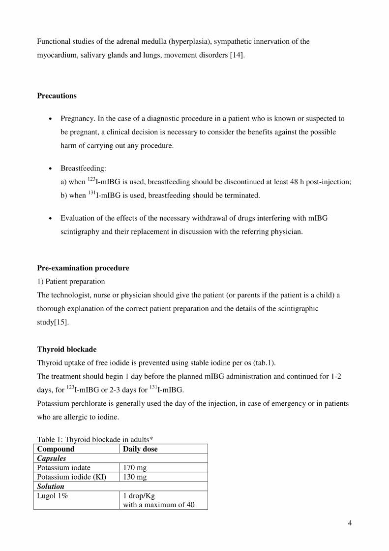
4
Functional studies of the adrenal medulla (hyperplasia), sympathetic innervation of the
myocardium, salivary glands and lungs, movement disorders [14].
Precautions
• Pregnancy. In the case of a diagnostic procedure in a patient who is known or suspected to
be pregnant, a clinical decision is necessary to consider the benefits against the possible
harm of carrying out any procedure.
• Breastfeeding:
a) when 123
I-mIBG is used, breastfeeding should be discontinued at least 48 h post-injection;
b) when 131
I-mIBG is used, breastfeeding should be terminated.
• Evaluation of the effects of the necessary withdrawal of drugs interfering with mIBG
scintigraphy and their replacement in discussion with the referring physician.
Pre-examination procedure
1) Patient preparation
The technologist, nurse or physician should give the patient (or parents if the patient is a child) a
thorough explanation of the correct patient preparation and the details of the scintigraphic
study[15].
Thyroid blockade
Thyroid uptake of free iodide is prevented using stable iodine per os (tab.1).
The treatment should begin 1 day before the planned mIBG administration and continued for 1-2
days, for 123
I-mIBG or 2-3 days for 131
I-mIBG.
Potassium perchlorate is generally used the day of the injection, in case of emergency or in patients
who are allergic to iodine.
Table 1: Thyroid blockade in adults*
Compound Daily dose
Capsules
Potassium iodate 170 mg
Potassium iodide (KI) 130 mg
Solution
Lugol 1% 1 drop/Kg
with a maximum of 40

5
(20 drops twice a day)
Capsules
Potassium perchlorate 400 mg
* in children, the dose should be reduced according to EANM Paediatric Committee guidelines
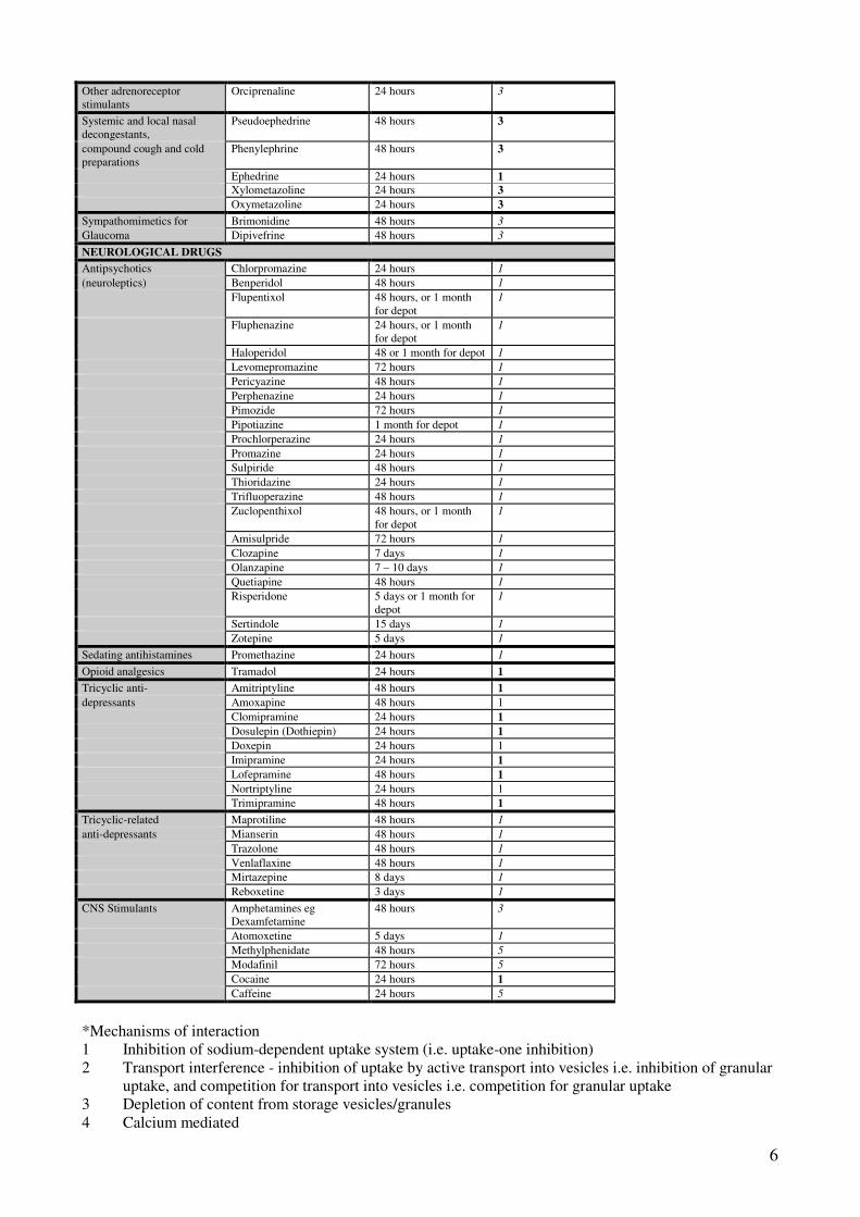
Drug interactions
Many classes of drugs are known (or may be expected) to interfere with the uptake and/or vesicular
storage of mIBG. Table 2 includes some of the most important medications that may affect the
results of mIBG scintigraphy [16,17].
Care must be taken to ensure that such drugs are discontinued (if possible) for an adequate time
prior imaging. Patients with metabolically active catecholamine secreting tumours (i.e.
phaechromocytoma, paraganglioma) often receive alpha- or beta-blocking treatment. Therefore,
drug interruption should be decided in consultation with the referring physician, who is able to
evaluate the patient’s condition and may postpone the study, or realise it without changing the
medication, although this could impair diagnostic accuracy [18,14,19].
Table 2: Drug interactions with mIBG
(adapted from the Radiopharmacy Protocol of the Nuclear Medicine Department, Queen Elizabeth Hospital,
Birmingham, UK)
Drug Group Approved name Recommended
withdrawal time
Mechanism of
interaction *
CARDIOVASCULAR AND SYMPATHOMIMETIC DRUGS
Anti-arrhythmics for
ventricular arrhythmias
Amiodarone Not practical to
withdraw
1,3
Combined ��blocker Labetalol 72 hours 1,3
Adrenergic neurone blockers Brethylium 48 hours 2,3
Guanethidine 48 hours 2,3
Reserpine 48 hours 2,3
�- blockers Phenoxybenzamine
(IV doses only)
15 days 5
Calcium channel blockers Amlodipine 48 hours 4,5
Diltiazem 24 hours 4,5
Felodipine 48 hours 4,5
Isradipine 48 hours 4,5
Lacidipine 48 hours 4,5
Lercanidipine 48 hours 4,5
Nicardipine 48 hours 4,5
Nifedipine 24 hours 4,5
Nimodipine 24 hours 4,5
Nisoldipine 48 hours 4,5
Verapamil 48 hours 4,5
Inotropic Dobutamine 24 hours 3
sympatho-mimetics Dopamine 24 hours 3
Dopexamine 24 hours 3
Vasoconstrictor Ephedrine 24 hours 1
sympathomimetics Metaraminol 24 hours 3
Norepinephrine 24 hours 3
Phenylephrine 24 hours 3
�2 stimulants Salbutamol 24 hours 3
(sympathomimetics) Terbutaline 24 hours 3
Eformoterol 24 hours 3
Bambuterol 24 hours 3
Fenoterol 24 hours 3
Salmeterol 24 hours 3
6
Other adrenoreceptor
stimulants
Orciprenaline 24 hours 3
Systemic and local nasal
decongestants,
Pseudoephedrine 48 hours 3
compound cough and cold
preparations
Phenylephrine 48 hours 3
Ephedrine 24 hours 1
Xylometazoline 24 hours 3
Oxymetazoline 24 hours 3
Sympathomimetics for Brimonidine 48 hours 3
Glaucoma Dipivefrine 48 hours 3
NEUROLOGICAL DRUGS
Antipsychotics Chlorpromazine 24 hours 1
(neuroleptics) Benperidol 48 hours 1
Flupentixol 48 hours, or 1 month
for depot
1
Fluphenazine 24 hours, or 1 month
for depot
1
Haloperidol 48 or 1 month for depot 1
Levomepromazine 72 hours 1
Pericyazine 48 hours 1
Perphenazine 24 hours 1
Pimozide 72 hours 1
Pipotiazine 1 month for depot 1
Prochlorperazine 24 hours 1
Promazine 24 hours 1
Sulpiride 48 hours 1
Thioridazine 24 hours 1
Trifluoperazine 48 hours 1
Zuclopenthixol 48 hours, or 1 month
for depot
1
Amisulpride 72 hours 1
Clozapine 7 days 1
Olanzapine 7 – 10 days 1
Quetiapine 48 hours 1
Risperidone 5 days or 1 month for
depot
1
Sertindole 15 days 1
Zotepine 5 days 1
Sedating antihistamines Promethazine 24 hours 1
Opioid analgesics Tramadol 24 hours 1
Tricyclic anti- Amitriptyline 48 hours 1
depressants Amoxapine 48 hours 1
Clomipramine 24 hours 1
Dosulepin (Dothiepin) 24 hours 1
Doxepin 24 hours 1
Imipramine 24 hours 1
Lofepramine 48 hours 1
Nortriptyline 24 hours 1
Trimipramine 48 hours 1
Tricyclic-related Maprotiline 48 hours 1
anti-depressants Mianserin 48 hours 1
Trazolone 48 hours 1
Venlaflaxine 48 hours 1
Mirtazepine 8 days 1
Reboxetine 3 days 1
CNS Stimulants Amphetamines eg
Dexamfetamine
48 hours 3
Atomoxetine 5 days 1
Methylphenidate 48 hours 5
Modafinil 72 hours 5
Cocaine 24 hours 1
Caffeine 24 hours 5
*Mechanisms of interaction
1 Inhibition of sodium-dependent uptake system (i.e. uptake-one inhibition)
2 Transport interference - inhibition of uptake by active transport into vesicles i.e. inhibition of granular
uptake, and competition for transport into vesicles i.e. competition for granular uptake
3 Depletion of content from storage vesicles/granules
4 Calcium mediated
7
5 Other, possible, unknown mechanisms
Patient preparation including children
The patients are encouraged to drink lots of fluids to facilitate excretion of the radiopharmaceutical.
As said above it is important that patients, when possible and with the supervision of the referring
physician, discontinue all medicaments that could interfere with tumour uptake of radiolabelled
mIBG. It is possible that some foods containing vanillin and catecholamine-like compounds (such
as chocolate and blue-veined cheeses) can interfere on the uptake of mIBG (depletion of granules).
Children need particular preparation, an adapted environment and an adequate staff of operators
who are expert and well trained in paediatric procedures. Parents should be involved in child
preparation and during the scintigraphic study (assistance, sedation, etc.). For paediatric patients see
Guidelines for Radioiodinated MIBG Scintigraphy in Children, which was published under the
auspices of the EANM Paediatric Committee
1) Pre-injection
Clinical evaluation by the nuclear medicine physician
The nuclear medicine physician should consider any information that could be useful for the
interpretation of scintigraphic images:
• relevant history of suspected or known primary tumour
• intake of possibly interfering drugs
• absence or presence of symptoms
• laboratory test results ( plasma and urinary catecholamine dosage, CEA, 5-HIAA, NSE,
chromogranin A, calcitonin, etc.)
• results of any other imaging studies (CT, MRI, US, X-rays)
• history of recent biopsy, surgery, chemotherapy, hormone therapy, radiation therapy.
2) Tracer injection, dosage and injected activity
mIBG, diluted in compliance with manufacturer’s instructions, is administered by slow intravenous
injection (at least 5 minutes) in a peripheral vein. The preparation should have a high specific
activity.
The activity of radiopharmaceutical to be administered should be determined taking into account
the Diagnostic Reference Levels (DRL) for radiopharmaceuticals; these are defined as levels of
activity for groups of standard-sized patients and for broadly defined types of equipment. It is
expected that these levels will not be exceeded for standard procedures when good and normal
8
practice regarding diagnostic and technical performance is applied. For the aforementioned reasons
the following activities for mIBG should be considered only as a general indication, based on the
data of the literature and the current experience. However, it should be noted that in each country
nuclear medicine physicians should respect the DRLs and the rules stated by the local law. The
injection of activities greater than local DRLs must be justified.
The activity administered to adults should be: for 131
I-mIBG: 40-80 MBq (1.2 - 2.2 mCi); for 123
I-
mIBG: 400 MBq (10.8 mCi). The activity administered to children should be calculated on the
basis of a reference dose for an adult, scaled to body weight according to the schedule proposed by
the EANM Paediatric Task Group. For minimum and maximum recommended activities in children
one should consult the above mentioned Guidelines for Radioiodinated MIBG Scintigraphy in
Children (minimum activity 20 MBq for 123
I-mIBG and 35 MBq for 131
I-mIBG; maximum activity
400 MBq for 123
I-mIBG and 80 MBq for 131
I-mIBG).
4) Post-injection
Patients should be encouraged to drink large volumes of fluids following mIBG injection and
should void immediately prior the study.
5) Side-effects
Adverse effects of mIBG (tachycardia, pallor, vomiting, abdominal pain), that are not related to
allergy but to pharmacologic effects of the molecule, are very rare when slow injection is used.
Injection via central venous catheters must be avoided if possible (imaging artefacts, potential
adverse effects).
Radiation dosimetry
The estimated radiation absorbed dose to various organs in healthy subjects following
administration of 123
I mIBG and 131
I mIBG is given in the following tables. The data for123
I mIBG
are quoted from ICRP 80 and for 131
I mIBG are calculated with approximation from ICRP 53 by
considering weighting factors (wt) from ICRP 60 [9,11].
Absorbed doses: 123
I mIBG
Absorbed dose per unit activity administered
(mGy/MBq)
Organ Adult 15 years 5 years
Adrenals 0.017 0,022 0.045
Bladder 0.048 0.061 0.084
Bone surfaces 0.011 0.014 0.034
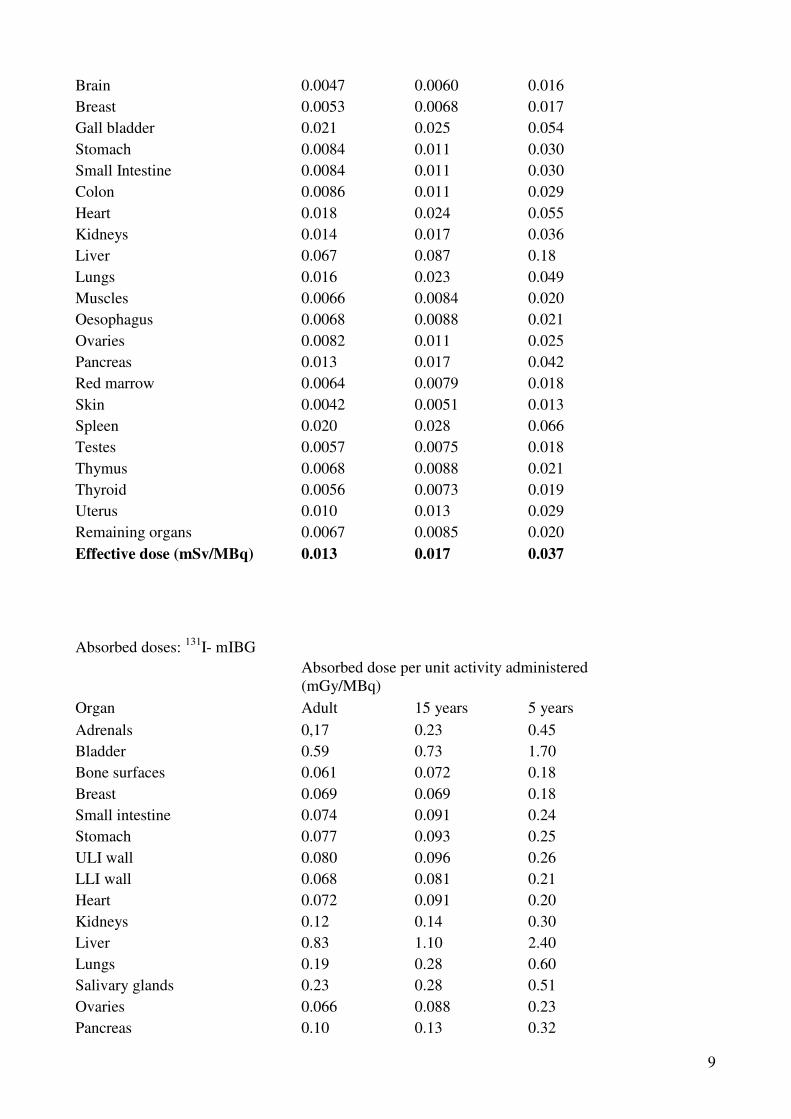
9
Brain 0.0047 0.0060 0.016
Breast 0.0053 0.0068 0.017
Gall bladder 0.021 0.025 0.054
Stomach 0.0084 0.011 0.030
Small Intestine 0.0084 0.011 0.030
Colon 0.0086 0.011 0.029
Heart 0.018 0.024 0.055
Kidneys 0.014 0.017 0.036
Liver 0.067 0.087 0.18
Lungs 0.016 0.023 0.049
Muscles 0.0066 0.0084 0.020
Oesophagus 0.0068 0.0088 0.021
Ovaries 0.0082 0.011 0.025
Pancreas 0.013 0.017 0.042
Red marrow 0.0064 0.0079 0.018
Skin 0.0042 0.0051 0.013
Spleen 0.020 0.028 0.066
Testes 0.0057 0.0075 0.018
Thymus 0.0068 0.0088 0.021
Thyroid 0.0056 0.0073 0.019
Uterus 0.010 0.013 0.029
Remaining organs 0.0067 0.0085 0.020
Effective dose (mSv/MBq) 0.013 0.017 0.037
Absorbed doses: 131
I- mIBG
Absorbed dose per unit activity administered
(mGy/MBq)
Organ Adult 15 years 5 years
Adrenals 0,17 0.23 0.45
Bladder 0.59 0.73 1.70
Bone surfaces 0.061 0.072 0.18
Breast 0.069 0.069 0.18
Small intestine 0.074 0.091 0.24
Stomach 0.077 0.093 0.25
ULI wall 0.080 0.096 0.26
LLI wall 0.068 0.081 0.21
Heart 0.072 0.091 0.20
Kidneys 0.12 0.14 0.30
Liver 0.83 1.10 2.40
Lungs 0.19 0.28 0.60
Salivary glands 0.23 0.28 0.51
Ovaries 0.066 0.088 0.23
Pancreas 0.10 0.13 0.32
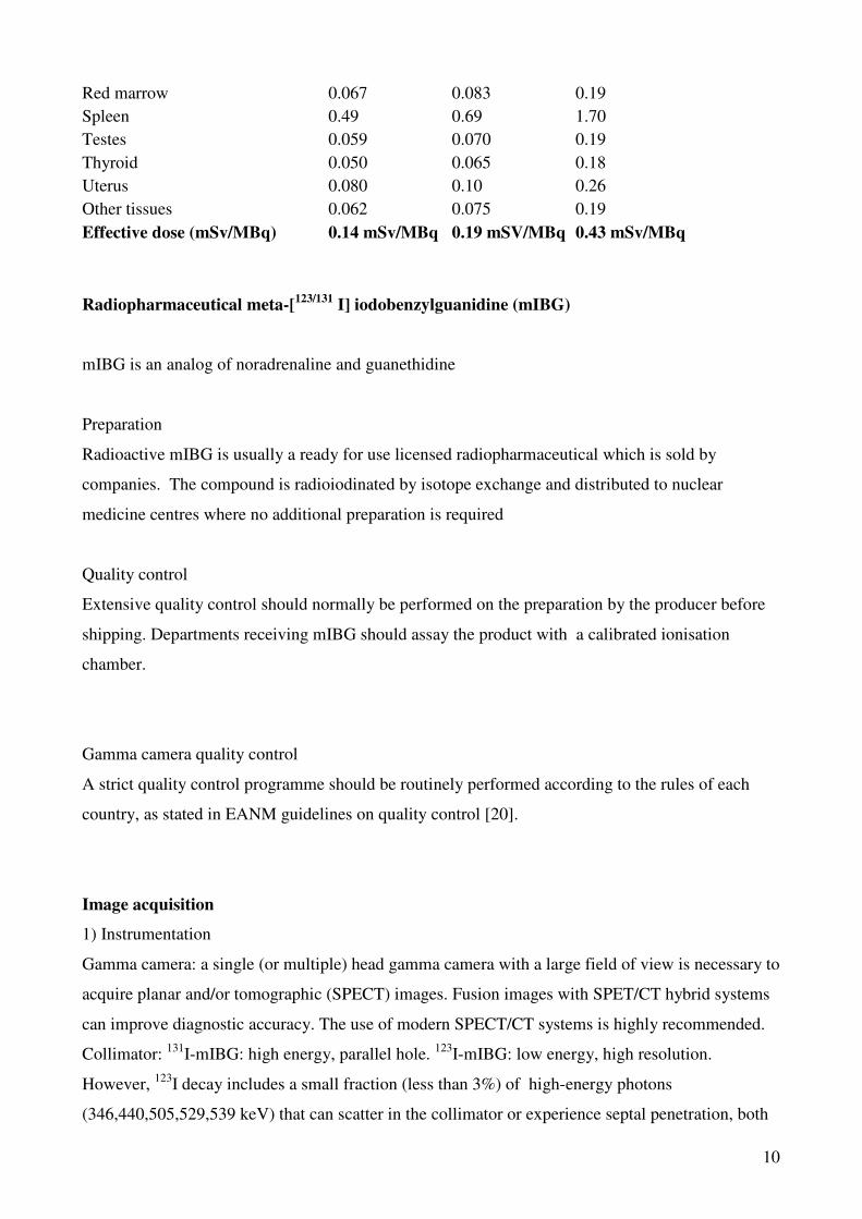
10
Red marrow 0.067 0.083 0.19
Spleen 0.49 0.69 1.70
Testes 0.059 0.070 0.19
Thyroid 0.050 0.065 0.18
Uterus 0.080 0.10 0.26
Other tissues 0.062 0.075 0.19
Effective dose (mSv/MBq) 0.14 mSv/MBq 0.19 mSV/MBq 0.43 mSv/MBq
Radiopharmaceutical meta-[123/131
I] iodobenzylguanidine (mIBG)
mIBG is an analog of noradrenaline and guanethidine
Preparation
Radioactive mIBG is usually a ready for use licensed radiopharmaceutical which is sold by
companies. The compound is radioiodinated by isotope exchange and distributed to nuclear
medicine centres where no additional preparation is required
Quality control
Extensive quality control should normally be performed on the preparation by the producer before
shipping. Departments receiving mIBG should assay the product with a calibrated ionisation
chamber.
Gamma camera quality control
A strict quality control programme should be routinely performed according to the rules of each
country, as stated in EANM guidelines on quality control [20].
Image acquisition
1) Instrumentation
Gamma camera: a single (or multiple) head gamma camera with a large field of view is necessary to
acquire planar and/or tomographic (SPECT) images. Fusion images with SPET/CT hybrid systems
can improve diagnostic accuracy. The use of modern SPECT/CT systems is highly recommended.
Collimator: 131
I-mIBG: high energy, parallel hole. 123
I-mIBG: low energy, high resolution.
However, 123
I decay includes a small fraction (less than 3%) of high-energy photons
(346,440,505,529,539 keV) that can scatter in the collimator or experience septal penetration, both
11
phenomena that degrade image quality when acquisition is performed with low energy collimators.
Medium energy collimators may thus improve image quality, by reducing scatter while preserving
acceptable sensitivity (i.e. without increasing acquisition time).
Given the variability in collimator characteristics and design from different manufacturers, the
choice of the collimator providing best image quality for 123
I-mIBG imaging should therefore be
left to each Nuclear Medicine department.
2) Acquisition modality
Timing of imaging: scanning with 131
I-mIBG is performed 1 and 2 days after injection and can be
repeated at day 3 or later. Scanning with 123I-mIBG is performed between 20 and 24 h. Selected
delayed images (never later than day 2) may be useful in case of equivocal findings at day 1.The
patient should be placed in the supine position. Views: whole body imaging with additional limited-
field images or spot images. Limited-field or spot images are recommended especially in paediatric
patients.
Imaging field:
131I-mIBG: total body scan (speed 4 cm/sec) or both anterior and posterior limited-field or static
spot views (>150 kcounts) of head, neck, chest, abdomen, pelvis, upper and lower extremities.
123I-mIBG: total body scan (speed 5 cm/sec) or both anterior and posterior limited-field or static
spot views (about 500 kcounts or 10 minutes acquisition) of head, neck, chest, abdomen, pelvis,
upper and lower extremities. In neuroblastoma patients for head imaging both antero-posterior and
lateral views are recommended.
In order to reduce acquisition time, for upper and lower limbs, spot views, 75-100 kcounts could be
sufficient for 123
I-mIBG.
Spot views are often superior to whole body scans in contrast and resolution, especially in low
count regions, and are therefore preferable in young children (who may also better bear this exam,
longer in total time, but with interruptions in between). However, the relative uptake intensity in
organs and lesions is more accurately depicted in whole body images.
Cooperative patients should be encouraged to void prior to imaging. It is recommended to start the
exam with abdomen/pelvis spot views when performing multiple spot views of the body.
Image parameters
A pixel size of about 2mm requires a 256x256 matrix or 128x128 matrix with zoom.
For quantification: different levels of approximation can be adopted to correct for attenuation. The
basic method of geometric mean between conjugate views can be improved using a standard
source-phantom based method.
12
Optional images
Single photon emission tomography (SPECT) can improve the diagnostic accuracy. SPECT is
useful mainly in cases where uncertainty exists regarding the localisation and interpretation of the
tracer uptake:
-SPECT can improve characterization of small lesions (soft tissue metastases and residual tumour
uptake), that may not be evident on planar images, especially in case of superimposed areas of high
physiological (i.e. liver, bladder) or pathological (i.e. primary tumour) uptake;
-SPECT can help distinguishing between soft tissue and skeletal lesions, especially in the spine
(that is fundamental in tumour grading);
SPECT can also facilitate the comparison with anatomical imaging: the integration of anatomical
and scintigraphic imaging is essential in clinical practice in order to interpret and identify the
topographic location and the nature of some doubtful lesions. For these reasons the
superimposition, fusion or co-registration of nuclear medicine with CT or MR anatomical images
have a significant impact on the diagnostic accuracy.
This is particularly true in the context of growing availability of hybrid SPECT-CT modality[20].
Thus, whenever possible, SPECT should performed, even if in young children sedation may be
required.
Acquisition parameters depend on the equipment available and the radioisotope used.
Ideally, SPECT should cover pelvis, abdomen and thorax.
Generally, SPECT protocol consists in 120 projections, in 3-degree steps, in continuous or step and
shoot mode, 25-35 seconds per step. Data are acquired on a 128x128 matrix. In case of non-
cooperative patients, it is possible to reduce acquisition time using 6 degree steps, or a 64x64 matrix
with shorter time per frame[22,23]. In SPECT/CT imaging the CT image should be taken with high
resolution in order to have a better characterization of the anatomical surroundings. These images
are also important for dosimetry calculations (uptake and size of the tumour).
Image processing
No particular processing procedure is needed for planar images.
In case of SPECT one should take into account the different types of gamma camera and software
available. Careful choice of processing parameters should be adopted in order to optimize image
quality.
Iterative reconstruction with a low pass post-filter often provides better images than filtered back
projection. Any reporting should clearly state the methodology adopted for image processing and
quantification
13
Interpretation criteria
To evaluate mIBG scintigraphy the following items should be taken into account:
• clinical issue raised in the request for mIBG scintigraphy
• clinical history of the patient
• presence of symptoms or syndrome
• topographical localisation of the uptake according to other imaging data
• uptake in non-physiological areas (this is suspicious for a neuroendocrine tumour or
metastatic localisations)
• intensity and features of the tracer uptake (mIBG uptake may be observed both in benign
and malignant tumours)
• clinical correlation with any other data from previous clinical, biochemical and
morphological examinations
• causes of false negative results (lesion size, tumour biology, physiological uptakes masking
cancer lesions, pharmaceutical interference, etc.)
• causes of false positive results (artefacts, uptake due to physiological processes, benign
uptakes, etc.).
Physiological distribution of mIBG
The uptake of radiolabelled mIBG in different organs depends on catecholamine excretion and/or
adrenergic innervation. After intravenous injection approximately 50% of the administered
radioactivity appears in the urine by 24 h, and 70–90% of the residual activity is recovered within
48 h. Since mIBG is excreted in the urine, the bladder and urinary tract show intense activity. mIBG
is normally taken up mainly by the liver; smaller uptake is described in spleen, lungs, salivary
glands, skeletal muscles and myocardium. Normal adrenal glands are usually not seen, but faint
uptake may be visible 48–72 h after injection in up to 15% of cases when using 131
I-mIBG.
However normal adrenals can be visualized in up to 75% of cases if using 123
I-mIBG (ref). mIBG
may accumulate to a variable degree in nasal mucosa, lungs, gallbladder, colon and uterus. Free
iodine in the bloodstream may cause some uptake in the digestive system and in the thyroid (if not
properly blocked). No skeletal uptake should be seen. Extremities show only slight muscular
activity.
14
In children, brown fat uptake is usually quite symmetric, along the edge of the trapezius
muscles[26]. However, it is seen also over the top of each lung, and along either side of the spine to
the level of the diaphragm, in children and in adults [1]
Pathology
mIBG soft tissue uptake is observed in primary tumour and in metastatic sites including lymph
nodes, liver, bone and bone marrow. Increased uptake in the skeleton (focal or diffuse) is indicative
of bone marrow involvement and/or skeletal metastases.
Sources of error [27,28]
• clinical and biochemical findings that are unknown or have not been considered
• insufficient knowledge of physiological mIBG biodistribution and kinetics
• small lesions, below the resolution power of scintigraphy
• incorrect patient preparation (e.g. pelvic views cannot be correctly interpreted if the patient
has not voided before the acquisition)
• lesions close to the areas of high physiological or pathological uptake
• tumour lesions that do not uptake mIBG (e.g. changes in differentiation, necrosis, interfering
drugs, etc.)
• patient motion (mainly in children)
• increased diffuse physiological uptake (hyperplastic adrenal gland after contralateral
adrenalectomy)
• increased focal physiological uptakes (mainly in the urinary tract or bowel)
• thyroid activity (if no adequate thyroid blockade is performed)
• urine contamination or any other external contamination (salivary secretion).
Reporting
The nuclear medicine physician should record all information regarding the patient, type of
examination, date, radiopharmaceutical (administered activity and route), concise patient history, all
correlated data from previous diagnostic studies, and the clinical question.
The report to the referring physician should describe:
15
• whether the distribution of mIBG is physiological or not;
• all abnormal areas of uptake (intensity, number and site; if necessary, retention of mIBG
over time);
• comparative analysis: the findings should be related to any previous information or results
from other clinical or instrumental examinations;
• interpretation: a clear diagnosis of malignant lesion should be made if possible,
accompanied by a differential diagnosis when appropriate;
• comments on factors that may limit the accuracy of scintigraphy are sometimes important
(lesion size, artefacts, interfering drugs, etc.).
Should gaining of definitive diagnosis require additional diagnostic examination or an adequate
follow-up, this must be recommended.
Standardised form
In order to evaluate the prognosis at diagnosis and to quantify treatment response in neuroblastoma,
different scoring systems have been proposed [29, 30,31,32].
Issues requiring further clarification
• Radiolabelled mIBG and pentetreotide can be used to visualise different neuroendocrine
tumours. In some of these tumours both modalities show a high diagnostic accuracy. Further
investigations are needed to accurately define the clinical indications of the single studies.
This evaluation should be based on diagnostic efficacy, costs, and clinical impact on patient
management [33,34].
Other imaging modalities
FDG-PET visualizes some neuroendoendocrine tumours. However the FDG uptake is satisfactory
only in cancer with high metabolic and proliferative rate. Several false negative results are
described in well-differentiated neoplasms [35]. In neuroblastoma, FDG-PET has been studied in
comparison with 123
I-mIBG. 123
I-mIBG was more sensitive for bone localizations, whereas FDG-
PET seemed to be more reliable for soft tissue lesions. [36]. These approaches showed a poor
concordance, therefore the two techniques could be used as complementary, although no definitive
data are available [36-39].
Some studies reported experience, in tumors known to be mIBG avid, with PET
radiopharmaceuticals like 124
I-mIBG, 18
F-L-DOPA, 18
F-Dopamine [40,41] and 68
Ga-DOTA-
peptides. Reported data are too limited to draw any obvious conclusion on their possible use,
16
although there is a strong rationale to forecast a future role of these radiopharmaceuticals in the
clinical practice.
References:
1. Nakajo M, Shapiro B, Copp J, et al. The normal and abnormal distribution of the
adrenomedullary imaging agent m-I123-iodobenzylguanidine (I-123 MIBG) in man:
evaluation by scintigraphy. J Nucl Med 1983; 24: 672-682.
2. Rubello D, Bui C, Casara d, et al. Functional scintigraphy of the adrenal gland. Eur J
Endocrinol 2002; 147: 13-28
3. Leung A, Shapiro B, Hattner R, et al. The specificity of radioiodinated MIBG for neural
crest tumors in childhood. J Nucl Med 1997; 38:1352-1357.
4. Sisson JC, Shulkin BL. Nuclear medicine imaging of pheochromocytoma and
neuroblastoma. Q J Nucl Med 1999; 43: 217-223.
5. Shapiro B, Gross MD. Radiochemistry, biochemistry, and kinetics of 131I-
metaiodobenzylguanidine (MIBG) and 123I-MIBG: clinical implications of the use of 123I-
MIBG. Med Pediatric Oncol 1987; 15:170-177.
6. Bombardieri E, Maccauro M, De Deckere E, et al. Nuclear medicine imaging of
neuroendocrine tumours. Ann Oncol 2001; 12: S51-61.
7. Troncone L, Rufini V. Radiolabeled metaiodobenzylguanidine in the diagnosis of neural
crest tumors. In: Murray IPC, Ell PJ, eds. Nuclear medicine in clinical diagnosis and
treatment. Edinburgh: Churchill Livingstone 1998; 843-857.
8. Staalman CR, Hoefnagel CA Imaging of neuroblastomas and metastasis in Neuroblastoma
303-329 Brodeur GM, Sawada T, Tsuchida Y and Voute PA Eds Elsevier Amsterdam 2000
9. ICRP Publication 80 Radiation dose to patients from radiopharmaceuticals. Annals of ICRP
1998 28: 3; Pergamon Press, Oxford.
10. Stabin MG, Gelfand MJ. Dosimetry of pediatric nuclear medicine procedures. Q J Nucl Med
1998; 42: 93-112.
11. ICRP Publication 53 Radiation dose to patients from radiopharmaceuticals. Annals of
ICRP.1987: 18: 1-4 Pergamon Press, Oxford.
12. Boubaker A, Bischof Delaloye A. Nuclear medicine procedures and neuroblastoma in
childhood. Their value in the diagnosis, staging and assessment of response to therapy. Q J
Nucl Med 2003; 47: 31-40.
17
13. Perel Y, Conway J, Kletzel M, et al. Clinical impact and prognostic value of
metaiodobenzylguanidine imaging in children with metastatic neuroblastoma. J Pediatr
Hematol Oncol 1999; 21: 13-18.
14. Wafelman AR, Hoefnagel CA, Maes RAA, et al. Radioiodinated metaiodobenzylguanidine:
a review of its biodistribution and pharmacokinetics, drug interaction, cytotoxicity and
dosimetry. Eur J Nucl Med 1994; 21: 545-559.
15. Giammarile F, Boneu A, Edeline V, et al. Guide de réalisation de la scintigraphie à la meta-
iodobenzylguanidine (MIBG) en oncologie pédiatrique. Med Nucl 2000; 24: 35-41.
16. Olivier P, Colarinha P, Fettich J, et al. Guidelines for radioiodinated MIBG scintigraphy in
children. Eur J Nucl Med Mol Imaging 2003; 30: B45-50.
17. Lassmann M, Biassoni L, Monsieurs M, Franzius C, Jacobs F; EANM Dosimetry and
Paediatrics Committees. The new EANM paediatric dosage card. Eur J Nucl Med Mol
Imaging. 2007;34:796-8
18. Solanki KK, Bomanji J, Moyes J, et al. A pharmacological guide to medicines which
interfere with the biodistribution of radiolabelled meta-iodobenzylguanidine (MIBG). Nucl
Med Commun 1992; 13: 513-521.
19. Khafagi FA, Shapiro B, Fig LM, et al. Labetalol reduces iodine-131-MIBG uptake by
pheochromocytoma and normal tissues. J Nucl Med 1989; 30: 481-489.
20. Sokole EB, Plachcinska A, Britten A. Routine quality control reccomendations for nuclear
medicine instrumentation. Eur J Nucl Med Mol Imaging 2010; 37:662-671
21. Goswin Y Meyer-Rochow, Geoff P Schembri, Diana E Benn, Mark S Sywak, Leigh W
Delbridge, Bruce G Robinson, Paul J Roach, and Stan B Sidhu. The utility of
metaiodobenzylguanidine single photon emission computed tomography/computed
tomography (mIBG SPECT/CT) for the diagnosis of pheochromocytoma. Ann Surg Oncol
2010;17:392–400.
22. Rufini V, Fisher GA, Shulkin BL, et al. Iodine-123-MIBG imaging of neuroblastoma: utility
of SPECT and delayed imaging. J Nucl Med 1996; 37: 1464-1468.
23. Rufini V, Giordano A, Di Giuda D, et al. 123MIBG scintigraphy in neuroblastoma: a
comparison between planar and SPECT imaging. Q J Nucl Med 1995; 4: 25-28.
24. Lynn MD, Shapiro B, Sisson JC et al. Portrayal of pheochromocytoma and normal human
adrenal medulla by m-[123I]iodobenzylguanidine: concise communication. J Nucl Med.
1984;25(4):436-440
25. Furuta N, Kiyota H, Yoshigoe F, Hasegawa N, Ohishi Y. Diagnosis of pheochromocytoma
using [123I]-compared with [131I]-metaiodobenzylguanidine scintigraphy. Int J Urol.
1999;6(3):119-124.
18
26. Okuyama C, Sakane N, Yoshida T, Shima K, Kurosawa H, Kumamoto K, Ushijima Y,
Nishimura T. (123)I- or (125)I-metaiodobenzylguanidine visualization of brown adipose
tissue. J Nucl Med.2002 Sept; 43 (9) 1234-40.
27. Peggi L, Liberti E, Pansini G, et al. Pitfalls in Scintigraphic detection of neuroendocrine
tumors. Eur J Nucl Med 1992;19:214-218.
28. Gordon I, Peters AM, Gutman A, et al. Skeletal assessment in neuroblastoma - the pitfalls
of iodine-123-MIBG scans. J Nucl Med 1990; 31: 129-134.
29. Ady N, Zucker JM, Asselain B, Edeline V, Bonnin F, Michon J, Gongora R, Manil L. A
new 123I-MIBG whole body scan scoring method--application to the prediction of the
response of metastases to induction chemotherapy in stage IV neuroblastoma. Eur J Cancer.
1995;31A(2):256-61.
30. Suc A, Lumbroso J, Rubie H, Hattchouel JM, Boneu A, Rodary C, Robert A, Hartmann O.
Metastatic neuroblastoma in children older than one year: prognostic significance of the
initial metaiodobenzylguanidine scan and proposal for a scoring system. Cancer. 1996 Feb
15;77(4):805-11.
31. Katzenstein HM, Cohn SL, Shore RM, Bardo DM, Haut PR, Olszewski M, Schmoldt J, Liu
D, Rademaker AW, Kletzel M. Scintigraphic response by 123I-metaiodobenzylguanidine
scan correlates with event-free survival in high-risk neuroblastoma. J Clin Oncol. 2004 Oct
1;22(19):3909-15.
32. Messina JA, Cheng SC, Franc BL, Charron M, Shulkin B, To B, Maris JM, Yanik G,
Hawkins RA, Matthay KK Evaluation of semi-quantitative scoring system for
metaiodobenzylguanidine (mIBG) scans in patients with relapsed neuroblastoma. Pediatr
Blood Cancer. 2006 Dec;47(7):865-74.
33. Taal BG, Hoefnagel CA, Valdes Olmos et al. Combined diagnostic imaging with 131I-
MIBG and111In-pentetreotide in carcinoid tumours Eur J Cancer 1996; 32: 1924-1932
34. Zuetenhorst JM, Hoefnagel CA, Boot H, et al. Evaluation of (111)In-pentetreotide, (131)I-
MIBG and bone scintigraphy in the detection and clinical management of bone metastases
in carcinoid disease. Nucl Med Commun 2002; 23: 735-41.
35. Adams S, Baum R, Rink T, et al. Limited value of fluorine-18fluoodeoxyglucose PET for
the imaging of neuroendocrine tumours. Eur J Nucl Med 1998; 25: 79-83.
36. Denah R Taggart, Myo M Han, Alekist Quach, Susan Groshen, Wei Ye, Judith G
Villablanca, Hollie A Jackson, Carina Mari Aparici, David Carlson, John Maris, Randall
Hawkins, and Katherine K Matthay. Comparison of iodine-123 metaiodobenzylguanidine
(mIBG) scan and [18F] FDG positron emission tomography to evaluate response after
iodine-131 mIBG therapy for relapsed neuroblastoma. J Clin Oncol 2009;27:5343–5349.
19
37. Kushner BH, Yeung HW, Larson SM, Kramer K, Cheung NK. Extending positron emission
tomography scan utility to high-risk neuroblastoma: fluorine-18 fluorodeoxyglucose (FDG)
positron emission tomography as sole imaging modality in follow-up of patients. J Clin
Oncol 2001; 19:3397–3405.
38. Sharp SE, Shulkin BL, Gelfand MJ, Salisbury S. 123I-mIBG versus 18F-FDG in
Neuroblastoma: Which is better, or which can be eliminated? J Nucl Med 2010 ;51:331.
39. Sharp SE, Shulkin BL, Gelfand MJ, Salisbury S, Furman WL. 123I-mIBG scintigraphy and
18F-FDG PET in neuroblastoma. J Nucl Med 2009; 50:1237–1243.
40. Timmers HJLM, Chen CC, Carrasquillo JA, Whatley M, Ling A, Havekes B, Eisenhofer G,
Martiniova L, Adams KT, Pacak K. Comparison of 18F-fluoro-l-DOPA, 18F-fluoro-
deoxyglucose, and 18F-fluorodopamine PET and 123I-mIBG scintigraphy in the
localization of pheochromocytoma and paraganglioma. J Clin Endocrinol Metab 2009;
94:4757–4767.
41. Ott RJ, Tait D, Flower MA, Babich JW, Lambrecht RM. Treatment planning for 131I-mIBG
radiotherapy of neural crest tumours using 124I-mIBG positron emission tomography. Br J
Radiol. 1992;65:787-91.
Disclaimer
The European Association has written and approved guidelines to promote the use of nuclear medicine procedures with
high quality. These general recommendations cannot be applied to all patients in all practice settings. The guidelines
should not be deemed inclusive of all proper procedures and exclusive of other procedures reasonably directed to
obtaining the same results. The spectrum of patients seen in a specialised practice setting may be different than the
spectrum usually seen in a more general setting. The appropriateness of a procedure will depend in part on the
prevalence of disease in the patient population. In addition, resource available for patient care may vary greatly from
one European country or one medical facility to another. For these reasons, guidelines cannot be rigidly applied.
Related Documents


