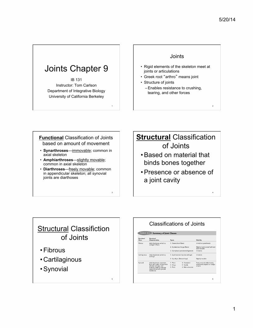
5/20/14 1 Joints Chapter 9 IB 131 Instructor: Tom Carlson Department of Integrative Biology University of California Berkeley 1 Joints • Rigid elements of the skeleton meet at joints or articulations • Greek root “arthro” means joint • Structure of joints – Enables resistance to crushing, tearing, and other forces 2 Functional Classification of Joints based on amount of movement • Synarthroses—immovable ; common in axial skeleton • Amphiarthroses—slightly movable ; common in axial skeleton • Diarthroses—freely movable ; common in appendicular skeleton; all synovial joints are diarthoses 3 Structural Classification of Joints • Based on material that binds bones together • Presence or absence of a joint cavity 4 Structural Classifiction of Joints • Fibrous • Cartilaginous • Synovial 5 Classifications of Joints 6

Welcome message from author
This document is posted to help you gain knowledge. Please leave a comment to let me know what you think about it! Share it to your friends and learn new things together.
Transcript
5/20/14
1
Joints Chapter 9 IB 131
Instructor: Tom Carlson Department of Integrative Biology University of California Berkeley
1
Joints
• Rigid elements of the skeleton meet at joints or articulations
• Greek root “arthro” means joint • Structure of joints
– Enables resistance to crushing, tearing, and other forces
2
Functional Classification of Joints based on amount of movement
• Synarthroses—immovable; common in axial skeleton
• Amphiarthroses—slightly movable; common in axial skeleton
• Diarthroses—freely movable; common in appendicular skeleton; all synovial joints are diarthoses
3
Structural Classification of Joints
• Based on material that binds bones together
• Presence or absence of a joint cavity
4
Structural Classifiction of Joints
• Fibrous • Cartilaginous • Synovial
5
Classifications of Joints
6
5/20/14
2
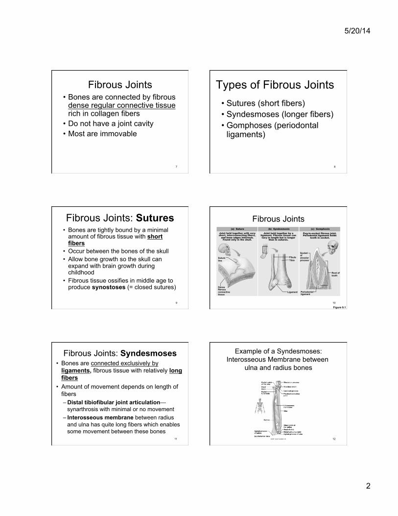
Fibrous Joints • Bones are connected by fibrous
dense regular connective tissue rich in collagen fibers
• Do not have a joint cavity • Most are immovable
7
Types of Fibrous Joints • Sutures (short fibers) • Syndesmoses (longer fibers) • Gomphoses (periodontal
ligaments)
8
Fibrous Joints: Sutures • Bones are tightly bound by a minimal
amount of fibrous tissue with short fibers
• Occur between the bones of the skull • Allow bone growth so the skull can
expand with brain growth during childhood
• Fibrous tissue ossifies in middle age to produce synostoses (= closed sutures)
9
Fibrous Joints
Figure 9.1
Dense fibrous connective tissue
Suture line
Root of tooth
Socket of alveolar process
Periodontal ligament
Fibula Tibia
Ligament
(a) Suture Joint held together with very short, interconnecting fibers,
and bone edges interlock. Found only in the skull.
(b) Syndesmosis Joint held together by a
ligament. Fibrous tissue can vary in length but is longer
than in sutures.
(c) Gomphosis Peg-in-socket fibrous joint. Periodontal ligament holds
tooth in socket.
10
Fibrous Joints: Syndesmoses • Bones are connected exclusively by
ligaments, fibrous tissue with relatively long fibers
• Amount of movement depends on length of fibers – Distal tibiofibular joint articulation—
synarthrosis with minimal or no movement – Interosseous membrane between radius
and ulna has quite long fibers which enables some movement between these bones
11
Example of a Syndesmoses: Interosseous Membrane between
ulna and radius bones
12
5/20/14
3
Fibrous Joints
Figure 9.1
Dense fibrous connective tissue
Suture line
Root of tooth
Socket of alveolar process
Periodontal ligament
Fibula Tibia
Ligament
(a) Suture Joint held together with very short, interconnecting fibers,
and bone edges interlock. Found only in the skull.
(b) Syndesmosis Joint held together by a
ligament. Fibrous tissue can vary in length but is longer
than in sutures.
(c) Gomphosis Peg-in-socket fibrous joint. Periodontal ligament holds
tooth in socket.
13
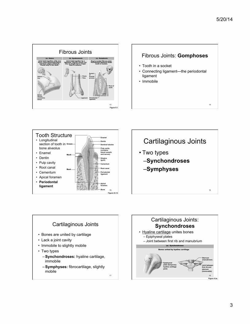
Fibrous Joints: Gomphoses
• Tooth in a socket • Connecting ligament—the periodontal
ligament • Immobile
14
Crown
Neck
Root
Enamel Dentin Dentinal tubules Pulp cavity (contains blood vessels and nerves)
Gingiva (gum)
Cementum
Root canal
Periodontal ligament
Apical foramen
Bone
Tooth Structure • Longitudinal
section of tooth in bone alveolus
• Enamel • Dentin • Pulp cavity • Root canal • Cementum • Apical foramen • Periodontal
ligament
Figure 23.14 15
Cartilaginous Joints • Two types
– Synchondroses – Symphyses
16
Cartilaginous Joints
• Bones are united by cartilage • Lack a joint cavity • Immobile to slightly mobile • Two types
– Synchondroses: hyaline cartilage, immobile
– Symphyses: fibrocartilage, slightly mobile
17
Figure 9.2a
Cartilaginous Joints: Synchondroses
• Hyaline cartilage unites bones – Epiphyseal plates – Joint between first rib and manubrium
Epiphyseal plate (temporary hyaline cartilage joint)
Sternum (manubrium)
Joint between first rib and sternum (immovable)
(a) Synchondroses
Bones united by hyaline cartilage
18
5/20/14
4
Cartilaginous Joints: Symphyses • Fibrocartilage unites bones • Fibrocartilage resists tension & compression
stresses and can act as a shock absorber • Slightly movable joints that provide strength
with flexibility – intervertebral discs – pubic symphysis
• Hyaline cartilage is also present as articular cartilage on the bony surfaces to reduce friction between bones during movement
19
Cartilaginous Joints: Symphyses Bones United by Fibrocartilage
20
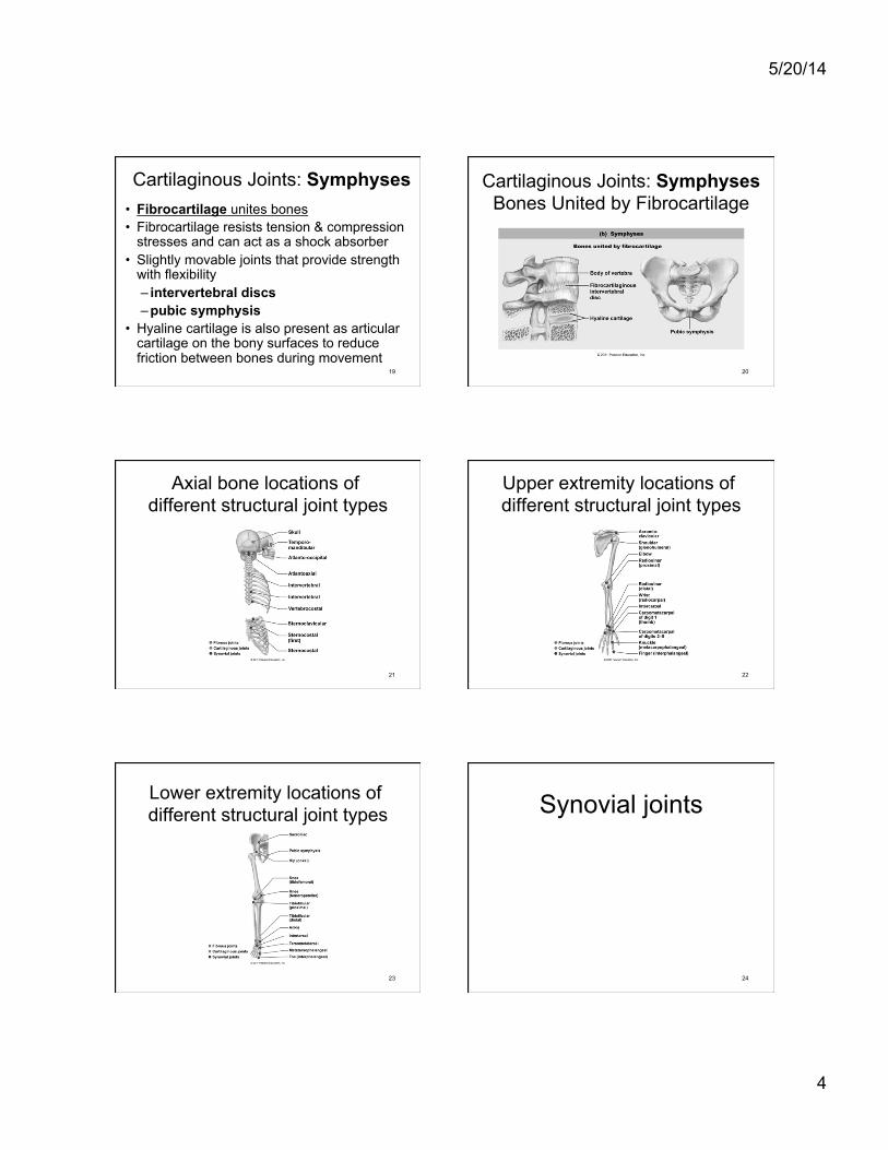
Axial bone locations of different structural joint types
21
Upper extremity locations of different structural joint types
22
Lower extremity locations of different structural joint types
23
Synovial joints
24

5/20/14
5
Synovial Joints • Most movable type of joint • All are diarthroses (movable joints)
and contain a fluid-filled joint cavity • Adjoining bones are covered with
articular cartilage and are separated by a joint cavity
• Joint cavity is enclosed within an articular capsule with the inner layer lined with synovial membrane
25
Periosteum
Ligament
Fibrous capsule Synovial membrane
Joint cavity (contains synovial fluid) Articular (hyaline) cartilage
Articular capsule
(a) A typical synovial joint
General Structure of Synovial Joints
Figure 9.3a 26
General Structure of Synovial Joints
• Articular cartilage – Ends of opposing bones are covered with
hyaline cartilage – Absorbs compression
• Joint cavity (synovial cavity) – Unique to synovial joints – Cavity is a space that contains synovial fluid
27
Periosteum
Ligament
Fibrous capsule Synovial membrane
Joint cavity (contains synovial fluid) Articular (hyaline) cartilage
Articular capsule
(a) A typical synovial joint
General Structure of Synovial Joints
Figure 9.3a 28
Ligaments
• Ligaments extend over the outer surface of the articular capsule and contribute to joint stability
• Ligaments are anchored in periosteum of adjacent bones
29
Articular Capsule of Synovial Joints is enclosed in a two-layered capsule
• Fibrous capsule—dense irregular connective tissue is continuous with periosteal layer of adjoining bones; strengthens joint
• Synovial membrane—loose connective tissue
• Lines inner layer of joint capsule and covers internal joint surfaces not covered by cartilage
• Functions to make synovial fluid 30

5/20/14
6
Synovial membrane • Rich blood supply to synovial membrane
forms extensive capillary beds that provide blood filtrate which forms synovial fluid
31
Synovial Fluid • Produced by synovial membrane • Present in joint cavity • Is a viscous fluid with consistency similar
to raw egg white • Contains glycoprotein molecules
secreted by fibroblasts
32
How Synovial Joints Function • Synovial joints—lubricating devices • Friction could overheat and destroy joint
tissue • As synovial joints are subjected to
compressive forces • Fluid is squeezed out as opposing
cartilages touch • Cartilages ride on the slippery film
33
Periosteum
Ligament
Fibrous capsule Synovial membrane
Joint cavity (contains synovial fluid) Articular (hyaline) cartilage
Articular capsule
(a) A typical synovial joint
General Structure of Synovial Joints
Figure 9.3a 34
Sensory Fibers Richly Innervate Synovial Joints
• Detect pain • Most monitor how much the capsule is
being stretched
35
Three Basic Movements of Synovial Joints
• Gliding—one bone across the surface of another
• Angular movement—movements change the angle between bones
• Rotation—movement around a bone's long axis
36
5/20/14
7
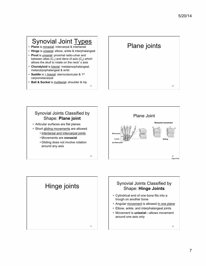
Synovial Joint Types • Plane is nonaxial: intercarpal & intertarsal • Hinge is uniaxial: elbow, ankle & interphalangeal • Pivot is uniaxial: proximal radio-ulnar and
between atlas (C1) and dens of axis (C2) which allows the skull to rotate on the neck’s axis
• Chondyloid is biaxial: metatarsophalangeal, metacarpophalangeal & wrist
• Saddle is > biaxial: sternoclavicular & 1st carpometacarpal
• Ball & Socket is multiaxial: shoulder & hip 37
Plane joints
38
Synovial Joints Classified by Shape: Plane joint
• Articular surfaces are flat planes • Short gliding movements are allowed
• Intertarsal and intercarpal joints • Movements are nonaxial • Gliding does not involve rotation
around any axis
39
Plane Joint
Figure 9.8a
(a) Plane joint
Gliding
Metacarpals
Carpals
Nonaxial movement
40
Hinge joints
41
Synovial Joints Classified by Shape: Hinge Joints
• Cylindrical end of one bone fits into a trough on another bone
• Angular movement is allowed in one plane • Elbow, ankle, and interphalangeal joints • Movement is uniaxial—allows movement
around one axis only
42

5/20/14
8
Hinge Joint
Figure 9.8b
(b) Hinge joint
Medial/ lateral axis
Flexion and extension
Humerus
Ulna
Uniaxial movement
43
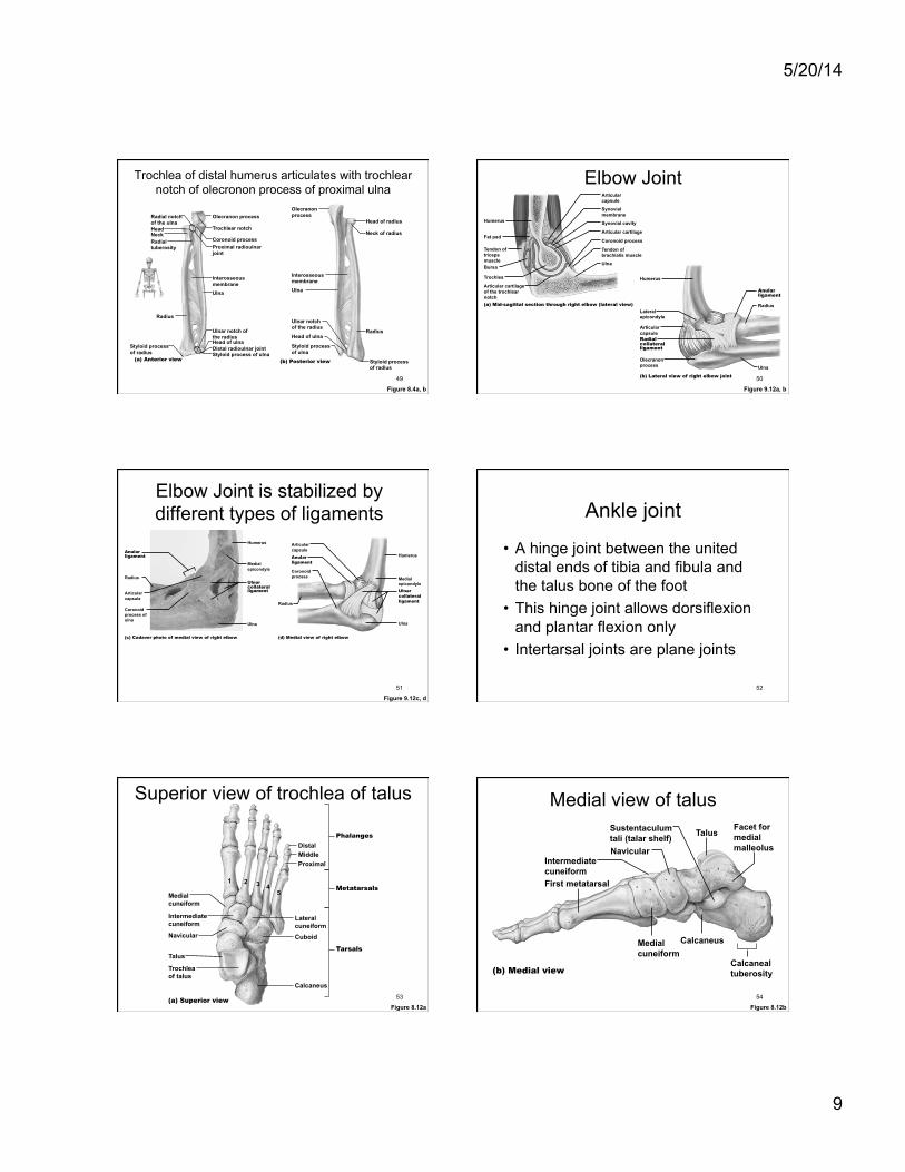
Trochlea of distal humerus articulates with trochlear notch of olecronon process of proximal ulna
Figure 8.3c, d
Coronoid fossa
Radius
Radial tuberosity
Head of radius
Capitulum
Trochlea
(c) Anterior view at the elbow region
Humerus
Medial epicondyle
Coronoid process of ulna
Ulna Radial notch
Olecranon fossa
Ulna
Olecranon process
Medial epicondyle
(d) Posterior view of extended elbow
Humerus
Lateral epicondyle
Head
Radius
Neck
44
Elbow joint • The trochlea of the distal humerus articulates
with the trochlear notch of the proximal ulna to form a hinge
• Allows flexion and extension • Tendons of biceps brachii, triceps brachii,
and brachialis provide stability • Anular ligament helps stabilize proximal
radius and ulna bones
45
Tendon of biceps brachii contributes to stability of elbow joint
46
Tendon of triceps brachii contributes to stability of elbow joint
47
Brachialis muscle attached to humerus and ulna helps
stabilize the elbow Anterior view
48
5/20/14
9
Trochlea of distal humerus articulates with trochlear notch of olecronon process of proximal ulna
Figure 8.4a, b
Radial notch of the ulna
Olecranon process
Trochlear notch
Coronoid process Proximal radioulnar joint
Distal radioulnar joint
Ulnar notch of the radius Head of ulna
Styloid process of ulna
Interosseous membrane Ulna
Head Neck Radial tuberosity
Radius
Styloid process of radius
(a) Anterior view
Olecranon process
Styloid process of radius
Radius
Neck of radius
Head of radius
Ulnar notch of the radius
Head of ulna
Styloid process of ulna
Interosseous membrane Ulna
(b) Posterior view
49
Elbow Joint
Figure 9.12a, b
Articular capsule
Synovial membrane
Synovial cavity
Articular cartilage
Coronoid process
Tendon of brachialis muscle
Ulna
Humerus
Fat pad
Tendon of triceps muscle Bursa
Trochlea
Articular cartilage of the trochlear notch (a) Mid-sagittal section through right elbow (lateral view)
Humerus
Lateral epicondyle
Articular capsule Radial collateral ligament
Olecranon process
(b) Lateral view of right elbow joint
Anular ligament
Radius
Ulna
50
Elbow Joint is stabilized by different types of ligaments
Figure 9.12c, d
Articular capsule Anular ligament
Coronoid process
(d) Medial view of right elbow
Radius
Humerus
Medial epicondyle Ulnar collateral ligament
Ulna
Anular ligament
Humerus
Medial epicondyle
Ulnar collateral ligament
Ulna
Articular capsule
Radius
Coronoid process of ulna
(c) Cadaver photo of medial view of right elbow
51
Ankle joint
• A hinge joint between the united distal ends of tibia and fibula and the talus bone of the foot
• This hinge joint allows dorsiflexion and plantar flexion only
• Intertarsal joints are plane joints
52
Medial cuneiform
Phalanges
Metatarsals
Tarsals
Navicular
Intermediate cuneiform
Talus
Calcaneus
(a) Superior view
Cuboid
Lateral cuneiform
Proximal Middle Distal
Trochlea of talus
5 4 3 2 1
Superior view of trochlea of talus
Figure 8.12a 53
Medial view of talus
Figure 8.12b
Facet for medial malleolus
Calcaneal tuberosity (b) Medial view
Intermediate cuneiform
Sustentaculum tali (talar shelf) Talus
Navicular
First metatarsal
Medial cuneiform
Calcaneus
54
5/20/14
10
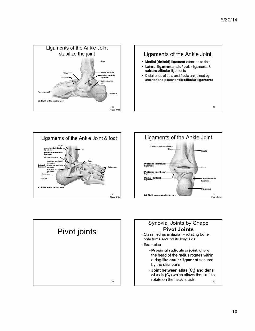
Ligaments of the Ankle Joint stabilize the joint
Figure 9.18b
Medial malleolus
Calcaneus
Sustentaculum tali
Medial (deltoid) ligament
Talus
Navicular
Tibia
1st metatarsal
(b) Right ankle, medial view
55
Ligaments of the Ankle Joint • Medial (deltoid) ligament attached to tibia • Lateral ligaments: talofibular ligaments &
calcaneofibular ligaments • Distal ends of tibia and fibula are joined by
anterior and posterior tibiofibular ligaments
56
Ligaments of the Ankle Joint & foot
Figure 9.18c
Anterior tibiofibular ligament
Anterior talofibular ligament
Calcaneus
Metatarsals
Lateral malleolus
Posterior tibiofibular ligament
Posterior talofibular ligament Calcaneofibular ligament
Tibia
Talus
Cuboid
Fibula
(c) Right ankle, lateral view
Lateral ligament
57
Ligaments of the Ankle Joint
Figure 9.18d
Talus
Posterior talofibular ligament
Posterior tibiofibular ligament
Interosseous membrane
Calcaneofibular ligament
Fibula Tibia
Medial (deltoid) ligament
Calcaneus
(d) Right ankle, posterior view 58
Pivot joints
59
Synovial Joints by Shape Pivot Joints
• Classified as uniaxial – rotating bone only turns around its long axis
• Examples • Proximal radioulnar joint where
the head of the radius rotates within a ring-like anular ligament secured by the ulna bone
• Joint between atlas (C1) and dens of axis (C2) which allows the skull to rotate on the neck’s axis
60
5/20/14
11
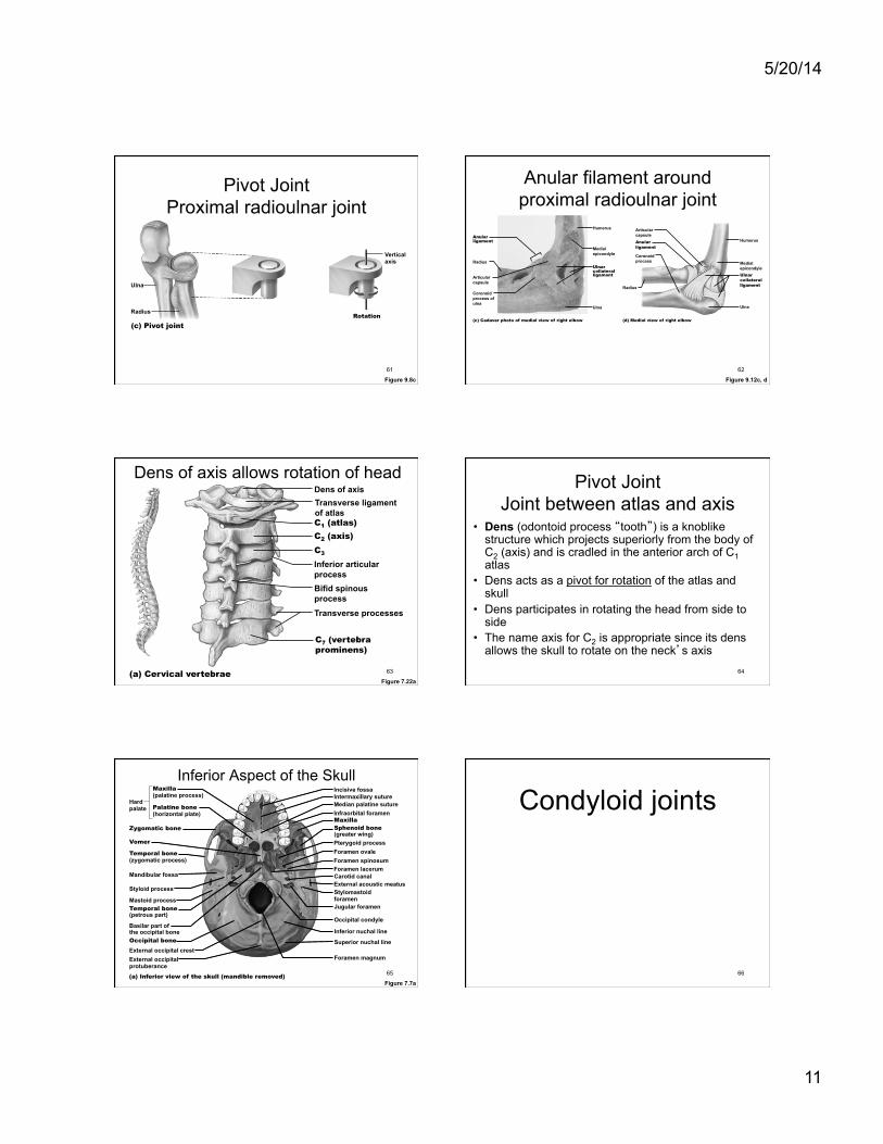
Pivot Joint Proximal radioulnar joint
Figure 9.8c
(c) Pivot joint
Ulna
Vertical axis
Rotation Radius
61
Anular filament around proximal radioulnar joint
Figure 9.12c, d
Articular capsule Anular ligament
Coronoid process
(d) Medial view of right elbow
Radius
Humerus
Medial epicondyle Ulnar collateral ligament
Ulna
Anular ligament
Humerus
Medial epicondyle
Ulnar collateral ligament
Ulna
Articular capsule
Radius
Coronoid process of ulna
(c) Cadaver photo of medial view of right elbow
62
Dens of axis Transverse ligament of atlas C1 (atlas) C2 (axis) C3
Bifid spinous process
Transverse processes
C7 (vertebra prominens)
(a) Cervical vertebrae
Inferior articular process
Dens of axis allows rotation of head
Figure 7.22a 63
Pivot Joint Joint between atlas and axis
• Dens (odontoid process “tooth”) is a knoblike structure which projects superiorly from the body of C2 (axis) and is cradled in the anterior arch of C1 atlas
• Dens acts as a pivot for rotation of the atlas and skull
• Dens participates in rotating the head from side to side
• The name axis for C2 is appropriate since its dens allows the skull to rotate on the neck’s axis
64
Maxilla (palatine process)
Hard palate
Zygomatic bone
Incisive fossa
Median palatine suture Intermaxillary suture
Infraorbital foramen Maxilla Sphenoid bone (greater wing)
Foramen ovale Pterygoid process
Foramen lacerum Carotid canal External acoustic meatus Stylomastoid foramen Jugular foramen
Foramen magnum
Occipital condyle Inferior nuchal line Superior nuchal line
Temporal bone (zygomatic process)
Mandibular fossa
Vomer
Styloid process
External occipital crest External occipital protuberance (a) Inferior view of the skull (mandible removed)
Mastoid process Temporal bone (petrous part) Basilar part of the occipital bone Occipital bone
Palatine bone (horizontal plate)
Foramen spinosum
Inferior Aspect of the Skull
Figure 7.7a 65
Condyloid joints
66
5/20/14
12
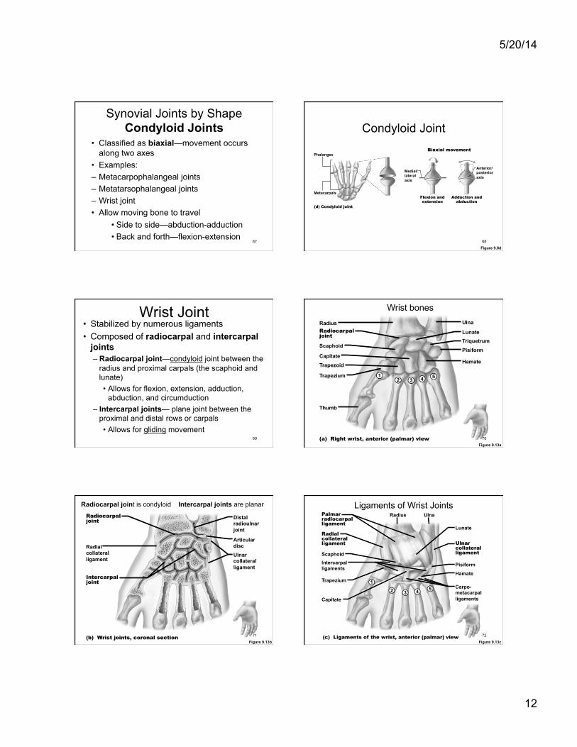
Synovial Joints by Shape Condyloid Joints
• Classified as biaxial—movement occurs along two axes
• Examples: – Metacarpophalangeal joints – Metatarsophalangeal joints – Wrist joint • Allow moving bone to travel
• Side to side—abduction-adduction • Back and forth—flexion-extension
67
Condyloid Joint
Figure 9.8d
(d) Condyloid joint
Medial/ lateral axis
Adduction and abduction
Flexion and extension
Metacarpals
Phalanges
Anterior/ posterior axis
Biaxial movement
68
Wrist Joint • Stabilized by numerous ligaments • Composed of radiocarpal and intercarpal
joints – Radiocarpal joint—condyloid joint between the
radius and proximal carpals (the scaphoid and lunate) • Allows for flexion, extension, adduction,
abduction, and circumduction – Intercarpal joints— plane joint between the
proximal and distal rows or carpals • Allows for gliding movement
69
Wrist Joint
Figure 9.13a
Radius Ulna
Lunate
Triquetrum
Pisiform
Hamate Capitate
Scaphoid
Trapezoid
Trapezium
Thumb
Radiocarpal joint
(a) Right wrist, anterior (palmar) view
Wrist bones
70
Wrist Joint
Figure 9.13b
Distal radioulnar joint
Ulnar collateral ligament
Articular disc Radial
collateral ligament
Radiocarpal joint
Intercarpal joint
(b) Wrist joints, coronal section
Radiocarpal joint is condyloid Intercarpal joints are planar
71
Ligaments of Wrist Joints
Figure 9.13c
Hamate
Carpo- metacarpal ligaments
Pisiform
Lunate
Radius Ulna
Ulnar collateral ligament
Radial collateral ligament
Palmar radiocarpal ligament
Intercarpal ligaments
Trapezium
Capitate
Scaphoid
(c) Ligaments of the wrist, anterior (palmar) view 72
5/20/14
13
Wrist Joint stabilized by numerous ligaments
• Radiocarpal ligaments • Intercarpal ligaments • Carpometacarpal ligaments • Collateral ligaments
73
Saddle joints
74
Synovial Joints by Shape Saddle Joints
• Each articular surface has concave and convex surfaces
• Essentially the same as condyloid joints • Often classified as biaxial joints, however have
the ability to move in more than two axes • 1st carpometacarpal joint is a good example
as it allows opposition of the thumb in addition to flexion and extension
• Sternoclavicular joint is another example 75
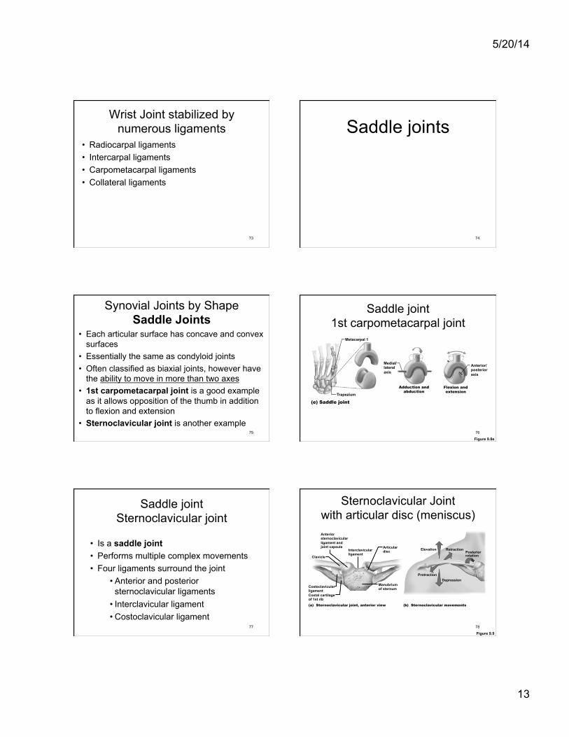
Saddle joint 1st carpometacarpal joint
Figure 9.8e
(e) Saddle joint
Anterior/ posterior axis
Medial/ lateral axis
Adduction and abduction
Metacarpal 1
Trapezium Flexion and extension
76
Saddle joint Sternoclavicular joint
• Is a saddle joint • Performs multiple complex movements • Four ligaments surround the joint
• Anterior and posterior sternoclavicular ligaments
• Interclavicular ligament • Costoclavicular ligament
77
Sternoclavicular Joint with articular disc (meniscus)
Figure 9.9
Anterior sternoclavicular ligament and joint capsule
Interclavicular ligament
Articular disc
Manubrium of sternum
Costal cartilage of 1st rib
Costoclavicular ligament
Clavicle
(a) Sternoclavicular joint, anterior view
Depression Protraction
Elevation Retraction Posterior rotation
(b) Sternoclavicular movements
78
5/20/14
14
Ball-in socket joints
79
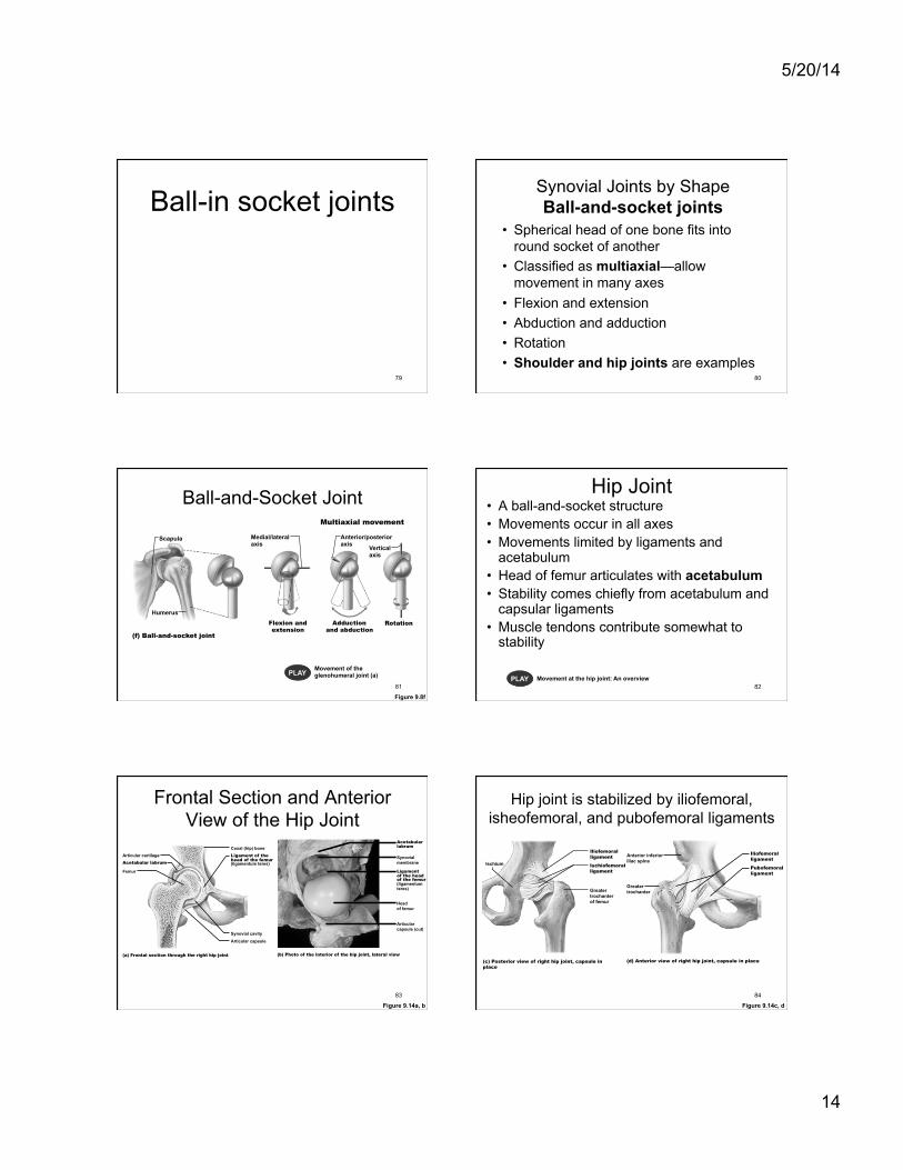
Synovial Joints by Shape Ball-and-socket joints
• Spherical head of one bone fits into round socket of another
• Classified as multiaxial—allow movement in many axes
• Flexion and extension • Abduction and adduction • Rotation • Shoulder and hip joints are examples
80
Ball-and-Socket Joint
Figure 9.8f
PLAY Movement of the glenohumeral joint (a)
(f) Ball-and-socket joint
Medial/lateral axis
Anterior/posterior axis Vertical
axis
Rotation Adduction and abduction
Flexion and extension
Scapula
Humerus
Multiaxial movement
81
Hip Joint • A ball-and-socket structure • Movements occur in all axes • Movements limited by ligaments and
acetabulum • Head of femur articulates with acetabulum • Stability comes chiefly from acetabulum and
capsular ligaments • Muscle tendons contribute somewhat to
stability
PLAY Movement at the hip joint: An overview 82
Frontal Section and Anterior View of the Hip Joint
Figure 9.14a, b
Articular cartilage Coxal (hip) bone Ligament of the head of the femur (ligamentum teres)
Synovial cavity Articular capsule
Acetabular labrum
Femur
(a) Frontal section through the right hip joint
Acetabular labrum
Synovial membrane
Ligament of the head of the femur (ligamentum teres)
Head of femur
Articular capsule (cut)
(b) Photo of the interior of the hip joint, lateral view
83
Anterior inferior iliac spine
Iliofemoral ligament
Pubofemoral ligament
Greater trochanter
(d) Anterior view of right hip joint, capsule in place
Hip joint is stabilized by iliofemoral, isheofemoral, and pubofemoral ligaments
Figure 9.14c, d
Ischium
Iliofemoral ligament
Ischiofemoral ligament
Greater trochanter of femur
(c) Posterior view of right hip joint, capsule in place
84
5/20/14
15
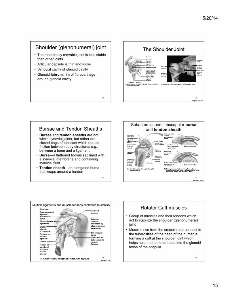
Shoulder (glenohumeral) joint • The most freely movable joint is less stable
than other joints • Articular capsule is thin and loose • Synovial cavity of glenoid cavity • Glenoid labrum: rim of fibrocartilage
around glenoid cavity
85
The Shoulder Joint
Figure 9.11d, e
Acromion
Coracoid process Articular capsule Glenoid cavity Glenoid labrum
Tendon of long head of biceps brachii muscle Glenohumeral ligaments Tendon of the subscapularis muscle Scapula
Posterior Anterior (d) Lateral view of socket of right shoulder joint, humerus removed
(e) Posterior view of an opened left shoulder joint
Head of humerus
Muscle of rotator cuff (cut)
Acromion (cut)
Glenoid cavity of scapula Capsule of shoulder joint (opened)
86
Bursae and Tendon Sheaths • Bursae and tendon sheaths are not
within synovial joints, but rather are closed bags of lubricant which reduce friction between body structures e.g., between a bone and a ligament
• Bursa—a flattened fibrous sac lined with a synovial membrane and containing synovial fluid
• Tendon sheath—an elongated bursa that wraps around a tendon
87
Subacromial and subscapular bursa and tendon sheath
Figure 9.5a, b
Acromion of scapula
Joint cavity containing synovial fluid
Synovial membrane Fibrous capsule
Humerus
Hyaline cartilage
Coracoacromial ligament Subacromial bursa Fibrous articular capsule
Tendon sheath
Tendon of long head of biceps brachii muscle (a) Frontal section through the right shoulder joint
Coracoacromial ligament Subacromial bursa
Cavity in bursa containing synovial fluid
(b) Enlargement of (a), showing how a bursa eliminates friction where a ligament (or other structure) would rub against a bone
Humerus resting
Humerus moving
Bursa rolls and lessens friction.
Humerus head rolls medially as arm abducts.
88
Multiple ligaments and muscle tendons contribute to stability
Figure 9.11c
Acromion Coracoacromial ligament Subacromial bursa Coracohumeral ligament Greater tubercle of humerus Transverse humeral ligament Tendon sheath Tendon of long head of biceps brachii muscle
Articular capsule reinforced by glenohumeral ligaments
Subscapular bursa Tendon of the subscapularis muscle Scapula
Coracoid process
(c) Anterior view of right shoulder joint capsule 89
Rotator Cuff muscles • Group of muscles and their tendons which
act to stabilize the shoulder (glenohumeral) joint
• Muscles rise from the scapula and connect to the tuberosities of the head of the humerus, forming a cuff at the shoulder joint which helps hold the humerus head into the glenoid fossa of the scapula
90
5/20/14
16
Glenohumeral (Shoulder) Joint • The rotator cuff is made up of four
muscles and their associated tendons – Subscapularis – Supraspinatus – Infraspinatus – Teres minor
• All four of these tendons contribute to the stability of the joint
• Rotator cuff injuries are common shoulder injuries
91
Rotator cuff muscles: - Supraspinatus muscle - Infraspinatus muscle - Teres minor muscle - Subscapularis muscle(
92
Subscapularis muscle attached to humerus Anterior view
93
Knee joint
94
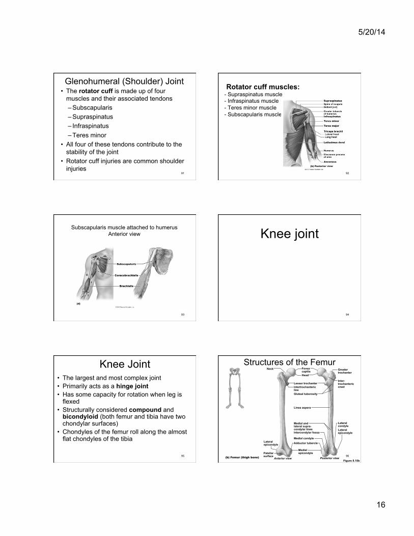
Knee Joint • The largest and most complex joint • Primarily acts as a hinge joint • Has some capacity for rotation when leg is
flexed • Structurally considered compound and
bicondyloid (both femur and tibia have two chondylar surfaces)
• Chondyles of the femur roll along the almost flat chondyles of the tibia
95
Neck Fovea capitis Greater
trochanter
Inter- trochanteric crest
Lateral condyle Lateral epicondyle
Head
Intertrochanteric line
Lesser trochanter
Gluteal tuberosity
Linea aspera
Intercondylar fossa
Medial and lateral supra- condylar lines
Medial condyle
Medial epicondyle
Adductor tubercle
Anterior view Posterior view (b) Femur (thigh bone)
Lateral epicondyle
Patellar surface
Structures of the Femur
Figure 8.10b 96
5/20/14
17
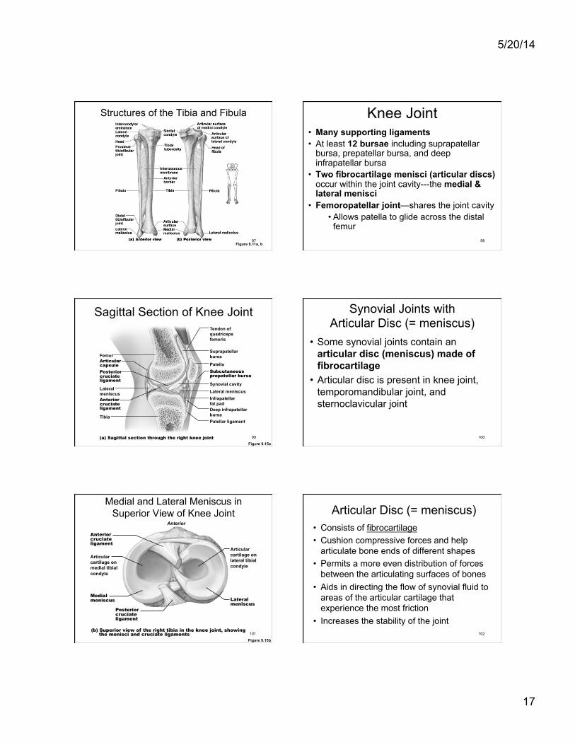
Structures of the Tibia and Fibula
Figure 8.11a, b 97
Knee Joint • Many supporting ligaments • At least 12 bursae including suprapatellar
bursa, prepatellar bursa, and deep infrapatellar bursa
• Two fibrocartilage menisci (articular discs) occur within the joint cavity---the medial & lateral menisci
• Femoropatellar joint—shares the joint cavity • Allows patella to glide across the distal
femur 98
Sagittal Section of Knee Joint
Figure 9.15a (a) Sagittal section through the right knee joint
Femur
Tendon of quadriceps femoris
Suprapatellar bursa
Patella Subcutaneous prepatellar bursa
Synovial cavity Lateral meniscus
Posterior cruciate ligament
Infrapatellar fat pad Deep infrapatellar bursa Patellar ligament
Articular capsule
Lateral meniscus Anterior cruciate ligament
Tibia
99
Synovial Joints with Articular Disc (= meniscus)
• Some synovial joints contain an articular disc (meniscus) made of fibrocartilage
• Articular disc is present in knee joint, temporomandibular joint, and sternoclavicular joint
100
Medial and Lateral Meniscus in Superior View of Knee Joint
Figure 9.15b
(b) Superior view of the right tibia in the knee joint, showing the menisci and cruciate ligaments
Medial meniscus
Articular cartilage on medial tibial condyle
Anterior
Anterior cruciate ligament
Articular cartilage on lateral tibial condyle
Lateral meniscus
Posterior cruciate ligament
101
Articular Disc (= meniscus) • Consists of fibrocartilage • Cushion compressive forces and help
articulate bone ends of different shapes • Permits a more even distribution of forces
between the articulating surfaces of bones • Aids in directing the flow of synovial fluid to
areas of the articular cartilage that experience the most friction
• Increases the stability of the joint 102
5/20/14
18
Medial and Lateral Meniscus in Anterior View of Flexed Knee
103
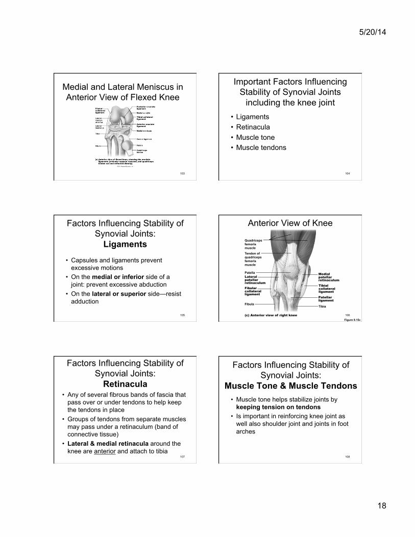
Important Factors Influencing Stability of Synovial Joints
including the knee joint • Ligaments • Retinacula • Muscle tone • Muscle tendons
104
Factors Influencing Stability of Synovial Joints:
Ligaments
• Capsules and ligaments prevent excessive motions
• On the medial or inferior side of a joint: prevent excessive abduction
• On the lateral or superior side—resist adduction
105
Anterior View of Knee
Figure 9.15c
Quadriceps femoris muscle Tendon of quadriceps femoris muscle
Patella Lateral patellar retinaculum
Medial patellar retinaculum
Tibial collateral ligament
Tibia
Fibular collateral ligament
Fibula
(c) Anterior view of right knee
Patellar ligament
106
Factors Influencing Stability of Synovial Joints:
Retinacula • Any of several fibrous bands of fascia that
pass over or under tendons to help keep the tendons in place
• Groups of tendons from separate muscles may pass under a retinaculum (band of connective tissue)
• Lateral & medial retinacula around the knee are anterior and attach to tibia
107
Factors Influencing Stability of Synovial Joints:
Muscle Tone & Muscle Tendons • Muscle tone helps stabilize joints by
keeping tension on tendons • Is important in reinforcing knee joint as
well also shoulder joint and joints in foot arches
108
5/20/14
19
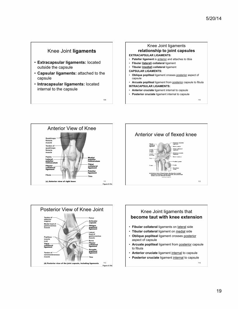
Knee Joint ligaments
• Extracapsular ligaments: located outside the capsule
• Capsular ligaments: attached to the capsule
• Intracapsular ligaments: located internal to the capsule
109
Knee Joint ligaments relationship to joint capsules
EXTRACAPSULAR LIGAMENTS: • Patellar ligament is anterior and attaches to tibia • Fibular (lateral) collateral ligament • Tibular (medial) collateral ligament CAPSULAR LIGAMENTS: • Oblique popliteal ligament crosses posterior aspect of
capsule • Arcuate popliteal ligament from posterior capsule to fibula INTRACAPSULAR LIGAMENTS: • Anterior cruciate ligament internal to capsule • Posterior cruciate ligament internal to capsule
110
Anterior View of Knee
Figure 9.15c
Quadriceps femoris muscle Tendon of quadriceps femoris muscle
Patella Lateral patellar retinaculum
Medial patellar retinaculum
Tibial collateral ligament
Tibia
Fibular collateral ligament
Fibula
(c) Anterior view of right knee
Patellar ligament
111
Anterior view of flexed knee
112
Posterior View of Knee Joint
Figure 9.15d
Articular capsule
Oblique popliteal ligament
Lateral head of gastrocnemius muscle
Fibular collateral ligament
Arcuate popliteal ligament
Tibia
Femur
Medial head of gastrocnemius muscle
Tendon of semimembranosus muscle
(d) Posterior view of the joint capsule, including ligaments
Popliteus muscle (cut)
Tendon of adductor magnus
Bursa
Tibial collateral ligament
113
Knee Joint ligaments that become taut with knee extension
• Fibular collateral ligaments on lateral side • Tibular collateral ligament on medial side • Oblique popliteal ligament crosses posterior
aspect of capsule • Arcuate popliteal ligament from posterior capsule
to fibula • Anterior cruciate ligament internal to capsule • Posterior cruciate ligament internal to capsule
114
5/20/14
20
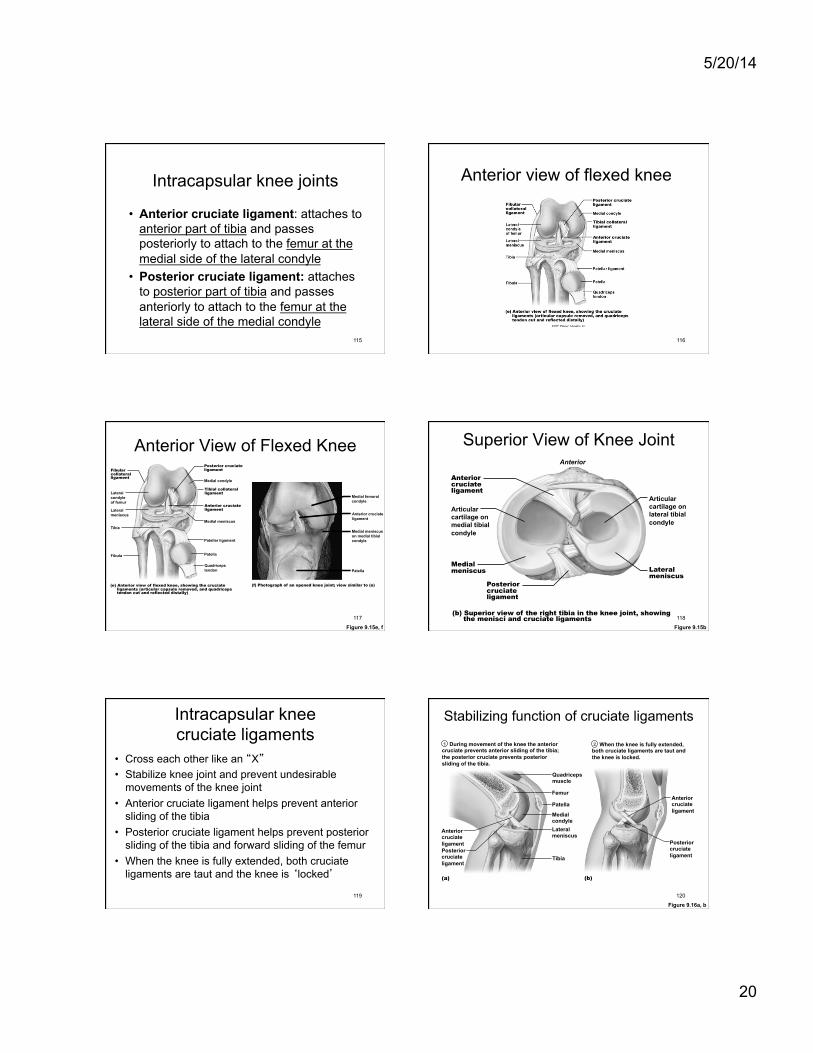
Intracapsular knee joints
• Anterior cruciate ligament: attaches to anterior part of tibia and passes posteriorly to attach to the femur at the medial side of the lateral condyle
• Posterior cruciate ligament: attaches to posterior part of tibia and passes anteriorly to attach to the femur at the lateral side of the medial condyle
115
Anterior view of flexed knee
116
Anterior View of Flexed Knee
Figure 9.15e, f
Fibular collateral ligament
Posterior cruciate ligament
Medial condyle
Tibial collateral ligament
Anterior cruciate ligament
Medial meniscus
Patellar ligament
Patella
Quadriceps tendon
Lateral condyle of femur
Lateral meniscus
Fibula
(e) Anterior view of flexed knee, showing the cruciate ligaments (articular capsule removed, and quadriceps tendon cut and reflected distally)
Tibia
Medial femoral condyle
Anterior cruciate ligament
Medial meniscus on medial tibial condyle
Patella
(f) Photograph of an opened knee joint; view similar to (e)
117
Superior View of Knee Joint
Figure 9.15b
(b) Superior view of the right tibia in the knee joint, showing the menisci and cruciate ligaments
Medial meniscus
Articular cartilage on medial tibial condyle
Anterior
Anterior cruciate ligament
Articular cartilage on lateral tibial condyle
Lateral meniscus
Posterior cruciate ligament
118
Intracapsular knee cruciate ligaments
• Cross each other like an “X” • Stabilize knee joint and prevent undesirable
movements of the knee joint • Anterior cruciate ligament helps prevent anterior
sliding of the tibia • Posterior cruciate ligament helps prevent posterior
sliding of the tibia and forward sliding of the femur • When the knee is fully extended, both cruciate
ligaments are taut and the knee is ‘locked’
119
Stabilizing function of cruciate ligaments During movement of the knee the anterior cruciate prevents anterior sliding of the tibia; the posterior cruciate prevents posterior sliding of the tibia.
Anterior cruciate ligament
(a)
Posterior cruciate ligament
Quadriceps muscle Femur Patella
Lateral meniscus
Tibia
Medial condyle
(b)
Anterior cruciate ligament
Posterior cruciate ligament
When the knee is fully extended, both cruciate ligaments are taut and the knee is locked.
1 2
Figure 9.16a, b 120
5/20/14
21
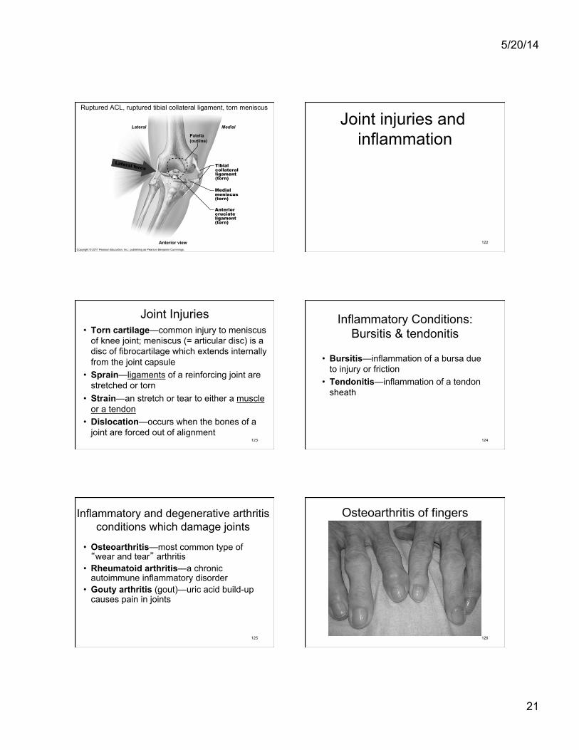
Copyright © 2011 Pearson Education, Inc., publishing as Pearson Benjamin Cummings.
Ruptured ACL, ruptured tibial collateral ligament, torn meniscus
Lateral Medial Patella (outline)
Tibial collateral ligament (torn)
Anterior view
Medial meniscus (torn) Anterior cruciate ligament (torn)
Lateral force
Joint injuries and inflammation
122
Joint Injuries • Torn cartilage—common injury to meniscus
of knee joint; meniscus (= articular disc) is a disc of fibrocartilage which extends internally from the joint capsule
• Sprain—ligaments of a reinforcing joint are stretched or torn
• Strain—an stretch or tear to either a muscle or a tendon
• Dislocation—occurs when the bones of a joint are forced out of alignment
123
Inflammatory Conditions: Bursitis & tendonitis
• Bursitis—inflammation of a bursa due to injury or friction
• Tendonitis—inflammation of a tendon sheath
124
Inflammatory and degenerative arthritis conditions which damage joints
• Osteoarthritis—most common type of “wear and tear” arthritis
• Rheumatoid arthritis—a chronic autoimmune inflammatory disorder
• Gouty arthritis (gout)—uric acid build-up causes pain in joints
125
Osteoarthritis of fingers
126
5/20/14
22
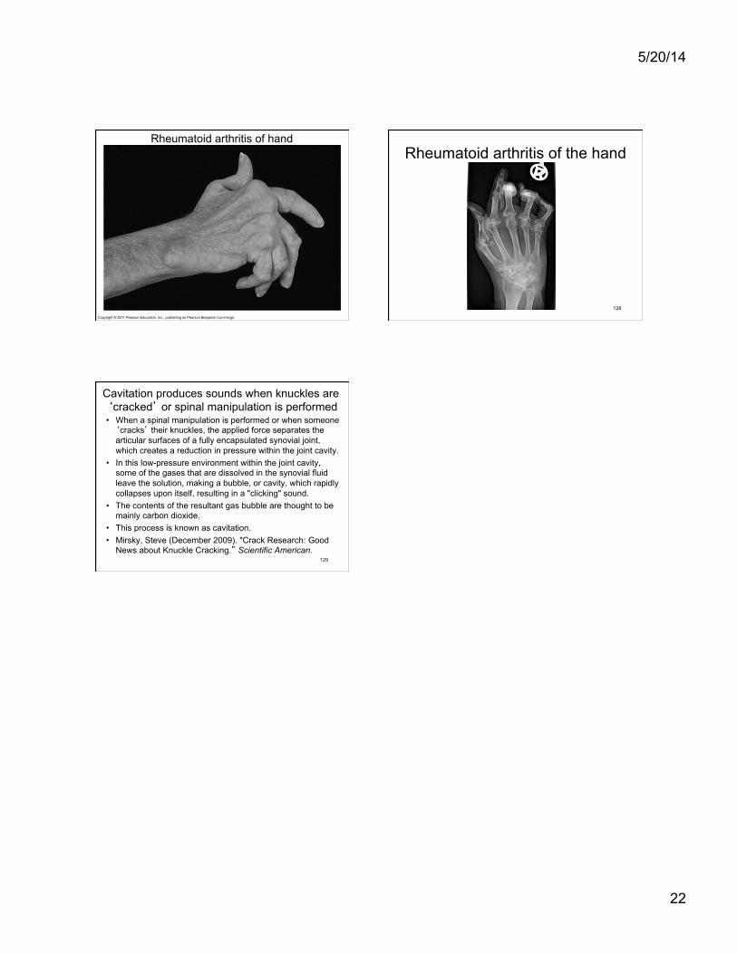
Copyright © 2011 Pearson Education, Inc., publishing as Pearson Benjamin Cummings.
Figure 9.21 A hand deformed by rheumatoid arthritis.
Rheumatoid arthritis of hand Rheumatoid arthritis of the hand
128
Cavitation produces sounds when knuckles are ‘cracked’ or spinal manipulation is performed • When a spinal manipulation is performed or when someone
‘cracks’ their knuckles, the applied force separates the articular surfaces of a fully encapsulated synovial joint, which creates a reduction in pressure within the joint cavity.
• In this low-pressure environment within the joint cavity, some of the gases that are dissolved in the synovial fluid leave the solution, making a bubble, or cavity, which rapidly collapses upon itself, resulting in a "clicking" sound.
• The contents of the resultant gas bubble are thought to be mainly carbon dioxide.
• This process is known as cavitation. • Mirsky, Steve (December 2009). "Crack Research: Good
News about Knuckle Cracking.” Scientific American. 129
Related Documents