www.ck12.org 359 13.1 Prokaryotes Lesson objectives • Outline the classification and evolution of prokaryotes. • Describe the structure of prokaryotes. • Identify different types of metabolism found in prokaryotes. • Describe the range of prokaryote habitats. • Explain how prokaryotes reproduce. • Identify important relationships between bacteria and humans. Vocabulary antibiotic drug drug that kills bacteria and cures bacterial infections and diseases antibiotic resistance ability to withstand antibiotic drugs that has evolved in some bacteria Archaea one of two prokaryote domains that includes organisms that live in extreme environments Bacteria domain of prokaryotes, some of which cause human diseases biofilm colony of prokaryotes that is stuck to a surface such as a rock or a host’s tissue cyanobacteria Gram-positive blue-green photosynthetic bacteria of the type that added oxygen to Earth’s early atmosphere and evolved into chloroplasts of eukaryotic cells endospore spores that form inside prokaryotic cells when they are under stress, enclosing the DNA and helping it survive conditions that may kill the cell extremophile any type of Archaea that lives in an extreme environment, such as a very salty, hot, or acidic environment flagella long, thin protein extensions of the plasma membrane in most prokaryotic cells that help the cells move genetic transfer method of increasing genetic variation in prokaryotes that involves cells “grabbing” stray pieces of DNA from their environment or exchanging DNA directly with other cells Gram-negative bacteria type of bacteria that stain red with Gram stain and have a thin cell wall with an outer membrane Chapter 13. Microorganisms: Prokaryotes and Viruses

Welcome message from author
This document is posted to help you gain knowledge. Please leave a comment to let me know what you think about it! Share it to your friends and learn new things together.
Transcript
www.ck12.org 359
13.1 Prokaryotes
Lesson objectives
• Outline the classification and evolution of prokaryotes.• Describe the structure of prokaryotes.• Identify different types of metabolism found in prokaryotes.• Describe the range of prokaryote habitats.• Explain how prokaryotes reproduce.• Identify important relationships between bacteria and humans.
Vocabulary
antibiotic drug drug that kills bacteria and cures bacterial infections and diseases
antibiotic resistance ability to withstand antibiotic drugs that has evolved in some bacteria
Archaea one of two prokaryote domains that includes organisms that live in extreme environments
Bacteria domain of prokaryotes, some of which cause human diseases
biofilm colony of prokaryotes that is stuck to a surface such as a rock or a host’s tissue
cyanobacteria Gram-positive blue-green photosynthetic bacteria of the type that added oxygen to Earth’s earlyatmosphere and evolved into chloroplasts of eukaryotic cells
endospore spores that form inside prokaryotic cells when they are under stress, enclosing the DNA and helping itsurvive conditions that may kill the cell
extremophile any type of Archaea that lives in an extreme environment, such as a very salty, hot, or acidicenvironment
flagella long, thin protein extensions of the plasma membrane in most prokaryotic cells that help the cells move
genetic transfer method of increasing genetic variation in prokaryotes that involves cells “grabbing” stray piecesof DNA from their environment or exchanging DNA directly with other cells
Gram-negative bacteria type of bacteria that stain red with Gram stain and have a thin cell wall with an outermembrane
Chapter 13. Microorganisms: Prokaryotes and Viruses

360 www.ck12.org
Gram-positive bacteria type of bacteria that stain purple with Gram stain and have a thick cell wall without anouter membrane
plasmid small, circular piece of DNA in a prokaryotic cell
vector organism such as an insect that spreads pathogens from host to host
Introduction
No doubt you’ve had a sore throat before, and you’ve probably eaten cheese or yogurt. If so, then you’ve encounteredthe fascinating world of prokaryotes. Prokaryotes are single-celled organisms that lack a nucleus. They also lackother membrane-bound organelles. Prokaryotes are tiny and sometimes bothersome, but they are the most numerousorganisms on Earth. Without them, the world would be a very different place.
An overview of bacteria can be seen at http://www.youtube.com/user/khanacademy#p/c/7A9646BC5110CF64/16/TDoGrbpJJ14.
MEDIAClick image to the left for more content.
Evolution and Classification of Prokaryotes
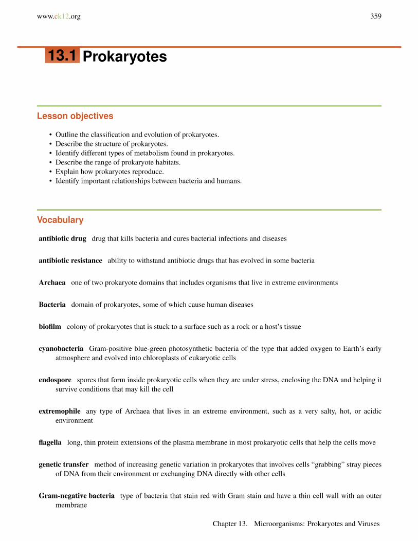
Prokaryotes are currently placed in two domains. A domain is the highest taxon, just above the kingdom. Theprokaryote domains are Bacteria and Archaea (see Figure 13.1). The third domain is Eukarya. It includes alleukaryotes. Unlike prokaryotes, eukaryotes have a nucleus in their cells.
Prokaryote Evolution
It’s not clear how the three domains are related. Archaea were once thought to be offshoots of Bacteria that wereadapted to extreme environments. For their part, Bacteria were considered to be ancestors of Eukarya. Scientistsnow know that Archaea share several traits with Eukarya that Bacteria do not share (see Table 13.1). How can thisbe explained? One hypothesis is that Eukarya arose when an Archaean cell fused with a Bacterial cell. The two cellsbecame the nucleus and cytoplasm of a new Eukaryan cell. How well does this hypothesis fit the evidence in Table13.1?
TABLE 13.1: Comparison of Bacteria, Archaea, and Eukarya
Characteristic Bacteria Archaea EukaryaFlagella Unique to Bacteria Unique to Archaea Unique to EukaryaCell Membrane Unique to Bacteria Like Bacteria and Eu-
karyaUnique to Eukarya
Protein Synthesis Unique to Bacteria Like Eukarya Like ArchaeaIntrons Absent in most Present Present
13.1. Prokaryotes
www.ck12.org 361
TABLE 13.1: (continued)
Characteristic Bacteria Archaea EukaryaPeptidoglycan (in cellwall)
Present Absent in most Absent
Domain Bacteria
Bacteria are the most diverse and abundant group of organisms on Earth. They live in almost all environments. Theyare found in the ocean, the soil, and the intestines of animals. They are even found in rocks deep below Earth’ssurface. Any surface that has not been sterilized is likely to be covered with bacteria. The total number of bacteriain the world is amazing. It’s estimated to be 5 ! 1030, or five million trillion trillion. You have more bacteria in andon your body than you have body cells!
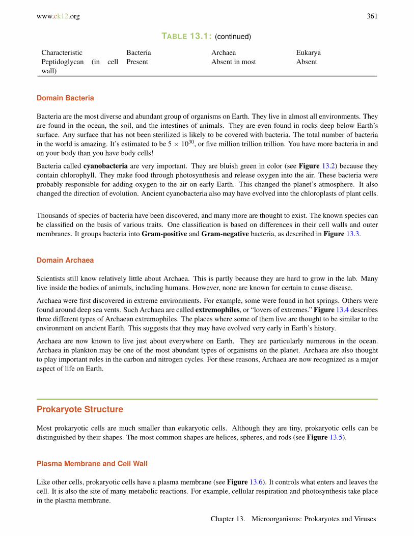
Bacteria called cyanobacteria are very important. They are bluish green in color (see Figure 13.2) because theycontain chlorophyll. They make food through photosynthesis and release oxygen into the air. These bacteria wereprobably responsible for adding oxygen to the air on early Earth. This changed the planet’s atmosphere. It alsochanged the direction of evolution. Ancient cyanobacteria also may have evolved into the chloroplasts of plant cells.
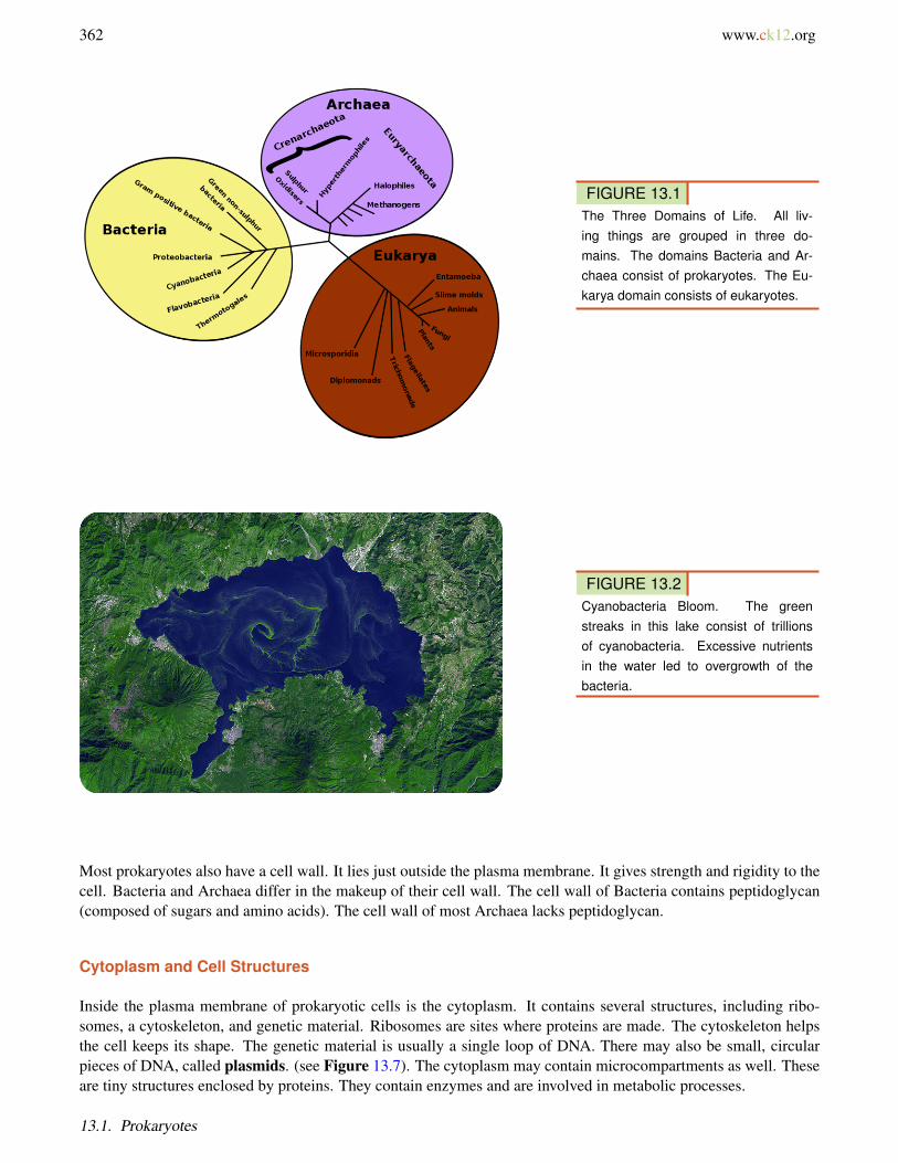
Thousands of species of bacteria have been discovered, and many more are thought to exist. The known species canbe classified on the basis of various traits. One classification is based on differences in their cell walls and outermembranes. It groups bacteria into Gram-positive and Gram-negative bacteria, as described in Figure 13.3.
Domain Archaea
Scientists still know relatively little about Archaea. This is partly because they are hard to grow in the lab. Manylive inside the bodies of animals, including humans. However, none are known for certain to cause disease.
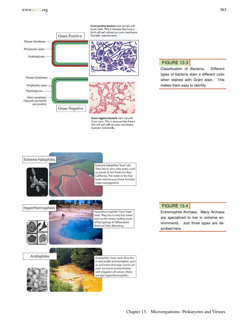
Archaea were first discovered in extreme environments. For example, some were found in hot springs. Others werefound around deep sea vents. Such Archaea are called extremophiles, or “lovers of extremes.” Figure 13.4 describesthree different types of Archaean extremophiles. The places where some of them live are thought to be similar to theenvironment on ancient Earth. This suggests that they may have evolved very early in Earth’s history.
Archaea are now known to live just about everywhere on Earth. They are particularly numerous in the ocean.Archaea in plankton may be one of the most abundant types of organisms on the planet. Archaea are also thoughtto play important roles in the carbon and nitrogen cycles. For these reasons, Archaea are now recognized as a majoraspect of life on Earth.
Prokaryote Structure
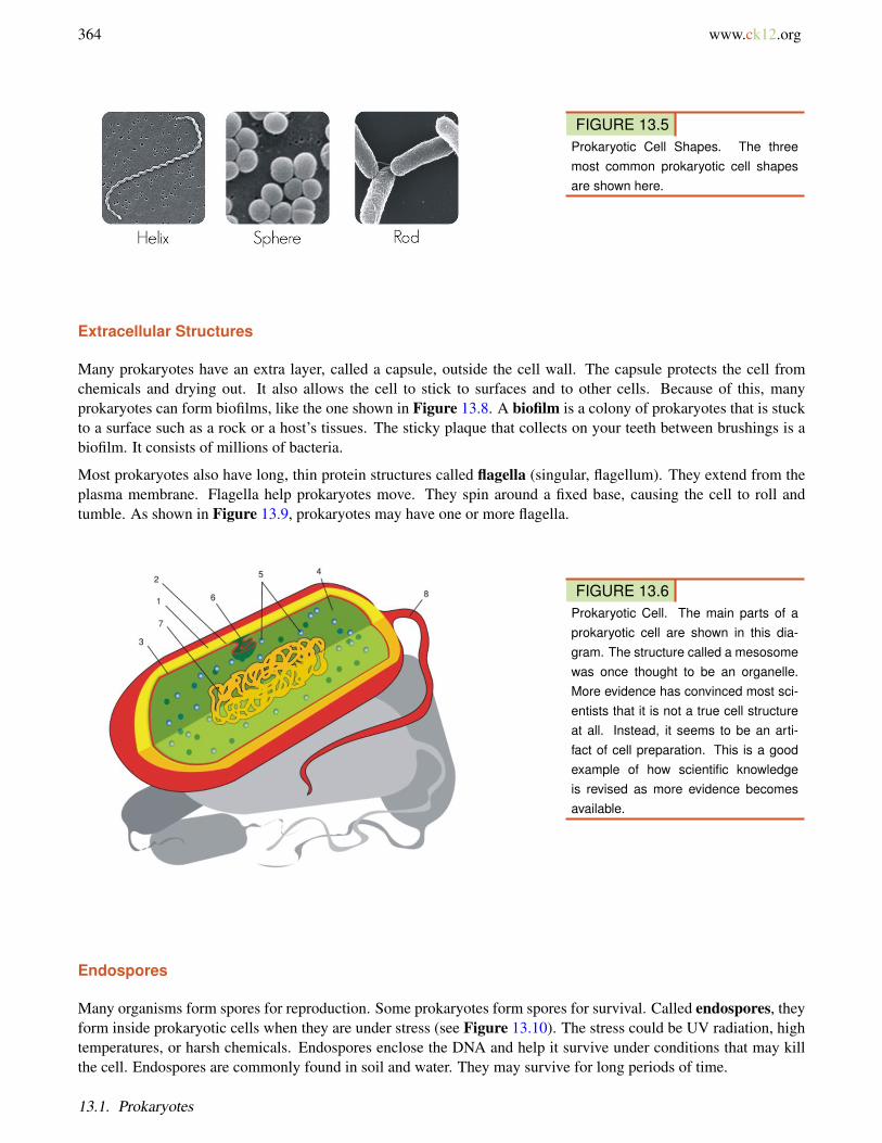
Most prokaryotic cells are much smaller than eukaryotic cells. Although they are tiny, prokaryotic cells can bedistinguished by their shapes. The most common shapes are helices, spheres, and rods (see Figure 13.5).
Plasma Membrane and Cell Wall
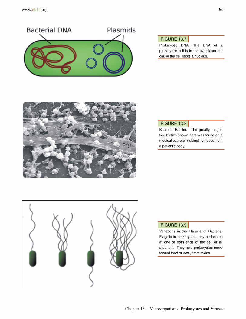
Like other cells, prokaryotic cells have a plasma membrane (see Figure 13.6). It controls what enters and leaves thecell. It is also the site of many metabolic reactions. For example, cellular respiration and photosynthesis take placein the plasma membrane.
Chapter 13. Microorganisms: Prokaryotes and Viruses
362 www.ck12.org
FIGURE 13.1The Three Domains of Life. All liv-ing things are grouped in three do-mains. The domains Bacteria and Ar-chaea consist of prokaryotes. The Eu-karya domain consists of eukaryotes.
FIGURE 13.2Cyanobacteria Bloom. The greenstreaks in this lake consist of trillionsof cyanobacteria. Excessive nutrientsin the water led to overgrowth of thebacteria.
Most prokaryotes also have a cell wall. It lies just outside the plasma membrane. It gives strength and rigidity to thecell. Bacteria and Archaea differ in the makeup of their cell wall. The cell wall of Bacteria contains peptidoglycan(composed of sugars and amino acids). The cell wall of most Archaea lacks peptidoglycan.
Cytoplasm and Cell Structures
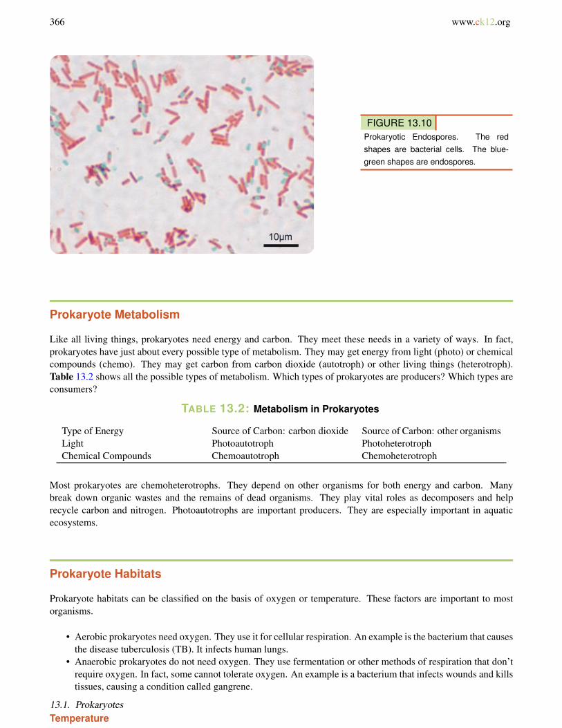
Inside the plasma membrane of prokaryotic cells is the cytoplasm. It contains several structures, including ribo-somes, a cytoskeleton, and genetic material. Ribosomes are sites where proteins are made. The cytoskeleton helpsthe cell keeps its shape. The genetic material is usually a single loop of DNA. There may also be small, circularpieces of DNA, called plasmids. (see Figure 13.7). The cytoplasm may contain microcompartments as well. Theseare tiny structures enclosed by proteins. They contain enzymes and are involved in metabolic processes.
13.1. Prokaryotes
www.ck12.org 363
FIGURE 13.3Classification of Bacteria. Differenttypes of bacteria stain a different colorwhen stained with Gram stain. Thismakes them easy to identify.
FIGURE 13.4Extremophile Archaea. Many Archaeaare specialized to live in extreme en-vironments. Just three types are de-scribed here.
Chapter 13. Microorganisms: Prokaryotes and Viruses
364 www.ck12.org
FIGURE 13.5Prokaryotic Cell Shapes. The threemost common prokaryotic cell shapesare shown here.
Extracellular Structures
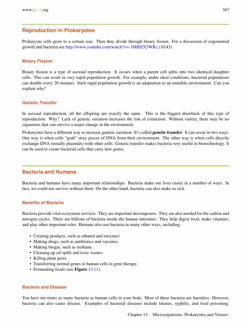
Many prokaryotes have an extra layer, called a capsule, outside the cell wall. The capsule protects the cell fromchemicals and drying out. It also allows the cell to stick to surfaces and to other cells. Because of this, manyprokaryotes can form biofilms, like the one shown in Figure 13.8. A biofilm is a colony of prokaryotes that is stuckto a surface such as a rock or a host’s tissues. The sticky plaque that collects on your teeth between brushings is abiofilm. It consists of millions of bacteria.
Most prokaryotes also have long, thin protein structures called flagella (singular, flagellum). They extend from theplasma membrane. Flagella help prokaryotes move. They spin around a fixed base, causing the cell to roll andtumble. As shown in Figure 13.9, prokaryotes may have one or more flagella.
FIGURE 13.6Prokaryotic Cell. The main parts of aprokaryotic cell are shown in this dia-gram. The structure called a mesosomewas once thought to be an organelle.More evidence has convinced most sci-entists that it is not a true cell structureat all. Instead, it seems to be an arti-fact of cell preparation. This is a goodexample of how scientific knowledgeis revised as more evidence becomesavailable.
Endospores
Many organisms form spores for reproduction. Some prokaryotes form spores for survival. Called endospores, theyform inside prokaryotic cells when they are under stress (see Figure 13.10). The stress could be UV radiation, hightemperatures, or harsh chemicals. Endospores enclose the DNA and help it survive under conditions that may killthe cell. Endospores are commonly found in soil and water. They may survive for long periods of time.
13.1. Prokaryotes
www.ck12.org 365
FIGURE 13.7Prokaryotic DNA. The DNA of aprokaryotic cell is in the cytoplasm be-cause the cell lacks a nucleus.
FIGURE 13.8Bacterial Biofilm. The greatly magni-fied biofilm shown here was found on amedical catheter (tubing) removed froma patient’s body.
FIGURE 13.9Variations in the Flagella of Bacteria.Flagella in prokaryotes may be locatedat one or both ends of the cell or allaround it. They help prokaryotes movetoward food or away from toxins.
Chapter 13. Microorganisms: Prokaryotes and Viruses
366 www.ck12.org
FIGURE 13.10Prokaryotic Endospores. The redshapes are bacterial cells. The blue-green shapes are endospores.
Prokaryote Metabolism
Like all living things, prokaryotes need energy and carbon. They meet these needs in a variety of ways. In fact,prokaryotes have just about every possible type of metabolism. They may get energy from light (photo) or chemicalcompounds (chemo). They may get carbon from carbon dioxide (autotroph) or other living things (heterotroph).Table 13.2 shows all the possible types of metabolism. Which types of prokaryotes are producers? Which types areconsumers?
TABLE 13.2: Metabolism in Prokaryotes
Type of Energy Source of Carbon: carbon dioxide Source of Carbon: other organismsLight Photoautotroph PhotoheterotrophChemical Compounds Chemoautotroph Chemoheterotroph
Most prokaryotes are chemoheterotrophs. They depend on other organisms for both energy and carbon. Manybreak down organic wastes and the remains of dead organisms. They play vital roles as decomposers and helprecycle carbon and nitrogen. Photoautotrophs are important producers. They are especially important in aquaticecosystems.
Prokaryote Habitats
Prokaryote habitats can be classified on the basis of oxygen or temperature. These factors are important to mostorganisms.
• Aerobic prokaryotes need oxygen. They use it for cellular respiration. An example is the bacterium that causesthe disease tuberculosis (TB). It infects human lungs.
• Anaerobic prokaryotes do not need oxygen. They use fermentation or other methods of respiration that don’trequire oxygen. In fact, some cannot tolerate oxygen. An example is a bacterium that infects wounds and killstissues, causing a condition called gangrene.
Temperature
Like most organisms, prokaryotes live and grow best within certain temperature ranges. Prokaryotes can be classifiedby their temperature preferences, as shown in Table 13.3. Which type of prokaryote would you expect to find insidethe human body?
TABLE 13.3: Classification of Prokaryotes by Temperature
Type of Prokaryote Preferred Temperature Where It Might Be FoundThermophile above 45°C (113°F) in compostMesophile about 37°C (98°F) inside animalsPsychrophile below 20°C (68°F) in the deep ocean
13.1. Prokaryotes
www.ck12.org 367
Reproduction in Prokaryotes
Prokaryote cells grow to a certain size. Then they divide through binary fission. For a discussion of exponentialgrowth and bacteria see http://www.youtube.com/watch?v=-3MI0ZX5WRc (10:43).
Binary Fission
Binary fission is a type of asexual reproduction. It occurs when a parent cell splits into two identical daughtercells. This can result in very rapid population growth. For example, under ideal conditions, bacterial populationscan double every 20 minutes. Such rapid population growth is an adaptation to an unstable environment. Can youexplain why?
Genetic Transfer
In asexual reproduction, all the offspring are exactly the same. This is the biggest drawback of this type ofreproduction. Why? Lack of genetic variation increases the risk of extinction. Without variety, there may be noorganisms that can survive a major change in the environment.
Prokaryotes have a different way to increase genetic variation. It’s called genetic transfer. It can occur in two ways.One way is when cells “grab” stray pieces of DNA from their environment. The other way is when cells directlyexchange DNA (usually plasmids) with other cells. Genetic transfer makes bacteria very useful in biotechnology. Itcan be used to create bacterial cells that carry new genes.
Bacteria and Humans
Bacteria and humans have many important relationships. Bacteria make our lives easier in a number of ways. Infact, we could not survive without them. On the other hand, bacteria can also make us sick.
Benefits of Bacteria
Bacteria provide vital ecosystem services. They are important decomposers. They are also needed for the carbon andnitrogen cycles. There are billions of bacteria inside the human intestines. They help digest food, make vitamins,and play other important roles. Humans also use bacteria in many other ways, including:
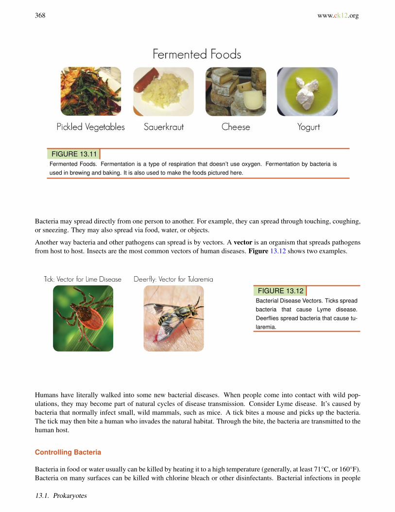
• Creating products, such as ethanol and enzymes.• Making drugs, such as antibiotics and vaccines.• Making biogas, such as methane.• Cleaning up oil spills and toxic wastes.• Killing plant pests.• Transferring normal genes to human cells in gene therapy.• Fermenting foods (see Figure 13.11).
Bacteria and Disease
You have ten times as many bacteria as human cells in your body. Most of these bacteria are harmless. However,bacteria can also cause disease. Examples of bacterial diseases include tetanus, syphilis, and food poisoning.
Chapter 13. Microorganisms: Prokaryotes and Viruses
368 www.ck12.org
FIGURE 13.11Fermented Foods. Fermentation is a type of respiration that doesn’t use oxygen. Fermentation by bacteria isused in brewing and baking. It is also used to make the foods pictured here.
Bacteria may spread directly from one person to another. For example, they can spread through touching, coughing,or sneezing. They may also spread via food, water, or objects.
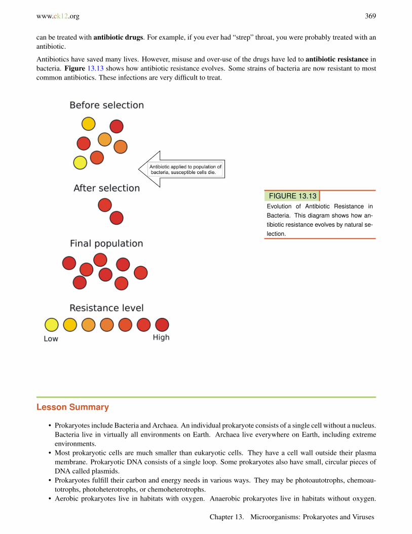
Another way bacteria and other pathogens can spread is by vectors. A vector is an organism that spreads pathogensfrom host to host. Insects are the most common vectors of human diseases. Figure 13.12 shows two examples.
FIGURE 13.12Bacterial Disease Vectors. Ticks spreadbacteria that cause Lyme disease.Deerflies spread bacteria that cause tu-laremia.
Humans have literally walked into some new bacterial diseases. When people come into contact with wild pop-ulations, they may become part of natural cycles of disease transmission. Consider Lyme disease. It’s caused bybacteria that normally infect small, wild mammals, such as mice. A tick bites a mouse and picks up the bacteria.The tick may then bite a human who invades the natural habitat. Through the bite, the bacteria are transmitted to thehuman host.
Controlling Bacteria
Bacteria in food or water usually can be killed by heating it to a high temperature (generally, at least 71°C, or 160°F).Bacteria on many surfaces can be killed with chlorine bleach or other disinfectants. Bacterial infections in people
13.1. Prokaryotes
www.ck12.org 369
can be treated with antibiotic drugs. For example, if you ever had “strep” throat, you were probably treated with anantibiotic.
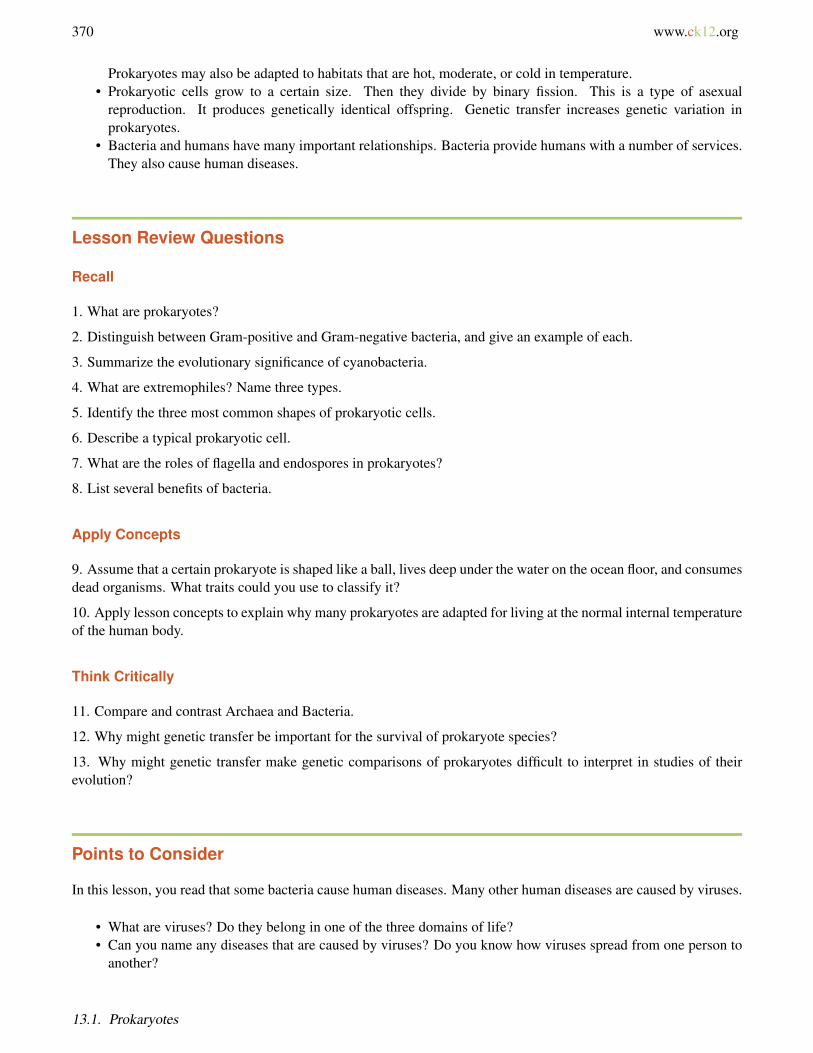
Antibiotics have saved many lives. However, misuse and over-use of the drugs have led to antibiotic resistance inbacteria. Figure 13.13 shows how antibiotic resistance evolves. Some strains of bacteria are now resistant to mostcommon antibiotics. These infections are very difficult to treat.
FIGURE 13.13Evolution of Antibiotic Resistance inBacteria. This diagram shows how an-tibiotic resistance evolves by natural se-lection.
Lesson Summary
• Prokaryotes include Bacteria and Archaea. An individual prokaryote consists of a single cell without a nucleus.Bacteria live in virtually all environments on Earth. Archaea live everywhere on Earth, including extremeenvironments.
• Most prokaryotic cells are much smaller than eukaryotic cells. They have a cell wall outside their plasmamembrane. Prokaryotic DNA consists of a single loop. Some prokaryotes also have small, circular pieces ofDNA called plasmids.
• Prokaryotes fulfill their carbon and energy needs in various ways. They may be photoautotrophs, chemoau-totrophs, photoheterotrophs, or chemoheterotrophs.
• Aerobic prokaryotes live in habitats with oxygen. Anaerobic prokaryotes live in habitats without oxygen.
Chapter 13. Microorganisms: Prokaryotes and Viruses
370 www.ck12.org
Prokaryotes may also be adapted to habitats that are hot, moderate, or cold in temperature.• Prokaryotic cells grow to a certain size. Then they divide by binary fission. This is a type of asexual
reproduction. It produces genetically identical offspring. Genetic transfer increases genetic variation inprokaryotes.
• Bacteria and humans have many important relationships. Bacteria provide humans with a number of services.They also cause human diseases.
Lesson Review Questions
Recall
1. What are prokaryotes?
2. Distinguish between Gram-positive and Gram-negative bacteria, and give an example of each.
3. Summarize the evolutionary significance of cyanobacteria.
4. What are extremophiles? Name three types.
5. Identify the three most common shapes of prokaryotic cells.
6. Describe a typical prokaryotic cell.
7. What are the roles of flagella and endospores in prokaryotes?
8. List several benefits of bacteria.
Apply Concepts
9. Assume that a certain prokaryote is shaped like a ball, lives deep under the water on the ocean floor, and consumesdead organisms. What traits could you use to classify it?
10. Apply lesson concepts to explain why many prokaryotes are adapted for living at the normal internal temperatureof the human body.
Think Critically
11. Compare and contrast Archaea and Bacteria.
12. Why might genetic transfer be important for the survival of prokaryote species?
13. Why might genetic transfer make genetic comparisons of prokaryotes difficult to interpret in studies of theirevolution?
Points to Consider
In this lesson, you read that some bacteria cause human diseases. Many other human diseases are caused by viruses.
• What are viruses? Do they belong in one of the three domains of life?• Can you name any diseases that are caused by viruses? Do you know how viruses spread from one person to
another?
13.1. Prokaryotes
www.ck12.org 371
13.2 Viruses
Lesson Objectives
• Describe the structure of viruses.• Outline the discovery and origins of viruses.• Explain how viruses replicate.• Explain how viruses cause human disease.• Describe how viruses can be controlled.• Identify how viruses are used in research and medicine.
Vocabulary
capsid protein coat that surrounds the DNA or RNA of a virus particle
latency period of dormancy of a virus inside a living body that may last for many years
vaccine substance containing modified pathogens that does not cause disease but provokes an immune responseand results in immunity to the pathogen
virion individual virus particle that consists of nucleic acid within a protein capsid
Introduction
At the end of the last lesson, you were asked which of the three domains of life do viruses belong to. Did you figureit out? None. Why? Viruses are usually considered to be nonliving. Viruses do not meet most of the criteria of life.They are not even cells.
An overview of viruses can be seen at http://www.youtube.com/user/khanacademy#p/c/7A9646BC5110CF64/17/0h5Jd7sgQWY.
MEDIAClick image to the left for more content.
Chapter 13. Microorganisms: Prokaryotes and Viruses
372 www.ck12.org
Characteristics of Viruses
An individual virus is called a virion. It is a tiny particle much smaller than a prokaryotic cell. Because viruses donot consist of cells, they also lack cell membranes, cytoplasm, ribosomes, and other cell organelles. Without thesestructures, they are unable to make proteins or even reproduce on their own. Instead, they must depend on a hostcell to synthesize their proteins and to make copies of themselves. Viruses infect and live inside the cells of livingorganisms. When viruses infect the cells of their host, they may cause disease. For example, viruses cause AIDS,influenza (flu), chicken pox, and the common cold.
Although viruses are not classified as living things, they share two important traits with living things. They havegenetic material, and they can evolve. This is why the classification of viruses has been controversial. It calls intoquestion just what it means to be alive. What do you think? How would you classify viruses?
Structure and Classification of Viruses
Viruses vary in their structure. The structure of a virus is one trait that determines how it is classified.
Structure of Viruses
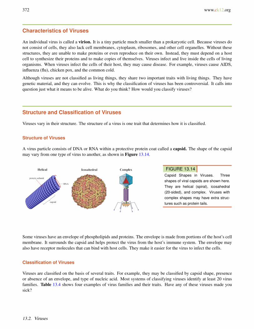
A virus particle consists of DNA or RNA within a protective protein coat called a capsid. The shape of the capsidmay vary from one type of virus to another, as shown in Figure 13.14.
FIGURE 13.14Capsid Shapes in Viruses. Threeshapes of viral capsids are shown here.They are helical (spiral), icosahedral(20-sided), and complex. Viruses withcomplex shapes may have extra struc-tures such as protein tails.
Some viruses have an envelope of phospholipids and proteins. The envelope is made from portions of the host’s cellmembrane. It surrounds the capsid and helps protect the virus from the host’s immune system. The envelope mayalso have receptor molecules that can bind with host cells. They make it easier for the virus to infect the cells.
Classification of Viruses
Viruses are classified on the basis of several traits. For example, they may be classified by capsid shape, presenceor absence of an envelope, and type of nucleic acid. Most systems of classifying viruses identify at least 20 virusfamilies. Table 13.4 shows four examples of virus families and their traits. Have any of these viruses made yousick?
13.2. Viruses
www.ck12.org 373
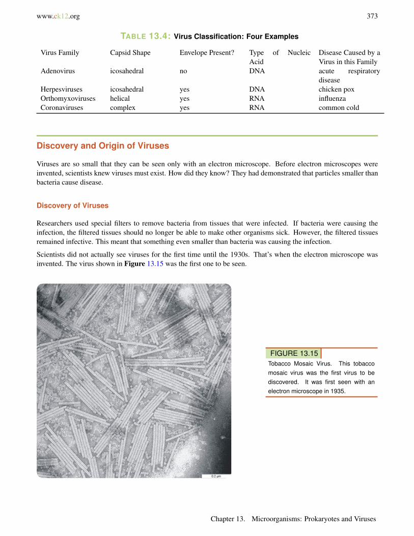
TABLE 13.4: Virus Classification: Four Examples
Virus Family Capsid Shape Envelope Present? Type of NucleicAcid
Disease Caused by aVirus in this Family
Adenovirus icosahedral no DNA acute respiratorydisease
Herpesviruses icosahedral yes DNA chicken poxOrthomyxoviruses helical yes RNA influenzaCoronaviruses complex yes RNA common cold
Discovery and Origin of Viruses
Viruses are so small that they can be seen only with an electron microscope. Before electron microscopes wereinvented, scientists knew viruses must exist. How did they know? They had demonstrated that particles smaller thanbacteria cause disease.
Discovery of Viruses
Researchers used special filters to remove bacteria from tissues that were infected. If bacteria were causing theinfection, the filtered tissues should no longer be able to make other organisms sick. However, the filtered tissuesremained infective. This meant that something even smaller than bacteria was causing the infection.
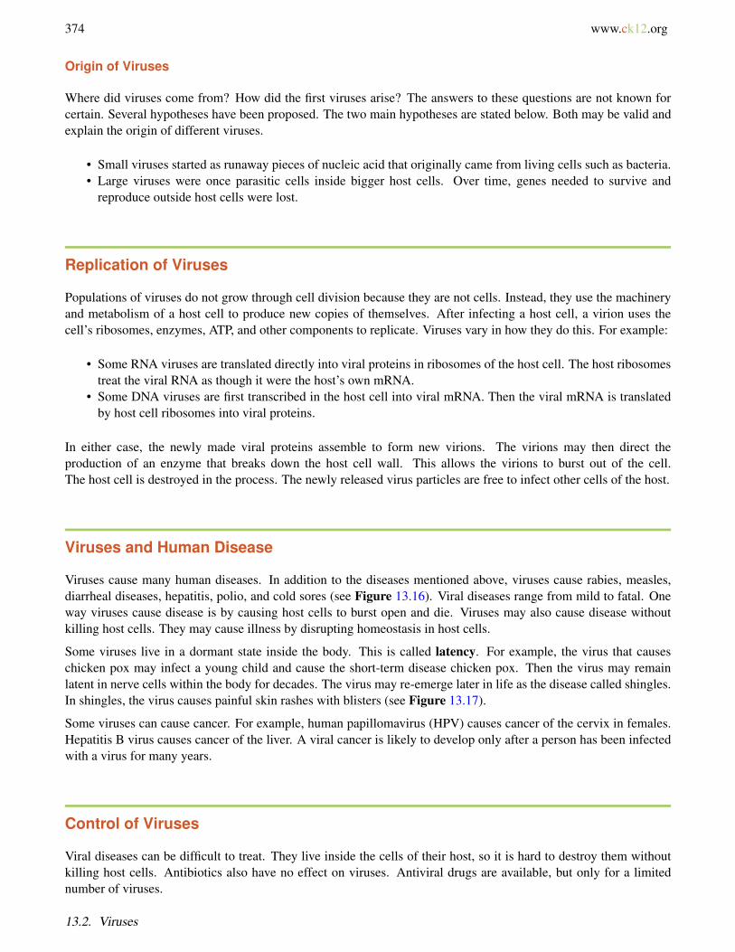
Scientists did not actually see viruses for the first time until the 1930s. That’s when the electron microscope wasinvented. The virus shown in Figure 13.15 was the first one to be seen.
FIGURE 13.15Tobacco Mosaic Virus. This tobaccomosaic virus was the first virus to bediscovered. It was first seen with anelectron microscope in 1935.
Chapter 13. Microorganisms: Prokaryotes and Viruses
374 www.ck12.org
Origin of Viruses
Where did viruses come from? How did the first viruses arise? The answers to these questions are not known forcertain. Several hypotheses have been proposed. The two main hypotheses are stated below. Both may be valid andexplain the origin of different viruses.
• Small viruses started as runaway pieces of nucleic acid that originally came from living cells such as bacteria.• Large viruses were once parasitic cells inside bigger host cells. Over time, genes needed to survive and
reproduce outside host cells were lost.
Replication of Viruses
Populations of viruses do not grow through cell division because they are not cells. Instead, they use the machineryand metabolism of a host cell to produce new copies of themselves. After infecting a host cell, a virion uses thecell’s ribosomes, enzymes, ATP, and other components to replicate. Viruses vary in how they do this. For example:
• Some RNA viruses are translated directly into viral proteins in ribosomes of the host cell. The host ribosomestreat the viral RNA as though it were the host’s own mRNA.
• Some DNA viruses are first transcribed in the host cell into viral mRNA. Then the viral mRNA is translatedby host cell ribosomes into viral proteins.
In either case, the newly made viral proteins assemble to form new virions. The virions may then direct theproduction of an enzyme that breaks down the host cell wall. This allows the virions to burst out of the cell.The host cell is destroyed in the process. The newly released virus particles are free to infect other cells of the host.
Viruses and Human Disease
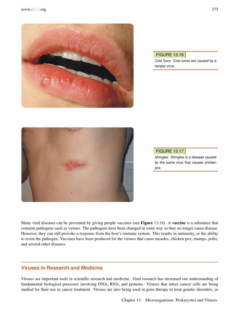
Viruses cause many human diseases. In addition to the diseases mentioned above, viruses cause rabies, measles,diarrheal diseases, hepatitis, polio, and cold sores (see Figure 13.16). Viral diseases range from mild to fatal. Oneway viruses cause disease is by causing host cells to burst open and die. Viruses may also cause disease withoutkilling host cells. They may cause illness by disrupting homeostasis in host cells.
Some viruses live in a dormant state inside the body. This is called latency. For example, the virus that causeschicken pox may infect a young child and cause the short-term disease chicken pox. Then the virus may remainlatent in nerve cells within the body for decades. The virus may re-emerge later in life as the disease called shingles.In shingles, the virus causes painful skin rashes with blisters (see Figure 13.17).
Some viruses can cause cancer. For example, human papillomavirus (HPV) causes cancer of the cervix in females.Hepatitis B virus causes cancer of the liver. A viral cancer is likely to develop only after a person has been infectedwith a virus for many years.
Control of Viruses
Viral diseases can be difficult to treat. They live inside the cells of their host, so it is hard to destroy them withoutkilling host cells. Antibiotics also have no effect on viruses. Antiviral drugs are available, but only for a limitednumber of viruses.
13.2. Viruses
www.ck12.org 375
FIGURE 13.16Cold Sore. Cold sores are caused by aherpes virus.
FIGURE 13.17Shingles. Shingles is a disease causedby the same virus that causes chickenpox.
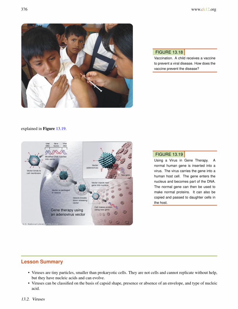
Many viral diseases can be prevented by giving people vaccines (see Figure 13.18). A vaccine is a substance thatcontains pathogens such as viruses. The pathogens have been changed in some way so they no longer cause disease.However, they can still provoke a response from the host’s immune system. This results in immunity, or the abilityto resist the pathogen. Vaccines have been produced for the viruses that cause measles, chicken pox, mumps, polio,and several other diseases.
Viruses in Research and Medicine
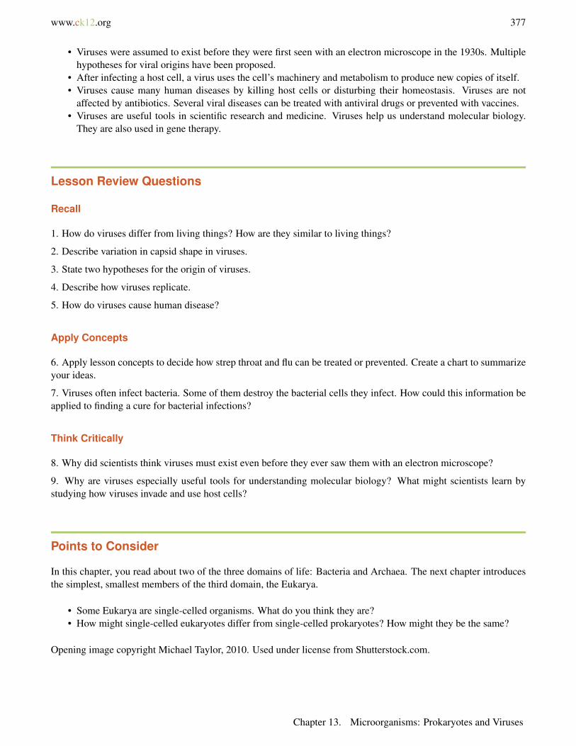
Viruses are important tools in scientific research and medicine. Viral research has increased our understanding offundamental biological processes involving DNA, RNA, and proteins. Viruses that infect cancer cells are beingstudied for their use in cancer treatment. Viruses are also being used in gene therapy to treat genetic disorders, as
Chapter 13. Microorganisms: Prokaryotes and Viruses
376 www.ck12.org
FIGURE 13.18Vaccination. A child receives a vaccineto prevent a viral disease. How does thevaccine prevent the disease?
explained in Figure 13.19.
FIGURE 13.19Using a Virus in Gene Therapy. Anormal human gene is inserted into avirus. The virus carries the gene into ahuman host cell. The gene enters thenucleus and becomes part of the DNA.The normal gene can then be used tomake normal proteins. It can also becopied and passed to daughter cells inthe host.
Lesson Summary
• Viruses are tiny particles, smaller than prokaryotic cells. They are not cells and cannot replicate without help,but they have nucleic acids and can evolve.
• Viruses can be classified on the basis of capsid shape, presence or absence of an envelope, and type of nucleicacid.
13.2. Viruses
www.ck12.org 377
• Viruses were assumed to exist before they were first seen with an electron microscope in the 1930s. Multiplehypotheses for viral origins have been proposed.
• After infecting a host cell, a virus uses the cell’s machinery and metabolism to produce new copies of itself.• Viruses cause many human diseases by killing host cells or disturbing their homeostasis. Viruses are not
affected by antibiotics. Several viral diseases can be treated with antiviral drugs or prevented with vaccines.• Viruses are useful tools in scientific research and medicine. Viruses help us understand molecular biology.
They are also used in gene therapy.
Lesson Review Questions
Recall
1. How do viruses differ from living things? How are they similar to living things?
2. Describe variation in capsid shape in viruses.
3. State two hypotheses for the origin of viruses.
4. Describe how viruses replicate.
5. How do viruses cause human disease?
Apply Concepts
6. Apply lesson concepts to decide how strep throat and flu can be treated or prevented. Create a chart to summarizeyour ideas.
7. Viruses often infect bacteria. Some of them destroy the bacterial cells they infect. How could this information beapplied to finding a cure for bacterial infections?
Think Critically
8. Why did scientists think viruses must exist even before they ever saw them with an electron microscope?
9. Why are viruses especially useful tools for understanding molecular biology? What might scientists learn bystudying how viruses invade and use host cells?
Points to Consider
In this chapter, you read about two of the three domains of life: Bacteria and Archaea. The next chapter introducesthe simplest, smallest members of the third domain, the Eukarya.
• Some Eukarya are single-celled organisms. What do you think they are?• How might single-celled eukaryotes differ from single-celled prokaryotes? How might they be the same?
Opening image copyright Michael Taylor, 2010. Used under license from Shutterstock.com.
Chapter 13. Microorganisms: Prokaryotes and Viruses
378 www.ck12.org
13.3 References
1. Samsara. . GNU-FDL 1.22. Jesse Allen/NASA. . Public Domain3. Gram cell wall: JulianOnions; Gram-positive: JA Jernigan et al./Centers for Disease Control and Prevention;
Gram-negative: William A. Clark/Centers for Disease and Control. . All three images are in the public domain4. From left to right, top to bottom: courtesy of NASA; Grombo; courtesy of PMEL/National Oceanic and At-
mospheric Administration; courtesy of US Geological Survey; courtesy of NASA; courtesy of EnvironmentalProtection Agency. . From left to right, top to bottom: Public Domain; CC-BY-SA 2.5; Public Domain; PublicDomain; Public Domain; Public Domain
5. (Helix) Janice Carr/Centers for Disease Control and Prevention; (Sphere): Janice Carr/Centers for DiseaseControl and Prevention; (Rod) Volker Brinkmann, Max Planck Institute for Infection Biology, Berlin, Ger-many. . (Helix): Public Domain; (Sphere): Public Domain; (Rod) CC-BY 2.5
6. Mariana Ruiz Villarreal. . Public Domain7. Spaully. . CC-BY-SA 2.58. Courtesy of Janice Carr/Centers for Disease Control and Prevention. . Public Domain9. Mike Jones. . CC-BY-SA 2.5
10. Y tambe. . GNU-FDL 1.211. (Left to right): ayustety; Jonathunder; Alex Anlicker; Omernos. . (Left to right): CC-BY-SA 2.0; GNU-FDL
1.2; CC-BY-SA 3.0; Public Domain12. Tick: Courtesy of Centers for Disease Control and Prevention; Deerfly image copyright Bruce MacQueen,
2010. . Tick: Public Domain; Deerfly: Used under license from Shutterstock.com13. Wykis, modified by CK-12 Foundation. . Public Domain14. Helical: Arionfx; Icosahedral: David S. Goodsell and the RCSB PDB; Complex: image copyright Blamb,
2010. . Helical: CC-BY-SA 3.0; Icosahedral: available for educational purposes with attribution; Complex:Used under license from Shuttesrtock.com
15. T. Moravec. . Public Domain16. Metju12. . Public Domain17. Preston Hunt. . CC-BY-SA 3.018. Andy Young. . CC-BY-SA 2.019. Courtesy of National Institutes of Health. . Public Domain
13.3. References
Related Documents