FORUM REVIEW ARTICLE Nutrition, Epigenetics, and Metabolic Syndrome Junjun Wang, 1 Zhenlong Wu, 1 Defa Li, 1 Ning Li, 2 Scott V. Dindot, 3–5 M. Carey Satterfield, 3,6 Fuller W. Bazer, 3,6 and Guoyao Wu 1,3,6 Abstract Significance: Epidemiological and animal studies have demonstrated a close link between maternal nutrition and chronic metabolic disease in children and adults. Compelling experimental results also indicate that adverse effects of intrauterine growth restriction on offspring can be carried forward to subsequent generations through covalent modifications of DNA and core histones. Recent Advances: DNA methylation is catalyzed by S-adenosylmethionine- dependent DNA methyltransferases. Methylation, demethylation, acetylation, and deacetylation of histone proteins are performed by histone methyltransferase, histone demethylase, histone acetyltransferase, and histone deacetyl- transferase, respectively. Histone activities are also influenced by phosphorylation, ubiquitination, ADP-ribosylation, sumoylation, and glycosylation. Metabolism of amino acids (glycine, histidine, methionine, and serine) and vitamins (B6, B12, and folate) plays a key role in provision of methyl donors for DNA and protein methylation. Critical Issues: Disruption of epigenetic mechanisms can result in oxidative stress, obesity, insulin resistance, diabetes, and vascular dysfunction in animals and humans. Despite a recognized role for epigenetics in fetal programming of metabolic syndrome, research on therapies is still in its infancy. Possible interventions include: 1) inhibition of DNA methyl- ation, histone deacetylation, and microRNA expression; 2) targeting epigenetically disturbed metabolic pathways; and 3) dietary supplementation with functional amino acids, vitamins, and phytochemicals. Future Directions: Much work is needed with animal models to understand the basic mechanisms responsible for the roles of specific nutrients in fetal and neonatal programming. Such new knowledge is crucial to design effective therapeutic strategies for preventing and treating metabolic abnormalities in offspring born to mothers with a previous experience of mal- nutrition. Antioxid. Redox Signal. 17, 282–301. Introduction T he United Nations Food and Agriculture Organization recently reported that 925 million people worldwide suffer from hunger (190). This population includes not only males and females of reproductive age, but also children who will grow to be adults and produce their own offspring. Currently, deficiencies in protein (amino acids), vitamin A, and iron remain major nutritional problems in developing nations (146), and a deficiency in folic acid can also occur when individuals have a low intake of fresh green vegetables or no consumption of folate-fortified foods (31). In both de- veloped and developing countries, gestating women can ex- perience undernutrition owing to: 1) severe nausea and vomiting known as hyperemesis gravidarum; 2) early or closely-spaced pregnancies; and 3) multi-fetal pregnancies resulting from assisted reproductive technologies (84, 104, 199). These conditions may result in intrauterine growth re- striction (IUGR) and impaired health of the offspring (Fig. 1). The other end of the nutrition spectrum is overnutrition, which can lead to overweight or obesity in human beings (115). One billion adults worldwide are overweight and more than 300 million are obese (126). Many overweight and obese women unknowingly enter pregnancy and continue over- eating during gestation (39). These women usually gain more weight during the first pregnancy and accumulate more fat during subsequent pregnancies (128). Maternal obesity before or during gestation may result in adverse pregnancy out- comes, including maternal insulin resistance and hypergly- cemia often associated with large-for-gestation age infants, IUGR, and impaired health of the offspring (Fig. 1). Thus, obesity is a global epidemic that negatively affects infants, children, and adults (48). Extensive epidemiological studies have linked maternal undernutrition and overnutrition with the etiology of many chronic diseases in offspring when they reach adulthood (108). The metabolic syndrome can be defined as a cluster of disorders that include obesity, hyperglycemia (fasting serum 1 State Key Laboratory of Animal Nutrition and 2 State Key Laboratory of AgroBiotechnology, China Agricultural University, Beijing, China. 3 Center for Animal Biotechnology and Genomics, Departments of 4 Veterinary Pathobiology, 5 Molecular and Cellular Medicine, and 6 Animal Science, Texas A&M University, College Station, Texas. ANTIOXIDANTS & REDOX SIGNALING Volume 17, Number 2, 2012 ª Mary Ann Liebert, Inc. DOI: 10.1089/ars.2011.4381 282

120 nutrition epigenetics_metabolic-syndrome
Jul 15, 2015
Welcome message from author
This document is posted to help you gain knowledge. Please leave a comment to let me know what you think about it! Share it to your friends and learn new things together.
Transcript
FORUM REVIEW ARTICLE
Nutrition, Epigenetics, and Metabolic Syndrome
Junjun Wang,1 Zhenlong Wu,1 Defa Li,1 Ning Li,2 Scott V. Dindot,3–5 M. Carey Satterfield,3,6
Fuller W. Bazer,3,6 and Guoyao Wu1,3,6
Abstract
Significance: Epidemiological and animal studies have demonstrated a close link between maternal nutrition andchronic metabolic disease in children and adults. Compelling experimental results also indicate that adverse effects ofintrauterine growth restriction on offspring can be carried forward to subsequent generations through covalentmodifications of DNA and core histones. Recent Advances: DNA methylation is catalyzed by S-adenosylmethionine-dependent DNA methyltransferases. Methylation, demethylation, acetylation, and deacetylation of histone proteinsare performed by histone methyltransferase, histone demethylase, histone acetyltransferase, and histone deacetyl-transferase, respectively. Histone activities are also influenced by phosphorylation, ubiquitination, ADP-ribosylation,sumoylation, and glycosylation. Metabolism of amino acids (glycine, histidine, methionine, and serine) and vitamins(B6, B12, and folate) plays a key role in provision of methyl donors for DNA and protein methylation. Critical Issues:Disruption of epigenetic mechanisms can result in oxidative stress, obesity, insulin resistance, diabetes, and vasculardysfunction in animals and humans. Despite a recognized role for epigenetics in fetal programming of metabolicsyndrome, research on therapies is still in its infancy. Possible interventions include: 1) inhibition of DNA methyl-ation, histone deacetylation, and microRNA expression; 2) targeting epigenetically disturbed metabolic pathways;and 3) dietary supplementation with functional amino acids, vitamins, and phytochemicals. Future Directions: Muchwork is needed with animal models to understand the basic mechanisms responsible for the roles of specific nutrientsin fetal and neonatal programming. Such new knowledge is crucial to design effective therapeutic strategies forpreventing and treating metabolic abnormalities in offspring born to mothers with a previous experience of mal-nutrition. Antioxid. Redox Signal. 17, 282–301.
Introduction
The United Nations Food and Agriculture Organizationrecently reported that 925 million people worldwide
suffer from hunger (190). This population includes not onlymales and females of reproductive age, but also children whowill grow to be adults and produce their own offspring.Currently, deficiencies in protein (amino acids), vitamin A,and iron remain major nutritional problems in developingnations (146), and a deficiency in folic acid can also occurwhen individuals have a low intake of fresh green vegetablesor no consumption of folate-fortified foods (31). In both de-veloped and developing countries, gestating women can ex-perience undernutrition owing to: 1) severe nausea andvomiting known as hyperemesis gravidarum; 2) early orclosely-spaced pregnancies; and 3) multi-fetal pregnanciesresulting from assisted reproductive technologies (84, 104,199). These conditions may result in intrauterine growth re-striction (IUGR) and impaired health of the offspring (Fig. 1).
The other end of the nutrition spectrum is overnutrition,which can lead to overweight or obesity in human beings(115). One billion adults worldwide are overweight and morethan 300 million are obese (126). Many overweight and obesewomen unknowingly enter pregnancy and continue over-eating during gestation (39). These women usually gain moreweight during the first pregnancy and accumulate more fatduring subsequent pregnancies (128). Maternal obesity beforeor during gestation may result in adverse pregnancy out-comes, including maternal insulin resistance and hypergly-cemia often associated with large-for-gestation age infants,IUGR, and impaired health of the offspring (Fig. 1). Thus,obesity is a global epidemic that negatively affects infants,children, and adults (48).
Extensive epidemiological studies have linked maternalundernutrition and overnutrition with the etiology of manychronic diseases in offspring when they reach adulthood(108). The metabolic syndrome can be defined as a cluster ofdisorders that include obesity, hyperglycemia (fasting serum
1State Key Laboratory of Animal Nutrition and 2State Key Laboratory of AgroBiotechnology, China Agricultural University, Beijing,China.
3Center for Animal Biotechnology and Genomics, Departments of 4Veterinary Pathobiology, 5Molecular and Cellular Medicine, and6Animal Science, Texas A&M University, College Station, Texas.
ANTIOXIDANTS & REDOX SIGNALINGVolume 17, Number 2, 2012ª Mary Ann Liebert, Inc.DOI: 10.1089/ars.2011.4381
282
Administrator
Underline
Administrator
Highlight
Administrator
Underline
Administrator
Highlight
Administrator
Underline
Administrator
Highlight
美国细胞修复系统医学中心
Sticky Note
美国细胞修复系统医学中心 US CytoThesis Systems Medicine Center www.CytoThesis.US 揭晓「癌症根本治疗」 www.oncotherapy.us/oncotherapy.pdf 重新思考癌症:「营养」与「治病」 www.oncotherapy.us/120.pdf 临床「转化医学」家庭健康管理系统 生命维护系统工程师‧健康系统(个性化)设计 http://www.health120years.com/120.pdf 美国肿瘤治疗系统生物医学集团 细胞修复生医工程研究集团
Administrator
Underline
Administrator
Highlight
Administrator
Underline
Administrator
Highlight
Administrator
Underline
Administrator
Highlight
Administrator
Underline
Administrator
Highlight
Administrator
Underline
Administrator
Highlight

glucose ( > 6.1 mM), hyperinsulinemia, hyperlipidemia, hy-pertension, and insulin resistance (an impaired response ofcells or tissues to physiological concentrations of insulin) (78).These factors, independently or collectively, contribute to ahigh risk for metabolic disease, a major cause of death in de-veloped nations and developing countries (126). This phe-nomenon led to the new concept of fetal programming that isdefined as an adaptive process whereby nutrition and otherenvironmental factors alter developmental pathways duringthe critical period of prenatal growth, thereby inducingchanges in postnatal metabolism and susceptibility of adultsto chronic disease (11). Such findings have prompted animaland human studies to identify the biological mechanisms re-sponsible for the effects of intrauterine nutrition on long-termhealth consequences of the offspring (156, 200). Results ofmolecular studies indicate that fetal programming can beexplained by epigenetics, which can be defined as stable andheritable alterations of gene expression through covalentmodifications of DNA and core histones without changes inthe DNA sequence (43). The present article highlights recentadvances in this exciting area of biomedical research.
Impacts of Maternal Nutrition on Fetal Programming
Nutritional requirements for embryonicand fetal development
The conceptus (embryo/fetus and associated membranes)requires water, amino acids, lipids, carbohydrates, minerals,and vitamins for growth and development (13). In humans,the blastocyst stage of development is reached on about Day 5
after fertilization of the oocyte and then enters the uteruswhere the embryo differentiates into the inner cell mass(which will develop into the fetus) and the outer layer of cells(trophoblasts which will develop into the placenta and asso-ciated membranes). This is followed by implantation (Days 7to 9 post-fertilization in humans), which is the first stage insequence of events leading to placentation (the formation andgrowth of the placenta within the uterus). By Week 4 afterfertilization, the basic structure of the mature placenta will hasbeen established. Before placentation, the embryo receivesnutrients from uterine secretions and oxygen from its sur-rounding environment (14). These secretions, including glu-cose and amino acids (e.g., arginine, leucine, proline, andglutamine), play crucial roles in activating cell signaling andmetabolic pathways necessary for protein synthesis and cy-toskeletal remodeling in the conceptus (196, 201). A deficiencyof nutrients during this period or an inability of the conceptusto respond to the nutrients can result in abnormal develop-ment or even death of the conceptus (13).
The placenta transports nutrients, respiratory gases, andthe products of their metabolism between the maternal andfetal circulations (186). Rates of uteroplacental blood flowsdepend on placental vascular growth (a result of angiogene-sis) and placental vascularization that are greatly influencedby the availability of nitric oxide and polyamines (143, 198).To support increased uterine and placental blood flows, pla-cental angiogenesis increases markedly from the first to thesecond third of gestation and continues to increase during lategestation. Uptake of nutrients by the uterus or the fetus isdetermined by both rate of blood flow and concentrations ofnutrients in the arterial and venous blood. To support therapid rate of placental and fetal growth, uptake of both macro-and micro-nutrients by the uterus is greater in pregnant wo-men relative to that for nonpregnant women (143). Thus,impaired placental blood flow contributes to IUGR in mam-malian pregnancies.
Energy is required for a variety of physiological processesin the fetus, including nutrient transport, cell motility, andbiosynthetic pathways (64). Dietary macronutrients (glucose,amino acids, and fatty acids) are the ultimate sources of en-ergy substrates during fetal growth (201). In the pregnantmother and her growing fetus, glucose is the major fuel for redblood cells, brain, retinal cells, and medullary cells of thekidney, while the maternal and fetal hearts utilize both glu-cose and lactate. Additionally, the small intestine of both themother and the fetus oxidizes glutamate, aspartate, and glu-tamine to meet most of their energy requirements (78).Through b-oxidation, fatty acids are the major energy sub-strates for the maternal liver, skeletal muscle, heart, and kid-neys (21). The fetal liver also oxidizes long-chain fatty acids(LCFA) to CO2 at relatively high rates. However, fetal musclehas a low capacity for oxidation of LCFA due to relatively lowconcentrations of carnitine and low carnitine palmitoyl-transferase-I activity (78).
The human placenta is permeable to LCFA and transfersthem to the fetus for metabolic utilization. In both maternaland fetal tissues (e.g., the liver), fatty acids are synthesizedfrom acetyl-CoA which is a product of oxidation of glucoseand amino acids (200). In most mammals (including humans),the fetus accumulates only a small amount of lipids prior tomid-gestation, but a relatively large amount of lipids accu-mulate exponentially thereafter (21). Because placental and
FIG. 1. Impacts of maternal and paternal nutrition onfetal programming. Either undernutrition or overnutrition ofthe mother or father affects expression of the fetal genome,which may have lifelong consequences. Thus, alterations infetal nutrition may result in developmental adaptations thatpermanently change the structure, physiology, and metabo-lism of offspring, thereby predisposing individuals to meta-bolic, endocrine, and cardiovascular diseases in adult life.
NUTRITION AND EPIGENETICS 283
Administrator
Highlight
Administrator
Underline
Administrator
Highlight
Administrator
Highlight
Administrator
Highlight
Administrator
Underline
fetal tissues haveD9 desaturase, they are able to synthesize thex9 (oleic acid) family of unsaturated fatty acids from palmi-tate (C16:0; a saturated fatty acid). Humans cannot synthesizelinoleic acid or a-linolenic acid, which are x6 and x3 fattyacids, respectively. Therefore, these two nutritionally essen-tial unsaturated fatty acids must be provided in the diet andused to synthesize the long-chain x-6 and x-3 polyunsatu-rated fatty acids. Physiologically and nutritionally importantlong chain x-6 and x-3 polyunsaturated fatty acids includearachidonic acid (20:4x6), eicosapentaenoic acid (20:5x3), anddocosahexaenoic acid (22:6x3).
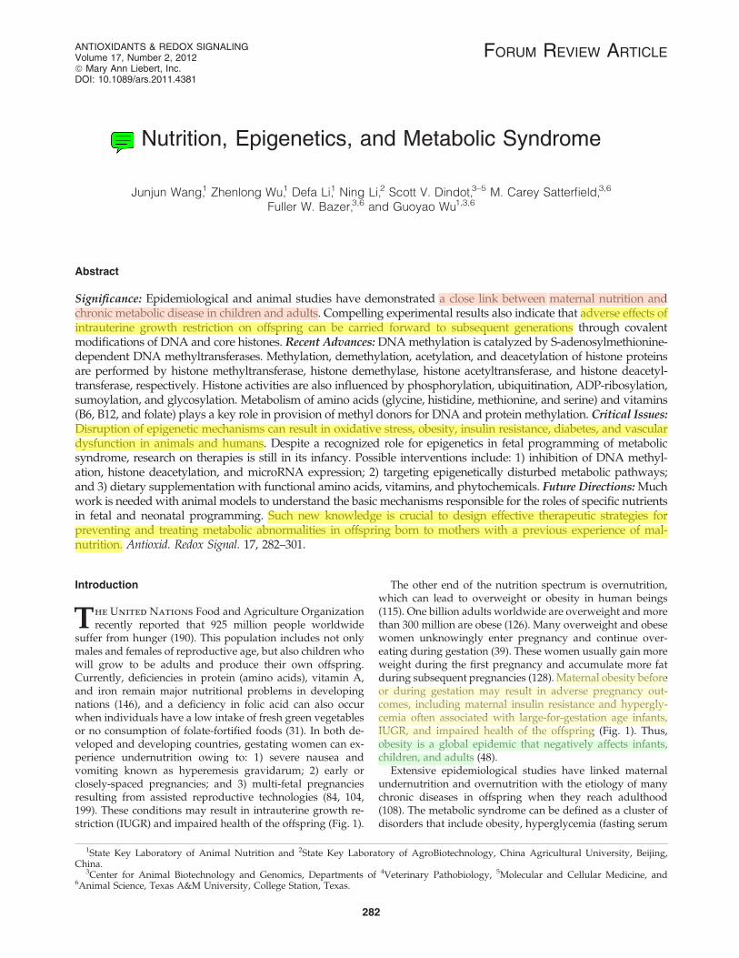
Amino acids serve not only as building blocks for proteins,but also as essential precursors for synthesis of a variety ofphysiologically important molecules, including hormones,small peptides (e.g., glutathione), neurotransmitters, nitricoxide, creatine, carnitine, and polyamines (196). Additionally,through multiple cell signaling pathways, amino acids regu-late key metabolic pathways that are vital to human health,growth, development, and reproduction (197). Notably, someamino acids (histidine, methionine, glycine, serine) partici-
pate in one-carbon-unit metabolism, which is essential forDNA synthesis, as well as cell growth and development (Fig.2). Based on nitrogen balance and growth, amino acids havebeen traditionally classified as either nutritionally essential(EAA) or nonessential (NEAA). The EAA are histidine, iso-leucine, leucine, lysine, methionine, phenylalanine, threonine,tryptophan, and valine. The NEAA are alanine, arginine, as-paragine, aspartate, cysteine, glutamate, glutamine, glycine,proline, serine, taurine, and tyrosine. However, the basis forclassification of an amino acid as a NEAA has conceptuallimitations because there is no compelling evidence that cer-tain NEAA (e.g., arginine and glutamine) can be adequatelysynthesized by the mother or fetus to support optimal sur-vival, growth, and development of the conceptus (203, 204).The synthesis of proteins in the fetus depends on the balancedprovision of all of the constituent amino acids, namely bothEAA and NEAA.
Nutritionally significant macro- and micro-minerals can-not be synthesized by the body and must, therefore, be pro-vided in the diet (195, 213). The macro-minerals are sodium,
FIG. 2. One-carbon unit metabolism for provision of methyl donors in cells. Folate, histidine, methionine, glycine andserine participate in the transfer of one-carbon units for the synthesis of purines and DNA as well as methylation reactions inanimals. The enzymes that catalyzed the indicated reactions are: 1) folate reductase; 2) N5-N10-methylene H4-folate dehy-drogenase (a trifunctional enzyme possessing N10-formyl H4-folate synthetase, N5-N10-methylene H4-folate dehydrogenase,and N5-N10-methenyl H4-folate cyclohydrolase activities); 3) N10-formyl H4-folate dehydrogenase; 4) serine hydroxymethyltransferase; 5) N5-N10-methylene H4-folate reductase; 6) methionine synthase; 7) S-adenosylmethionine synthase; 8)S-adenosylmethionine as a major methyl group donor in methyltransferase reactions; 9) formiminotransferase-cyclodeami-nase (a bifunctional enzyme possessing glutamate formiminotransferase and formimidoyl H4-folate cyclodeaminase activi-ties); 10) serine hydroxymethyl transferase, N5-N10-methenyl H4-folate cyclohydrolase, and spontaneous reaction at pH 4 to7.0; 11) N5-N10-methenyl H4-folate synthetase; 12) a sequential series of enzymes: histidase, urocanase, and imidazolonepropionate hydrolase; 13) thymidylate synthase; 14) formyltransferase; 15) an oxidant for cells; 16) betaine:homocysteine me-thyltransferase; 17) choline dehydrogenase; and 18) S-adenosylhomocysteine hydrolase. dTMP, deoxythymidine 5’-monophosphate;dUMP, deoxyuridine 5’-monophosphate; Gly, glycine; H4-folate, tetrahydrofolate; SAH, S-adenosylhomocysteine; SAM,S-adenosylmethionine; Ser, serine; Vit, vitamin.
284 WANG ET AL.
Administrator
Highlight
potassium, chloride, calcium, phosphorus, magnesium, andsulfur (177). The micro-minerals are iodine, iron, zinc, sele-nium, manganese, copper, cobalt, chromium, molybdenum,silicon, fluoride, vanadium, and nickel (177). The inorganicnutrients are the backbone for development, maintenance,and function of the skeleton system, and they serve as secondmessengers in cell signaling, and maintain polarity of theplasma membrane. Minerals also regulate extracellular andintracellular osmolality, which is important for maintainingcell and blood volume as well as cell viability and shape (213).Most minerals are either cofactors for enzymes or componentsof metalloproteins, and also participate in gene expression,electron transport, and redox reactions (21).
Vitamins are organic nutrients that are required in smallamounts for biochemical reactions. Water-soluble vitaminsinclude vitamin C and members of the B complex that areabsorbed by the small intestine into the portal circulation(127). Members of the B complex vitamins are vitamin B1
(thiamin), vitamin B2 (riboflavin), vitamin B3 (niacin), vitaminB5 (pantothenic acid), vitamin B6 (pyridoxal, pyridoxine, andpyridoxamine), vitamin B12 (cobalamin), biotin, and folate(21). Because of their water solubility, there is limited storageof these vitamins in the body (127). As a result, these micro-nutrients must be provided regularly in the diet. Lipid-solublevitamins include vitamin A, vitamin D, vitamin E, and vita-min K. Most vitamins, except niacin and vitamin D, cannot besynthesized by humans and must be supplied in the diet (177).Vitamins serve as cofactors for enzymes involved in geneexpression, metabolism of nutrients (e.g., fatty acids, glucose,and amino acids), one-carbon-unit transfer (Fig. 2), and ATPproduction.
Impacts of maternal undernutrition on offspringmetabolism and health
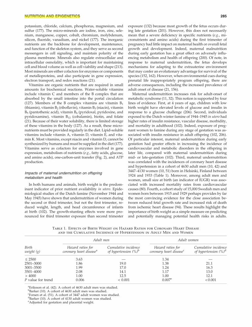
In both humans and animals, birth weight is the predom-inant indicator of prior nutrient availability in utero. Epide-miological studies of the Dutch famine (November 1944 andMay 1945) have shown that undernutrition of women duringthe second or third trimester, but not the first trimester, re-duced weight, length, and head circumference of infantsat birth (102). The growth-stunting effects were more pro-nounced for third trimester exposure than second trimester
exposure (132) because most growth of the fetus occurs dur-ing late gestation (201). However, this does not necessarilymean that a severe deficiency in specific nutrients (e.g., mi-cronutrients and amino acids) during the first trimester ofpregnancy had little impact on maternal health or overall fetalgrowth and development. Indeed, maternal malnutritionduring early gestation has a great effect on adversely influ-encing metabolism and health of offspring (200). Of note, inresponse to maternal undernutrition, the fetus developsmechanisms for adapting to the extrauterine environmentthat may confer an evolutionary advantage for survival of thespecies (152, 162). However, when environmental cues duringprenatal life inappropriately program offspring, there areadverse consequences, including the increased prevalence ofadult onset of disease (21, 156).
Maternal undernutrition increases risk for adult-onset ofmetabolic syndrome (11, 32). This notion is supported by fourlines of evidence. First, at 4 years of age, children with lowbirth weight have elevated levels of glucose and insulin inresponse to a glucose challenge (206). Second, individualsexposed to the Dutch winter famine of 1944–1945 in utero hadhigher rates of insulin resistance, vascular disease, morbidity,and mortality in adulthood (102). Indeed, exposure of preg-nant women to famine during any stage of gestation was as-sociated with insulin resistance in adult offspring (102, 206).Of particular interest, maternal undernutrition during earlygestation had greater effects in increasing the incidence ofcardiovascular and metabolic disorders in the offspring inlater life, compared with maternal undernutrition duringmid- or late-gestation (102). Third, maternal undernutritionwas correlated with the incidences of coronary heart diseaseand hypertension in a cohort of 4630 adult men (10, 42) and3447–4130 women (10, 51) born in Helsinki, Finland between1924 and 1933 (Table 1). Moreover, among adult men andwomen, small size at birth (an indicator of IUGR) was asso-ciated with increased mortality rates from cardiovascularcauses (80). Fourth, a cohort study of 15,000 Swedish men andwomen born between 1915 and 1929 perhaps provides by farthe most convincing evidence for the close association be-tween reduced fetal growth rate and increased risk of deathfrom ischemic heart disease (94). These results highlight theimportance of birth weight as a simple measure on predictingand potentially managing potential health risks in adults.
Table 1. Effects of Birth Weight on Hazard Ratios for Coronary Heart Disease
and the Cumulative Incidence of Hypertension in Adult Men and Women
Adult men Adult women
Birthweight (g)
Hazard ratios forcoronary heart diseasea
Cumulative incidenceof hypertension (%)b
Hazard ratios forcoronary heart diseasec
Cumulative incidenceof hypertension (%)d
£ 2500 3.63 — 1.34 —2501–3000 1.86 19.0 1.38 21.13001–3500 1.99 17.0 1.24 16.33501–4000 2.08 14.1 1.17 13.0> 4000 1.00 12.5 1.00 12.1P value for trend 0.006 < 0.001 0.007e < 0.001
aEriksson et al. (42). A cohort of 4630 adult men was studied.bBarker (10). A cohort of 4630 adult men was studied.cForsen et al. (51). A cohort of 3447 adult women was studied.dBarker (10). A cohort of 4130 adult women was studied.eAdjusted for gestation and placental weight.
NUTRITION AND EPIGENETICS 285
Administrator
Highlight
Administrator
Underline
Thus, in terms of long-term health, the embryo/fetus is mostvulnerable to nutritional insults during early gestation.
Experimental evidence from a variety of species and animalmodels has provided a wealth of knowledge regarding effectsof reduced nutrient availability in utero on the development ofadult disease (3). For example, global undernutrition in-creases the risk for dyslipidemia, obesity, and type-2 diabetesmellitus in rats (156). Also, maternal protein restriction in-duces hypertension and vascular dysfunction in adult femaleoffspring of rats (65, 149). Extensive research with large ani-mals (e.g., sheep and nonhuman primates) provides furtherevidence for fetal programming of vascular dysfunction andmetabolic abnormalities. For example, in sheep, maternalundernutrition induces left ventricular hypertrophy and al-ters gene expression in the left ventricle of the fetal heart (62,180). Additionally, maternal nutrient restriction results in in-creased myocardial lipid and altered gene expression in off-spring at 1 year of age (28). Moreover, in sheep, maternalnutrient restriction impairs renal function, increases the de-velopment of glomerulosclerosis, and enhances apoptosis inkidneys, while altering the expression of proteins involved inregulating inflammatory processes (155, 192). In both pigs andsheep, maternal undernutrition also results in decreasedskeletal muscle fiber number, increased deposition of adiposetissue, and increased connective tissue content (15, 58). Col-lectively, these alterations in normal development result in: a)reduced postnatal growth, including both whole-body andskeletal muscle growth; b) reduced efficiency of nutrient uti-lization; and c) suboptimal function of multiple organs.
Impacts of maternal overnutrition on metabolismand health of offspring
Maternal obesity has been linked to metabolic perturba-tions, obesity, and cardiovascular disorders in the offspring ofa number of mammalian species, including humans, mice,rats, and sheep (6, 83, 85, 156). In nonhuman primates, amaternal high fat diet promotes the development of nonal-coholic fatty liver and atherosclerosis (110) and alteration ofseven metabolites in the fetus related to one-carbon unit me-tabolism (33). Notably, changes in the fetal metabolome re-sult, at least in part, from an altered fetal hepatic chromatinstructure that leads to aberrant gene expression in the liver (1)and other insulin-sensitive tissues (162).
Feeding a high-fat diet during pregnancy or the neonatalperiod stimulates adipogenesis, induces cardiovascular dys-function characterized by elevated systolic blood pressure,and impairs endothelium-dependent relaxation in rats (3, 61,83). Importantly, the response to maternal dietary treatmentdiffered between male and female offspring in that femaleoffspring were more susceptible to elevated systolic and dia-stolic blood pressure when their mothers were fed a high-fatdiet during gestation and/or lactation (145). This interestingphenomenon may be explained by imprinting of genesand sex-related differences in hormone secretion. In contrast,endothelium-dependent relaxation of blood vessels in re-sponse to acetylcholine was consistently blunted in bothmales and females exposed to a high-fat diet either in utero orduring lactation (137). A major underlying mechanism for thehigh fat-induced vascular dysfunction is likely the reducedrelease of nitric oxide (a major vasodilator) from endothelialcells (198).
Maternal obesity due to a high-fat diet reduced mitochon-drial copy number in the kidney in 1-year-old rats, while al-tering expression of a number of mitochondrial genes in therat aorta (166). These findings suggest that mitochondria playa central role in developmental programming of vascularfunction. Similar results were observed in the offspring ofpregnant mice fed a high-fat diet, including increased adi-posity, dyslipidemia, hypertension, and insulin resistance (38,148). In sheep, maternal obesity downregulates expression ofgenes involved in myogenesis and placental angiogenesis(212) and fetal skeletal muscle (173), while reducing thephosphorylation of AMP-activated kinase in the fetal andneonatal liver (134) as well as energy metabolism (185). Im-portantly, these changes in gene and protein expression inresponse to maternal overnutrition result in increased fetaland/or neonatal adiposity (50, 121) and increased expressionof the leptin gene in peri-renal and subcutaneous adiposetissue depots (121). The consequences of maternal overnutri-tion continue to manifest in offspring as evidenced by im-paired insulin sensitivity and reduced glucose utilization onpostnatal Day 210 despite growing in an identical nutritionalenvironment from birth (150). Similarly, a maternal high-fatdiet during pregnancy results in impaired glucose homeo-stasis in rat offspring characterized by elevated concentra-tions of insulin in plasma at 1 year of age (166).
Impact of Maternal Nutritionon Gene Expression in Offspring
Maternal nutrition and gene expression in offspring
As noted previously, studies of offspring born to womenduring the Dutch famine in the 1940s suggested a linkbetween the fetal environment (including nutrition) andpostnatal health, particularly cardiovascular function ordysfunction, in humans (9, 30, 69, 133). Interestingly, 60 yearsafter birth, offspring with early prenatal experience of thefamine exhibited less DNA methylation of the imprinted IGF2gene, as compared to the same-sex siblings without exposureto prenatal malnutrition (67). Furthermore, individuals withpericonceptional exposure to the famine had lower methyla-tion of the INSIGF gene, but higher methylation levels forseveral other genes (IL10, LEP, ABCA1, GNASAS, and MEG3)(171). These genes are closely linked with nutrient metabo-lism, cardiovascular function, and inflammation. Interactionsbetween undernutrition and sex also affect methylation of theINSIGF, LEP, and GNASAS genes (171).
In monkeys, a 30% reduction in maternal nutrient intakedid not alter fetal weight at stages 0.5 and 0.9 of gestation (fullterm being 1.0) (176). However, tissue-specific global meth-ylation status (176) and the availability of methyl group do-nors (153) were altered in the kidneys of nonhuman primatesat both 0.5 and 0.9 of gestation. Furthermore, emerging evi-dence indicates modifications of hepatic phosphoenolpyr-uvate carboxykinase (a key enzyme in gluconeogenesis) afterexposure of fetal baboons to moderately reduced nutrientavailability (124). These results indicate that even a relativelymild nutrient restriction can induce alterations in gene ex-pression and provide a potential mechanism responsible forthe increased incidence of endothelial dysfunction, renaldysfunction, and hypertension in offspring with previousexperience of malnutrition during the period of fetal growth.Such an effect of fetal programming on gene expression in
286 WANG ET AL.
Administrator
Highlight
offspring has been experimentally tested in a number ofspecies, including nonhuman primates (124), rats (5), mice(38), cattle (23), and sheep (162). Thus, effects of changes innutrition or endocrine status during the fetal can negativelyimpact subsequent development of the offspring.
Transgenerational effects of maternal nutrition
Epidemiological studies have indicated the presence oftransgenerational effects of maternal malnutrition in humans(209). For example, starvation in young boys (7–11 years ofage) resulted in increased incidence of metabolic syndrome inthem as adult men, and in their children and grandchildren(79). Likewise, food deprivation in female fetuses for 7–12weeks was associated with increased prevalence of metabolicsyndrome in them as adult women, and in their children andgrandchildren (22). Results of animal studies also indicatedthat the effect of maternal undernutrition on hormone secre-tion and metabolism can be passed transgenerationally frommother (F0) to daughter (F1) and to the F2 progeny (16). Forexample, maternal calorie restriction in rats caused: a) de-creases in the mass and number of pancreatic b-cells inthe first generation of female offspring; and b) impaired re-sponses of F2 pancreatic b-cells to pregnancy, leading to in-sulin resistance and gestational hyperglycemia (107, 168).Similar results were obtained for female rats fed a low-proteindiet (210) or female mice subjected to caloric undernutrition(76) during gestation. In guinea pigs, even modest maternalundernutrition (70% of normal food intake) resulted in alter-ations in the cardiac structure (e.g., increases in wall thicknessand mass of the left ventricle), hypothalamo-pituitary-adrenalfunction (e.g., increased basal levels of cortisol), and elevatedblood pressure in adult male F1 and F2 offspring through thematernal line (4, 16). Interestingly, some of the transgenera-tional effects of maternal undernutrition-induced IUGR anddevelopmental defects on diabetes and cardiovascular func-tion are sex-dependent (168, 187), which can be explained wellby differential genomic imprinting (35). Likewise, chronichigh-fat feeding in fathers resulted in hypomethylation of theII13ra2 gene and b-cell dysfunction in female, but not male,offspring (123).
Fetal Programming
The mechanism of fetal programming: Epigenetics
The genetic code established by the DNA sequence exhibitsminimal rates of change after the formation of the diploidchromatin state at fertilization (43). Therefore, an extensivesearch for alternative mechanisms of gene regulation resultedin the now widely accepted notion that changes in gene ex-pression can be manifest by mitotically and/or meioticallyheritable alterations in the DNA–protein complex withoutany change in the DNA sequence (156). This phenomenonwas originally termed ‘‘epigenetics’’ by Conrad Waddingtonin 1940 (182) based on the Greek prefix ‘‘epi’’ meaning over orabove. Molecular mechanisms about epigenetics and genomicimprinting have evolved over the past 40 years since the firstidentification in the 1970s of DNA methylation as a regulatoryfactor (70). The 1980s (109, 183, 193) and 1990s (12, 18, 63)witnessed pioneering research on the maintenance and reg-ulation of DNA methylation patterns in mammals. In the mid1990s, histone modifications were discovered as an epigenetic
determinant of chromatin structure and, therefore, genomeactivity (29, 174). The identification of noncoding microRNAsand their functions early in the 2000s further expanded thefield of epigenetic research (4, 95). Excitingly, the first wholeepigenome analysis was completed for yeast in 2005 (136). It isnow known that epigenetic modifications can be carriedforward from one generation of cells to the next (mitotic in-heritance) and between generations in a species (meiotic in-heritance) (156).
At present, the four mechanisms responsible for mediatingepigenetic effects are: a) chromatin modifications, b) DNAmethylation (occurring at the 5’-position of cytosine residueswithin CpG dinucleotides throughout the mammalian ge-nome), c) histone modifications (acetylation, methylation,phosphorylation, ubiquitination, and sumoylation), and d)RNA-based mechanisms such as small noncoding RNAs orinhibitory RNAs (Fig. 3). The enzymes involved in these re-actions include specific DNA and protein methyltransferases,DNA demethylases, histone acetylase (lysine acetyltransfer-ase), GCN5-related N-acetyltransferase (a super family ofacetyltransferase), and histone deacetyltransferase (18, 26, 30).
DNA methylation
The methylation of DNA is catalyzed by DNA methyl-transferases (DNMT1, DNMT3a, and DNMT3b) and is a re-versible physiological process in the eukaryotic genome (45).This biochemical event involves the donation of a methylgroup from S-adenosylmethionine to the 5’-position of a cy-tosine nucleotide linked to a guanine nucleotide (CpG dinu-cleotide) by a phosphodiester bond. Regions with a high CpGdinucleotide content form CpG islands, which are located inthe regulatory regions of many genes, including promotersand enhancers (37). Due to their presence in these regulatoryregions, changes in methylation status can either facilitate(hypomethylation) or inhibit (hypermethylation) the expres-sion of a gene (106). In normal mammalian cells, over 85% ofCpG dinucleotides are methylated, which plays a role inmaintaining the integrity of chromatin structure and tran-scriptional regulation (56). Except for imprinted genes, CpGislands present in the promoter regions of genes are usuallyunmethylated. Approximately 1% to 2% of the human ge-nome consists of CpG dinucleotide whose methylation is in-versely related to transcriptional activity in cells (187).
Epigenetic modifications are erased and re-established in atissue-specific manner during embryonic development (184).The haploid genomes of the sperm and oocyte possess dif-ferent patterns of DNA methylation that arise from sex-specific patterns of de novo methylation. After fertilization, thepaternal genome is rapidly demethylated prior to the first celldivision of the zygote. In contrast, the maternal genome isprotected from this demethylation event, and, instead, isgradually demethylated during the development of the blas-tocyst. At the blastocyst stage, most methyl marks are re-moved, remaining present at elements regulating genomicimprinting and retroviral elements (43, 56). After implanta-tion, de novo DNA methylation occurs, giving rise to tissue-specific patterns of DNA methylation (36). The process ofdemethylation and remethylation during the development ofgerm cells is referred to as ‘‘reprogramming’’ and this physi-ological event is regulated, in part, by the phosphatidylino-sitol 3-kinase pathway (161).
NUTRITION AND EPIGENETICS 287
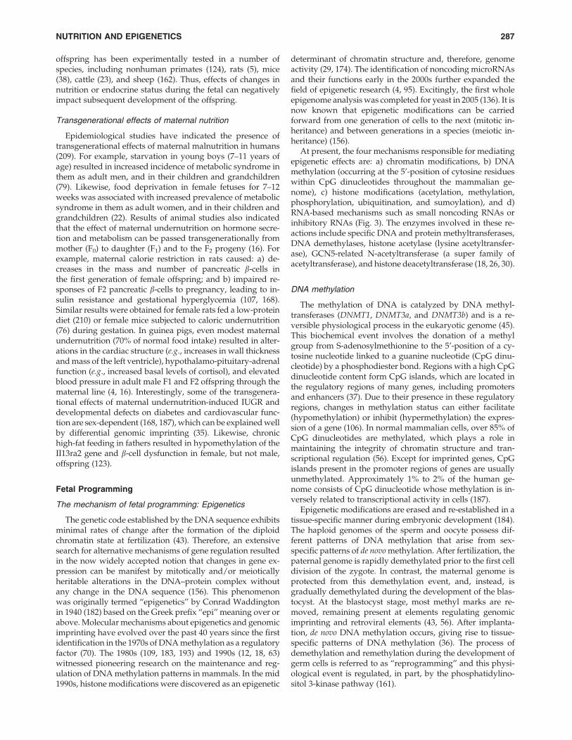
Epigenetic information is heritable between cell genera-tions through the action of the methyltransferase, DNMT1(184). In somatic cells, this enzyme recognizes and methylatesCpG dinucleotides present on the newly synthesized strand.As DNA synthesis occurs, the parental strand remainsmethylated, whereas the newly synthesized daughter strandis not. DNMT1 ‘‘reads’’ the parental strand and methylatesCpG dinucleotides on the daughter strand that are comple-mentary to methylated CpG dinucleotides on the parentalDNA strand (Fig. 4). The importance of DNA methylation forsuccessful pregnancy outcome is evident by the fact that allhomozygous DNMT1 knockout mice die at the morula stageof embryonic development (20).
Histone modifications
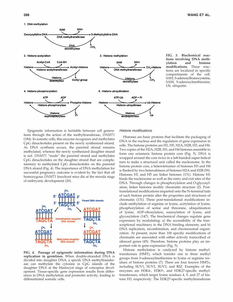
Histones are basic proteins that facilitate the packaging ofDNA in the nucleus and the regulation of gene expression incells. The histone proteins are H1, H5, H2A, H2B, H3, and H4.Two copies of the H2A, H2B, H3, and H4 histones assemble toform one octameric histone protein core (Fig. 5). DNA iswrapped around the core twice in a left-handed super-helicalturn to make a structural unit called the nucleosome. In thehistone protein core, a heterotetramer of histones H3 and H4is flanked by two heterodimers of histones H2A and H2B (59).Histones H1 and H5 are linker histones (131). Histone H1binds the nucleosome as well as the entry and exit sites of theDNA. Through changes in phosphorylation and O-glycosyl-ation, linker histones modify chromatin structure (2). Post-translational modifications imparted onto the N-terminal tailsof each histone protein alter the properties and structures ofchromatin (131). These post-translational modifications in-clude methylation of arginine or lysine, acetylation of lysine,phosphorylation of serine and threonine, ubiquitinationof lysine, ADP-ribosylation, sumoylation of lysine, andglycosylation (147). The biochemical changes regulate geneexpression by modulating: a) the accessibility of the tran-scriptional machinery to the DNA binding elements; and b)DNA replication, recombination, and chromosomal organi-zation. At present, more than 100 specific modifications ofchromatin are associated with either actively transcribed orsilenced genes (45). Therefore, histone proteins play an im-portant role in gene expression (Fig. 5).
Histone methylation is catalyzed by histone methyl-transferases (HMT), which transfer one to three methylgroups from S-adenosylmethionine to lysine or arginine res-idues of histone proteins (7). There are four known HMTs,including SUV1, SUV2, SUV3, and RIZ. Examples of theenzymes are H3K4-, H3K9-, and H3K27-specific methyl-transferases, which target lysine residues 4, 9, and 27 of his-tone H3, respectively. The H3K27-specific methyltransferase
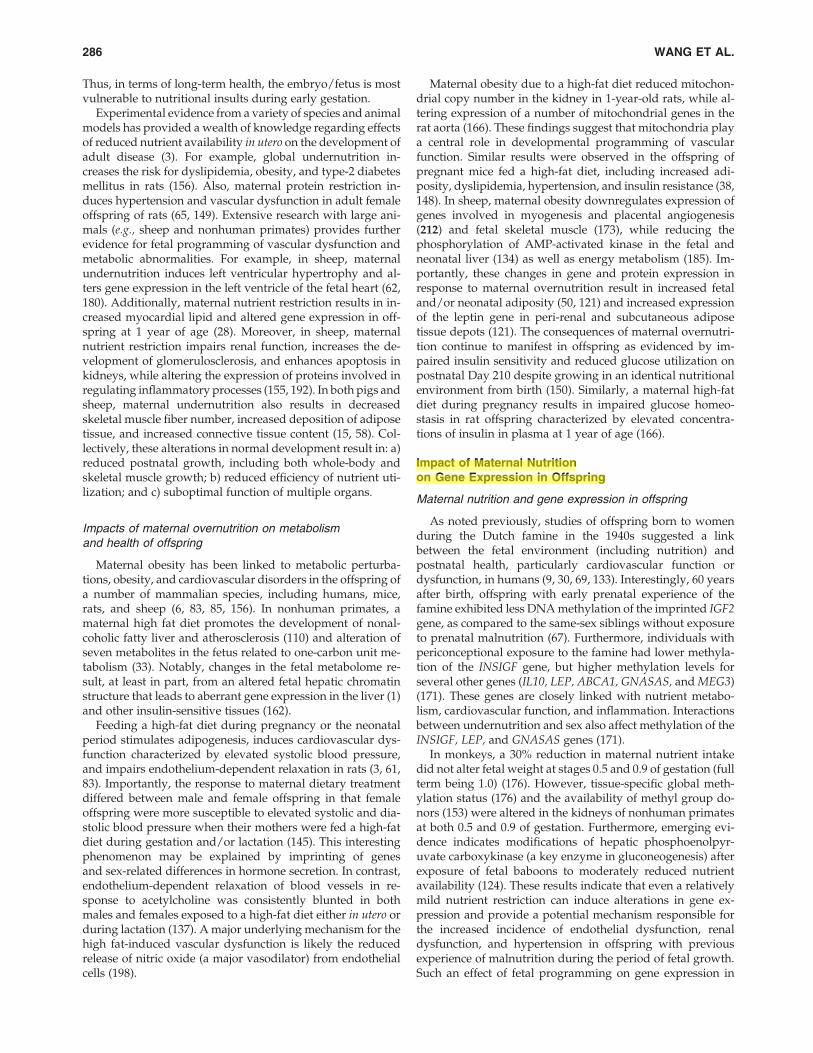
FIG. 3. Biochemical reac-tions involving DNA meth-ylation and histonemodifications. These reac-tions are localized in specificcompartments of the cell.SAH, S-adenosylhomocysteine;SAM, S-adenosylmethionine;Ub, ubiquitin.
FIG. 4. Passage of epigenetic information during DNAreplication in germlines. When double-stranded DNA isdivided into daughter DNA, a specifc DNA methyltransfer-ase can methylate the cytosine in CpG islands of thedaughter DNA at the blastocyst stage of conceptus devel-opment. Tissue-specific gene expression results from differ-ences in DNA methylation and promoter activity, leading todifferentiated somatic cells.
288 WANG ET AL.
is also known as the enhancer of zeste homologue 2 (EZH2).Other significant histone methylases are H3K79- and H4H20-specific (131). Silencing of differentiation genes is associatedwith the methylated H3K27 chromatin mark. In a variety ofcell types, histone methylation is generally associated withtranscriptional repression, but methylation of some lysine(e.g., lysine 4 of histone 3) and arginine residues (e.g., those onH3 and H4) of histones can result in transcriptional activation(7). While histone methylation was previously thought to be apermanent modification, recent studies uncovered his-tone demethylating enzymes (45). It is now known thatdemethylation of histone proteins is performed by his-tone demethylases. Lysine-specific demethylase 1 (a flavin-dependent monoamine oxidase) can demethylate mono- anddi-methylated lysines, specifically lysines 4 and 9 of histone 3(H3K4 and H3K9) (54). This enzyme cannot demethylate tri-methylated lysines. However, the Jumonji domain-containing( JmjC) histone demethylases can demethylate mono-, di-, ortri-methylated lysines (187). It is possible that arginine-specific demethylase exists in cells to demethylate mono- anddi-methylated arginines in histone proteins (19). Alternatively,methylated arginine residues can be converted into citrullineby peptidyl arginine deiminase 4, which acts on unmodifiedand mono-methylated arginine residues in proteins (7).
Histone acetylation also influences gene expression (43).Acetylation of lysine residues at the N terminus of histoneproteins is catalyzed by histone acetyltransferases (HAT) (59).Superfamilies of HAT include GNAT, CBP/p300, and MYST.Examples of histone acetyltransferases are HAT1, HATp300,CDY1, and CLOCK. The source of the acetyl group in histoneacetylation is acetyl-CoA. Thus, metabolism of energy sub-strates or the energy status of the cell can modulate histoneacetylation (184). Notably, acetylation of lysine residuesneutralizes the positive charge on histones, thereby decreas-ing the interaction of the N termini of histones with the neg-atively charged phosphate groups of DNA (187). Such amodification of histone proteins transforms the condensedchromatin into a more relaxed structure and makes RNApolymerase and transcription factors easier to access thepromoter region of the DNA, leading to enhanced genetranscription. Thus, histone acetylation is usually associatedwith activation of gene transcription (59). Histone deacetyla-
tion is catalyzed by histone deacetylases (HDAC; also knownas lysine deacetylases). In this reaction, the acetyl group istransferred from the acetylated histone protein to coenzymeA. Based on intracellular localization, three classes of HDACare I: HDAC 1, 2, 3, and 8 (exclusively in the nucleus); II:HDAC 4, 5, 6, 7, 9, and 10 (predominantly residing in thecytoplasm and shuttling in and out of the nucleus); and III:surtuin enzymes (SIRT) 1, 2, 3, 4, 5, 6, and 7 (localized in eithernucleus, cytoplasm, or mitochondria) (187). The histone dea-cetylase CBP/p300 can interact with numerous transcriptionregulators and likely plays a major role in repressing genetranscription (57).
The biological activity of histones is also regulated byphosphorylation. Histone H3 phosphorylation, which occursduring both mitosis and meiosis, is catalyzed by ATP-dependent protein kinases (26, 207). This event is coordinatedin both space and time as a signaling mechanism to modulatenucleosome structure and thus DNA accessibility (73). Phos-phorylation of H2A (at serine 1 and threonine T119), H3 (atserine 10 and 28; threonine 3 and 11), and H4 (at serine 1)results in transcriptional activation, as well as mitosis andmeiosis (26, 131). Phosphorylation of H2A also enhancesDNA repair, while phosphorylation of H4 promotes sper-matogenesis. Likewise, histone H1 phosphorylation is asso-ciated with decondensation of chromatin and activation oftranscription (2), and histone-3 phosphorylation is critical forcytokine-induced gene expression (207). In contrast, phos-phorylation of H2B (at serine 14) stimulates apoptosis, whichplays an important role in cell metabolism and growth (101).Thus, histone phosphorylation can allow cells to rapidly re-spond and adapt to changes in maternal nutritional status byregulating numerous processes, including transcription, DNArepair, and apoptosis (Fig. 5).
The monoubiquitination of histones H2A and H2B plays acritical role in regulating DNA repair, gene transcription, andmRNA translation (188). In this process, ubiquitin (a 76-aminoacid polypeptide) is conjugated to histones H2A and H2B in aseries of reactions involving three separate enzymatic activi-ties: a) ATP-dependent ubiquitin activating enzyme (E1; ac-tivation of ubiquitin), b) ubiquitin-conjugating enzyme (E2;conjugation via a thioester bond to a cysteine residue); and c)ubiquitin-protein isopeptide ligase (E3; transfer of ubiquitin
FIG. 5. Roles of histone modificationsin the regulation of gene transcription.Methylation of histones, acetylation, orubiquitination can either activate or re-press gene expression, depending onspecific histone proteins and the sites ofmodifications. In general, phosphoryla-tion of histones promotes transcription,DNA repair, and apoptosis. H, histone;K, lysine residue; R, arginine residue.
NUTRITION AND EPIGENETICS 289
from the E2 enzyme to a target lysine residue in a histone)(135). Monoubiquitination of H2B promotes transcriptioninitiation and elongation through efficient reassembly of nu-cleosomes (49). There is evidence that H2B monoubiquitina-tion is a prerequisite for Lys-4 H3 and Lys-79 H3 methylation,whereas H2A monoubiquitination is associated with tran-scriptional silencing (187). Although H2A and H2B ubiquiti-nation appear to perform separate functions, there is cross talkbetween ubiquitinated H2A and H2B proteins to modulateLys-4 H3 methylation and RNA polymerase II activity duringtranscription elongation (49, 169).
RNA-based epigenetic mechanisms
Epigenetic regulation of gene expression by RNA-basedmechanisms can occur at both the post-transcriptional leveland the level of chromatin (187). These mechanisms are me-diated by small-interfering RNAs or small non-coding RNAswhich act through their respective pathways to induce DNAmethylation or histone modifications to silence or enhancegene expression (82, 169). In mammals, the small non-codingRNAs include Piwi RNA and microRNA (miRNA) (74). PiwiRNA is restricted to the germ line (e.g., testes in males andoocytes in females), and regulates gene expression possiblythrough sequence-specific targeting of heterochromatin for-mation factors to the mobile elements in the genome and thedegradation of mobile element transcripts (172). In contrast,miRNAs are widespread in cells and tissue to regulate geneexpression at the post-transcriptional level (44). Since thediscovery of lin-4 as the first miRNA in 1993 (93), approxi-mately 1000 human miRNAs have been identified, annotatedand catalogued (103, 113).
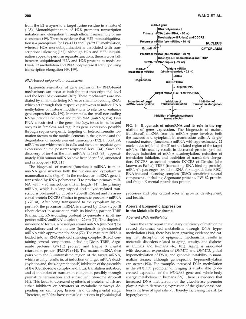
The biogenesis of mature (functional) miRNA from itsmiRNA gene involves both the nucleus and cytoplasm inmammalian cells (Fig. 6). In the nucleus, an miRNA gene istranscribed by RNA polymerase II to produce primary miR-NA with *80 nucleotides (nt) in length (44). The primarymiRNA, which is a long capped and polyadenylated tran-script, is processed by Drosha (type-III RNase) and its asso-ciated protein DGCR8 (Pasha) to generate precursor miRNA(*70 nt). After being transported to the cytoplasm by ex-portin-5, the precursor miRNA is cleaved by Dicer (type-IIIribonuclease) in association with its binding partner TRBP(transacting RNA-binding protein) to generate a small im-perfect miRNA:miRNA* duplex (* 22 nt) (74). This duplex isunwound to form a) a passenger strand miRNA (miRNA*) fordegradation; and b) a mature (functional) single-strandedmiRNA with approximately 22 nt (72). The mature miRNA isloaded into an RNA-induced silencing complex (RISC) con-taining several components, including Dicer, TRBP, Argo-naute proteins, GW182 protein, and fragile X mentalretardation protein (FMRP1) (44). The mature miRNA thenpairs with the 3’-untranslated region of the target mRNA,which usually results in: a) induction of target mRNA dead-enylation and, thus, degradation; b) inhibition of the assemblyof the 80S ribosome complex and, thus, translation initiation;and c) inhibition of translation elongation possibly throughpremature termination and subsequent ribosome drop-off(44). This leads to decreased synthesis of proteins which areeither inhibitors or activators of metabolic pathways de-pending on cell types, tissues, and developmental stage.Therefore, miRNAs have versatile functions in physiological
processes and play crucial roles in growth, development,and health.
Aberrant Epigenetic Expressionin the Metabolic Syndrome
Aberrant DNA methylation
Since the early report that dietary deficiency of methioninecaused abnormal cell metabolism through DNA hypo-methylation (194), there has been growing evidence indicat-ing that disruption of epigenetic mechanisms results inmetabolic disorders related to aging, obesity, and diabetesin animals and humans (46, 101). Aging is associatedwith decreased expression of DNMT1 and DNMT3, globalhypomethylation of DNA, and genomic instability in mam-malian tissues, although gene-specific hypermethylationcan occur (193). For example, increased DNA methylationin the NDUFB6 promoter with aging is attributable to de-creased expression of the NDUFB6 gene and whole-bodyenergy metabolism in humans (99). There is evidence thatincreased DNA methylation of the glucokinase promoterplays a role in decreasing expression of the glucokinase pro-tein in the liver of aged rats (75), thereby increasing the risk forhyperglycemia.
FIG. 6. Biogenesis of microRNA and its role in the reg-ulation of gene expression. The biogenesis of mature(functional) miRNA from its miRNA gene involves boththe nucleus and cytoplasm in mammalian cells. A single-stranded mature (functional) miRNA with approximately 22nucleotides (nt) binds the 3’-untranslated region of the targetmRNA. This usually results in decreased protein synthesisthrough induction of mRNA deadenylation, reduction oftranslation initiation, and inhibition of translation elonga-tion. DGCR8, associated protein DGCR8 of Drosha (alsoknown as Pasha); TRBP (transacting RNA-binding protein);miRNA*, passenger strand miRNA for degradation; RISC,RNA-induced silencing complex (RISC) containing severalcomponents, including Argonaute proteins, PW182 protein,and fragile X mental retardation protein.
290 WANG ET AL.
Epigenetic defects (epimutations) can play a role in thedevelopment of obesity (3). Of interest, loss-of-function of thehistone demethylase, Jhdm2a, is associated with reduced ex-pression of peroxisome proliferator-activated receptor-a andmedium-chain acyl-CoA dehydrogenase in skeletal muscle,impairment of cold-induced expression of uncoupling pro-tein-1, and obesity in mice (165). Accordingly, hypomethyla-tion increases risk of obesity in humans and animals (71). Forexample, maternal high-fat diet increased global and gene(dopamine and opioid) specific hypomethylation of DNA inthe brains of offspring that resulted in alterations in gene ex-pression and behavior (181). In addition, secretion of leptin bywhite adipose tissue is controlled by epigenetic mechanisms.Specifically, the leptin promoter that contains a CpG islandhas a high degree of methylation in preadipocytes (116, 208),and the promoter is demethylated in association with induc-tion of leptin expression in differentiated adipocytes (208).Furthermore, a high-fat diet increases DNA methylation ofone CpG site in the leptin promoter of adult rat adipocytes(119). Finally, NAD-dependent sirtuins (class III HDAC),which target both histones and nonhistone proteins, regulatenutrient metabolism, mitogenesis, adipogenesis, and insulinsecretion (154). Additionally, defects in orchestration of cir-cadian rhythm by HDAC3 lead to abnormal lipid metabolismin the liver (46).
Diabetes may also have an epigenetic basis (99, 100). First,recent work revealed that COX7A1 (part of complex 4 of therespiratory chain) is a target of DNA methylation (144). In-terestingly, methylation of the DNA for the COX7A1 promoteris increased in skeletal muscle of diabetic animals, and its genetranscription is reduced in association with reduced glucoseuptake by muscle (144). Second, genetic mutation in the Pdx1/insulin promoter factor-1, which is a transcription factor regu-lating development of the pancreas and b-cell differentiation,results in a monogenic form of diabetes (178). Interestingly,IUGR resulting from uteroplacental insufficiency is associatedwith the silencing of Pdx1 and the development of type 2 dia-betes in adult offspring of rats (130). Third, DNA methylationof the PPARGC1A gene is elevated in pancreatic islets frompatients with type 2 diabetes and the degree of DNA methyl-ation is inversely correlated with PPARGC1A expression (100).Fourth, hypomethylation at chromosome 6q24 is associatedwith transient neonatal diabetes in humans, and these subjectsdevelop type 2 diabetes as adults (167).
Aberrant histone modifications
Abnormal modifications of histone proteins contribute tometabolic disorders (101). GLUT4 is a major glucose trans-porter in insulin-sensitive tissues; therefore, intensive re-search on epigenetic regulation of diabetes has focused on thisprotein. In animal studies, reduced expression of GLUT4 isassociated with elevated HDAC activity and hyperglycemia(112). Normally, GLUT4 expression is regulated by the tran-scription factor myocyte enhancer factor 2 (MEF2), whichinteracts with HDAC5 in the nucleus. However, when thehistone tails that are colocalized with the GLUT4 gene aredeacetylated by HDAC5 in skeletal muscle, the formation ofcondense chromatin structures impairs GLUT4 gene expres-sion (111). In contrast, enhanced HAT activity to acetylatehistones is positively correlated with oxygen consumption inhuman skeletal muscle (129). Similarly, Class II HDAC en-
zymes limit GLUT4 gene expression during adipocyte dif-ferentiation (187). Likewise, histone code modifications byHDAC1 and HDAC4 repress GLUT4 expression in IUGRoffspring (140). Furthermore, phosphorylation of HDAC5 hasa stimulatory effect on GLUT4 transcription mediated byAMP-activated protein kinase in skeletal muscle (111).
A novel finding in epigenetic research is the regulationof insulin secretion by histone modifications (125, 156). Innormal pancreatic b-cells, the insulin gene exhibits hyper-acetylation of H4 and hypermethylation of H3K4. This patternis not observed for the insulin gene in other cells (e.g., HeLacells) that do not produce insulin (27). Recruitment of histoneacetyltransferase and the histone methyltransferase SET7/9 tothe insulin promoter is necessary to activate the gene for in-sulin (27). HDAC may inhibit pancreatic development in thefetus (66). Also, reduced levels of histone H3K27 trimethyla-tion and the histone methyltransferase Ezh2, as well as theloss of H2A ubiquitination are associated with elevated ex-pression of Ink4a/Arf and reduced beta-cell regeneration inaging mice (88). Thus, reduced activity of HAT (e.g., CBP/p300) is associated with a monogenic form of diabetes (8).
Another exciting new development in histone modifica-tions is their crucial roles in cellular antioxidative reactionsand cellular signaling. Poor glycemic control increases NF-kBactivity in monocytes and expression of genes encoding in-flammatory cytokines through an interaction between NF-kBand HAT (e.g., CBP/p300) (117). The H3K4 methyltransferaseSET7/9 affects the recruitment of NF-kB p65 to genepromoters and thus expression of NF-kB-dependent inflam-matory genes (98). Histone methylase (SET7) and histonedemethylase (LSD1) can regulate epigenetic changes in theNF-kB p65 promoter induced by hyperglycemia (19). Vas-cular smooth muscle cells from diabetic mice display reducedactivities of H3K9 methyltransferase and lysine-specific his-tone demethylase-1, as well as reduced levels of H3K9 tri-methylation and elevated levels of H3K4 dimethylation (179).Abnormal H3K9 dimethylation has also been reported forlymphocytes from patients with type-2 diabetes (118).
Aberrant expression of small non-coding RNAs
Aberrant expression of miRNAs has been implicated inobesity, insulin resistance, diabetes, cardiovascular compli-cations, and placental dysfunction (72, 120). For example, highfat feeding results in upregulation of miR-143 but down-regulation of primary miR-27a in mouse mesenteric adiposetissue via mechanisms involving expression of the PPAR-family of genes (113). Also, enhanced levels of miR-335 havebeen reported for the liver and white adipose tissues of threemurine models of obesity (leptin-deficient ob/ob mice, leptinreceptor-deficient db/db mice, and KKAy 44 mice) (122). Si-milarly, type-2 diabetes in humans is associated with downexpression of miR-27a and miR-378, which promotes theformation of lipid droplets, adipocyte differentiation, andadipogenesis in white adipocytes (113). Furthermore, miR-17-5P and miR-132 in omental fat and blood are positively cor-related with human obesity (68). Interestingly, emergingevidence indicates that miR-17-92, miR-21, miR-103, miR-143,miR-371, and miR-378 promote, but let-7, miR-27, miR-130,miR-138, miR 369-5P, and miR-448 inhibit, oxidative stressand inflammation in animals with obesity and atherosclero-sis (72). Finally, mis-expression of miRNAs contributes to
NUTRITION AND EPIGENETICS 291

inflammatory processes in placental dysfunction (120), car-diovascular diseases (e.g., coronary artery disease, ischemicinjury, hypertension, and atherosclerosis) (158), the metabolicsyndrome (103), and aging (158). Besides microRNAs, little isknown about effects of nutrition on expression of other smallnon-coding RNAs in animal tissues.
Inhibition to Regulate Epigeneticsand Ameliorate Metabolic Syndrome
Environmental cues appear to be the primary trigger toinduce changes in the epigenome and include such factors asnutrients, stress, environmental pollutants, and toxins (55).The extent to which cells are able to respond to these cues liesin large part with the plasticity of cells. Thus, the developingembryo/fetus is a prime target for epigenetic modifications(43) and interventions during pregnancy may be an importantstrategy for ameliorating or preventing metabolic disorders(including vascular insulin resistance) in adult life (Fig. 7).Despite a recognized role for epigenetics in fetal program-ming for metabolic syndrome (175), work in this field remainsvery limited.
Inhibition of DNA methylationand histone deacetylation
Based on epigenetic research, chemically synthesized drugshave been used to target deleterious effects of DNA methyl-
ation and histone deacetylation to reverse metabolic disorders(164). The classic DNMT inhibitor is 5-azacytidine, a deriva-tive of cytidine. Results of both in vitro and in vivo studiesindicate that 5-azacytidine increases expression of the PPARcgene in white adipocytes in a dose-dependent manner andthe metabolic pattern of the cells (53). Also of particular in-terest is the application of isoform-specific HDAC inhibitors(e.g., short-chain fatty acids, hydroxamic acids, cyclic tetra-peptides, and benzamides) to treat vascular complicationsassociated with diabetes (17, 60). For example, myocardialischemia-reperfusion injury is associated with increasedHDAC activity, but decreased acetylation of histones H3 andH4 in the heart of mice (57). Notably, administration of syn-thetic HDAC inhibitors (trichostatin A and Scriptaid) to theseanimals reduced myocardial infarction and cell death (57).Similarly, HDAC inhibitors attenuated load-and agonist-induced cardiac hypertrophy and abolished the associatedactivation of autophagy in cardiomyocytes (24).
Inhibition of miRNA expression and activity
Because abnormally high levels of some miRNAs causetranslational repression in tissues (particularly insulin-sensitive tissues) and are associated with the development ofchronic diseases (72, 113), modulation of their expression andactivity may be a potentially effective therapeutic means forprevention and treatment of the metabolic syndrome. At
FIG. 7. Roles of macro- and micro-nutrients in epigenetics and physiological responses. Nutrients, particularly aminoacids, regulate cellular redox state, the secretion of hormones (e.g., insulin and insulin-like growth factors), physiologicalfunctions, and whole-body homeostasis in humans and animals through three mechanisms: (1) the expression of genes; (2)the production of signaling gases and other metabolites; and (3) MTOR activation. S-Adenosylmethionine is the major methylgroup donor in cells and its synthesis is affected by amino acids (e.g., methionine, serine, glycine, and histidine), B vitamins(including folate, vitamin B12, and vitamin B6), choline, and creatine. Methylation of DNA and protein contributes toepigenetics, which results in transcriptional activation or inhibition of select genes. Changes in intracellular protein turnover(protein synthesis and degradation) and protein kinase cascades can alter physiological responses in the fetus and offspring.CO, carbon monoxide; 4EBP1, eIF4E-binding protein-1; GH, growth hormone; H2S, hydrogen sulfide; IGF, insulin-likegrowth factor; MTOR, mechanistic target of rapamycin; NO, nitric oxide; SAM, S-adenosylmethionine; S6K1, ribosomalprotein S6 kinase-1; SPP1, secreted phosphoprotein 1.
292 WANG ET AL.
Administrator
Highlight
Administrator
Highlight

present, the expression or activity of a specific miRNA can beinhibited or silenced using: a) a locked nucleic acid oligonu-cleotide (a Morpholino oligonucleotide); b) an antisense 2’-O-methyl RNA oligonucleotide; c) an antagomir (a small syn-thetic RNA oligonucleotide that is perfectly complementaryto the specific miRNA target), or d) a steric-blocking oligo-nucleotide to prevent the maturation of miRNA or block themiRNA-binding site of a target mRNA (40, 89). Althoughresearch in this area is still limited, results from both in vitroand in vivo studies have been promising. For example, specificinhibition of miR-375 (a pancreatic islet-specific microRNA)by transfection with an antisense 2’-O-methyl oligonucleotidecomplementary to miR-375 decreased the expression of themyotrophin and PDK1 genes and increased glucose-stimulatedinsulin secretion in cultured murine pancreatic b-cells (138).The use of a similar technology led to the discovery that miR-143 plays a key role in the differentiation of cultured humanpre-adipocytes by inhibiting expression of the ERK5 gene (41).Based on the in vitro studies, intravenous administration of anantagomir against miR-122 three times within 24 h (80 mg/kgbody weight/day) to mice reduced expression of 11 genesinvolved in cholesterol synthesis (including hydroxy-3-methylglutaryl-CoA reductase) in the liver, thereby decreas-ing concentrations of cholesterol in plasma (89). Likewise,subcutaneous injection of an anti-miR-33 oligonucleotide tomice weekly for 4 weeks promoted the reversal transport ofcholesterol from peripheral tissues to the plasma, liver, andfeces, as well as regression of established atherosclerosis, andameliorated dyslipidemia (as indicated by reduced concen-trations of triacyglycerols and cholesterol in plasma) (141).Employing the similar technology, Rayner et al. (142) recentlyreported that inhibition of miR-33a/b increased the expres-sion of: a) the sterol-response-element-binding protein genesSREBF1 and SREBF2; b) the cholesterol transporter geneABCA1; and c) genes involved in fatty acid oxidation (CROT,CPT1A, HADHB, and PRKAA1), while reducing the expres-sion of genes involved in fatty acid synthesis (SREBF1, FASN,ACLY, and ACACA) in nonhuman primates. This anti-microRNA strategy resulted in increased concentrations ofhigh-density lipoproteins and reduced concentrations of very-low-density lipoproteins in plasma to reduce risk for cardio-vascular disease.
Targeting epigenetically disturbed metabolic pathways
Maternal malnutrition may affect growth and health of F1and subsequent offspring (200). It is now known that IUGR isassociated with reduced expression of the imprinted geneIGF2 in animals (52). Interestingly, loss of imprinting of theIGF2 gene results in enhanced signaling of IGF2/IGF1 at thereceptor level due to a positive feedback mechanism (35)which leads to abnormal metabolism of nutrients (e.g., un-controlled utilization of glucose via glycolysis) (170). Inhibi-tion of the IGF2 receptor can reverse the epigeneticallydisturbed metabolic pathways in animals with loss of im-printing of the IGF2 gene (81). Additionally, results of recentstudies indicate that alterations in DNA methylation andhistone modifications in cardiovascular diseases all resultin elevated levels of hyperhomocysteinemia due to reducedexpression of the enzymes involved in the utilization ofhomocysteine (25, 47, 87). Accordingly, reducing circulatinglevels of homocysteine through dietary supplementation with
folic acid can improve function of the cardiovascular system(139, 191).
Nutritional interventions with amino acids and vitamins
Besides serving as building blocks of proteins, amino acidsare signaling molecules, regulators of gene expression and theprotein phosphorylation cascade, and they are key precursorsfor syntheses of hormones and low-molecular weight ni-trogenous substances with enormous biological importance(Fig. 7). Amino acids also modulate cellular redox state andthe secretion of hormones from endocrine organs (e.g., insulin,growth hormone, lactogen, and insulin-like factors) (197).Physiological concentrations of metabolites (e.g., nitric oxide,polyamines, glutathione, taurine, thyroid hormones, and se-rotonin) of amino acids are required for the functions of cellsand whole-body homeostasis (197). It is noteworthy thatmaintenance and regulation of the epigenetic state, whichdepends on one-carbon unit metabolism, require adequateprovision of methionine, serine, glycine, histidine, choline,creatine, and B vitamins (including folate, vitamin B12, andvitamin B6) (Fig. 2). These nutrients play an important role inregulating the availability of S-adenosylmethionine, a majormethyl donor for DNA and protein methylation by specificDNA and protein methytransferases (200). Thus, restriction ofessential B vitamins, folate, and methionine during the peri-conceptual period in sheep resulted in altered DNA methyl-ation, insulin resistance, and elevated blood pressureobserved most notably in adult male offspring (157). Con-versely, supplementing the maternal diet with folate, vitaminB12, choline, or betaine increases DNA methylation of theagouti gene in the offspring, thereby leading to low agoutiexpression and preventing the development of obesity (77).Interestingly, the effect of maternal supplementation withmethyl donors can be inherited in the F2 generation throughgermline epigenetic modifications (34).
S-Adenosylmethionine is synthesized from methionine byS-adenosylmethionine synthase (also known as methionineadenosyltransferase) (197). In addition to its role as a methyldonor, S-adenosylmethionine is required for the synthesis ofpolyamines, cysteine, taurine, and creatine. Polyamines arerequired for the proliferation of endothelial cells and the re-modeling of the vasculature (96). Importantly, the nutritionaland physiological state of the animal will alter the productionof these bioactive substances, thus regulating the availabilityof methyl donors. When cysteine or taurine is deficient in thediet, their synthesis from methionine will be increased in vivo,thus decreasing total S-adenosylmethionine availability forDNA or protein methylation. Inadequate synthesis of glycineand serine, coupled with low supplies from the diet, can alsoimpair one-carbon unit metabolism (200). Therefore, an aminoacid deficiency can alter the epigenetic code through changesin DNA methylation as well as histone modifications (127).
Both epidemiological and experimental evidence indi-cate that IUGR contributes to a plethora of metabolic disor-ders and chronic diseases in adults (Fig. 1). These problemsinclude: 1) hormonal imbalances (e.g., increased plasma levelsof glucocorticoids and renin; decreased plasma levels of in-sulin, growth hormone, and insulin-like growth factor-I);2) metabolic disorders (e.g., insulin resistance, b-cell dys-function, dyslipidemia, glucose intolerance, impaired en-ergy homeostasis, obesity, type-II diabetes, oxidative stress,
NUTRITION AND EPIGENETICS 293
Administrator
Highlight
Administrator
Highlight
mitochondrial dysfunction, and aging); 3) organ dysfunctionand abnormal development (e.g., testes, ovaries, brain, heart,skeletal muscle, liver, thymus, and small intestine); and 4)cardiovascular disorders (e.g., coronary heart disease, hyper-tension, stroke, and atherosclerosis) (3, 156, 162, 199). Thesenovel findings led to the design of nutritional means (in-cluding supplementation with functional amino acids) toenhance fetal growth and development (200).
Enteral feeding or intravenous administration of aminoacids (e.g., arginine and citrulline) is effective in increasingtheir circulating concentrations in the mother and her fetus(es)(90–92). Therefore, these nutrients may provide an effectivesolution to fetal growth restriction in underfed and overfeddams and postnatal metabolic disorders. The importance ofamino acids in supporting fetal growth, development, andhealth is an emerging area of investigation and will un-doubtedly shape the future of nutritional management inmedicine and animal production (201). The translation ofbasic research on amino acid nutrition into practice has yiel-ded fruitful outcomes. For example, in pigs, arginine sup-plementation to pregnant sows during gestation increasesplacental vascularity (97) and embryonic/fetal survival (105).Interestingly, piglets from gilts that received arginine sup-plementation during gestation have higher rates of neonatalsurvival and growth (203). Additionally, supplementing thegestational diet for gilts with 0.4% L-arginine plus 0.6% L-glutamine between Days 30 and 114 of gestation enhanced theefficiency of nutrient utilization, reduced variation in pigletbirth weight, and increased litter birth weight (203). In sheep,maternal arginine administration during late gestation in-creased the development of fetal peri-renal brown adiposetissue (152), which is enriched with endothelial cells. The in-creased mass of brown adipose tissue may lead to enhance-ment of blood flow to tissues and the ability of neonates tocombat cold exposure at birth. Moreover, intravenous ad-ministration of L-arginine-HCl (3 x 27 mg/kg body weightper day) enhanced fetal growth in ovine models of both un-dernutrition-induced and naturally-occurring IUGR (91). Fi-nally, during late (Week 33) gestation, daily intravenousinfusion of L-arginine (20 g/day) for 7 days to women withunknown causes of IUGR increased birth weight at term(Week 39) by 6.4% (205). The beneficial effects of argininelikely result from increases in angiogenesis, the number andsize of blood vessels, and ultimately the transfer of nutrientsfrom the mother to the conceptus through utero-placentalblood flows (151, 202). Future studies are required to deter-mine cardiovascular function and health of offspring frommothers supplemented with arginine during pregnancy.
Physiological effects of supplemental nutrients depend onmany factors, including species, developmental stage, nutri-tional status, dose, duration, window of receptivity, and thecontent of dietary components. Thus, too much of a good thingcan be detrimental to health and care must be taken to ensurethe safety of use of nutrients in epigenetic interventions. Forexample, a high dose of supplemental arginine can disturbacid-base balance and cause antagonism among basic aminoacids to yield undesirable effects (e.g., reduced growth, skinlesions, and organ dysfunction) on animals and humans (196).For this reason, it is important that arginine be administered individed doses on each day to: a) prevent gastrointestinal tractdisorders due to abrupt production of large amounts of NO; b)increase the availability of circulating arginine over a longer
period of time; and c) avoid a potential imbalance among die-tary amino acids. Likewise, dietary supplementation with highdoses of folate may not benefit all the people and rather maycause harm to some subjects. For example, high intake of die-tary folate by animals and humans may reduce the cytotoxicityof natural killer cells, impair the response to antifolate-baseddrugs, and result in phenotypic changes in subsequent gener-ations (159). Additionally, because folate interferes with theactions of vitamin B12-dependent enzymes, excess folate in dietcan worsen vitamin B12 deficiency in subjects whose diets areinadequate in vitamin B12, which may increase risks for: a)cognitive impairment and anemia in the elderly; and b) inpregnant mothers, adverse fetal programming on insulin re-sistance and obesity in their offspring (159).
Nutritional interventions with bioactive phytochemicals
Besides specific nutrients, botanicals have been identifiedas possible epigenetic modulators to ameliorate metabolicsyndrome by regulating expression and activity of DNMT,HMT, HAT, and HDAC (86). For example, PMI 5011 (A dra-cunculus L), which has hypoglycemic activity in diabetic ani-mals, reduced DNMT1 and DNMT3b gene expression in NIH3T3 fibroblasts. Similar results were obtained for ALT-S (Atuberosum L) (86). There are also reports that phytochemicals(including epigallocatechingallate, resveratrol, genistein,curcumin, and isothiocyanates) can interfere with enzymaticactivities of DNMT, Class I, II, IV HDAC, HAT, and Class IIIHDAC sirtuins (SIRT), thereby beneficially modulating in-flammatory responses and immunological senescence (163).Plant extracts may also affect interactions among histoneproteins or between nucleosomes and binding factors. Thus,dietary ingredients in food can be epigenetic medicine toremedy abnormalities in DNA methylation, histone modifi-cations, chromatin remodeling, and microRNA patterns.Based on current knowledge of epigenetics, we proposepharmacological and nutritional interventions to prevent andtreat metabolic disorders caused by epigenetic defects (Fig. 8).However, as noted above for folate supplementation, cautions
FIG. 8. Pharmacological and nutritional interventions ofepigenetic defects. Epigenetic defects can occur in responseto environmental stress (e.g., malnutrition and heat stress),abnormal endocrine status, and abnormal levels of metabo-lites in the body. Chemically synthesized drugs (e.g., 5’-azacytidine as an inhibitor of DNA methyltransferase),physiological metabolites (e.g., butyrate as an inhibitor ofhistone deacetylase), specific nutrients (e.g., amino acids andvitamins), or bioactive phytochemicals may be used to pre-vent and treat epimutations.
294 WANG ET AL.
Administrator
Highlight

(e.g,, timing, dose, frequency, and length of administration)should be taken when using epigenetic compounds to treathuman disorders.
Conclusion
Either undernutrition or overnutrition during pregnancy(particularly the periconception period), remains a significantproblem in both medicine and animal agriculture as it canresult in epigenetic changes of some genes in both animalsand humans. These changes are affected by multiple factors(e.g., sex, gestational period, and the severity of malnutrition)and may persist in offspring throughout postnatal life, andmay carry over to the next generation. A unified explanationfor impairment of fetal growth and development in responseto both maternal undernutrition and overnutrition may bereduced utero-placental blood flow and, therefore, the re-duced transfer of nutrients from mother to fetus (Fig. 9). Thishypothesis is gaining support from results of studies withanimal models, including rats, pigs, and sheep (114, 145, 160,211). Nutrients, particularly amino acids and B vitamins, areessential for the regulation of epigenetics and vascular func-tion. Compelling evidence indicates that the fetal and earlyneonatal periods of development are extremely sensitive toenvironmental cues, which have long-lasting consequences topostnatal growth, health, and likely athletic performance.Much research is needed with both cell and animal models tounderstand basic mechanisms responsible for effects medi-ated by specific nutrients in fetal and neonatal programming.Such new knowledge is expected to aid in designing effectivetherapeutic means for metabolic abnormalities in offspringthat experience an adverse intrauterine environment.
Acknowledgments
This work was supported, in part, by grants from theThousand-People-Talent program at China AgriculturalUniversity, National Natural Science Foundation of China(3081010390, 30972156, 31129006, and 31172217), NationalInstitutes of Health (1R21 HD049449), American Heart As-sociation (10GRNT4480020), National Research InitiativeCompetitive Grants from the Animal Reproduction Program(2008-35203-19120), and Animal Growth & Nutrient Utiliza-tion Program (2008-35206-18764 and 2009-35206-05211) of theUSDA National Institute of Food and Agriculture, and TexasAgriLife Research Hatch Project (H-8200).
Author Disclosure Statement
For all authors, there are no financial or other contractualagreements that might cause conflicts of interest or be per-ceived as causing conflicts of interest.
References
1. Aagaard-Tillery KM, Grove K, Bishop J, Ke X, Fu Q,McKnight R, and Lane RH. Developmental origins of dis-ease and determinants of chromatin structure: Maternaldiet modifies the primate fetal epigenome. J Mol Endocrinol41: 91–102, 2008.
2. Ahmad W, Shabbiri K, Nazar N, Nazar S, Qaiser S, andMughal MAS. Human linker histones: Interplay betweenphosphorylation and O-b-GlcNAc to mediate chromatinstructural modifications. Cell Div 6: 15, 2011.
3. Ainge H, Thompson C, Ozanne SE, and Rooney KB. Asystematic review on animal models of maternal high fatfeeding and offspring glycaemic control. Int J Obes 35: 325–335, 2011.
4. Ambros V. microRNAs: Tiny regulators with great poten-tial. Cell 107: 823–826, 2001.
5. Anderson CM, Lopez F, Zimmer A, and Benoit JN. Placentalinsufficiency leads to developmental hypertension andmesenteric artery dysfunction in two generations of Spra-gue-Dawley rat offspring. Biol Reprod 74: 538–544, 2006.
6. Armitage JA, Lakasing L, Taylor PD, Balachandran AA,Jensen RI, Dekou V, Ashton N, Nyengaard JR, and PostonL. Developmental programming of aortic and renal struc-ture in offspring of rats fed fat-rich diets in pregnancy.J Physiol 565: 171–184, 2005.
7. Bannister AJ and Kouzarides T. Reversing histone meth-ylation. Nature 436: 1103–1106, 2005.
8. Barbacci E, Chalkiadaki A, Masdeu C, Haumaitre C, Lok-mane L, Loirat C, Cloarec S, Talianidis I, Bellanne-Chan-telot C, and Cereghini S. HNF1beta/TCF2 mutationsimpair transactivation potential through altered cor-egulator recruitment. Hum Mol Genet 13: 3139–3149, 2004.
9. Barker DJP and Osmond C. Infant mortality, childhoodnutrition, and ischaemic heart disease in England andWales. Lancet 8489: 1077–1081, 1986.
10. Barker DJP. Growth and living conditions in childhood andhypertension in adult life: A longitudinal study. J Hypertens20: 1951–1956, 2002.
11. Barker DJP. The origins of the developmental origins the-ory. J Intern Med 261: 412–417, 2007.
12. Barlow DP. Gametic imprinting in mammals. Science 270:1610–1613, 1995.
13. Bazer FW, Wu G, Spencer TE, Johnson GA, Burghardt RC,and Bayless K. Novel pathways for implantation and
FIG. 9. Role of L-arginine in enhancing fetal growth anddevelopment in response to maternal undernutrition orovernutrition. Through the tetrahydrobiopterin (BH4)-de-pendent synthesis of nitric oxide (NO) and production ofpolyamines from arginine-derived ornithine as well as acti-vation of the mechanistic target of rapamycin (MTOR) sig-naling pathway, L-arginine can promote embryogenesis andplacental vascular growth. Antioxidants are essential tomaintain sufficient levels of BH4 in cells for NO synthase(NOS). Enhancement of uteroplacental blood flow ensuresadequate transport of nutrients and oxygen from mother tofetus to support fetal growth and development under theconditions of maternal malnutrition. ODC, ornithine dec-arboxylase.
NUTRITION AND EPIGENETICS 295
Administrator
Highlight
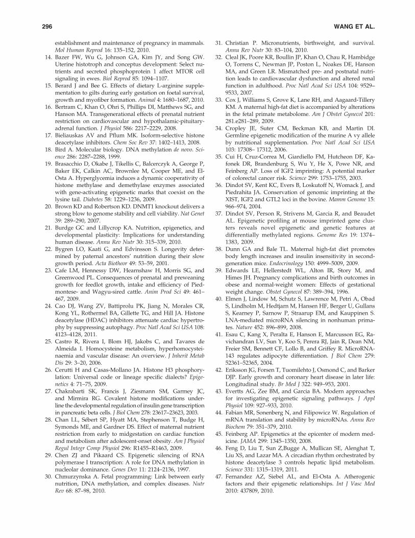
establishment and maintenance of pregnancy in mammals.Mol Human Reprod 16: 135–152, 2010.
14. Bazer FW, Wu G, Johnson GA, Kim JY, and Song GW.Uterine histotroph and conceptus development: Select nu-trients and secreted phosphoprotein 1 affect MTOR cellsignaling in ewes. Biol Reprod 85: 1094–1107.
15. Berard J and Bee G. Effects of dietary L-arginine supple-mentation to gilts during early gestation on foetal survival,growth and myofiber formation. Animal 4: 1680–1687, 2010.
16. Bertram C, Khan O, Ohri S, Phillips DI, Matthews SG, andHanson MA. Transgenerational effects of prenatal nutrientrestriction on cardiovascular and hypothalamic-pituitary-adrenal function. J Physiol 586: 2217–2229, 2008.
17. Bieliauskas AV and Pflum MK. Isoform-selective histonedeacetylase inhibitors. Chem Soc Rev 37: 1402–1413, 2008.
18. Bird A. Molecular biology. DNA methylation de novo. Sci-ence 286: 2287–2288, 1999.
19. Brasacchio D, Okabe J, Tikellis C, Balcerczyk A, George P,Baker EK, Calkin AC, Brownlee M, Cooper ME, and El-Osta A. Hyperglycemia induces a dynamic cooperativity ofhistone methylase and demethylase enzymes associatedwith gene-activating epigenetic marks that coexist on thelysine tail. Diabetes 58: 1229–1236, 2009.
20. Brown KD and Robertson KD. DNMT1 knockout delivers astrong blow to genome stability and cell viability. Nat Genet39: 289–290, 2007.
21. Burdge GC and Lillycrop KA. Nutrition, epigenetics, anddevelopmental plasticity: Implications for understandinghuman disease. Annu Rev Nutr 30: 315–339, 2010.
22. Bygren LO, Kaati G, and Edvinsson S. Longevity deter-mined by paternal ancestors’ nutrition during their slowgrowth period. Acta Biotheor 49: 53–59, 2001.
23. Cafe LM, Hennessy DW, Hearnshaw H, Morris SG, andGreenwood PL. Consequences of prenatal and preweaninggrowth for feedlot growth, intake and efficiency of Pied-montese- and Wagyu-sired cattle. Anim Prod Sci 49: 461–467, 2009.
24. Cao DJ, Wang ZV, Battiprolu PK, Jiang N, Morales CR,Kong YL, Rothermel BA, Gillette TG, and Hill JA. Histonedeacetylase (HDAC) inhibitors attenuate cardiac hypertro-phy by suppressing autophagy. Proc Natl Acad Sci USA 108:4123–4128, 2011.
25. Castro R, Rivera I, Blom HJ, Jakobs C, and Tavares deAlmeida I. Homocysteine metabolism, hyperhomocystei-naemia and vascular disease: An overview. J Inherit MetabDis 29: 3–20, 2006.
26. Cerutti H and Casas-Mollano JA. Histone H3 phosphory-lation: Universal code or lineage specific dialects? Epige-netics 4: 71–75, 2009.
27. Chakrabarti SK, Francis J, Ziesmann SM, Garmey JC,and Mirmira RG. Covalent histone modifications under-line the developmental regulation of insulin gene transcriptionin pancreatic beta cells. J Biol Chem 278: 23617–23623, 2003.
28. Chan LL, Sebert SP, Hyatt MA, Stephenson T, Budge H,Symonds ME, and Gardner DS. Effect of maternal nutrientrestriction from early to midgestation on cardiac functionand metabolism after adolescent-onset obesity. Am J PhysiolRegul Integr Comp Physiol 296: R1455–R1463, 2009.
29. Chen ZJ and Pikaard CS. Epigenetic silencing of RNApolymerase I transcription: A role for DNA methylation innucleolar dominance. Genes Dev 11: 2124–2136, 1997.
30. Chmurzynska A. Fetal programming: Link between earlynutrition, DNA methylation, and complex diseases. NutrRev 68: 87–98, 2010.
31. Christian P. Micronutrients, birthweight, and survival.Annu Rev Nutr 30: 83–104, 2010.
32. Cleal JK, Poore KR, Boullin JP, Khan O, Chau R, HambidgeO, Torrens C, Newman JP, Poston L, Noakes DE, HansonMA, and Green LR. Mismatched pre- and postnatal nutri-tion leads to cardiovascular dysfunction and altered renalfunction in adulthood. Proc Natl Acad Sci USA 104: 9529–9533, 2007.
33. Cox J, Williams S, Grove K, Lane RH, and Aagaard-TilleryKM. A maternal high-fat diet is accompanied by alterationsin the fetal primate metabolome. Am J Obstet Gynecol 201:281.e281–289, 2009.
34. Cropley JE, Suter CM, Beckman KB, and Martin DI.Germline epigenetic modification of the murine A vy alleleby nutritional supplementation. Proc Natl Acad Sci USA103: 17308– 17312, 2006.
35. Cui H, Cruz-Correa M, Giardiello FM, Hutcheon DF, Ka-fonek DR, Brandenburg S, Wu Y, He X, Powe NR, andFeinberg AP. Loss of IGF2 imprinting: A potential markerof colorectal cancer risk. Science 299: 1753–1755, 2003.
36. Dindot SV, Kent KC, Evers B, Loskutoff N, Womack J, andPiedrahita JA. Conservation of genomic imprinting at theXIST, IGF2 and GTL2 loci in the bovine. Mamm Genome 15:966–974, 2004.
37. Dindot SV, Person R, Strivens M, Garcia R, and BeaudetAL. Epigenetic profiling at mouse imprinted gene clus-ters reveals novel epigenetic and genetic features atdifferentially methylated regions. Genome Res 19: 1374–1383, 2009.
38. Dunn GA and Bale TL. Maternal high-fat diet promotesbody length increases and insulin insensitivity in second-generation mice. Endocrinology 150: 4999–5009, 2009.
39. Edwards LE, Hellerstedt WL, Alton IR, Story M, andHimes JH. Pregnancy complications and birth outcomes inobese and normal-weight women: Effects of gestationalweight change. Obstet Gynecol 87: 389–394, 1996.
40. Elmen J, Lindow M, Schutz S, Lawrence M, Petri A, ObadS, Lindholm M, Hedtjarn M, Hansen HF, Berger U, GullansS, Kearney P, Sarnow P, Straarup EM, and Kauppinen S.LNA-mediated microRNA silencing in nonhuman prima-tes. Nature 452: 896–899, 2008.
41. Esau C, Kang X, Peralta E, Hanson E, Marcusson EG, Ra-vichandran LV, Sun Y, Koo S, Perera RJ, Jain R, Dean NM,Freier SM, Bennett CF, Lollo B, and Griffey R. MicroRNA-143 regulates adipocyte differentiation. J Biol Chem 279:52361–52365, 2004.
42. Eriksson JG, Forsen T, Tuomilehto J, Osmond C, and BarkerDJP. Early growth and coronary heart disease in later life:Longitudinal study. Br Med J 322: 949–953, 2001.
43. Evertts AG, Zee BM, and Garcia BA. Modern approachesfor investigating epigenetic signaling pathways. J ApplPhysiol 109: 927–933, 2010.
44. Fabian MR, Sonenberg N, and Filipowicz W. Regulation ofmRNA translation and stability by microRNAs. Annu RevBiochem 79: 351–379, 2010.
45. Feinberg AP. Epigenetics at the epicenter of modern med-icine. JAMA 299: 1345–1350, 2008.
46. Feng D, Liu T, Sun Z,Bugge A, Mullican SE, Alenghat T,Liu XS, and Lazar MA. A circadian rhythm orchestrated byhistone deacetylase 3 controls hepatic lipid metabolism.Science 331: 1315–1319, 2011.
47. Fernandez AZ, Siebel AL, and El-Osta A. Atherogenicfactors and their epigenetic relationships. Int J Vasc Med2010: 437809, 2010.
296 WANG ET AL.
48. Flegal KM, Carroll MD, Ogden CL, and Curtin LR. Pre-valence and trends in obesity among US adults, 1999–2008.JAMA 303: 235–241, 2010.
49. Fleming AB, Kao CF, Hillyer C, Pikaart M, Osley MA. H2Bubiquitylation plays a role in nucleosome dynamics duringtranscription elongation. Mol Cell 31: 57–66, 2008.
50. Ford SP, Zhang L, Zhu M, Miller MM, Smith DT, Hess BW,Moss GE, Nathanielsz PW, and Nijland MJ. Maternalobesity accelerates fetal pancreatic beta- cell but not alpha-cell development in sheep: Prenatal consequences. Am JPhysiol Regul Integr Comp Physiol 297: R835–843, 2009.
51. Forsen T, Eriksson JG, Tuomilehto J, Osmond C, and Bar-ker, DJP. Growth in utero and during childhood amongwomen who develop coronary disease: Longitudinal study.Br Med J 319: 1403–1407, 1999.
52. Fowden AL, Ward JW, Wooding FP, Forhead AJ, andConstancia M. Programming placental nutrient transportcapacity. J Physiol 572: 5–15, 2006.
53. Fujiki K, Kano F, Shiota K, and Murata M. Expression ofthe peroxisome proliferator activated receptor gamma geneis repressed by DNA methylation in visceral adipose tissueof mouse models of diabetes. BMC Biol 7: 38, 2009.
54. Godmann M, Auger V, Ferraroni-Aguiar V, Di Sauro A,Sette C, Behr R, and Kimmins S. Dynamic regulation ofhistone H3 methylation at lysine 4 in mammalian sper-matogenesis. Biol Reprod 77: 754–764, 2007.
55. Godfrey KM, Lillycrop KA, Burdge GC, Gluckman PD, andHanson MA. Epigenetic mechanisms and the mismatchconcept of the developmental origins of health and disease.Pediatr Res 61: 5R–10R, 2007.
56. Golding MC, Williamson GL, Stroud TK, Westhusin ME,and Long CR. Examination of DNA methyltransferaseexpression in cloned embryo reveals an essential role forDnmt1 in bovine development. Mol Reprod Dev 78: 306–317, 2011.
57. Granger A, Abdullah I, Huebner F, Stout A, Wang T,Huebner T, Epstein JA, and Gruber PJ. Histone deacetylaseinhibition reduces myocardial ischemia-reperfusion injuryin mice. FASEB J 22: 35–49, 2008.
58. Greenwood PL, Hunt AS, Hermanson JW, and Bell AW.Effects of birth weight and postnatal nutrition on neonatalsheep: II. Skeletal muscle growth and development. J AnimSci 78: 50–61, 2000.
59. Grunstein M. Histone acetylation in chromatin structureand transcription. Nature 389: 349–352, 1997.
60. Haberland M, Montgomery RL, and Olsen EN. The manyroles of histone deacetylation in development and physi-ology: Implications for disease and therapy. Nat Rev Genet10: 32–42, 2009.
61. Hales CN and Barker DJ. Type 2 (non-insulin-dependent)diabetes mellitus: The thrifty phenotype hypothesis. Dia-betologia 35: 595–601, 1992.
62. Han HC, Austin KJ, Nathanielsz PW, Ford SP, Nijland MJ,and Hansen TR. Maternal nutrient restriction altersgene expression in the ovine fetal heart. J Physiol 558: 111–121, 2004.
63. Hansen RS, Gartler SM, Scott CR, Chen SH, and Laird CD.Methylation analysis of CGG sites in the CPG island of thehuman FMR1 gene. Hum Mol Genet 1: 571–578, 1992.
64. Hardie DG, Hawley SA, and Scott JW. AMP-activatedprotein kinase: Development of the energy sensor concept.J Physiol 574: 7–15, 2006.
65. Harrison M and Langley-Evans SC. Intergenerational pro-gramming of impaired nephrogenesis and hypertension in
rats following maternal protein restriction during preg-nancy. Br J Nutr 101: 1020–1030, 2009.
66. Haumaitre C, Lenoir O, and Scharfmann R. Histone dea-cetylase inhibitors modify pancreatic cell fate determina-tion and amplify endogenous progenitors. Mol Cell Biol 28:6373– 6383, 2008.
67. Heijmans BT, Tobi EW, Stein AD, Putter H, Blauw GJ,Susser ES, Slagboom PE, and Lumey LH. Persistent epi-genetic differences associated with prenatal exposure tofamine in humans. Proc Natl Acad Sci USA 105: 17046–17049, 2008.
68. Heneghan FM, Miller N, McAnena OJ, O’Brien T, andKerin MJ. Differential miRNA expression in omental adi-pose tissue and in the circulation of obese patients identifiesnovel metabolic biomarkers. J Clin Endocrinol Metab 96:E846–850, 2011.
69. Hill DJ and Duville B. Pancreatic development and adultdiabetes. Pediatr Res 48: 269–274, 2000.
70. Holliday R and Pugh JE. DNA modification mechanismsand gene activity during development. Science 187: 226–232, 1975.
71. Hosoki K, Ogata T, Kagami M, Tanaka T, and Saitoh S.Epimutation (hypomethylation) affecting the chromosome14q32.2 in a girl with upd914)mat-like phenotype. Eur JHum Genet 16: 1019–1023, 2008.
72. Hulsmans M, De Keyzer D, and Holvoet P. MicroRNAsregulating oxidative stress and inflammation in relation toobesity and atherosclerosis. FASEB J 25: 2515–2527, 2011.
73. Hurd PJ, Bannister AJ, Halls K, Dawson MA, VermeulenM, Olsen JV, Ismail H, Somers J, Mann M, Owen-Hughes T,Gout I, and Kouzarides T. Phosphorylation of histone H3Thr-45 is linked to apoptosis. J Biol Chem 284: 16575–16583,2009.
74. Ilnytskyy Y and Kovalchuk O. Non-targeted radiation ef-fects—An epigenetic connection. Mutat Res 714: 113-25,2011.
75. Jiang MH, Fei J, Lan MS, Lu ZP, Liu M, Fan WW, Gao X,and Lu DR. Hypermethylation of hepatic Gck promoter inageing rats contributes to diabetogenic potential. Diabeto-logia 51: 1525–1533, 2008.
76. Jimenez-Chillaron JC, Isganaitis E, Charalambons M,Gesta S, Pentinat-Pelegrin T, Faucette RR, Otis JP, Chow A,Diaz R, Ferguson-Smith A, and Patti ME. Intergenera-tional transmission of glucose intolerance and obesityby in utero undernutrition in mice. Diabetes 58: 460–468,2009.
77. Jirtle RL and Skinner MK. Environmental epigenomics anddisease susceptibility. Nat Rev Genet 8: 253–262, 2007.
78. Jobgen WS, Fried SK, Fu WJ, Meininger CJ, and Wu G.Regulatory role for the arginine-nitric oxide pathway inmetabolism of energy substrates. J Nutr Biochem 17: 571–588, 2006.
79. Kaati G, Bygren LO, Pembrey M, and Sjostrom M. Trans-generational response to nutrition, early life circumstancesand longevity. Eur J Hum Genet 15: 784–790, 2007.
80. Kajantie E, Osmond C, Barker DJP, Forsen T, Phillips DIW,and Eriksson JG. Size at birth as a predictor of mortality inadulthood: A follow-up of 350 000 person- years. Int JEpidemiol 34: 655–663, 2005.
81. Kaneda A, Wang CJ, Cheong R, Timp W, Onyango P,Wen B, Iacobuzio-Donahue CA, Ohlsson R, Andraos R,Pearson MA, Sharov AA, Longo DL, Ko MS, Levchenko A,and Feinberg AP Enhanced sensitivity to IGF-II signal-ing links loss of imprinting of IGF2 to increased cell pro-
NUTRITION AND EPIGENETICS 297
liferation and tumor risk. Proc Natl Acad Sci USA 104:20926–20931, 2007.
82. Kawasaki H, Taira K, and Morris KV. siRNA inducedtranscriptional gene silencing in mammalian cells. CellCycle 4: 442–448, 2005.
83. Khan IY, Dekou V, Douglas G, Jensen R, Hanson MA,Poston L, and Taylor PD. A high-fat diet during rat preg-nancy or suckling induces cardiovascular dysfunction inadult offspring. Am J Physiol Regul Integr Comp Physiol 288:R127–133, 2005.
84. Khan KS, Wojdyla D, Say L, Gulmezoglu AM, and VanLook PF. WHO analysis of causes of maternal death: Asystematic review. Lancet 367: 1066–1074, 2006.
85. Kiel DW, Dodson EA, Artal R, Boehmer TK, and Leet TL.Gestational weight gain and pregnancy outcomes in obesewomen. Obstet Gynecol 110: 752–758, 2007.
86. Kirk H, Cefalu WT, Ribnicky D, Liu Z, and Eilertsen KJ.Botanicals as epigenetic modulators for mechanisms con-tributing to development of metabolic syndrome. Metabo-lism 57: S16–S23, 2008.
87. Krishna SM, Dear AE, Norman PE, and Golledge J. Geneticand epigenetic mechanisms and their possible role in ab-dominal aortic aneurysm. Atherosclerosis 212: 16–29, 2010.
88. Krishnamurthy J, Ramsey MR, Ligon KL, Torrice C, Koh A,Bonner-Weir S, and Sharpless NE. p16INK4a induces anage-dependent decline in islet regeneration potential. Nat-ure 443: 453–457, 2006.
89. Krutzfeldt M, Rajewsky N, Braich R, Rajeev KG, Tuschl T,Manoharan M, and Stoffel M. Silencing of miRNAs in vivowith ‘antagomirs’’. Nature 438: 685–689, 2005.
90. Lassala A, Bazer FW, Cudd TA, Li P, Li XL, Satterfield MC,Spencer TE, and Wu G. Intravenous administration of L-citrulline to pregnant ewes is more effective than L-argininefor increasing arginine availability in the fetus. J Nutr 139:660–665, 2009.
91. Lassala A, Bazer FW, Cudd TA, Datta S, Keisler DH, Sat-terfield MC, Spencer TE, and Wu G. Parenteral adminis-tration of L-arginine prevents fetal growth restriction inundernourished ewes. J Nutr 140: 1242–1248, 2010.
92. Lassala A, Bazer FW, Cudd TA, Datta S, Keisler DH,Satterfield MC, Spencer TE, and Wu G. Parenteral admin-istration of L-arginine enhances fetal survival and growthin sheep carrying multiple pregnancies. J Nutr 141: 849–855,2011.
93. Lee RC, Feinbaum RL, and Ambross V. The C. elegan het-erochronic gene lin-4 encodes small RNAs with antisensecomplementary to lin-14. Cell 75: 843–854, 1993.
94. Leon DA, Lithell HO, Vagero D, Koupilova I, Mohsen R,Berglund L, Lithell UB, and McKeigue PM. Reduced fetalgrowth rate and increased risk of death from ischaemicheart disease: Cohort study of 15000 Swedish men andwomen born 1915–29. Br Med J 317: 241–245, 1998.
95. Lewis BP, Shih IH, Jones-Rhoades MW, Bartel DP, andBurge CB. Prediction of mammalian microRNA targets.Cell 115: 787–798, 2003.
96. Li H, Meininger CJ, Hawker JR, Kelly KA, Morris SM, andWu G. Activities of arginase I and II are limiting for en-dothelial cell proliferation. Am J Physiol Regul Integr CompPhysiol 282: R64–R69, 2002.
97. Li XL, Bazer FW, Johnson GA, Burghardt RC, Erikson DW,Frank JW, Spencer TE, Shinzato I, and Wu G. Dietarysupplementation with 0.8% L-arginine between days 0 and25 of gestation reduces litter size in gilts. J Nutr 140: 1111–1116, 2010.
98. Li Y, Reddy MA, Miao F, Shanmugam N, Yee JK, HawkinsD, Ren B, and Natarajan R. Role of the histone H3 lysine4 methyltransferase, SET7/9, in the regulation of NF-kB-dependent inflammatory genes. J Biol Chem 283: 26771–26781, 2008.
99. Ling C, Poulsen P, Simonsson S, Ronn T, Holmkvist J,Almgren P, Hagert P, Nilsson E, Mabey AG, Nilsson P,Vaag A, and Groop L. Genetic and epigenetic factors areassociated with expression of respiratory chain compo-nent NDUFB6 in human skeletal muscle. J Clin Invest 117:3427–3435, 2007.
100. Ling C, Del Guerra S, Lupi R, Ronn T, Granhall C, LuthmanH, Masiello P, Marchetti P, Groop L, and Del Prato S.Epigenetic regulation of PPARGC1A in human type 2 dia-betic islets and effect on insulin secretion. Diabetologia 51:615–622, 2008.
101. Ling C and Groop L. Epigenetics: A molecular link betweenenvironmental factors and type 2 diabetes. Diabetes 58:2718–2725, 2009.
102. Lumey LH. Reproductive outcome in women prenatallyexposed to undernutrition: a review of findings from theDutch famine birth cohort. Proc Nutr Soc 57: 129–135, 1998.
103. Lynn FC. Meta-regulation: MicroRNA regulation of glu-cose and lipid metabolism. Trends Endocrinol Metab 20: 452–459, 2009.
104. Marsal K. Intrauterine growth restriction. Curr Opin ObstetGynecol 14: 127–135, 2002.
105. Mateo RD, Wu G, Bazer FW, Park JC, Shinzato I, and KimSW. Dietary L- arginine supplementation enhances the re-productive performance of gilts. J Nutr 137: 652–656, 2007.
106. Matouk CC and Marsden PA. Epigenetic regulationof vascular endothelial gene expression. Circ Res 102: 873–887, 2008.
107. Martin JF, Johnston CS, Han CT, and Benyshek DC. Nu-tritional origins of insulin resistance: A rat model for dia-betes-prone human populations. J Nutr 130: 741–744, 2000.
108. Maulik N and Maulik G. (Eds). Nutrition, Epigenetic Me-chanisms, and Human Disease. New York: CRC Press, 2011,pp. 426.
109. McClelland M and Nelson M. The effect of site-specificDNA methylation on restriction endonucleases and DNAmodification methyltransferases. Gene 74: 291–304, 1988.
110. McCurdy CE, Bishop JM, Williams SM, Grayson BE, SmithMS, Friedman JE, and Grove KL. Maternal high-fat diettriggers lipotoxicity in the fetal livers of nonhuman pri-mates. J Clin Invest 119: 323–335, 2009.
111. McGee SL, van Denderen BJ, Howlett KF, Mollica J,Schertzer JD, Kemp BE, and Hargreaves M. AMP-activatedprotein kinase regulates GLUT4 transcription by phos-phorylating histone deacetylase 5. Diabetes 57: 860–867,2008.
112. McGee SL and Hargreaves M. Histone modifications andexercise adaptations. J Appl Physiol 110: 258–263, 2011.
113. McGregor RA and Choi MS. MicroRNAs in the regulationof adipogenesis and obesity. Curr Mol Med 11: 304–316,2011.
114. McKnight JR, Satterfield MC, Jobgen WS, Smith SB,Spencer TE, Meininger CJ, McNeal, CJ, and Wu G. Bene-ficial effects of L-arginine on reducing obesity: Potentialmechanisms and important implications for human health.Amino Acids 39: 349–357, 2010.
115. McKnight JR, Satterfield MC, Li XL, Gao HJ, Wang JJ, LiDF, and Wu G. Obesity in pregnancy: Problems and po-tential solutions. Front Biosci E3: 442–452, 2011.
298 WANG ET AL.
116. Melzner I, Scott V, Dorsch K, Fischer P, Wabitsch M, Bru-derlein S, Hasel C, and Moller P. Leptin gene expression inhuman preadipocytes is switched on by maturation-in-duced demethylation of distinct CpGs in its proximalpromoter. J Biol Chem 277: 45420–45427, 2002.
117. Miao F, Gonzalo IG, Lanting L, and Natarajan R. In vivochromatin remodeling events leading to inflammatory genetranscription under diabetic conditions. J Biol Chem 279:18091–18097, 2004.
118. Miao F, Smith DD, Zhang L, Min A, Feng W, and NatarajanR. Lymphocytes from patients with type 1 diabetes displaya distinct profile of chromatin histone H3 lysine 9 di-methylation: An epigenetic study in diabetes. Diabetes 57:3189–3198, 2008.
119. Milagro FI, Campion J, Garcia-Diaz DF, Goyenechea E,Paternain L, and Martinez JA. High fat diet-induced obe-sity modifies the methylation pattern of leptin promoter inrats. J Physiol Biochem 65: 1–9, 2009.
120. Mouillet JF, Chu T, and Sadovsky Y. Expression patternsof placental microRNAs. Birth Defects Res A 91: 737–743,2011.
121. Muhlhausler BS, Duffield JA, and McMillen IC. Increasedmaternal nutrition increases leptin expression in perirenaland subcutaneous adipose tissue in the postnatal lamb.Endocrinology 148: 6157–6163, 2007.
122. Nakanishi N, Nakagawa Y, Tokushige N, Aoki N, Matsu-zaka T, Ishii K, Yahagi N, Kobayashi K, Yatoh S, TakahashiA, Suzuki H, Urayama O, Yamada N, and Shimano H. Theupregulation of microRNA-335 is associated with lipid me-tabolism in liver and white adipose tissue of geneticallyobese mice. Biochem Biophys Res Commun 385: 492–496, 2009.
123. Ng SF, Lin RC, Laybutt DR, Barres R, Owens JA, and MorrisMJ. Chronic high-fat diet in fathers programs b-cell dys-function in female rat offspring. Nature 467: 963–966, 2010.
124. Nijland MJ, Mitsuya K, Li C, Ford SP, McDonald TJ, Na-thanielsz PW, and Cox LA. Epigenetic modification of fetalbaboon hepatic phosphoenolpyruvate carboxykinase fol-lowing exposure to moderately reduced nutrient avail-ability. J Physiol 588: 1349–1359, 2010.
125. Nolan CJ, Damm P, and Prentki M. Type 2 diabetes acrossgenerations: From pathophysiology to prevention andmanagement. Lancet 378: 169–181, 2011.
126. Ogden CL, Yanovski SZ, Carroll MD, and Flegal KM. Theepidemiology of obesity. Gastroenterology 132: 2087–2102,2007.
127. Oommen AM, Griffin JB, Sarath G, and Zempleni J.Roles for nutrients in epigenetic events. J Nutr Biochem 16:74–77, 2005.
128. Ovesen P, Rasmussen S, and Kesmodel U. Effect of pre-pregnancy maternal overweight and obesity on pregnancyoutcome. Obstet Gynecol 118: 305–312, 2011.
129. Parikh H, Nilsson E, Ling C, Poulsen P, Almgren P, NittbyH, Eriksson KF, Vaag A, and Croop LC. Molecular corre-lates for maximal oxygen uptake and type 1 fibers. Am JPhysiol Endocrinol Metab 294: E1152–E1159, 2008.
130. Park JH, Stoffers DA, Nicholls RD, and Simmons RA. De-velopment of type 2 diabetes following intrauterine growthretardation in rats is associated with progressive epigeneticsilencing of Pdx1. J Clin Invest 118: 2316–2324, 2008.
131. Peterson CL and Laniel MA. Histones and histone modi-fications. Curr Biol 14: R546–R551, 2004.
132. Philbin VJ and Levy O. Developmental biology of the in-nate immune response: Implications for neonatal and infantvaccine development. Pediatr Res 65: 98R–105R, 2009.
133. Phillips DIW. Programming of the stress response: A fun-damental mechanism underlying the long-term effects ofthe fetal environment. J Intern Med 261: 453–460, 2007.
134. Philp LK, Muhlhausler BS, Janovska A, Wittert GA, Duf-field JA, and McMillen IC. Maternal overnutrition sup-presses the phosphorylation of 5’-AMP-activated proteinkinase in liver, but not skeletal muscle, in the fetal andneonatal sheep. Am J Physiol Regul Integr Comp Physiol 295:R1982–1990, 2008.
135. Pickart CM. Mechanisms underlying ubiquitination. AnnuRev Biochem 70: 503–533, 2001.
136. Pokholok DK, Harbison CT, Levine S, Cole M, HannettNM, Lee TI, Bell GW, Walker K, Rolfe PA, HerbolsheimerE, Zeitlinger J, Lewitter F, Gifford DK, and Young RA.Genome-wide map of nucleosome acetylation and meth-ylation in yeast. Cell 122: 517–527, 2005.
137. Poston L, Harthoorn LF, and Van Der Beek EM, Con-tributors to the ILSI Europe Workshop. Obesity in preg-nancy: Implications for the mother and lifelong health ofthe child. A consensus statement. Pediatr Res 69: 175–180,2011.
138. Poy MN, Eliasson L, Krutzfeldt J, Kuwajima S, Ma X,Macdonald PE, Pfeffer S, Tuschl T, Rajewsky N, RorsmanP, and Stoffel M. A pancreatic islet-specific microRNAregulates insulin secretion. Nature 432: 226–230, 2004.
139. Qipshidze N, Metreveli N, Lominadze D, and Tyagi SC.Folic acid improves acetylcholine- induced vasoconstrictionof coronary vessels isolated from hyperhomocysteinemicmice: An implication to coronary vasospasm. J Cell Physiol226: 2712–2720, 2011.
140. Raychaudhuri N, Raychaudhuri S, Thamotharan M, andDevaskar SU. Histone code modifications repress glucosetransporter 4 expression in the intrauterine growth-restricted offspring. J Biol Chem 283: 13611–13626, 2008.
141. Rayner KJ, Sheedy FJ, Esau CC, Hussain FN, Temel RE,Parathath S, van Gils JM, Rayner AJ, Chang AN, Suarez Y,Fernandez-Hernando C, Fisher EA, and Moore KJ. Antag-onism of miR-33 in mice promotes reverse cholesteroltransport and regression of atherosclerosis. J Clin Invest 121:2921–2931, 2011.
142. Rayner KJ, Esau CC, Hussain FN, McDaniel AL, MarshallSM, van Gils JM, Ray TD, Sheedy FJ, Goedeke L, Liu X,Khatsenko OG, Kaimal V, Lees CJ, Fernandez-Hernando C,Fisher EA, Temel RE, Moore KJ. Inhibition of miR-33a/b innonhuman primates raises plasma HDL and lowers VLDLtriglycerides. Nature 478: 404–407, 2011.
143. Reynolds LP, Caton JS, Redmer DA, Grazul-Bilska AT,Vonnahme KA, Borowicz PB, Luther JS, Wallace JM, Wu G,and Spencer TE. Evidence for altered placental blood flowand vascularity in compromised pregnancies. J Physiol 572:51–58, 2006.
144. Ronn T, Poulsen P, Hansson O, Holmkvist J, Almgren P,Nilsson P, Tuomi T, Isomaa B, Groop L, Vaag A, and LingC. Age influences DNA methylation and gene expression ofCOX7A1 in human skeletal muscle. Diabetologia 51: 1159–1168, 2008.
145. Rooney K and Ozanne SE. Maternal over-nutrition andoffspring obesity predisposition: Targets for preventativeinterventions. Int J Obes 35: 883–890, 2011.
146. Rush D. Nutritional and maternal mortality in the devel-oping world. Am J Clin Nutr 72: 212S–240S, 2000.
147. Sakabe K, Wang ZH, and Hart GW. b-N-acetylglucosamine(O-GlcNAc) is part of the histone code. Proc Natl Acad SciUSA 107: 19915–19920, 2010.
NUTRITION AND EPIGENETICS 299

148. Samuelsson AM, Matthews PA, Argenton M, Christie MR,McConnell JM, Jansen EH, Piersma AH, Ozanne SE, TwinnDF, Remacle C, Rowlerson A, Poston L, and Taylor PD.Diet-induced obesity in female mice leads to offspring hy-perphagia, adiposity, hypertension, and insulin resistance:A novel murine model of developmental programming.Hypertension 51: 383–392, 2008.
149. Sathishkumar K, Elkins R, Yallampalli U, and YallampalliC. Protein restriction during pregnancy induces hyperten-sion and impairs endothelium-dependent vascular functionin adult female offspring. J Vasc Res 46: 229–239, 2009.
150. Satterfield MC, McKnight JR, Li XL, and Wu G. Nutrition,epigenetics, and vascular function. In: Nutrition, EpigeneticMechanisms, and Human Disease, edited by Maulik N andMaulik G. New York: CRC Press, 2011, pp. 125–139.
151. Satterfield MC, Bazer FW, Spencer TE, and Wu G.Sildenafil citrate treatment enhances amino acid avail-ability in the conceptus and fetal growth in an ovinemodel of intrauterine growth restriction. J Nutr 140: 251–258, 2010.
152. Satterfield MC and Wu G. Growth and development ofbrown adipose tissue: Significance and nutritional regula-tion. Front Biosci 16: 1589–1608, 2011.
153. Schlabritz-Loutsevitch N, Farley D, Tejero ME, ComuzzieAG, Higgins PB, Cox L, Werner SL, Jenkins SL, Li C, Choj J,Dick EJ, Hubbard GB, Frost P, Dudley DD, Wu G, andNathanielsz PW. Feto-placental adaptations to maternalobesity in the baboon. Placenta 30: 752–760, 2009.
154. Schwer B and Verdin E. Conserved metabolic regulatoryfunctions of sirtuins. Cell Metab 7: 104–112, 2008.
155. Sharkey D, Gardner DS, Symonds ME, and Budge H.Maternal nutrient restriction during early fetal kidney de-velopment attenuates the renal innate inflammatory re-sponse in obese young adult offspring. Am J Physiol RenalPhysiol 297: F1199–1207, 2009.
156. Simmons R. Epigenetics and maternal nutrition: Nature v.nurture. Proc Nutr Soc 70: 73– 81, 2011.
157. Sinclair KD, Allegrucci C, Singh R, Gardner DS, SebastianS, Bispham J, Thurston A, Huntley JF, Rees WD, MaloneyCA, Lea RG, Craigon J, McEvoy TG, and Young LE. DNAmethylation, insulin resistance, and blood pressure in off-spring determined by maternal periconceptional B vitaminand methionine status. Proc Natl Acad Sci USA 104: 19351–19356, 2007.
158. Schroen B and Heymans S. Small but smart—MicroRNAsin the center of inflammatory processes during cardiovas-cular diseases, the metabolic syndrome, and aging. Cardi-ovasc Res doi.10.1093/cvr/cvr268, 2011.
159. Smith AD, Kim YI, Refsum H. Is folic acid good for ev-eryone? Am J Clin Nutr 87: 517–533, 2008.
160. Sullivan EL, Smith MS, and Grove KL. Perinatal exposureto high-fat diet programs energy balance, metabolism andbehavior in adulthood. Neuroendocrinol 93: 1–8, 2011.
161. Sun LD, Zhao HB, Xu ZB, Liu QL, Liang W, Wang LT, CaiX, Zhang L, Hu L, Wang G, and Zha X. Phosphatidylino-sitol 3- kinase/protein kinase B pathway stabilizes DNAmethyltransferase I protein and maintains DNA methyla-tion. Cell Signal 19: 2255–2263, 2007.
162. Symonds ME, Sebert SP, Hyatt MA, and Budge H. Nutri-tional programming of the metabolic syndrome. Nat RevEndocrinol 5: 604–610, 2009.
163. Szic KSV, Ndlovu MN, Haegeman G, and Vanden BergheW. Nature or nurture: Let food be your epigenetic medicine
in chronic inflammatory disorders. Biochem Pharmacol 80:1816–1832, 2010.
164. Szyf M. Epigenetics, DNA methylation, and chromatin mod-ifying drugs. Annu Rev Pharmacol Toxicol 49: 243–263, 2009.
165. Tateishi K, Okada Y, Kallin EM, and Zhang Y. Role ofJhdm2a in regulating metabolic gene expression and obe-sity resistance. Nature 458: 757–761, 2009.
166. Taylor PD, McConnell J, Khan IY, Holemans K, LawrenceKM, Asare-Anane H, Persaud SJ, Jones PM, Petrie L,Hanson MA, and Poston L. Impaired glucose homeostasisand mitochondrial abnormalities in offspring of rats fed afat-rich diet in pregnancy. Am J Physiol Regul Integr CompPhysiol 288: R134–R139, 2005.
167. Temple IK and Shield JP. Transient neonatal diabetes, adisorder of imprinting. J Med Genet 39: 872–875, 2002.
168. Thamotharan M, Garg M, Oak S, Rogers LM, Pan A, SangiorgiF, Lee PN, and Devaskar SU. Transgenerational inheritance ofthe insulin resistant phenotypes in embryo- transferred in-trauterine growth-restricted adult female rat offspring. Am JPhysiol Endocrinol Metab 292: E1270–E1279, 2007.
169. Thambirajah AA, Li A, Ishibashi T, and Ausio J. Newdevelopments in post- translational modifications and func-tions of histone H2A variants. Biochem Cell Biol 87: 7–17, 2009.
170. Timp W, Levchenko A, and Feinberg AP. A new linkbetween epigenetic progenitor lesions in cancer and the dy-namics of signal transduction. Cell Cycle 8: 383–390, 2009.
171. Tobi EW, Lumey LH, Talens RP, Kremer D, Putter H, SteinAD, Slagboom PE, and Heijmans BT. DNA methylation dif-ferences after exposure to prenatal famine are common andtiming- and sex-specific. Hum Mol Genet 18: 4046–4053, 2009.
172. Todeschini AL, Teysset L, Delmarre V, and Ronsseray S. Theepigenetic trans-silencing effect in Drosophila involves ma-ternally-transmitted small RNAs whose production dependson the piRNA pathway and HP1. PLoS One 5: e11032, 2010.
173. Tong JF, Yan X, Zhu MJ, Ford SP, Nathanielsz PW, and DuM. Maternal obesity downregulates myogenesis and beta-catenin signaling in fetal skeletal muscle. Am J Physiol En-docrinol Metab 296: E917–E924, 2009.
174. Turner BM. Histone acetylation as an epigenetic determi-nant of long-term transcriptional competence. Cell Mol LifeSci 54: 21–31, 1998.
175. Turunen MP, Aavik E, and Yla-Herttuala S. Epigenetics andatherosclerosis. Biochim Biophys Acta 1790: 886–891, 2009.
176. Unterberger A, Szyf M, Nathanielsz PW, and Cox LA.Organ and gestational age effects of maternal nutrient re-striction on global methylation in fetal baboons. J MedPrimatol 38: 219–227, 2009.
177. USDA. Dietary Guidance. 2010. DRI Tables. http://fnic.nal.usda.gov. Accessed on July 26, 2011.
178. Vaxillaire M and Froguel P. Monogenic diabetes in theyoung, pharmacogenetics and relevance to multifactorialforms of type 2 diabetes. Endocr Rev 29: 254–264, 2008.
179. Villeneuve LM, Reddy MA, Lanting LL, Wang M, Meng L,and Natarajan R. Epigenetic histine H3 lysine 9 methyla-tion in metabolic memory and inflammatory phenotype ofvascular smooth muscle cells in diabetes. Proc Natl Acad SciUSA 105: 9047–9052, 2008.
180. Vonnahme KA, Hess BW, Hansen TR, McCormick RJ, RuleDC, Moss GE, Murdoch WJ, Nijland MJ, Skinner DC, Na-thanielsz PW, and Ford SP. Maternal undernutrition fromearly- to mid-gestation leads to growth retardation, cardiacventricular hypertrophy, and increased liver weight in thefetal sheep. Biol Reprod 69: 133–140, 2003.
300 WANG ET AL.
181. Vucetic Z, Kimmel J, Totoki K, Hollenbeck E, and ReyesTM. Maternal high-fat diet alters methylation and geneexpression of dopamine and opioid-related genes. En-docrinology 151: 4756–4764, 2010.
182. Waddington CH. Organizers and Genes. Cambridge Uni-versity Press. 1940.
183. Waechter DE and Baserga R. Effect of methylation on ex-pression of microinjected genes. Proc Natl Acad Sci USA 79:1106–1110, 1982.
184. Wallace DC and Fan WW. Energetics, epigenetics, mito-chondrial genetics. Mitochondrion 10: 12–31, 2010.
185. Wallace JM, Milne JS, and Aitken RP. The effect of over-nourishing singleton-bearing adult ewes on nutrient parti-tioning to the gravid uterus. Br J Nutr 94: 533–539, 2005.
186. Wallace JM, Luther JS, Milne JS, Aitken RP, Redmer DA, Rey-nolds LP, and Hay WW. Nutritional modulation of adolescentpregnancy outcome. A review. Placenta 27: S61–S68, 2006.
187. Warner MJ and Ozanne SE. Mechanisms involved in thedevelopmental programming of adulthood disease. Bio-chem J 427: 333–347, 2010.
188. Weake VM and Workman JL. Histone ubiquitination:Triggering gene activity. Mol Cell 29: 653–663, 2008.
189. Weems J and Olson AL. Class II histone deacetylases limitGLUT4 gene expression during adipocyte differentiation. JBiol Chem 286: 460–468, 2011.
190. WHO. United Nations Food and Agriculture Organization.http://www.fao.org/publications/sofi/en.2010. Accessedon July 20, 2011.
191. Williams KT and Schalinske KL. Homocysteine metabolismand its relation to health and disease. Biofactors 36: 19–24,2010.
192. Williams PJ, Kurlak LO, Perkins AC, Budge H, StephensonT, Keisler D, Symonds ME, and Gardner DS. Hypertensionand impaired renal function accompany juvenile obesity:The effect of prenatal diet. Kidney Int 72: 279–289, 2007.
193. Wilson VL and Jones PA. DNA methylation decreases in ag-ing but not in immortal cells. Science 220: 1055–1057, 1983.
194. Wilson MJ, Shivapurkar N, and Poirier LA. Hypomethy-lation of hepatic nuclear DNA in rats fed with a carcino-genic methyl-deficient diet. Biochem J 218: 987–990, 1984.
195. Winzerling JJ and Law JH. Comparative nutrition of ironand copper. Annu Rev Nutr 17: 501–526, 1997.
196. Wu G. Amino acids: Metabolism, functions, and nutrition.Amino Acids 37: 1–17, 2009.
197. Wu G. Functional amino acids in growth, reproduction andhealth. Adv Nutr 1: 31–37, 2010.
198. Wu G and Meininger CJ. Nitric oxide and vascular insulinresistance. BioFactors 35: 21–27, 2009.
199. Wu G, Bazer FW, Cudd TA, Meininger CJ, and Spencer TE.Maternal nutrition and fetal development. J Nutr 134: 2169–2172, 2004.
200. Wu G, Bazer FW, Wallace JM, and Spencer TE. Board-invited review: Intrauterine growth retardation: iImplica-tions for the animal sciences. J Anim Sci 84: 2316–2337, 2006.
201. Wu G, Bazer FW, Datta S, Johnson GA, Li P, Satterfield,MC, and Spencer TE. Proline metabolism in the conceptus:Implications for fetal growth and development. AminoAcids 35: 691–702, 2008.
202. Wu G, Bazer FW, Davis TA, Kim SW, Li P, Rhoads JM,Satterfield MC, Smith SB, Spencer TE, and Yin YL. Argininemetabolism and nutrition in growth, health and disease.Amino Acids 37: 153–168, 2009.
203. Wu G, Bazer FW, Burghardt RC, Johnson GA, Kim SW, Li XL,Satterfield MC, and Spencer TE. Impacts of amino acid nutri-
tion on pregnancy outcome in pigs: Mechanisms and impli-cations for swine production. J Anim Sci 88: E195–E204, 2010.
204. Wu G, Bazer FW, Burghardt RC, Johnson GA, Kim SW,Knabe DA, Li P, Li XL, McKnight JR, Satterfield MC, andSpencer TE. Proline and hydroxyproline metabolism: Im-plications for animal and human nutrition. Amino Acids 40:1053–1063, 2011.
205. Xiao XM and Li LP. L-arginine treatment for asymmetricfetal growth restriction. Int J Gynecol Obstet 88: 15–18, 2005.
206. Yajnik CS and Deshmukh US. Maternal nutrition, intra-uterine programming and consequential risks in the off-spring. Rev Endocr Metab Disord 9: 203–211, 2008.
207. Yamamoto Y, Verma UN, Prajapati S, Kwak YT, and GaynorRB. Histone H3 phosphorylation by IKK-a is critical for cy-tokine-induced gene expression. Nature 423: 655–659, 2003.
208. Yokomori N, Tawata M, and Onaya T. DNA demethylationmodulates mouse leptin promoter activity during the dif-ferentiation of 3T3-L1 cells. Diabetologia 45: 140–148, 2002.
209. Youngson NA and Whitelaw E. Transgenerational epige-netic effects. Annu Rev Genomics Hum Genet 9: 233-257, 2008.
210. Zambrano E, Martnez-Samayoa PM, Bautista CJ, Deas M,Guillen L, Rodriguez- Gonzales GL, Guzman C, Larrea F,and Nathanielsz PW. Sex differences in transgenerationalalterations of growth and metabolism in progeny (F2) offemale offspring (F1) of rats fed a low protein diet duringpregnancy and lactation. J Physiol 566: 225–236, 2005.
211. Zeng XF, Wang FL, Fan X, Yang WJ, Zhou B, Li PF, Yin YL,Wu G, and Wang JJ. Dietary arginine supplementationduring early pregnancy enhances embryonic survival inrats. J Nutr 138: 1421–1425, 2008.
212. Zhu MJ, Du M, Nijland MJ, Nathanielsz PW, Hess BW,Moss GE, and Ford SP. Down- regulation of growth sig-naling pathways linked to a reduced cotyledonary vascu-larity in placentomes of over-nourished, obese pregnantewes. Placenta 30: 405-410, 2009.
213. Zimmermann MB. The influence of iron status on iodineutilization and thyroid function. Annu Rev Nutr 26: 367–389, 2006.
Address correspondence to:Dr. Guoyao Wu
Room 212, Kleberg Center2471 TAMU
Texas A&M UniversityCollege Station, TX 77843-2471
E-mail: [email protected]
Date of first submission to ARS Central, October 31, 2011; dateof acceptance, November 1, 2011.
Abbreviations Used
DNMT¼DNA methyltransferaseEAA¼ essential amino acids
H3K4¼histone 3 lysine residue 4HAT¼histone acetyltransferase
HDAC¼histone deacetyltransferaseHMT¼histone methyltransferaseIUGR¼ intrauterine growth restrictionLCFA¼ long-chain fatty acids
miR¼microRNANEAA¼nonessential amino acids
NUTRITION AND EPIGENETICS 301
Related Documents