- 1 - Introduction 1 Introduction 1.1 Pancreas anatomy, histology and function The pancreas a part of the digestive system is a long, flat gland (Figure 1), that is located deep in the abdomen sandwiched between the stomach and the spine. 1 It lies partially behind the stomach. The other part is nestled in the curve of the small intestine called the duodenum. Pancreas is described as having a head, body and tail. Fig. 1: Morphological structure and location of pancreas in the body Histologically, the pancreatic parenchyma is divided into two components: (i) the exocrine portion, which is composed of ducts and acini and (ii) the endocrine component, (Figure 2) which is composed of hormone-secreting cells arranged in islets (islets of Langerhans). The pancreatic exocrine cells, the larger part (98-99 %), is composed of tubulo-acinar glands that drain, via a highly branched duct system, into the main pancreatic duct. This duct runs the whole length of the gland and opens into the duodenum through the ampulla of Vater. The acinar cells produce digestive enzymes and some duct lining cells yield a fluid rich in sodium and bicarbonate. 2 The enzymes are responsible for the breakdown of proteins mainly (trypsin, chymotrypsin, elastase and others), fats (lipase) and

Welcome message from author
This document is posted to help you gain knowledge. Please leave a comment to let me know what you think about it! Share it to your friends and learn new things together.
Transcript
- 1 - Introduction
1 Introduction
1.1 Pancreas anatomy, histology and function
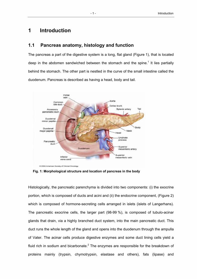
The pancreas a part of the digestive system is a long, flat gland (Figure 1), that is located
deep in the abdomen sandwiched between the stomach and the spine.1 It lies partially
behind the stomach. The other part is nestled in the curve of the small intestine called the
duodenum. Pancreas is described as having a head, body and tail.
Fig. 1: Morphological structure and location of pancreas in the body
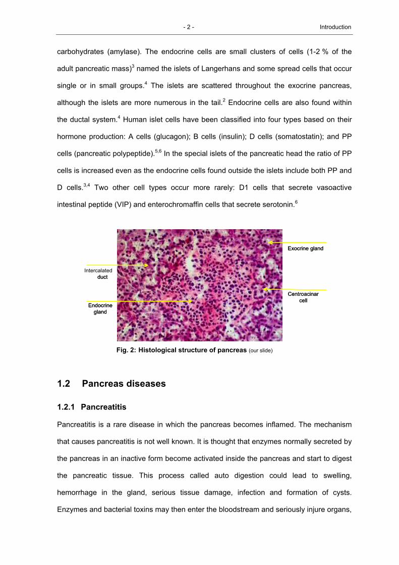
Histologically, the pancreatic parenchyma is divided into two components: (i) the exocrine
portion, which is composed of ducts and acini and (ii) the endocrine component, (Figure 2)
which is composed of hormone-secreting cells arranged in islets (islets of Langerhans).
The pancreatic exocrine cells, the larger part (98-99 %), is composed of tubulo-acinar
glands that drain, via a highly branched duct system, into the main pancreatic duct. This
duct runs the whole length of the gland and opens into the duodenum through the ampulla
of Vater. The acinar cells produce digestive enzymes and some duct lining cells yield a
fluid rich in sodium and bicarbonate.2 The enzymes are responsible for the breakdown of
proteins mainly (trypsin, chymotrypsin, elastase and others), fats (lipase) and
- 2 - Introduction
carbohydrates (amylase). The endocrine cells are small clusters of cells (1-2 % of the
adult pancreatic mass)3 named the islets of Langerhans and some spread cells that occur
single or in small groups.4 The islets are scattered throughout the exocrine pancreas,
although the islets are more numerous in the tail.2 Endocrine cells are also found within
the ductal system.4 Human islet cells have been classified into four types based on their
hormone production: A cells (glucagon); B cells (insulin); D cells (somatostatin); and PP
cells (pancreatic polypeptide).5,6 In the special islets of the pancreatic head the ratio of PP
cells is increased even as the endocrine cells found outside the islets include both PP and
D cells.3,4 Two other cell types occur more rarely: D1 cells that secrete vasoactive
intestinal peptide (VIP) and enterochromaffin cells that secrete serotonin.6
Fig. 2: Histological structure of pancreas (our slide)
1.2 Pancreas diseases
1.2.1 Pancreatitis
Pancreatitis is a rare disease in which the pancreas becomes inflamed. The mechanism
that causes pancreatitis is not well known. It is thought that enzymes normally secreted by
the pancreas in an inactive form become activated inside the pancreas and start to digest
the pancreatic tissue. This process called auto digestion could lead to swelling,
hemorrhage in the gland, serious tissue damage, infection and formation of cysts.
Enzymes and bacterial toxins may then enter the bloodstream and seriously injure organs,
duct
Endocrine gland
Exocrine gland
Centroacinar cell
Intercalated duct
Endocrine gland
Exocrine gland
Centroacinar cell
- 3 - Introduction
such as the heart, lungs and kidney. Most commonly caused by alcohol abuse or
gallstones, the disease affects men more often than women. There are two forms of
pancreatitis: acute and chronic. The acute form occurs suddenly and may result in life-
threatening complications; nevertheless the majority of patients (80 %) recover
completely. Chronic pancreatitis is a long-term inflammation of the pancreas, that is
primarily marked by severe pain and loss of pancreatic function.7
1.2.2 Pancreatic cancer
Like all organs, the pancreas is made up of individual living cells. These cells are joined to
form the tissues of the pancreas. There are several different types of cells in the pancreas
to accomplish its functions as mentioned above. The cells divide rapidly while the
pancreas is growing in the womb, childhood and through puberty. In adults, they only
divide rarely to replace old, dying cells or injured ones. Cell division is normally under very
tight control by the genetic material (genes) such as cyclines, p21, p53 etc, that control all
mechanisms like proliferation, differentiation and other functions inside each cell.
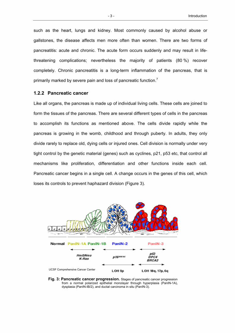
Pancreatic cancer begins in a single cell. A change occurs in the genes of this cell, which
loses its controls to prevent haphazard division (Figure 3).
UCSF Comprehensive Cancer CenterUCSF Comprehensive Cancer Center
Fig. 3: Pancreatic cancer progression. Stages of pancreatic cancer progression from a normal polarized epithelial monolayer through hyperplasia (PanIN-1A), dysplasia (PanIN-IB/2), and ductal carcinoma in situ (PanIN-3).
- 4 - Introduction
The abnormal cell starts dividing rapidly, makes millions and billions of copies of it. Soon a
clump of abnormal cells is produced, called adenocarcinoma. Adenocarcinoma simply
means a swelling and is not necessarily malignant or cancerous. When the cells in the
adenocarcinoma just grow in their local area and do not spread elsewhere, then the
adenocarcinoma is benign. However, when the adenocarcinoma's cells, which are dividing
out of control, gain the capacity to spread to distant body areas, this is a malignancy
called cancer. Malignant adenocarcinomas can spread to any area of the body; the
process of distant spread is called metastasis (Table 1). The cancer at first grows in its
local area and interferes with the pancreas functions. When cancer metastasizes, it can
grow in vital organs, cause symptoms there and eventually kill the patient.
1.2.3 Risk factors
Pancreatic cancer is the fourth most common cause of cancer death in Western society
and is a leading cause of cancer death worldwide. Its incidence and mortality rates are
almost identical. The 5-year survival rate is approximately 1-2 % and the median survival
time after diagnosis is 4-6 months.
Pancreatic cancer is more common among males than females, with peak incidence
occurring at age sixty.8 The etiology of the disease remains unclear, but cigarette
smoking, obesity and alcohol abuse have been related with an increased incidence of
pancreatic cancer. Pancreatic cancer is more common among individuals with histories of
the following conditions: cirrhosis (a chronic liver disease), chronic pancreatitis, diabetes
and a history of surgery to the upper digestive tract.8 Long term exposure to certain
chemical carcinogens, such as dry cleaning chemicals, gasoline or metallurgic fumes,
also appears to increase the risk of this cancer.8 Inherited DNA changes that may
increase a person's risk of developing cancer of the pancreas can also cause an
increased risk for certain other cancers. Mutation in oncogene (K-ras in codon 12), tumor
suppressor genes (CDKN2A/INK4A, TP53 and DPC4/SMAD4/MADH4) and caretaker
genes (BRCA2) also have a higher rate of pancreatic cancer. Current studies on

- 5 - Introduction
pancreatic adenocarcinoma tissues and cell lines have shown that multiple subsets of
genes undergo activation or inactivation during development and progression of disease.9
Cancer of the endocrine pancreas includes a highly treatable and often curable collection
of tumors. They are uncommon cancers with 200 to 1.000 new cases per year and occur
in only 1.5 % of detailed autopsy series. About 95% of pancreatic cancers begin in the
exocrine pancreas. Here in this study only exocrine pancreas cell lines will being
investigated.
1.2.4 Cellular cancer classification of pancreatic adenocarcinoma
Cancer of the pancreas can be classified according to the UICC and TNM . Once cancer
of the pancreas is found, more tests will be done to find out whether the cancer has
spread from the pancreas to the tissues around it or to other parts of the body. This is
called staging (Table 1).
Table 1: The UICC TNM classification for staging of cancers
Involvement of lymph nodes N
Primary adenocarcinoma T N0 regional node N1 distant nodes M
T1 With in the pancreas 2 cm or smaller Stage 1a Stage 2b Stage 4
T2 With in the pancreas larger than 2 cm Stage 1b Stage 2b Stage 4
T3 Beyond the pancreas Stage 2a Stage 2b Stage 4
T4 Invasion to the celiac artery or SMA* Stage 3 Stage 3 Stage 4
Liver metastasis, lung metastasis, peritoneal dissemination12 Stage 4:
*Smooth muscle actins (SMA) are commonly used to elucidate mammary myoepithelial (ME) cells, whose presence or absence is a reliable criterion for differentiating in situ and invasive adenocarcinomas.
Cancers of the exocrine pancreas can be grouped according to where in the pancreas the
cancer is or according to the type of cell the cancer has originated from.
duct cell adenocarcinoma (90 % of all cases)
acinar cell adenocarcinoma
papillary adenocarcinoma
adenosquamous adenocarcinoma
- 6 - Introduction
undifferentiated adenocarcinoma
mucinous adenocarcinoma
giant cell adenocarcinoma
mixed type
small cell adenocarcinoma
cyst adenocarcinoma
unclassified
pancreatoblastoma
papillary-cystic neoplasm (this adenocarcinoma has lower malignant potential and may be cured with surgery alone)10,11
1.3 Ectoenzymes
Ectoenzymes are integral membrane proteins anchored by hydrophobic interactions with
the lipids of the plasma membrane, that have their enzymatically active site outside the
plasma membrane in the extracellular environment. Many ectoenzymes are type II integral
membrane proteins with a short amino terminus in the cytosol or are glycosylphosphatidyl-
inositol-linked molecules. But all other possible kinds of anchoring in cell membranes have
been found. One of the first ectoenzymes to be identified was cholinesterase, which
hydrolyses acetylcholine at neuronal synapses.13 Ectoenzymes can be classified
according to their enzymatic activities. Many of them are peptidases and proteinases, see
below table 3. Other enzyme species include hydrolases and nucleotidases which
hydrolyse extracellular nucleotides, NAD and NADP, or oxidases which oxidize various
substrates (Table 2).14
- 7 - Introduction
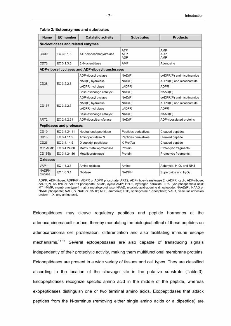
Table 2: Ectoenzymes and substrates
Name EC number Catalytic activity Substrates Products
Nucleotidases and related enzymes
CD39 EC 3.6.1.5 ATP diphosphohydrolase ATP ATP ADP
AMP ADP AMP
CD73 EC 3.1.3.5 5.-Nucleotidase AMP Adenosine
ADP-ribosyl cyclases and ADP-ribosyltransferases ADP-ribosyl cyclase NAD(P) cADPR(P) and nicotinamide
NAD(P) hydrolase NAD(P) ADPR(P) and nicotinamide
cADPR hydrolase cADPR ADPR CD38 EC 3.2.2.5
Base-exchange catalyst NAD(P) NAAD(P)
ADP-ribosyl cyclase NAD(P) cADPR(P) and nicotinamide
NAD(P) hydrolase NAD(P) ADPR(P) and nicotinamide
cADPR hydrolase cADPR ADPR CD157 EC 3.2.2.5
Base-exchange catalyst NAD(P) NAAD(P)
ART2 EC 2.4.2.31 ADP-ribosyltransferase NAD(P) ADP-ribosylated proteins
Peptidases and proteases CD10 EC 3.4.24.11 Neutral endopeptidase Peptides derivatives Cleaved peptides
CD13 EC 3.4.11.2 Aminopeptidase N Peptides derivatives Cleaved peptide
CD26 EC 3.4.14.5 Dipeptidyl peptidase X-Pro/Ala Cleaved peptide
MT1-MMP EC 3.4.24.80 Matrix metalloproteinase Protein Proteolytic fragments
CD156b EC 3.4.24.86 Metalloproteinase Protein Proteolytic fragments
Oxidases VAP1 EC 1.4.3.6 Amine oxidase Amine Aldehyde, H2O2 and NH3 NADPH oxidase EC 1.6.3.1 Oxidase NADPH Superoxide and H2O2
ADPR, ADP-ribose; ADPR(P), ADPR or ADPR phosphate; ART2, ADP-ribosyltransferase 2; cADPR, cyclic ADP-ribose; cADR(P), cADPR or cADPR phosphate; cAMP, cyclic AMP; H2O2, hydrogen peroxide; LPA, lyso-phosphatidic acid; MT1-MMP, membrane-type-1 matrix metalloproteinase; NAAD, nicotinic-acid-adenine dinucleotide; NAAD(P), NAAD or NAAD phosphate; NAD(P), NAD or NADP; NH3, ammonia; S1P, sphingosine 1-phosphate; VAP1, vascular adhesion protein 1; X, any amino acid.
Ectopeptidases may cleave regulatory peptides and peptide hormones at the
adenocarcinoma cell surface, thereby modulating the biological effect of these peptides on
adenocarcinoma cell proliferation, differentiation and also facilitating immune escape
mechanisms.15-17 Several ectopeptidases are also capable of transducing signals
independently of their proteolytic activity, making them multifunctional membrane proteins.
Ectopeptidases are present in a wide variety of tissues and cell types. They are classified
according to the location of the cleavage site in the putative substrate (Table 3).
Endopeptidases recognize specific amino acid in the middle of the peptide, whereas
exopeptidases distinguish one or two terminal amino acids. Exopeptidases that attack
peptides from the N-terminus (removing either single amino acids or a dipeptide) are

- 8 - Introduction
termed (dipeptidyl) aminopeptidases, whereas peptidases attacking the C-terminus are
termed carboxypeptidases.18
Table 3: Ectopeptidases and substrates
Peptidase Specificity* Posssible natural substrates
Aminopeptidases APN ○ ÷ ● ─ = Ala, Leu fMLP, opioid peptides, enkephalins
APA ○ ÷ ● ─ = Glu, Asp Angiotensins
APP ● ÷ ○ ─ = Pro BK, SP
DPP IV ● ÷ ○ ─ = Pro, Ala SP, BK, NPY, chemokines (CXCL9, 10)
Carboxy peptidases CPN ─ ● ÷ ○ = Arg, Lys Anaphylatoxins
Endopeptidases
NEP ─● ÷ ○ ─ ● ─ = Phe, Leu, Ile, Val, Thr, Trp, Ala BK, SP, NKA, NPY, VIP, ANF, enkephalins ET-1, BLP, angiotensins
ACE ● ─ ● ÷ ○─ ○ ─ = Relatively non-specific Angiotensins, enkephalins, SP
ECE ─ ○─ ○ ─ ÷ ● ─ = Ile-Ile-Trp Big ET-1
The peptidase cleaves peptides in which the open circle represents (one of) the mentioned amino acids. The closed circle can be any amino acid. The cleaved bond is represented by ‘÷’. Peptidases: APN, aminopeptidase N; APA, aminopeptidase A; APP, aminopeptidase P; DPP IV, dipeptidyl(amino)peptidase IV; CPN, carboxypeptidase N; NEP, neprilysin; ACE, angiotensin-converting enzyme; ECE, endothelin-converting enzyme. Substrates:fMLP, formyl-metheonyl-leucyl-phenylalanine; BK, bradykinin; SP, substance P; NKA, neurokinin A ; NPY, neuropeptide Y ; VIP, vasoactive intestinal peptide ; ANF, atrial natriuretic factor ; ET-1, endothelin-1 ; BLP, bombesin-like peptides.
1.3.1 Neutral endopeptidase
Membrane metalloendopeptidase EC 3.4.24.11 (Enkephalinase, neprilysin, neutral
endopeptidase, NEP) is a mammalian type II integral membrane zinc-containing
endopeptidase ectoenzyme. Immunophenotypically it is identified as the leukocyte cluster
of differentiation CD10 or CALLA (common acute lymphoblastic leukemia antigen), which
has specificity for cleaving oligopeptides (≤ 40 amino acids in length) at the amino side of
hydrophobic amino acids (Val, Ile, Phe, Leu, Ala).19
1.3.1.1 Biochemical and molecular characteristics
Neutral endopeptidase was first characterized and isolated from rabbit kidney tubule
brush borders. Cloning of the NEP/CD10 gene and subsequent cloning of the common
acute lymphoblastic leukemia antigen (CALLA, CD10) showed that both sequences are
identical.20-22 NEP/CD10 is a glycoprotein with 750 amino acids.20 It has a single 24 amino
- 9 - Introduction
acid hydrophobic segment that functions both as a transmembrane region and a signal
peptide. The C-terminal 700 amino acids compose the extracellular domain which
contains the pentapeptide consensus sequence (His-Glu-[Ile, Leu, Met]-X-His) of zinc-
binding metalloproteases, whereas the 25 N-terminal amino acids form the cytoplasmic
tail.
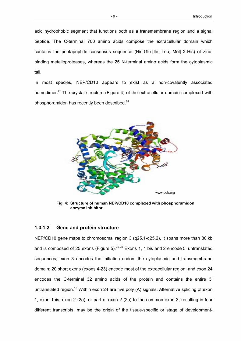
In most species, NEP/CD10 appears to exist as a non-covalently associated
homodimer.23 The crystal structure (Figure 4) of the extracellular domain complexed with
phosphoramidon has recently been described.24
www.pdb.orgwww.pdb.org
Fig. 4: Structure of human NEP/CD10 complexed with phosphoramidon enzyme inhibitor.
1.3.1.2 Gene and protein structure
NEP/CD10 gene maps to chromosomal region 3 (q25.1-q25.2), it spans more than 80 kb
and is composed of 25 exons (Figure 5).25,26 Exons 1, 1 bis and 2 encode 5’ untranslated
sequences; exon 3 encodes the initiation codon, the cytoplasmic and transmembrane
domain; 20 short exons (exons 4-23) encode most of the extracellular region; and exon 24
encodes the C-terminal 32 amino acids of the protein and contains the entire 3’
untranslated region.18 Within exon 24 are five poly (A) signals. Alternative splicing of exon
1, exon 1bis, exon 2 (2a), or part of exon 2 (2b) to the common exon 3, resulting in four
different transcripts, may be the origin of the tissue-specific or stage of development-

- 10 - Introduction
specific expression of NEP/CD10.27 Indeed, two separate regulatory elements have been
found in the NEP/CD10 promoter region and these elements may be regulated by the
transcription factor CBF/ NF-Y in a tissue specific manner. A cDNA clone lacking the
complete exon 16 has been isolated from human lung tissue.28
Fig. 5: Variant gene transcripts structure of NEP/CD10 confer four transcripts
that present only one protein. Copied from gene data bank.
1.3.1.3 Enzymatic activity and biological function
NEP/CD10 is able to hydrolyse peptide bonds on the N-terminal site of hydrophobic amino
acids, including Phe, Leu, Ile, Val, Tyr, Ala and Trp. However, sub-site interactions and
conformational factors greatly influence the efficiency of hydrolysis. It hydrolyses a variety
of physiologically active peptides including opioid peptides (Met- and Leu-enkephalin),
substance P, atrial natriuretic factor (ANF), endothelin, neurotensin, oxytocin, bradykinin,
angiotensin 1, 2, chemotactic peptide formyl-Met-Leu-Phe (f - MLP) , and bombesine like
peptides.19 Thus, one of the main roles of NEP/CD10 seems to be reducing the local
concentration of biologically active peptides available for receptor binding and signal
transduction. The ubiquitous occurrence of NEP/CD10 in mammalian organs renders
possible a broad field of physiological functions as illustrated by more or less organ-
- 11 - Introduction
specific peptides proven to be cleft by the enzymes. In kidneys possible physiological
substrates of NEP/CD10 are ANP, adrenomedullin and PAMP (see the list of
abbreviations); in the brain, the substrates probably are enkephalins and oxytocin.
Similarly, possible substrates in the lung are bombesin, BLP, GRP, neuromedin C,
substance P and neurokinin A; in the cardiovascular system, angiotenisin II, bradykinin
and CGRP; in the gut, VIP; on the neutrophil membrane, fMLP etc. Some substrates are
not strictly tissue-specific, e.g. substance P. Preclinical and clinical trials explored
possibilities of therapeutic application of the inhibitors of NEP/CD10, such as thiorphan in
the management of pain, diarrhea, depression, arterial hypertension and asthma.29-37 It
has been reported that NEP/CD10 may also be able to hydrolyse certain larger
substrates, including cytokines such as IL-1β and IL-6.18 A role for NEP/CD10 in lymphoid
development has been suggested by studies showing that inhibition of NEP/CD10 results
in increased proliferation and maturation of B cells, both in vitro and in vivo.38
1.3.1.4 Distribution in tissues
The common acute lymphoblastic leukemia antigen (CALLA; CD10) was originally found
on lymphoblasts from patients with pre-B type acute lymphoblastic leukemia.39 Later it has
been found to be widely associated with precursor B-cell acute lymphoblastic leukemia, T-
cell acute lymphoblastic leukemia, as well as various types of lymphomas including
follicular lymphomas and Burkitt lymphomas.40-45
Subsequently, immunohistochemical studies demonstrated the expression of this antigen
on a variety of non hematopoietic normal and adenocarcinoma tissues, such as kidney,
breast, fetal small intestine, fibroblasts, gliomas, melanomas, retinoblastomas, various
mesenchymal adenocarcinomas, breast, colon adenocarcinoma cells, genitourinary tract,
such as renal cell, transitional cell and prostate adenocarcinomas.46-59 In the female
genital tract, NEP/CD10 is a marker of trophoblastic adenocarcinomas and endometrial
stromal neoplasms.60 It is also expressed by serous ovarian adenocarcinomas.60-62 In the
gastrointestinal system, NEP/CD10 is expressed in hepatocellular adenocarcinomas.63 It
also has been detected in pancreatic ductal adenocarcinomas and is frequently observed
- 12 - Introduction
in solid pseudopapillary adenocarcinomas.64-66 It has been reported to be positive in some
pancreatic endocrine adenocarcinomas (PETs) in a small series.64,67 This wide tissue
distribution, suggests a crucial role in vivo of this antigen.
1.3.1.5 NEP/CD10 function in normal and carcinoma tissues
The various functions certainly depend on cell type or tissue origin. In muscles,
NEP/CD10 may play an important role during muscle cell differentiation, possibly through
the regulation, either directly or indirectly, of the insulin-like growth factor I driven
myogenic program.68 Furthermore, other results showed that elevated cardiac NEP/CD10
activity may increase the local degradation of bradykinin and natriuretic peptides promote
to pressure loaded and failing human hearts.69 Neutral endopeptidase may terminate the
proinflammatory and mitogenic actions of neuropeptides in normal skin and wounds.70 In
addition it plays a specific role in promoting early T-cell development.71 In human bronchi,
it may regulate peptide-induced inflammation.72 In human thymocytes, expression of
functional NEP/CD10 suggests a role for this enzyme in the maturation of human
thymocytes by hydrolysing thymopentin, a thymic peptide known to induce the maturation
of prothymocytes into thymocytes.73 NEP/CD10 of epithelial cells within human digestive
tract (in the stomach, duodenum, small intestine, ascending, descending sigmoid colon
and rectum) confirms the hypothesis of its participation in protein hydrolysis processes.74
In the human respiratory mucosa, in addition to the modulating functions of NEP/CD10 on
neuropeptide-mediated activities on vessels and glands, it is possible that in secreted
form it plays a role in regulating mucosal responses to luminal neuropeptides or other as
yet uncharacterized NEP/CD10 substrates.75 In spleen, results suggest that NEP/CD10
inhibition promotes the reconstitution and maturation of splenic B-cells. Therefore, it may
function to regulate B-cell ontogeny in vivo by hydrolyzing a peptide substrate that
stimulates B-cell proliferation and or differentiation.76
In tumors, NEP/CD10 function seems to be also variable from one tissue to others. In
stromal cells, it seems to promote invasion and metastasis of differentiated gastric
carcinoma.77 In malignant melanoma the expression is also associated with tumor
- 13 - Introduction
progression.78 In epithelial ovarian carcinomas, it may play a role in the regulation of
neoplastic transformation and tumor differentiation,79 however other evidence suggests
that NEP/CD10 functionally suppresses the progression of ovarian carcinoma.80 In an
integral part of colorectal carcinogenesis it seems to contribute to the invasion and thus
probably facilitates metastasis.81 In placenta, induction of choriocarcinoma cell
differentiation is associated with an increase of NEP/CD10 expression at the cell surface,
suggesting a role of this enzyme in regulating differentiated trophoblast functions such as
human chronic gonadotrophin (hCG) secretion.82 In brain, a partial reduction of
extracellular amyloid-beta peptide (Abeta) levels by NEP/CD10 may facilitate
development of human neuronal progenitor cells (HNPCs) in treatment of
neurodegenerative diseases like Alzheimer's disease (AD).83,84 Downregulation of
NEP/CD10 in the progression of urothelial bladder carcinomas, probably facilitates
invasion, especially into muscle.85 In nasopharyngeal carcinoma it was revealed that
NEP/CD10 expression by stromal cells in this malignancy plays an important role in tumor
progression, particularly in older patients.86 In cervical carcinoma cells results indicated
that NEP/CD10 functions as a tumor-suppressor gene and its expression may have
prognostic significance,87 and in oral squamous cell carcinoma may have an important
role in tumor invasion, probably facilitating the occurrence of metastases.88

- 14 - Introduction
1.4 NEP/CD10 expression in pancreatitis and carcinoma tissues
We investigated NEP/CD10 expression in pancreatitis and pancreatic tumor lesions.89
Interestingly, there exists no significant difference in NEP/CD10 mRNA expression levels
in pancreatic carcinoma compared to that of pancreatitis tissues (data not shown).
Regarding protein expression, immunoreactivity for NEP/CD10 was detectable in a subset
of pancreatic carcinomas exhibiting a membranous staining pattern in tumor cells
(Figure 6). A positive staining was found in 6 of 24 pancreatic ductal adenocarcinomas
(25 %). However, the heterogeneous NEP/CD10 expression pattern was not associated
with tumor grading, staging and metastasis formation. In chronic pancreatitis 3 of 6
samples demonstrated a focal staining of residual ducts.
*b
c
d*
b
c
d
Fig.6: Immunohistochemical detection of NEP/CD10 expression in pancreatic adenocarcinomas as well as in chronic pancreatitis. Perineural (arrow indicating nerve) infiltrating ductal adenocarcinoma with membranous expression of NEP/CD10 (a). Ductal adenocarcinoma negative for NEP/CD10 (*), normal duodenal mucosa served as internal positive control (b). Chronic pancreatitis with focal expression of NEP/CD10 in residual ducts and intravascular neutrophils (arrow) (c, d). (Erhuma et. al.)89
- 15 - Introduction
1.5 Aim of the present investigation
Neutral endopeptidase is a membrane bound enzyme with various functions depending
on cell type or tissue origin. Only limited information however exists about the NEP/CD10
expression in physiological and patho-physiological conditions of the pancreas.
In this study, the main purpose is in the following steps explained:
NEP/CD10 expression: investigation of the relevance of NEP/CD10 expression in
pancreatic adenocarcinoma cell lines.
NEP/CD10 promoters methylation status: Examination of a subset of cases for
evidence of two NEP/CD10 promoters methylation and correlateion the results with
NEP/CD10 expression to determine the mechanism for the lack of NEP/CD10 expression
in pancreatic adenocarcinoma cell lines.
NEP/CD10 regulation: Either hypermethylation of the 5' CpG island or deacetylation of
chromatin results in a loss of NEP/CD10 expression in pancreatic adenocarcinoma cell
lines. To differentiate between both mechanisms pancreatic carcinoma cell lines were
exposed in separate to either the demethylating agent 5-aza-2'-deoxycytidine or
acetylation agents, butyric acid and valproic acid.
Biological function of NEP/CD10: Investigation of the biological function of NEP/CD10
in pancreatic exocrine carcinoma using pancreatic carcinoma cell line as a model,
transfected with pEGFP-C3 (Mock), NEP-pEGFP-C3 and mutated NEPH587E-pEGFP-C3 in
enzyme active site, as well as their effects on proliferation, motility and invasion via
induction or repression of diverse proteins that play a crucial role in biological
mechanisms.
Affecting of NEP/CD10 on signal cascades: Investigation of the influence of NEP/CD10
over expression on the proteins that play crucial roles in signal cascades that regulate
proliferation, motility and invasion via stimulation through cell membrane receptors
concluded our series of experiments.
Related Documents