1. 1 Normal Anterior Chamber Angle Schlemm’s canal Schwalbe’s line

1.1 Normal Anterior Chamber Angle Schlemms canal Schwalbes line.
Dec 10, 2015
Welcome message from author
This document is posted to help you gain knowledge. Please leave a comment to let me know what you think about it! Share it to your friends and learn new things together.
Transcript

1.1
Normal Anterior Chamber Angle
Schlemm’s canal
Schwalbe’s line

1.1
Narrow Anterior Chamber Angle

1.1
Angle Scans
•Scleral spur (red arrow)
•Schlemm’s canal (blue arrow)
•Schwalbe’s line (green arrow)

1.1
Closed angle with peripheral anterior synechiae
OD OS
1.1
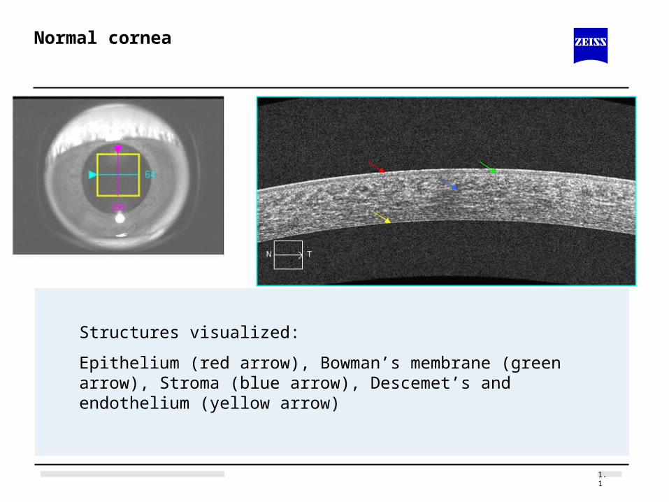
Normal cornea
Structures visualized:
Epithelium (red arrow), Bowman’s membrane (green arrow), Stroma (blue arrow), Descemet’s and endothelium (yellow arrow)

1.1
Central Corneal Thickness Measurement

1.1
LASIK
One month post-op LASIK flap edge visible in scan

1.1
Corneal Scar
50 y/o male, bungee cord injury 2 months priorTraumatic corneal ulcerationThinned cornea with epithelial defect. Tear film visible over defect.

1.1
Descemet’s Stripping Endothelial Keratoplasty (DSEK)
Note gap between edge of DSEK and Descemet’s membrane of recipient

1.1
Peripheral corneal degeneration
Related Documents