Copyright © 2008 Pearson Education, Inc., publishing as Pearson Benjamin Cummings PowerPoint ® Lecture Presentations for Biology Eighth Edition Neil Campbell and Jane Reece Lectures by Chris Romero, updated by Erin Barley with contributions from Joan Sharp Chapter 11 Chapter 11 Cell Communication

Welcome message from author
This document is posted to help you gain knowledge. Please leave a comment to let me know what you think about it! Share it to your friends and learn new things together.
Transcript

Copyright © 2008 Pearson Education, Inc., publishing as Pearson Benjamin Cummings
PowerPoint® Lecture Presentations for
BiologyEighth Edition
Neil Campbell and Jane Reece
Lectures by Chris Romero, updated by Erin Barley with contributions from Joan Sharp
Chapter 11Chapter 11
Cell Communication

Overview: The Cellular Internet
• Cell-to-cell communication is essential for multicellular organisms
• Biologists have discovered some universal mechanisms of cellular regulation
• The combined effects of multiple signals determine cell response
• For example, the dilation of blood vessels is controlled by multiple molecules
Copyright © 2008 Pearson Education, Inc., publishing as Pearson Benjamin Cummings
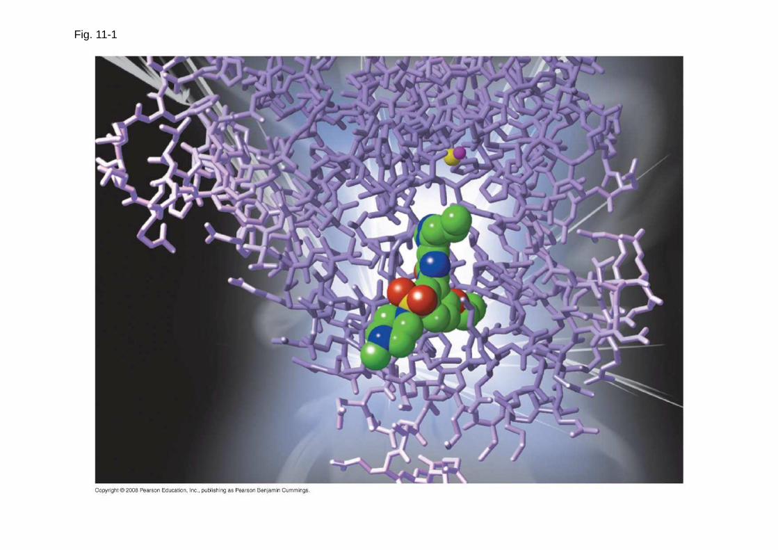
Fig. 11-1

Concept 11.1: External signals are converted to responses within the cell
• Microbes are a window on the role of cell signaling in the evolution of life
Copyright © 2008 Pearson Education, Inc., publishing as Pearson Benjamin Cummings

Evolution of Cell Signaling
• A signal transduction pathway is a series of steps by which a signal on a cell’s surface is converted into a specific cellular response
• Signal transduction pathways convert signals on a cell’s surface into cellular responses
Copyright © 2008 Pearson Education, Inc., publishing as Pearson Benjamin Cummings

Fig. 11-2
Receptorαααα factor
a factor
a αααα
ααααa
Exchangeof matingfactors
Yeast cell,mating type a
Yeast cell,mating type αααα
Mating
New a/ααααcell
a/αααα
1
2
3
• Pathway similarities suggest that ancestral signaling molecules evolved in prokaryotes and were modified later in eukaryotes
• The concentration of signaling molecules allows bacteria to detect population density
Copyright © 2008 Pearson Education, Inc., publishing as Pearson Benjamin Cummings

Fig. 11-3
Individual rod-shaped cells
Spore-formingstructure(fruiting body)
Aggregation inprocess
Fruiting bodies
0.5 mm
1
3
2

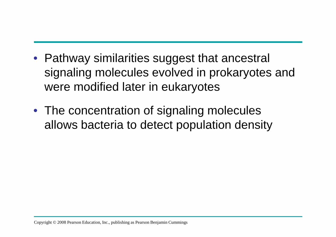
Local and Long-Distance Signaling
• Cells in a multicellular organism communicate by chemical messengers
• Animal and plant cells have cell junctions that directly connect the cytoplasm of adjacent cells
• In local signaling, animal cells may communicate by direct contact, or cell-cell recognition
Copyright © 2008 Pearson Education, Inc., publishing as Pearson Benjamin Cummings
Fig. 11-4Plasma membranes
Gap junctionsbetween animal cells
(a) Cell junctions
Plasmodesmatabetween plant cells
(b) Cell-cell recognition

• In many other cases, animal cells communicate using local regulators , messenger molecules that travel only short distances
• In long-distance signaling, plants and animals use chemicals called hormones
Copyright © 2008 Pearson Education, Inc., publishing as Pearson Benjamin Cummings
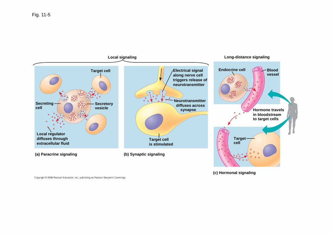
Fig. 11-5
Local signaling
Target cell
Secretingcell
Secretoryvesicle
Local regulatordiffuses throughextracellular fluid
(a) Paracrine signaling (b) Synaptic signaling
Target cellis stimulated
Neurotransmitterdiffuses across
synapse
Electrical signalalong nerve celltriggers release ofneurotransmitter
Long-distance signaling
Endocrine cell Bloodvessel
Hormone travelsin bloodstreamto target cells
Targetcell
(c) Hormonal signaling

Fig. 11-5ab
Local signaling
Target cell
Secretoryvesicle
Secretingcell
Local regulatordiffuses throughextracellular fluid
(a) Paracrine signaling (b) Synaptic signaling
Target cellis stimulated
Neurotransmitterdiffuses across
synapse
Electrical signalalong nerve celltriggers release ofneurotransmitter
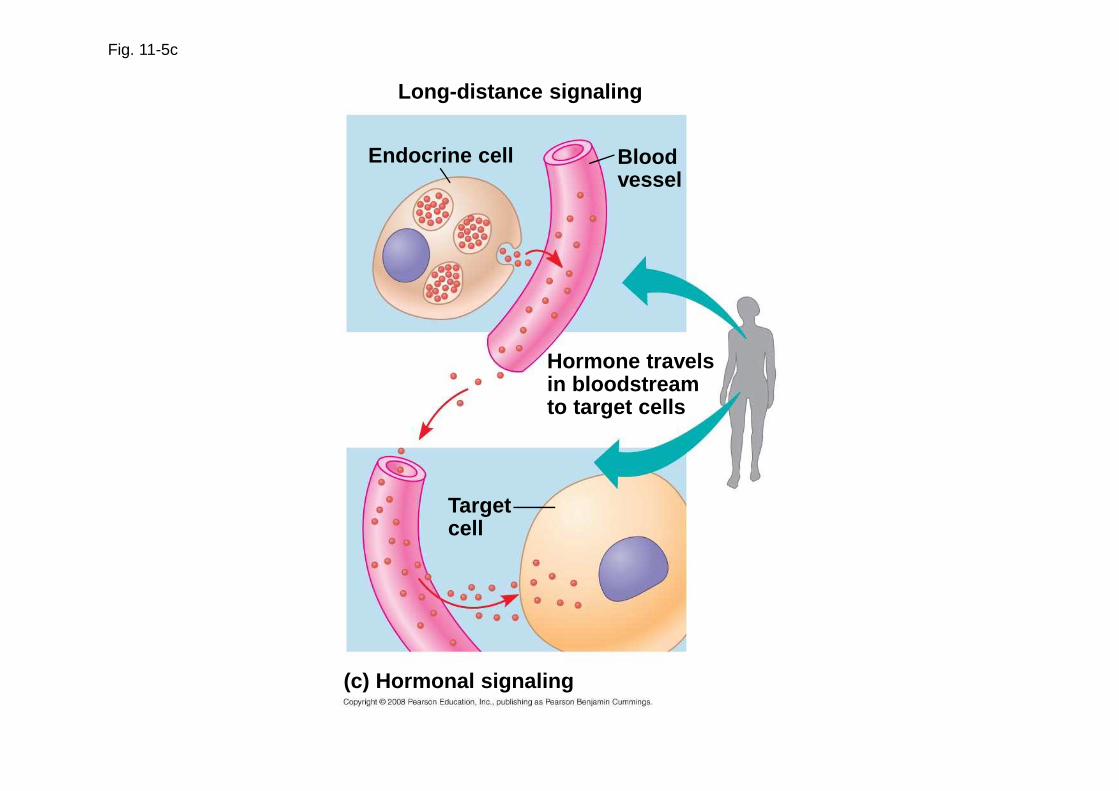
Fig. 11-5c
Long-distance signaling
Endocrine cell Bloodvessel
Hormone travelsin bloodstreamto target cells
Targetcell
(c) Hormonal signaling

The Three Stages of Cell Signaling: A Preview
• Earl W. Sutherland discovered how the hormone epinephrine acts on cells
• Sutherland suggested that cells receiving signals went through three processes:
– Reception
– Transduction
– Response
Animation: Overview of Cell Signaling
Copyright © 2008 Pearson Education, Inc., publishing as Pearson Benjamin Cummings
Fig. 11-6-1
Reception1
EXTRACELLULARFLUID
Signalingmolecule
Plasma membrane
CYTOPLASM
1
Receptor

Fig. 11-6-2
1
EXTRACELLULARFLUID
Signalingmolecule
Plasma membrane
CYTOPLASM
Transduction2
Relay molecules in a signal transduction pathway
Reception1
Receptor

Fig. 11-6-3
EXTRACELLULARFLUID
Plasma membrane
CYTOPLASM
Receptor
Signalingmolecule
Relay molecules in a signal transduction pathway
Activationof cellularresponse
Transduction Response2 3Reception1
Concept 11.2: Reception: A signal molecule binds to a receptor protein, causing it to change shape
• The binding between a signal molecule (ligand ) and receptor is highly specific
• A shape change in a receptor is often the initial transduction of the signal
• Most signal receptors are plasma membrane proteins
Copyright © 2008 Pearson Education, Inc., publishing as Pearson Benjamin Cummings

Receptors in the Plasma Membrane
• Most water-soluble signal molecules bind to specific sites on receptor proteins in the plasma membrane
• There are three main types of membrane receptors:
– G protein-coupled receptors
– Receptor tyrosine kinases
– Ion channel receptors
Copyright © 2008 Pearson Education, Inc., publishing as Pearson Benjamin Cummings
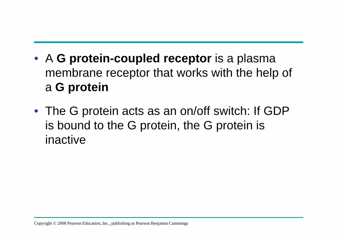
• A G protein -coupled receptor is a plasma membrane receptor that works with the help of a G protein
• The G protein acts as an on/off switch: If GDP is bound to the G protein, the G protein is inactive
Copyright © 2008 Pearson Education, Inc., publishing as Pearson Benjamin Cummings

Fig. 11-7a
Signaling -molecule binding site
Segment thatinteracts withG proteins
G protein -coupled receptor
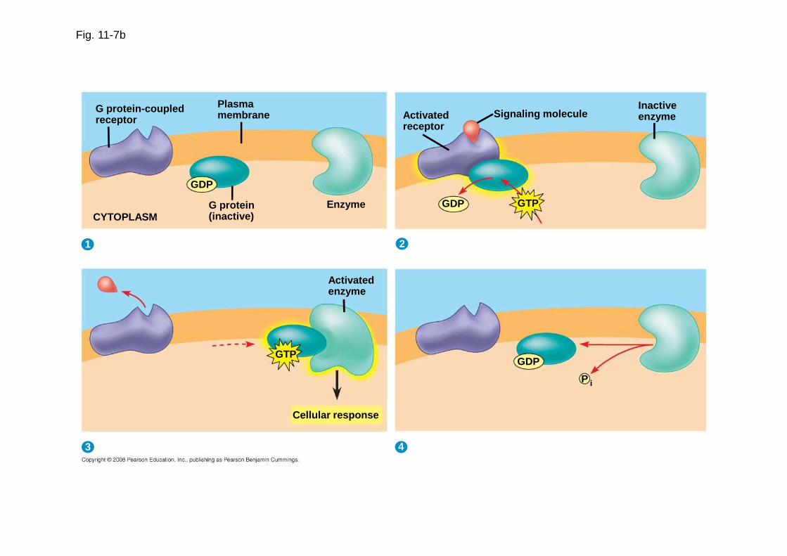
Fig. 11-7b
G protein-coupledreceptor
Plasmamembrane
EnzymeG protein(inactive)
GDP
CYTOPLASM
Activatedenzyme
GTP
Cellular response
GDP
P i
Activatedreceptor
GDP GTP
Signaling moleculeInactiveenzyme
1 2
3 4
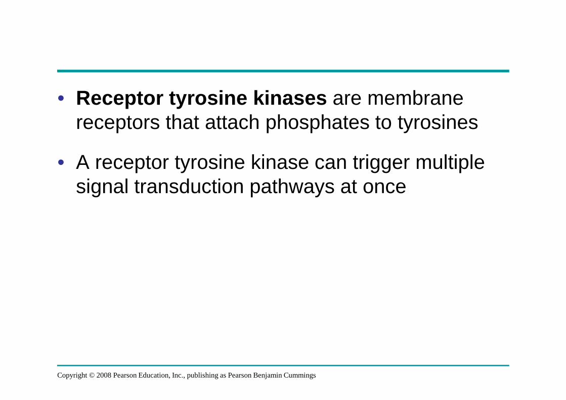
• Receptor tyrosine kinases are membrane receptors that attach phosphates to tyrosines
• A receptor tyrosine kinase can trigger multiple signal transduction pathways at once
Copyright © 2008 Pearson Education, Inc., publishing as Pearson Benjamin Cummings
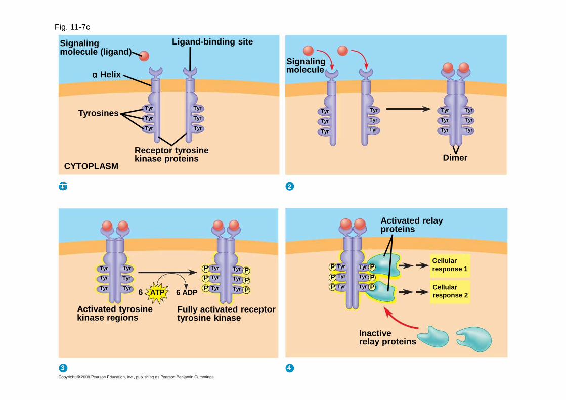
Fig. 11-7c
Signalingmolecule (ligand)
Ligand-binding site
αααα Helix
TyrosinesTyr
Tyr
Tyr
Tyr
Tyr
Tyr
Receptor tyrosinekinase proteins
CYTOPLASM
Signalingmolecule
Tyr
Tyr
Tyr
Tyr
Tyr
Tyr
Tyr
Tyr
Tyr
Tyr
Tyr
Tyr
Dimer
Activated relayproteins
Tyr
Tyr
Tyr
Tyr
Tyr
Tyr
P
PP
P
PP
Cellularresponse 1
Cellularresponse 2
Inactiverelay proteins
Activated tyrosinekinase regions
Fully activated receptortyrosine kinase
6 6 ADPATP
Tyr
Tyr
Tyr
Tyr
Tyr
Tyr
Tyr
Tyr
Tyr
Tyr
Tyr
Tyr
PP
P
PPP
1 2
3 4
• A ligand -gated ion channel receptor acts as a gate when the receptor changes shape
• When a signal molecule binds as a ligand to the receptor, the gate allows specific ions, such as Na+ or Ca2+, through a channel in the receptor
Copyright © 2008 Pearson Education, Inc., publishing as Pearson Benjamin Cummings

Fig. 11-7dSignalingmolecule(ligand)
Gateclosed Ions
Ligand-gatedion channel receptor
Plasmamembrane
Gate open
Cellularresponse
Gate closed3
2
1
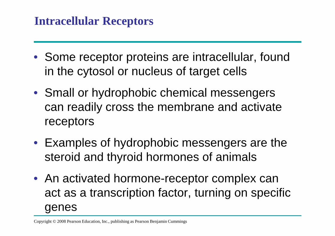
Intracellular Receptors
• Some receptor proteins are intracellular, found in the cytosol or nucleus of target cells
• Small or hydrophobic chemical messengers can readily cross the membrane and activate receptors
• Examples of hydrophobic messengers are the steroid and thyroid hormones of animals
• An activated hormone-receptor complex can act as a transcription factor, turning on specific genes
Copyright © 2008 Pearson Education, Inc., publishing as Pearson Benjamin Cummings

Fig. 11-8-1
Hormone(testosterone)
Receptorprotein
Plasmamembrane
EXTRACELLULARFLUID
DNA
NUCLEUS
CYTOPLASM
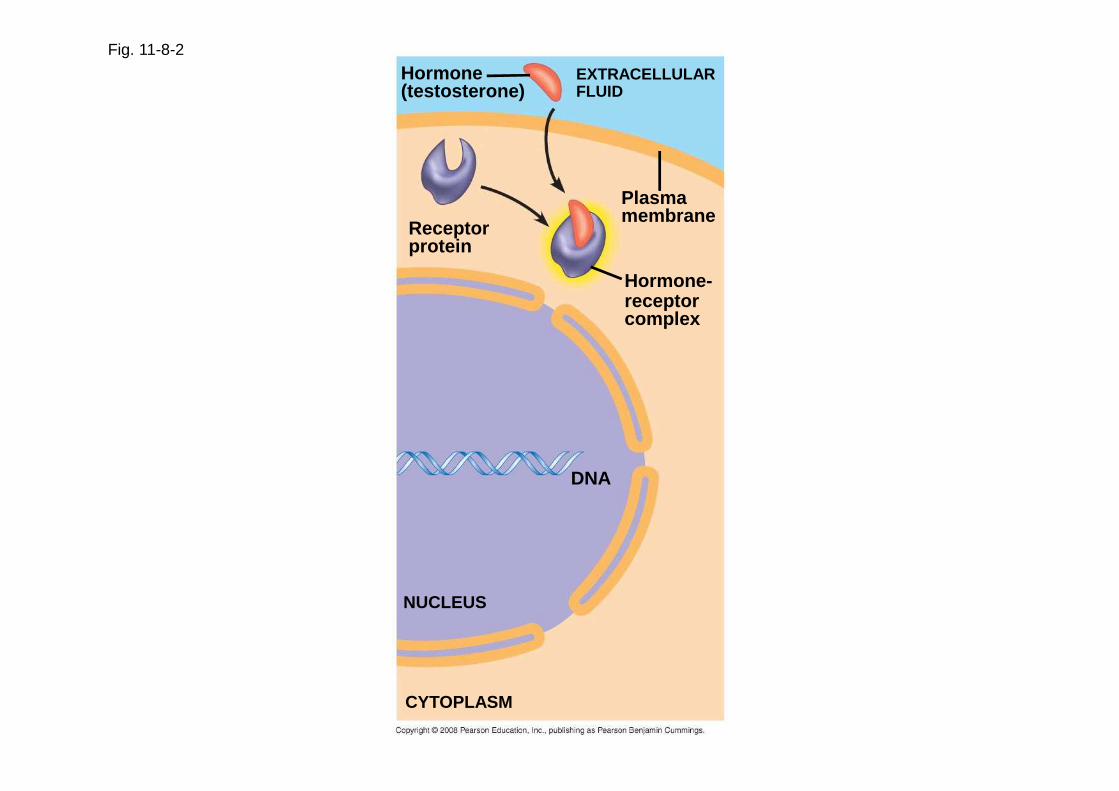
Fig. 11-8-2
Receptorprotein
Hormone(testosterone)
EXTRACELLULARFLUID
Plasmamembrane
Hormone-receptorcomplex
DNA
NUCLEUS
CYTOPLASM

Fig. 11-8-3
Hormone(testosterone)
EXTRACELLULARFLUID
Receptorprotein
Plasmamembrane
Hormone-receptorcomplex
DNA
NUCLEUS
CYTOPLASM
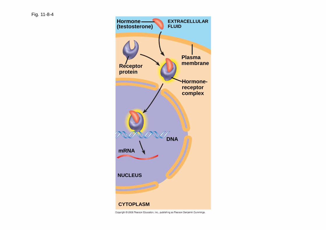
Fig. 11-8-4
Hormone(testosterone)
EXTRACELLULARFLUID
PlasmamembraneReceptor
protein
Hormone-receptorcomplex
DNA
mRNA
NUCLEUS
CYTOPLASM

Fig. 11-8-5
Hormone(testosterone)
EXTRACELLULARFLUID
Receptorprotein
Plasmamembrane
Hormone-receptorcomplex
DNA
mRNA
NUCLEUS New protein
CYTOPLASM

Concept 11.3: Transduction: Cascades of molecular interactions relay signals from receptors to target molecules in the cell
• Signal transduction usually involves multiple steps
• Multistep pathways can amplify a signal: A few molecules can produce a large cellular response
• Multistep pathways provide more opportunities for coordination and regulation of the cellular response
Copyright © 2008 Pearson Education, Inc., publishing as Pearson Benjamin Cummings
Signal Transduction Pathways
• The molecules that relay a signal from receptor to response are mostly proteins
• Like falling dominoes, the receptor activates another protein, which activates another, and so on, until the protein producing the response is activated
• At each step, the signal is transduced into a different form, usually a shape change in a protein
Copyright © 2008 Pearson Education, Inc., publishing as Pearson Benjamin Cummings

Protein Phosphorylation and Dephosphorylation
• In many pathways, the signal is transmitted by a cascade of protein phosphorylations
• Protein kinases transfer phosphates from ATP to protein, a process called phosphorylation
Copyright © 2008 Pearson Education, Inc., publishing as Pearson Benjamin Cummings

• Protein phosphatases remove the phosphates from proteins, a process called dephosphorylation
• This phosphorylation and dephosphorylation system acts as a molecular switch, turning activities on and off
Copyright © 2008 Pearson Education, Inc., publishing as Pearson Benjamin Cummings
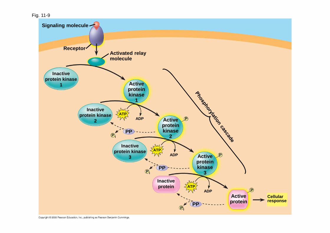
Fig. 11-9
Signaling molecule
ReceptorActivated relaymolecule
Inactiveprotein kinase
1 Activeproteinkinase
1
Inactiveprotein kinase
2
ATPADP Active
proteinkinase
2
P
PPP
Inactiveprotein kinase
3
ATPADP Active
proteinkinase
3
P
PPP
i
ATPADP P
ActiveproteinPP
P i
Inactiveprotein
Cellularresponse
i
Small Molecules and Ions as Second Messengers
• The extracellular signal molecule that binds to the receptor is a pathway’s “first messenger”
• Second messengers are small, nonprotein, water-soluble molecules or ions that spread throughout a cell by diffusion
• Second messengers participate in pathways initiated by G protein-coupled receptors and receptor tyrosine kinases
• Cyclic AMP and calcium ions are common second messengers
Copyright © 2008 Pearson Education, Inc., publishing as Pearson Benjamin Cummings
Cyclic AMP
• Cyclic AMP (cAMP) is one of the most widely used second messengers
• Adenylyl cyclase , an enzyme in the plasma membrane, converts ATP to cAMP in response to an extracellular signal
Copyright © 2008 Pearson Education, Inc., publishing as Pearson Benjamin Cummings
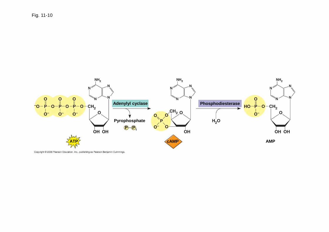
Adenylyl cyclase
Fig. 11-10
Pyrophosphate
P P i
ATP cAMP
Phosphodiesterase
AMP
• Many signal molecules trigger formation of cAMP
• Other components of cAMP pathways are G proteins, G protein-coupled receptors, and protein kinases
• cAMP usually activates protein kinase A, which phosphorylates various other proteins
• Further regulation of cell metabolism is provided by G-protein systems that inhibit adenylyl cyclase
Copyright © 2008 Pearson Education, Inc., publishing as Pearson Benjamin Cummings
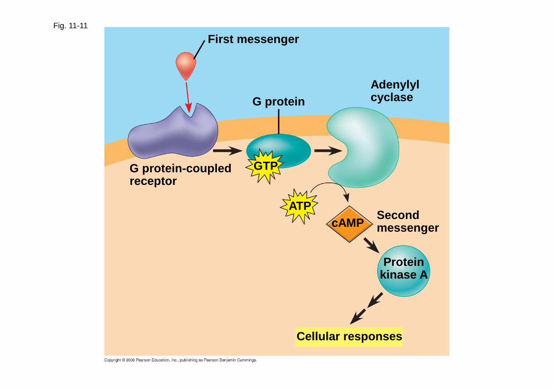
First messengerFig. 11-11
G proteinAdenylylcyclase
GTP
ATPcAMP
Secondmessenger
Proteinkinase A
G protein-coupledreceptor
Cellular responses

Calcium Ions and Inositol Triphosphate (IP3)
• Calcium ions (Ca2+) act as a second messenger in many pathways
• Calcium is an important second messenger because cells can regulate its concentration
Copyright © 2008 Pearson Education, Inc., publishing as Pearson Benjamin Cummings

EXTRACELLULARFLUID
Fig. 11-12
ATP
Nucleus
Mitochondrion
Ca2+ pump
Plasmamembrane
CYTOSOL
Ca2+
pumpEndoplasmicreticulum (ER)
Ca2+
pumpATP
Key
High [Ca 2+]Low [Ca 2+]

• A signal relayed by a signal transduction pathway may trigger an increase in calcium in the cytosol
• Pathways leading to the release of calcium involve inositol triphosphate (IP 3) and diacylglycerol (DAG) as additional second messengers
Animation: Signal Transduction Pathways
Copyright © 2008 Pearson Education, Inc., publishing as Pearson Benjamin Cummings

Fig. 11-13-1
EXTRA-CELLULARFLUID
Signaling molecule(first messenger)
G protein
GTP
G protein-coupledreceptor Phospholipase C PIP2
IP3
DAG
(second messenger)
IP3-gatedcalcium channel
Endoplasmicreticulum (ER) Ca2+
CYTOSOL

Fig. 11-13-2
G protein
EXTRA-CELLULARFLUID
Signaling molecule(first messenger)
G protein-coupledreceptor Phospholipase C PIP2
DAG
IP3(second messenger)
IP3-gatedcalcium channel
Endoplasmicreticulum (ER) Ca2+
CYTOSOL
Ca2+
(secondmessenger)
GTP

Fig. 11-13-3
G protein
EXTRA-CELLULARFLUID
Signaling molecule(first messenger)
G protein-coupledreceptor Phospholipase C PIP2
DAG
IP3(second messenger)
IP3-gatedcalcium channel
Endoplasmicreticulum (ER) Ca2+
CYTOSOL
Variousproteinsactivated
Cellularresponses
Ca2+
(secondmessenger)
GTP

Concept 11.4: Response: Cell signaling leads to regulation of transcription or cytoplasmic activities
• The cell’s response to an extracellular signal is sometimes called the “output response”
Copyright © 2008 Pearson Education, Inc., publishing as Pearson Benjamin Cummings
Nuclear and Cytoplasmic Responses
• Ultimately, a signal transduction pathway leads to regulation of one or more cellular activities
• The response may occur in the cytoplasm or may involve action in the nucleus
• Many signaling pathways regulate the synthesis of enzymes or other proteins, usually by turning genes on or off in the nucleus
• The final activated molecule may function as a transcription factor
Copyright © 2008 Pearson Education, Inc., publishing as Pearson Benjamin Cummings
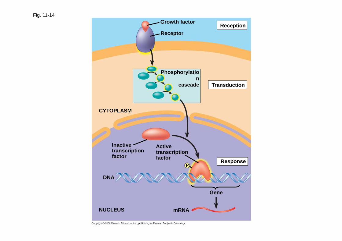
Fig. 11-14Growth factor
Receptor
Phosphorylation
cascade
Reception
Transduction
Activetranscriptionfactor
ResponseP
Inactivetranscriptionfactor
CYTOPLASM
DNA
NUCLEUS mRNA
Gene

• Other pathways regulate the activity of enzymes
Copyright © 2008 Pearson Education, Inc., publishing as Pearson Benjamin Cummings

Fig. 11-15
Reception
Transduction
Response
Binding of epinephrine to G protein-coupled recepto r (1 molecule)
Inactive G protein
Active G protein (10 2 molecules)
Inactive adenylyl cyclaseActive adenylyl cyclase (10 2)
ATPCyclic AMP (10 4)
Inactive protein kinase AActive protein kinase A (10 4)
Inactive phosphorylase kinaseActive phosphorylase kinase (10 5)
Inactive glycogen phosphorylase
Active glycogen phosphorylase (10 6)
GlycogenGlucose-1-phosphate
(108 molecules)
• Signaling pathways can also affect the physical characteristics of a cell, for example, cell shape
Copyright © 2008 Pearson Education, Inc., publishing as Pearson Benjamin Cummings

Fig. 11-16 RESULTS
CONCLUSION
Wild-type (shmoos) ∆Fus3 ∆formin
Shmoo projection forming
ForminP
ActinsubunitP
PForminFormin
Fus3
Phosphory-lationcascade
GTP
G protein-coupledreceptor
Matingfactor
GDP
Fus3 Fus3
P
Microfilament
1
2
3
4
5

Fig. 11-16a
RESULTS
Wild-type (shmoos) ∆Fus3 ∆formin

Fig. 11-16b
CONCLUSION
Matingfactor G protein-coupled
receptor
GDP GTP
Phosphory-lation
cascade
Shmoo projectionforming
Fus3
Fus3 Fus3
Formin Formin
P
P
P
ForminP
Actinsubunit
Microfilament
1
2
3
4
5
Fine-Tuning of the Response
• Multistep pathways have two important benefits:
– Amplifying the signal (and thus the response)
– Contributing to the specificity of the response
Copyright © 2008 Pearson Education, Inc., publishing as Pearson Benjamin Cummings
Signal Amplification
• Enzyme cascades amplify the cell’s response
• At each step, the number of activated products is much greater than in the preceding step
Copyright © 2008 Pearson Education, Inc., publishing as Pearson Benjamin Cummings

The Specificity of Cell Signaling and Coordination of the Response
• Different kinds of cells have different collections of proteins
• These different proteins allow cells to detect and respond to different signals
• Even the same signal can have different effects in cells with different proteins and pathways
• Pathway branching and “cross-talk” further help the cell coordinate incoming signals
Copyright © 2008 Pearson Education, Inc., publishing as Pearson Benjamin Cummings
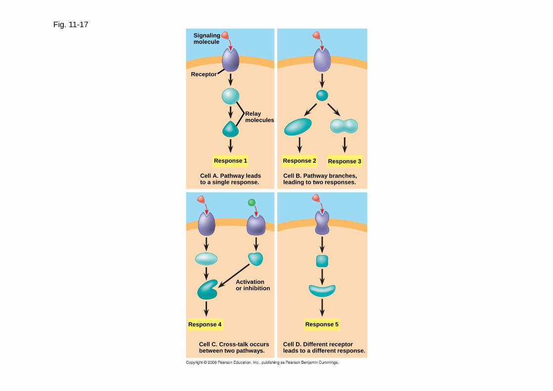
Fig. 11-17Signalingmolecule
Receptor
Relaymolecules
Response 1
Cell A. Pathway leadsto a single response.
Response 2 Response 3
Cell B. Pathway branches,leading to two responses.
Response 4 Response 5
Activationor inhibition
Cell C. Cross-talk occursbetween two pathways.
Cell D. Different receptorleads to a different response.

Fig. 11-17a
Signalingmolecule
Receptor
Relaymolecules
Response 1
Cell A. Pathway leadsto a single response.
Cell B. Pathway branches,leading to two responses.
Response 2 Response 3

Fig. 11-17b
Response 4 Response 5
Activationor inhibition
Cell C. Cross-talk occursbetween two pathways.
Cell D. Different receptorleads to a different response.

Signaling Efficiency: Scaffolding Proteins and Signaling Complexes
• Scaffolding proteins are large relay proteins to which other relay proteins are attached
• Scaffolding proteins can increase the signal transduction efficiency by grouping together different proteins involved in the same pathway
Copyright © 2008 Pearson Education, Inc., publishing as Pearson Benjamin Cummings
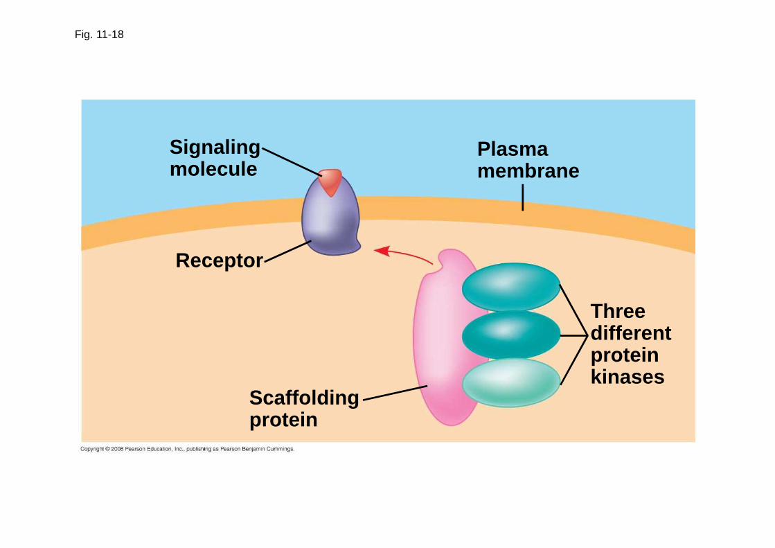
Fig. 11-18
Signalingmolecule
Receptor
Scaffoldingprotein
Plasmamembrane
Threedifferentproteinkinases
Termination of the Signal
• Inactivation mechanisms are an essential aspect of cell signaling
• When signal molecules leave the receptor, the receptor reverts to its inactive state
Copyright © 2008 Pearson Education, Inc., publishing as Pearson Benjamin Cummings
Concept 11.5: Apoptosis (programmed cell death) integrates multiple cell-signaling pathways
• Apoptosis is programmed or controlled cell suicide
• A cell is chopped and packaged into vesicles that are digested by scavenger cells
• Apoptosis prevents enzymes from leaking out of a dying cell and damaging neighboring cells
Copyright © 2008 Pearson Education, Inc., publishing as Pearson Benjamin Cummings
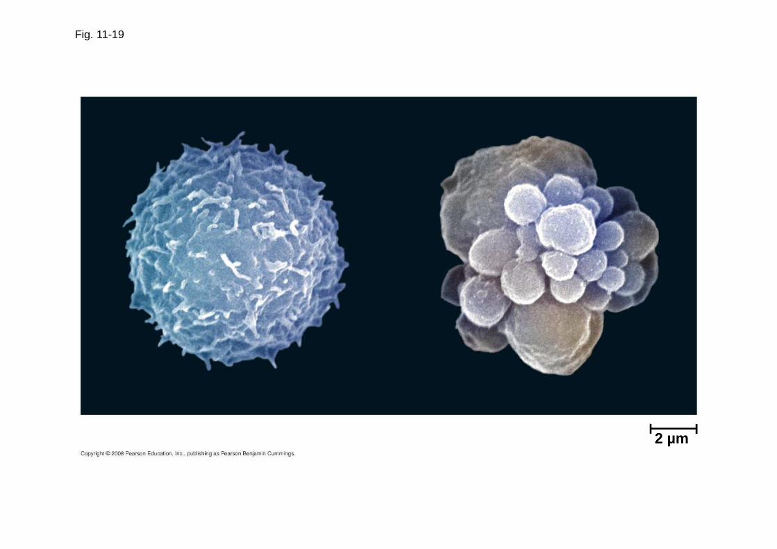
Fig. 11-19
2 µm
Apoptosis in the Soil Worm Caenorhabditis elegans
• Apoptosis is important in shaping an organism during embryonic development
• The role of apoptosis in embryonic development was first studied in Caenorhabditis elegans
• In C. elegans, apoptosis results when specific proteins that “accelerate” apoptosis override those that “put the brakes” on apoptosis
Copyright © 2008 Pearson Education, Inc., publishing as Pearson Benjamin Cummings

Fig. 11-20
Ced-9protein (active)inhibits Ced-4activity
Mitochondrion
Receptorfor death-signalingmolecule
Ced-4 Ced-3
Inactive proteins
(a) No death signal
Ced-9(inactive)
Cellformsblebs
Death-signalingmolecule
Otherproteases
ActiveCed-4
ActiveCed-3
NucleasesActivationcascade
(b) Death signal

Fig. 11-20a
Ced-9protein (active)inhibits Ced-4activity
Mitochondrion
Ced-4 Ced-3Receptorfor death-signalingmolecule
Inactive proteins
(a) No death signal

Fig. 11-20b
(b) Death signal
Death-signalingmolecule
Ced-9(inactive)
Cellformsblebs
ActiveCed-4
ActiveCed-3
Activationcascade
Otherproteases
Nucleases
Apoptotic Pathways and the Signals That Trigger Them
• Caspases are the main proteases (enzymes that cut up proteins) that carry out apoptosis
• Apoptosis can be triggered by:
– An extracellular death-signaling ligand
– DNA damage in the nucleus
– Protein misfolding in the endoplasmic reticulum
Copyright © 2008 Pearson Education, Inc., publishing as Pearson Benjamin Cummings

• Apoptosis evolved early in animal evolution and is essential for the development and maintenance of all animals
• Apoptosis may be involved in some diseases (for example, Parkinson’s and Alzheimer’s); interference with apoptosis may contribute to some cancers
Copyright © 2008 Pearson Education, Inc., publishing as Pearson Benjamin Cummings
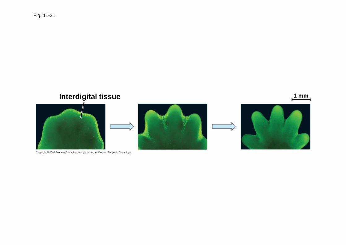
Fig. 11-21
Interdigital tissue 1 mm

Fig. 11-UN1
Reception Transduction Response
Receptor
Relay molecules
Signalingmolecule
Activationof cellularresponse
1 2 3

Fig. 11-UN2

You should now be able to:
1. Describe the nature of a ligand-receptor interaction and state how such interactions initiate a signal-transduction system
2. Compare and contrast G protein-coupled receptors, tyrosine kinase receptors, and ligand-gated ion channels
3. List two advantages of a multistep pathway in the transduction stage of cell signaling
4. Explain how an original signal molecule can produce a cellular response when it may not even enter the target cell
Copyright © 2008 Pearson Education, Inc., publishing as Pearson Benjamin Cummings
5. Define the term second messenger; briefly describe the role of these molecules in signaling pathways
6. Explain why different types of cells may respond differently to the same signal molecule
7. Describe the role of apoptosis in normal development and degenerative disease in vertebrates
Copyright © 2008 Pearson Education, Inc., publishing as Pearson Benjamin Cummings
Related Documents