Homeostasis • Homeostasis – the maintenance of normal physiological functions – Regulation of body temperature – Maintenance of blood pressure – Cell turnover – Tissue repair – Immune system fighting off diseases

Welcome message from author
This document is posted to help you gain knowledge. Please leave a comment to let me know what you think about it! Share it to your friends and learn new things together.
Transcript
Homeostasis
• Homeostasis – the maintenance of normal physiological functions– Regulation of body temperature
– Maintenance of blood pressure
– Cell turnover
– Tissue repair
– Immune system fighting off diseases

What Does Healing Involve?
• The body’s ability to repair itself is essential
• It involves:– prevents excessive bleeding
– removes exogenous debris
– promotes new tissue deposition
– allows resumption of normal physiological processes

Regeneration vs. Scarring
There are two types of tissue repair
– Regeneration
– Formation of scar tissue
The type of repair depends on the tissue

How Is Healing Handled?
Through activation of a series of processes that
– limit bleeding (blood coagulation)
– lead to clot removal (fibrinolysis)
– target foreign bacteria for removal (complement system)
– stimulate dilatation of the vasculature (kinin system)

Inflammation and Wound Healing
The immediate response to tissue injury is inflammation and wound healing which leads to...
• migration of inflammatory cells to the wound site
• release of cytokines and growth factors
• activation of kinin, complement, blood clotting, and fibrinolytic cascades
If not controlled this can lead to the deposition of fibrous scar tissue.

Inflammation and Wound Healing
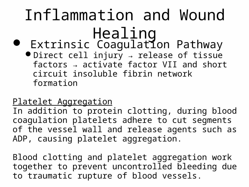
Blood Clotting & Platelet AggregationSoluble Blood Proteins involved in wound
healing Hageman factor (factor XII) fibrinogen (factor II)
Intrinsic Coagulation Pathwaytorn blood vessels → exposed fragments of collagen fibers →
activated Hageman factorActivated Hageman factor → the intrinsic blood coagulation cas
cade → an insoluble fibrin network that binds proteins and cells together.
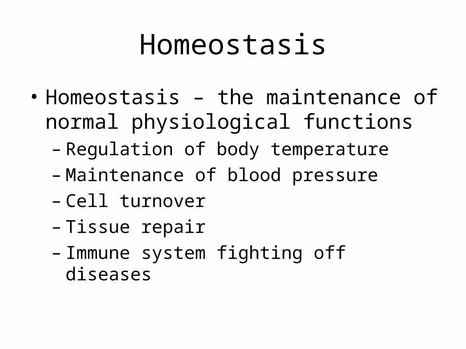
Inflammation and Wound Healing
Extrinsic Coagulation PathwayDirect cell injury → release of tissue factors → activate
factor VII and short circuit insoluble fibrin network formation
Platelet AggregationIn addition to protein clotting, during blood coagulation platelets adhere to cut segments of the vessel wall and release agents such as ADP, causing platelet aggregation.
Blood clotting and platelet aggregation work together to prevent uncontrolled bleeding due to traumatic rupture of blood vessels.
Video clip of Coagulation Cascadehttp://www.youtube.com/watch?v=cy3a__OOa2M
Inflammation and Wound Healing
Fig 13.1
Inflammation and Wound Healing
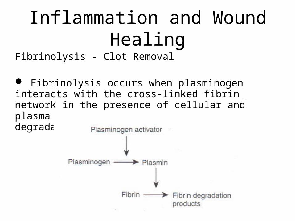
Fibrinolysis - Clot Removal
Fibrinolysis occurs when plasminogen interacts with the cross-linked fibrin network in the presence of cellular and plasma factors , which leads to the degradation of the fibrin network

The Complement System
Triggered during contact with foreign materials
Involves about 11 soluble serum proteins They can lyse bacteriaAntibody response to foreign cell surface macromolecules.
Induces an inflammatory response, so excessive complement activation is undesirable.
Inflammation and Wound Healing
Complement System
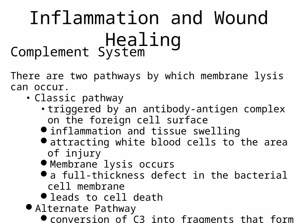
There are two pathways by which membrane lysis can occur.• Classic pathway
• triggered by an antibody-antigen complex on the foreign cell surface
inflammation and tissue swell ing attracting white blood cells to the area of injuryMembrane lysis occurs a full-thickness defect in the bacterial cell membraneleads to cell death
Alternate Pathwayconversion of C3 into fragments that form active C3b.
Inflammation and Wound Healing

Inflammation and Wound Healing
See this complement viedo: http://www.youtube.com/watch?v=_iMy50AxyZE
Red Blood Cells
enucleated cellssurrounded by a deformable membranein the form of a diskfilled with the protein hemoglobincomprise about 45% of the blood volumecirculate for about 120 dayThen they are removed by the reticuloendothelial systemGive clots thier characteristic red color
Cells Involved In Wound Healing

White Blood Cells [Leukocytes]
found at a concentration of 4,000 to 10,000 per milliliter of blood
several sub-classifications of leukocytesNeutrophils - 40 to 75% Lymphocytes - 20 to 45%Monocytes - 2 to 10%Eosinophils - 1 to 6%Basophils - less than 1%
Cells Involved In Wound Healing

White Blood Cells [Leukocytes] – Neutrophils
Phagocytic cells that digest foreign and dead cellular and noncellular material
Neutrophils contain granules in the cytoplasm, called lysosomesThey are used to break down bacteriadenatured proteins, and other foreign or worn-out materials
Neutrophils are found in peripheral blood and small blood vessels
Within 12 hours of injury, they migrate to the injury sitesUndergo phagocytosis for a period of four to five daysIf the injury persists for more than five days, then neutrophils are
replaced by monocytes at the wound site.
Cells Involved In Wound Healing

White Blood Cells [Leukocytes] – Monocytes
Phagocytic cells that digest foreign and dead cellular and noncellular material
Larger than other leukocytes Released into the circulation from the bone marrowmigrate into the liver, spleen, lymph nodes, and lungs where
they comprise the macrophages of the reticuloendothelial systemIn sites of injury, macrophages remove foreign and worn-out
materials by the process of phagocytosis.
Cells Involved In Wound Healing

White Blood Cells [Leukocytes] – Lymphocytes
associated with immunological responses to foreign materials subclassified as B and T cells
B (bone marrow derived) cellsare transformed on contact with foreign antigens into
plasma cells that make antibodies which bind to foreign cells and promote cell death, or they form antibody
T (thymus derived) cellsparticipate in cell-mediated immunity by helping or
suppressing antibody production or by promoting cell lysis
Cells Involved In Wound Healing

Plateletslack a nucleus and stick to cut blood vessels
present at a concentration of 150,000 to 400,000 per milliliter of blood
play an active role in plugging leaky vessels in conjunction with fibrin
Cells Involved In Wound Healing

Injury to cells and tissues as well as activation of the kinin, complement, and fibrinolytic systems causes vasodilation, an influx of inflammatory cells
Results in vascular permeability and the ability of white blood cells to permeate through blood vessel walls into the wound area
Denatured collagen and fibrin degradation products help promote migration of the white blood cells
Wound Healing clips:http://www.youtube.com/watch?v=0TvTyj5FAaQ http://www.youtube.com/watch?v=-_wddQWG32A
Wound Healing

The normal defense against foreign matter consists of natural and acquired immunity.
Natural immunity involves no prior exposure to the foreign material and is not enhanced by exposure. This type of immunity involves the inflammatory process, including neutrophils and macrophages.
Acquired immunity is specific and requires exposure to a foreign material, an antigen, and is magnified by a subsequent exposure to the antigen.
Wound Healing - Immunity

T cells are characterized at different stages of development by specific cell-surface markers including T-helper cells, T-suppressor cells, CD4+, and CD8+.
Antibody-producing cells in the presence of T-helper cells up regulate antibody production to foreign cells. T-suppressor cells down regulate the production of antibodies by plasma cells. CD4+ T cells secrete proinflammatory lymphokines such as interleukin 1 (IL-1) and recognize antigens on foreign cells containing class II major histocompatibility complex (MHC) markers CD8+ T cells secrete molecules that exert suppressor and cyto-toxic functions, and these cells recognize class I MHC markers on foreign cells.
Wound Healing – T Cells

Cytolytic T cells are another subset of T cells that kill target cells expressing specific antigens on the cell membrane. Most cytolytic T cells are CD8+ and recognize class I MHC products expressed on the surface of foreign cells. Cytolytic T cells react with class I MHC markers and release a cytotoxin that leads to target cell lysis.
In comparison, a third type of lymphocyte, called null cells, include natural killer cells (NK). Found in blood and lymphoid tissue (lymph nodes and spleen) that lack specific receptors for antigen recognition on the surface of foreign cells. NK cells possess the ability to kill certain tumor cells or cells infected by viruses and are not antigen dependent.
Wound Healing – Natural Killer Cells
Related Documents


![$1RYHO2SWLRQ &KDSWHU $ORN6KDUPD +HPDQJL6DQH … · 1 1 1 1 1 1 1 ¢1 1 1 1 1 ¢ 1 1 1 1 1 1 1w1¼1wv]1 1 1 1 1 1 1 1 1 1 1 1 1 ï1 ð1 1 1 1 1 3](https://static.cupdf.com/doc/110x72/5f3ff1245bf7aa711f5af641/1ryho2swlrq-kdswhu-orn6kdupd-hpdqjl6dqh-1-1-1-1-1-1-1-1-1-1-1-1-1-1.jpg)