APPLIED AND ENVIRONMENTAL MICROBIOLOGY, Nov. 2010, p. 7635–7640 Vol. 76, No. 22 0099-2240/10/$12.00 doi:10.1128/AEM.01188-10 Copyright © 2010, American Society for Microbiology. All Rights Reserved. Stable, Site-Specific Fluorescent Tagging Constructs Optimized for Burkholderia Species Michael H. Norris, 2 Yun Kang, 2 Bruce Wilcox, 3 and Tung T. Hoang 1,2 * Department of Microbiology, 1 Department of Molecular Biosciences and Bioengineering, 2 and Department of Ecology and Health, 3 University of Hawaii at Manoa, Honolulu, Hawaii 96822 Received 18 May 2010/Accepted 4 September 2010 Several vectors that facilitate stable fluorescent labeling of Burkholderia pseudomallei and Burkholderia thailandensis were constructed. These vectors combined the effectiveness of the mini-Tn7 site-specific transpo- sition system with fluorescent proteins optimized for Burkholderia spp., enabling bacterial tracking during cellular infection. Burkholderia pseudomallei is a highly infectious Gram-nega- tive bacterium and a facultative intracellular pathogen. The ability to observe infectious processes of this bacterium at various stages is critical to understand its pathogenesis. Fluo- rescent proteins facilitate bacterial tagging and have been pow- erful investigative tools in deciphering biological processes (3, 23, 24, 29, 31, 32). However, the lack of optimized fluorescent constructs used to label B. pseudomallei for visualization ne- cessitates further development. Although there are many flu- orescent tools available besides the green fluorescent protein (GFP), they are not optimized for use in B. pseudomallei or the less pathogenic model species Burkholderia thailandensis. Commercially available fluorescent proteins are optimized for eukaryotic expression or, at best, for bacteria with low-G/C- content genomes, and hence codon preference may cause problems (13, 19) during protein expression in Burkholderia spp. Also, there is usually an ineffective promoter driving tran- scription in Burkholderia spp., and available constructs usually replicate plasmids that require selective maintenance. The re- stricted use of antibiotic markers in select agents (e.g., B. pseudomallei) adds another level of complexity to the genetic manipulation of these species (27). Hence, these obstacles have limited the applications of fluorescent proteins in patho- genesis studies of Burkholderia spp. The well-established mini-Tn7 system (7, 14, 18) inserts it- self at a unique neutral site(s) in the bacterial genome with the aid of a nonreplicating helper plasmid encoding the trans- posase (1). The B. pseudomallei chromosome contains three insertion sites downstream of three glmS genes, whereas B. thailandensis contains two insertion sites downstream of two glmS genes (8). Mini-Tn7 inserted in bacterial genomes is quite stable (7, 20), and selective maintenance is not required. In this study, we constructed and demonstrated the use of fluorescent proteins encoded on mini-Tn7-based site-specific transposition vectors for fluorescent tagging of B. pseudomallei and B. thai- landensis. We optimized for B. pseudomallei the fluorescent protein genes (cyan, red, and yellow), which were synthesized through Genscript Corporation based on the amino acid se- quences from Evrogen, by driving their transcription with a P S12 promoter (36) and changing the codons to those preferred by B. pseudomallei. Since enhanced GFP (eGFP) is sufficiently bright in Burkholderia spp., we utilized the gfp gene driven by the P S12 promoter (Table 1). The four new fluorescent proteins (green, cyan, red, and yellow fluorescent proteins [GFP, CYP, RFP, and YFP, respectively]) were combined with two mini- Tn7 backbones to produce eight fluorescent tagging vectors (Fig. 1 and Table 1). The first series of four vectors encode the nonantibiotic selectable marker gat (resistance to glyphosate) (5, 21), and the other series of vectors encode kanamycin resistance (Kan r ) (Fig. 1). Both are currently approved select- able markers for B. pseudomallei (8, 21). Nevertheless, prior CDC/USDA approval for each laboratory to use these markers is necessary and must be sought. All manipulations of B. pseudomallei were carried out in a CDC/USDA-approved bio- safety level 3 laboratory by following the guidelines presented in Biosafety in Microbiological and Biomedical Laboratories, 5th ed. (34). The four vectors based on gat were used to tag B. pseudoma- llei 1026b (Fig. 1). To introduce the fluorescent tags, triparen- tal matings were conducted using an Escherichia coli donor (E1354) (Table 1) carrying a helper plasmid (pTNS3-asd Ec ), B. pseudomallei 1026b, and one of the four fluorescent vectors shown in Fig. 1, as previously described (21). B. pseudomallei containing the inserted transposon was selected for on 1 M9 minimal glucose medium containing 0.3% (vol/vol) glyphosate, as previously described (21). Colonies appeared 2 days later and were purified on the same medium. Insertion at one of the three glmS sites in the chromosome was verified by PCR as described previously (8, 17, 21). B. pseudomallei could be la- beled, and all four colors could be observed as shown in Fig. 2. To visualize fluorescently labeled bacteria, all samples were fixed in 1% paraformaldehyde based on previously published protocols for 30 min (6, 25). Fluorescent microscopy was car- ried out using the suggested filter cube sets shown in Table 2. As our laboratory has not applied for approval to introduce Kan r genes into B. pseudomallei, we used the gat-based con- structs to tag B. pseudomallei for the rest of our experiments. Regardless, the four other fluorescent vectors based on Kan r * Corresponding author. Mailing address: Department of Molecular Biosciences and Bioengineering, University of Hawaii at Manoa, Ho- nolulu, HI 96822. Phone: (808) 956-3522. Fax: (808) 956-5339. E-mail: [email protected]. Published ahead of print on 17 September 2010. 7635 Downloaded from https://journals.asm.org/journal/aem on 25 December 2021 by 179.97.19.193.

Welcome message from author
This document is posted to help you gain knowledge. Please leave a comment to let me know what you think about it! Share it to your friends and learn new things together.
Transcript

APPLIED AND ENVIRONMENTAL MICROBIOLOGY, Nov. 2010, p. 7635–7640 Vol. 76, No. 220099-2240/10/$12.00 doi:10.1128/AEM.01188-10Copyright © 2010, American Society for Microbiology. All Rights Reserved.
Stable, Site-Specific Fluorescent Tagging ConstructsOptimized for Burkholderia Species�
Michael H. Norris,2 Yun Kang,2 Bruce Wilcox,3 and Tung T. Hoang1,2*Department of Microbiology,1 Department of Molecular Biosciences and Bioengineering,2 and Department of
Ecology and Health,3 University of Hawaii at Manoa, Honolulu, Hawaii 96822
Received 18 May 2010/Accepted 4 September 2010
Several vectors that facilitate stable fluorescent labeling of Burkholderia pseudomallei and Burkholderiathailandensis were constructed. These vectors combined the effectiveness of the mini-Tn7 site-specific transpo-sition system with fluorescent proteins optimized for Burkholderia spp., enabling bacterial tracking duringcellular infection.
Burkholderia pseudomallei is a highly infectious Gram-nega-tive bacterium and a facultative intracellular pathogen. Theability to observe infectious processes of this bacterium atvarious stages is critical to understand its pathogenesis. Fluo-rescent proteins facilitate bacterial tagging and have been pow-erful investigative tools in deciphering biological processes (3,23, 24, 29, 31, 32). However, the lack of optimized fluorescentconstructs used to label B. pseudomallei for visualization ne-cessitates further development. Although there are many flu-orescent tools available besides the green fluorescent protein(GFP), they are not optimized for use in B. pseudomallei or theless pathogenic model species Burkholderia thailandensis.Commercially available fluorescent proteins are optimized foreukaryotic expression or, at best, for bacteria with low-G/C-content genomes, and hence codon preference may causeproblems (13, 19) during protein expression in Burkholderiaspp. Also, there is usually an ineffective promoter driving tran-scription in Burkholderia spp., and available constructs usuallyreplicate plasmids that require selective maintenance. The re-stricted use of antibiotic markers in select agents (e.g., B.pseudomallei) adds another level of complexity to the geneticmanipulation of these species (27). Hence, these obstacleshave limited the applications of fluorescent proteins in patho-genesis studies of Burkholderia spp.
The well-established mini-Tn7 system (7, 14, 18) inserts it-self at a unique neutral site(s) in the bacterial genome with theaid of a nonreplicating helper plasmid encoding the trans-posase (1). The B. pseudomallei chromosome contains threeinsertion sites downstream of three glmS genes, whereas B.thailandensis contains two insertion sites downstream of twoglmS genes (8). Mini-Tn7 inserted in bacterial genomes is quitestable (7, 20), and selective maintenance is not required. In thisstudy, we constructed and demonstrated the use of fluorescentproteins encoded on mini-Tn7-based site-specific transpositionvectors for fluorescent tagging of B. pseudomallei and B. thai-landensis. We optimized for B. pseudomallei the fluorescent
protein genes (cyan, red, and yellow), which were synthesizedthrough Genscript Corporation based on the amino acid se-quences from Evrogen, by driving their transcription with aPS12 promoter (36) and changing the codons to those preferredby B. pseudomallei. Since enhanced GFP (eGFP) is sufficientlybright in Burkholderia spp., we utilized the gfp gene driven bythe PS12 promoter (Table 1). The four new fluorescent proteins(green, cyan, red, and yellow fluorescent proteins [GFP, CYP,RFP, and YFP, respectively]) were combined with two mini-Tn7 backbones to produce eight fluorescent tagging vectors(Fig. 1 and Table 1). The first series of four vectors encode thenonantibiotic selectable marker gat (resistance to glyphosate)(5, 21), and the other series of vectors encode kanamycinresistance (Kanr) (Fig. 1). Both are currently approved select-able markers for B. pseudomallei (8, 21). Nevertheless, priorCDC/USDA approval for each laboratory to use these markersis necessary and must be sought. All manipulations of B.pseudomallei were carried out in a CDC/USDA-approved bio-safety level 3 laboratory by following the guidelines presentedin Biosafety in Microbiological and Biomedical Laboratories, 5thed. (34).
The four vectors based on gat were used to tag B. pseudoma-llei 1026b (Fig. 1). To introduce the fluorescent tags, triparen-tal matings were conducted using an Escherichia coli donor(E1354) (Table 1) carrying a helper plasmid (pTNS3-asdEc), B.pseudomallei 1026b, and one of the four fluorescent vectorsshown in Fig. 1, as previously described (21). B. pseudomalleicontaining the inserted transposon was selected for on 1� M9minimal glucose medium containing 0.3% (vol/vol) glyphosate,as previously described (21). Colonies appeared �2 days laterand were purified on the same medium. Insertion at one of thethree glmS sites in the chromosome was verified by PCR asdescribed previously (8, 17, 21). B. pseudomallei could be la-beled, and all four colors could be observed as shown in Fig. 2.To visualize fluorescently labeled bacteria, all samples werefixed in 1% paraformaldehyde based on previously publishedprotocols for 30 min (6, 25). Fluorescent microscopy was car-ried out using the suggested filter cube sets shown in Table 2.As our laboratory has not applied for approval to introduceKanr genes into B. pseudomallei, we used the gat-based con-structs to tag B. pseudomallei for the rest of our experiments.Regardless, the four other fluorescent vectors based on Kanr
* Corresponding author. Mailing address: Department of MolecularBiosciences and Bioengineering, University of Hawaii at Manoa, Ho-nolulu, HI 96822. Phone: (808) 956-3522. Fax: (808) 956-5339. E-mail:[email protected].
� Published ahead of print on 17 September 2010.
7635
Dow
nloa
ded
from
http
s://j
ourn
als.
asm
.org
/jour
nal/a
em o
n 25
Dec
embe
r 20
21 b
y 17
9.97
.19.
193.
(Fig. 1) were constructed for use in B. pseudomallei by thoselaboratories with appropriate USDA/CDC approval, and wehave validated proper transposition and fluorescence in B.thailandensis, as well as in Burkholderia cenocepacia strainK56-2 and Pseudomonas aeruginosa strain PAO1 (data notshown).
The ability to tag Burkholderia spp. with different colorscould facilitate studies where one or more strains expressingdifferent colors could be located or tracked. To demonstrate
this, we took advantage of the intracellular replication of B.pseudomallei by using two of the fluorescent strains engineeredabove (e.g., green and red) to infect the murine macrophagecell line RAW 264.7 in a modified aminoglycoside protectionassay (16). Briefly, the respective B. pseudomallei cultures weregrown overnight and used to infect the RAW 264.7 cell mono-layers at a multiplicity of infection (MOI) of 10:1 for 1 h (4, 26,28, 30, 33). Afterwards, the extracellular bacteria were re-moved and the monolayers were washed twice with 1� phos-
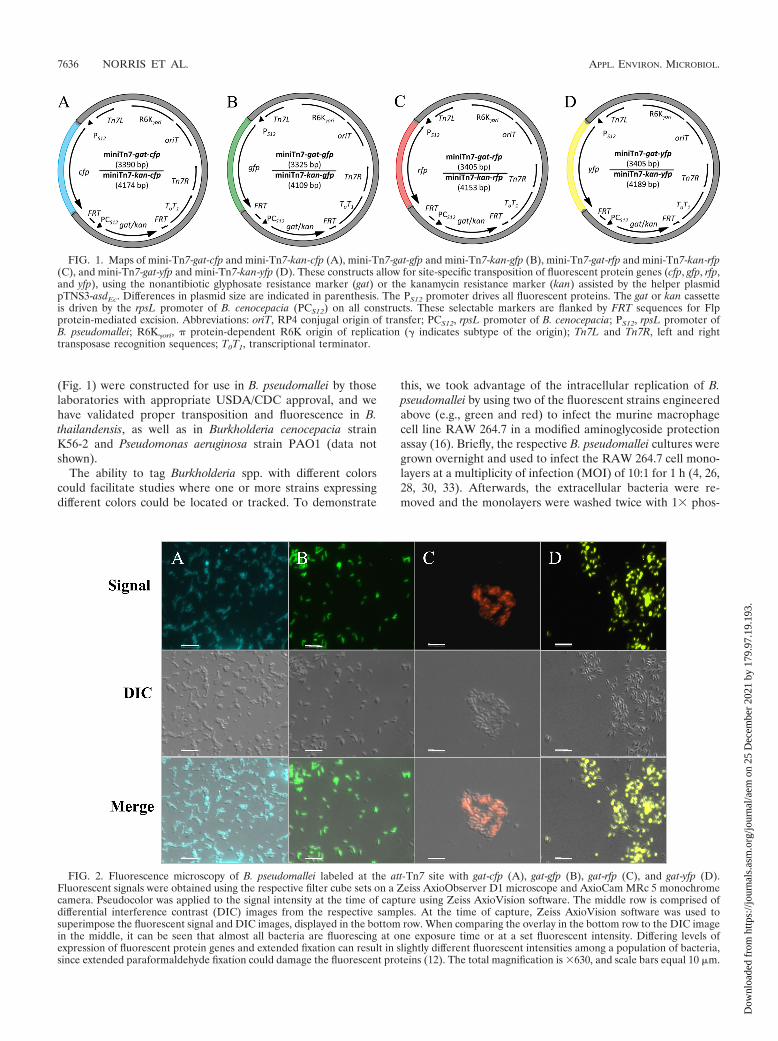
FIG. 1. Maps of mini-Tn7-gat-cfp and mini-Tn7-kan-cfp (A), mini-Tn7-gat-gfp and mini-Tn7-kan-gfp (B), mini-Tn7-gat-rfp and mini-Tn7-kan-rfp(C), and mini-Tn7-gat-yfp and mini-Tn7-kan-yfp (D). These constructs allow for site-specific transposition of fluorescent protein genes (cfp, gfp, rfp,and yfp), using the nonantibiotic glyphosate resistance marker (gat) or the kanamycin resistance marker (kan) assisted by the helper plasmidpTNS3-asdEc. Differences in plasmid size are indicated in parenthesis. The PS12 promoter drives all fluorescent proteins. The gat or kan cassetteis driven by the rpsL promoter of B. cenocepacia (PCS12) on all constructs. These selectable markers are flanked by FRT sequences for Flpprotein-mediated excision. Abbreviations: oriT, RP4 conjugal origin of transfer; PCS12, rpsL promoter of B. cenocepacia; PS12, rpsL promoter ofB. pseudomallei; R6K�ori, � protein-dependent R6K origin of replication (� indicates subtype of the origin); Tn7L and Tn7R, left and righttransposase recognition sequences; T0T1, transcriptional terminator.
FIG. 2. Fluorescence microscopy of B. pseudomallei labeled at the att-Tn7 site with gat-cfp (A), gat-gfp (B), gat-rfp (C), and gat-yfp (D).Fluorescent signals were obtained using the respective filter cube sets on a Zeiss AxioObserver D1 microscope and AxioCam MRc 5 monochromecamera. Pseudocolor was applied to the signal intensity at the time of capture using Zeiss AxioVision software. The middle row is comprised ofdifferential interference contrast (DIC) images from the respective samples. At the time of capture, Zeiss AxioVision software was used tosuperimpose the fluorescent signal and DIC images, displayed in the bottom row. When comparing the overlay in the bottom row to the DIC imagein the middle, it can be seen that almost all bacteria are fluorescing at one exposure time or at a set fluorescent intensity. Differing levels ofexpression of fluorescent protein genes and extended fixation can result in slightly different fluorescent intensities among a population of bacteria,since extended paraformaldehyde fixation could damage the fluorescent proteins (12). The total magnification is �630, and scale bars equal 10 �m.
7636 NORRIS ET AL. APPL. ENVIRON. MICROBIOL.
Dow
nloa
ded
from
http
s://j
ourn
als.
asm
.org
/jour
nal/a
em o
n 25
Dec
embe
r 20
21 b
y 17
9.97
.19.
193.
TABLE 1. Bacterial strains and plasmids used in this studya
Strain or plasmid Lab IDb GenBank IDno. Relevant properties Source or
reference(s)
StrainsE. coli
EPMax10B-pir116 �asd�trp::Gmr mob-Kanr
E1354 Gmr Kanr F� �� mcrA�(mrr-hsdRMS-mcrBC) 80dlacZ�M15�lacX74 deoR recA1 endA1 araD139 �(ara leu)7697 galUgalK rpsL nupG Tn-pir116-FRT2 �asd::wFRT�trp::Gmr-FRT5 mobrecA::RP4-2 Tc::Mu-Kanr�
Available labstrain
EPMax10B-lacIq pir E1869 F� �� mcrA �(mrr-hsdRMS-mcrBC) 80dlacZ�M15 �lacX74deoR recA1 endA1 araD139 �(ara leu)7697 galU galK rpsLnupG lacIq-FRT8 pir-FRT4
Available labstrain
B. pseudomallei1026b B0004 Clinical melioidosis isolate 101026b/att-Tn7-gat-cfp B0036 GSr, 1026b with mini-Tn7-gat-cfp inserted This study1026b/att-Tn7-gat-gfp B0037 GSr, 1026b with mini-Tn7-gat-gfp inserted This study1026b/att-Tn7-gat-rfp B0038 GSr, 1026b with mini-Tn7-gat-rfp inserted This study1026b/att-Tn7-gat-yfp B0039 GSr, 1026b with mini-Tn7-gat-yfp inserted This study
B. thailandensisE264 E1298 Prototroph, environmental isolate 2E264/att-Tn7-kan-cfp E2552 Kanr, E264 with mini-Tn7-kan-cfp inserted This studyE264/att-Tn7-kan-gfp E2491 Kanr, E264 with mini-Tn7-kan-gfp inserted This studyE264/att-Tn7-kan-rfp E2553 Kanr, E264 with mini-Tn7-kan-rfp inserted This studyE264/att-Tn7-kan-yfp E2554 Kanr, E264 with mini-Tn7-kan-yfp inserted This study
Plasmidsc
mini-Tn7-gat E1981 GSr, mini-Tn7 integration vector based on gat 21mini-Tn7-gat-cfp E2460 HM150704 GSr, mini-Tn7-gat harboring cfp This studymini-Tn7-gat-gfp E2462 HM150705 GSr, mini-Tn7-gat harboring gfp This studymini-Tn7-gat-rfp E2326 HM150706 GSr, mini-Tn7-gat harboring rfp This studymini-Tn7-gat-yfp E2468 HM150707 GSr, mini-Tn7-gat harboring yfp This studymini-Tn7-kan-cfp E2480 HM150708 Kanr, mini-Tn7-kan harboring cfp This studymini-Tn7-kan-gfp E2482 HM150709 Kanr, mini-Tn7-kan harboring gfp This studymini-Tn7-kan-rfp E2486 HM150710 Kanr, mini-Tn7-kan harboring rfp This studymini-Tn7-kan-yfp E2488 HM150711 Kanr, mini-Tn7-kan harboring yfp This studypPS747 E0042 Apr, plasmid harboring eGFP 9, 15pRK2013 E1358 Kanr, helper plasmid encoding conjugative proteins 11pTNS3-asdEc E2237 Suicidal helper plasmid containing E. coli asd and transposase
for the Tn7 site-specific transposition system17
pUC57-PS12-cfp E1739 HM150701 Apr, plasmid harboring B. pseudomallei codon-optimized cfpdriven by PS12
This study
pUC57-PS12-rfp E1735 HM150702 Apr, plasmid harboring B. pseudomallei codon-optimized rfpdriven by PS12
This study
pUC57-PS12-yfp E1738 HM150703 Apr, plasmid harboring B. pseudomallei codon-optimized yfpdriven by PS12
This study
a Abbreviations: Apr, ampicillin resistant; asdEc, E. coli asd gene; gat, gene encoding glyphosate acetyltransferase; Gmr, gentamicin resistant; GSr, glyphosate resistant;kan, gene encoding kanamycin resistance; Kanr, kanamycin resistant; PS12, rpsL promoter from B. pseudomallei; w, wild type.
b Please use the laboratory identification number (Lab ID) when requesting strains and plasmids.c The mini-Tn7 plasmid backbone is based on pUC18R6KT-mini-Tn7T (7). Fluorescent protein genes (cfp, rfp, yfp) were synthesized by GeneScript and cloned into
the EcoRV site of pUC57. These genes were removed by digestion with KpnI and PstI and then subcloned into mini-Tn7-gat cut with the same enzymes. Two-step PCRwas performed to incorporate the PS12 promoter upstream of the gfp gene, which was digested with KpnI and PstI and cloned into mini-Tn7-gat that had been digestedwith the same enzymes. The mini-Tn7-kan constructs were created by replacing the gat cassette with the kan cassette.
TABLE 2. Fluorescent-protein characteristicsa
Fluorescent protein (original source)Excitationmaximum
(nm)
Emissionmaximum
(nm)Recommended fluorescence microscopy filter set(s)
Speed ofmaturation at
37°C
Tag CFP (Aequorea macrodactyla) 458 480 Omega Optical set XF114-2 or XF130-2 FasteGFP (Aequorea victoria) 488 509 Omega Optical set XF116-2 or Chroma Technology
set 41017Fast
Turbo YFP (Phialidium sp.) 525 538 Omega Optica set XF104-3 or Chroma Technologyset 42003
Superfast
Turbo RFP (Entacmaea quadricolor) 553 574 Omega Optical sets QMAX-Yellow, XF108-2,XF101-2, and XF111-2
Superfast
a These characteristics were found on the Evrogen website.
VOL. 76, 2010 FLUORESCENT TAGGING CONSTRUCTS FOR BURKHOLDERIA SPP. 7637
Dow
nloa
ded
from
http
s://j
ourn
als.
asm
.org
/jour
nal/a
em o
n 25
Dec
embe
r 20
21 b
y 17
9.97
.19.
193.

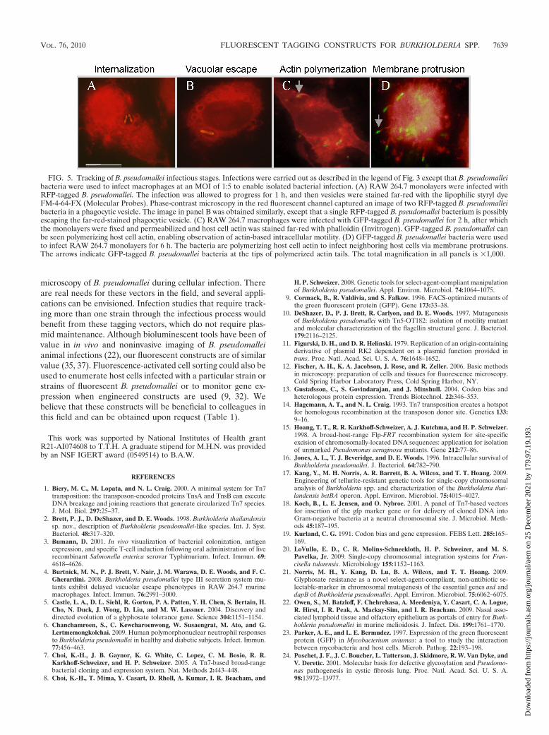
phate-buffered saline (PBS). Fresh Dulbecco’s modified Ea-gle’s medium (DMEM) containing 750 �g/ml amikacin and750 �g/ml kanamycin was then added to inhibit extracellularbacterial replication. At 7 h postinfection, the monolayers werefixed with 1% (wt/vol) paraformaldehyde for 30 min and visu-alized using fluorescence microscopy (6, 25). As Fig. 3 indi-cates, B. pseudomallei cells tagged with different colors wereeasily distinguishable within the murine macrophage monolay-ers and can be seen inside host cells as the bacteria replicate.When both green and red fluorescent B. pseudomallei cells aremixed together and used to infect a murine macrophage mono-layer at a total MOI of 10:1, differently colored bacteria can bedistinguished and neighboring bacteria of either color can bedifferentiated from one another (Fig. 4). To observe the dif-ferent infectious stages with fluorescently tagged B. pseudoma-llei, RAW 264.7 macrophages were infected at an MOI of 1:5(1 bacterium per 5 host cells). In Fig. 5A and B, the host cellswere infected with RFP-tagged B. pseudomallei, fixed, perme-abilized, and then stained with the far-red lipophilic styryl dyeFM 4-64-FX (Molecular Probes). This stains all lipid bilayersfar-red, including vacuoles, leaving the slightly orange color ofthe RFP-tagged B. pseudomallei visible (i) in the macrophagevesicle (Fig. 5A) and (ii) during vesicular escape (Fig. 5B).Alternatively, B. pseudomallei could be labeled with GFP forvisualization (Fig. 5C and D). By infecting the macrophages atan MOI of 1:5 with GFP-tagged B. pseudomallei and thenstaining host cell actin far-red, one can visualize bacterial rep-lication in the cytoplasm (Fig. 5C) and the formation of actintails during protrusion from the host cell (Fig. 5D).
In summary, we have constructed and demonstrated the useof transposon vectors for site-specific stable fluorescent taggingof B. pseudomallei with four unique colors. These tools will bebeneficial for microbiological studies involving the tracking or
FIG. 3. Fluorescence microscopy of B. pseudomallei labeled at the att-Tn7 site with gat-cfp (A), gat-gfp (B), gat-rfp (C), and gat-yfp (D) to infectthe murine macrophage-like cell line RAW 264.7. Cell monolayers were seeded overnight onto poly-L-lysine-coated coverslips at the bottom of a6-well plate and infected with fluorescently tagged B. pseudomallei. Images were obtained as described in the legend of Fig. 2. The totalmagnification is �630, and scale bars equal 10 �m.
FIG. 4. Dual infection of RAW 264.7 macrophages by differentiallylabeled (green and red) B. pseudomallei bacteria. Infections were carriedout identically to those described in the legend of Fig. 3. (A) The greenfluorescent signal indicates where gfp-tagged B. pseudomallei bacteria arereplicating inside macrophages. (B) The red fluorescent signal was ob-tained from the same field and shows where rfp-tagged B. pseudomalleibacteria are replicating within macrophages. (C) A DIC image was thencaptured and is presented. (D) Overlay of images captured sequentially inpanels A, B, and C. Images were superimposed at the time of captureusing Zeiss AxioVision software. (E and F) Close-ups of the two macro-phages indicated by arrows in panel D, where the two differently fluoresc-ing B. pseudomallei strains are clearly visible and distinguishable withinthe macrophages and even within the same host cell. The total magnifi-cation in panels A to D is �630, and all scale bars equal 10 �m.
7638 NORRIS ET AL. APPL. ENVIRON. MICROBIOL.
Dow
nloa
ded
from
http
s://j
ourn
als.
asm
.org
/jour
nal/a
em o
n 25
Dec
embe
r 20
21 b
y 17
9.97
.19.
193.
microscopy of B. pseudomallei during cellular infection. Thereare real needs for these vectors in the field, and several appli-cations can be envisioned. Infection studies that require track-ing more than one strain through the infectious process wouldbenefit from these tagging vectors, which do not require plas-mid maintenance. Although bioluminescent tools have been ofvalue in in vivo and noninvasive imaging of B. pseudomalleianimal infections (22), our fluorescent constructs are of similarvalue (35, 37). Fluorescence-activated cell sorting could also beused to enumerate host cells infected with a particular strain orstrains of fluorescent B. pseudomallei or to monitor gene ex-pression when engineered constructs are used (9, 32). Webelieve that these constructs will be beneficial to colleagues inthis field and can be obtained upon request (Table 1).
This work was supported by National Institutes of Health grantR21-AI074608 to T.T.H. A graduate stipend for M.H.N. was providedby an NSF IGERT award (0549514) to B.A.W.
REFERENCES
1. Biery, M. C., M. Lopata, and N. L. Craig. 2000. A minimal system for Tn7transposition: the transposon-encoded proteins TnsA and TnsB can executeDNA breakage and joining reactions that generate circularized Tn7 species.J. Mol. Biol. 297:25–37.
2. Brett, P. J., D. DeShazer, and D. E. Woods. 1998. Burkholderia thailandensissp. nov., description of Burkholderia pseudomallei-like species. Int. J. Syst.Bacteriol. 48:317–320.
3. Bumann, D. 2001. In vivo visualization of bacterial colonization, antigenexpression, and specific T-cell induction following oral administration of liverecombinant Salmonella enterica serovar Typhimurium. Infect. Immun. 69:4618–4626.
4. Burtnick, M. N., P. J. Brett, V. Nair, J. M. Warawa, D. E. Woods, and F. C.Gherardini. 2008. Burkholderia pseudomallei type III secretion system mu-tants exhibit delayed vacuolar escape phenotypes in RAW 264.7 murinemacrophages. Infect. Immun. 76:2991–3000.
5. Castle, L. A., D. L. Siehl, R. Gorton, P. A. Patten, Y. H. Chen, S. Bertain, H.Cho, N. Duck, J. Wong, D. Liu, and M. W. Lassner. 2004. Discovery anddirected evolution of a glyphosate tolerance gene. Science 304:1151–1154.
6. Chanchamroen, S., C. Kewcharoenwong, W. Susaengrat, M. Ato, and G.Lertmemongkolchai. 2009. Human polymorphonuclear neutrophil responsesto Burkholderia pseudomallei in healthy and diabetic subjects. Infect. Immun.77:456–463.
7. Choi, K.-H., J. B. Gaynor, K. G. White, C. Lopez, C. M. Bosio, R. R.Karkhoff-Schweizer, and H. P. Schweizer. 2005. A Tn7-based broad-rangebacterial cloning and expression system. Nat. Methods 2:443–448.
8. Choi, K.-H., T. Mima, Y. Casart, D. Rholl, A. Kumar, I. R. Beacham, and
H. P. Schweizer. 2008. Genetic tools for select-agent-compliant manipulationof Burkholderia pseudomallei. Appl. Environ. Microbiol. 74:1064–1075.
9. Cormack, B., R. Valdivia, and S. Falkow. 1996. FACS-optimized mutants ofthe green fluorescent protein (GFP). Gene 173:33–38.
10. DeShazer, D., P. J. Brett, R. Carlyon, and D. E. Woods. 1997. Mutagenesisof Burkholderia pseudomallei with Tn5-OT182: isolation of motility mutantand molecular characterization of the flagellin structural gene. J. Bacteriol.179:2116–2125.
11. Figurski, D. H., and D. R. Helinski. 1979. Replication of an origin-containingderivative of plasmid RK2 dependent on a plasmid function provided intrans. Proc. Natl. Acad. Sci. U. S. A. 76:1648–1652.
12. Fischer, A. H., K. A. Jacobson, J. Rose, and R. Zeller. 2006. Basic methodsin microscopy: preparation of cells and tissues for fluorescence microscopy.Cold Spring Harbor Laboratory Press, Cold Spring Harbor, NY.
13. Gustafsson, C., S. Govindarajan, and J. Minshull. 2004. Codon bias andheterologous protein expression. Trends Biotechnol. 22:346–353.
14. Hagemann, A. T., and N. L. Craig. 1993. Tn7 transposition creates a hotspotfor homologous recombination at the transposon donor site. Genetics 133:9–16.
15. Hoang, T. T., R. R. Karkhoff-Schweizer, A. J. Kutchma, and H. P. Schweizer.1998. A broad-host-range Flp-FRT recombination system for site-specificexcision of chromosomally-located DNA sequences: application for isolationof unmarked Pseudomonas aeruginosa mutants. Gene 212:77–86.
16. Jones, A. L., T. J. Beveridge, and D. E. Woods. 1996. Intracellular survival ofBurkholderia pseudomallei. J. Bacteriol. 64:782–790.
17. Kang, Y., M. H. Norris, A. R. Barrett, B. A. Wilcox, and T. T. Hoang. 2009.Engineering of tellurite-resistant genetic tools for single-copy chromosomalanalysis of Burkholderia spp. and characterization of the Burkholderia thai-landensis betBA operon. Appl. Environ. Microbiol. 75:4015–4027.
18. Koch, B., L. E. Jensen, and O. Nybroe. 2001. A panel of Tn7-based vectorsfor insertion of the gfp marker gene or for delivery of cloned DNA intoGram-negative bacteria at a neutral chromosomal site. J. Microbiol. Meth-ods 45:187–195.
19. Kurland, C. G. 1991. Codon bias and gene expression. FEBS Lett. 285:165–169.
20. LoVullo, E. D., C. R. Molins-Schneekloth, H. P. Schweizer, and M. S.Pavelka, Jr. 2009. Single-copy chromosomal integration systems for Fran-cisella tularensis. Microbiology 155:1152–1163.
21. Norris, M. H., Y. Kang, D. Lu, B. A. Wilcox, and T. T. Hoang. 2009.Glyphosate resistance as a novel select-agent-compliant, non-antibiotic se-lectable-marker in chromosomal mutagenesis of the essential genes asd anddapB of Burkholderia pseudomallei. Appl. Environ. Microbiol. 75:6062–6075.
22. Owen, S., M. Batzloff, F. Chehrehasa, A. Meedeniya, Y. Casart, C. A. Logue,R. Hirst, I. R. Peak, A. Mackay-Sim, and I. R. Beacham. 2009. Nasal asso-ciated lymphoid tissue and olfactory epithelium as portals of entry for Burk-holderia pseudomallei in murine melioidosis. J. Infect. Dis. 199:1761–1770.
23. Parker, A. E., and L. E. Bermudez. 1997. Expression of the green fluorescentprotein (GFP) in Mycobacterium aviumas: a tool to study the interactionbetween mycobacteria and host cells. Microb. Pathog. 22:193–198.
24. Poschet, J. F., J. C. Boucher, L. Tatterson, J. Skidmore, R. W. Van Dyke, andV. Deretic. 2001. Molecular basis for defective glycosylation and Pseudomo-nas pathogenesis in cystic fibrosis lung. Proc. Natl. Acad. Sci. U. S. A.98:13972–13977.
FIG. 5. Tracking of B. pseudomallei infectious stages. Infections were carried out as described in the legend of Fig. 3 except that B. pseudomalleibacteria were used to infect macrophages at an MOI of 1:5 to enable isolated bacterial infection. (A) RAW 264.7 monolayers were infected withRFP-tagged B. pseudomallei. The infection was allowed to progress for 1 h, and then vesicles were stained far-red with the lipophilic styryl dyeFM-4-64-FX (Molecular Probes). Phase-contrast microscopy in the red fluorescent channel captured an image of two RFP-tagged B. pseudomalleibacteria in a phagocytic vesicle. The image in panel B was obtained similarly, except that a single RFP-tagged B. pseudomallei bacterium is possiblyescaping the far-red-stained phagocytic vesicle. (C) RAW 264.7 macrophages were infected with GFP-tagged B. pseudomallei for 2 h, after whichthe monolayers were fixed and permeabilized and host cell actin was stained far-red with phalloidin (Invitrogen). GFP-tagged B. pseudomallei canbe seen polymerizing host cell actin, enabling observation of actin-based intracellular motility. (D) GFP-tagged B. pseudomallei bacteria were usedto infect RAW 264.7 monolayers for 6 h. The bacteria are polymerizing host cell actin to infect neighboring host cells via membrane protrusions.The arrows indicate GFP-tagged B. pseudomallei bacteria at the tips of polymerized actin tails. The total magnification in all panels is �1,000.
VOL. 76, 2010 FLUORESCENT TAGGING CONSTRUCTS FOR BURKHOLDERIA SPP. 7639
Dow
nloa
ded
from
http
s://j
ourn
als.
asm
.org
/jour
nal/a
em o
n 25
Dec
embe
r 20
21 b
y 17
9.97
.19.
193.

25. Reckseidler, S. L., D. DeShazer, P. A. Sokol, and D. E. Woods. 2001. De-tection of bacterial virulence genes by subtractive hybridization: identifica-tion of capsular polysaccharide of Burkholderia pseudomallei as a majorvirulence determinant. Infect. Immun. 69:34–44.
26. Ribot, W. J., and R. L. Ulrich. 2006. The animal pathogen-like type IIIsecretion system is required for the intracellular survival of Burkholderiamallei within J774.2 macrophages. Infect. Immun. 74:4349–4353.
27. Schweizer, H. P., and S. J. Peacock. 2008. Antimicrobial drug-selectionmarkers for Burkholderia pseudomallei and B. mallei. Emerg. Infect. Dis.14:1689–1692.
28. Stevens, J. M., R. L. Ulrich, L. A. Taylor, M. W. Wood, D. Deshazer, M. P.Stevens, and E. E. Galyov. 2005. Actin-binding proteins from Burkholderiamallei and Burkholderia thailandensis can functionally compensate for theactin-based motility defect of a Burkholderia pseudomallei bimA mutant. J.Bacteriol. 187:7857–7862.
29. Tan, M.-W., S. Mahajan-Miklos, and F. M. Ausubel. 1999. Killing of Cae-norhabditis elegans by Pseudomonas aeruginosa used to model mammalianbacterial pathogenesis. Proc. Natl. Acad. Sci. U. S. A. 96:715–720.
30. Utaisincharoen, P., S. Arjcharoen, I. Lengwehasatit, K. Limposuwan, and S.Sirisinha. 2005. Burkholderia pseudomallei invasion and activation of epithe-lial cells requires activation of p38 mitogen-activated protein kinase. Microb.Pathog. 38:107–112.
31. Valdivia, R. H., and S. Falkow. 1998. Flow cytometry and bacterial patho-genesis. Curr. Opin. Microbiol. 1:359–363.
32. Valdivia, R. H., and S. Falkow. 1997. Fluorescence-based isolation of bac-terial genes expressed within host cells. Science 277:2007–2011.
33. van Schaik, E. J., M. Tom, and D. E. Woods. 2009. Burkholderia pseudomalleiisocitrate lyase is a persistence factor in pulmonary melioidosis: implicationsfor the development of isocitrate lyase inhibitors as novel antimicrobials.Infect. Immun. 77:4275–4283.
34. Wilson, D. E., and L. C. Chosewood (ed.). 2007. Biosafety in microbiologicaland biomedical laboratories, 5th ed. Centers for Disease Control and Pre-vention, Atlanta, GA.
35. Yang, M., E. Baranov, A. R. Moossa, S. Penman, and R. M. Hoffman. 2000.Visualizing gene expression by whole-body fluorescence imaging. Proc. Natl.Acad. Sci. U. S. A. 97:12278–12282.
36. Yu, M., and J. S. H. Tsang. 2006. Use of ribosomal promoters from Burk-holderia cenocepacia and Burkholderia cepacia for improved expression oftransporter protein in Escherichia coli. Protein Expr. Purif. 49:219–227.
37. Zhao, M., M. Yang, E. Baranov, X. Wang, S. Penman, A. R. Moossa, andR. M. Hoffman. 2001. Spatial-temporal imaging of bacterial infection andantibiotic response in intact animals. Proc. Natl. Acad. Sci. U. S. A. 98:9814–9818.
7640 NORRIS ET AL. APPL. ENVIRON. MICROBIOL.
Dow
nloa
ded
from
http
s://j
ourn
als.
asm
.org
/jour
nal/a
em o
n 25
Dec
embe
r 20
21 b
y 17
9.97
.19.
193.
Related Documents