Cell, Vol. 110, 673–687, September 20, 2002, Copyright ©2002 by Cell Press Review Integrins: Bidirectional, Al lo st er ic Si gnaling Machines and Hynes, 2002). The simplest metazo a, spong es and cni dar ia, hav e int egr ins (Burke, 1999 ; Hug hes, 2001) and it is clear that primitive bilateria had at least two integrin Richard O. Hynes 1 Howard Hughes Medical Institute Center for Cancer Research Department of Biology heterodimers, the descendents of which persist to this day in organisms as diverse as flies, nematodes, Massachusetts Institute of Technology Cambridge, Massa chusetts 02139 an d verte bra t es (Hynes and Zhao, 2000). In deed, that is the enti re set of integr ins in Caenorhabdit is elegans; one subunit and two subuni ts forming two integ rins. Orthologs of these two integrins are recognized in Dro- In their roles as major adhesion receptors, integrins sophila melanogaster and in vertebrates, although ver- signa l acros s the plasma membrane in both direc tions . tebrates have expanded each set (Figure 1). One set Rece nt struct ural and cell biologic al data suggest (blue in Figure 1) recognizes the tripeptide sequence, models for how inte grins transmit signals between RGD, in molecules such as fibronectin and vitronectin their extracellular ligand binding adhesion sites and in vertebrates and tiggrin in Drosophila, whereas the their cytoplasmic domain s, which link to th e cytos kel- oth er set(purpl e in Figure 1) mediates adhesion to base- eton and to sig nal transd uct ion pat hwa ys. Lon g-r ang e ment membrane laminins. It is plausible that evolution conformat ional changes coupl e these functions via of int egr ins was necessary to allow the cel l-matr ix adhe- allosteric equilibria. sion intrinsic to metazoa, and as diploblastic organisms evolved, the two cell layers may have evolved separate Integrinsare themajormetazo an recept orsfor cel l adh e- integrins to mediate their asymmetric interactions with sion to extracellular matrix proteins and, in vertebrates, the basal lamin a; repres entat ivesof thesetwo primordial also play important roles in certain cell-cell adhesions. integrins are detected in all higher metazoan phyla. In addition to mediating cell adhesion, integrins make transmemb rane connecti ons to the cyt osk elet on and Expansions of the int egr in subunit set have occurr ed activate many intracellular signaling pathways. Since in dif fer ent phy la. Fig ure 1 sho ws the complete mamma- the recognition of the integrin receptor family around lian set (based on extensive searches of the human and 15 years ago (Hynes, 1987), they have become the best- mouse genomic sequences, C.A. Whittaker and R.O.H., understood cell adhesion receptors. Integrins and their unpu blished data), compr ising 8 and 18 subunits, lig ands play key roles in developme nt, immune re- so far known to assemble into 24 distinct integrins. Or- sponses, leukocyte traffic, hemostasis, and cancer and tholo gs o f mor e tha n ha lf these subunits hav e, so far, are at theheart of many human dis eases—geneti c, auto- been foun d on ly i n ch ordate s, i nclud ing most of t he immune, and others. They are the target of effective subun its and all the nine subunits that have an extra therapeutic drugs agains t thrombosis and inflammati on, insert ed do main, known as an I or A domai n (see later). and integrins are receptors for many viruses and bac- In addition to the anci ent RGD and laminin receptor teria. subfamilies mentioned above, vertebrates have a set of Becaus e of these multifarious functions, integ rins collag en receptors with inser ted I/A domai ns ( 1, 2, have been and are being studied intensively and there 10, 11) and a pair of related integrins ( 41, 91), has been continuous, rapid progress over the past 15 which recognize both ECM proteins such as fibronectin or so years (averaging over a 1000 papers a year for and Ig-su perfamily cell surfa ce co unter receptors such the past decade). Over the past year there have been as VCAM-1. Verteb rates also have a se t of leuko cyte- particularl y rapid advanc es in understand ing integ rin specif ic integrins (Figure 1), which also recognize Ig- structure and function because of the elucidation of the superfamily counterreceptors and mediate heterotypic 3D structures of one integrin (Xiong et al., 2001, 2002) cell-ce ll adhesio n. Mo st i ntegr ins recogn ize relati vely and parts of other s. These struc tural analys es have re- short pepti de mo tifs and, in g eneral , a key const ituen t vealed some surprises but are also beginning to make residue is an a cidic amino acid (see more below). T he sense of an enormous body of prior data on integrins. ligan d spe cificities rely on both subunits of a given In this review, I will first give a brief overview of the heterodimer and are signi fican tly more c omple x th an integrin family to set the context and then review the sho wn in F igu re 1 (se e reviews cited in Fi gur e 1 for recent structural data and experiment s arisin g fro m more d etails about the d iverse ligan d spec ifici ties of them, which give insight into some long-standing ques- integrins). tions concerning integrin functions. It is impossible to Each of the 24 integ rins shown in Figu re 1 appear s be exhaustive in such a review of integrins, so I have to h ave a specif ic, n onred undan t fu nctio n. In part, this resorted to summary figures and tables and cited other is evident from the de tails of their li gand s pecif iciti es reviews for more details on specific aspects. (not shown in Figure 1) but is most clearly shown by the phenotypes of knockout mice (Table 1). Genes for the The Integrin Receptor Family: Evolution subunits and all but four of the subunits have been and Complexity knocke d out and each phen otype is disti nct, reflec ting Integrins are restricted to the metazoa; no homologs the d iffer ent roles of th e vari ous integ rins. The pheno - are detected in prokaryotes, plants, or fungi (Whittaker types range from a complete block in p reimp lantat ion develo pment ( 1), throu gh major develo pmental defect s ( 4, 5, v, 8), to perinatal lethality ( 3, 6, 8, v, 4, 1 Correspondence: [email protected]

Welcome message from author
This document is posted to help you gain knowledge. Please leave a comment to let me know what you think about it! Share it to your friends and learn new things together.
Transcript

7/21/2019 1-s2.0-S0092867402009716-main
http://slidepdf.com/reader/full/1-s20-s0092867402009716-main 1/15
Cell, Vol. 110, 673–687, September 20, 2002, Copyright ©2002 by Cell Press
ReviewIntegrins: Bidirectional, Allosteric Signaling Machines
and Hynes, 2002). The simplest metazoa, sponges and
cnidaria, have integrins (Burke, 1999; Hughes, 2001) and
it is clear that primitive bilateria had at least two integrin
Richard O. Hynes1
Howard Hughes Medical Institute
Center for Cancer Research
Department of Biology heterodimers, the descendents of which persist to
this day in organisms as diverse as flies, nematodes,Massachusetts Institute of Technology
Cambridge, Massachusetts 02139 and vertebrates (Hynes and Zhao, 2000). Indeed, that is
the entire set of integrins in Caenorhabditis elegans; one
subunit and two subunits forming two integrins.
Orthologs of these two integrins are recognized in Dro-In their roles as major adhesion receptors, integrins
sophila melanogaster and in vertebrates, although ver-signal across the plasma membrane in both directions.
tebrates have expanded each set (Figure 1). One setRecent structural and cell biological data suggest
(blue in Figure 1) recognizes the tripeptide sequence,models for how integrins transmit signals between
RGD, in molecules such as fibronectin and vitronectintheir extracellular ligand binding adhesion sites and
in vertebrates and tiggrin in Drosophila, whereas thetheir cytoplasmic domains, which link to the cytoskel-
other set (purple in Figure 1) mediates adhesion to base-eton and to signal transduction pathways. Long-range
ment membrane laminins. It is plausible that evolutionconformational changes couple these functions via
of integrins was necessary to allow the cell-matrix adhe-allosteric equilibria.
sion intrinsic to metazoa, and as diploblastic organisms
evolved, the two cell layers may have evolved separateIntegrinsare themajormetazoan receptorsfor cell adhe-integrins to mediate their asymmetric interactions withsion to extracellular matrix proteins and, in vertebrates,the basal lamina; representatives of thesetwo primordialalso play important roles in certain cell-cell adhesions.
integrins are detected in all higher metazoan phyla.In addition to mediating cell adhesion, integrins make
transmembrane connections to the cytoskeleton and Expansions of the integrin subunit set have occurred
activate many intracellular signaling pathways. Since in different phyla. Figure 1 shows the complete mamma-the recognition of the integrin receptor family around lian set (based on extensive searches of the human and15 years ago (Hynes, 1987), they have become the best- mouse genomic sequences, C.A. Whittaker and R.O.H.,understood cell adhesion receptors. Integrins and their unpublished data), comprising 8 and 18 subunits,ligands play key roles in development, immune re- so far known to assemble into 24 distinct integrins. Or-sponses, leukocyte traffic, hemostasis, and cancer and thologs of more than half these subunits have, so far,are at theheart of many human diseases—genetic, auto- been found only in chordates, including most of the
immune, and others. They are the target of effective subunits and all the nine subunits that have an extratherapeutic drugs against thrombosis and inflammation, inserted domain, known as an I or A domain (see later).and integrins are receptors for many viruses and bac- In addition to the ancient RGD and laminin receptor teria. subfamilies mentioned above, vertebrates have a set of
Because of these multifarious functions, integrins collagen receptors with inserted I/A domains ( 1, 2,have been and are being studied intensively and there 10, 11) and a pair of related integrins ( 41, 91),has been continuous, rapid progress over the past 15 which recognize both ECM proteins such as fibronectinor so years (averaging over a 1000 papers a year for and Ig-superfamily cell surface counterreceptors suchthe past decade). Over the past year there have been as VCAM-1. Vertebrates also have a set of leukocyte-particularly rapid advances in understanding integrin specific integrins (Figure 1), which also recognize Ig-structure and function because of the elucidation of the superfamily counterreceptors and mediate heterotypic3D structures of one integrin (Xiong et al., 2001, 2002) cell-cell adhesion. Most integrins recognize relativelyand parts of others. These structural analyses have re- short peptide motifs and, in general, a key constituentvealed some surprises but are also beginning to make residue is an acidic amino acid (see more below). The
sense of an enormous body of prior data on integrins. ligand specificities rely on both subunits of a given In this review, I will first give a brief overview of the heterodimer and are significantly more complex thanintegrin family to set the context and then review the shown in Figure 1 (see reviews cited in Figure 1 for recent structural data and experiments arising from more details about the diverse ligand specificities ofthem, which give insight into some long-standing ques- integrins).tions concerning integrin functions. It is impossible to Each of the 24 integrins shown in Figure 1 appearsbe exhaustive in such a review of integrins, so I have to have a specific, nonredundant function. In part, thisresorted to summary figures and tables and cited other is evident from the details of their ligand specificitiesreviews for more details on specific aspects. (not shown in Figure 1) but is most clearly shown by the
phenotypes of knockout mice (Table 1). Genes for theThe Integrin Receptor Family: Evolution subunits and all but four of the subunits have beenand Complexity knocked out and each phenotype is distinct, reflectingIntegrins are restricted to the metazoa; no homologs the different roles of the various integrins. The pheno-are detected in prokaryotes, plants, or fungi (Whittaker types range from a complete block in preimplantation
development ( 1), through major developmental defects( 4, 5, v, 8), to perinatal lethality ( 3, 6, 8, v, 4,1Correspondence: [email protected]

7/21/2019 1-s2.0-S0092867402009716-main
http://slidepdf.com/reader/full/1-s20-s0092867402009716-main 2/15
Cell674
in controlling integrin functions but there is not space
here to review the integrin-cytoskeleton links; details
can be found in recent reviews (Zamir and Geiger, 2001;
van der Flier and Sonnenberg, 2001).
In part related (both in cause and in effect) to the
integrin-mediated assembly of cytoskeletal linkages, li-
gation of integrins also triggers a large variety of signal
transduction events (Figure 2) that serve to modulate
many aspects of cell behavior including proliferation,
survival/apoptosis, shape, polarity, motility, gene ex-
pression, and differentiation. These signal transduction
pathways are complex, like those emanating from re-
ceptors for soluble factors (e.g., G protein-coupled and
kinase receptors). Indeed, many integrin-stimulated
pathways are very similar to those triggered by growth
factor receptors and are intimately coupled with them
(Figure 2). In fact, many cellular responses to soluble
growth factors, such as EGF, PDGF, LPA, and thrombin,Figure 1. The Integrin Receptor Family etc., are dependent on the cell’s being adherent to a
substrate via integrins. That is the essence of anchorageIntegrins are heterodimers; each subunit crosses the membrane
once, with most of each polypeptide ( 1600 amino acids in total) dependence of cell survival and proliferation and integ-in theextracellular space andtwo short cytoplasmic domains (20–50 rins lie at the basis of these phenomena (Assoian, 1997;amino acids). The figure depicts the mammalian subunits and their
Schwartz and Assoian, 2001; Frisch and Screaton, associations; 8 subunits can assort with 18 subunits to form
2001). It is now very well established that integrin-medi-24 distinct integrins. These canbe considered in several subfamiliesated signals are necessary in normal cells to block apo-based on evolutionary relationships (coloring of subunits), ligand
ptosis(via PI3-kinase and Akt) and to stimulate cell cyclespecificity and, in the case of 2 and 7 integrins, restricted expres-
sion on white blood cells. subunits with gray hatching or stippling progression (via ERK and cyclin D1, etc.). In oncogeni-have inserted I/A domains (see text). Such subunits are restricted cally transformed cells, these anchorage (integrin)-to chordates, as are 4 and 9 (green) and subunits 2-8. In con-
dependent signals are instead provided by oncogenestrast, subunits with specificity for laminins (purple) or RGD (blue)
or by loss of tumor suppressor genes. Again, this is tooare found throughout the metazoa and are clearly ancient (see text).complex an area to review here in detail and the reader Asterisks denote alternatively spliced cytoplasmic domains. A few
is referred to more specialized reviews; see Figure 2,extracellular domains are also alternatively spliced (not shown). Fur-
ther information on integrin subunit structures and details of ligand which summarizes the main messages—integrins arespecificity are given in several extensive reviews (Hemler, 1999; full-fledged signal transduction receptors, at least asPlow et al., 2000; van der Flier and Sonnenberg, 2001). important to cells as more traditional growth factor re-
ceptors.
8) and defects in leukocyte function ( L, M, E, 2,Regulation of Integrin Function: Activation
7), inflammation ( 6), hemostasis ( IIb, 3, 2), boneand Inactivation
remodeling ( 3), and angiogenesis ( 1, 3) as well asMany integrins are not constitutively active; they can
others (see Table 1; Hynes, 1996, 2002; DeArcangelisbe, and often are, expressed on cell surfaces in an inac-
and Georges-Labouesse, 2000; Sheppard, 2000; Bou-tive or “OFF” state, in which they do not bind ligands and
vard et al., 2001). There is not space here to discussdo not signal. This is very important for their biological
the details of all these phenotypes; the relevant point isfunctions, as is most evident from considering integrins
that the integrins play diverse and important roles inon circulating blood cells.
most biological processes. How do they accomplishThe major platelet integrin, IIb3, also known as
this?GPIIb/IIIa, is present at high density on circulating plate-
lets where it is inactive. If it were not, platelets wouldbind their major ligand, fibrinogen, from the plasma andTransmembrane Connections and Signaling
In addition to their roles in adhesion to ECM ligands or aggregate, leading to thrombosis. On platelet activation,
IIb3 is activated from within the cell, so that it cancounterreceptors on adjacent cells, integrins serve as
transmembrane mechanical links from those extracellu- bind fibrinogen, von Willebrand factor, and fibronectin,
leading to strong adherence to the vessel wall and, bylarcontacts to thecytoskeleton insidecells.For allinteg-
rins except64, thelinkage is to theactin-based micro- crosslinking via fibrinogen, to aggregation with other
platelets. The importance of this activation of IIb3 for filament system, which integrins also regulate and
modulate. The 4 subunit differs from all the others; its hemostasis is clear from the phenotypes of mice lacking
either subunit (see Table 1); these mice show major cytoplasmic domain being much larger, 1000 amino
acids long instead of around 50, and making connec- defects in hemostasis and have a bleeding disorder that
is an excellent model of the human genetic diseasetions to intermediate filaments instead of to actin. The
submembrane linker proteins connecting the cyto- Glanzmann thrombasthenia (GT), which arises from mu-
tations in the genes for IIb or 3 (Kato, 1997). Antago-plasmic domains of integrins to the cytoskeleton are
multiple and their interactions are complex. We will re- nists of IIb3/fibrinogen binding, either antibodies or low molecular reagents based on the integrin recogni-turn later to a discussion of the roles of some of them
7/21/2019 1-s2.0-S0092867402009716-main
http://slidepdf.com/reader/full/1-s20-s0092867402009716-main 3/15
Review675
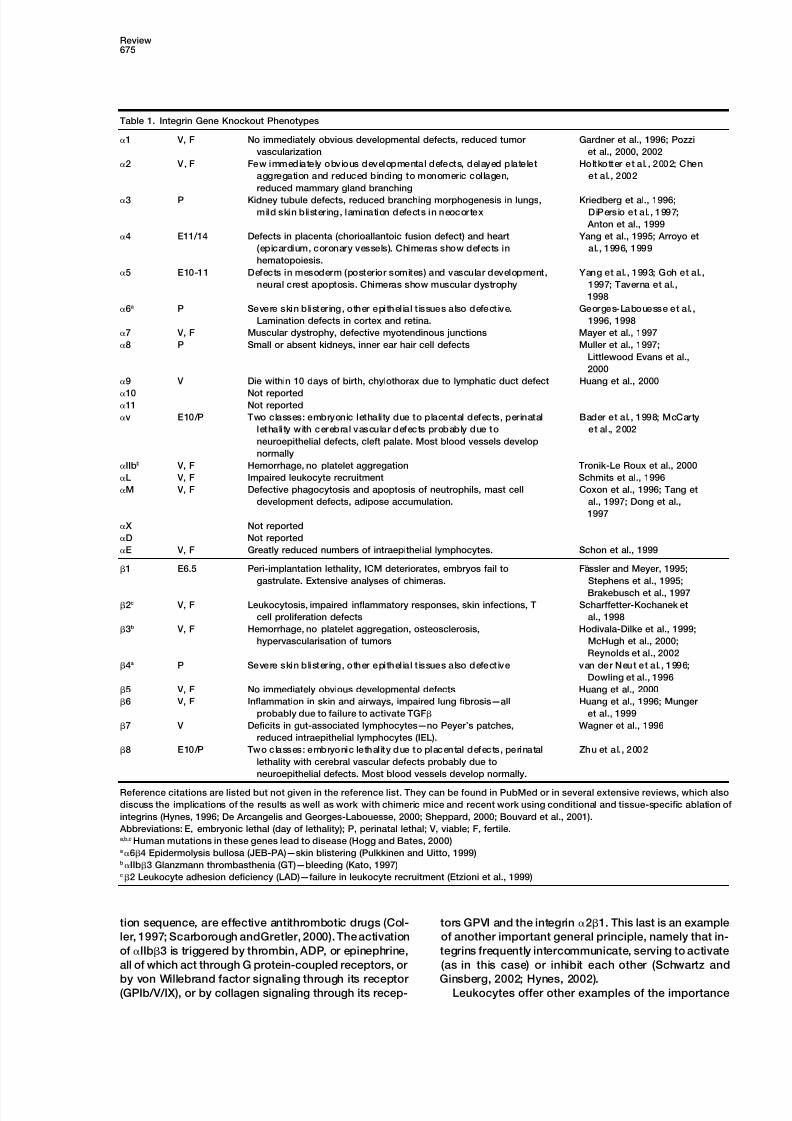
Table 1. Integrin Gene Knockout Phenotypes
1 V, F No immediately obvious developmental defects, reduced tumor Gardner et al., 1996; Pozzi
vascularization et al., 2000, 2002
2 V, F Few immediately obvious developmental defects, delayed platelet Holtkotter et al. , 2002; Chen
aggregation and reduced binding to monomeric collagen, et al., 2002
reduced mammary gland branching
3 P Kidney tubule defects, reduced branching morphogenesis in lungs, Kriedberg et al., 1996;
mild skin blistering, lamination defects in neocortex DiPersio et al. , 1997;
Anton et al., 1999
4 E11/14 Defects in placenta (chorioallantoic fusion defect) and heart Yang et al., 1995; Arroyo et
(epicardium, coronary vessels). Chimeras show defects in al., 1996, 1999
hematopoiesis.
5 E10-11 Defects in mesoderm (posterior somites) and vascular development, Yang et al., 1993; Goh et al.,
neural crest apoptosis. Chimeras show muscular dystrophy 1997; Taverna et al.,
1998
6a P Severe skin blistering, other epithel ial t issues also defective. Georges-Labouesse et al. ,
Lamination defects in cortex and retina. 1996, 1998
7 V, F Muscular dystrophy, defective myotendinous junctions Mayer et al., 1997
8 P Small or absent kidneys, inner ear hair cell defects Muller et al., 1997;
Littlewood Evans et al.,
2000
9 V Die within 10 days of birth, chylothorax due to lymphatic duct defect Huang et al., 2000
10 Not reported11 Not reported
v E10/P Two classes: embryonic lethality due to placental defects, perinatal Bader et al., 1998; McCarty
lethality with cerebral vascular defects probably due to et al ., 2002
neuroepithelial defects, cleft palate. Most blood vessels develop
normally
IIbb V, F Hemorrhage, no platelet aggregation Tronik-Le Roux et al., 2000
L V, F Impaired leukocyte recruitment Schmits et al., 1996
M V, F Defective phagocytosis and apoptosis of neutrophils, mast cell Coxon et al., 1996; Tang et
development defects, adipose accumulation. al., 1997; Dong et al.,
1997
X Not reported
D Not reported
E V, F Greatly reduced numbers of intraepithelial lymphocytes. Schon et al., 1999
1 E6.5 Peri-implantation lethality, ICM deteriorates, embryos fail to Fa ¨ ssler and Meyer, 1995;
gastrulate. Extensive analyses of chimeras. Stephens et al., 1995;
Brakebusch et al., 1997
2c V, F Leukocytosis, impaired inflammatory responses, skin infections, T Scharffetter-Kochanek et
cell proliferation defects al., 1998
3b V, F Hemorrhage, no platelet aggregation, osteosclerosis, Hodivala-Dilke et al., 1999;
hypervascularisation of tumors McHugh et al., 2000;
Reynolds et al., 2002
4a P Severe skin blistering, other epithel ial t issues also defective van der Neut et al. , 1996;
Dowling et al., 1996
5 V, F No immediately obvious developmental defects Huang et al., 2000
6 V, F Inflammation in skin and airways, impaired lung fibrosis—all Huang et al., 1996; Munger
probably due to failure to activate TGF et al., 1999
7 V Deficits in gut-associated lymphocytes—no Peyer’s patches, Wagner et al., 1996
reduced intraepithelial lymphocytes (IEL).
8 E10/P Two classes: embryonic lethality due to placental defects, perinatal Zhu et al. , 2002
lethality with cerebral vascular defects probably due to
neuroepithelial defects. Most blood vessels develop normally.
Reference citations are listed but not given in the reference list. They can be found in PubMed or in several extensive reviews, which also
discuss the implications of the results as well as work with chimeric mice and recent work using conditional and tissue-specific ablation of
integrins (Hynes, 1996; De Arcangelis and Georges-Labouesse, 2000; Sheppard, 2000; Bouvard et al., 2001).
Abbreviations: E, embryonic lethal (day of lethality); P, perinatal lethal; V, viable; F, fertile.a,b,c Human mutations in these genes lead to disease (Hogg and Bates, 2000)a64 Epidermolysis bullosa (JEB-PA)—skin blistering (Pulkkinen and Uitto, 1999)bIIb3 Glanzmann thrombasthenia (GT)—bleeding (Kato, 1997)c2 Leukocyte adhesion deficiency (LAD)—failure in leukocyte recruitment (Etzioni et al., 1999)
tion sequence, are effective antithrombotic drugs (Col- tors GPVI and the integrin 21. This last is an example
of another important general principle, namely that in-ler, 1997; Scarborough andGretler, 2000). The activation
of IIb3 is triggered by thrombin, ADP, or epinephrine, tegrins frequently intercommunicate, serving to activate
(as in this case) or inhibit each other (Schwartz andall of which act through G protein-coupled receptors, or
by von Willebrand factor signaling through its receptor Ginsberg, 2002; Hynes, 2002).Leukocytes offer other examples of the importance(GPIb/V/IX), or by collagen signaling through its recep-

7/21/2019 1-s2.0-S0092867402009716-main
http://slidepdf.com/reader/full/1-s20-s0092867402009716-main 4/15
Cell676
Figure 2. Integrin Signaling
A decade ago, ideas about integrin signaling
were in their infancy (Hynes, 1992). It was
clear that integrins synergized with other cell
surface receptors including growth factor re-
ceptors to activate largely unknown signaling
pathways to affect cell proliferation and dif-ferentiation, cell shape and migration, and
other events. These signal transduction
mechanisms could be subverted by onco-
genessuch aspp60sre to give anchorageinde-
pendence of growth. Our current view re-
mains the same in outline but many detailed
signal transduction pathways have now been
elucidated.
The major signal transduction pathways and
many of the key players in them are shown,
leading to the major effects on cell behavior
mediated by integrins, often acting in concert
with G protein-coupled or kinase receptors
for soluble factors. The major submembra-
nous, integrin-associated links between integrins and these signal transduction pathways are contained within the pink-purple pentagon
beneath the clustered integrins. Details of the interactions of these linker/adaptor proteins and of the signal transduction pathways are omitted,
as are other known players in these processes. Readers are referred to several excellent reviews for further details (Clark and Brugge, 1995;
Schwartz et al., 1995; Yamada and Miyamoto, 1995; Clark and Hynes, 1997; Giancotti and Ruoslahti, 1999; Danen and Yamada, 2001; Wu
and Dedhar, 2001; Schwartz and Ginsberg, 2002; Miranti and Brugge, 2002).
of inactive integrinsand their regulated activation. Mem- lation of integrin function from within the cell has com-
monly been called “inside-out” signaling to distinguishbers of the 2 integrin subfamily (also known as CD11/
18) are expressed on most white blood cells but, when it from “outside-in” signaling, as depicted in Figure 2
(Hynes, 1992; Ginsberg et al., 1992). Both obviously in-these cells are “resting,” these integrins become inac-
tive. When the cells become activated, for example by volve transmembrane signals, the nature of which has
been difficult to decipher. Major insights come fromcytokines, the 2 integrins are rapidly activated and the
cells become adhesive for their counterreceptors, in this recent structural information on integrins and from ex-
periments stimulated by and/or reinterpreted in lightcase, Ig superfamily molecules such as ICAMs. These
are expressed on endothelial cells, allowing attachment of the structural results, and we will return later to a
discussion of the nature of integrin activation.of leukocytes to the vessel wall, or on other cells,allowing phenomena such as phagocytosis, cytotoxic
killing, or lymphocyte help. As in the case of platelets, Integrin Structure: Extracellular Domains
Thefirstdomain of integrinsto be crystallizedwas theI/A it is important that the 2 integrins are inactive on the
surfaces of resting leukocytes (to avoid inflammation) domain inserted into half of the mammalian subunits
(Figure 1). Lee et al. (1995a) determined the structure ofand that they can be rapidly activated (to allow immune
function). Defects in either have pathological conse- thisdomainfromM2 (CD11b/CD18, CR3)and showed
it to be a Rossmann fold with a core of parallel sheetsquences. Clear support for the importance of these pro-
cesses comes from the phenotypes of mice lacking one surrounded by amphipathic helices. Within the ex-
tended family of Rossmann folds, the integrin I/A do-or more of the 2 integrins or their ligands (Table 1;
Rosenkranz and Mayadas, 1999) and from the genetic mains form a subset of the larger group of VWA domains
found in a wide variety of proteins (Tuckwell, 1999; Whit-disease leukocyte adhesion deficiency (LAD), which
arises from mutations in the gene for 2 integrin. LAD taker and Hynes, 2002). VWA domains are around 180
amino acids long and many appear to be involved inpatients suffer from leukocytosis and the failure to re-
cruit leukocytes to sites of infection, leading to early protein-protein interactions. The I/A domains of integrin subunits comprise the ligand binding sites of thesedeath (Etzioni et al., 1999). In contrast, blockade of 2
integrins, and of 4 integrins, which mediate similar integrins.
Lee et al. (1995a) defined a metal ion coordination sitefunctions on lymphocytes, is a very promising avenue
for therapy of a variety of inflammatory and autoimmune at the “top” of the I/A domain of M, involving residues
from three separate loops of the I/A domain. Interest-diseases (Gottlieb et al., 2000; Jackson, 2002).
While these vascular processes offer particularly clear ingly, a glutamate from an adjacent molecule inthe crys-
tal formedpart of thecoordinationsphere. It was alreadyexamples of the importance of inactivation and activa-
tion of integrin function, not all integrins have been well established that integrins require divalent cations
for ligand binding and that an aspartate (D) or glutamateshown to undergo such extremes of activity. However, it
is believed that many, perhaps all, integrins may behave (E) residue is key to the integrin recognition site of all
ligands (including ICAM-1, a ligand for M2). This hadsimilarly, albeit in a less absolute and more localized
fashion, duringprocesses such as cell migration, neurite led to the idea that the ligand D/E might participate
together with residues from theintegrinin joint coordina-outgrowth, and so forth, when it is important for cells to
regulate their adhesion in a temporal and spatial fashion tion of a divalent cation. The structure determined byLee et al. (1995a) fitted this idea very well and they(Lauffenburger andHorwitz, 1996). This conceptof regu-

7/21/2019 1-s2.0-S0092867402009716-main
http://slidepdf.com/reader/full/1-s20-s0092867402009716-main 5/15
Review677
Figure 3. Integrin I/A Domain Structure and
Conformational Change
(A) Comparison of I/A domain structures of
2 (left) and M (right). In each case, regions
showing large changes between the two
states; open/liganded (blue) and closed/unli-
ganded (yellow) are indicated, and the shiftson ligation are shown by red arrows. Note the
shift from theC helix (red; specific to collagen
binding I/A domains) into the 6 helix and
the large downward shift of the C-terminal 7
helix on binding of ligand to 2. M shows a
very similar downward shift of the C-terminal
helix.
(B) Close-up of the movements of the metal
ion and loops around the MIDAS site in 2
(left) and M (right) with color-coding as in
(A). Againnote thestrong similarity inthe con-
formational changes occurring in the two do-
mains. The movement of the loops is coordi-
nated with the movement of the metal ion,
which switches its coordination from a D in
loop L3 to a T in loop L1. Changes in L1 and
L2 lead to the reorganization of C and 7
shown in (A).
(C) Stereo diagram of the MIDAS motif of 2
with the glutamate residue (E) from the ligand
(yellow) coordinating the metal ion (blue).
Residues from the loops of the I/A domain
coordinate the metal ion either directly or
through water molecules ( ). An additional
residue (E256 from L3) has been omitted for
clarity.
All panels from Emsley et al. (2000).
coined the term metal ion-dependent adhesion site mational change within the I/A domain and this was
elegantly confirmed by the determination of the struc-(MIDAS). They also pointed out a homologous segment
embedded within the subunit and sharing hydropathy ture of the I/A domain of 2 with and without a model
ligand based on the recognition sequence in collagenand secondary structure predictions and a MIDAS motif.
This segment of the 3 subunit had already been impli- (Figure 3; Emsley et al., 1997, 2000). Comparisons
among all the I/A domain structures lead to the clear cated in ligand binding by crosslinking, genetic, andmutagenesis data (D’Souza et al., 1988; Bajt and Loftus, deduction that the ligand does indeed coordinate the
metal ion in the MIDAS site via a carboxylate group and1994; Loftus et al., 1994). This prediction was followed
up by more elaborate secondary structure predictions this is coupled to alterations in metal coordination by
residues within the integrin MIDAS motif. These in turn(Tozer et al., 1996; Tuckwell and Humphries, 1997;
Huang et al., 2000), and refined HMM models now reli- are coupled to conformational shifts within the domain:
lateral movements of the loops containing the MIDASably predict a VWA domain within integrin subunits.
These conclusions have been confirmed within the last residues and longer-range movements in the C-terminal
helix of the I/A domain, which moves around 10 A ˚ downyear by the determination of the structure of the entire
extracellular domain of integrin v3 (see below). the side of the domain when ligand binds (Figure 3).
Liddington and colleagues (Lee et al., 1995b; Loftus and Additional structures of I/A domains followed and it
became clear that the domains could take on two con- Liddington, 1997) noted the strong parallels between
these conformational changes in I/A domains and thoseformations, “open” and “closed,” differing in the coordi-
nation of the metal at the MIDAS site (Lee et al., 1995b; occurring in GTPases such as ras and G proteins, which
also contain Rossmann nucleotide binding folds. It isQu and Leahy, 1995, 1996; Emsley et al., 1997). It wasproposed that ligand binding was coupled to a confor- easy to imagine how such conformational changes
7/21/2019 1-s2.0-S0092867402009716-main
http://slidepdf.com/reader/full/1-s20-s0092867402009716-main 6/15
Cell678
could propagate to the rest of the molecule, to which refer to as I-EGF repeats. The four I-EGF repeats are
followed by a C-terminal disulfide-bonded sheet do-the I/A domain is coupled via its adjacent N and C ter-
mini, and we will return later to this important allosteric main termed the -tail domain.
As mentioned, this structure confirmed many predic-property of integrins.
Xiong et al. (2000) expressed I/A domains of M that tions and conformed with much preexisting data con-
cerning integrin structure (see Humphries, 2000, 2002;adopt each of the two forms (open or closed) and
showed that only the open form binds ligands. Springer Shimaoka et al., 2002, forrelevantreviews relatingearlier
data to the structure). The big surprise was that, insteadand colleagues have also exploited the structural infor-
mation to produce I/A domains of L locked in the open of being extended as depicted in Figure 4B and as ex-
pected from published EM images of integrins, the v3and closed states by disulfide bonds engineered into
the C-terminal helix to lock it into the up (closed) or integrin in the crystal structure was bent over at a 135
angle with a “genu” between the thigh and calf domainsdown (open) position and shown that these two forms
differ markedly in affinity for ligand (Lu et al., 2001a; of v and a similar bend in the I-EGF 2/3 region of the
3 leg (Figure 4A). This surprising structure raises veryShimaoka et al., 2001, 2002). The open form is high
affinity or “active” and the closed form is low affinity or interesting questionsand has already stimulated experi-
ments to which I will return below.“inactive,” and the conformational switch between them
is coupled with ligand binding or with known activation The structure determined by Xiong et al. (2001) was
obtained in a Ca2 buffer and lacked bound ligand, con-stimuli such as activating antibodies or Mn2 ions.
Half the mammalian subunits and all known non- ditions usually yielding inactive integrins. The MIDAS
motif did not have a clear cation engaged, although anchordate integrins lack an inserted I/A domain (Figure
1), but it is clear that these subunits also contribute adjacent site (ADMIDAS) did and other cations bind atother sites within both subunits. Subsequent structuresligandbinding specificity.How do they do that? Springer
(1997) predictedthat the7-fold repeatin theextracellular obtained after diffusing cycloRGDF and Mn2 into the
crystal showed cycloRGDF bound at the interfacedomain of all subunits folds into a 7-bladed propeller
like that in the subunit of G proteins (Wall et al., 1995; with the arginine residue binding the propeller domain of
the subunit and the aspartate joining the coordinationLambrightet al., 1996; Sondeket al., 1996) andpredicted
that this might complex with the I/A domain embedded sphere of a Mn2 ion bound at the MIDAS site (Figure
4C; Xiong et al., 2002). Changes occurred in the loopswithin the integrin subunit by analogy with the G /G
complex in G proteins. This prediction has also been at the top of the I/A domains, similar to those seen in
-I/A domains, but the 10 A ˚ shift in the C-terminal helixconfirmed by the v3 structure.
The solution by Arnaout and colleagues of the crystal characteristic of ligand bound I/A domains from sub-
units was not observed in the 3 I/A domain. Severalstructure of the extracellular domain of v3 (Xiong et
al., 2001) represents a truly major advance in the integrin possibilities have been suggested: (1) the3 I/A domain
is constitutively active, even in the absence of ligandfield. In addition to confirming the predictions of an I/A
domain within the subunit and of a -propeller domain (Xiong et al., 2001, 2002), (2) the lattice contacts in thecrystal prevent the full conformational change and acti-within the subunit in an association very like that of
G and G, it revealed the structure of much of the vation (Liddington, 2002), or (3) activation of the I/A do-
main in subunits occurs somewhat differently (Mouldrest of the extracellular domains of both subunits (Figure
4; Xiong et al., 2001, 2002). The propeller domain and the et al., 2002; Liddington, 2002). Mould et al. (2002) report
an activation-dependent antibody that bindsthe 1 helix-I/A domain are complexed to form the ligand binding
head of the integrin, which is attached to two legs, one at the base of the -I/A domain near the contact with the
hybrid domain. Many function-blocking and -activatingfrom each subunit, as predicted from a large body of
electron microscopic, biophysical, and other data. The antibodies bind the 1 and 2 helices in this part of the
-I/Adomain (Takada andPuzon, 1993), also suggestingN-terminal propeller domain of the subunit is attached
to an elongated leg formed of three sandwich domains a propagated conformational change in this region not
seen in the cycloRGDF-v3 crystal.termed thigh, calf1, and calf2. The subunit domain
organization is a bit more complex; although the -I/A Much of the top surface of the propeller is occluded
by the apposed -I/A domain in the crystal structuredomain is at the distal end of the molecule (furthest from
the C-terminal membrane insertion site), it is not at the (Xiong et al., 2001), including residues known to be in-volved in interactions with ligands and to contain epi-N terminus of the primary sequence. Instead, it is in-
serted into a loop in a so-called hybrid domain, another topes for blocking antibodies against several integrins
(Humphries, 2000, 2002). It has been known for a long sandwich domain with some homology with I-set Ig
domains. The hybrid-I/A domain unit is preceded in the time that RGDpeptidesand small ligands canbind integ-
rins that are not fully activated, whereas larger ligandssequence by an N-terminal 54-residue PSI domain,
which in the 3D structure lies below the hybrid-I/A do- such as fibrinogen and fibronectin cannot (Coller, 1986;
Beer et al., 1992). Mould et al. (1997) showed that themain “head” and is disulfide bonded to the distal end
of the subunit leg. This leg is made up of four tandem RGD of fibronectin interacts with the -I/A domain,
whereas the synergy site in the adjacent Fn3 repeatcystine-rich repeats highly characteristic of integrin
subunits. The first and second are poorly resolved in interacts with the propeller domain. Dual interaction of
these two sites appears to be necessary for strong bind-the crystal, but the third and fourth are clearly folded
into EGF-like folds. An NMR structure of the second and ing of 51 integrin to fibronectin (Garcia et al., 2002).
These data suggest that a fully active ligand-engagedthird cystine-rich repeats of 2 (Beglova et al., 2002)
confirms their EGF-like pattern including an extra fourth integrin must undergo some opening up at the interfacebetween the -I/A domain and the propeller domain.cystine pair characteristic of these repeats, which I will

7/21/2019 1-s2.0-S0092867402009716-main
http://slidepdf.com/reader/full/1-s20-s0092867402009716-main 7/15
Review679
Figure 4. Three-Dimensional Structure of the
Integrin v3
(A) The structure of the unliganded v3 is
shown as a ribbon diagram with the v sub-
unit in blue and the 3 subunit in red. In the
crystal the integrin is folded over at a bend
or “genu,” with thehead (propeller,-I/A, andhybrid domains) bent over toward the C ter-
mini of the legs which would be inserted into
the membrane in an intact integrin. The do-
mains are hard to see in this view and are
more readily visualized in (B).
(B) The structure in (A) has been unfolded by
straightening it out at the “genu” of the v
subunit by 135 and rotating the thigh 120
around its axis, with similar adjustments to
the 3 structure. The structures of the linker
segments (1 in the v, 2 and 3 in the 3) and
of the PSI domain and I-EGF repeats 1 and
2 are not well resolved and are approximate
estimatesonly. Thestructurereveals two legs
( 160 A ˚ 20 A ˚ ) extending from the mem-
brane insertion site at the C termini to the
head at the top. The head is 90 A ˚ 60 A ˚
45 A ˚ and comprises three domains: a pro-
peller domain at the N terminus of the v
subunit andan I/Adomain insertedinto a loop
on the top of the hybrid domain in the sub-
unit. The N-terminal PSI domain is curled in
below the hybrid domain and is known to be
linked by a disulfide bond to the I-EGF-1 re-
peat,although this connection is not resolved
in the crystal structure. The apposition of the propeller and I/A domains is highly similar to that of G proteins. A 3 10 helix from the I/A domain
reaches out to the propeller and inserts an arginine residue into the central channel of the propeller. This arrangement is very similar to the
arrangement of a lysine in the 2 helix of the switch II region of G inserted into the propeller domain of G. The asterisk marks the loop into
which I/A domains are inserted in some integrin subunits, although not v.
(C) Surface representation of the cyclo RGDF peptide bound to the interface between the subunit propeller (blue) and the subunit I/A
domain (red). The aspartate (D) of the ligand coordinates a Mn 2 ion (cyan) and the arginine (R) binds to aspartate residues in loops on top
of the propeller. The second Mn2 ion (violet) is in the ADMIDAS site.
(A) and (B) are from Xiong et al. (2001); (C) is from Xiong et al. (2002).
This would resemble the separation of the homologous activation of the extracellular domains after we have
reviewed recent data on the cytoplasmic domains ofG and G domains in activated G proteins and seems
a very reasonable working hypothesis for integrins (Lid- integrinsto which eventsat theligand binding sites must
be coupled.dington, 2002; Liddington and Ginsberg, 2002). Such a
model receives some support from EM images of integ-
rins in the presence of ligand peptides. Hantgan et al. Cytoplasmic Domains: Structures
and Interactions(1999) report some separation of the and heads of
IIb3 in the presence of RGD peptides and Takagi et Despite the fact that integrins’ cytoplasmic domains are
much smaller than their extracellular domains (generallyal. (2002) detect changes in the relationship between
the head and the hybrid domain of v3 as a conse- less than 50 amino acids) they play a vital role in integrin
functions and have been the subject of intensive analy-quence of RGD binding. Since the C-terminal helix of
the -I/A domain connects to the hybrid domain, if it sis. Paradoxically we have a less clear picture of their 3Dstructure than we do for the large extracellular domains,were to undergo a downward shift like that shown by
the corresponding helix in -I/A domains, that would although recent work has produced some major in-
sights.necessarily be coupled to changes in -I/A-hybrid do-
main organization that could well include rotation away The cytoplasmic domains are the sites of interaction
with, and linkage to, the cytoskeletal and signaling part-from contact with the propeller domain, opening it up
for further interactions with ligands (Figure 5). ners of integrins (see Figure 2). There is an extensive
literature on the many proteins that have been reportedThe v3 integrin lacks an -I/A domain, but the site
of insertion of I/A domains in those subunits that have to interact with or cytoplasmic domains but I will
not attempt to review most of that work (see Burridgeonefallsbetween blades 2 and3 of thepropeller domain,
and this position is marked in Figures 4 and 5. Since and Chrzanowska-Wodnicka, 1996; Critchley et al.,
1999; Calderwood et al., 2000; Zamir and Geiger, 2001,-I/A domains contain the ligand binding sites of the
corresponding integrins, we need to consider how li- for reviews). For our present considerations, it is most
relevant to consider data that indicate that integrin cyto-gand binding may differ between the two classes of
integrin, those with and without -I/A domains. We will plasmic domains can regulate the activation state ofintegrins; that is, affect the structure and function of thereturn to consider further models for ligand binding and

7/21/2019 1-s2.0-S0092867402009716-main
http://slidepdf.com/reader/full/1-s20-s0092867402009716-main 8/15
Cell680
Figure 6. Interactions between and with Integrin Cytoplasmic Do-mainsFigure 5. Hypothetical Models for Ligand binding to Integrin Heads(A) Sequences of the cytoplasmic tails of IIb and 3. The mem-(A) An integrin without an I/A domain in the subunit, such as v3brane-spanning segment is usually considered to end at the W(note; only the head region is shown). A small ligand such as cyclowithin the darker gray shaded area (lipid bilayer). The immediatelyRGDF binds at the interface between the propeller and the -I/A membrane-proximal segments are highly conserved (red denotesdomain (see Figure 4). The model proposes that the C-terminal helixconservation in thevast majority of subunits,lilac denotes conserva-(orange) moves down, causing the -I/A domain (pink) to rotatetion in more than half). Conserved NxxY motifs are highlighted inaway from the propeller domain opening up the top of the propeller yellow. Deletion of the conserved membrane-proximal segmentto engage larger ligands such as fibronectin (lilac). It is known thatfrom eithersubunitleadsto activation, asdo pointmutations markedthe RGD motif in fibronectin engages the -I/A domain while theby asterisks (see text). The proposed salt bridge between R995 andsynergy site in the adjacent Fn3 domain engages the propeller D723 is marked by a red bar (Hughes et al., 1996). The pink bars(Mould et al., 1997), consistent with this model, although the degreedenote regions showing interaction between subunits and the greenof opening shown is hypothetical and could easily vary among in-bars denote -helical segments, both deduced from NMR data (Vi-tegrins.nogradova et al., 2002). The purple bar denotes segment of 3(B) An integrin with an -I/A domain such as 21. The ligand,showing interaction with talin head by NMR (Vinogradova et al.,collagen, binds to the top of the -I/A domain (pale blue) causing2002)consistent with cell biological results (Calderwoodet al., 1999,a 10 A ˚ downward shift of the C-terminal helix (Figure 3), which is2002; Patil et al., 1999). Talin binding also requires Y747 (hatchattached to an extended loop containing a conserved glutamatemark). Since the affinity of talin head for 3 tail is much higher than(red dot). It is proposed that this could bind to the MIDAS site inthat between thetwo tails,bindingof talin undoesthe clasp betweenthe -I/A domain (Alonso et al., 2002) and act upon it as a ligandthe cytoplasmic domains in the same way as mutations in the mem-relay. The -I/A domain is proposed to transmit conformationalbrane-proximal region (asterisks). Armulik et al. (1999) report thatchange to the hybrid domain as in (A). Springer and colleaguesthe conserved membrane-proximal segments can be buried in the(Shimaoka et al., 2002) have concentrated on inside-out activationlipid bilayer (lighter gray shading). If so, then the transmembraneof 2 integrins and thus have focused on how the -I/A domainssegments are atypically long (28–30 residues) and Armulik et al.become activated. They have suggested that the C-terminal helixsuggest that interactions with cytoplasmic proteins could pull theacts like a bell rope to pull open theI/A domain. This is thereciprocalconserved segments out of the bilayer, offering an alternative or of the ligand-relay model. The change is an allosteric one and theadditional way in which binding of proteins such as talin could alter equilibrium can be driven from either end.integrin conformation leading to activation (see also Figure 7B).
(B) Talin can be activated for binding to tails by cleavage (Yan et
al., 2001) to release the FERM domain-containing head (blue) or byextracellular domains. There is a considerable body of interaction with PIP2 (Martel et al., 2001). In each case, the talin
data indicating that the cytoplasmic domains of the head binds the cytoplasmic domain leading to separation of thetails (see [A] and text). Intact talin does not interact with integrinand subunits can interact to control the activationtails and is depicted as folded upon itself with the head domainstates of integrins. These analyses have proceeded fur-occluded by the tail of talin, by analogy with ERM proteins (Pearsonthest for theplatelet integrin,IIb3, which as discussedet al., 2000), although the tertiary structure of talin is unknown. The
earlier is tightly regulated so that it is inactive on restingtalin tail comprises a series of short -helical segments (yellow) and
platelets but rapidly activated by thrombogenic stimuli. an actin binding domain (red).Ginsberg and colleagues have investigated the roles of
the IIb and 3 cytoplasmic domains in this regulation.
They have shown that the shortIIbcytoplasmic domain tion of either one to alanine yields a constitutively active
integrin, whereas a charge reversal, IIbR995D/ 3D723R,acts as a negative regulator of activation. Deletion of
the entire domain (see Figure 6A) or of just the highly restored the inactive state (Hughes et al., 1996). Based
on these and other results, Ginsberg and colleaguesconserved GFFKR sequence produces a constitutively
active integrin (O’Toole et al., 1991, 1994). Similarly, the suggested several models, all relying on interaction be-
tween the membrane-proximal segments of IIb and 3conserved membrane-proximal segment of 3 is also
necessary (Hughes et al., 1995). They proposed that to restrain the integrin in an inactive state (Williams etal., 1994; Woodside et al., 2001). Separation, twisting,R995 of IIb forms a salt bridge with D723 of 3; muta-

7/21/2019 1-s2.0-S0092867402009716-main
http://slidepdf.com/reader/full/1-s20-s0092867402009716-main 9/15
Review681
pistoning, and hinging of the tails were all considered integrin tails, most often those of subunits. Others of
as mechanisms to allow activation. More recent data these could act similarly to talin head or, alternatively,
favor models involving separation of the cytoplasmic could bind elsewhere in the tail, such as the distal por-domains as a key step in integrin activation. Evidence tion of tail, which does not appear to interact with thecomes from recent NMR analyses and from cell biologi- tail (Figure 6A).cal studies.
Binding between the cytoplasmic domains ofIIb and Integrin Activation: Transmembrane Connections3 could be detected by surface plasmon resonance and Long-Range Conformational Changesand was ablated bydeletionof KVGFFKR orby anR995A If activation of integrins by inside-out signaling involvesmutation (Vallar et al., 1999). The affinity was low (K d separation of the and cytoplasmic tails, how is that7–50 M depending on divalent cation concentration), signal transmitted to the ligand binding site(s) 10–20 nmwhich may explain why initial efforts to determine struc- away at the far end of the extracellular domain? Recentturesof the interactingdomains were largely unsuccess- results are beginning to reveal possible mechanisms,ful (Ulmer et al., 2001; Li et al., 2001). However, Weljie despite the fact that there is not a structure for an intactet al. (2002) detected-helical structure and intersubunit integrin, only for the separate intracellular and extracel-interactions using synthetic peptides representing the lular domains.membrane-proximal segments. Vinogradova et al. 2 integrins, like IIb3, are dependent on their mem-(2002) demonstrated interactions between membrane- brane-proximal cytoplasmic domains to maintain an in-proximal helices in both subunits, using the entire cyto- active state; deletion of either or segments yieldsplasmic domains, and also demonstrated that they were
active integrins (Lu et al., 2001b). Furthermore, replace-disrupted by point mutations (F992A or R995D) already ment of the L and 2 tails by, respectively, acidic andknown to interfere with inactivation by IIb cytoplasmic basic coiled-coil domains restored the inactive state.domain in the intact integrin (see earlier discussion). This is analogousto thecharge-reversalexperiment withThese data are summarized in Figure 6A, which also IIb3 and confirms that tail associations also re-collects together information from a different, comple- strain 2 integrins in an inactive state. To take the analy-mentary set of experiments. sis further, Takagi et al. (2001) eliminated both the tails
Calderwood et al. (1999, 2002) showed that the head and the transmembrane domains from 51 and re-domain of talin binds to the cytoplasmic domains of
placed them with acidic and basic coiled coils joined3 and other subunits via a PTB domain within the
by a disulfide bond. This generated a soluble 51 di-conserved FERM domain of talin; Y747 of 3 is neces-
mer. As predicted, this clamped, soluble 51 did notsary for this interaction. The NPLY motif is believed to
bind its ligand, fibronectin, but it could be activated byform a turn, and NMR data on 3 cytoplasmic domain
cleaving the C-terminal clamp; that is, by allowing thesupport this idea (Ulmer et al., 2001). Vinogradova et al.
and stalks (legs) to separate, which was confirmed(2002) therefore analyzed the effects of talin head on
by EM. This experiment shows that the C-terminal cyto-theNMR signals of theIIband3 cytoplasmic domains; plasmic domain clasp or the engineered C-terminaltalin head bound to 3 but not to IIb. The interactions
clamp, whether inside or outside the membrane, con-extended from K716 to N744, completely overlapping
strain integrins in an inactive state but release of thesethe region of 3 interaction with IIb (see Figure 6A).
constraints, allowing separation of the stalks/legs of theFurthermore, talin head ablated the interaction between
extracellular domains, leads to activation of the ligandtheIIband3 tails (Vinogradovaet al., 2002), consistent
binding site in the head.with its much higher affinity for 3 tail (K d 100 nM;
The idea that conformational changes in the extracel-Calderwood et al., 1999, 2002). Thus, the head of talin
lular domain near the membrane can be linked tobinds to the 3 tail and separates it from the IIb tail.
changes in the ligand binding domain in the head ofTalin head was also shown to bind to and activateintegrins is far from a new one. A decade ago Weisel etintegrins(Calderwood et al., 1999, 2002), entirely consis-al. (1992) demonstrated that IIb3 bound to fibrinogentent with the model that interactions between IIb tailtends to show widely separated tails. This is effectivelyand 3 tail keep the integrin in an inactive state andthe reciprocal result of the experiment of Takagi et al.separation is necessary for activation (see Figure 6B).
(2001) with51 and fibronectin. In another early experi-In order for talin’s head domain to trigger this activation, ment, Du et al. (1993) showed cooperative activationit must be exposed. This can be accomplished by ex-between the binding of fibrinogen to the head of thepressing recombinant fragments of talin (CalderwoodIIb3 integrin and binding of a monoclonal antibody toet al., 1999, 2002; Patil et al., 1999), by calpain cleavage,the first 90 amino acids of the 3 stalk adjacent to thewhich separates the head from the tail (Yan et al., 2001),membrane. The distance between these two sites asor by phosphatidyl inositols (Martel et al., 2001) as de-revealed by EM was 16 nm. The antibody had originallypicted in Figure 6B. The mapping of the interaction tobeen isolated as recognizing a ligand-induced bindinga PTB domain within the talin head (Calderwood et al.,site (anti-LIBS) and its binding was enhanced by fibrino-2002), which binds to the NPxY motif conserved in 3gen binding to the head of the integrin. Importantly,and in most other integrin subunits, raises the verybinding of the antibody also enhanced the affinity of theinteresting possibility that other PTB-containing pro-integrin for fibrinogen, i.e., the activation was reciprocal .teins may also interact with tails leading to activation
So we now have a picture of long-range conforma-of integrins (Liddington and Ginsberg, 2002). Amongtional changes linking the C-terminal ends of an integ-candidates for such a role is FAK, which like talin has
rin’s legs, i.e., the membrane-proximal regions both out-a FERM domaincontaining a PTBdomain. As mentionedearlier, multiple proteins have been reported to bind to side and inside the membrane, to ligand binding at the

7/21/2019 1-s2.0-S0092867402009716-main
http://slidepdf.com/reader/full/1-s20-s0092867402009716-main 10/15
Cell682
head. Thereis,in fact, a greatdeal ofevidence insupport
of this concept, including many activating and activa-
tion-sensitive monoclonal antibodies that frequently
map in the stalk regions (reviewed in Humphries, 2000,
2002; Shimaoka et al., 2002), as well as biophysical data
(e.g., Hantgan et al., 1999, and earlier work) and the
electron microscopy already mentioned. The challenge
is to understand how conformational changes in the
head domains associated with ligand binding are cou-
pled reciprocally with alterations, probably separation,
at the base of the legs and in the cytoplasm. How can
we fit these results with the newly available structural
data? The structure offers some potential solutions but
also the complication represented by the bent structure
observed in the crystal (Figure 4A).
Xiong et al. (2001, 2002) suggested that the bent form
is the active form of the integrin. However, others have
argued that it is more likely to be the inactive state,
based on details of the conformationof the-I/A domain
and the fact that it was crystallized in the absence
of ligand (Liddington, 2002; Liddington and Ginsberg,2002; Shimaoka et al., 2002). The latter interpretation
would fit much better with EM images of ligand bound
integrins (Weisel et al., 1992; Du et al., 1993), which
show an extended structure like that shown in Figure
4B. Beglova et al. (2002) mappedepitopesfor activation-
specific monoclonal antibodies to specific residues inFigure 7. Models for Long-Range Allosteric Changes Giving Bidi-
I-EGF repeats 2 and 3 and noted that these residues rectional Signaling by Integrinswould be buried in the bent form of the integrin. They
(A) Integrin in its bent form is presumed to be inactive. Activationproposed, therefore, that the bent form represents the can occur either by ligand binding or by effects on the cytoplasmicinactive state and that activation occurs by a “switch- domains, leading to straightening and separation of the legs. Alter-
ationsin theorientationof thepropeller andI/A domains arecoupledblade” opening of the integrin into an extended shapeto changes in the hybrid domain (yellow) by movement of the C-ter-and a separation of the legs. Such a conformationalminal helix of the I/A domain (orange). The hybrid domain, in turn,change could expose the epitopes for activation-spe-
is linked to the I-EGF domains (purple) via the PSI domain (green),cific antibodies, many of which are known to bind to which is disulfide bonded (yellow line) to the first I-EGF domain.the I-EGF repeats or to the PSI domain, which is also Straightening and separation of the legs exposes activation epi-buried in the genu (the structure is not well resolved topes in the I-EGF domains (red stars) and in the PSI domain (not
there). Takagi et al. (2002) went on to show that integrins shown). Separation of the cytoplasmic domains is accompanied by
conformational changes in them, allowing binding of cytoplasmicclamped in the inactive state predominantly adopt aproteins (see Figure 2) and signaling (lightning). All changes arebent shape as seen by EM, whereas integrins activatedreversible equilibria and can operate in either direction, allowingby Mn2 or by cyclo RGDfV were predominantly in anboth outside-in and inside-out signaling. See text for discussion
extended form. They showed that the clamped, bentand references.
form did not bind ligand, whereas the activated, ex- (B)Two modelsfor theproposedstraightening up of integrinsduringtended form did. Finally they presented evidence that activation. The switch-blade or flick-knife model (Beglova et al.,
2002) andan alternative angle-poise model differ in theway in whichintegrins on cell surfaces can be trapped in a bent andthe C termini of the legs relate to the transmembrane segmentsinactive state by an engineered disulfide bond that,(which is unknown). The angle-poise model incorporates the possi-when released, allows their activation. These resultsbility that the transmembrane helices may be especially long andconform well with the idea that the bent form seen incould change orientation and/or move in and out of the membrane
the crystal represents the inactive state of the integrin during activation (Armulik et al., 1999; see Figure 6A). The angle-and that activation comprises straightening and separa- poise model would place theligand binding site in a more accessibletion of the legs. This is, of course, also in good agree- position for macromolecular ligands.
ment with the data on cytoplasmic domain separation
(Figure 6, see prior discussion). These concepts aretion. The latter model would place the head domain in aschematized in Figure 7, which shows twoways in whichbetter position to interact with macromolecular ligands.the bent form might be related to the membrane. TheseThere are currently no data available to distinguish be-differ in the orientation of the membrane-proximaltween these two possibilities.“ankles” of the legs relative to the membrane; this is of
Thus, the preponderance of the evidence strongly fa-course unknown at present. In the switch-blade (Beg-vors models in which activation of the ligand bindinglova et al. 2002; Shimaoka et al., 2002) or “flick-knife”domain in the head and binding of ligand are coupled,(Liddington, 2002) model, the “calves” of each leg arevia long-range conformational changes in the legsperpendicular to the membrane and the head domain(probably including straightening and separation), tois very close to the cell surface. In a variant “angle-
separation of the bases of the legs and the attachedpoise” model, the legs are bent over closer to the mem-brane andextendlike an angle-poise lamp duringactiva- transmembrane and cytoplasmic domains. This cou-

7/21/2019 1-s2.0-S0092867402009716-main
http://slidepdf.com/reader/full/1-s20-s0092867402009716-main 11/15
Review683
pling is bidirectional and reciprocal and is best viewed Open Questions and Future Directions
Although the models presented (Figures 5–7) are consis-in terms ofan allosteric equilibrium,or seriesof equilibria
tent with a broad range of data, including the 3D struc-(Figure 7). Binding of extracellular ligand wouldthereforetures, they remain working hypotheses and require ex-enhance separation of the cytoplasmic domains,perimental tests. Foremost among these is the pressingallowing their interaction with cytoskeletal and signalneed for integrin/ligand cocrystals to investigate thetransduction molecules, that is, outside-in signalingconformation of ligand bound integrins. Does an acti-(Figure 2). Reciprocally, separation of the cytoplasmicvated integrin actually stand up as implied by Figuresdomains by talin and perhaps other activators would4B and 7? Do the legs separate? What exactly are theactivate the head for ligand binding, that is, inside-outconformational changes in the -I/A domain and thesignaling (Figure 6). The distinction between these twodomains in the legs? Much immunological evidenceforms of integrin signaling has been conceptually usefuldemonstrates the existence of conformational changesover thepast decade,but they shouldactuallybe viewedin these domains, but what exactly are they? In fact, weas two reflections of the same allosteric equilibrium.lack any definitive structures for the PSI domain andEither cytoplasmic or extracellular interactions can trapseveral of the I-EGF domains. There are inconsistenciesthe equilibrium in the active state, enhancing therebybetween the disulfide bonding patterns for I-EGF-3 de-the function at the opposite end of the integrin. Oneduced from the X-ray crystallography (Xiong et al., 2001)could also imagine cytoplasmic interactors that couldand by NMR (Beglova et al., 2002). Could these perhapstrap the equilibrium in the inactive state, stabilizing thereflect possible disulfide interchanges within the integ-integrin in the “off” state as on resting platelets or leuko-rin, as has been suggested (Yan and Smith, 2000, 2001;cytes. Similarly, antibodies that activate integrins or rec-O’Neill et al., 2000)? What is the significance of the other ognize only the active state presumably trap the equilib-divalent cations bound to integrins? Does the -I/A do-rium in the active state, and some function-blockingmain change conformation in the same way as the -I/A antibodies are also known to act allosterically rather domain? Does its C-terminal helix move down, alteringthan at the actual ligand binding site and presumablythe relationship between the -I/A and hybrid domains?act by trapping the equilibrium in the inactive state; IDoes the -I/A domain rotate away from the propeller,will discuss an example of this below.opening up the top of the integrin for more extensiveFigure 7 depicts an integrin lacking an -I/A domain.interactions with macromolecular ligands? Are there in-We need to consider how the situation might differ for termediate, stable conformers and different activationthose integrins with an inserted -I/A domain. In thesestates, as suggested by some data? Do all integrinsintegrins, ligand is wholly or largely bound by the -I/A undergo extreme changes in conformation or are somedomain. As discussed earlier, recombinant -I/A do-more subtle in their approach? Is there linkage betweenmains can bind ligand with the same affinity and speci--I/A and -I/A domains as suggested in Figure 5B?ficity as intact integrins, especially when locked in theWhy do some integrins have -I/A domains, anyway?active conformation. Many inactivating mutations and
Although the current data favor the model that theepitopes for function-blocking antibodies lie in the -I/A inactive state of integrins is a bent form as seen in thedomains and deletion of these domains inactivates the
X-ray structure (or something very like it), this requiresintegrins. As discussed above, the active conformationfurther investigation and its generalityneeds testing. Theof -I/A domains shows a downward shift of the C-ter-results of Takagiet al. (2002) clearly show that soluble3minal helix (Figure 3). This could clearly propagate aintegrins and those on the cell surface do adopt a bent,
conformational change to other domains of the integrin.inactive form, which can be induced to extend by appro-
Alonso et al. (2002) have suggested that a highly con-priate manipulations, However, it would be helpful to be
served glutamate just C-terminal to the C-terminal helixable to monitor this process in livingcells,perhaps using
could act as a pseudoligand for the -I/A MIDAS site,conformation-specific antibodies or FRET. We also do
acting as a ligand relay (Figure 5B). They present muta-not know exactly how the bent extracellular part of an
genesis data in support of this model, although it, likeintegrin is connected to the membrane. What is the
all the other models discussed here, will need further significance of the fact that most integrins lacking an
confirmation. Consistent with the model is the fact that-I/A domainare cleaved near the base oftheir subunit
there exist both mutations in, and function-blocking an- legs (in the calf2 domain), whereas none of those withtibodies against, the -I/A domain that preclude bindingan -I/A domain is so cleaved? We know essentially
of ligand at the -I/A domain unless the -I/A domain nothing about the transmembrane domain structuresis locked in the open position when it becomes immune and their interactions. They are always assumed to beto such inhibition (Lu et al., 2001c). Based on these and helical but are they and, if so, how long are they? Theother results, Shimaoka et al. (2002) proposed that the results of Armulik et al. (1999) suggest that the TM seg--I/A domain is activated by allosteric interactions with ments may be longer than necessary for a straight, per-the -I/A domain and proposed that the C-terminal helix pendicular helix and could even move in and out ofacts like a bell rope to open the -I/A domain. This is the membrane to some degree. There are intriguing con-entirely compatible with the ligand-relay model (Figure servations in primary sequence among integrin TM do-5). Thus, it appears likely that integrins containing -I/A mains; what do they mean? Do the and TM segmentsdomains function in essentially the same way as those interact? Do other integral membrane proteins knownwhich lack that extra domain, differing only in that there to interact with integrins perhaps interact within theis an extra step in the chain of linked conformational membrane? Could this affect activation and signaling?
changes connecting the ligand binding site with the cy- Possible candidates for such interactions and effectsinclude IAP/CD47 (Brown and Frazier, 2001), tetraspa-toplasmic domains.

7/21/2019 1-s2.0-S0092867402009716-main
http://slidepdf.com/reader/full/1-s20-s0092867402009716-main 12/15
Cell684
nins (Hemler, 2001), CD98 (Fenczik et al., 1997; Kolesni- tors will continue in the next few years as these ques-
kova et al., 2001), and others (Hemler, 1998; Hughes tions and others are addressed, incorporating structural
and Pfaff, 1998), importantly including the growth factor information along with the cell biological data and newreceptors with which integrins cooperate in signal trans- techniques such as proteomic analysis of complexesduction (Figure 2). and real-time imaging of molecules and their interac-
The role of cytoplasmic domain separation in integrin tions in situ. One hope is that the insights obtained willsignaling seems well established, but we need to know lead to better therapeutic agents targeting integrins inmore about exactly what happens. What are the precise human diseases as diverse as thrombosis, hemorrhage,structures of the cytoplasmic domains in the inactive inflammation, atherosclerosis, osteoporosis, cancer,and active states? If the talin head PTB domain binds and infectious diseases. It is, after all, the biological tails and activates integrins, as seems clear, do other importance of these receptors that makes them particu-proteins with PTB domains do the same or do some of larly interesting; the elegance of their allosteric signalthose proteins only bind a previously activated tail? Do transduction mechanisms is an extra bonus.some such proteins prefer a phosphorylated NPxY se-
Acknowledgmentsquence? There are typically two NPxY sequences in
tails. Do both work analogously? Do different proteinsI would like to thank all those whose research, writings, and discus-bind differentones? What about themany other proteinssion have contributed to the ideas in this review, including the refer-
that have been reported to bind to integrin cytoplasmicees of the manuscript. I apologize to all those in the field whose
domains? It seems likely that, like anti-integrin antibod- work could not be discussed in the context of a brief review, includ-ies, there will be activating, activation-specific, and in- ing many importantstudies on thebiology of integrins. I thank Gene-
vieve Hendrey for help with manuscript preparation, Charlie Whitta-hibitory interactors among them. Plausible models existker for help with figures, and the Howard Hughes Medical Institute,for activation of talin to allow it to bind to tails (FiguretheNationalCancer Institute, andthe National Heart Lung andBlood6), but how is that controlled? Evidence exists for smallInstitute for support.
GTPases as intermediates in pathways leading to integ-
rin activation (Zhang et al., 1996; Hughes et al., 1997;References
Schoenwaelder and Burridge, 1999; Katagiri et al., 2000;
Bos et al., 2001; Bertoni et al., 2002). Phosphatidyl inosi- Alonso, J.L., Essafi, M., Xiong, J.P., Stehle, T., and Arnaout, M.A.
tols are also likely to be significant, since they activate (2002). Does the integrin alphaA domain act as a ligand for its betaA
domain? Curr. Biol. 12, R340–342.many of the proteins that might interact with integrin Armulik, A., Nilsson, I., von Heijne, G., and Johansson, S. (1999).tails (talin, vinculin, ERM proteins) and many of the pro-Determination of the border between the transmembrane and cyto-teins are phosphorylated, so they could be regulatedplasmic domains of human integrin subunits. J. Biol. Chem. 274,by kinases and phosphatases. Somewhere in this net-37030–37034.
work of regulators must be the mechanisms by which Assoian, R.K. (1997). Anchorage-dependent cell cycle progression.integrins regulate each other.
J. Cell Biol. 136 , 1–4.We should also not forget that active integrins typi-Bajt, M.L., and Loftus, J.C. (1994). Mutation of a ligand binding
cally cluster in the plane of the membrane, and thisdomain of beta 3 integrin. Integral role of oxygenated residues in
“avidity modulation” of cell adhesion has long been a alpha IIb beta 3 (GPIIb-IIIa) receptor function. J. Biol. Chem. 269 ,competitive model (Bazzoni and Hemler, 1998) with the 20913–20919.“affinity modulation” models that I have reviewed here. Bazzoni, G., and Hemler, M.E. (1998). Are changes in integrin affinity
Although it is clear that affinity modulation of integrins and conformation overemphasized? Trends Biochem. Sci. 23,
plays a central role in regulating their functions, that 30–34.
certainly does not exclude clustering from also making Beer, J.H., Springer, K.T., and Coller, B.S. (1992). Immobilized Arg-
Gly-Asp (RGD) peptides of varying lengths as structural probes ofmajor contributions; the two are not mutually exclusivethe platelet glycoprotein IIb/IIIa receptor. Blood 79, 117–128.and usually occur in concert (Hato et al., 1998). CouldBeglova, N., Blacklow, S.C., Takagi, J., and Springer, T.A. (2002).the conformational changes intrinsic to the allosteric,Cysteine-rich module structure reveals a fulcrum for integrin re-bidirectional control of integrins’ affinities and signalingarrangement upon activation. Nat. Struct. Biol. 9, 282–287.also regulate their ability to cluster? Clustering could beBertoni, A., Tadokoro, S., Eto, K., Pampori, N., Parise, L.V., White,via integrin-integrin interactions, regulated interactions
G.C., and Shattil, S.J. (2002). Relationships between Rap1b, affinitywith integrin-associated proteins, altered associations modulation of integrin aIIbB3 and the actin cytoskeleton. J. Biol.with lipid domains in the membrane, or contributions of
Chem. 277 , 25715–25721.any or all of these, not to mention the well-established
Bos, J.L., de Rooij, J., and Reedquist, K.A. (2001). Rap1 signalling:cytoskeletal interactions of integrins (Schoenwaelder
adhering to new models. Nat. Rev. Mol. Cell. Biol. 2, 369–377.and Burridge, 1999). There are hints in the literature
Bouvard, D., Brakebusch, C., Gustafsson,E., Aszodi, A., Bengtsson,about allof these possibilities; progress would be greatly
T., Berna, A., and Fassler, R. (2001). Functional consequences ofenhanced by a better understanding of the transmem- integrin gene mutations in mice. Circ. Res. 89, 211–223.brane domains of integrins.
Brown, E.J., and Frazier, W.A. (2001). Integrin-associated proteinIt is clear that these fascinating and important recep- (CD47) and its ligands. Trends Cell Biol. 11, 130–135.
tors have many secrets yet to be discovered. The struc- Burke, R.D. (1999). Invertebrate integrins: structure, function, andtural information has made sense of a lot of prior data evolution. Int. Rev. Cytol. 191, 257–284.and offered possible answers to some long-standing Burridge, K., and Chrzanowska-Wodnicka, M. (1996). Focal adhe-questions about integrin functions. It has also raised or, sions, contractility, and signaling. Annu. Rev. Cell Dev. Biol. 12,
rather, refocused many additional questions and mod- 463–518.
els, a few of which I have touched upon. We can expect Calderwood, D.A., Zent, R., Grant, R., Rees, D.J., Hynes, R.O., andGinsberg, M.H. (1999). The Talin head domain binds to integrin betathat the rapid advances in understanding these recep-

7/21/2019 1-s2.0-S0092867402009716-main
http://slidepdf.com/reader/full/1-s20-s0092867402009716-main 13/15
Review685
subunit cytoplasmic tails and regulates integrin activation. J. Biol. Hemler, M.E. (1998). Integrin associated proteins. Curr. Opin. Cell
Biol. 10, 578–585.Chem. 274, 28071–28074.
Hemler, M.E. (1999). Integrins. In Guidebook to the Extracellular Calderwood, D.A., Shattil, S.J., and Ginsberg, M.H. (2000). IntegrinsMatrix, Anchor and Adhesion Proteins, T. Kreis and R. Vale, eds.andactin filaments:reciprocal regulationof celladhesionand signal-(Oxford: Sambrook and Tooze Publication at Oxford Universitying. J. Biol. Chem. 275, 22607–22610.Press), pp. 196–212.
Calderwood, D.A., Yan, B., de Pereda, J.M., Alvarez, B.G., Fujioka,
Hemler, M.E. (2001). Specific tetraspanin functions. J. Cell Biol. 155, Y., Liddington, R.C., and Ginsberg, M.H. (2002). The phosphotyro-1103–1107.sine binding-like domain of talin activates integrins. J. Biol. Chem.
277 , 21749–21758. Hogg,N., andBates, P.A.(2000). Genetic analysis of integrin function
in man: LAD-1 and other syndromes. Matrix Biol. 19, 211–222.Clark, E.A.,and Brugge, J.S.(1995). Integrinsand signal transduction
Huang, C., Zang, Q., Takagi, J., and Springer, T.A. (2000). Structuralpathways: the road taken. Science 268, 233–239.
and functional studies with antibodies to the integrin beta 2 subunit.Clark, E.A., and Hynes, R.O. (1997). Meeting report. 1997 Keystone A model for the I-like domain. J. Biol. Chem. 275, 21514–21524.symposium on signal transduction by cell adhesion receptors. BBA Hughes, A.L. (2001). Evolution of the integrin alpha and beta proteinReviews on Cancer. 1333: R9–R16.families. J. Mol. Evol. 52, 63–72.
Coller, B.S. (1986). Activation affects access to the platelet receptor Hughes, P.E., and Pfaff, M. (1998). Integrin affinity modulation.for adhesive glycoproteins. J. Cell Biol. 103, 451–456.Trends Cell Biol. 8, 359–364.
Coller, B.S.(1997). Platelet GPIIb/IIIaantagonists: the firstanti-integ-Hughes, P.E., O’Toole, T.E., Ylanne, J., Shattil, S.J., and Ginsberg,rin receptor therapeutics. J. Clin. Invest. 99, 1467–1471.M.H. (1995). The conserved membrane-proximal region of an integ-
Critchley, D.R., Holt, M.R., Barry,S.T.,Priddle,H., Hemmings,L., andrin cytoplasmic domain specifies ligand binding affinity. J. Biol.
Norman, J. (1999). Integrin-mediated cell adhesion: the cytoskeletalChem. 270, 12411–12417.
connection. Biochem. Soc. Symp. 65, 79–99.
Hughes, P.E., Diaz-Gonzalez, F., Leong, L., Wu, C., McDonald, J.A.,Danen, E.H., and Yamada, K.M. (2001). Fibronectin, integrins, andShattil, S.J., and Ginsberg, M.H. (1996). Breaking the integrin hinge.
growth control. J. Cell. Physiol. 189, 1–13. A defined structural constraint regulates integrin signaling. J. Biol.
De Arcangelis, A., and Georges-Labouesse, E. (2000). Integrin and Chem. 271, 6571–6574.ECM functions: roles in vertebrate development. Trends Genet. 16 , Hughes, P.E., Renshaw, M.W., Pfaff, M., Forsyth, J., Keivens, V.M.,389–395. Schwartz, M.A., and Ginsberg, M.H. (1997). Suppression of integrin
activation: a novel function of a Ras/Raf-initiated MAP kinase path-D’Souza, S.E., Ginsberg, M.H., Burke, T.A., Lam, S.C., and Plow,
E.F. (1988). Localization of an Arg-Gly-Asp recognition site within way. Cell 88, 521–530.
an integrin adhesion receptor. Science 242, 91–93. Humphries, M.J. (2000). Integrin structure. Biochem. Soc. Trans. 28,
311–339.Du,X., Gu,M., Weisel, J.W.,Nagaswami, C.,Bennett, J.S.,Bowditch,
R.,and Ginsberg,M.H. (1993). Long range propagation of conforma- Humphries, M.J. (2002). Insights into integrin-ligand binding andtionalchanges in integrin alpha IIbbeta 3. J. Biol. Chem. 268, 23087– activation from the first crystal structure. Arthritis Res. 4, S69–S78.23092. Hynes, R.O. (1987). Integrins: a family of cell surface receptors. Cell
Emsley, J., King, S.L., Bergelson, J.M., and Liddington, R.C. (1997). 48, 549–554.Crystal structure of the I domain from integrin alpha2beta1. J. Biol. Hynes, R.O. (1992). Integrins: versatility, modulation, and signalingChem. 272, 28512–28517.
in cell adhesion. Cell 69, 11–25.Emsley, J., Knight, C.G., Farndale, R.W., Barnes, M.J., and Lidding- Hynes,R.O. (1996). Targeted mutationsin cell adhesion genes:whatton, R.C. (2000). Structural basis of collagen recognition by integrin have we learned from them? Dev. Biol. 180, 402–412.alpha2beta1. Cell 101, 47–56.
Hynes,R.O. (2002). A reevaluationof integrins as regulatorsof angio-Etzioni, A., Doerschuk, C.M., and Harlan, J.M. (1999). Of man and genesis. Nat. Med. 8, 918–921.mouse: leukocyte and endothelial adhesion molecule deficiencies.
Hynes, R.O., and Zhao, Q. (2000). The evolution of cell adhesion. J.Blood 94, 3281–3288. Cell Biol. 150, F89–96.Fenczik, C.A., Sethi, T., Ramos, J.W., Hughes, P.E., and Ginsberg, Jackson, D.Y.(2002). Alpha4 integrin antagonists. Curr.Pharm. Des.M.H. (1997). Complementation of dominant suppression implicates 8, 1229–1253.CD98 in integrin activation. Nature 390, 81–85.
Katagiri, K., Hattori, M., Minato, N., Irie, S., Takatsu, K., and Kinashi,Frisch, S.M., and Screaton, R.A. (2001). Anoikis mechanisms. Curr. T. (2000). Rap1 is a potent activation signal for leukocyte function-Opin. Cell Biol. 13, 555–562. associated antigen 1 distinct from protein.kinase C and phosphati-
dylinositol-3-OH kinase. Mol. Cell. Biol. 20, 1956–1969.Garcia, A.J., Schwarzbauer, J.E., and Boettiger, D. (2002). Distinct
activation states of alpha5beta1 integrin show differential binding Kato, A. (1997). The biologic and clinical spectrum of Glanzmann’sto RGD and synergy domains of fibronectin. Biochemistry 41, 9063– thrombasthenia: implications of integrin alpha IIb beta 3 for its
9069. pathogenesis. Crit. Rev. Oncol. Hematol. 26 , 1–23.Giancotti, F.G., and Ruoslahti, E. (1999). Integrin signaling. Science Kolesnikova, T.V., Mannion, B.A., Berditchevski, F., and Hemler, 285, 1028–1032. M.E. (2001). Beta1 integrins show specific association with CD98
protein in low density membranes. BMC Biochem. 2, 10–20.Ginsberg, M.H., Du, X., and Plow, E.F. (1992). Inside-out integrin
signalling. Curr. Opin. Cell Biol. 4, 766–771. Lambright, D.G., Sondek, J., Bohm, A., Skiba, N.P., Hamm, H.E.,
andSigler, P.B. (1996). The2.0 A ˚ crystal structure of a heterotrimericGottlieb, A., Krueger, J.G., Bright, R., Ling, M., Lebwohl, M., Kang,G protein. Nature 379, 311–319.S., Feldman, S., Spellman, M., Wittkowski, K., Ochs, H.D., et al.Lauffenburger, D.A.,and Horwitz, A.F.(1996). Cellmigration:a physi-(2000). Effects of administration of a single dose of a humanizedcally integrated molecular process. Cell 84, 359–369.monoclonal antibody to CD11a on the immunobiology and clinical
activity of psoriasis. J. Am. Acad. Dermatol. 42, 428–435. Lee, J.O., Bankston, L.A., Arnaout, M.A., and Liddington, R.C.
(1995a). Two conformations of the integrin A-domain (I-domain): aHantgan,R.R.,Paumi, C.,Rocco, M.,and Weisel, J.W. (1999). Effectspathway for activation? Structure 3, 1333–1340.of ligand-mimetic peptides Arg-Gly-Asp-X (X Phe, Trp, Ser) on
alphaIIbbeta3 integrin conformation and oligomerization. Biochem- Lee, J.O., Rieu, P.,Arnaout,M.A., andLiddington,R. (1995b). Crystalistry 38, 14461–14474. structure of the A domain from the alpha subunit of integrin CR3
(CD11b/CD18). Cell 80, 631–638.Hato, T., Pampori, N., and Shattil, S.J. (1998). Complementary roles
for receptor clustering and conformational change in the adhesive Li,R., Babu, C.R., Lear,J.D.,Wand,A.J.,Bennett, J.S., andDeGrado,W.F. (2001). Oligomerization of the integrin alphaIIbbeta3: roles ofand signaling function of integrin IIb 3. J. Cell Biol. 141, 1685–1695.

7/21/2019 1-s2.0-S0092867402009716-main
http://slidepdf.com/reader/full/1-s20-s0092867402009716-main 14/15
Cell686
the transmembrane and cytoplasmic domains. Proc. Natl. Acad. from the CD11a/CD18 (LFA-1, alpha L beta 2) integrin. Proc. Natl.
Acad. Sci. USA 92, 10277–10281.Sci. USA 98, 12462–12467.
Qu, A., and Leahy, D.J. (1996). The role of the divalent cation in theLiddington, R.C. (2002). Will thereal integrin pleasestandup? Struc-
structure of the I domain from the CD11a/CD18 integrin. Structureture 10, 605–607.
4, 931–942.Liddington, R.C.,and Ginsberg,M.H. (2002). Integrin activationtakes
Rosenkranz, A.R., and Mayadas, T.N. (1999). Leukocyte-endothelialshape. J. Cell Biol. 158, 833–839.
cell interactions—lessons from knockout mice. Exp. Nephrol. 7 ,Loftus, J.C., and Liddington, R.C. (1997). Cell adhesion in vascular 125–136.biology. New insights into integrin-ligand interaction. J. Clin. Invest.
Scarborough, R.M., and Gretler, D.D. (2000). Platelet glycoprotein99, 2302–2306.
IIb-IIIa antagonists as prototypical integrin blockers: novel paren-Loftus, J.C., Smith, J.W., and Ginsberg, M.H. (1994). Integrin-medi-teral and potential oral antithrombotic agents. J. Med. Chem. 43,ated cell adhesion: the extracellular face. J. Biol. Chem. 269, 25235–3453–3473.
25238.Schoenwaelder, S.M., and Burridge, K. (1999). Bidirectional signal-
Lu, C., Shimaoka, M., Ferzly, M., Oxvig, C., Takagi, J., and Springer,ing between the cytoskeleton and integrins. Curr. Opin. Cell Biol.
T.A. (2001a). An isolated, surface-expressed I domain of the integrin11, 274–286.
alphaLbeta2 is sufficient for strong adhesive function when lockedSchwartz,M.A., and Assoian, R.K. (2001). Integrins andcell prolifera-in the open conformation with a disulfide bond. Proc. Natl. Acad.tion: regulation of cyclin-dependent kinases via cytoplasmic signal-Sci. USA 98, 2387–2392.ing pathways. J. Cell Sci. 114, 2553–2560.
Lu, C., Takagi, J., and Springer, T.A. (2001b). Association of theSchwartz, M.A., and Ginsberg, M.H. (2002). Networks and crosstalk:membrane proximal regions of the alpha and beta subunit cyto-integrin signalling spreads. Nat. Cell Biol. 4, E65–68.plasmic domains constrains an integrin in the inactive state. J. Biol.
Chem. 276 , 14642–14648. Schwartz, M.A., Schaller, M.D.,and Ginsberg, M.H. (1995). Integrins:
emerging paradigms of signal transduction. Annu. Rev. Cell Dev.Lu,C., Shimaoka,M., Zang, Q.,Takagi, J.,and Springer,T.A. (2001c).Biol. 11, 549–599.Locking in alternate conformations of the integrin alphaLbeta2 I
domain with disulfide bonds reveals functional relationships among Sheppard, D. (2000). In vivo functions of integrins: lessons from null
integrin domains. Proc. Natl. Acad. Sci. USA 98, 2393–2398. mutations in mice. Matrix Biol. 19, 203–209.
Martel, V., Racaud-Sultan, C., Dupe, S., Marie, C., Paulhe, F., Gal- Shimaoka, M., Lu, C., Palframan, R.T., von Andrian, U.H., McCor-
miche, A., Block, M.R., and Albiges-Rizo, C. (2001). Conformation, mack, A., Takagi, J., and Springer, T.A. (2001). Reversibly locking a
localization, and integrin binding of talin depend on its interaction protein fold in an active conformation with a disulfide bond: integrin
with phosphoinositides. J. Biol. Chem. 276 , 21217–21227. alphaL I domains with high affinity and antagonist activity in vivo.
Proc. Natl. Acad. Sci. USA 98, 6009–6014.Miranti, C.K., and Brugge, J.S. (2002). Sensing the environment: a
historical perspective on integrin signal transduction. Nat. Cell Biol. Shimaoka, M., Takagi, J., and Springer, T.A. (2002). Conformational
regulation of integrin structure and function. Annu. Rev. Biophys.4, E83–90.
Biomol. Struct. 31, 485–516.Mould, A.P., Askari, J.A., Aota, S., Yamada, K.M., Irie, A., Takada,
Sondek, J., Bohm, A., Lambright, D.G., Hamm, H.E., and Sigler, P.B. Y., Mardon, H.J., and Humphries, M.J. (1997). Defining the topology
(1996). Crystal structure of a G-protein beta gamma dimer at 2.1 A ˚of integrin alpha5beta1-fibronectin interactions usinginhibitory anti-
resolution. Nature 379, 369–374.alpha5 and anti-beta1 monoclonal antibodies. Evidence that the
synergy sequence of fibronectin is recognizedby the amino-terminal Springer, T.A. (1997). Folding of the N-terminal, ligand-binding re-repeats of the alpha5 subunit. J. Biol. Chem. 272, 17283–17292. gion of integrin alpha-subunits into a beta-propeller domain. Proc.
Natl. Acad. Sci. USA 94, 65–72.Mould, A.P.,Askari, J.A.,Barton,S., Kline,A.D., McEwan, P.A.,Craig,
S.E., and Humphries, M.J. (2002). Integrin activation involves a con- Takada, Y., andPuzon, W. (1993). Identificationof a regulatoryregionformationalchangein thealpha1 helix ofthe beta subunit A-domain. of integrin beta 1 subunit using activating and inhibiting antibodies.J. Biol. Chem. 277 , 19800–19805. J. Biol. Chem. 268, 17597–17601.
O’Neill, S., Robinson, A., Deering, A., Ryan, M., Fitzgerald, D.J., Takagi, J., Erickson, H.P., and Springer, T.A. (2001). C-terminaland Moran, N. (2000). The platelet integrin alpha IIbbeta 3 has an opening mimics ‘inside-out’ activation of integrin alpha5beta1. Nat.endogenous thiol isomerase activity. J. Biol. Chem. 275, 36984– Struct. Biol. 8, 412–416.36990.
Takagi, J., Petre, B.M., Walz, T., and Springer, T.A. (2002). GlobalO’Toole, T.E., Mandelman, D., Forsyth, J., Shattil, S.J., Plow, E.F., conformational rearrangements in integrin extracellular domains inand Ginsberg, M.H. (1991). Modulation of the affinity of integrin outside-in and inside-out signaling. Cell 110, 599–611.alpha IIb beta 3 (GPIIb-IIIa) by the cytoplasmic domain of alpha IIb.
Tozer, E.C., Liddington, R.C., Sutcliffe, M.J., Smeeton, A.H., andScience 254, 845–847.
Loftus, J.C. (1996). Ligand binding to integrin alphaIIbbeta3 is de-O’Toole, T.E.,Katagiri,Y., Faull,R.J., Peter,K., Tamura, R., Quaranta, pendent on a MIDAS-like domainin thebeta3subunit.J. Biol. Chem.
V., Loftus, J.C., Shattil, S.J., and Ginsberg, M.H. (1994). Integrin 271, 21978–21984.cytoplasmic domains mediate inside-out signal transduction. J. Cell
Tuckwell, D. (1999). Evolution of von Willebrand factor A (VWA)Biol. 124, 1047–1059.
domains. Biochem. Soc. Trans. 27 , 835–840.Patil, S., Jedsadayanmata, A., Wencel-Drake, J.D., Wang, W., Knez- Tuckwell, D.S., and Humphries, M.J. (1997). A structure predictionevic, I., and Lam, S.C. (1999). Identification of a talin-binding site in for the ligand-binding region of the integrin beta subunit: evidencethe integrin beta(3) subunit distinct from the NPLY regulatory motif
for the presence of a von Willebrand factor A domain. FEBS Lett.of post-ligand binding functions. The talin n-terminal head domain
400, 297–303.interacts with the membrane-proximal region of the beta(3) cyto-
Ulmer, T.S., Yaspan, B., Ginsberg, M.H., and Campbell, I.D. (2001).plasmic tail. J. Biol. Chem. 274, 28575–28583.NMR analysis of structure and dynamics of the cytosolic tails of
Pearson, M.A., Reczek, D., Bretscher, A., and Karplus, P.A. (2000). integrin alpha IIb beta 3 in aqueous solution. Biochemistry 40, 7498–Structure of the ERM protein moesin reveals the FERM domain fold 7508.maskedby an extended actin binding tail domain. Cell 101, 259–270.
Vallar, L., Melchior, C., Plancon, S., Drobecq, H., Lippens, G., Reg-Plow, E.F., Haas, T.A., Zhang, L., Loftus, J., and Smith, J.W. (2000). nault, V., and Kieffer, N. (1999). Divalent cations differentially regu-Ligand binding to integrins. J. Biol. Chem. 275, 21785–21788. late integrin alphaIIb cytoplasmic tail binding to beta3 and to calci-
Pulkkinen, L., and Uitto, J. (1999). Mutation analysis and molecular um- and integrin-binding protein. J. Biol. Chem. 274, 17257–17266.
genetics of epidermolysis bullosa. Matrix Biol. 18, 29–42. van der Flier, A., and Sonnenberg, A. (2001). Function and interac-tions of integrins. Cell Tissue Res. 305, 285–298.Qu, A., and Leahy, D.J. (1995). Crystal structure of the I-domain

7/21/2019 1-s2.0-S0092867402009716-main
http://slidepdf.com/reader/full/1-s20-s0092867402009716-main 15/15
Review687
Vinogradova, O., Velyvis, A., Velyviene, A., Hu, B., Haas, T.A., Plow,
E.F., and Qin, J. (2002). A structural mechanism of integrin IIb3
“inside-out” activation as regulated by its cytoplasmic face. Cell
110, 587–597.
Wall, M.A., Coleman, D.E., Lee, E., Iniguez-Lluhi, J.A., Posner, B.A.,
Gilman, A.G., and Sprang, S.R. (1995). The structure of the G protein
heterotrimer Gi alpha 1 beta 1 gamma 2. Cell 83, 1047–1058.Weisel, J.W., Nagaswami, C., Vilaire, G., and Bennett, J.S. (1992).
Examination of the platelet membrane glycoprotein IIb-IIIa complex
and its interaction with fibrinogen and other ligands by electron
microscopy. J. Biol. Chem. 267 , 16637–16643.
Weljie, A.M.,Hwang, P.M.,and Vogel, H.J.(2002). Solution structures
of the cytoplasmic tail complex from platelet integrin alpha IIb- and
beta 3-subunits. Proc. Natl. Acad. Sci. USA 99, 5878–5883.
Whittaker, C.A., and Hynes, R.O. (2002). Distribution and evolution
of the von Willebrand/Integrin A domain: a widely dispersed domain
with roles in cell adhesion and elsewhere. Mol. Biol. Cell, in press,
published online August 6, 2002. 10.1091/mbc.E02-05-0259
Williams, M.J., Hughes, P.E., O’Toole, T.E., and Ginsberg, M.H.
(1994). The inner world of cell adhesion: integrin cytoplasmic do-
mains. Trends Cell Biol. 4, 109–112.
Woodside, D.G., Liu, S., and Ginsberg, M.H. (2001). Integrin activa-
tion. Thromb. Haemost. 86 , 316–323.
Wu, C., and Dedhar, S. (2001). Integrin-linked kinase (ILK) and its
interactors: a new paradigm for the coupling of extracellular matrix
to actin cytoskeleton and signaling complexes. J. Cell Biol. 155,
505–510.
Xiong, J.P., Li, R., Essafi, M., Stehle, T., and Arnaout, M.A. (2000).
An isoleucine-based allosteric switch controls affinity and shape
shifting in integrin CD11b A-domain. J. Biol. Chem. 275, 38762–
38767.
Xiong, J.P., Stehle, T., Diefenbach, B., Zhang, R., Dunker, R., Scott,
D.L.,Joachimiak, A., Goodman, S.L.,and Arnaout, M.A. (2001). Crys-
tal structure of the extracellular segment of integrin alpha Vbeta3.
Science 294, 339–345.
Xiong, J.P., Stehle, T., Zhang, R., Joachimiak, A., Frech, M., Good-
man, S.L.,and Arnaout, M.A. (2002). Crystal structureof theextracel-
lular segment of integrin alpha Vbeta3 in complex with an Arg-Gly- Asp ligand. Science 296 , 151–155.
Yamada, K.M., and Miyamoto, S. (1995). Integrin transmembrane
signaling and cytoskeletal control. Curr. Opin. Cell Biol. 7 , 681–689.
Yan, B., and Smith, J.W. (2000). A redox site involved in integrin
activation. J. Biol. Chem. 275, 39964–39972.
Yan, B., and Smith, J.W. (2001). Mechanism of integrin activation
by disulfide bond reduction. Biochemistry 40, 8861–8867.
Yan, B., Calderwood, D.A., Yaspan, B., and Ginsberg, M.H. (2001).
Calpain cleavage promotes talin binding to the beta 3 integrin cyto-
plasmic domain. J. Biol. Chem. 276 , 28164–28170.
Zamir,E., andGeiger, B. (2001). Molecularcomplexity anddynamics
of cell-matrix adhesions. J. Cell Sci. 114, 3583–3590.
Zhang, Z., Vuori, K., Wang, H., Reed, J.C., and Ruoslahti, E. (1996).
Integrin activation by R-ras. Cell 85, 61–69.
Related Documents