Review A review of the endometrial histologic effects of progestins and progesterone receptor modulators in reproductive age women Anh Dinh a, ⁎ , Intira Sriprasert a,b , Alistair R. Williams c , David F. Archer a a Clinical Research Center, Department of Obstetrics and Gynecology, Eastern Virginia Medical School, Norfolk, VA 23507, USA b Department of Obstetrics and Gynecology, Faculty of Medicine, Chiang Mai University, Chiang Mai 50200, Thailand c Division of Pathology, University of Edinburgh Medical School, 51 Little France Crescent, Edinburgh EH16 4SA, Scotland Received 28 May 2014; revised 6 January 2015; accepted 8 January 2015 Abstract This review compares the histologic changes that occur in the endometrium following ovulation and progesterone secretion with contraceptive progestins and progesterone receptor modulators (PRMs) that may be used as contraceptive agents in women. The morphologic endometrial changes vary by the progestin type, dosage and duration; are often subtle and difficult to interpret; and may also vary depending on whether or not estrogen is used. The prolonged use of ethinyl estradiol and a progestin as a combined oral contraceptive results in common endometrial histologic findings that include glandular and stromal atrophy and spiral arteriole underdevelopment. Intrauterine systems releasing levonorgestrel have similar changes that are related to the proximity of the device to the endometrium, while progestin-only implants result in atrophy with marked vascular changes characterized by underdevelopment of spiral arterioles and dilated, thin-walled vessels near the surface epithelium. Lower doses of levonorgestrel delivered by a vaginal ring allow ovulation, and the endometrial changes appear to reflect the impact of the endogenous hormones. PRMs have been investigated as potential female contraceptives. PRM-associated endometrial changes include an inactive endometrium with cystically dilated glands, lined by epithelium with increased apoptosis in a background of compact nondecidualized stroma. Histologic differences between PRMs appear to depend on the degree of progesterone receptor agonistic activity. © 2015 Elsevier Inc. All rights reserved. Keywords: Progesterone; Progestins; Selective progesterone receptor modulators; Endometrium; Histology; Contraceptive 1. Introduction Exogenous steroids with estrogenic, androgenic and progestogenic activity are commonly used as female contra- ceptives. These steroids induce a spectrum of histologic changes in endometrial glandular and stromal architecture, blood vessels and cytology that differ from those that occur during the normal menstrual cycle. The morphologic changes vary by hormone type, dosage and duration; are often subtle and difficult to interpret; and may also vary depending on whether or not estrogen is used. The progesterone receptor modulators (PRMs) mifepristone and ulipristal acetate (UPA) have the potential to be used as female contraceptives but they result in unique endometrial histology known as PRM-asso- ciated endometrial changes (PAECs). The endometrial morphologic changes may be reflected in unscheduled bleeding, contraceptive efficacy or both. The purpose of this review is to describe the varying endometrial histologic changes of progestins used as hormonal contraceptive, used with or without estrogen and PRMs. 2. Normal menstrual cycle endometrial changes Endometrial tissues undergo continual changes resulting from endogenous ovarian hormonal secretion and the interaction of these hormones with their respective receptors [1]. The endometrial histology is further modified by the ratio of estradiol to progesterone, their receptor concentrations [2] and the location within the uterus, since the lower uterine Contraception xx (2015) xxx – xxx ⁎ Corresponding author. Pathology Department, Beth Israel Deaconess Medical Center, 330 Brookline Avenue, Boston, MA 02215, USA. Tel.: + 1-617-667-4344. E-mail address: [email protected] (A. Dinh). http://dx.doi.org/10.1016/j.contraception.2015.01.008 0010-7824/© 2015 Elsevier Inc. All rights reserved.

1-s2.0-S0010782415000098-main
Dec 22, 2015
endometrial efects of progestines
Welcome message from author
This document is posted to help you gain knowledge. Please leave a comment to let me know what you think about it! Share it to your friends and learn new things together.
Transcript
Contraception xx (2015) xxx–xxx
Review
A review of the endometrial histologic effects of progestins andprogesterone receptor modulators in reproductive age women
Anh Dinha,⁎, Intira Sripraserta,b, Alistair R. Williamsc, David F. ArcheraaClinical Research Center, Department of Obstetrics and Gynecology, Eastern Virginia Medical School, Norfolk, VA 23507, USA
bDepartment of Obstetrics and Gynecology, Faculty of Medicine, Chiang Mai University, Chiang Mai 50200, ThailandcDivision of Pathology, University of Edinburgh Medical School, 51 Little France Crescent, Edinburgh EH16 4SA, Scotland
Received 28 May 2014; revised 6 January 2015; accepted 8 January 2015
Abstract
This review compares the histologic changes that occur in the endometrium following ovulation and progesterone secretion withcontraceptive progestins and progesterone receptor modulators (PRMs) that may be used as contraceptive agents in women. The morphologicendometrial changes vary by the progestin type, dosage and duration; are often subtle and difficult to interpret; and may also vary dependingon whether or not estrogen is used.
The prolonged use of ethinyl estradiol and a progestin as a combined oral contraceptive results in common endometrial histologic findingsthat include glandular and stromal atrophy and spiral arteriole underdevelopment. Intrauterine systems releasing levonorgestrel have similarchanges that are related to the proximity of the device to the endometrium, while progestin-only implants result in atrophy with markedvascular changes characterized by underdevelopment of spiral arterioles and dilated, thin-walled vessels near the surface epithelium. Lowerdoses of levonorgestrel delivered by a vaginal ring allow ovulation, and the endometrial changes appear to reflect the impact of theendogenous hormones.
PRMs have been investigated as potential female contraceptives. PRM-associated endometrial changes include an inactive endometriumwith cystically dilated glands, lined by epithelium with increased apoptosis in a background of compact nondecidualized stroma. Histologicdifferences between PRMs appear to depend on the degree of progesterone receptor agonistic activity.© 2015 Elsevier Inc. All rights reserved.
Keywords: Progesterone; Progestins; Selective progesterone receptor modulators; Endometrium; Histology; Contraceptive
1. Introduction
Exogenous steroids with estrogenic, androgenic andprogestogenic activity are commonly used as female contra-ceptives. These steroids induce a spectrum of histologicchanges in endometrial glandular and stromal architecture,blood vessels and cytology that differ from those that occurduring the normal menstrual cycle. The morphologic changesvary by hormone type, dosage and duration; are often subtleand difficult to interpret; and may also vary depending onwhether or not estrogen is used. The progesterone receptormodulators (PRMs) mifepristone and ulipristal acetate (UPA)
⁎ Corresponding author. Pathology Department, Beth Israel DeaconessMedical Center, 330 Brookline Avenue, Boston, MA 02215, USA. Tel.:+1-617-667-4344.
E-mail address: [email protected] (A. Dinh).
http://dx.doi.org/10.1016/j.contraception.2015.01.0080010-7824/© 2015 Elsevier Inc. All rights reserved.
have the potential to be used as female contraceptives but theyresult in unique endometrial histology known as PRM-asso-ciated endometrial changes (PAECs). The endometrialmorphologic changes may be reflected in unscheduledbleeding, contraceptive efficacy or both. The purpose of thisreview is to describe the varying endometrial histologicchanges of progestins used as hormonal contraceptive, usedwith or without estrogen and PRMs.
2. Normal menstrual cycle endometrial changes
Endometrial tissues undergo continual changes resultingfrom endogenous ovarian hormonal secretion and theinteraction of these hormones with their respective receptors[1]. The endometrial histology is further modified by the ratioof estradiol to progesterone, their receptor concentrations [2]and the location within the uterus, since the lower uterine
2 A. Dinh et al. / Contraception xx (2015) xxx–xxx
segment is less responsive to hormonal stimulation comparedto the fundus [3,4].
The endometrial functional layer (stratum functionalis) isthe upper two thirds [5] that undergoes characteristic changesof proliferation, secretion and degeneration during theovarian cycle [5,6]. The basal layer is retained duringmenstruation and is the source of stem cells, epithelial cellsand stromal cells that regenerate the functional layer [7]. Thedemarcation between functional and basal layers is welldescribed both in primates [8] and humans with the onset ofmenstruation [9,10], but there is no clear-cut morphologicseparation between the two layers.
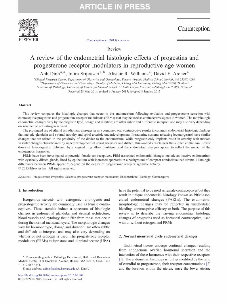
The epithelium (glandular and luminal) and mesenchyme(stroma and vasculature) undergo specific morphologic chang-es, with proliferation in response to estradiol, and secretorydifferentiation attributed to progesterone [11] (Figs. 1 and 2).
The withdrawal of estradiol and progesterone results inendometrial breakdown with bleeding, cellular dissolutionand shedding (menstruation) [12]. Recent studies implicatelocal inflammation in initiating endometrial breakdown andmenstruation. Progesterone withdrawal initiates an influx ofleukocytes, which result in local release of proteases,chemokines and cytokines [13,14]. The increased synthesisof endometrial matrix metalloproteinases leads to breakdownof the interstitial collagen, with associated loss of vascularsupport resulting in local thrombosis and loss of endometrialintegrity followed by endometrial shedding [15].
The cessation of menstruation is thought to be due tore-epithelization of the luminal epithelium, which is broughtabout by a rapid proliferation of the glandular and luminalepithelial cells initiated through local growth factors andlater by endogenous estradiol [16]. The glands and vesselsbecome sinuous in the proliferative phase as their growth“outstrips” that of the stroma [17].
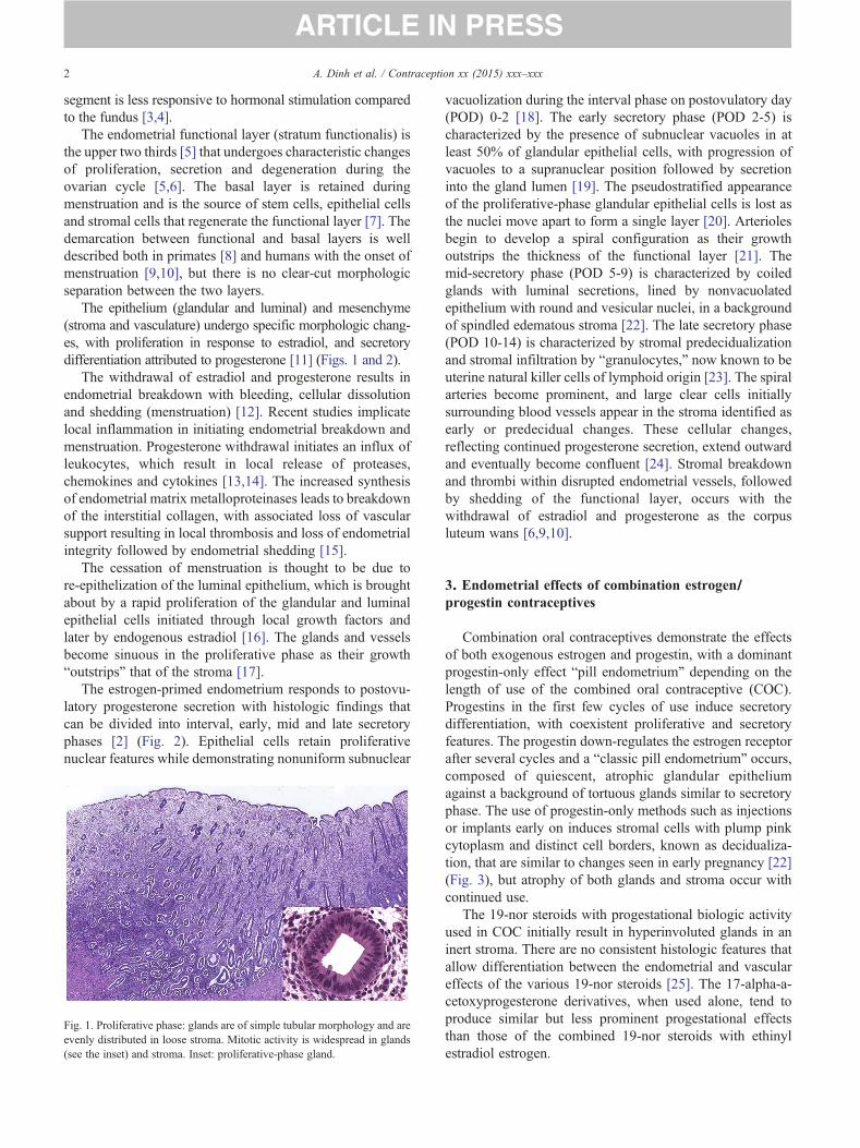
The estrogen-primed endometrium responds to postovu-latory progesterone secretion with histologic findings thatcan be divided into interval, early, mid and late secretoryphases [2] (Fig. 2). Epithelial cells retain proliferativenuclear features while demonstrating nonuniform subnuclear
Fig. 1. Proliferative phase: glands are of simple tubular morphology and areevenly distributed in loose stroma. Mitotic activity is widespread in glands(see the inset) and stroma. Inset: proliferative-phase gland.
vacuolization during the interval phase on postovulatory day(POD) 0-2 [18]. The early secretory phase (POD 2-5) ischaracterized by the presence of subnuclear vacuoles in atleast 50% of glandular epithelial cells, with progression ofvacuoles to a supranuclear position followed by secretioninto the gland lumen [19]. The pseudostratified appearanceof the proliferative-phase glandular epithelial cells is lost asthe nuclei move apart to form a single layer [20]. Arteriolesbegin to develop a spiral configuration as their growthoutstrips the thickness of the functional layer [21]. Themid-secretory phase (POD 5-9) is characterized by coiledglands with luminal secretions, lined by nonvacuolatedepithelium with round and vesicular nuclei, in a backgroundof spindled edematous stroma [22]. The late secretory phase(POD 10-14) is characterized by stromal predecidualizationand stromal infiltration by “granulocytes,” now known to beuterine natural killer cells of lymphoid origin [23]. The spiralarteries become prominent, and large clear cells initiallysurrounding blood vessels appear in the stroma identified asearly or predecidual changes. These cellular changes,reflecting continued progesterone secretion, extend outwardand eventually become confluent [24]. Stromal breakdownand thrombi within disrupted endometrial vessels, followedby shedding of the functional layer, occurs with thewithdrawal of estradiol and progesterone as the corpusluteum wans [6,9,10].
3. Endometrial effects of combination estrogen/progestin contraceptives
Combination oral contraceptives demonstrate the effectsof both exogenous estrogen and progestin, with a dominantprogestin-only effect “pill endometrium” depending on thelength of use of the combined oral contraceptive (COC).Progestins in the first few cycles of use induce secretorydifferentiation, with coexistent proliferative and secretoryfeatures. The progestin down-regulates the estrogen receptorafter several cycles and a “classic pill endometrium” occurs,composed of quiescent, atrophic glandular epitheliumagainst a background of tortuous glands similar to secretoryphase. The use of progestin-only methods such as injectionsor implants early on induces stromal cells with plump pinkcytoplasm and distinct cell borders, known as decidualiza-tion, that are similar to changes seen in early pregnancy [22](Fig. 3), but atrophy of both glands and stroma occur withcontinued use.
The 19-nor steroids with progestational biologic activityused in COC initially result in hyperinvoluted glands in aninert stroma. There are no consistent histologic features thatallow differentiation between the endometrial and vasculareffects of the various 19-nor steroids [25]. The 17-alpha-a-cetoxyprogesterone derivatives, when used alone, tend toproduce similar but less prominent progestational effectsthan those of the combined 19-nor steroids with ethinylestradiol estrogen.
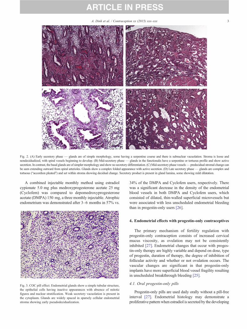
Fig. 2. (A) Early secretory phase — glands are of simple morphology, some having a serpentine course and there is subnuclear vacuolation. Stroma is loose andnondecidualized, with spiral vessels beginning to develop. (B) Mid-secretory phase — glands in the functionalis have a serpentine or tortuous profile and show activesecretion. In contrast, the basal glands are of simplermorphology and showno secretory differentiation. (C)Mid-secretory phase vessels— predecidual stromal change canbe seen extending outward from spiral arterioles. Glands show a complex folded appearance with active secretion. (D) Late secretory phase— glands are complex andtortuous (“accordion pleated”) and set within stroma showing decidual change. Secretory product is present in gland lumina, some showing mild dilatation.
3A. Dinh et al. / Contraception xx (2015) xxx–xxx
A combined injectable monthly method using estradiolcypionate 5.0 mg plus medroxyprogesterone acetate 25 mg(Cyclofem) was compared to depomedroxyprogesteroneacetate (DMPA) 150 mg, a three monthly injectable. Atrophicendometrium was demonstrated after 3–6 months in 57% vs.
Fig. 3. COC pill effect. Endometrial glands show a simple tubular structure,the epithelial cells having inactive appearances with absence of mitoticfigures and nuclear stratification. Weak secretory vacuolation is present inthe cytoplasm. Glands are widely spaced in sparsely cellular endometrialstroma showing early pseudodecidualization.
34% of the DMPA and Cyclofem users, respectively. Therewas a significant decrease in the density of the endometrialblood vessels in both DMPA and Cyclofem users, whichconsisted of dilated, thin-walled superficial microvessels butwere associated with less unscheduled endometrial bleedingthan in progestin-only users [26].
4. Endometrial effects with progestin-only contraceptives
The primary mechanism of fertility regulation withprogestin-only contraception consists of increased cervicalmucus viscosity, as ovulation may not be consistentlyinhibited [27]. Endometrial changes that occur with proges-tin-only therapy are highly variable and depend on dose, typeof progestin, duration of therapy, the degree of inhibition offollicular activity and whether or not ovulation occurs. Thevascular changes are significant in that progestin-onlyimplants have more superficial blood vessel fragility resultingin unscheduled breakthrough bleeding [25].
4.1. Oral progestin-only pills
Progestin-only pills are used daily orally without a pill-freeinterval [27]. Endometrial histology may demonstrate aproliferative patternwhen estradiol is secreted by the developing

ig. 4. Progestin (LNG-IUS; Mirena) effect — the stroma shows confluentecidual change. Vessels are thin-walled and ectatic, and clusters ofmphoid cells are present.
4 A. Dinh et al. / Contraception xx (2015) xxx–xxx
follicle and/or corpus luteum progesterone is decreased orabsent. The secretory pattern, when present, superficiallyresembles the secretory phase of the normal menstrual cycle,but glandular and stromal development lag compared to thatexpected on a given day of a normal cycle. Glands appearunderdeveloped and the stroma lacks the intermediate edema-tous changes found in a normal secretory phase. An atrophicpattern is common after prolonged use of progestin-onlycontraceptives, and it consists of small and scant glands thatlose their tortuosity and are lined by low columnar epithelium ina background of enlarged ovoid stromal cells with indistinctborders. The vessels of the superficial endometrium arethin-walled and ectatic. The glands are small and indistinct inmore abundant stroma compared to physiologic endometrialatrophy [28]. Norethindrone (NET) 300 mcg/day for a total of3 months resulted in a variety of endometrial histologic changesoccurring irrespective of ovarian function, including prolifera-tive activity, suppressed proliferation, irregular secretion andatrophy [29]. A study of desogestrel 75 mcg/day for a total of6 weeks showed a spectrum of endometrial changes in biopsies:proliferative endometrium, decidual transformation and glan-dular atrophy [30]. The differences in these two studies reflectthe fact that desogestrel highly suppresses ovulation whileNETat this dose has been found to inhibit ovulation only 35%ofthe time.
4.2. Subcutaneous implants: etonogestrel or levonorgestrel
Levonorgestrel tends to induce a wider spectrum ofendometrial effects than etonogestrel. A study in users oflevonorgestrel subcutaneous implants demonstrated histologyranging from atrophic changes, evidence of endometrialshedding, effects of progestins to proliferation. A few tissuesamples demonstrated early, mid or late secretory changes [31].There is a lack of specific histologic effect between theprogestins due to subjective interpretation of the endometriumand a lack of quantitative assessment of the endometrium [32].Both etonogestrel and levonorgestrel have similar endometrialhistologic findings after 12 months of use [33]. Levonorgestrelimplants result in an increased density of dilated, thin-walledfragile blood vessels that lead to the early onset of unscheduledbleeding [25].
4.3. Injectable DMPA
DMPA, an injectable contraceptive, is administeredintramuscularly every 3 months, and it induces endometrialchanges that initially have an exaggerated gestational-typeglandular hyperplasia with marked nuclear changes, in abackground of decidualized stroma. The stroma displaysprogestational effects after the first 6 months of use, butfurther treatment results in endometrial atrophy [34].
As mentioned above, DMPA was shown to result in thedecrease of the density of the endometrial blood vessels, whichconsisted of dilated, thin-walled superficial microvessels [26].Women using DMPA were found to have increased number ofleukocytes in the endometrium associated with unscheduled
bleeding compared to those with amenorrhea, suggesting a rolefor inflammation in the unscheduled bleeding [23,35].
4.4. Intrauterine systems releasing levonorgestrel
The levonorgestrel-releasing intrauterine system (LNG-IUS)produces atrophy of the glandular epithelium, prominentdecidualization of the stroma and suppression of spiral arteryformation as well as large, thin-walled, dilated vessels (Fig. 4)[29]. Women using the LNG-IUS had endometrial tissueobtained between 3 months and 7 years after insertion. Theendometrial histology demonstrated glandular atrophy anddecidualized stroma in all samples with evidence of inflamma-tion and necrosis. Endometrial morphology returned to normal1–3 months after removal of the LNG-IUS [36]. The mostcommon histologic features with use of the LNG-IUS arestromal decidualization (96%), glandular atrophy (87%) andstromal inflammatory cell infiltration (79%), with other stromalfindings including myxoid change and hemosiderin pigment[37]. The histologic findings vary according to the distance fromand contact with the IUS, reflecting a concentration gradient oflevonorgestrel within the tissue. The endometrium closest to theIUS had cushions of edematous, spindle-celled pseudodecidua-lization, and occasionally surface micropapillae, while theendometrium remote from the device displayed a less intenselydecidualized stroma with spindle cells. Endometrium closest tothe device showed glandswith a cuboidal-to-columnar epitheliallining in a background of stromal decidualization characterizedby rounder cells as opposed to spindled. A few of the specimenswere interpreted as having hemorrhagic infarction, necrobiosisor coagulative necrosis [38].
4.5. Intravaginal rings
An intravaginal ring (IVR) releasing levonorgestrel 20 mcg/day was studied for the effect on the endometrium after 90 daysof exposure. Endometrial tissues were obtained at days 23–25,
Fdly

5A. Dinh et al. / Contraception xx (2015) xxx–xxx
6 weeks and 10 weeks of use. At pretreatment, all endometrialbiopsies were normal secretory phase. During active treatment,the endometrial histology was as follows: 6 secretory, 4suppressed, 2 proliferative and 4 could not be dated due tobleeding, from 16 individual participants. There seemed to be acorrelation with the endogenous hormonal profile, as normalsecretory endometrium was associated with a normal serumprogesterone levels [39]. Menstrual disturbances were theprincipal reason for discontinuation of the method althoughwomen with ovulation appeared to have less unscheduledbleeding [18,40].
5. Progesterone receptor modulators
Mifepristone and UPA are PRMs that have beenevaluated in menstruating women for contraception and themanagement of uterine fibroids and endometriosis. Thesecompounds have the potential to be used as long-termcontraceptive agents [41]. PRMs bind to the progesteronereceptor and elicit tissue specific agonist, antagonist ormixed agonist/antagonist activity. Ligand binding to theprogesterone receptor causes dimerization and alteredconformation of the ligand-receptor complex, resulting in aunique DNA-binding configuration with subsequent bindingof the ligand-receptor complex to the promoter region knownas the progesterone response element. Depending on thetissue environment and ligand-receptor complex, recruitmentof coregulators, coactivators and coinhibitors results inprogesterone agonist or antagonist biologic activity [42].
The interpretation of the endometrial effects of PRMs hasvaried, from hyperplasia initially to a current consensus that theendometrial changes are specific to the class and do notrepresent hyperplasia or neoplasia. One study of mifepristonereported a 28% incidence of endometrial hyperplasia, whichupon subsequent review was revised to 14% [43–45]. A panelof seven gynecologic pathologists reviewed 84 endometrialspecimens from women receiving four different PRMs(mifepristone, asoprisnil, UPA and a Johnson & Johnsoncompound), including those from the abovementioned mifep-ristone study. The pathologists were blinded to PRM type, dose,exposure, exposure interval, patient group and indication. Thegoal was to identify endometrial histologic changes common toPRMs (a “class effect”) and to develop consensus terminologyand recommendations. The appearance of cystically dilatedglands was a frequent architectural finding. Cystic dilation ofglands is usually found in proliferative states such as unopposedestrogen effect and hyperplasia, but it is also seen inpostmenopausal and inactive states. Glands in the PRMspecimens, whether showing cystic dilation or not, had anarchitecture suggesting proliferative phase, but the glandularepithelial cells were only weakly mitotic and had secretoryvacuoles reflecting a progestational stimulus. This discordanceof epithelial architecture has been termed a nonphysiologicsecretory effect. Other findings included abnormal vasculature(thick-walled and thin-walled blood vessels and anastomosing
capillaries). The panel labeled this novel constellation ofchanges as PRM-associated endometrial changes or PAEC.The extent towhich PAECchanges are generic to the PRMclassor are specific to particular PRM compounds was unclear at thattime, and it was acknowledged that more evidence was requiredto define the endometrial changes [46].
5.1. Mifepristone
Short-term use of mifepristone induces endometrial changesthat may contribute to its use as a contraceptive, includingregression, retarded development, inhibited glandular secretoryactivity and decidual necrosis. A study evaluating the effects of50 mg of mifepristone daily on cycle days 7–10 or 20–23showed endometrial effects dependent on the time ofadministration during the cycle. Preovulatory administrationresulted in no bleeding episodes during treatment, and themorphology of the endometrium was identical to that found innormal menstruation. In contrast, postovulatory administrationresulted in bleeding on the third or fourth day of treatment, andendometrial histology showed early regressive changes corre-sponding to late postovulatory changes in a normal cycle.Likewise, postovulatory administration induces vascular chang-es that include capillary endothelial cell necrosis, which mayoccur without regression of adjacent stroma [47].
Forty-eight percent of women who took mifepristone for6 months had cystically dilated glands but no evidence ofhyperplasia or atypia [48]. A study of mifepristone in doses of2.5 and 5.0 mg for 6 months for treatment of uterine myomas(fibroids) evaluated the endometrium compared to untreatedwomen. Cystically dilated glands were seen in 86% ofendometrial tissue in the treated women and only 4% ofuntreated women, while few mitotic figures were seen inglandular epithelium, and apoptotic bodies were numerous.Abnormal vessels were variable and seen in 45% of exposedcases. The stroma ranged from compacted cellular to edematousareas, often within the same sample. No endometrial polyps,complex hyperplasia, premalignant lesions, atypical hyperpla-sia, endometrial intraepithelial neoplasia or carcinoma werenoted in any of the samples [21,49].
ZK230211 is a progesterone receptor antagonist that, whenadministered as an IUS, was shown to induce “estrogeniceffects,” including tortuous glands, cellular stratification andmitotic activity. The endometrium displayed dilated glandslined by epithelium of secretory morphology, in a backgroundof nondecidualized stroma. The authors noted that, while thefindings are consistent with previous results for mifepristone,application of nomenclature used for physiologic processes(“estrogen or progestin-like”) may not be justified when dealingwith synthetic progesterone antagonists [50].
5.2. Ulipristal acetate
UPA, a PRM in oral doses of 5.0 or 10.0 mg/day, was usedto treat women with heavy menstrual bleeding and fibroids intwo clinical trials [51,52]. The endometrium after 13 weeks ofUPA treatment demonstrated cystic dilation of the glands

Fig. 5. UPA effect. (A) The endometrial glands show frequent cystic dilatation but have an inactive appearance. Thick-walled arteriolar vessels are present instroma. The stroma is compact but nondecidualized. (B) Glands have a complex, distorted morphology, sometimes with cystic dilatation. Abortive secretoryvacuolation is present. The stroma is compact and nondecidualized with areas of prominent vascularity.
6 A. Dinh et al. / Contraception xx (2015) xxx–xxx
(Fig. 5), similar to other PRMs [53]. The glandular epitheliumappeared inactive or contained abortive subnuclear vacuoliza-tion and few mitoses. Many glands had a tortuous appearancesimilar to that seen in the secretory phase. The stroma was ofvariable cellularity but without predecidual change. Abnormalstromal vasculature was commonly seen, including “chick-en-wire” capillaries, aggregates of thickened arterioles andthin-walled ectatic vessels. There was one case of endometrialhyperplasia without atypia and four polyps. Six months aftertreatment, the endometrium had returned to a normal histologicpattern in the majority of the patients. There was one polyp andno cases of hyperplasia in the UPA-treated groups [53]. Thesefindings indicate that the endometrial changes associated withUPA are reversible upon discontinuation of treatment.
An IVR releasing UPA 600–800 mcg/day for contraceptionwas studied in 37 women. There were no reports of endometrialhyperplasia or cytologic atypia after 12 weeks of use. Inactiveendometrium was found in 38% of cases, with widelyinterspersed tubular glands in a compact stroma. Benignglandular changes described as PAECs were present in 41%of cases [54].
5.3. Summary of endometrial PRM changes
PRMs alter the configuration of the progesterone receptorresulting in endometrial features resembling an unopposedestrogenic effect. This finding of weakly proliferative glandularepithelium might suggest susceptibility to developing endome-trial hyperplasia, but to date, this has not been substantiated inthe clinical trials. A defining feature of PAECs is relativelyinactive appearing endometrium, with low levels of mitoticactivity, elevated incidence of apoptosis in the glandularepithelium and compact stroma that is rarely decidualized.The endometrial glandular mitotic activity is low in thePRM-treated women compared to the normal proliferative
phase, a consequence of well-recognized but still unexplainedendometrial antiproliferative effects of selective progesteronereceptor modulators (SPRMs) [55]. There is variation in thepattern of PAEC according to the PRM used, which can beexplained by different activities varying from almost pureantagonist effect (e.g., mifepristone) to a mixed agonist–antagonist effect (UPA) [56]. The variability is seen particularlyin the nonphysiologic secretory effects, which are frequentlyprominent with UPA but less common with mifepristone.
6. Conclusion
Exogenous hormones used as contraceptives induce histo-logic changes with effects on endometrial glandular and stromalarchitecture, blood vessels and cytology that differ from thosethat occur during the menstrual ovarian cycle (Table 1).
The common histologic findings with prolonged use ofcombined oral contraception are atrophy of both glands andstroma and spiral arteriole underdevelopment. There are fewmorphologic differences between effects of individual proges-togenic agents. Injectable contraception with depot medrox-yprogesterone induces endometrial changes that includeatrophic glands in a background of predecidualized stroma.Intrauterine devices releasing levonorgestrel induce atrophicglands in a background of decidualized stroma and suppressionof spiral artery formation with thin-walled dilated vessels. Theprominence of such features depends on proximity of theaffected endometrium to the device. The IVR with levonorges-trel induces variable secretory and suppressed histologic featuresthat appear to correspond with the hormone profile duringtreatment. A common feature of all progestin-only contracep-tives is the effects on vasculature, inwhich underdevelopment ofspiral arterioles and dilated, thin-walled vessels may contributeto the irregular bleeding common with such contraceptives.

Table 1Summary histologic changes.
Duration of therapy General Glands Stroma Vasculature
E+P Early — first few cycles Secretory differentiation,proliferative and secretoryfeatures coexist.
S
Several cycles “Pill endometrium” A D Spiral arteriole underdevelopmentLate A A A Spiral arteriole underdevelopment
Progestin (systemic) Early A D Vessels of the superficialendometrium arethin-walled and ectatic
Late A A Vessels of the superficialendometrium are thin-walledand ectatic
Progestin (local) Close to IUS:cuboidal-to-columnarepitheliumRemote from IUS:flattened cuboidalepithelium
D Suppression of spiral arteryformation and large, thin-walled,dilated vessels
SPRM PAECs Cystically dilated Nondecidualized,compact stroma
Abnormal, three types: dilated,thin-walled, thick-walled,anastomosing capillary
Progestogenic changes — secretory (S), decidualization (D) and atrophy (A).
7A. Dinh et al. / Contraception xx (2015) xxx–xxx
PAECs include an inactive endometrium with characteristiccystically dilated glands, lined by epithelium with increasedapoptosis in a background of compact nondecidualized stroma.Histologic differences between agents appear to depend on thelevel of antagonism to the progesterone receptor, in whichmifepristone does not display the nonphysiologic secretoryeffect found in UPA. To date, the consensus is that endometrialhyperplasia is not a histologic consequence of PRM treatment.
References
[1] Lessey BA, Killam AP, Metzger DA, Haney AF, Greene GL, McCartyKS. Immunohistochemical analysis of human uterine estrogen andprogesterone receptors throughout the menstrual cycle. J ClinEndocrinol Metab 1988;67:334–40.
[2] Noyes RW, Hertig AT, Rock J. Dating the endometrial biopsy. Am JObstet Gynecol 1975;122:262–3.
[3] Ferenczy A, Bertrand G, Gelfand MM. Proliferation kinetics of humanendometrium during the normal menstrual cycle. Am J Obstet Gynecol1979;133:859–67.
[4] Noyes RW. Uniformity of secretory endometrium. Obstet Gynecol1956;7:221–8.
[5] Kurman RJEH. Blaustein's pathology of the female genital tract. 6thed. New York: Springer; 2011.
[6] Kristen A, Atkins MRH, Kempson Richard L. Chapter 41: normalhistology of the uterus and fallopian tubes. In: &Mills SE, editor. Histologyfor the pathologist. Wolters Kluwer, Health; 2012, pp. 1088–99.
[7] Ferenczy A, Bertrand G, Gelfand MM. Studies on the cytodynamics ofhuman endometrial regeneration. III. In vitro short-term incubationhistoradioautography. Am J Obstet Gynecol 1979;134:297–304.
[8] Brenner RM, Slayden OD. Molecular and functional aspects ofmenstruation in the macaque. Rev Endocr Metab Disord 2012;13:309–18.
[9] Christiaens GC, Sixma JJ, Haspels AA. Morphology of haemostasis inmenstrual endometrium. Br J Obstet Gynaecol 1980;87:425–39.
[10] Christiaens GC, Sixma JJ, Haspels AA. Hemostasis in menstrualendometrium: a review. Obstet Gynecol Surv 1982;37:281–303.
[11] Richart RM, Ferenczy A. Endometrial morphologic response tohormonal environment. Gynecol Oncol 1974;2:180–97.
[12] Hall JE, Schoenfeld DA, Martin KA, Crowley WF. Hypothalamicgonadotropin-releasing hormone secretion and follicle-stimulatinghormone dynamics during the luteal-follicular transition. J ClinEndocrinol Metab 1992;74:600–7.
[13] Salamonsen LA. Current concepts of the mechanisms of menstruation:a normal process of tissue destruction. Trends Endocrinol Metab1998;9:305–9.
[14] Salamonsen LA, Lathbury LJ. Endometrial leukocytes and menstru-ation. Hum Reprod Update 2000;6:16–27.
[15] Hickey M, Fraser IS. Iatrogenic unscheduled (breakthrough) endome-trial bleeding. Rev Endocr Metab Disord 2012;13:301–8.
[16] Ferenczy A, Bergeron C. Histology of the human endometrium: frombirth to senescence. Ann N Y Acad Sci 1991;622:6–7.
[17] Johnson JV, Grubb GS, Constantine GD. Endometrial histologyfollowing 1 year of a continuous daily regimen of levonorgestrel90 micro g/ethinyl estradiol 20 micro g. Contraception 2007;75:23–6.
[18] Koetsawang S, Gao J, Krishna U, Cuadros A, Dhall GI, Wyss R, et al.Microdose intravaginal levonorgestrel contraception: a multicentreclinical trial. I. Contraceptive efficacy and side effects. World HealthOrganization. Task Force on Long-Acting Systemic Agents forFertility Regulation. Contraception 1990;41:105–24.
[19] Chwalisz K, Larsen L, Mattia-Goldberg C, Edmonds A, Elger W,Winkel CA. A randomized, controlled trial of asoprisnil, a novelselective progesterone receptor modulator, in women with uterineleiomyomata. Fertil Steril 2007;87:1399–412.
[20] Song JY, Fraser IS. Effects of progestogens on human endometrium.Obstet Gynecol Surv 1995;50:385–94.
[21] Fiscella J, Bonfiglio T, Winters P, Eisinger SH, Fiscella K.Distinguishing features of endometrial pathology after exposure tothe progesterone receptor modulator mifepristone. Hum Pathol2011;42:947–53.
[22] Bentley GLM Rex C, Robboy Stanley J. Chapter 12: the normalendometrium. Robboy's pathology of the female reproductive tract.2nd ed. Elsevier Health Sciences; 2009297–324.
[23] Salamonsen LA, Zhang J, Brasted M. Leukocyte networks and humanendometrial remodelling. J Reprod Immunol 2002;57:95–08.
8 A. Dinh et al. / Contraception xx (2015) xxx–xxx
[24] Bulten J, Grefte J, Siebers B, Dieben T. The combined contraceptivevaginal ring (NuvaRing) and endometrial histology. Contraception2005;72:362–5.
[25] Smith OP, Critchley HO. Progestogen only contraception andendometrial break through bleeding. Angiogenesis 2005;8:117–26.
[26] Simbar M, Tehrani FR, Hashemi Z, Zham H, Fraser IS. A comparativestudy of Cyclofem and depot medroxyprogesterone acetate (DMPA)effects on endometrial vasculature. J Fam Plann Reprod Health Care2007;33:271–6.
[27] Amy JJ, Tripathi V. Contraception for women: an evidence basedoverview. BMJ 2009;339:b2895.
[28] Mazur M, Kurman Robert J. Diagnosis of endometrial biopsies andcurettings: a practical approach. 2nd ed. New York: Springer Scienceand Business Media, Inc.; 2005.
[29] Guttinger A, Critchley HO. Endometrial effects of intrauterinelevonorgestrel. Contraception 2007;75:S93–8.
[30] Haimovich S, Mancebo G, Alameda F, Agramunt S, Hernandez JL,Carreras R. Endometrial preparation with desogestrel before Essurehysteroscopic sterilization: preliminary study. J Minim InvasiveGynecol 2013;20:591–4.
[31] MarshMM, Butt AR, Riley SC, Rogers PAW, Susil B, Affandi B, et al.Immunolocalization of endothelin and neutral endopeptidase in theendometrium of users of subdermally implanted levonorgestrel(Norplant). Hum Reprod 1995;10:2584–9.
[32] Laban M, Abd Alhamid M, Ibrahim EA, Elyan A, Ibrahim A.Endometrial histopathology, ovarian changes and bleeding patternsamong users of long-acting progestin-only contraceptives in Egypt.Eur J Contracept Reprod Health Care 2012;17:451–7.
[33] Mascarenhas L, vanBeekA,BenninkHC,Newton J.A 2-year comparativestudy of endometrial histology and cervical cytology of contraceptiveimplant users in Birmingham, UK. Hum Reprod 1998;13:3057–60.
[34] Rex C, Bentley SJR. Chapter 13: exogenous hormones and their effectson the endometrium. Robboy's pathology of the female reproductivetract2nd ed. . Elsevier Health Sciences; 2009325–42.
[35] Thurman AR, Soper DE. Endometrial histology of depomedroxypro-gesterone acetate users: a pilot study. Infect Dis Obstet Gynecol2006;2006:69402.
[36] Silverberg SG, Haukkamaa M, Arko H, Nilsson CG, Luukkainen T.Endometrial morphology during long-term use of levonorgestrel-releasing intrauterine devices. Int J Gynecol Pathol 1986;5:235–41.
[37] Phillips V, Graham CT, Manek S, McCluggage WG. The effects of thelevonorgestrel intrauterine system (Mirena coil) on endometrialmorphology. J Clin Pathol 2003;56:305–7.
[38] Critchley HO, Wang H, Jones RL, et al. Morphological and functionalfeatures of endometrial decidualization following long-term intrauter-ine levonorgestrel delivery. Hum Reprod 1998;13:1218–24.
[39] Landgren BM, Johannisson E, Masironi B, Diczfalusy E. Pharmaco-kinetic and pharmacodynamic investigations with vaginal devicesreleasing levonorgestrel at a constant, near zero order rate. Contra-ception 1982;26:567–85.
[40] Koetsawang S, Gao J, Krishna U, Cuadros A, Dhall GI, Wyss R, et al.Microdose intravaginal levonorgestrel contraception: a multicentreclinical trial. IV. Bleeding patterns. World Health Organization. TaskForce on Long-Acting Systemic Agents for Fertility Regulation.Contraception 1990;41:151–67.
[41] Bouchard P, Chabbert-Buffet N, Fauser BC. Selective progesteronereceptor modulators in reproductive medicine: pharmacology, clinicalefficacy and safety. Fertil Steril 2011;96:1175–89.
[42] Chabbert-Buffet N, Meduri G, Bouchard P, Spitz IM. Selectiveprogesterone receptor modulators and progesterone antagonists:mechanisms of action and clinical applications. Hum Reprod Update2005;11:293–307.
[43] Eisinger SH, Meldrum S, Fiscella K, le Roux HD, Guzick DS. Low-dose mifepristone for uterine leiomyomata. Obstet Gynecol2003;101:243–50.
[44] Eisinger SH, Bonfiglio T, Fiscella K, Meldrum S, Guzick DS. Twelve-month safety and efficacy of low-dose mifepristone for uterinemyomas. J Minim Invasive Gynecol 2005;12:227–33.
[45] Spitz IM. Clinical utility of progesterone receptor modulators and theireffect on the endometrium. Curr Opin Obstet Gynecol 2009;21:318–24.
[46] Mutter GL, Bergeron C, Deligdisch L, Ferenczy A, Glant M, MerinoM, et al. The spectrum of endometrial pathology induced byprogesterone receptor modulators. Mod Pathol 2008;21:591–8.
[47] Johannisson E, Oberholzer M, Swahn ML, Bygdeman M. Vascularchanges in the human endometrium following the administration of theprogesterone antagonist RU 486. Contraception 1989;39:103–17.
[48] Lakha F, Ho PC, Van der Spuy ZM, Dada K, Elton R, Glasier AF, et al.A novel estrogen-free oral contraceptive pill for women: multicentre,double-blind, randomized controlled trial of mifepristone andprogestogen-only pill (levonorgestrel). Hum Reprod 2007;22:2428–36.
[49] Sathya G, Jansen MS, Nagel SC, Cook CE, McDonnell DP.Identification and characterization of novel estrogen receptor-beta-sparing antiprogestins. Endocrinology 2002;143:3071–82.
[50] Heikinheimo O, Vani S, Carpen O, Tapper A, Harkki P, Rutanen E, et al.Intrauterine release of progesterone antagonist ZK230211 is feasible andresults in novel endometrial effects: a pilot study. Hum Reprod2007;22:2515–22.
[51] Donnez J, Tomaszewski J, Vazquez F, Bouchard P, Lemieszczuk B, BaroF, et al. Ulipristal acetate versus leuprolide acetate for uterine fibroids. NEngl J Med 2012;366:421–32.
[52] Donnez J, TatarchukTF, Bouchard P, Puscasiu L, ZakharenkoNF, IvanovaT, et al. Ulipristal acetate versus placebo for fibroid treatment beforesurgery. N Engl J Med 2012;366:409–20.
[53] Williams AR, Bergeron C, Barlow DH, Ferenczy A. Endometrialmorphology after treatment of uterine fibroids with the selectiveprogesterone receptor modulator, ulipristal acetate. Int J GynecolPathol 2012;31:556–69.
[54] Brache V, Sitruk-Ware R, Williams A, Blithe D, Croxatto H, Kumar N,et al. Effects of a novel estrogen-free, progesterone receptor modulatorcontraceptive vaginal ring on inhibition of ovulation, bleeding patternsand endometrium in normal women. Contraception 2012;85:480–8.
[55] Chwalisz K, Brenner RM, Fuhrmann UU, Hess-Stumpp H, Elger W.Antiproliferative effects of progesterone antagonists and progesteronereceptor modulators on the endometrium. Steroids 2000;65:741–51.
[56] Schubert G, Elger W, Kaufmann G, Schneider B, Reddersen G,Chwalisz K. Discovery, chemistry, and reproductive pharmacology ofasoprisnil and related 11beta-benzaldoxime substituted selectiveprogesterone receptor modulators (SPRMs). Semin Reprod Med2005;23:58–73.
Related Documents