Defensins play a crucial role in protecting mice against oral Shigella flexneri infection Doo-Hee Shim a,b , Sangryeol Ryu b , Mi-Na Kweon a,⇑ a Mucosal Immunology Section, International Vaccine Institute, Seoul 151-818, Republic of Korea b Department of Food and Animal Biotechnology, School of Agricultural Biotechnology and Center for Agricultural Biomaterials, Seoul National University, Seoul 151-742, Republic of Korea article info Article history: Received 17 September 2010 Available online 1 October 2010 Keywords: Shigella Inflammation Mucosa Neonate Defensin abstract An earlier study revealed that 4-day-old mice, but not older mice, were infected with invasive Shigella strains. Here we attempted to determine the underlying mechanism that induces inflammation in the intestines of neonate mice after oral Shigella infection. Wild-type BALB/c mice of different ages (7, 14, and 35 days old) were orally administered GFP-expressing Shigella flexneri 5a M90T strain (5 Â 10 9 CFU) and analyzed for colonization 6 h following infection. We found that Shigella localized in the epithe- lium, lamina propria, and crypt regions of the small intestines of 7-day-old BALB/c mice. Microarray anal- ysis revealed that expression levels of cryptdin and various types of cryptdin-related mRNA (e.g., cryptrs- 2, -5, -7, -12 and lysozyme) in the small intestines were significantly lower in 7-day-old than in older mice regardless of Shigella infection status. Interestingly, matrix metalloprotease-7 (matrilysin)-deficient (MAT / ) mice of B6 background had more colonies and more severe symptoms of inflammation in the intestines than did wild-type B6 mice after oral Shigella challenge. This suggests that cryptdin-related antimicrobial molecules are indispensable for efficient protection against oral Shigella infection. Ó 2010 Elsevier Inc. All rights reserved. 1. Introduction Shigella species, Gram-negative enteroinvasive bacteria, cause bacillary dysentery by invading the large intestinal epithelium and promoting a severe intestinal inflammatory response accom- panied by fever, abdominal cramps, and tenesmus in human and non-human primates [1–4]. Shigellosis is a huge global health problem and in developing countries is a common cause of infant mortality [3–6]. A murine shigellosis model would facilitate under- standing the pathogenesis and characteristics of the disease and enable screening of vaccine candidates. A previous study demon- strated that 4-day-old mice were susceptible to oral invasive Shi- gella challenge but not older mice [7]. Histologically, 4-day-old mice challenged with an invasive Shigella strain showed bacteria and inflammation in the intestinal region that mimicked human shigellosis [7]. To date, the underlying mechanism that enables older mice to acquire resistance against virulent Shigella infection is not clear. We know there are key innate and adaptive immune system differences between human neonates/young infants and adults, which may explain why infants are unduly susceptible to infection and tend to respond less rapidly and vigorously to immunization than older children or adults [8]. Kollmann et al. [9] showed that in neonatal cells the TLR-mediated innate immunity produces few- er multiple cytokines simultaneously than found in adult cells. T-cell-dependent and -independent antibody responses, although inducible by the third trimester of pregnancy, develop slowly. Thus, disease may take hold before effective immunity is induced [10]. In addition, T-cell-independent antibody responses to poly- saccharide antigens in neonates are absent and therefore cannot be used for intervention [11,12]. It seems likely that antigen-pre- senting cells (e.g., macrophages and dendritic cells [DC]) in the gas- trointestinal mucosa of neonatal mice lack the ability to recognize bacteria as dangerous [13]. Innate immunity is the first line of defense against virulent bac- teria and is important for normal microbiota in intestinal environ- ments and crucial at early stages of life [14]. A recent study showed that Paneth cells, major producers of multiple peptides and pro- teins with antimicrobial activity in the small intestine, develop after age 7 days [15,16]. Antimicrobial peptides (AMPs) efficiently control microbial growth in the gastrointestinal tract without 0006-291X/$ - see front matter Ó 2010 Elsevier Inc. All rights reserved. doi:10.1016/j.bbrc.2010.09.100 Abbreviations: AMPs, antimicrobial peptides; CFU, colony-forming units; Cyptrs, cryptdin-related mRNA sequence; DC, dendritic cells; FAE, follicle-associated epithelium; LP, lamina propria; MAT, matrilysin; PLA(2)-IIA, phospholipase A2 group IIA; PP, Peyer’s patch; SIgA, secretory IgA; TSB, trypticase soybean-casein digest broth; Zo-1, zonula occludens protein 1. ⇑ Corresponding author. Address: Mucosal Immunology Section, International Vaccine Institute, Seoul National University Research Park, Nakseongdae-dong Kwanak-Gu, Seoul 151-818, Republic of Korea. Fax: +82 2 881 1211. E-mail address: [email protected] (M.-N. Kweon). Biochemical and Biophysical Research Communications 401 (2010) 554–560 Contents lists available at ScienceDirect Biochemical and Biophysical Research Communications journal homepage: www.elsevier.com/locate/ybbrc

Welcome message from author
This document is posted to help you gain knowledge. Please leave a comment to let me know what you think about it! Share it to your friends and learn new things together.
Transcript

Biochemical and Biophysical Research Communications 401 (2010) 554–560
Contents lists available at ScienceDirect
Biochemical and Biophysical Research Communications
journal homepage: www.elsevier .com/locate /ybbrc
Defensins play a crucial role in protecting mice against oralShigella flexneri infection
Doo-Hee Shim a,b, Sangryeol Ryu b, Mi-Na Kweon a,⇑a Mucosal Immunology Section, International Vaccine Institute, Seoul 151-818, Republic of Koreab Department of Food and Animal Biotechnology, School of Agricultural Biotechnology and Center for Agricultural Biomaterials, Seoul National University, Seoul151-742, Republic of Korea
a r t i c l e i n f o
Article history:Received 17 September 2010Available online 1 October 2010
Keywords:ShigellaInflammationMucosaNeonateDefensin
0006-291X/$ - see front matter � 2010 Elsevier Inc. Adoi:10.1016/j.bbrc.2010.09.100
Abbreviations: AMPs, antimicrobial peptides; CFU,cryptdin-related mRNA sequence; DC, dendritic cepithelium; LP, lamina propria; MAT, matrilysin; Pgroup IIA; PP, Peyer’s patch; SIgA, secretory IgA; TSdigest broth; Zo-1, zonula occludens protein 1.⇑ Corresponding author. Address: Mucosal Immun
Vaccine Institute, Seoul National University ResearKwanak-Gu, Seoul 151-818, Republic of Korea. Fax: +
E-mail address: [email protected] (M.-N. Kweon).
a b s t r a c t
An earlier study revealed that 4-day-old mice, but not older mice, were infected with invasive Shigellastrains. Here we attempted to determine the underlying mechanism that induces inflammation in theintestines of neonate mice after oral Shigella infection. Wild-type BALB/c mice of different ages (7, 14,and 35 days old) were orally administered GFP-expressing Shigella flexneri 5a M90T strain (5 � 109
CFU) and analyzed for colonization 6 h following infection. We found that Shigella localized in the epithe-lium, lamina propria, and crypt regions of the small intestines of 7-day-old BALB/c mice. Microarray anal-ysis revealed that expression levels of cryptdin and various types of cryptdin-related mRNA (e.g., cryptrs-2, -5, -7, -12 and lysozyme) in the small intestines were significantly lower in 7-day-old than in oldermice regardless of Shigella infection status. Interestingly, matrix metalloprotease-7 (matrilysin)-deficient(MAT�/�) mice of B6 background had more colonies and more severe symptoms of inflammation in theintestines than did wild-type B6 mice after oral Shigella challenge. This suggests that cryptdin-relatedantimicrobial molecules are indispensable for efficient protection against oral Shigella infection.
� 2010 Elsevier Inc. All rights reserved.
1. Introduction
Shigella species, Gram-negative enteroinvasive bacteria, causebacillary dysentery by invading the large intestinal epitheliumand promoting a severe intestinal inflammatory response accom-panied by fever, abdominal cramps, and tenesmus in human andnon-human primates [1–4]. Shigellosis is a huge global healthproblem and in developing countries is a common cause of infantmortality [3–6]. A murine shigellosis model would facilitate under-standing the pathogenesis and characteristics of the disease andenable screening of vaccine candidates. A previous study demon-strated that 4-day-old mice were susceptible to oral invasive Shi-gella challenge but not older mice [7]. Histologically, 4-day-oldmice challenged with an invasive Shigella strain showed bacteriaand inflammation in the intestinal region that mimicked humanshigellosis [7]. To date, the underlying mechanism that enables
ll rights reserved.
colony-forming units; Cyptrs,ells; FAE, follicle-associatedLA(2)-IIA, phospholipase A2B, trypticase soybean-casein
ology Section, Internationalch Park, Nakseongdae-dong82 2 881 1211.
older mice to acquire resistance against virulent Shigella infectionis not clear.
We know there are key innate and adaptive immune systemdifferences between human neonates/young infants and adults,which may explain why infants are unduly susceptible to infectionand tend to respond less rapidly and vigorously to immunizationthan older children or adults [8]. Kollmann et al. [9] showed thatin neonatal cells the TLR-mediated innate immunity produces few-er multiple cytokines simultaneously than found in adult cells.T-cell-dependent and -independent antibody responses, althoughinducible by the third trimester of pregnancy, develop slowly.Thus, disease may take hold before effective immunity is induced[10]. In addition, T-cell-independent antibody responses to poly-saccharide antigens in neonates are absent and therefore cannotbe used for intervention [11,12]. It seems likely that antigen-pre-senting cells (e.g., macrophages and dendritic cells [DC]) in the gas-trointestinal mucosa of neonatal mice lack the ability to recognizebacteria as dangerous [13].
Innate immunity is the first line of defense against virulent bac-teria and is important for normal microbiota in intestinal environ-ments and crucial at early stages of life [14]. A recent study showedthat Paneth cells, major producers of multiple peptides and pro-teins with antimicrobial activity in the small intestine, developafter age 7 days [15,16]. Antimicrobial peptides (AMPs) efficientlycontrol microbial growth in the gastrointestinal tract without

D.-H. Shim et al. / Biochemical and Biophysical Research Communications 401 (2010) 554–560 555
any induction of potentially harmful inflammatory responses [17].Among AMPs, a-defensin plays a crucial role in enteric antibacte-rial defense together with other molecules such as lysozyme, b-defensin, cryptdin-related peptides, and the phospholipase A2group IIA (PLA(2)-IIA). These are mainly produced by Paneth andepithelial cells in the small intestine [18,19]. In children and adultswith diarrhea caused by Shigella spp., synthesis of colonic entero-cyte b-defensin HBD-1 and the cathelicidin LL37 is markedly de-pressed but expression recovers as the illness resolves [20]. Inaddition, a-defensins, termed cryptdin in mice, are abundant con-stituents of secretory granules in Paneth cells in the small intestine[17] and are produced from day 14 after birth [21].
In the present study, we found that Shigella organisms can in-vade the small intestine of neonatal mice until age 7 days butnot later. Microarray analysis revealed lower expression levels ofvarious types of cryptdin-related mRNA in the small intestine of7-day-old mice than in the older mice that were less susceptibleto oral Shigella challenge. These data suggest the importance ofdefensin (e.g., cryptdin and cryptdin-related molecules) for hostdefense against oral Shigella infection in mice and further implythat modulation of those molecules in adult mice could be usefulfor developing a murine shigellosis model.
2. Materials and methods
2.1. Mice
BALB/c and C57BL/6 mice were purchased from Charles RiverCo. (Seoul, Korea). MAT�/� mice of C57BL/6 background were pur-chased from the Jackson Laboratory (Bar Harbor, ME). The animalswere maintained in the animal care facilities of the InternationalVaccine Institute (Seoul, Korea). All experiments described wereapproved by the ethical committees for animal experiments ofSeoul National University and the International Vaccine Institute.
2.2. Oral infection and survival
We grew the virulent form of streptomycin-resistant, GFP-expressing Shigella flexneri 5a M90T strain in trypticase soybean-casein digest broth (TSB; Difco, Sparks, MD) containing 20 lg/mlstreptomycin (Sigma–Aldrich, St. Louis, MO) [7]. Neonate micewere inoculated orally with 5 � 109 colony-forming units (CFU)of virulent Shigella strain without any starvation or antibiotic treat-ment. After oral infection, mice were separated from their mothers.Mice were monitored for tenesmus, diarrhea at 6 h, and survival at24 h after oral Shigella infection.
2.3. Bacteria count
To assess the numbers of bacteria in the ileal region of smallintestine (one-fourth of the small intestine beginning at the ce-cum) and whole large intestine of non-infected and Shigella-in-fected mice, dissected samples were placed in PBS withgentamycin (50 lg/ml). Tissues were homogenized and platedonto TSB agar plates that included streptomycin, and green-col-ored colonies were enumerated after overnight culture at 37 �C.
2.4. Histology
Randomly selected tissues in the ileal region were washed withPBS including gentamycin and fixed in 4% formaldehyde for 1 h at4 �C. For immunohistochemical study, frozen intestines were cutinto 5-lm sections and stained with PE-conjugated anti-CD11c(BD Pharmingen, San Diego, CA) or Alexa 647-conjugatedNKM16-2-4 mAb (kindly provided by Prof. Hiroshi Kiyono, Univer-
sity of Tokyo, Japan) [22]. The sections were mounted with Perma-Fluor medium (Thermo, Houston, TX) and viewed under a confocalscanning laser microscope (Zeiss, Germany).
2.5. Microarray analysis
At 0 and 6 h following Shigella infection, we obtained about1 cm of the small intestine ileal region from 7-, 14-, and 35-day-old BALB/c mice. Specimens were homogenized after washes withPBS with gentamycin to remove attached bacteria in the cell or tis-sue surface. RNA was extracted by RNA isolation kit (Qiagen,Valencia, CA). cDNA microarray was performed by Macrogen Co.(Seoul, Korea) using an illumina BeadStation 500 X (Illumina,Inc., San Diego, CA).
2.6. Statistics
Data are expressed as the mean ± S.D. Statistical comparisonsbetween experimental groups were performed using the Studentt-test.
3. Results
3.1. Virulent Shigella organisms invade small and large intestines oforally challenged newborn mice
In order to determine whether Shigella organisms invade andcolonize mouse intestines, we adopted 4-, 7-, 14-, and 21-day-old BALB/c mice and orally administered GFP-expressed S. flexneri5a M90T strain (5 � 109 CFU). At 6 h after oral challenge, we deter-mined bacteria colonization in the ileal region of the small intes-tine and in the whole large intestine (Fig. 1A). We confirmed ourfindings by confocal microscopy (Fig. 1B). In contrast to findingsin an earlier study [7], we found that orally administered S. flexneri5a M90T invaded and colonized both small and large intestines in4-, 7-, and 14-day-old mice and gradually decreased (Fig. 1A). Or-ally administered GFP-expressing S. flexneri 5a M90T was detectedin both epithelial and crypt regions in the intestines of 7-day-oldmice (Fig. 1B). Next, we used NKM16-4-2 mAb specific for murineM cells to clarify the invasion patterns of Shigellae in the murinesmall intestine [22]. Interestingly, virulent GFP-expressing S. flex-neri 5a M90T were translocated not only by M cells (Fig. 1C-I)but also by epithelial cells (Fig. 1C-II) in the follicle-associated epi-thelium (FAE) of Peyer’s patches (PP), and lamina propria (LP) ofthe small intestine of 7-day-old mice (Fig. 1C). Because a previousstudy showed DCs penetrate epithelia to sample bacteria in the gutlumen [23], we next determined localization of CD11c+ DCs in thesmall intestine following oral challenge with GFP-expressingS. flexneri 5a M90T strain. We found no direct evidence that intes-tinal DCs are actively involved in sampling Shigella from lumen ineither PP or LP of the small intestine of 7-day-old mice (Fig. 1C).Thus, when our findings are considered together, we believeGFP-expressing virulent S. flexneri 5a M90T administered by oralchallenge might invade through both M and epithelial cells andcolonize in the small intestine of 7-day-old of BALB/c mice.
3.2. Lack of cryptdins in the small intestine of 7-day-old mice
In order to clarify which molecules are crucial for bacterial inva-sion and onset of inflammation in 7-day-old neonate mice, totalgene expression levels were compared in the small intestine of7-, 14- and 35-day-old mice by microarray analysis (Fig. 2). Among40,000 probes, we found approximately 20- and 60-fold increasesof intelectin 2 expression, which is known as goblet cell lectin[24], in 14- and 35-day-old mice, respectively. We also found a

Fig. 1. After oral challenge with virulent GFP-expressing S. flexneri 5a, the bacteria invade both epithelial cells and crypt regions and colonize the intestines of 7-day-old mice.BALB/c mice of different ages were inoculated intragastrically with 5 � 109 CFU of S. flexneri 5a and sacrificed 6 h later for count of surviving bacteria in the intestines (A). GFP-expressed S. flexneri 5a was observed by confocal microscopy (B). Shigella was localized in the small and large intestines of 7-day-old mice. Results are expressed as meanvalues ± S.D. for two to four mice per group and are representative of three separate experiments. *p < 0.05; compared to non-infected age-matched mice. n.d., Not detected.(C) GFP-expressed bacteria in the Peyer’s patch (PP) and lamina propria (LP) in the small intestine of 7-day-old mice were visualized by confocal microscopy after staining forM cells (A-I) and CD11c+ cells (A-II). Bar indicates 20 lm.
556 D.-H. Shim et al. / Biochemical and Biophysical Research Communications 401 (2010) 554–560
15-fold increase of clusterin expression, an inflammation-associ-ated protein, in 35-day-old mice [25]. In addition, the intestinalantimicrobial enzymes [26], lysozyme and PLA (2)-IIA encodingmRNA expression, were increased by 25- and 50-fold in the smallintestine of 14- and 35-day-old mice when compared with 7-day-old mice. Further, pIgR expression also increased 4- and 40-fold inthe 14- and 35-day-old mice when compared with 7-day-old mice.Interestingly, cryptdin and cryptdin-related mRNA (cyptrs) such ascryptdin-4, and cyptrs-1, -7, -2, -5, -12, -10 (mainly secreted by Pa-neth cells) were significantly lacking in the small intestine of 7-day-old mice when compared with those of 14- and 35-day-oldmice regardless of oral Shigella challenge (Fig. 2). Approximately20- to 50-fold increases of those cryptdin-related molecules werefound in 14- and 35-day-old mice compared with 7-day-old miceat steady-state. Expression of cyptrs-7, -10, and -2 were 200-,
160-, and 100-fold higher in the 14-day-old mice and cyptrs-10, -12, and -7 expression levels were up to 540-, 330-, and 220-fold in-creased in 35-day-old mice compared with 7-day-old mice afteroral Shigella infection. Other molecules related to T and B cell acti-vation were not significantly different in mice aged 7, 14, and35 days. Among the cell junction and cytoskeleton-related mole-cules, glypican3, claudin2, and tubulin-b5 expression were 50-, 15-, and 6-fold higher in the small intestine of 7- and 14-day-old micethan in 35-day-old mice regardless of infection status (Fig. 2). Incontrast, expression levels of claudin15 and Muc3 were 15- and20-fold higher in the small intestine of 35-day-old mice than in7- and 14-day-old mice. These findings collectively suggest thatlack of cryptdin and cryptdin-related molecules in the small intes-tine of 7-day-old mice could be a crucial clue for the high suscep-tibility of young mice to Shigella infection.

6.47 11.09 15.71
Fig. 2. Oral challenge of Shigella induces antimicrobial peptides and innate immunity mediator in 14-day-old but not in 7-day-old mice. Total RNA was prepared fromterminal ileum of the corresponding mice 6 h after S. flexneri 5a infection for Illumina microarray analysis. Results show hierarchical clustering analysis for six groups andgene expression, especially those related to antimicrobial peptides and innate immune response before and after Shigella infection in 7-, 14-, and 35-day-old mice. Green,probes Pthreefold down-regulated; red, probes Pthreefold up-regulated. Each column represents one individual mouse.
D.-H. Shim et al. / Biochemical and Biophysical Research Communications 401 (2010) 554–560 557
3.3. Severe intestinal inflammation in MAT�/� mice after oral Shigellainfection
To confirm the importance of AMPs against Shigella infection in7-day-old mice, MAT�/�mice, which cannot secrete active forms ofcryptdin, were adopted [27]. Since BALB/c background MAT�/�
mice are not commercially available, we used mice with a B6 back-ground for the comparison group. First we compared bacterialinvasion patterns between BALB/c and B6 background mice follow-ing oral challenge with S. flexneri 5a M90T. The B6 mice had muchless colonization in their small and large intestines than the BALB/cmice (Figs. 1A and 3C). Of note, however, the 7-day-old (Fig. 3A)and 14-day-old (Fig. 3B) MAT�/� mice of B6 background had muchhigher CFU counts in their small and large intestines than the age-matched B6 mice at 6 h after oral challenge with S. flexneri 5aM90T. These results were confirmed by confocal microscopy(Fig. 3C). We further checked for other inflammatory symptomssuch as tenesmus and watery diarrhea using wild-type andMAT�/� mice of B6 background. As expected, the 7-day-oldMAT�/� mice of B6 background had much more severe tenesmusand watery diarrhea than age-matched wild-type B6 mice at 6 h
after oral challenge with S. flexneri 5a M90T (Fig. 3D). When com-bined our findings show that cryptdin and cryptdin-related mole-cules play a critical role in protecting mice from entericpathogenic Shigella infection.
4. Discussion
In the present study, an orally administered S. flexneri 5a straininvaded and colonized the small and large intestines of 4- and 7-day-old BALB/c mice. The low expression of defense moleculessuch as lysozyme and several cryptdins in the intestine of neonatemice might be an important clue for the cause of the intestinalinflammations. The higher susceptibility of MAT�/� mice of B6background to oral infection with S. flexneri 5a (compared withwild-type B6 mice) further demonstrates the indispensable roleof cryptdin and cryptdin-related molecules in development of oralShigella infection in mice.
The innate immune system is important for maintaininghomeostasis of the intestines, the first line of defense againstpathogens and microbiota [21]. Many AMPs that have an impor-

Fig. 3. 7- (A) and 14-day-old (B) matrilysin (MAT)�/� mice have more GFP-expressing S. flexneri 5a colonies in both the small and large intestines than do wild-type C57BL/6(B6) mice after oral challenge with S. flexneri 5a. *p < 0.05; **p < 0.01; compared to Shigella-infected B6 mice. (C) Bacterial colonization in the small and large intestines wasvisualized by confocal microscopy. Bar indicates 20 lm. (D) Tenesmus was evaluated for 30 min at 6 h after infection in MAT�/� and wild-type B6 mice. *p < 0.05; **p < 0.01;compared to wild-type B6 mice. All data are expressed as mean values ± S.D. and are representative of two independent experiments using four mice per group.
558 D.-H. Shim et al. / Biochemical and Biophysical Research Communications 401 (2010) 554–560
tant role in innate immunity employ sophisticated and dynamicmechanisms of action to carry out their roles in antimicrobial hostdefense. AMPs in the neonatal intestine and meconium exhibitbroad-spectrum killing of Gram-positive and Gram-negative bacte-ria including Bacillus megaterium, Escherichia coli, and group B.streptococci [28]. Defensin is the most abundant and diverse ofthe AMPs and has a pivotal role not only in defense from ingestedand commensal microbes in the intestinal lumen but also in thenumber and composition of the colonizing microbiota [15,29].Cryptdin is produced by the Paneth cells in the small intestine ofmice while defensin is produced by leukocytes in humans, rats,guinea pigs, and hamsters [30]. Ayabe et al. demonstrated thatMAT�/� mice [27], which cannot process procryptidin precursors,have more severe symptoms after Salmonella or Listeria infectionthan found in cryptdin-compromised mice [31]. The synthesis ofthose peptides is impaired in early life in wild-type mice and re-sults in sensitivity to pathogenic infection [21]. In our currentstudy, we found a significant defect of cryptdin-related moleculesin 7-day-old mice (Fig. 2) and detected large numbers of Shigella
organisms in the small intestine of MAT�/� mice at age 7 daysand even at age 14 days (Fig. 3). Several lines of evidence fromother researchers together with our findings lead us to speculatethat oral infection with S. flexneri 5a provokes severe inflammationin 7-day-old mice, likely because they lack the ability to expressAMPs.
Defensin granules are continuously released into the lumen ofthe crypt, a process that may be stimulated by cholinergic agonists[15]. These interact with negatively charged phospholipid groupson the target cell outer membrane through electrostatic attractionand/or hydrophobic interaction. This interaction of the peptidewith the bacteria membrane results in displacement of lipids andalteration of the membrane structure, which consequently breaksthe cell wall, and in certain cases entry of the peptide into the inte-rior of the target cells and reduction of their activities [29,32].
It is well known that Shigellae initially translocate from theintestinal lumen region through the M cells in the FAE region[33]. After translocation from the intestinal lumen, infiltrated poly-morphonuclear cells contribute to the destabilization of epithelial
D.-H. Shim et al. / Biochemical and Biophysical Research Communications 401 (2010) 554–560 559
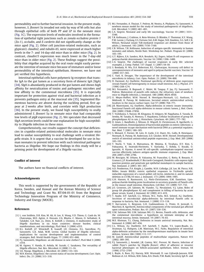
permeability and to further bacterial invasion. In the present study,however, S. flexneri 5a invaded not only by way of M cells but alsothrough epithelial cells of both PP and LP in the neonate mice(Fig. 1C). The expression levels of molecules involved in the forma-tion of epithelial tight junctions such as zonula occludens protein 1(Zo-1), occludin, and E-carderin did not significantly change as themice aged (Fig. 2). Other cell junction-related molecules, such asglypican3, claudin2, and tubulin-b5, were expressed at much higherlevels in the 7- and 14-day-old mice than in older mice (Fig. 2). Incontrast, claudin15 expression was much lower in the 7-day-oldmice than in older mice (Fig. 2). These findings suggest the possi-bility that shigellae acquired by the oral route might easily perme-ate the intestine of neonate mice because of immature and/or loosepermeability of the intestinal epithelium. However, we have notyet verified this hypothesis.
Intestinal epithelial cells have polymeric Ig receptors that trans-fer IgA to the gut lumen as a secretory form of dimeric IgA (SIgA)[34]. SIgA is abundantly produced in the gut lumen and shows highaffinity for neutralization of toxins and pathogenic microbes andlow affinity to the commensal microbiota [35]. It is especiallyimportant for protection against viral and bacterial infection as itprevents pathogen entry at the mucosal site [35]. Segmented fila-mentous bacteria are absent during the suckling period, first ap-pear at 2 weeks after birth, and correlate with SIgA production[36]. In the present study, we found high susceptibility to orallyadministered S. flexneri 5a in neonate mice that had comparablylow levels of pIgR expression (Fig. 2). We speculate that decreasedSIgA secretion levels could be one explanation for high susceptibil-ity to Shigella infection in these mice.
Comprehensively, our results support the concept that deficien-cies in cryptdin-related antimicrobial molecules in neonate micelead to undue susceptibility to oral challenge with a virulent Shi-gella strain. It is urgent that a vaccine be developed for use in hu-man neonates to prevent infection with such intestinal pathogenicbacteria as Shigellae. We hope our findings in this study will be apriming point for development of a Shigella vaccine.
Conflict of interest
The authors have no financial conflicts of interest.
Acknowledgments
This work is supported by the governments of the Republic ofKorea, Sweden, and Kuwait and the Korean Ministry of Scienceand Technology and Grant No. RT104-01-01 from the RegionalTechnology Innovation Program of the Ministry of Commerce,Industry and Energy (MOCIE).
References
[1] L. von Seidlein, D.R. Kim, M. Ali, H. Lee, X. Wang, V.D. Thiem, G. Canh do, W.Chaicumpa, M.D. Agtini, A. Hossain, Z.A. Bhutta, C. Mason, O. Sethabutr, K.Talukder, G.B. Nair, J.L. Deen, K. Kotloff, J. Clemens, A multicentre study ofShigella diarrhoea in six Asian countries: disease burden, clinicalmanifestations, and microbiology, PLoS Med. 3 (2006) e353.
[2] K.L. Kotloff, J.P. Winickoff, B. Ivanoff, J.D. Clemens, D.L. Swerdlow, P.J.Sansonetti, G.K. Adak, M.M. Levine, Global burden of Shigella infections:implications for vaccine development and implementation of controlstrategies, Bull. World Health Organ. 77 (1999) 651–666.
[3] P.J. Sansonetti, Shigellosis: an old disease in new clothes?, PLoS Med 3 (2006)e354.
[4] M. Ogawa, Y. Handa, H. Ashida, M. Suzuki, C. Sasakawa, The versatility ofShigella effectors, Nat. Rev. Microbiol. 6 (2008) 11–16.
[5] S.K. Niyogi, Shigellosis, J. Microbiol. 43 (2005) 133–143.[6] M.N. Kweon, Shigellosis: the current status of vaccine development, Curr. Opin.
Infect. Dis. 21 (2008) 313–318.
[7] M.I. Fernandez, A. Thuizat, T. Pedron, M. Neutra, A. Phalipon, P.J. Sansonetti, Anewborn mouse model for the study of intestinal pathogenesis of shigellosis,Cell. Microbiol. 5 (2003) 481–491.
[8] C.A. Siegrist, Neonatal and early life vaccinology, Vaccine 19 (2001) 3331–3346.
[9] T.R. Kollmann, J. Crabtree, A. Rein-Weston, D. Blimkie, F. Thommai, X.Y. Wang,P.M. Lavoie, J. Furlong, E.S. Fortuno 3rd, A.M. Hajjar, N.R. Hawkins, S.G. Self, C.B.Wilson, Neonatal innate TLR-mediated responses are distinct from those ofadults, J. Immunol. 183 (2009) 7150–7160.
[10] C.B. Wilson, T.R. Kollmann, Induction of antigen-specific immunity in humanneonates and infants, Nestle Nutr. Workshop Ser. Pediatr. Program 61 (2008)183–195.
[11] G.T. Rijkers, E.A. Sanders, M.A. Breukels, B.J. Zegers, Infant B cell responses topolysaccharide determinants, Vaccine 16 (1998) 1396–1400.
[12] C.A. Siegrist, The challenges of vaccine responses in early life: selectedexamples, J. Comp. Pathol. 137 (Suppl. 1) (2007) S4–S9.
[13] S. Bondada, H. Wu, D.A. Robertson, R.L. Chelvarajan, Accessory cell defect inunresponsiveness of neonates and aged to polysaccharide vaccines, Vaccine 19(2000) 557–565.
[14] C. Vael, K. Desager, The importance of the development of the intestinalmicrobiota in infancy, Curr. Opin. Pediatr. 21 (2009) 794–800.
[15] D. Darmoul, A.J. Ouellette, Positional specificity of defensin gene expressionreveals Paneth cell heterogeneity in mouse small intestine, Am. J. Physiol. 271(1996) G68–G74.
[16] M.I. Fernandez, B. Regnault, C. Mulet, M. Tanguy, P. Jay, P.J. Sansonetti, T.Pedron, Maturation of paneth cells induces the refractory state of newbornmice to Shigella infection, J. Immunol. 180 (2008) 4924–4930.
[17] U. Meyer-Hoffert, M.W. Hornef, B. Henriques-Normark, L.G. Axelsson, T.Midtvedt, K. Putsep, M. Andersson, Secreted enteric antimicrobial activitylocalises to the mucus surface layer, Gut 57 (2008) 764–771.
[18] J.R. Mastroianni, A.J. Ouellette, Alpha-defensins in enteric innate immunity:functional Paneth cell alpha-defensins in mouse colonic lumen, J. Biol. Chem.284 (2009) 27848–27856.
[19] T. Yoshikawa, S. Naruse, M. Kitagawa, H. Ishiguro, M. Nagahama, E. Yasuda, R.Semba, M. Tanaka, K. Nomura, T. Hayakawa, Cellular localization of group IIAphospholipase A2 in rats, J. Histochem. Cytochem. 49 (2001) 777–782.
[20] D. Islam, L. Bandholtz, J. Nilsson, H. Wigzell, B. Christensson, B. Agerberth, G.Gudmundsson, Downregulation of bactericidal peptides in enteric infections: anovel immune escape mechanism with bacterial DNA as a potential regulator,Nat. Med. 7 (2001) 180–185.
[21] S. Menard, V. Forster, M. Lotz, D. Gutle, C.U. Duerr, R.L. Gallo, B. Henriques-Normark, K. Putsep, M. Andersson, E.O. Glocker, M.W. Hornef, Developmentalswitch of intestinal antimicrobial peptide expression, J. Exp. Med. 205 (2008)183–193.
[22] T. Nochi, Y. Yuki, A. Matsumura, M. Mejima, K. Terahara, D.Y. Kim, S.Fukuyama, K. Iwatsuki-Horimoto, Y. Kawaoka, T. Kohda, S. Kozaki, O.Igarashi, H. Kiyono, A novel M cell-specific carbohydrate-targeted mucosalvaccine effectively induces antigen-specific immune responses, J. Exp. Med.204 (2007) 2789–2796.
[23] M. Rescigno, M. Urbano, B. Valzasina, M. Francolini, G. Rotta, R. Bonasio, F.Granucci, J.P. Kraehenbuhl, P. Ricciardi-Castagnoli, Dendritic cells express tightjunction proteins and penetrate gut epithelial monolayers to sample bacteria,Nat. Immunol. 2 (2001) 361–367.
[24] A.D. Pemberton, P.A. Knight, J. Gamble, W.H. Colledge, J.K. Lee, M. Pierce, H.R.Miller, Innate BALB/c enteric epithelial responses to Trichinella spiralis:inducible expression of a novel goblet cell lectin, intelectin-2, and its naturaldeletion in C57BL/10 mice, J. Immunol. 173 (2004) 1894–1901.
[25] G.H. Hansen, K. Rasmussen, L.L. Niels-Christiansen, E.M. Danielsen, Lipo-polysaccharide-binding prote localization in secretory granules of Paneth cellsin the mouse small intestine, Histochem. Cell Biol. 131 (2009) 727–732.
[26] J.O. Gronroos, J.H. Salonen, M. Viander, T.J. Nevalainen, V.J. Laine, Roles ofgroup IIA phospholipase A2 and complement in killing of bacteria by acutephase serum, Scand. J. Immunol. 62 (2005) 413–419.
[27] T. Ayabe, D.P. Satchell, C.L. Wilson, W.C. Parks, M.E. Selsted, A.J. Ouellette,Secretion of microbicidal alpha-defensins by intestinal Paneth cells inresponse to bacteria, Nat. Immunol. 1 (2000) 113–118.
[28] Y. Kai-Larsen, G. Bergsson, G.H. Gudmundsson, G. Printz, H. Jornvall, G.Marchini, B. Agerberth, Antimicrobial components of the neonatal gut affectedupon colonization, Pediatr. Res. 61 (2007) 530–536.
[29] U.M. Salzman NH, U.M. Salzman NH, C.L. Bevins, Paneth cells, defensins, andthe commensal microbiota: a hypothesis on intimate interplay at theintestinal mucosa, Semin. Immunol. 19 (2007) 70–83.
[30] M.E. Klotman, T.L. Chang, Defensins in innate antiviral immunity, Nat. Rev.Immunol. 6 (2006) 447–456.
[31] C.L. Wilson, A.J. Ouellette, D.P. Satchell, T. Ayabe, Y.S. Lopez-Boado, J.L.Stratman, S.J. Hultgren, L.M. Matrisian, W.C. Parks, Regulation of intestinalalpha-defensin activation by the metalloproteinase matrilysin in innate hostdefense, Science 286 (1999) 113–117.
[32] M. Zasloff, Antimicrobial peptides of multicellular organisms, Nature 415(2002) 389–395.
[33] P.J. Sansonetti, J. Arondel, J.R. Cantey, M.C. Prevost, M. Huerre, Infection ofrabbit Peyer’s patches by Shigella flexneri: effect of adhesive or invasivebacterial phenotypes on follicle-associated epithelium, Infect. Immun. 64(1996) 2752–2764.
[34] L. Royle, A. Roos, D.J. Harvey, M.R. Wormald, D. van Gijlswijk-Janssen, R.M.Redwan el, I.A. Wilson, M.R. Daha, R.A. Dwek, P.M. Rudd, Secretory IgA N- and

560 D.-H. Shim et al. / Biochemical and Biophysical Research Communications 401 (2010) 554–560
O-glycans provide a link between the innate and adaptive immune systems, J.Biol. Chem. 278 (2003) 20140–20153.
[35] A.J. Macpherson, K.D. McCoy, F.E. Johansen, P. Brandtzaeg, The immunegeography of IgA induction and function, Mucosal Immunol. 1 (2008) 11–22.
[36] H.Q. Jiang, N.A. Bos, J.J. Cebra, Timing, localization and persistence ofcolonization by segmented filamentous bacteria in the neonatal mouse gutdepend on immune status of mothers and pups, Infect. Immun. 69 (2001)3611–3617.
Related Documents