1

1. Protective structures: Vertebral column and the meninges provide protect the spinal cord and provide physical stability. a. Dura mater, b. Arachnoid,
Dec 14, 2015
Welcome message from author
This document is posted to help you gain knowledge. Please leave a comment to let me know what you think about it! Share it to your friends and learn new things together.
Transcript

1

Protective structures: Vertebral column and the meninges provide protect the spinal cord and provide physical stability. a. Dura mater, b. Arachnoid, c. Pia mater
Epidural space, subdural space and subarachnoid space
2

3
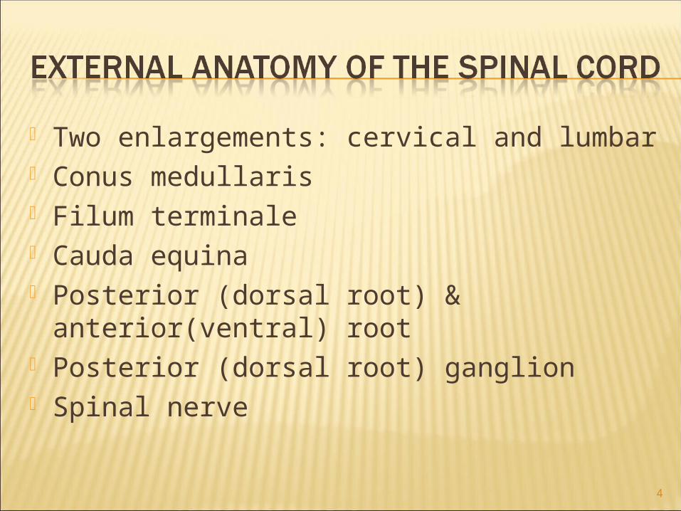
Two enlargements: cervical and lumbar Conus medullaris Filum terminale Cauda equina Posterior (dorsal root) &
anterior(ventral) root Posterior (dorsal root) ganglion Spinal nerve
4

5

Anterior median fissure Posterior median sulcus Gray and white commissures Central canal Anterior, posterior & lateral gray horns Anterior, posterior & lateral white
columns
6

7
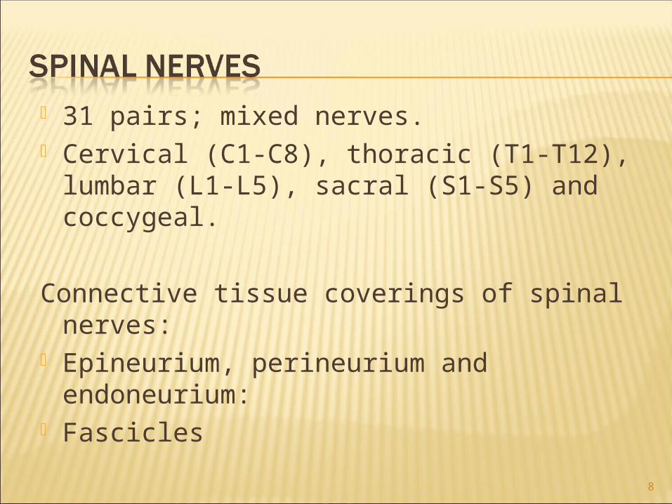
31 pairs; mixed nerves. Cervical (C1-C8), thoracic (T1-T12),
lumbar (L1-L5), sacral (S1-S5) and coccygeal.
Connective tissue coverings of spinal nerves:
Epineurium, perineurium and endoneurium:
Fascicles8

9

Spinal nerves branch and their braches are called rami: Posterior (dorsal) ramusAnterior (ventral) ramus
Plexuses: a network of axonsAnterior rami except T1-T11 form plexuses.
10

Formed by the anterior rami of C1-C5.
Phrenic nerves- important nerves from the cervical plexuses.
Formed by the anterior rami of C1-C5.
Phrenic nerves- important nerves from the cervical plexuses.
11

Formed by the anterior rami of C5-C8 & T1.
Supplies the shoulders and upper limbs.
Roots → trunks → divisions → cords → nerves.
12
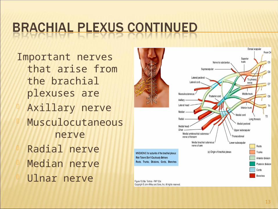
Important nerves that arise from the brachial plexuses are
Axillary nerve Musculocutaneous
nerve Radial nerve Median nerve Ulnar nerve
13
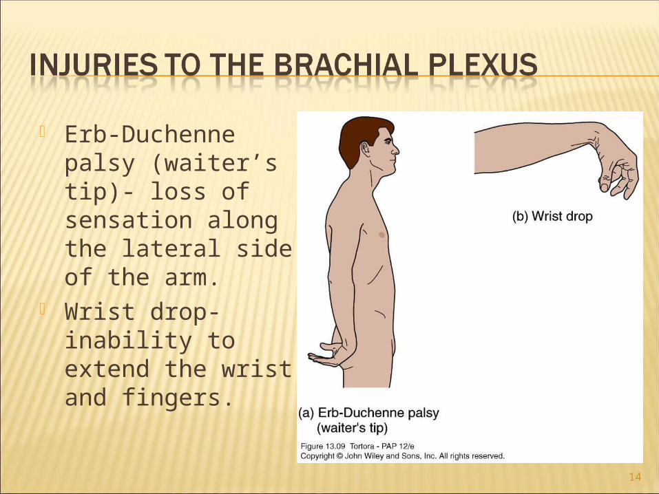
Erb-Duchenne palsy (waiter’s tip)- loss of sensation along the lateral side of the arm.
Wrist drop- inability to extend the wrist and fingers.
14

Median nerve palsy- numbness, tingling and pain in the palm and fingers.
Ulnar nerve palsy- inability to abduct or adduct fingers
Winged scapula- the arm cannot be abducted beyond the horizontal position.
15

16

Formed by the anterior rami of L1-L4.
Supplies the anterolateral abdominal wall, external genitals, and part of the lower limbs.
Femoral nerves, obturator nerves.
17

Formed by the anterior rami of L4-L5 and S1-S4.
Supplies the buttocks, perineum, and lower limbs.
Gives rise to the largest nerve in the body- the sciatic nerve.
18

19

Formed by the anterior rami of S4-S5 and the coccygeal nerves.
Supplies a small area of skin in the coccygeal region.
20
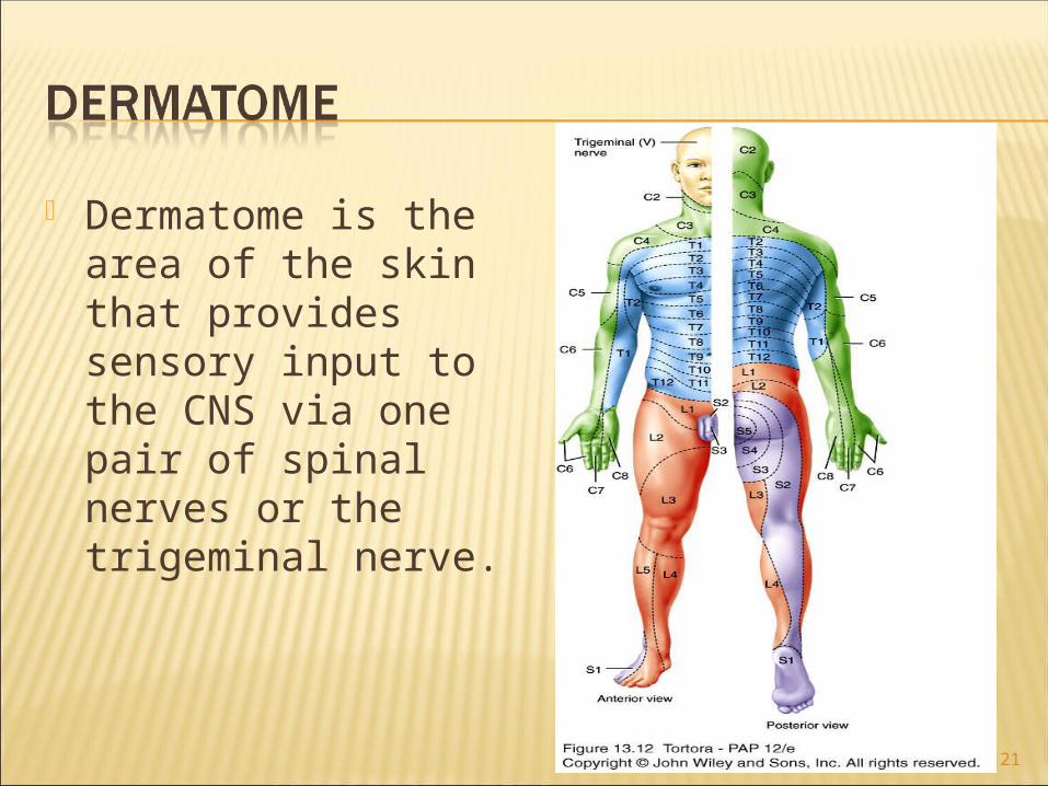
Dermatome is the area of the skin that provides sensory input to the CNS via one pair of spinal nerves or the trigeminal nerve.
21

The name of the tract often indicates its location in the white matter and where it begins and ends.
The white matter contains both sensory and motor tracts.
22

A reflex is an automatic, sudden, involuntary response to a stimulus.
When the integration takes place in the spinal cord, the reflex is a spinal reflex.
23

The pathway followed by nerve impulses that produce a reflex is a reflex arc.
A reflex arc includes:a. sensory receptorb. sensory neuronc. integrating centerd. motor neurone. effector
24

1 SENSORY RECEPTOR(responds to a stimulusby producing a generatoror receptor potential)
1SENSORY NEURON(axon conducts impulses from receptor to integrating center)
SENSORY RECEPTOR(responds to a stimulusby producing a generatoror receptor potential)
2 1SENSORY NEURON(axon conducts impulses from receptor to integrating center)
SENSORY RECEPTOR(responds to a stimulusby producing a generatoror receptor potential)
INTEGRATING CENTER(one or more regions within the CNSthat relay impulses from sensory tomotor neurons)
Interneuron
2
3
1SENSORY NEURON(axon conducts impulses from receptor to integrating center)
SENSORY RECEPTOR(responds to a stimulusby producing a generatoror receptor potential)
INTEGRATING CENTER(one or more regions within the CNSthat relay impulses from sensory tomotor neurons)
MOTOR NEURON(axon conducts impulses fromintegrating center to effector)
Interneuron
2
3
4
1SENSORY NEURON(axon conducts impulses from receptor to integrating center)
SENSORY RECEPTOR(responds to a stimulusby producing a generatoror receptor potential)
INTEGRATING CENTER(one or more regions within the CNSthat relay impulses from sensory tomotor neurons)
MOTOR NEURON(axon conducts impulses fromintegrating center to effector)
EFFECTOR(muscle or gland thatresponds to motornerve impulses)
Interneuron
2
3
4 5
25

Causes contraction of a skeletal muscle in response to stretching of the muscle.
Monosynaptic reflex. Patellar or knee-jerk reflex: Stretching of
a muscle →activation of muscle spindles →sensory neuron →spinal cord→motor neuron → muscle contraction.
Ipsilateral.
26

1 Stretching stimulatesSENSORY RECEPTOR(muscle spindle)
Antagonisticmuscles relax
1 Stretching stimulatesSENSORY RECEPTOR(muscle spindle)
SENSORYNEURONexcited
To brain
SpinalNerve
+
+
2
1 Stretching stimulatesSENSORY RECEPTOR(muscle spindle)
SENSORYNEURONexcited
MOTORNEURONexcited
EFFECTOR(same muscle)contracts andrelieves thestretching
Antagonisticmuscles relax
Motor neuron toantagonistic musclesis inhibited
Within INTEGRATINGCENTER (spinal cord),sensory neuron activatesmotor neuron
Inhibitoryinterneuron
To brain
SpinalNerve
+
–+
+
2
3
45
27

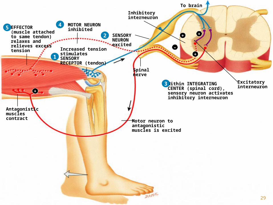
Polysynaptic reflex. Control muscle tension by causing muscle
relaxation when muscle tension is great. Sensory receptors- Golgi tendon organs. ↑ Tension applied to the tendon → tendon
organ stimulation → nerve impulse → spinal cord →motor neuron causes muscle relaxation and relieves tension.
28
1
Increased tensionstimulatesSENSORYRECEPTOR (tendon)
1
Spinalnerve
SENSORYNEURONexcited
To brain
Increased tensionstimulatesSENSORYRECEPTOR (tendon)
2 ++
1
Within INTEGRATINGCENTER (spinal cord),sensory neuron activatesinhibitory interneuron
Excitatoryinterneuron
Spinalnerve
Inhibitoryinterneuron
SENSORYNEURONexcited
+
To brain
Increased tensionstimulatesSENSORYRECEPTOR (tendon)
++2
3
–
+1
–
Within INTEGRATINGCENTER (spinal cord),sensory neuron activatesinhibitory interneuron
Excitatoryinterneuron
Antagonisticmusclescontract
Spinalnerve
MOTOR NEURONinhibited
Inhibitoryinterneuron
SENSORYNEURONexcited
+
To brain
Increased tensionstimulatesSENSORYRECEPTOR (tendon)
Motor neuron toantagonisticmuscles is excited
+
+
+
+2
3
4
1
–
EFFECTOR(muscle attachedto same tendon)relaxes andrelieves excesstension
Within INTEGRATINGCENTER (spinal cord),sensory neuron activatesinhibitory interneuron
Excitatoryinterneuron
Antagonisticmusclescontract
Spinalnerve
MOTOR NEURONinhibited
Inhibitoryinterneuron
SENSORYNEURONexcited
+
To brain
Increased tensionstimulatesSENSORYRECEPTOR (tendon)
Motor neuron toantagonisticmuscles is excited
+
+
+2
3
45
+
29

Polysynaptic reflex Ipsilateral. Stepping on a tack (stimulus) →
nerve impulse → activation of the interneuron → activation of the motor neuron →muscle contraction →withdrawal of the leg.
30

1 Stepping on tack stimulatesSENSORY RECEPTOR (dendritesof pain-sensitive neuron)
1
+
Stepping on tack stimulatesSENSORY RECEPTOR (dendritesof pain-sensitive neuron)
SENSORYNEURONexcited
+
2
1
+
Stepping on tack stimulatesSENSORY RECEPTOR (dendritesof pain-sensitive neuron)
SENSORYNEURONexcited
Within INTEGRATING CENTER(spinal cord), sensory neuronactivates interneurons in severalspinal cord segments
Ascendinginterneuron
Interneuron
Descendinginterneuron
Spinalnerve
+
+
+
+
+
+
2
3
1
+
Stepping on tack stimulatesSENSORY RECEPTOR (dendritesof pain-sensitive neuron)
SENSORYNEURONexcited
MOTORNEURONSexcited
MOTORNEURONexcited
Within INTEGRATING CENTER(spinal cord), sensory neuronactivates interneurons in severalspinal cord segments
Ascendinginterneuron
Interneuron
Descendinginterneuron
Spinalnerve
+
+
+
+
+
+
+
+
+
2
3
4
4
1
+
Stepping on tack stimulatesSENSORY RECEPTOR (dendritesof pain-sensitive neuron)
SENSORYNEURONexcited
MOTORNEURONSexcited
MOTORNEURONexcited
EFFECTORS(flexor muscles)contract andwithdraw leg
Within INTEGRATING CENTER(spinal cord), sensory neuronactivates interneurons in severalspinal cord segments
Ascendinginterneuron
Interneuron
Descendinginterneuron
Spinalnerve
+
+
+
+
+
+
+
+
+
23
4
5
4
31

Polysynaptic reflex. Contralateral reflex. Contraction of muscles that extend joints
in the opposite limb in response to a painful stimulus.
Stepping on a tack (stimulus) → nerve impulse →activation of several interneurons → activation of the motor neurons → muscle contraction causing flexion of the leg stepping on a tack & extension on the opposite side.
32

1
Withdrawal of right leg(flexor reflex)
Stepping on a tackstimulates SENSORYRECEPTOR (dendrites ofpain-sensitive neuron) inright foot
1
SENSORYNEURONexcited
Withdrawal of right leg(flexor reflex)
Stepping on a tackstimulates SENSORYRECEPTOR (dendrites ofpain-sensitive neuron) inright foot
2
+
1
+
Ascendinginterneurons
SENSORYNEURONexcited
Spinalnerve
Within INTEGRATING CENTER(spinal cord), sensory neuronactivates several interneurons
Descendinginterneurons
Withdrawal of right leg(flexor reflex)
Stepping on a tackstimulates SENSORYRECEPTOR (dendrites ofpain-sensitive neuron) inright foot
+
+ +
2
3
+
+ +
+ +
+ +
Interneuronsfrom other side
1
+
Ascendinginterneurons
SENSORYNEURONexcited
Spinalnerve
Within INTEGRATING CENTER(spinal cord), sensory neuronactivates several interneurons
MOTORNEURONSexcited
Descendinginterneurons
Withdrawal of right leg(flexor reflex)
Stepping on a tackstimulates SENSORYRECEPTOR (dendrites ofpain-sensitive neuron) inright foot Extension of left leg
(crossed extensor reflex)
MOTORNEURONSexcited
+
+ +
2
3
4
4
+
+ +
+ +
+ +
+
+
+
Interneuronsfrom other side
1
+
Ascendinginterneurons
EFFECTORS(extensor muscles)contract, and extendleft leg
SENSORYNEURONexcited
Spinalnerve
Within INTEGRATING CENTER(spinal cord), sensory neuronactivates several interneurons
MOTORNEURONSexcited
Descendinginterneurons
Withdrawal of right leg(flexor reflex)
Flexor musclescontract and with-drawright leg
Stepping on a tackstimulates SENSORYRECEPTOR (dendrites ofpain-sensitive neuron) inright foot Extension of left leg
(crossed extensor reflex)
MOTORNEURONSexcited
+
+ +
2
3
4
4
5
+
+ +
+ +
+ +
+
+
+
Interneuronsfrom other side
+
+
+
33

34
Portions of the above presentation are copy-writed by John Wiley & Sons, Inc. For those portions, all rights are reserved. Reproduction or translation of those portions beyond that permitted in section 117 of the 1976 United States Copyright Act without express permission of the copyright owner is unlawful. Request for further information should be addressed to the Permission Department, John Wiley & Sons, Inc. The Publishers assumes no responsibility for errors, omissions, or damages caused by the use of theses programs or from the use of the information herein. Copyright 2009 John Wiley & Sons, Inc.
Related Documents


![Repair of Tegmen Tympani Defect Presenting with ...€¦ · aberrant arachnoid granulations [3, 8]. According to the arachnoid theory, some arachnoid granulations may not find venous](https://static.cupdf.com/doc/110x72/606db78183041435125f357b/repair-of-tegmen-tympani-defect-presenting-with-aberrant-arachnoid-granulations.jpg)