1 Pathophysiology of portal hypertension TARUN K. GUPTA MD Assistant Adjunct Professor Hepatic Hemodynamic Laborator), VA Medical Center; West Haven, CT 06516; Yale University School of Medicine, New Haven, CT 06510 and Bridgeport Hospital, Bridgeport, CT 06610, USA LISA CHEN MD Post-doctoral Fellow Hepatic Hemodynamic Laboratoq, VA Medical Centec 9 West Haven. CT06516 and Yale universiv School of Medicine, New Haven, CT 06510, USA ROBERTO J. GROSZMANN” MD, FRCP Professor of Medicine and Chief, Digestive Diseases Hepatic Hemodynamic Laboratoy, VA Medical Centel; West Haven, CT 06516 and Yale Uni\‘er.yiQ School of Medicine, New Haven, CT 06510, USA Portal hypertension is a common clinical syndrome associated with chronic liver diseases and is characterized by a pathological increase in portal pressure. Increase in portal pressure is because of an increase in vascular resistance and an elevated portal blood flow. The site of increased intrahepatic resistance is variable and is dependent on the disease process. The site of obstruction may be: pre-hepatic, hepatic, and/or post-hepatic. In addition, part of the increased intrahepatic resistance is because of increased vascular tone. Another important factor contributing to increased portal pressure is elevated blood flow. Peripheral vasodilatation initiates the classical profile of decreased systemic resistance, expanded plasma volume, elevated splanchnic blood flow and elevated cardiac index. The elevated portal pressure leads to formation of portosystemic collaterals and oesophageal varices. Pharmacotherapy for portal hypertension is aimed at reducing both intrahepatic vascular tone and elevated splanchnic blood flow. Key words: portal hypertension; porto-systemic collaterals; hyperdynamic circulation; vasodilators; vasoconstrictors; oesophageal varices; variceal haemorrhage; plasma volume; intrahepatic resistance; pharmacotherapy. Portal hypertension is a common clinical syndrome associated with chronic liver diseases and is characterized by a pathological increase in portal pressure. Elevated portal pressure results in extensive formation of porto- * Address for correspondence: R. J. Groszmann Yale University School of Medicine, Hepatic Hemodynamic Laboratories/l 1IH, VA Connecticut Healthcare System, 950 Campbell Avenue, West Haven, CT 06516, USA. BaiUit?re k Clinical Gastroenterology- Vol. 11, No. 2, June 1997 ISBN o-702&2339-6 0950-3528/97/020203 + 17 $12.00/00 203 Copyright 0 1997, by Baillitre Tindall All rights of reproduction in any form reserved

Welcome message from author
This document is posted to help you gain knowledge. Please leave a comment to let me know what you think about it! Share it to your friends and learn new things together.
Transcript
1
Pathophysiology of portal hypertension
TARUN K. GUPTA MD Assistant Adjunct Professor Hepatic Hemodynamic Laborator), VA Medical Center; West Haven, CT 06516; Yale University School of Medicine, New Haven, CT 06510 and Bridgeport Hospital, Bridgeport, CT 06610, USA
LISA CHEN MD Post-doctoral Fellow Hepatic Hemodynamic Laboratoq, VA Medical Centec 9 West Haven. CT06516 and Yale universiv School of Medicine, New Haven, CT 06510, USA
ROBERTO J. GROSZMANN” MD, FRCP Professor of Medicine and Chief, Digestive Diseases Hepatic Hemodynamic Laboratoy, VA Medical Centel; West Haven, CT 06516 and Yale Uni\‘er.yiQ School of Medicine, New Haven, CT 06510, USA
Portal hypertension is a common clinical syndrome associated with chronic liver diseases and is characterized by a pathological increase in portal pressure. Increase in portal pressure is because of an increase in vascular resistance and an elevated portal blood flow. The site of increased intrahepatic resistance is variable and is dependent on the disease process. The site of obstruction may be: pre-hepatic, hepatic, and/or post-hepatic. In addition, part of the increased intrahepatic resistance is because of increased vascular tone. Another important factor contributing to increased portal pressure is elevated blood flow. Peripheral vasodilatation initiates the classical profile of decreased systemic resistance, expanded plasma volume, elevated splanchnic blood flow and elevated cardiac index. The elevated portal pressure leads to formation of portosystemic collaterals and oesophageal varices. Pharmacotherapy for portal hypertension is aimed at reducing both intrahepatic vascular tone and elevated splanchnic blood flow.
Key words: portal hypertension; porto-systemic collaterals; hyperdynamic circulation; vasodilators; vasoconstrictors; oesophageal varices; variceal haemorrhage; plasma volume; intrahepatic resistance; pharmacotherapy.
Portal hypertension is a common clinical syndrome associated with chronic liver diseases and is characterized by a pathological increase in portal pressure. Elevated portal pressure results in extensive formation of porto-
* Address for correspondence: R. J. Groszmann Yale University School of Medicine, Hepatic Hemodynamic Laboratories/l 1 IH, VA Connecticut Healthcare System, 950 Campbell Avenue, West Haven, CT 06516, USA.
BaiUit?re k Clinical Gastroenterology- Vol. 11, No. 2, June 1997 ISBN o-702&2339-6 0950-3528/97/020203 + 17 $12.00/00
203 Copyright 0 1997, by Baillitre Tindall
All rights of reproduction in any form reserved

204 T. K. GUPTA ET AL
systemic collaterals that divert portal blood to the systemic circulation. This causes many haemodynamic disturbances, which result in complications of portal hypertension. The major consequences of portal hypertension are gastrointestinal bleeding from ruptured oesophageal varices, ascites formation and hepatic encephalopathy. A grasp of the biological mech- anisms involved in the pathogenesis of portal hypertension is essential to an understanding of the complications of chronic liver disease and to the development of rational therapies. This chapter is an overview of the basic pathophysiological mechanisms of the splanchnic and systemic circulatory derangements that lead to portal hypertension and cause the portosystemic collaterals to develop.
Some recapitulation of general principles is essential to the understand- ing of the pathophysiology of portal hypertension. When Ohm’s law is applied to the vascular system, the pressure gradient between two points (Pl - P2) in a blood vessel can be described as the product of blood flow (Q) and resistance to flow (R)
PI-P2=QxR
Unlike pressure and flow, resistance can not be directly measured, but it can be derived from pressure and flow. However, resistance to the flow of blood in a vessel is best understood when expressed according to Pouseuille’s law:
R- 8qL 7cr4
in which IJ = coefficient of viscosity L = length of vessel r = radius of vessel
Expressed in these terms, substitution of resistance (R) into Ohm’s equation yields
p, -p2=e 7v4
Under physiological conditions, the length of blood vessel (L) can be assumed to be constant. Similarly, unless there are large changes in haematocrit, the viscosity (rl) is taken as constant. Hence, the changes in the pressure (PI - P2) are largely accounted for by changes in flow (Q) and are inversely related to the changes in the radius (r) of the vessel. The contribution of these factors to the portal hypertensive syndrome will be discussed in the subsequent sections (Figure 1).
INCREASED RESISTANCE
Portal hypertension is associated with increased resistance to portal blood flow. Increased vascular resistance is because of an increase in both intra-

PATHOPHYSIOLOGY OF PORTAL HYPERTENSION 205
Elevated portal pressure
Increased resistance Increased systemickplanchnic to portal blood flow blood flow
(hyperdynamic circulation) l Systemic vasodilatation 0 t Plasma volume
lntrahepatic Portosystemic resistance collaterals
l Anatomical 0 Opening of pre-existing (irreversible component) vascular channels
l Functional/vascular tone l Formation of new (reversible component) vascular channels
Figure 1. Contributing factors to portal hypertension.
hepatic and portosystemic collateral resistance in comparison with the low resistance of the normal liver.
Intrahepatic resistance
The normal liver is a very compliant organ. Hence, the intrahepatic resist- ance decreases with increase in blood flow because of distension of the vascular tree in response to increased inflow. This compensatory mech- anism maintains the portal pressure within normal limits with a wide range of portal flow in normal livers (Greenway and Stark, 1971). This phenomenon is not seen in the portal hypertensive states in which the intra- hepatic resistance becomes fixed due to fibrosis and mechanical distortion of the vascular tree and the hepatic vascular compliance is greatly reduced. Moreover, the splanchnic flow is increased as discussed in the subsequent sections. Decreased compliance and increased blood flow are probably instrumental in initiating and perpetuating the portal hypertension.
As stated above, there is little resistance in the normal liver, and the portal pressure remains low (4-8 mmHg) over a wide range of portal flows. The main site of resistance in normal livers is somewhat controversial. The hepatic sinusoids (where stellate cells are present), terminal hepatic venules and also portal venules have been suggested as possible sites of resistance. However, in view of the minimal contribution of intrahepatic resistance to portal pressure in the normal liver, this issue is of little importance. The flow into the portal system is actively regulated by changes in vascular resistance at the level of splanchnic arterioles and not by the liver itself (Greenway and Stark, 1971). In portal hypertensive syndromes, increased resistance to portal venous flow may be localized to pre-hepatic, post- hepatic, or intrahepatic (pre-sinusoidal, sinusoidal or post-sinusoidal) sites (Genecin and Groszmann, 1993). In pre-hepatic and post-hepatic portal hypertension, increased resistance is secondary to obstruction of portal venous inflow or hepatic venous outflow, respectively. Unlike pre- and
206 T. K. GUPTA ETAL
post-hepatic portal hypertension, the intrahepatic syndromes are more complex and rarely can be classified according to a single site of resistance.
An early view of vascular resistance in cirrhotic livers hypothesized that portal hypertension is the consequence of a vascular obliterative process with scar tissue and regenerative nodules, both occluding and compressing vascular structures (Popper, 1958; Baldus and Hoffbauer, 1963). Thus earlier understanding of intrahepatic portal hypertension emphasized the role of anatomical alterations leading to mechanical obstruction (irreversible component) in the increased intrahepatic resistance. However, recently it has been shown that there is also an increased vascular tone in the cirrhotic liver (reversible component) (Bhathal, 1985).
In intrahepatic portal hypertension, there may be several areas of obstruction, and as the disease progresses, new sites may become involved. For example, in hepatic schistosomiasis, the increased intrahepatic resist- ance results from granulomas located in the presinusoidal areas (Beker and Valencia-Parparcen, 1968). However, in late stages, an elevated hepatic wedge venous pressure gradient may be observed, reflecting increased sinusoidal resistance. Chronic hepatitis seems to have both pre-sinusoidal and sinusoidal abnormalities, which increasingly contribute to vascular resistance as the lesion progresses towards cirrhosis (van Leeuwen et al, 1991). In ethanol-induced liver disease, the resistance is increased because of lesions in sinusoidal and post-sinusoidal sites. The terminal hepatic vein fibrosis (or sclerosis), encroachment on sinusoids by enlarged hepatocytes, collagen deposition in the perisinusoidal region or the space of Disse, result in elevated portal pressure even in a pre-cirrhotic stage (Schaffner and Popper, 1963; Reynolds et al, 1969; Lieber et al, 1976). However, in advanced stage or cirrhosis, regenerating nodules and pruning of the vascular tree contribute to increased vascular resistance. A variety of other non-alcoholic liver diseases cause portal hypertension because of increased sinusoidal vascular resistance. In hepatic amyloidosis, resistance is increased due to deposition of amyloid in the space of Disse (Brion et al, 1991). The capillarization process and occlusion of the fenestrae are postulated to increase the resistance of passage of fluid across the endo- thelium, but the extent of their effect on resistance to blood flow is unknown.
The morphological changes that occur in chronic liver diseases are undoubtedly the most important factor involved in the increased intra- hepatic resistance. However, recent data also suggest a role of functional factors that lead to increased vascular tone, similar to that which is seen in the arterial hypertension. In chronic liver disease and also during acute liver injury, hepatic stellate cells acquire contractile properties and may contribute to the dynamic modulation of intrahepatic resistance (Pinzani et al, 1992). These cells may act as pericytes, a type of cell that has been shown to regulate blood flow in other organs. The hepatic stellate cells, which are also the main source of collagen synthesis, may contribute to the regulation of hepatic blood flow at the microcirculatory level. Stellate cells are strategically located in the sinusoids with perisinusoidal and inter- hepatocellular branching processes that contain actin-like filaments. They
PATHOPHYSIOLOGY OF PORTAL HYPERTENSION 207
also express the alpha smooth-muscle actin gene, which is characteristic of vascular smooth muscle cells. The characteristics of these cells make them similar to myofibroblast. Myofibroblasts are intermediate in structure between smooth muscle cells and fibroblasts. Myofibroblast-like cells have been shown to exist in fibrous septa around the sinusoids and terminal hepatic venules in cirrhotic livers (Rudolph et al, 1979). These cells are postulated to play a role in the regulation of vascular resistance in the cirrhotic rat liver (Bhathal and Grossman, 1985).
The vascular endothelium synthesizes vasodilators such as nitric oxide, prostacyclins, hyperpolarizing factor and vasoconstrictors such as endo- thelins and prostanoids (Rubnayi, 1990; Vane et al, 1990). These vasoactive substances act in a paracrine fashion on the underlying vascular smooth muscle and modulate vascular tone. Normal vascular tone is maintained by a delicate balance between these vasodilatory and vasoconstrictive substances. Perturbation of this balance leads to abnormal vascular tone. Increased vascular tone seen in cirrhotic livers could be because of a deficit of endothelial vasodilators or an increase in the vasoconstrictors, or combination of both. Nitric oxide is a potent endothelial vasodilator that has been shown to play an important role in the modulation of intrahepatic vascular tone in normal livers (Mittal et al, 1994). Preliminary evidence from our laboratory suggest that there may be deficit of nitric oxide in the cirrhotic intrahepatic microcirculation as seen in other hypertensive regional microcirculations (Gupta and Groszmann, 1994). Using an isolated rat liver perfusion model, we have demonstrated that there is endothelial dysfunction in the intrahepatic microcirculation of cirrhotic livers (Gupta et al, 1995). Endothelial dysfunction leads to impaired release of endothelial vasodilators which, in part, may be responsible for the increased vascular tone observed in cirrhotic livers. More recently, it has been shown that the stellate cells (myofibroblasts) from cirrhotic livers exhibit an enhanced response to endothelins (Rockey and Weisiger, 1996). An imbalance between endothelial vasodilators and vasoconstrictors can affect the acti- vated stellate cells (myofibroblasts), which modulate intrahepatic vascular tone. It is possible that both a deficit of vasodilators and an increase in vaso- constrictors may be responsible for the increased vascular tone.
In summary, there are multiple factors that may lead to increased resist- ance to portal blood flow. Some of these are irreversible, such as fibrosis, capillarization, regenerating nodules, and some are quite dynamic, such as the imbalance between endothelial factors that leads to increased vascular tone.
Portosystemic collateral resistance
Although the collateral circulation begins as a consequence of portal hyper- tension, it evolves into an important mediator of the circulatory derange- ments of portal hypertension in its own right. The portosytemic collaterals provide a route to decompress the hypertensive portal system; however, the vascular resistance of this collateral bed is still greater than the resistance of the normal liver. Hence, portosystemic collaterals do not permit a
208 T. K. GUPTA ET AL
complete portal decompression. Development of portosystemic collaterals is discussed in detail later.
HYPERDYNAMIC CIRCULATION IN PORTAL HYPERTENSION
Chronic elevations in systemic and splanchnic blood flow have been documented as key elements of the hyperdynamic circulatory state (HCS) of portal hypertensive animals and humans. Peripheral vasodilatation initiates the development of the classic profile of decreased systemic vascular resistance and mean arterial pressure, plasma volume expansion, elevated splanchnic blood flow, and elevated cardiac index that character- izes this state (Colombato et al, 1991).
Systemic vasodilatation At least three mechanisms are thought to contribute to this peripheral vasodilation: (i) increased concentrations of circulating vasodilators; (ii) increased endothelial production of local vasodilators; and (iii) decreased vascular responsiveness to endogenous vasoconstrictors. The last mech- anism is probably because of the effect of the first two components. The relative importance of each of these potential causes of peripheral vaso- dilation is unknown.
Increased circulating vasodilators
In portal hypertensive states, there is increase in both endothelium- dependent and independent vasodilators. Possible aetiologies for increased circulatory concentrations of vasodilatory substances include increased production, decreased catabolism secondary to impaired hepatic function, and portosystemic shunting.
Circulating bile acids and glucagon increase splanchnic flow. Circulating bile acids, routinely cleared by the liver, are present in elevated concen- trations when liver function is impaired. Bile acid depletion has been shown to be associated with decrease in splanchnic hyperaemia. Experimental evidence, however, suggests that an increase in circulating bile acids is not essential for maintaining the HCS in portal hypertension. More specifically, cholestyramine-induced reduction of bile acids to concentrations seen in placebo-treated controls did not ameliorate the haemodynamic changes of the HCS in portal hypertensive animals (Genecin et al, 1990b)
Elevated concentrations of circulating glucagon also have been docu- mented in both animals and humans with portal hypertension. Rats with portal hypertension induced experimentally by either partial portal vein- ligation (PVL) or carbon tetrachloride inhalation had significantly higher glucagon and insulin concentrations compared with control rats (Gomis et al, 1994; Pizcueta et al, 1995). In addition, pancreatic islet isolates from these animals exhibited significantly higher glucagon secretion in response
PATHOPHYSIOLOGY OF PORTAL HYPERTENSION 209
to glucose and arginine administration compared with controls, although insulin secretion appeared to be impaired. Of note, this heightened pancreatic alpha cell glucagon secretion was not inhibited by increasing the glucose concentration in the incubation medium. Intraoperative glucagon concentrations measured from the portal vein and inferior vena cava increased significantly after surgical portosystemic shunting in Budd- Chiari patients and slightly, but not significantly, in cirrhotic patients (Sitzmann et al, 1993). Glucagon concentrations in this study, however, did not correlate with portal pressure. By infusing glucagon into normal rats to achieve levels seen in portal hypertensive rats, Benoit and co-workers demonstrated that glucagon significantly reduces splanchnic vascular resistance (Benoit et al, 1984).
Pizcueta and co-workers (1991) have demonstrated in PVL rats that the administration of somatostatin, which inhibits glucagon secretion, was asso- ciated with a marked decrease in glucagon secretion, a significant decrease in portal pressure and portal venous inflow. They postulated that this decrease in portal inflow was secondary to increased splanchnic vaso- constriction (Pizcueta et al, 1991). Administration of glucagon and somato- statin in these animals abolishes the above effects of somatostatin suggesting that somatostatin’s haemodynamic effects, probably, are elicited via inhibition of glucagon secretion. In an earlier study, we found no correlation between glucagon levels and portal venous inflow, thus questioning a major role for glucagon in mediating the hyperdynamic circulation (Sikuler and Groszmann, 1986). A recent study from our laboratory has not found increased levels of glucagon in cirrhotic patients, nor could a correlation be demonstrated between changes in their levels and changes in forearm haemodynamics (Rodriguez-Perez et al, 1993). Therefore, whether glucagon plays a role in the production of the HCS remains unclear.
Increased endothelial production of vasodilators
Recently, increasing evidence has pointed towards a major role for the endothelium in the maintenance of basal vascular tone and the development of local and generalized vasodilatation in portal hypertension. The endo- thelium produces at least two substances that are known to contribute to the development of systemic and splanchnic vasodilatation in portal hyper- tension: nitric oxide (NO) and prostaglandins (Casadevall et al, 1993).
Nitric oxide. Nitric oxide, previously known as endothelial derived relaxing factor, is synthesized from L-arginine by the enzyme nitric oxide synthase (NOS), which has constitutive and inducible forms in different cell types. Nitric oxide mediates its potent vasodilatory action on smooth muscle cells through soluble guanylate cyclase. Evidence exists that elevated production of NO is essential to the development of portal hypertension; treatment with N-nitro-L-arginine (L-NNA), a competitive inhibitor of NOS, prevented the development of peripheral vasodilatation, decreased systemic arterial pressure, and plasma volume expansion in PVL rats (Lee et al, 1993b). Similarly, chronic continuous administration of N-nitro-L-

210 T. K. GUPTA ETAL
arginine (L-NAME), another inhibitor of NOS, recently has been shown to delay splanchnic vasodilatation, increase splanchnic blood flow and the development of collaterals in PVL rats (Garcia-Pagan et al, 1994a). The vasodilatory effects of NO in portal hypertension are not limited to the splanchnic circulation. In PVL rats, isolated aortic rings demonstrated increased relaxation to acetylcholine, an endothelial agonist, compared with sham controls. This increased response to acetylcholine was partly reversed with L-NAME (Karatapanis et al, 1994).
Whether production of NO is a primary stimulus in the development of vasodilatation or a subsequent phenomenon that results from shear, and secondarily contributes to increased flow, has yet to be determined. Valiance and co-workers hypothesized that chronic endotoxaemia in portal hyper- tension and cirrhosis may upregulate inducible NOS, thus causing the increased NO production that leads to splanchnic vasodilation (Valiance and Moncada, 1991). Tumour necrosis factor (TNF)-a may play an important role in the vasodilatation by upregulating the inducible NOS. Treatment with anti-TNFa polyclonal antibodies and thalidomide have been shown to ameliorate the hyperdynamic circulation (Lopez-Talavera et al, 1995, 1996). The relative contributions of the constitutive and inducible forms of NOS are still under investigation. Although NO plays a definite role in vaso- dilation in portal hypertension, clearly other factors are also involved.
Prostuglundins. Several studies in animals and humans have implicated increased endothelial production of prostaglandins as a cause of splanchnic vasodilatation in portal hypertensive states. Portal vein concentrations of prostacyclin (PGI,), for example, have been found to be elevated in PVL rabbits and rats, as well as in patients with cirrhosis and Budd-Chiari syndrome. These PGI, concentrations have correlated with portal pressure (Wu et al, 1993). In addition, increased concentrations of the prostaglandin metabolite 2,3-dinor- keto PGF,, have been observed in cirrhotic patients, and elevated gastric PGE, synthesis has been seen in cirrhotic humans with severe portal hypertensive gastropathy (Casadevall et al, 1993).
In the PVL animal model of portal hypertension, increased prostaglandin production appears to have a more prominent role in rabbits than in rats. Inhibition of prostaglandin synthesis through pharmacological cyclo- oxygenase blockade has been shown to prevent development of the HCS in portal hypertensive rabbits. Wu and co-workers (1993) measured splanchnic flow and portal pressure in PVL rabbits in the presence and absence of the respective inhibitors of cycle-oxygenase and NO synthase, indomethacin and L-NAME; their results were consistent with mediation of splanchnic hyperaemia predominantly by a prostaglandin, possibly prosta- cyclin, with a limited role for NO as a mediator of basal vascular tone. In addition, the effects of NO blockade with the NO synthase competitive inhibitor L-NAME, and reversal of that blockade with the naturally occur- ring substrate for NO synthase, L-arginine, were not significantly different between normal and PVL rabbits, thus implying that, at least in rabbits, increased NO production may not be responsible for the HCS associated with portal hypertension.

PATHOPHYSIOLOGY OF PORTAL HYPERTENSION 211
Similar studies performed in PVL rats, however, suggest that both NO and prostaglandins contribute to gastric hyperaemia. In PVL rats, haemo- dynamic measurements in the presence and absence of indomethacin and L-NAME demonstrated that prostaglandins and NO do not appear to act synergistically. In addition, an in vitro endothelial study suggested that prostacyclin release may be markedly suppressed by NO (Doni et al, 1988), and other researchers have found that NO inhibition may be associated with increased prostacyclin production (Claria et al, 1993).
Thus, both NO and prostaglandins appear to act through separate path- ways to contribute to the vasodilatation that leads to increased splanchnic flow in portal hypertension, although the relative contribution of each may vary among different species. Although prostaglandins appear to have a more prominent role than NO in the rabbit model of portal hypertension, both of these endothelial-derived vasodilators appear to play central roles in the development of the HCS in portal hypertensive humans and rats.
Decreased response to vasoconstrictors
Basal vascular tone is regulated by the complex balance between endogenous vasodilators and vasoconstrictors. A blunted response to vaso- constrictors, therefore, should also contribute to vasodilatation and, sub- sequently, hyperdynamic flow. In portal hypertensive states, in vitro hyporesponsiveness to the endogenous vasopressors norepinephrine, arginine vasopressin, and angiotensin II has been reported to contribute to the HCS (Sieber and Groszmann, 1992a).
This hyporeactivity to vasopressors appears to be mediated largely by NO. In portal hypertensive rats, inhibition of NO in isolated, perfused superior mesenteric artery beds has been shown to prevent the development of vascular hyporeactivity to the endogenous vasoconstrictors norepi- nephrine, and vasopressin (Sieber and Groszmann, 1992a), the exogenous alpha-agonist methoxamine, and the receptor-independent vasoconstrictor potassium chloride (Sieber and Groszmann, 1992b). These observations are consistent with the previous hypothesis that the decreased response to vaso- constrictors in portal hypertension is mediated by receptor-independent mechanisms.
In portal hypertensive rats, a role for prostaglandins in hyporesponsive- ness to vasoconstrictors has not been substantiated. In fact, cyclo- oxygenase inhibition with indomethacin did not ameliorate vascular hyporeactivity in superior mesenteric artery preparations in partial portal vein ligated rats (Sieber and Groszmann, 1992b). Therefore, at least in the rat model of portal hypertension, NO appears to cause the vascular hypo- reactivity to endogenous and exogenous vasoconstrictors that contributes to the generalized vasodilation seen in the HCS.
Plasma volume
The hyperdynamic circulation is mediated in part by vasodilatation, but this alone is not sufficient to cause the circulation to become hyperdynamic.
212 T. K. GUPTA ET AL
Plasma volume expansion has been recognized in portal hypertension for many years (Liebermann and Reynolds, 1967). In the PVL rat model of portal hypertension, plasma volume failed to expand in animals on sodium restricted diet as compared with those on normal diet. Moreover, the fully developed hyperdynamic circulation is found to be nearly reversible by sodium restriction (Genecin et al, 1990a). Subsequent studies have demon- strated that in the PVL rats, vasodilatation, expansion of plasma volume by sodium retention, and development of the hyperdynamic circulation follow each other in a stepwise fashion (Albillos et al, 1992: Colombato et al, 1992). Vascular resistance in the systemic circulation dropped significantly within 1 day of partial portal vein ligation, followed on day 2 by parallel increases in plasma volume and a progressive increase in systemic and regional blood flows. The fully expanded plasma volume was observed on day 4 and coincided with maximally hyperkinetic cardiac index. These studies provided important evidence for the existence of several events in the patho- genesis of hyperdynamic circulation. These include initial vasodilatation (induced by humoral and local endothelial factors) and subsequently, plasma volume expansion. Our laboratory have demonstrated that both octreotide, which suppresses the secretion of vasodilatory peptides, and nitric oxide blockers decrease renal sodium retention and plasma volume expansion by diminishing vasodilatation, thereby preventing the full expression of the hyperdynamic circulation (Albillos et al, 1993; Lee et al, 1993b).
The studies that examine the role of vasodilatation and plasma volume expansion in the hyperdynamic circulation provide support for the peripheral vasodilatation hypothesis. According to this hypothesis, portal hypertension leads to a relative hypovolaemia induced by the dilatation of the systemic and splanchnic circulation. This results in under-filling of the systemic vascular space with the consequent decrease in central blood volume. This in turn leads to activation of the sympathetic nervous system, renin-angiotensin system and release of anti-diuretic hormone. Mediators from these systems result in sodium and water retention by the kidneys, which results in increased plasma volume. Peripheral vasodilatation and increased plasma volume result in a hyperdynamic circulatory state (Figure 2).
DEVELOPMENT OF PORTOSYSTEMIC COLLATERALS AND OESOPHAGEAL VARICES
Portosystemic collaterals develop as a result of portal hypertension. The development of collaterals is the central pathophysiological event that leads to variceal bleeding and portosystemic encephalopathy in patients with portal hypertension.
The exact nature of the physiological stimuli responsible for initiating the collateral formation remains controversial. Propranolol and clonidine have been shown to ameliorate the development of the collateralization of the portal system thus implicating increased portal pressure in the pathophysiology of collateral formation (Halvorsen and Myking, 1974). Lee et al (1993a) however, demonstrated that collateral formation could be
PATHOPHYSIOLOGY OF PORTAL HYPERTENSION 213
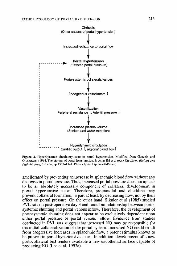
Cirrhosis (Other causes of portal hypertension)
i Increased resistance to portal flow
1 ------
* Portal hypertension
r----- I (Elevated portal pressure) 1
I Porto-systemic collaterals/varices
I
+ Endogenous vasodilators 7
1 Vasodilatation
Peripheral resistance 1, Arterial pressure 1
1 Increased plasma volume
(Sodium and water retention) I
I I I I Hyperdynamic circulation L.--------- Cardiac output 7, regional blood flow?
Figure 2. Hyperdynamic circulatory state in portal hypertension. Modified from Genecin and Groszmann (1994, The biology of portal hypertension. In Arias IM et al (eds) The Liver: Biology and Parhobiology, 3rd edn, pp 1327-1341. Philadelphia: Lippincott-Raven).
ameliorated by preventing an increase in splanchnic blood flow without any decrease in portal pressure. Thus, increased portal pressure does not appear to be an absolutely necessary component of collateral development in portal hypertensive states. Therefore, propranolol and clonidine may prevent collateral formation, in part at least, by decreasing flow, not by their effect on portal pressure. On the other hand, Sikuler et al (1985) studied PVL rats on post-operative day 3 and found no relationship between porto- systemic shunting and portal venous inflow. Therefore, the development of portosystemic shunting does not appear to be exclusively dependent upon either portal pressure or portal venous inflow. Evidence from studies conducted in PVL rats suggest that increased NO may be responsible for the initial collateralization of the portal system. Increased NO could result from progressive increases in splanchnic flow, a potent stimulus known to be present in portal hypertensive states. In addition, development of a new portocollateral bed renders available a new endothelial surface capable of producing NO (Lee et al, 1993a).
214 T. K. GUPTA ET AL
Mosca et al (1992) determined the vascular responsiveness of collateral vessels to various vasoconstrictors and vasodilators using an in situ perfused animal model. By administering norepinephrine, 5-hydroxy- tryptamine, isoproterenol and acetylcholine in the presence and absence of their respective blockers, phentolamine, propranolol, and L-NNA, they determined that collateral vessels have functional a-adrenoreceptors, 5-hydroxytryptamine receptors, and P-adrenoreceptors. In addition, collateral veins appear to be as sensitive to NO as arteries.
Regardless of the specific initiating stimulus, at least at the beginning, the mechanism of collateral formation appears to be recruitment of preformed channels, as opposed to de IZOVO formation of new vessels. Portosystemic shunting can be detected almost immediately after the induction of portal hypertension, a pattern of timing that is consistent with the rapid dilatation of preformed vessels (Mosca et al, 1992). Whether these preformed vessels expand as the result of active dilatation mediated by increased flow or as a passive response to increased portal pressure is unclear. Neoformation of vessels cannot be excluded.
In humans, the portosystemic shunting of blood occurs between the short gastric, coronary veins, and the oesophageal azygos and the intercostal veins; superior and the middle and inferior haemorrhoidal veins; the paraumbilicus venous plexus and the venous system of abdominal organs juxtaposed with the retroperitoneum and abdominal wall; the left renal vein and the splanchnic, adrenal and spermatic veins. As discussed earlier, the dilatation of the pre-existing embryonic channels is thought to be the main mechanism of evolution of these collaterals. With progression of portal hypertension, the number and diameter of these collaterals increases.
The development of gastro-oesophageal vat-ices constitute a major complication of portal hypertension. A threshold portal pressure gradient (HPVG) of 11 to 12 mmHg has been shown to exist in humans with oesophageal varices, below which varices are not encountered (Garcia- Tsao et al, 1985). Elevation above this pressure, however, does not always result in vat-ices, since many patients with HPVG in excess of 12 mmHg do not have vat-ices.
PATHOPHYSIOLOGY OF VARICEAL HAEMORRHAGE
The earlier concept of erosion theory, which suggested that the oesophageal varices bleed due to external trauma to thin-walled varices, has been abandoned because of lack of supporting objective evidence. Factors involved in the variceal haemorrhage appear to be multiple. A certain increase in the portal venous pressure is required for the initiation of oesophageal collateral formation. The volume of blood traversing the collateral vein has a role in the enlarging collateral as does the perivascular tissue, which gives support to the vessel. Tissue support is particularly important in the oesophageal and rectal varices since the surrounding tissue provides very little support to the variceal wall. With continuing increase in intravariceal pressure, the varices expand in size and the variceal wall
PATHOPHYSIOLOGY OF PORTAL HYPERTENSION 215
thickness decreases. Bleeding probably occurs when the expanding force can no longer be counter-balanced by the variceal wall tension; at this point the varices ruptures and bleed (Polio and Groszmann, 1986). The factors regulating the tension exerted by the wall of vat-ices to contain the expand- ing force can be described according to the Laplace’s law (Figure 3):
T=TPxrlw
in which T is the wall tension, TP the transmural pressure, r the radius and w the wall thickness of the varices. The transmural pressure is the differ- ence between intravariceal pressure and the pressure in the oesophageal lumen. The entire varix is in equilibrium between the outwardly directed force (TPx r/w) and the inwardly directed force of wall tension. The inwardly directed force is augmented by the surrounding tissue structures, which provide reinforcement for the vessel wall.
The larger variceal size multiplies the deleterious effect of high intravariceal pressure by increasing the expanding force. When varix distension increases, the radius increases and wall thickness decreases (Figure 3). The wall tension increases in an attempt to contain the expand- ing force. Since venous structures have little musculature, this limits their capacity to develop wall tension. Eventually, the expanding force in a enlarging vat-ix exceeds the limit of wall tension and variceal rupture occurs.
Varix
Mucosa
mucosa
Lamina propria
Varix
Figure 3. Relative position and size of varices with respect to the oesophagus and oesophageal lumen. At equal transmural pressure (TP), the tension (T) developed will be greater in T, than in T, due to increased radius (r) and smaller wall thickness (w). Reproduced from Jensen and Groszmann (1992, Complications of liver disease: pathophysiology and management. In Kaplowitz N. ed. Liver and B&IV Diseases, p 502. Baltimore: Williams & Wilkins).
216 T. K. GUPTA ET AL
The sequence of events leading to portal variceal bleeding are initiated by increased portal pressure, which results in the opening of collaterals. With progression of portal hypertension, the variceal blood flow and intravariceal pressure increases. The variceal size increases and the wall becomes thinner. Further increase in portal pressure, or a defect in the variceal wall leads to variceal haemorrhage. Portal pressure and blood flow are not static and vary markedly with various physiological stimuli. Portal flow increases transiently after meals because of postprandial hyperaemia. Similarly, fluctuations in portal pressure has been observed during circadian rhythm: portal pressure being higher at night and lower during afternoon and evening (Garcia-Pagan et al, 1994b). These variations in portal pressure may influence the onset of bleeding in patients with already elevated baseline portal pressure.
RATIONAL BASIS OF PHARMACOTHERAPY OF PORTAL HYPERTENSION
Drug therapy of portal hypertension is primarily the treatment and prevention of the complications of portal hypertension, mainly variceal haemorrhage. The aim of therapy is to reduce oesophageal vat-ix wall tension, which is accomplished pharmacologically by decreasing transmural variceal pressure by a reduction in portal and intravariceal venous pressure. Since the portal pressure is elevated due to an increase in both resistance and portal venous inflow, the reduction in portal pressure can be achieved pharmacologically by decreasing portal venous and/or portocollateral blood flow or by reducing intrahepatic or portocollateral resistance. As discussed in earlier sections, part of the increased intrahepatic resistance is because of an increased vascular tone, which can be modulated. Vasodilators can reduce intrahepatic and/or portocollateral resistance.
The pharmacological agents currently available for the treatment of portal hypertension primarily act by modulation of the hyperdynamic splanchnic circulation. Vasoconstrictors decrease portal pressure by reducing splanchnic arterial flow. Since increased plasma volume is an important component of hyperdynamic circulation, drugs that modulate plasma volume like diuretics may help in reducing splanchnic blood flow. The last 5 years have clearly seen a general acceptance of drug therapy for portal hypertension. This is not a fad that will wane away with time, but is an important advance in medical treatment of a syndrome that has a mortality as high as an acute myocardial infarction.
REFERENCES
Albillos A, Colombato LA & Groszmann RJ (1992) Vasodilatation and sodium retention in prehepatic portal hypertension. Gastroenterology 102: 931-935.
Albillos A, Colombato LA, Lee FY et al (1993) Octreotide ameliorates vasodilatation and sodium retention in portal hypertensive rats. Gasrroenterology 104: 575-579.

PATHOPHYSIOLOGY OF PORTAL HYPERTENSION 217
Baldus WP & Hoffbauer FW (1963) Vascular changes in the cirrhotic liver as studied by injection technique. American Journal of Digestive Disease 8: 689-692.
Beker S & Valencia-Parparcen J (1968) Portal hypertension syndrome. A comparative analysis of bilharzial fibrosis and hepatic cirrhosis. American Journal of Digestive Disease 13: 1047- 1054.
Bhathal PS & Grossman HJ (1985) Reduction of the increased portal vascular resistance of the isolated perfused cirrhotic rat liver by vasodilators. Journal of Hepatology 1: 325-337.
Benoit JN, Barrowman JA, Harper SL et al (1984) Role of humoral factors in the intestinal hyperemia associated with chronic portal hypertension. American Journal of Physiology 247: G486- G493.
Brion E, Brenard R, Pariente EA et al (1991) Sinusoidal portal hypertension in hepatic amyloidosis. Gut 32: 227-230.
*Casadevall M, Panes J, Pique JM et al (1993) Involvement of nitric oxide and prostaglandins in gastric mucosal hyperemia of portal-hypertensive anesthetized rats. Hepafology 18: 628-634.
Claria J, Jimenez W, Ros J et al (1993) Pathogenesis of arterial hypotension in cirrhotic rats with ascites: role of endogenous nitric oxide. Heparology 1.5: 343-349.
*Colombato LA, Albillos A & Groszmann RJ (1992) Temporal relationship of peripheral vaso- dilatation, plasma volume expansion and the hyperdynamic circulatory state in portal-hyper- tensive rats. Hepatology 15: 323-328.
Doni MG, Whittle BJ, Palmer RM et al (1988) Action of nitric oxide on the release of prosta- cyclin from bovine endothelial cells in culture. European Journal of Pharmacology 151: 19-25.
Garcia-Pagan JC, Femandez M, Bemadich C et al (1994a) Effect of continued NO inhibition on portal hypertensive syndrome after portal vein stenosis in rat. American Journal of Physiology 267: G984-G990.
Garcia-Pagan JC, Feu F, Castells A et al (1994b) Circadian variations of portal pressure and variceal hemorrhage in patients with cirrhosis. Hepafology 19: 595-601.
*Garcia-Tsao G, Groszmann RJ, Fisher RL et al (1985) Portal pressure, presence of gastroesophageal varices and variceal bleeding. Hepatology 5: 419-424.
*Genecin P & Groszmann RJ (1993) Portal hypertension. In Schiff E & Schiff L (eds) Diseases of the Liver, pp 935-973. Philadelphia: JB Lippincott Company.
Genecin P, & Groszmann RJ (1994) The biology of portal hypertension. In Arias IM et al (eds) The Liver: Biology and Pathobiology, 3rd edn, pp 1327-1341. Philadelphia: Lippincott-Raven.
Genecin P, Polio J & Groszmann RJ (1990a) Sodium restriction blunts expansion of plasma volume and ameliorates hyperdynamic circulation in portal hypertension. American Journal of Physiology 259: G498-G503.
Genecin P, Polio J, Ferraioli G et al (199Ob) Bile acids do not mediate the hyperdynamic circulation in portal hypertensive rats. American Journal of Physiology 259: G21-G25.
Gomis R, Femandez-Alvarez J, Pizcueta Pet al (1994) Impaired function of pancreatic islets from rats with portal hypertension resulting from cirrhosis and partial portal vein ligation. Hepafology 19: 1257-1261.
Greenway CV & Stark RD (1971) Hepatic vascular bed. Physiological Review 51: 23-65. Groszmann RJ & Atterbury CE (1982) The pathophysiology of portal hypertension. A basis for
classification. Seminars in Liver Disease 2: 177-186. Gupta TK & Groszmann RJ (1994) Administration of L-arginine, the physiological precursor of nitric
oxide, reduces portal perfusion pressure and ameliorates hepatic vascular hyperreactivity in experimental cirrhosis. Hepatology 20(4): 200A.
Gupta TK, Chung M, Sessa WC et al (1995) Impaired endothelial function in the intrahepatic circulation in cirrhosis, Hepatology 22(4): 156A.
Halvorsen JF & Myking A0 (1974) The portosysfemic collateral pattern in the rat. European Surgical Research 6: 183-195.
Jensen JE and Groszmann RJ (1992) Complications of liver disease: pathophysiology and manage- ment In Kaplowitz N (ed.) Liver and Biliary Diseases, pp 494-504. Baltimore: Williams & Wilkins.
Johnson SJ, Hines JE & Burt AD (1992) Phenotypic modulation of p&sinusoidal cells following acute liver injury-a quantitative analysis. Internaiional Journal of Experimental Pathology 73: 165-772.
Karatapanis S, McCormick PA, Kakad S et al (1994) Alteration in vascular reactivity in isolated aortic rings from portal vein-constricted rats. Hepatology 20: 1516-1521.

218 T. K. GUPTA ET AL
Lee F-Y, Colombato LA, Albillos A et al (1993a) Administration of N-omega-nitro-L-arginine ameliorates portal-systemic shunting in portal-hypertensive rats. Gasrroenterology 105: 1464-1470.
*Lee F-Y, Colombato LA, Albillos A et al (1993b) N”-nitro-t-arginine administration corrects vaso- dilatation and systemic capillary hypotension, and ameliorates plasma volume expansion and sodium retention in portal hypertensive rats. Hepatology 17: 84-90.
van Leeuwen DJ, Howe SC, Scheuer PJ, et al (1991) Portal hypertension in chronic hepatitis: relationship to morphological changes. Gur 31: 339-343.
Lieber CS, Zimmon DS, Kessler RE et al (1976) Portal hypertension in experimental alcoholic liver injury. Clinical Research 24: 478(a).
Liebermann FL & Reynolds TB (1967) Plasma volume in cirrhosis of the liver. Journal of Clinical Invesfigafion 46: 1279-1308.
*Lopez-Talavera JC, Merrill WW & Groszmann RJ (1995) Tumor necrosis factor CL: a major contributor to the hyperdynamic circulation in prehepatic portal-hypertensive rats. Gusrro- enrerology 108: 761-767.
Lopez-Talavera JC, Cadelina GW, Olchowski J et al (1996) Thalidomide inhibits tumor necrosis factor-n, decreases nitric oxide synthesis and ameliorates the hyperdynamic circulatory syndrome in portal hypertensive rats. Hepatology 23(6): 16161621.
Mittal MK, Gupta TK, Lee FY et al (1994) Nitric oxide modulates vascular tone in normal rat liver. American Journal of Physiology 267: G416-G422.
Mosca P, Lee F-Y, Kaumann AJ et al (1992) Pharmacology of portal-systemic collaterals in portal hypertensive rats: role of endothelium. Ameriwn Journal of Physiology 263: G544-G550.
Pinzani M, Failli P, Ruocco C et al (1992) Fat-storing cells as liver-specific pericytes-spatial dynamics of agonist-stimulated intracellular calcium transients. Journal of Clinical Investigarion 90: 642-646.
Pizcueta P, Garcia-Pagan JC, Femandez M et al (1991) Glucagon hinders the effects of somatostatin on portal hypertension. Gastroenterology 101: 1710-1715.
Pizcueta MP, Casamitjana R, Bosch J et al (1995) Decreased systemic vascular sensitivity to norepineprine in portal hypertensive rats: role of hyperglucagonemia. American Journal of Physiology 258: G191-G195.
*Polio J & Groszmann RJ (1986) Hemodynamic factors involved in the development and rupture of esophageal varices: a pathophysiologic approach to treatment. Seminars of Liver Disease 6: 318-331.
Popper H & Zak FG (1958) Pathological aspects of cirrhosis. American Journal of Medicine 24: 593-625.
Reynolds TB, Hidmura R, Mitchell H et al (1969) Portal hypertension without cirrhosis in alcoholic liver disease. Annals of Inrernal Medicine 70: 497-506
Rockey DC & Weisiger RA (1996) Endothelin induced contractility of stellate cell from normal and cirrhotic rat liver: implication for regulation of portal pressure and resistance. Heparology 24(l): 233-240.
Rodriguez-Perez F, Isales CM & Groszmann RJ (1993) Platelet cytosolic calcium, peripheral hemo- dynamics, and vasodilatory peptides in liver cirrhosis. Gastroenterology 105: 863-867.
Rubanyi GM (1990) Endothelium-derived relaxing and contracting factors. Journal of Cellular Biochemistry 46: 27-36.
Rudolph R, McClure WJ & Woodward M (1979) Contractile fibroblasts in chronic alcoholic cirrhosis. Gasrroenrerology 76: 704-709.
Schaffner F & Popper H (1963) Capilhuization of the hepatic sinusoids in man. Gastroenrerology 44: 239-251.
*Sieber CC & Groszmann RJ (1992a) Nitric oxide mediates hyporeactivity to vasopressors in mesenteric vessels of portal hypertensive rats. Gasrroenrerology 103: 235-239.
Sieber CC & Groszmann RJ (1992b) In vitro hyporeactivity to methoxamine in portal hypertensive rats: reversal by nitric oxide blockade. American Journal ofPhysiology 262: G996GlOOl.
Sikuler E & Groszmann RJ (1986) Hemodynamic studies in long- and short-term portal hypertensive rats: the relation of systemic glucagon levels. Heparology 6(3): 414-418.
Sikuler E, Kravetz D & Groszmann RJ (1985) Evolution of portal hypertension and mechanisms involved in its maintenance in a rat model. American Journal of Physiology 248: G618- G625.
Sitzmann JV, Campbell KA, Wu Y et al (1993) Effect of portosystemic shunting on PGIi and glucagon levels in humans. Annuls @Surgery 217: 248-252.

PATHOPHYSIOLOGY OF PORTAL HYPERTENSION 219
*Vallauce P & Moncada S (1991) Hyperdynamic circulation in cirrhosis: a role for nitric oxide? Lancer 337: 176-118.
Vane JR, Anggacd EE & Botting RM (1990) Regulatory functions of vascular endothelium. New England Journal of Medicine 323: 21-36.
Wu Y, Bums C & Sitzmann JV (1993) Effects of nitric oxide and cyclooxygenase inhition on splanchnic hemodynamics in portal hypertension. Hepuroiogg 18: 1416-1421.
Related Documents