1-Methoxy-Canthin-6-One Induces c-Jun NH 2 -Terminal Kinase– Dependent Apoptosis and Synergizes with Tumor Necrosis Factor–Related Apoptosis-Inducing Ligand Activity in Human Neoplastic Cells of Hematopoietic or Endodermal Origin Massimo Ammirante, Rita Di Giacomo, Laura De Martino, Alessandra Rosati, Michela Festa, Antonio Gentilella, Maria Carmela Pascale, Maria Antonietta Belisario, Arturo Leone, Maria Caterina Turco, and Vincenzo De Feo Department of Pharmaceutical Sciences (DiFarma), University of Salerno, Salerno, Italy Abstract We investigated the effects of 1-methoxy-canthin-6-one, isolated from the medicinal plant Ailanthus altissima Swingle, on apoptosis in human leukemia (Jurkat), thyroid carcinoma (ARO and NPA), and hepatocellular carcinoma (HuH7) cell lines. Cultures incubated with the compound showed >50% of sub-G 1 (hypodiploid) elements in flow cytometry analysis; the apoptosis-inducing activity was evi- dent at <10 Mmol/L and half-maximal at about 40 Mmol/L 1-methoxy-canthin-6-one. The appearance of hypodiploid elements was preceded by mitochondrial membrane depo- larization, mitochondrial release of cytochrome c , and Smac/DIABLO and procaspase-3 cleavage. We subsequently investigated the effect of 1-methoxy-canthin-6-one in com- bination with human recombinant tumor necrosis factor– related apoptosis-inducing ligand (TRAIL) in the four cell lines. Suboptimal concentrations (10 Mmol/L 1-methoxy- canthin-6-one and 0.25 ng/mL TRAIL, respectively) of the two agents, unable to elicit apoptosis when used alone, induced mitochondrial depolarization, activation of cas- pase-3, and 45% to 85% of sub-G 1 elements when added together to the cells. The synergism seemed to rely partly on the enhanced expression of TRAIL receptor 1 (TRAIL-R1; DR4), analyzed by immunofluorescence, by 1-methoxy- canthin-6-one. Cell incubation with 1-methoxy-canthin-6- one resulted in activating c-Jun NH 2 -terminal kinase (JNK), as revealed by Western blotting; induction of apoptosis and TRAIL-R1 up-regulation by 1-methoxy-canthin-6-one were >80% prevented by the addition of the JNK inhibitor (JNKI) SP600125JNKI, indicating that both effects were almost completely mediated by JNK activity. On the other hand, synergism with TRAIL was reduced by about 50%, suggest- ing that besides up-regulating TRAIL-R1, 1-methoxy-can- thin-6-one could influence other factor(s) that participated in TRAIL-induced apoptosis. These findings indicate that 1-methoxy-canthin-6-one can represent a candidate for in vivo studies of monotherapies or combined antineoplas- tic therapies. (Cancer Res 2006; 66(8): 4385-93) Introduction Species belonging to the genus Ailanthus (Simaroubaceae) have long been used in Chinese traditional medicine for their antitumoral properties (1), and isolated compounds from their extracts have been observed to induce apoptosis in human cell lines in vitro (2, 3). In particular, extracts from Ailanthus altissima Swingle display, in addition to their antigerminative properties (4), antiproliferative effects in human cell cultures (3, 5). 1-Methoxy- canthin-6-one, isolated from the plant, can induce cell death in human osteosarcoma SaOS-2 cells (3). The mechanism of apoptosis induction by this compound was not previously identified. c-Jun NH 2 -terminal kinase (JNK), a member of the mitogen- activated protein kinase (MAPK) family, is a key regulator of apo- ptosis (6). Modulation of its activity can either promote or inhibit apoptotic processes, depending on cell system and contexts. Indeed, the kinase acts on a variety of targets, including, in addition to c-Jun, other transcription factors (ATF2, Elk-1, p53, and c-Myc) and proapoptotic and antiapoptotic members of the Bcl-2 family (Bcl-2, Bcl-xL, Bim, and BAD), thereby influencing levels and activities of molecules that variously participate in cell death mechanisms (6). In the present study, we showed that 1-methoxy-canthin-6-one induced apoptosis via a JNK-dependent mechanism. Furthermore, the compound synergized with human recombinant tumor necrosis factor (TNF)–related apoptosis-inducing ligand (hrTRAIL) in apo- ptosis induction. TRAIL, a member of the TNF gene superfamily, interacts with a complex system of receptors consisting of two proapoptotic death receptors (TRAIL-R1 and TRAIL-R2) and three decoy receptors (TRAIL-R3, TRAIL-R4, and osteoprotegerin). As a stable soluble trimer, the cytokine selectively induces apoptosis in many transformed cells but not in normal cells; differential expression levels of decoy versus proapoptotic receptors and other mechanisms seem to account for normal cell resistance to TRAIL- induced cell death. TRAIL action involves the formation of a death- inducing signaling complex (DISC) and activation of caspase-8; the apoptotic processes then follow two pathways: the mitochondrial- independent activation of caspase-3 and mitochondrial-dependent apoptosis due to cleavage of BID by caspase-8 (7, 8). The JNK-activating and proapoptotic properties of 1-methoxy- canthin-6-one render this molecule a candidate for in vivo studies of its activity in monotherapies or combined antineoplastic therapies. Materials and Methods Extraction and isolation of 1-methoxy-canthin-6-one. Roots of A. altissima were collected in June 2002 at the University Campus in Fisciano Note: M. Ammirante and R. Di Giacomo contributed equally to this work. Requests for reprints: Maria Caterina Turco, Department of Pharmaceutical Sciences (DiFarma), University of Salerno, via ponte don Melillo, 84084 Fisciano, Salerno, Italy. Phone: 89-962-076; Fax: 89-962-828; E-mail: [email protected]. I2006 American Association for Cancer Research. doi:10.1158/0008-5472.CAN-05-3895 www.aacrjournals.org 4385 Cancer Res 2006; 66: (8). April 15, 2006 Research Article Research. on August 10, 2015. © 2006 American Association for Cancer cancerres.aacrjournals.org Downloaded from

Welcome message from author
This document is posted to help you gain knowledge. Please leave a comment to let me know what you think about it! Share it to your friends and learn new things together.
Transcript

1-Methoxy-Canthin-6-One Induces c-Jun NH2-Terminal Kinase–
Dependent Apoptosis and Synergizes with Tumor Necrosis
Factor–Related Apoptosis-Inducing Ligand Activity in Human
Neoplastic Cells of Hematopoietic or Endodermal Origin
Massimo Ammirante, Rita Di Giacomo, Laura De Martino, Alessandra Rosati, Michela Festa,Antonio Gentilella, Maria Carmela Pascale, Maria Antonietta Belisario, Arturo Leone,Maria Caterina Turco, and Vincenzo De Feo
Department of Pharmaceutical Sciences (DiFarma), University of Salerno, Salerno, Italy
Abstract
We investigated the effects of 1-methoxy-canthin-6-one,isolated from the medicinal plant Ailanthus altissimaSwingle, on apoptosis in human leukemia (Jurkat), thyroidcarcinoma (ARO and NPA), and hepatocellular carcinoma(HuH7) cell lines. Cultures incubated with the compoundshowed >50% of sub-G1 (hypodiploid) elements in flowcytometry analysis; the apoptosis-inducing activity was evi-dent at <10 Mmol/L and half-maximal at about 40 Mmol/L1-methoxy-canthin-6-one. The appearance of hypodiploidelements was preceded by mitochondrial membrane depo-larization, mitochondrial release of cytochrome c , andSmac/DIABLO and procaspase-3 cleavage. We subsequentlyinvestigated the effect of 1-methoxy-canthin-6-one in com-bination with human recombinant tumor necrosis factor–related apoptosis-inducing ligand (TRAIL) in the four celllines. Suboptimal concentrations (10 Mmol/L 1-methoxy-canthin-6-one and 0.25 ng/mL TRAIL, respectively) of thetwo agents, unable to elicit apoptosis when used alone,induced mitochondrial depolarization, activation of cas-pase-3, and 45% to 85% of sub-G1 elements when addedtogether to the cells. The synergism seemed to rely partly onthe enhanced expression of TRAIL receptor 1 (TRAIL-R1;DR4), analyzed by immunofluorescence, by 1-methoxy-canthin-6-one. Cell incubation with 1-methoxy-canthin-6-one resulted in activating c-Jun NH2-terminal kinase (JNK),as revealed by Western blotting; induction of apoptosis andTRAIL-R1 up-regulation by 1-methoxy-canthin-6-one were>80% prevented by the addition of the JNK inhibitor (JNKI)SP600125JNKI, indicating that both effects were almostcompletely mediated by JNK activity. On the other hand,synergism with TRAIL was reduced by about 50%, suggest-ing that besides up-regulating TRAIL-R1, 1-methoxy-can-thin-6-one could influence other factor(s) that participatedin TRAIL-induced apoptosis. These findings indicate that1-methoxy-canthin-6-one can represent a candidate forin vivo studies of monotherapies or combined antineoplas-tic therapies. (Cancer Res 2006; 66(8): 4385-93)
Introduction
Species belonging to the genus Ailanthus (Simaroubaceae) havelong been used in Chinese traditional medicine for theirantitumoral properties (1), and isolated compounds from theirextracts have been observed to induce apoptosis in human celllines in vitro (2, 3). In particular, extracts from Ailanthus altissimaSwingle display, in addition to their antigerminative properties (4),antiproliferative effects in human cell cultures (3, 5). 1-Methoxy-canthin-6-one, isolated from the plant, can induce cell death inhuman osteosarcoma SaOS-2 cells (3). The mechanism of apoptosisinduction by this compound was not previously identified.
c-Jun NH2-terminal kinase (JNK), a member of the mitogen-activated protein kinase (MAPK) family, is a key regulator of apo-ptosis (6). Modulation of its activity can either promote or inhibitapoptotic processes, depending on cell system and contexts. Indeed,the kinase acts on a variety of targets, including, in addition to c-Jun,other transcription factors (ATF2, Elk-1, p53, and c-Myc) andproapoptotic and antiapoptotic members of the Bcl-2 family (Bcl-2,Bcl-xL, Bim, and BAD), thereby influencing levels and activities ofmolecules that variously participate in cell death mechanisms (6).
In the present study, we showed that 1-methoxy-canthin-6-oneinduced apoptosis via a JNK-dependent mechanism. Furthermore,the compound synergized with human recombinant tumor necrosisfactor (TNF)–related apoptosis-inducing ligand (hrTRAIL) in apo-ptosis induction. TRAIL, a member of the TNF gene superfamily,interacts with a complex system of receptors consisting of twoproapoptotic death receptors (TRAIL-R1 and TRAIL-R2) and threedecoy receptors (TRAIL-R3, TRAIL-R4, and osteoprotegerin). As astable soluble trimer, the cytokine selectively induces apoptosis inmany transformed cells but not in normal cells; differentialexpression levels of decoy versus proapoptotic receptors and othermechanisms seem to account for normal cell resistance to TRAIL-induced cell death. TRAIL action involves the formation of a death-inducing signaling complex (DISC) and activation of caspase-8; theapoptotic processes then follow two pathways: the mitochondrial-independent activation of caspase-3 and mitochondrial-dependentapoptosis due to cleavage of BID by caspase-8 (7, 8).
The JNK-activating and proapoptotic properties of 1-methoxy-canthin-6-one render this molecule a candidate for in vivo studies ofits activity in monotherapies or combined antineoplastic therapies.
Materials and Methods
Extraction and isolation of 1-methoxy-canthin-6-one. Roots of A.altissima were collected in June 2002 at the University Campus in Fisciano
Note: M. Ammirante and R. Di Giacomo contributed equally to this work.Requests for reprints: Maria Caterina Turco, Department of Pharmaceutical
Sciences (DiFarma), University of Salerno, via ponte don Melillo, 84084 Fisciano,Salerno, Italy. Phone: 89-962-076; Fax: 89-962-828; E-mail: [email protected].
I2006 American Association for Cancer Research.doi:10.1158/0008-5472.CAN-05-3895
www.aacrjournals.org 4385 Cancer Res 2006; 66: (8). April 15, 2006
Research Article
Research. on August 10, 2015. © 2006 American Association for Cancercancerres.aacrjournals.org Downloaded from

(Salerno, Italy). A voucher specimen of the plant (DF.2002.193) is kept atthe Herbarium of Pharmaceutical Botany Chair at the State University of
Salerno. Air-dried roots of A. altissima were successively extracted at room
temperature with solvents of increasing polarity [petroleum ether,
chloroform, chloroform-methanol (9:1), methanol, and water]. Chloroformextract of roots, the most active in biological assays, was fractionated on a
silica gel column, eluting with chloroform and mixtures of chloroform and
methanol of increasing polarity. Seven hundred sixty-two fractions of
10 mL were obtained and pooled in 13 major fractions based on their TLCsimilarity. Fraction pool XXIII-XXVII was further purified on a silica gel
column, collecting 127 fractions of 5 mL, pooled in nine major
subfractions (A-I). Subfraction C, showing a strong biological activity,
was shown to contain, in TLC assays, a single substance, positive toDragendorff reagent. The identification of this active substance, recog-
nized as 1-methoxy-canthin-6-one, was done by accurate analyses of its1H-NMR, 13C-NMR, and 13C-NMR DEPT data and by comparison withliterature data (ref. 9; Fig. 1).
Cell lines and peripheral blood mononuclear cells from normaldonors. Human leukemia (Jurkat), thyroid carcinoma (ARO and NPA),
and hepatocellular carcinoma (HuH7) cell lines were obtained fromthe American Type Culture Collection (Rockville, MD). Jurkat and ARO
cells were maintained in 5% CO2 at 37jC in RPMI 1640 (Sigma-Aldrich,
St. Louis, MO) containing 10% FCS, 1% penicillin, and 1% streptomycin.
HuH7 cells and NPA cells were maintained in 5% CO2 at 37jC in DMEMcontaining 10% FCS, 1% penicillin, and 1% streptomycin. Peripheral blood
mononuclear cells (PBMC) from normal donors were isolated from
heparinized blood samples through centrifugation on Ficoll densitygradient (10).
Antibodies, inhibitors, and other reagents. Anti- human cyto-
chrome c and Smac/DIABLO mouse monoclonal antibodies (mAb) were
obtained from BD PharMingen (San Diego, CA). Anti-human Hsp60(H-1) and phycoerythrin-conjugated anti-integrin a4 (SG31) and anti-
integrin h1 (P5D2) mouse mAbs were purchased from Santa Cruz Bio-
technology (Santa Cruz, CA). Anti-human a-tubulin mAb was obtained
from Sigma (St. Louis, MO). Anti-human phospho-c-Jun (Ser63) andphospho-JNK (G-7) rabbit polyclonal antibodies were obtained from Cell
Signaling, Inc. (Beverly, MA). Anti-human caspase-3 rabbit polyclonal
antibody was purchased from StressGen (Victoria, British Columbia,
Canada). Etoposide and staurosporine were purchased from Sigma.Human recombinant Superkiller TRAIL and extracellular signal-regulated
kinase inhibitor U0126 were purchased from Alexis Biochemicals (San
Diego, CA). JNK inhibitor SP600125 was obtained from Calbiochem (San
Diego, CA).Immunofluorescence. Cells (1 � 105) were incubated with saturating
amounts of phycoerythrin-conjugated mAb or, in indirect tests, primary
mAb followed, after washing with PBS, by FITC-conjugated rabbit anti-mouse antibodies. After further washing with PBS, the cells were
resuspended in 1% formaldehyde-PBS and analyzed with a FACScan
(Becton Dickinson, San Jose, CA) flow cytometer.
Analysis of hypodiploid (apoptotic) nuclei and caspase-3 activity.Apoptosis was analyzed by propidium iodide incorporation in permeabi-
lized cells and flow cytometry as described (10). Briefly, cells (5 � 105) were
washed in PBS and resuspended in 500 AL of a solution containing 0.1%
sodium citrate, 0.1% Triton X-100, and 50 Ag/mL propidium iodide (Sigma).Following incubation at 4jC for 30 minutes in the dark, cell nuclei were
analyzed with a Becton Dickinson FACScan flow cytometer. Cellular debris
was excluded from analysis by raising the forward scatter threshold, and theDNA content of the nuclei was registered on a logarithmic scale. The
percentage of the elements in the hypodiploid region was calculated.
Caspase-3 activity was evaluated either by Western blotting (see below) or
Figure 1. Extraction procedure for 1-methoxycanthin-6-one.
Cancer Research
Cancer Res 2006; 66: (8). April 15, 2006 4386 www.aacrjournals.org
Research. on August 10, 2015. © 2006 American Association for Cancercancerres.aacrjournals.org Downloaded from

by using Caspase-3 Fluorometric Detection kit (Assay Designs, Ann Arbor,
MI; ref. 11).
Analysis of mitochondrial transmembrane potential. Mitochondrial
membrane potential (DWm) was assessed using a cytofluorometrictechnique using tetramethylrhodamine ethyl ester (TMRE; ref. 12). Cells
were exposed to TMRE (5 nmol/L; Molecular Probes, Eugene, OR) for 1 hour
at 37jC. Changes in dye fluorescence were analyzed in a Becton Dickinson
FACScan flow cytometer.Cell lysates, subcellular fractionation, and Western blotting. Cell
total protein lysates were prepared in sample buffer [2% SDS, 10% glycerol,2% mercaptoethanol, and 60 mmol/L Tris-HCl (pH 6.8) in demineralizedwater] on ice. Cell cytosolic and mitochondrial fractions were generatedusing a digitonin-based subcellular fractionation technique as described(13). For Western blotting analysis, proteins from whole cell or fractionlysates were run on 12% SDS-PAGE gels and electrophoretically transferredto nitrocellulose. Nitrocellulose blots were blocked with 5% bovine serumalbumin (BSA) in TBS/Tween 20 (TBST) buffer [20 mmol/L Tris-HCl (pH7.4), 500 mmol/L NaCl, and 0.01% Tween 20] and incubated with primaryantibody in TBST/5% BSA overnight at 4jC. Immunoreactivity was detectedby sequential incubation with horseradish peroxidase–conjugated second-ary antibody and enhanced chemiluminescence reagents following standardprotocols.
Statistical analysis. Statistical analysis was done using GraphPad Prismversion 4.00 for Windows (GraphPad Software, San Diego, CA).1
Results
Induction of apoptosis in human cell lines of differentorigins by 1-methoxy-canthin-6-one. We have recently observedthat 1-methoxy-canthin-6-one induced cell death in the humanosteosarcoma cell line SaOS-2 (3). To verify and characterize theproapoptotic effect of the compound in human cells of diverseorigins, we analyzed the dose-dependent induction of apoptosis inleukemia (Jurkat), thyroid carcinoma (NPA and ARO), or hepato-cellular carcinoma (HuH7) cells. Following a 24-hour incubationwith 1-methoxy-canthin-6-one, the four tested lines showedpercentages of apoptotic (sub-G1) cells ranging from 50% to>70%, as evaluated by flow cytometry (ref. 10; see Materials andMethods). The apoptosis- inducing activity was evident at <10Amol/L 1-methoxy-canthin-6-one and half-maximal at a compoundconcentration of about 40 Amol/L. The compound did not showany proapoptotic or toxic activity in PBMCs from two differentdonors (Fig. 2). The appearance of sub- G1 elements was evidentin cells incubated with 1-methoxy-canthin-6-one for <15 hours
Figure 2. Apoptosis induction by1-methoxycanthin-6-one. Jurkat (A ), NPA(B), ARO (C ), and HuH7 (D) cells wereplated in RPMI supplemented with 10%of heat-inactivated FCS (10% FCS-RPMI)at a density of 1 � 106 per mL andincubated with the indicated concentrationsof 1-methoxycanthin-6-one. After 24 hours,apoptosis was measured as percentage ofsub-G1 nuclei by flow cytometry (10).Points, means of triplicate determinations;bars, SE. E, PBMCs from two (A and B )healthy donors (1 � 106 per mL) wereincubated in 10% FCS-RPMI with40 Amol/L 1-methoxycanthin-6-one.Following 24 hours, cell death andpercentages of sub-G1 nuclei wereanalyzed by trypan blue exclusion and flowcytometry, respectively. Columns, meansof triplicate determinations; bars, SE.
1 http://www.graphpad.com.
Apoptosis-Inducing Activity of 1-Methoxy-Canthin-6-One
www.aacrjournals.org 4387 Cancer Res 2006; 66: (8). April 15, 2006
Research. on August 10, 2015. © 2006 American Association for Cancercancerres.aacrjournals.org Downloaded from

(Fig. 3A). To investigate whether mitochondrial events wereinvolved in the induction of apoptosis, we analyzed mitochondrialmembrane depolarization by flow cytometry (12) and the release ofcytochrome c and Smac/DIABLO (13) by Western blotting in cellsincubated with the compound. The cells clearly showed a loss ofmitochondrial membrane polarity 4 to 8 hours following cellincubation with 1-methoxy-canthin-6-one (Fig. 3B); in parallel, wedetected a decrease in the intramitochondrial and an increase inthe cytosolic amounts of cytochrome c and Smac/DIABLO proteins(Fig. 3C). These findings indicated that the mitochondrial pathwayof apoptosis was triggered by this molecule. As expected,procaspase-3 was accordingly cleaved, leading to the appearanceof a faint p20/19 and an intense p17 signal (13), in cells exposed tothe drug (Fig. 3D).
Synergy of 1-methoxy-canthin-6-one and hrTRAIL. To furtherexplore the potential use of 1-methoxy-canthin-6-one as antineo-plastic agent, we investigated its proapoptotic effect in combinationwith TRAIL, a molecule that induces apoptosis in a vast series ofdifferent neoplasias (7, 8). To this purpose, we analyzed apoptosis byflow cytometry in cells treated with suboptimal doses of both com-pounds. When different cell lines were incubated with 10 Amol/L1-methoxy-canthin-6-one and/or suboptimal TRAIL, the singleagents induced <10% of apoptotic elements, whereas their com-bination elicited apoptotic responses ranging from 45% to >80%.Such effect was not detectable in normal PBMCs (Fig. 4A). Thesynergistic effect in neoplastic cells was evident at the levels ofcaspase-3 activation (Fig. 4C) and mitochondrial depolarization(Fig. 4B), indicating that its mechanism was to be investigated in
Figure 3. Effect of 1-methoxycanthin-6-one on mitochondrial membranedepolarization, cytochrome c , and Smac/DIABLO release and caspase-3 cleavage.A, Jurkat cells were plated at a density of1 � 106 per mL in 10% FCS-RPMI, in theabsence or presence of 1-methoxycanthin-6-one (40 Amol/L), in triplicates. Apoptosiswas measured at the indicated timeintervals as percentage of sub-G1 nuclei byflow cytometry. B, Jurkat cells wereincubated at 5 � 105 per mL in 10%FCS-RPMI, in the absence or presence of1-methoxy-canthin-6-one (40 Amol/L). Atthe indicated time intervals, mitochondrialmembrane depolarization was evaluatedby cell staining with TMRE (5 nmol/l) for1 hour at 37jC followed by flow cytometricanalysis (12). Experiments were done atleast twice. C, Jurkat cells were incubatedat 5 � 105 per mL in 10% FCS-RPMI, inthe absence or presence of 1-methoxy-canthin-6-one (40 Amol/L). Cell incubationwith staurosporine (250 nmol/L) was usedas a positive control for cytochrome cand Smac/DIABLO release (47). At theindicated time intervals, cells wereharvested, cytosolic and mitochondrialfractions were generated (13), andsubcellular fractions were analyzed withanti-cytochrome c , anti-Smac/DIABLO,and anti-Hsp60 (as intramitochondrialmarker protein) antibodies by Westernblotting. Experiments were done at leasttwice. D, Jurkat cells were incubated at5 � 105 per mL in 10% FCS-RPMI, in theabsence or presence of 1-methoxy-canthin-6-one (40 Amol/L). Cell incubationwith etoposide (50 Amol/L) was used as apositive control for caspase-3 activationand appearance of the caspase-3 activecleaved forms p20/19 and/or p17 (48, 49).At the indicated time intervals, cell lysateswere obtained and analyzed with ananti-caspase-3 antibody by Westernblotting. Experiments were done at leasttwice.
Cancer Research
Cancer Res 2006; 66: (8). April 15, 2006 4388 www.aacrjournals.org
Research. on August 10, 2015. © 2006 American Association for Cancercancerres.aacrjournals.org Downloaded from

earlier step(s) of the apoptotic program. Therefore, we verifiedwhether incubation with 1-methoxy-canthin-6-one modulated theexpression of TRAIL receptors. As shown in Fig. 5, Jurkat cellsincubated with the molecule (10 Amol/L) showed an enhanced
binding to an anti-TRAIL-R1 antibody, as detected by immunoflu-orescence: indeed, the mean intensity of fluorescence increased from24.1 (Fig. 5, TRAIL-R1, green) to 44.8 after 15 hours of cell incubationwith the drug (Fig. 5, TRAIL-R1, orange). This effect is specific
Figure 4. Effect of 1-methoxycanthin-6-one on TRAIL-induced cell apoptosis. A , Normal PBMC (a ), HuH7 (b), NPA (c ), Jurkat (d), and ARO (e) cells were incubatedwith 1-methoxycanthin-6-one at a concentration of 10 Amol/L, with or without TRAIL at the indicated suboptimal concentrations, in triplicates. The suboptimalconcentration of TRAIL was higher (3 ng/mL) in ARO cells, that were less sensitive than the other analyzed neoplastic cell lines to the proapoptotic activity of thecytokine. After 24 hours (a-d ) or the indicated times (e ), apoptosis was measured as percentage of sub-G1 nuclei by flow cytometry. B, Jurkat cells were incubated with1-methoxycanthin-6-one (10 Amol/L), without or with TRAIL (0.25 ng/mL), for 4 hours. Then cells were harvested, incubated with TMRE, and analyzed by flowcytometry. C, Jurkat cells were incubated with 1-methoxycanthin-6-one (10 Amol/L), without or with TRAIL (0.25 ng/mL), in triplicates, for the indicated time intervals.Then caspase-3 activity was measured by fluorimetric assay.
Apoptosis-Inducing Activity of 1-Methoxy-Canthin-6-One
www.aacrjournals.org 4389 Cancer Res 2006; 66: (8). April 15, 2006
Research. on August 10, 2015. © 2006 American Association for Cancercancerres.aacrjournals.org Downloaded from

because we could not detect any increase in the expression of otherTRAIL receptors or unrelated proteins (i.e., integrins; Fig. 5).Role of JNK in 1-methoxy-canthin-6-one- induced apopto-
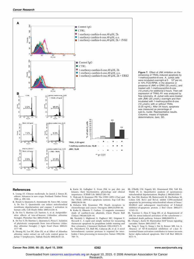
sis. The activity of JNK influences cell apoptosis (6, 14–18) andmediates the proapoptotic effects of some antineoplastic com-pounds (19–27). Furthermore, it regulates the expression of TRAIL-R2 (DR5; refs. 28–31). We therefore investigated the role of thekinase in 1-methoxy-canthin-6-one–induced apoptosis and syner-gism with hrTRAIL. Incubation of Jurkat cells with 1-methoxy-canthin-6-one induced JNK activation, as revealed by theappearance of the phosphorylated forms of JNK and its substratec-Jun, analyzed by Western blot (Fig. 6A). To verify whether JNKactivation was required for apoptosis induction, we used the JNKinhibitor (JNKI) SP600125 (32). The addition of this molecule to cellcultures inhibited apoptosis by >80% (Fig. 6C), indicating that JNKactivity mediated the effect of the drug. Furthermore, the inhibitorreduced TRAIL-R1 up-regulation (Fig. 7A) and the synergisticactivity of 1-methoxy-canthin-6-one with hrTRAIL (Fig. 7B ).Indeed, whereas apoptosis induced by the two agents together incell culture was >45%, it was reduced to 21% in the presence ofJNKI. Because the synergism was not completely abrogated by theinhibitor, other element(s), in addition to JNK activation,apparently contributed to the enhancement of TRAIL- inducedapoptosis by 1-methoxy-canthin-6-one.
Discussion
This study identifies and characterizes the apoptosis-inducingactivity of 1-methoxy-canthin-6-one, isolated from A. altissima , inhuman neoplastic cell lines of hematopoietic or endodermal origin.
Understanding of such an effect of this molecule supports itspotential therapeutic application. The mechanism of 1-methoxy-canthin-6-one activity seemed to entirely rely on JNK activation;indeed, the inhibition of the kinase was able to completely preventapoptosis induced by the compound. In this respect, these findingsprovide one of the clearest evidence attesting that JNK activation canexert a major role in the activity of antineoplastic molecules (19–27).
The MAPK subfamily JNK has three isoforms (JNK1, JNK2, andJNK3); JNK1 and JNK2 are ubiquitously expressed, whereas JNK3 ismainly expressed in neuronal and heart tissues (6, 15, 33). AmongMAPKs, JNKs are preferentially activated by cytotoxic stresses(oxidative/nitrosative stress, heat and osmotic shock, X-ray, andUV irradiation) and proinflammatory cytokines, including TNF-a,interleukin-1 (IL-1), and IL-18 (34–38). Although initially identifiedby its ability to phosphorylate the transcription factor c-Jun, JNKwas subsequently found to act also on other substrates, such as thetranscription factors ATF2, Elk-1, p53 (33), c-Myc and proapoptotic(Bim and BAD) or antiapoptotic (Bcl-2 and Bcl-xL) proteinsbelonging to the Bcl-2 family. This justifies the opposite effects(proapoptotic or antiapoptotic) that JNK has been shown to exertin diverse systems (39–42). Genetic disruption of jnk1 and jnk2alleles in mice resulted in increasing apoptosis in hindbrain andforebrain regions (39, 40), suggesting that JNK may be required forcell survival. Furthermore, some reports showed that inhibition ofJNK2 activity suppressed tumorigenesis through promotingapoptosis (41). One of the molecular mechanisms by which JNKcan suppress apoptosis is inhibition of BAD proapoptotic activitythrough phosphorylation at Thr201 (18). On the other hand,numerous evidence indicate that JNK is involved in proapop-totic pathways (14, 16, 17, 42, 43). JNK activation is required for
Figure 5. Effect of 1-methoxycanthin-6-one on theexpression of TRAIL receptors. After 2 hours(pink ), 7 hours (blue ), or 15 hours (orange ) oftreatment with 1-methoxycanthin-6-one(10 Amol/L), Jurkat cells were harvested andwashed once with PBS 1�. Then cells wereanalyzed with anti-TRAIL-R1, anti-TRAIL-R2,anti-TRAIL-R3, or TRAIL-R4 mAb in indirect andphycoerythrin-conjugated anti-integrin a4(SG31) or anti-integrin h1 (P5D2) in directimmunofluorescence by flow cytometry. Green,untreated cells (CTRL ).
Cancer Research
Cancer Res 2006; 66: (8). April 15, 2006 4390 www.aacrjournals.org
Research. on August 10, 2015. © 2006 American Association for Cancercancerres.aacrjournals.org Downloaded from

UV-induced apoptosis, and its absence caused cell failure torelease cytochrome c (14). In TNF-a-induced apoptosis, in theabsence of nuclear factor-nB activation, prolonged JNK activa-tion promotes cell death (16, 42), probably via its contributionto the proteolysis of the proapoptotic molecule BID (17). In theDNA damage response, JNK activation is responsible for 14-3-3protein phosphorylation followed by release of sequestered c-Abland its nuclear targeting, leading to apoptosis (43). Therefore,it seems that the proapoptotic activity of JNK can be exertedthrough more than one mechanism.
Various antineoplastic agents, including topoisomerase inhib-itors (25), histone deacetylase inhibitors (23), rapamycin (27), andothers (19–22, 24, 26), have been shown to activate JNK. Inhibitionof the kinase mostly reduced the proapoptotic activity of theseagents (19–27), whereas completely abrogated that displayed by 1-methoxy-canthin-6-one. In this case, therefore, JNK activation didnot simply participated in promoting or amplifying the proapop-totic signal but was decisive for subverting the cell survival/deathbalance. These findings strongly contribute in indicating that levelsof JNK activity can determine cell fate in some systems (6, 44). Theyintroduce an element of caution in evaluating antineoplasticapproaches based on the use of JNK inhibitors (45) and might
instead support the potential utility of strategies aimed atspecifically activating the kinase, to trigger apoptosis in neoplasticcells (46).
JNK activity has been shown to influence the levels of TRAIL-R2(28–31). We found that it can also up-regulate TRAIL-R1 becausethe levels of this receptor were enhanced by 1-methoxy-canthin-6-one in a JNK-dependent manner: indeed, JNK inhibition completelyprevented the effect. However, the inhibition of the kinase onlypartially reduced the synergism of 1-methoxy-canthin-6-one withTRAIL. Therefore, 1-methoxy-canthin-6-one apparently regulated,in addition to JNK and TRAIL receptor levels, other factor(s) thatparticipated in TRAIL-induced apoptosis. A more extensive ana-lysis of such factors (DISC components, IAPs, etc.) could contributeto deeply evaluating the proapoptotic activity of 1-methoxy-canthin-6-one and its synergism with agents that induce tumorcell death.
Acknowledgments
Received 11/1/2005; revised 1/9/2006; accepted 2/14/2006.The costs of publication of this article were defrayed in part by the payment of page
charges. This article must therefore be hereby marked advertisement in accordancewith 18 U.S.C. Section 1734 solely to indicate this fact.
Figure 6. Effect of 1-methoxycanthin-6-one on JNK activation. A, cells (1 � 106 per mL in 10% FCS-RPMI) were incubated with 1-methoxycanthin-6-one (40 Amol/L)for the indicated times. Then cell lysates were obtained and analyzed with anti-phospho-JNK, anti-phospho-c-Jun, or anti-a-tubulin antibodies in Western blot. B, cells(1 � 106 per mL in 10% FCS-RPMI) were incubated with 1-methoxycanthin-6-one (40 Amol/L) for the indicated times, with or without overnight pretreatment withJNKI or ERKI (20 Amol/L). Then cell lysates were obtained and analyzed with anti-phospho-JNK or anti-a-tubulin antibodies in Western blot. C, cells (1 � 106 per mL in10% FCS-RPMI), preincubated with medium alone or JNKI or ERKI (20 Amol/L) overnight, were incubated with 1-methoxycanthin-6-one (40 Amol/L) for 16 hours. Thenapoptosis was measured as percentage of sub-G1 nuclei. Representative results. Columns, means of triplicate determinations; bars, SD.
Apoptosis-Inducing Activity of 1-Methoxy-Canthin-6-One
www.aacrjournals.org 4391 Cancer Res 2006; 66: (8). April 15, 2006
Research. on August 10, 2015. © 2006 American Association for Cancercancerres.aacrjournals.org Downloaded from
References1. Leung AY. Chinese medicinals. In: Janick J, Simon JE,
editors. Advances in new crops. Portland: Timber Press;1988. p. 499–510.2. Rosati A, Quaranta E, Ammirante M, Turco MC, Leone
A, De Feo V. Quassinoids can induce mitochondrialmembrane depolarisation and caspase 3 activation inhuman cells. Cell Death Differ 2004;11:216–8.3. De Feo V, Martino LD, Santoro A, et al. Antiprolifer-
ative effects of tree-of-heaven (Ailanthus altissimaSwingle). Phytother Res 2005;19:226–30.4. De Feo V, De Martino L, Quaranta E, Pizza C. Isolation
of phytotoxic compounds from tree-of-heaven (Ailan-thus altissima Swingle). J Agric Food Chem 2003;51:1177–80.5. Hwang SG, Lee HC, Kim CK, et al. Effect of Ailanthusaltissima water extract on cell cycle control genes inJurkat T lymphocytes. Yakhak Hoechi 2002;46:18–23.
6. Karin M, Gallagher E. From JNK to pay dirt: junkinases, their biochemistry, physiology and clinicalimportance. IUBMB Life 2005;57:283–95.7. Walczak H, Krammer PH. The CD95 (APO-1/Fas) and
the TRAIL (APO-2L) apoptosis systems. Exp Cell Res2000;256:58–66.8. Debatin KM, Krammer PH. Death receptors in
chemotherapy and cancer. Oncogene 2004;23:2950–66.9. Koike K, Ohmoto T. Carbon- 13 magnetic resonance
study of canthin-6-one alkaloids. Chem Pharm Bull(Tokyo) 1985;66:5239–44.10. Nicoletti I, Migliorati G, Pagliacci MC, Grignani F,
Riccardi C. A rapid and simple method for measuringthymocyte apoptosis by propidium iodide staining andflow cytometry. J Immunol Methods 1991;139:271–9.11. Thornberry NA, Bull HG, Calaycay JR, et al. A novel
heterodimeric cysteine protease is required for inter-leukin-1 beta processing in monocytes. Nature 1992;356:768–74.
12. O’Reilly CM, Fogarty KE, Drummond RM, Tuft RA,Walsh JV, Jr. Quantitative analysis of spontaneousmitochondrial depolarizations. Biophys J 2003;85:3350–7.13. Sun XM, Bratton SB, Butterworth M, MacFarlane M,
Cohen GM. Bcl-2 and Bcl-xL inhibit CD95-mediatedapoptosis by preventing mitochondrial release of Smac/DIABLO and subsequent inactivation of X-linkedinhibitor-of-apoptosis protein. J Biol Chem 2002;277:11345–51.14. Tournier C, Hess P, Yang DD, et al. Requirement of
JNK for stress-induced activation of the cytochrome c -mediated death pathway. Science 2000;288:870–4.15. Chang L, Karin M. Mammalian MAP kinase signaling
cascades. Nature 2001;410:37–40.16. Tang F, Tang G, Xiang J, Dai Q, Rosner MR, Lin A.
Absence of NF-B-mediated inhibition of c-Jun N-terminal kinase activation contributes to tumor necrosisfactor alpha-induced apoptosis. Mol Cell Biol 2002;24:8571–9.
Figure 7. Effect of JNK inhibition on theenhancing of TRAIL-induced apoptosis by1-methoxycanthin-6-one. A, Jurkat cellswere incubated overnight at 5 � 105 per mLin 10% FCS-RPMI, in the absence orpresence of JNKI or ERKI (20 Amol/L), andtreated with 1-methoxycanthin-6-one(10 Amol/L) for additional 2 hours. Then cellexpression of TRAIL-R1 was analyzed byflow cytometry. B, Jurkat cells were treatedwith JNKI (20 Amol/L) overnight and thenincubated with 1-methoxycanthin-6-one(10 Amol/L) with or without TRAIL(0.25 ng/mL). After 24 hours, apoptosiswas measured as percentage ofsub-G1 nuclei. Representative results.Columns, means of triplicatedeterminations; bars, SD.
Cancer Research
Cancer Res 2006; 66: (8). April 15, 2006 4392 www.aacrjournals.org
Research. on August 10, 2015. © 2006 American Association for Cancercancerres.aacrjournals.org Downloaded from

Apoptosis-Inducing Activity of 1-Methoxy-Canthin-6-One
www.aacrjournals.org 4393 Cancer Res 2006; 66: (8). April 15, 2006
17. Deng Y, Ren X, Yang L, Lin Y, Wu X. A JNK-dependentpathway is required for TNF-induced apoptosis. Cell2003;115:61–70.18. Yu C, Minemoto Y, Zhang J, et al. JNK suppresses
apoptosis via phosphorylation of the proapoptotic Bcl-2family protein BAD. Mol Cell 2004;13:329–40.19. Fan M, Goodwin ME, Birrer MJ, Chambers TC. The c-
Jun NH(2)-terminal protein kinase/AP-1 pathway isrequired for efficient apoptosis induced by vinblastine.Cancer Res 2001;61:4450–8.20. Filomeni G, Aquilano K, Rotilio G, Ciriolo MR.
Reactive oxygen species-dependent c-Jun NH2-terminalkinase/c-Jun signaling cascade mediates neuroblastomacell death induced by diallyl disulfide. Cancer Res 2003;63:5940–9.21. Hideshima T, Chauhan D, Hayashi T, et al. Antitumor
activity of lysophosphatidic acid acyltransferase-betainhibitors, a novel class of agents, in multiple myeloma.Cancer Res 2003;63:8428–36.22. Ikeda T, Sporn M, Honda T, Gribble GW, Kufe D. The
novel triterpenoid CDDO and its derivatives induceapoptosis by disruption of intracellular redox balance.Cancer Res 2003;63:5551–8.23. Yu C, Subler M, Rahmani M, et al. Induction of
apoptosis in BCR/ABL+ cells by histone deacetylaseinhibitors involves reciprocal effects on the RAF/MEK/ERK and JNK pathways. Cancer Biol Ther 2003;2:544–51.24. Xiao D, Pinto JT, Soh JW, et al. Induction of apoptosis
by the garlic-derived compound S -allylmercaptocysteine(SAMC) is associated with microtubule depolymeriza-tion and c-Jun NH(2)-terminal kinase 1 activation.Cancer Res 2003;63:6825–37.25. Catley L, Tai YT, Shringarpure R, et al. Proteasomal
degradation of topoisomerase I is preceded by c-JunNH2-terminal kinase activation, Fas up-regulation, andpoly(ADP-ribose) polymerase cleavage in SN38-mediatedcytotoxicity against multiple myeloma. Cancer Res 2004;64:8746–53.26. Dai Y, Rahmani M, Pei XY, et al. Farnesyltransferase
inhibitors interact synergistically with the Chk1 inhib-itor UCN-01 to induce apoptosis in human leukemiacells through interruption of both Akt and MEK/ERKpathways and activation of SEK1/JNK. Blood 2005;105:1706–16.
27. Hahn M, Li W, Yu C, Rahmani M, Dent P, Grant S.Rapamycin and UCN-01 synergistically induce apo-ptosis in human leukemia cells through a processthat is regulated by the Raf-1/MEK/ERK, Akt, and JNKsignal transduction pathways. Mol Cancer Ther 2005;4:457–70.28. Ohtsuka T, Zhou T. Bisindolylmaleimide VIII enhan-
ces DR5-mediated apoptosis through the MKK4/JNK/p38 kinase and the mitochondrial pathways. J Biol Chem2002;277:29294–303.29. Higuchi H, Grambihler A, Canbay A, Bronk SF, Gores
GJ. Bile acids up-regulate death receptor 5/TRAIL-receptor 2 expression via a c-Jun N-terminal kinase-dependent pathway involving Sp1. J Biol Chem 2004;279:51–60.30. Zou W, Liu X, Yue P, et al. c-Jun NH2-terminal kinase-
mediated up-regulation of death receptor 5 contributesto induction of apoptosis by the novel synthetictriterpenoid methyl-2-cyano-3,12-dioxooleana-1, 9-dien-28-oate in human lung cancer cells. Cancer Res 2004;64:7570–8.31. Zhu H, Guo W, Zhang L, et al. Proteasome inhibitors-
mediated TRAIL resensitization and Bik accumulation.Cancer Biol Ther 2005;4:781–6.32. Bennett BL, Sasaki DT, Murray BW, et al. SP600125,
an anthrapyrazolone inhibitor of Jun N-terminal kinase.Proc Natl Acad Sci U S A 2001;98:13681–6.33. Hu MC, Qiu WR, Wang YP. JNK1, JNK2 and JNK3 are
p53 N-terminal serine 34 kinases. Oncogene 1997;15:2277–87.34. Minden A, Lin A, McMahon M, et al. Differential
activation of ERK and JNK mitogen-activated proteinkinases by Raf-1 and MEKK. Science 1994;266:1719–23.35. Westwick JK, Weitzel C, Minden A, Karin M, Brenner
DA. Tumor necrosis factor alpha stimulates AP-1activity through prolonged activation of the c-Junkinase. J Biol Chem 1994;269:26396–401.36. Gupta S, Campbell D, Derijard B, Davis RJ. Tran-
scription factor ATF2 regulation by the JNK signaltransduction pathway. Science 1995;267:389–93.37. Whitmarsh AJ, Shore P, Sharrocks AD, Davis RJ.
Integration of MAP kinase signal transduction pathwaysat the serum response element. Science 1995;269:403–7.38. Chandrasekar B, Mummidi S, Valente AJ, et al. The
pro-atherogenic cytokine interleukin-18 induces
CXCL16 expression in rat aortic smooth muscle cellsvia MyD88, interleukin-1 receptor-associated kinase,tumor necrosis factor receptor-associated factor 6, c-Src, phosphatidylinositol 3-kinase, Akt, c-Jun N-terminalkinase, and activator protein-1 signaling. J Biol Chem2005;280:26263–77.39. Kuan CY, Yang DD, Samanta Roy DR, et al. The Jnk1
and Jnk2 protein kinases are required for regionalspecific apoptosis during early brain development.Neuron 1999;22:667–76.40. Sabapathy K, Jochum W, Hochedlinger K, Chang L,
Karin M, Wagner EF. Defective neural tube morpho-genesis and altered apoptosis in the absence of bothJNK1 and JNK2. Mech Dev 1999;89:115–24.41. Bost F, McKay R, Bost M, Potapova O, Dean NM,
Mercola D. The Jun kinase 2 isoform is preferentiallyrequired for epidermal growth factor-induced transfor-mation of human A549 lung carcinoma cells. Mol CellBiol 1999;19:1938–49.42. Tang G, Minemoto Y, Dibling B, et al. Inhibition of
JNK activation through NF-B target genes. Nature 2001;414:313–7.43. Yoshida K, Yamaguchi T, Natsume T, Kufe D, Miki Y.
JNK phosphorylation of 14–3-3 proteins regulatesnuclear targeting of c-Abl in the apoptotic response toDNA damage. Nat Cell Biol 2005;7:278–85.44. Franzoso G, Zazzeroni F, Papa S. JNK: a killer on a
transcriptional leash. Cell Death Differ 2003;10:13–5.45. Ennis BW, Fultz KE, Smith KA, et al. Inhibition of
tumor growth, angiogenesis, and tumor cell prolifera-tion by a small molecule inhibitor of c-Jun N-terminalkinase. J Pharmacol Exp Ther 2005;313:325–32.46. Asakura T, Ohkawa K. Chemotherapeutic agents that
induce mitochondrial apoptosis. Curr Cancer DrugTargets 2004;4:577–90.47. Luetjens CM, Kogel D, Reimertz C, et al. Multiple
kinetics of mitochondrial cytochrome c release in drug-induced apoptosis. Mol Pharmacol 2001;60:1008–19.48. Dubrez L, Savoy I, Hamman A, Solary E. Pivotal role
of a DEVD-sensitive step in etoposide-induced and Fas-mediated apoptotic pathways. EMBO J 1996;15:5504–12.49. Erhardt P, Cooper GM. Activation of the CPP32
apoptotic protease by distinct signaling pathways withdifferential sensitivity to Bcl-xL. J Biol Chem 1996;271:17601–4.
Research. on August 10, 2015. © 2006 American Association for Cancercancerres.aacrjournals.org Downloaded from

2006;66:4385-4393. Cancer Res Massimo Ammirante, Rita Di Giacomo, Laura De Martino, et al. Originin Human Neoplastic Cells of Hematopoietic or Endodermal
Related Apoptosis-Inducing Ligand Activity−Necrosis Factor Dependent Apoptosis and Synergizes with Tumor−Kinase
-Terminal21-Methoxy-Canthin-6-One Induces c-Jun NH
Updated version
http://cancerres.aacrjournals.org/content/66/8/4385
Access the most recent version of this article at:
Cited articles
http://cancerres.aacrjournals.org/content/66/8/4385.full.html#ref-list-1
This article cites 48 articles, 23 of which you can access for free at:
Citing articles
http://cancerres.aacrjournals.org/content/66/8/4385.full.html#related-urls
This article has been cited by 3 HighWire-hosted articles. Access the articles at:
E-mail alerts related to this article or journal.Sign up to receive free email-alerts
Subscriptions
Reprints and
To order reprints of this article or to subscribe to the journal, contact the AACR Publications
Permissions
To request permission to re-use all or part of this article, contact the AACR Publications
Research. on August 10, 2015. © 2006 American Association for Cancercancerres.aacrjournals.org Downloaded from
Related Documents


![The natural compound forskolin synergizes with ... · PDF fileThe natural compound forskolin synergizes with dexamethasone to ... (50mM Tris [pH7.5], 150mM NaCl ... The natural compound](https://static.cupdf.com/doc/110x72/5abc4b217f8b9ab1118e03fe/the-natural-compound-forskolin-synergizes-with-natural-compound-forskolin-synergizes.jpg)