Electrical Activity of the Heart

Welcome message from author
This document is posted to help you gain knowledge. Please leave a comment to let me know what you think about it! Share it to your friends and learn new things together.
Transcript
Electrical Activity of the Heart
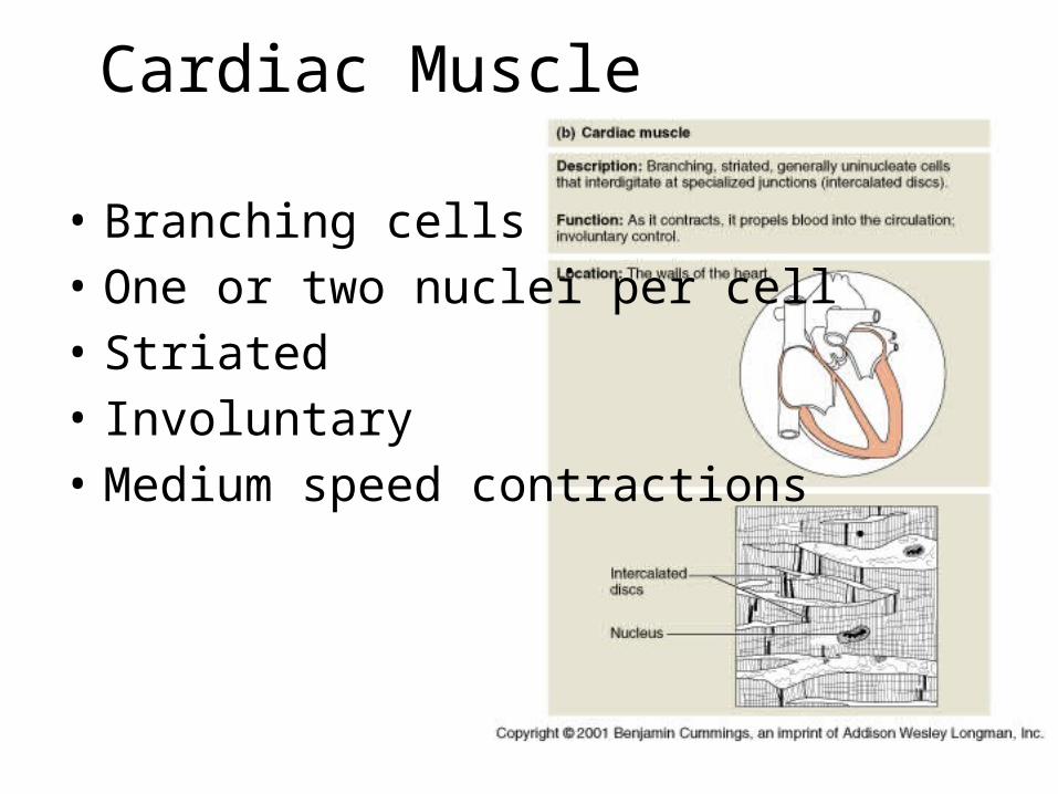
Cardiac Muscle
• Branching cells• One or two nuclei per cell• Striated• Involuntary• Medium speed contractions

Excitation-Contraction Coupling and Relaxation of Cardiac Muscle

Excitation-Contraction Coupling
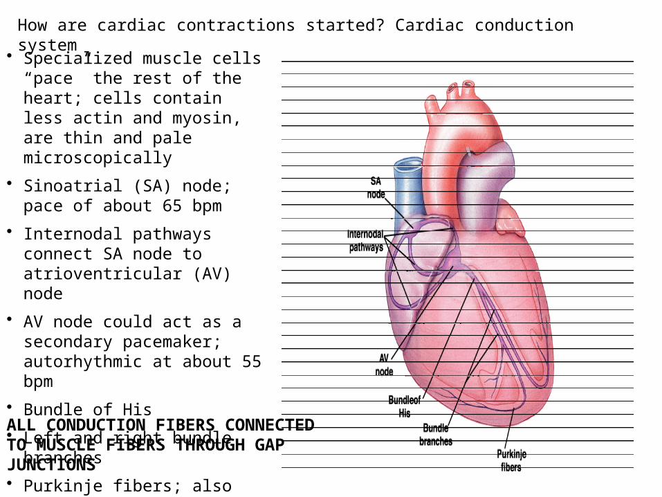
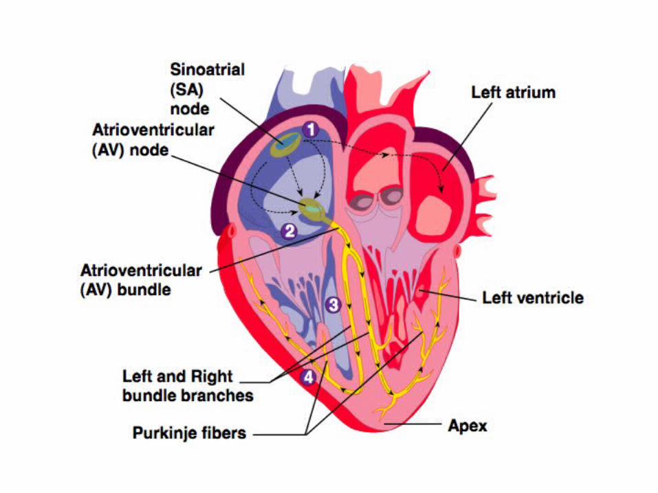
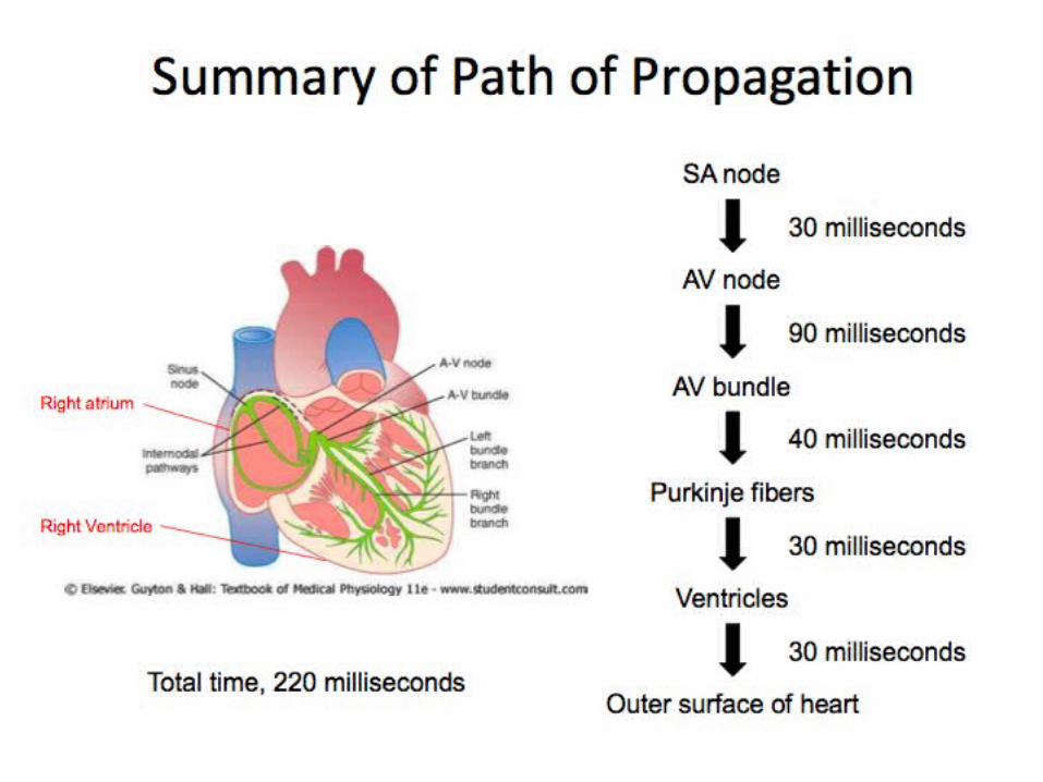
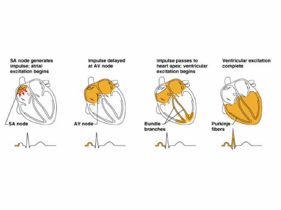
How are cardiac contractions started? Cardiac conduction system
• Specialized muscle cells “pace” the rest of the heart; cells contain less actin and myosin, are thin and pale microscopically
• Sinoatrial (SA) node; pace of about 65 bpm
• Internodal pathways connect SA node to atrioventricular (AV) node
• AV node could act as a secondary pacemaker; autorhythmic at about 55 bpm
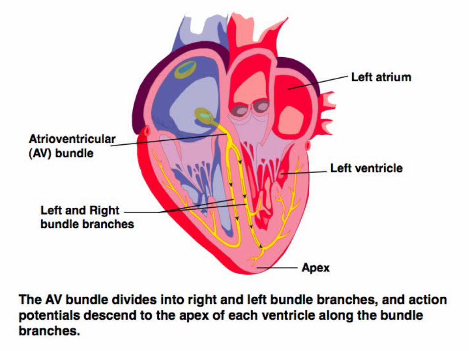
• Bundle of His
• Left and right bundle branches
• Purkinje fibers; also autorhythmic at about 45 bpm
ALL CONDUCTION FIBERS CONNECTED TO MUSCLE FIBERS THROUGH GAP JUNCTIONS
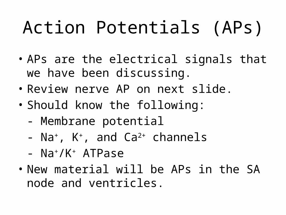
Action Potentials (APs)
• APs are the electrical signals that we have been discussing.
• Review nerve AP on next slide.• Should know the following:
- Membrane potential- Na+, K+, and Ca2+ channels- Na+/K+ ATPase
• New material will be APs in the SA node and ventricles.
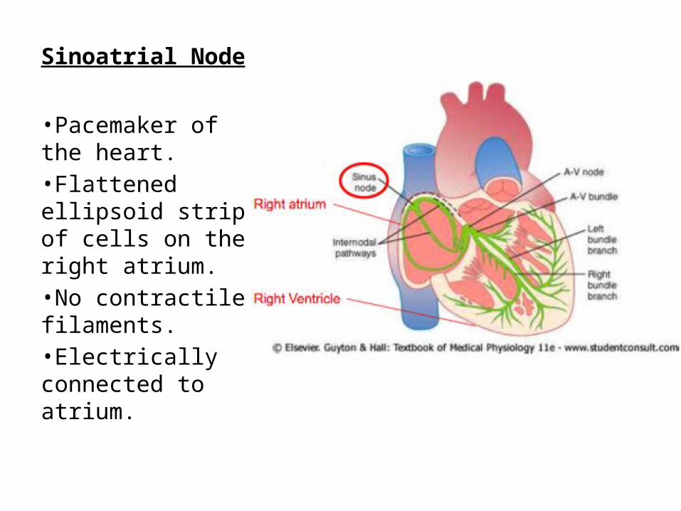
Sinoatrial Node
•Pacemaker of the heart.•Flattened ellipsoid strip of cells on the right atrium.•No contractile filaments.•Electrically connected to atrium.

Sinoatrial Node Action Potential
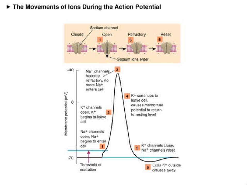
•Phase 4: slow depolarization due to Na+ and Ca2+ leak until threshold. Note fast Na+ channels are inactive at -60 to -40 mV.•Phase 0: at threshold, Ca2+ channels open.•Phase 3: As in nerves, K+ channels open during repolarization.•Finally, note the slow rise and fall of the SA AP compared to that of the nerve AP, and the rhythmic firing.
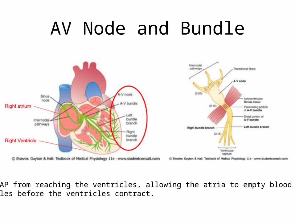
AV Node and Bundle
Delays AP from reaching the ventricles, allowing the atria to empty blood into ventricles before the ventricles contract.

Purkinje Fibres
Receives the AP fromthe AV bundle and rapidly transmits theimpulse through theventricles.

Impulses in Ventricles
•At the termination of the Purkinje fibres, the impulse rapidly travels through the ventricle muscle fibres via gap junctions, from the inside (endocardium) to the outside (epicardium).•The rapid propagation of the cardiac impulse through the Purkinje fibres and ventricles is important for an effective contraction.

Ventricular AP•Phase 4: resting membrane potential near the K+ equilibrium potential.•Phase 0: depolarizing impulse activates fast Na+ channels and inactivates K+ channels.•Phase 1: Transient opening of K+ channels and Na+ channels begin to close.•Phase 2: Ca2+ channels are open, key difference between nerve AP.•Phase 3: repolarization, Ca2+ inactivate and K+ channels open.•Refractory period: Na+ channels are inactive until membrane is repolarized.


1
2
3
4

Sequence of Excitation
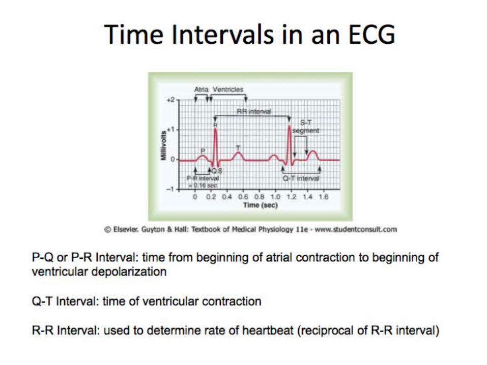
Electrocardiography (EKG)
Examines how Depolarization occurs in the Heart
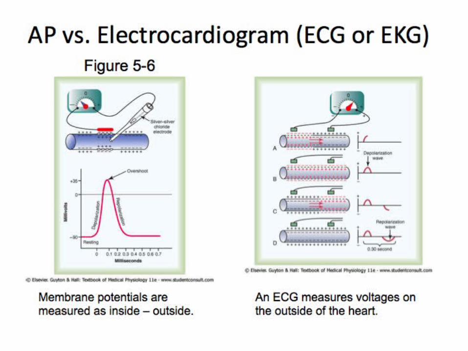

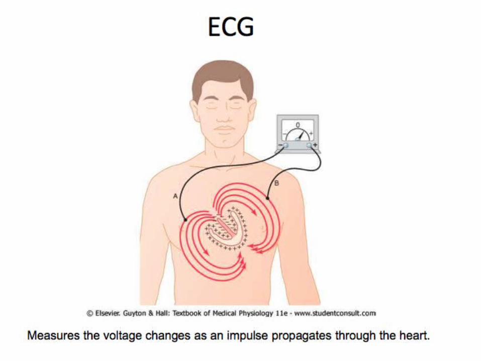
• If a wavefront of depolarization travels towards the electrode attached to the + input terminal of the ECG amplifier and away from the electrode attached to the - terminal, a positive deflection will result.
• If the waveform travels away from the + terminal lead towards the - terminal, a negative going deflection will be seen.
• If the waveform is travelling in a direction perpendicular to the line joining the sites where the two leads are placed, no deflection or a biphasic deflection will be produced.
ECG examines how depolarization events occur in the heart
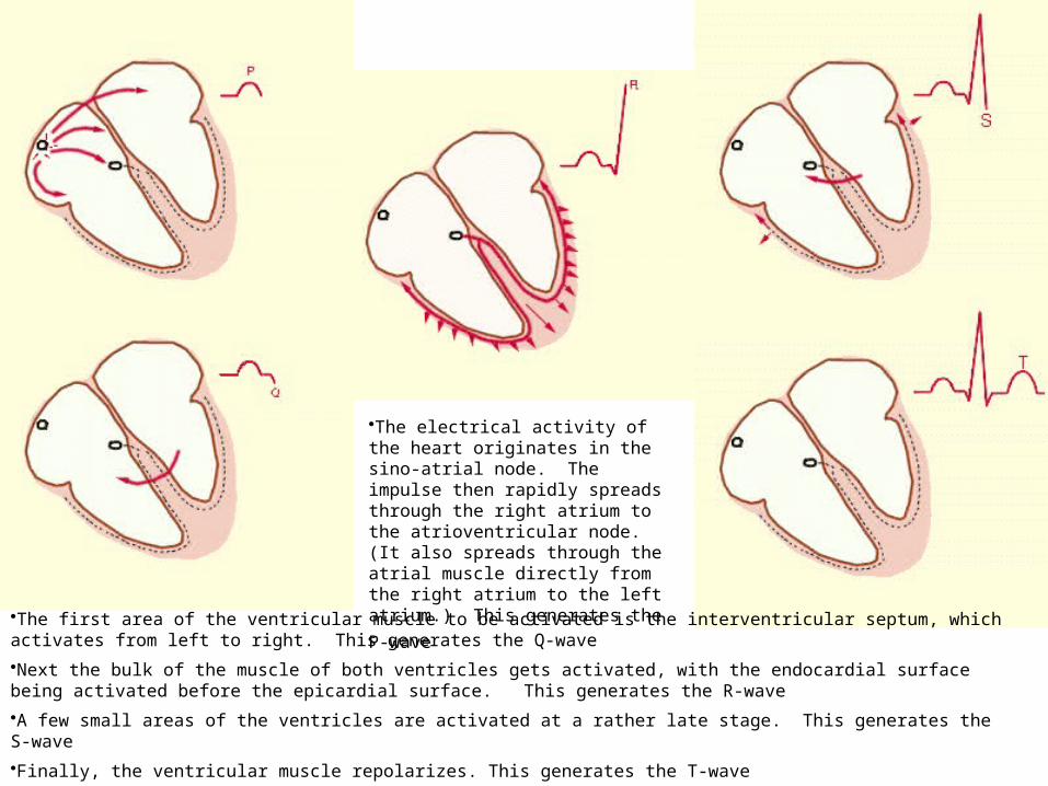
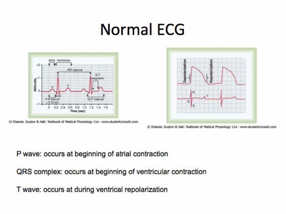
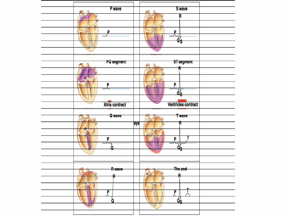
•The electrical activity of the heart originates in the sino-atrial node. The impulse then rapidly spreads through the right atrium to the atrioventricular node. (It also spreads through the atrial muscle directly from the right atrium to the left atrium.) This
generates the P-wave
•The first area of the ventricular muscle to be activated is the interventricular septum, which activates from left to right. This generates the Q-wave
•Next the bulk of the muscle of both ventricles gets activated, with the endocardial surface being activated before the epicardial surface. This generates the R-wave
•A few small areas of the ventricles are activated at a rather late stage. This generates the S-wave
•Finally, the ventricular muscle repolarizes. This generates the T-wave
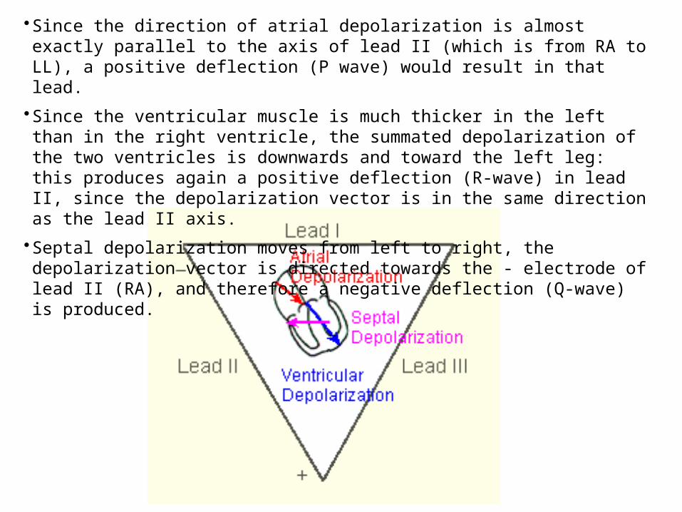
•Since the direction of atrial depolarization is almost exactly parallel to the axis of lead II (which is from RA to LL), a positive deflection (P wave) would result in that lead.
•Since the ventricular muscle is much thicker in the left than in the right ventricle, the summated depolarization of the two ventricles is downwards and toward the left leg: this produces again a positive deflection (R-wave) in lead II, since the depolarization vector is in the same direction as the lead II axis.
•Septal depolarization moves from left to right, the depolarization vector is directed towards the - electrode of lead II (RA), and therefore a negative deflection (Q-wave) is produced.


Electrocardiography
Related Documents