Biofilms in the Food Environment, Second Edition. Edited by Anthony L. Pometto III and Ali Demirci. © 2015 John Wiley & Sons, Ltd. Published 2015 by John Wiley & Sons, Ltd. 1 Current Knowledge and Perspectives on Biofilm Formation and Remediation Lynne A. McLandsborough Department of Food Science, University of Massachusetts, Amherst, MA, USA 1.1 INTRODUCTION Biofilms are formed by almost every type of microorganism under suitable condi- tions. Biofilm food associated organisms include food spoilage microorganisms, such as Pseudomonas sp. and thermophilic sporeformers, and pathogens, including the genera of Bacillus, Cronobacter, Campylobacter, Vibrio, Listeria, Escherichia, and Salmonella (Burgess et al. 2010; Hartmann et al. 2010; Marriott 1999; Poulsen 1999; Sommer et al. 1999). Simplistically, biofilms are microorganisms growing on a solid surface. However, biofilms are generally defined as matrix‐enclosed bacte- rial populations that adhere to a surface and/or to each other producing a dynamic environment in which the component microbial cells appear to reach homeostasis, optimally organized to make use of all available nutrients (An and Friedman 1997; Doyle 2001; O’Toole et al. 2002; Poulsen 1999; Sutherland 2001). Throughout natural ecosystems, biofilms can be found on almost any surface with a high enough level of moisture to support growth (Kim and Frank 1995). Interfaces where biofilms may grow in food processing environments include solid/ liquid, gas/liquid, or in the case of solid foods at the gas/ solid interfaces (Jenkinson and Lappin‐Scott 2001; Poulsen 1999). Over the past 15 or more years, researchers have realized that bacteria growing on surfaces, either alone or in a community containing a diversity of different organisms, have a greater resistance to a large variety of environmental stresses (Costerton 1995; Jessen and Lammert 2003). Thus, the biofilm physiology and organization enables organisms to survive within the food processing environment. In order to control environmental bacterial con- tamination, cleaning and sanitation of this environment is indispensable in order to ensure safety of all commercially produced foods. 1.1.1 General properties of biofilms When growing as a biofilm, bacteria are known to have a different growth rate and physiology than their planktonic (free growing broth cultures) counterparts, and may exhibit varied physiological responses to nutrient conditions (Hodgson et al. 1995; COPYRIGHTED MATERIAL

Welcome message from author
This document is posted to help you gain knowledge. Please leave a comment to let me know what you think about it! Share it to your friends and learn new things together.
Transcript

Biofilms in the Food Environment, Second Edition. Edited by Anthony L. Pometto III and Ali Demirci. © 2015 John Wiley & Sons, Ltd. Published 2015 by John Wiley & Sons, Ltd.
Chapter No.: 2 Title Name: PometoComp. by: EAravally Date: 03 Jul 2015 Time: 07:55:18 AM Stage: Printer WorkFlow:CSW Page Number: 1
1 Current Knowledge and Perspectives on Biofilm Formation and Remediation
Lynne A. McLandsboroughDepartment of Food Science, University of Massachusetts, Amherst, MA, USA
1.1 INTRODUCTION
Biofilms are formed by almost every type of microorganism under suitable condi-tions. Biofilm food associated organisms include food spoilage microorganisms, such as Pseudomonas sp. and thermophilic sporeformers, and pathogens, including the genera of Bacillus, Cronobacter, Campylobacter, Vibrio, Listeria, Escherichia, and Salmonella (Burgess et al. 2010; Hartmann et al. 2010; Marriott 1999; Poulsen 1999; Sommer et al. 1999). Simplistically, biofilms are microorganisms growing on a solid surface. However, biofilms are generally defined as matrix‐enclosed bacte-rial populations that adhere to a surface and/or to each other producing a dynamic environment in which the component microbial cells appear to reach homeostasis, optimally organized to make use of all available nutrients (An and Friedman 1997; Doyle 2001; O’Toole et al. 2002; Poulsen 1999; Sutherland 2001).
Throughout natural ecosystems, biofilms can be found on almost any surface with a high enough level of moisture to support growth (Kim and Frank 1995). Interfaces where biofilms may grow in food processing environments include solid/liquid, gas/liquid, or in the case of solid foods at the gas/ solid interfaces (Jenkinson and Lappin‐Scott 2001; Poulsen 1999). Over the past 15 or more years, researchers have realized that bacteria growing on surfaces, either alone or in a community containing a diversity of different organisms, have a greater resistance to a large variety of environmental stresses (Costerton 1995; Jessen and Lammert 2003). Thus, the biofilm physiology and organization enables organisms to survive within the food processing environment. In order to control environmental bacterial con-tamination, cleaning and sanitation of this environment is indispensable in order to ensure safety of all commercially produced foods.
1.1.1 General properties of biofilms
When growing as a biofilm, bacteria are known to have a different growth rate and physiology than their planktonic (free growing broth cultures) counterparts, and may exhibit varied physiological responses to nutrient conditions (Hodgson et al. 1995;
0002524477.indd 1 7/3/2015 7:55:18 AM
COPYRIG
HTED M
ATERIAL

2 Biofilms in the Food Environment
Chapter No.: 2 Title Name: PometoComp. by: EAravally Date: 03 Jul 2015 Time: 07:55:18 AM Stage: Printer WorkFlow:CSW Page Number: 2
Kim and Frank 1995; Kuchma and O’Toole 2000; O’Toole and Kolter 1998; Sauer et al. 2002). Although gases and liquid nutrients are transported to, from, and through the biofilm matrix via diffusion, studies have indicated that biofilm‐forming bacteria can grow with less oxygen and fewer nutrients than cells in suspension. Surprisingly, this leads to advantages in growth, altered physiology, and increased resistance to a variety of stress compared to their planktonic forms (Fox et al. 2011; Frank and Chmielewski 1997; Frank and Koffi 1990; Sutherland 2001; Vatanyoopaisarn et al. 2000). Through diffusional mass transport, biophysical interactions, and cell‐to‐cell interactions, commensal and mutual communities of organisms survive in the low nutrient and decreased temperature conditions that are often found in food processing and storage environments. The ability to resist anti-microbial agents is of particular concern to both the medical and food processing communities, since once a biofilm has been established on a surface, it becomes exceedingly difficult to clean and sanitize (Bolton et al. 1988; Bower and Daeschel 1999; Bridier et al. 2011; Carpentier and Cerf 2011; Donlan 2002; Donlan and Costerton 2002; Frank and Koffi 1990; LeChevallier et al. 1988; Lowry 2010; Simoes et al. 2010; Tompkin 2002).
1.1.2 Biofilm formation and propagation
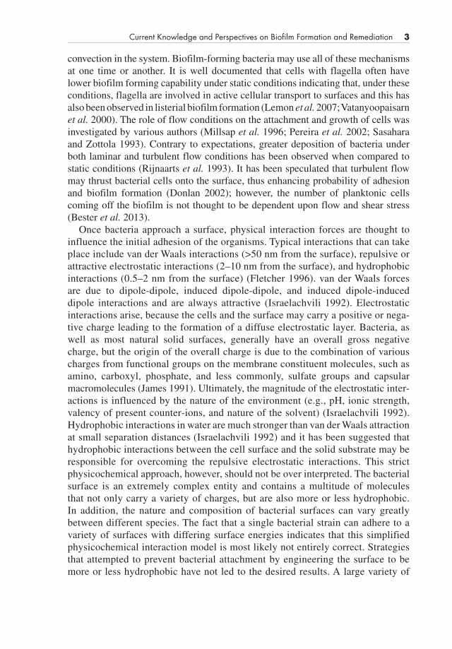
There are several steps in the formation of bacterial biofilms: (1) transport (2) initial adhesion, (3) substrate attachment, and (4) microcolony formation (cell‐cell adhesion) leading to mature biofilms consisting of cells and a surrounding extracellular polymer matrix with the last step being the dissemination or disruption of the bio-film (Figure 1.1) (O’Toole et al. 2002; Purevdorj‐Gage et al. 2005; Simoes et al. 2010; van Loosdrecht et al. 1997). The first step in biofilm formation consists of the transport of the organism to a solid surface. This can occur via motility of the organism, diffusion of the organism through the environment, or natural and forced
Transport/Adhesion Microcolony formation Three dimensional biofilm Dispersal and Re-colonization
ReversibleNon-reversible
Cells and exopolymericmatrix
Figure 1.1 Stages in biofilm formation.
0002524477.indd 2 7/3/2015 7:55:18 AM
Current Knowledge and Perspectives on Biofilm Formation and Remediation 3
Chapter No.: 2 Title Name: PometoComp. by: EAravally Date: 03 Jul 2015 Time: 07:55:18 AM Stage: Printer WorkFlow:CSW Page Number: 3
convection in the system. Biofilm‐forming bacteria may use all of these mechanisms at one time or another. It is well documented that cells with flagella often have lower biofilm forming capability under static conditions indicating that, under these conditions, flagella are involved in active cellular transport to surfaces and this has also been observed in listerial biofilm formation (Lemon et al. 2007; Vatanyoopaisarn et al. 2000). The role of flow conditions on the attachment and growth of cells was investigated by various authors (Millsap et al. 1996; Pereira et al. 2002; Sasahara and Zottola 1993). Contrary to expectations, greater deposition of bacteria under both laminar and turbulent flow conditions has been observed when compared to static conditions (Rijnaarts et al. 1993). It has been speculated that turbulent flow may thrust bacterial cells onto the surface, thus enhancing probability of adhesion and biofilm formation (Donlan 2002); however, the number of planktonic cells coming off the biofilm is not thought to be dependent upon flow and shear stress (Bester et al. 2013).
Once bacteria approach a surface, physical interaction forces are thought to influence the initial adhesion of the organisms. Typical interactions that can take place include van der Waals interactions (>50 nm from the surface), repulsive or attractive electrostatic interactions (2–10 nm from the surface), and hydrophobic interactions (0.5–2 nm from the surface) (Fletcher 1996). van der Waals forces are due to dipole‐dipole, induced dipole‐dipole, and induced dipole‐induced dipole interactions and are always attractive (Israelachvili 1992). Electrostatic interactions arise, because the cells and the surface may carry a positive or nega-tive charge leading to the formation of a diffuse electrostatic layer. Bacteria, as well as most natural solid surfaces, generally have an overall gross negative charge, but the origin of the overall charge is due to the combination of various charges from functional groups on the membrane constituent molecules, such as amino, carboxyl, phosphate, and less commonly, sulfate groups and capsular macromolecules (James 1991). Ultimately, the magnitude of the electrostatic inter-actions is influenced by the nature of the environment (e.g., pH, ionic strength, valency of present counter‐ions, and nature of the solvent) (Israelachvili 1992). Hydrophobic interactions in water are much stronger than van der Waals attraction at small separation distances (Israelachvili 1992) and it has been suggested that hydrophobic interactions between the cell surface and the solid substrate may be responsible for overcoming the repulsive electrostatic interactions. This strict physicochemical approach, however, should not be over interpreted. The bacterial surface is an extremely complex entity and contains a multitude of molecules that not only carry a variety of charges, but are also more or less hydrophobic. In addition, the nature and composition of bacterial surfaces can vary greatly between different species. The fact that a single bacterial strain can adhere to a variety of surfaces with differing surface energies indicates that this simplified physicochemical interaction model is most likely not entirely correct. Strategies that attempted to prevent bacterial attachment by engineering the surface to be more or less hydrophobic have not led to the desired results. A large variety of
0002524477.indd 3 7/3/2015 7:55:18 AM

4 Biofilms in the Food Environment
Chapter No.: 2 Title Name: PometoComp. by: EAravally Date: 03 Jul 2015 Time: 07:55:18 AM Stage: Printer WorkFlow:CSW Page Number: 4
bacterial cells have no difficulty attaching to both hydrophobic and hydrophilic surfaces (Fletcher 1996).
After the initial adhesion occurs, bacteria begin to anchor themselves to the surface by synthesizing extracellular polymeric substances (EPS) that facilitate irreversible bacterial attachment to a surface and help maintain the microcolony and biofilm structure (Doyle 2001; O’Toole et al. 2002; Sutherland 2001; Wimpenny et al. 2000; Wirtanen and Mattila‐Sandholm 1993). Azaredo and Olivera (2000) found that the exopolymers produced by Sphingomonas paucimobilis possess surface‐active properties that aided bacteria in their attachment to hydrophilic surfaces (Azaredo and Oliveira 2000). Interestingly, the presence of pre‐adsorbed proteins on a surface prior to inoculation generally reduced the adhesion of Listeria mono-cytogenes regardless of the surface composition or free energy (Al‐Makhlafi et al. 1994; Al‐Makhlafi et al. 1995; Barnes et al. 1999; Cunliffe et al. 1999), although others have reported that the type of food soil or preconditioning film can influence the final cell density of this organism within the biofilm (Verghese et al. 2011); therefore, there may be a difference upon initial adhesion and ultimate density of biofilm formation. EPS have been shown to enhance nutrient capture and resistance to environmental stress and anti‐microbial agents (Costerton 1995; Costerton et al. 1987; Jenkinson and Lappin‐Scott 2001; O’Toole et al. 2002; Poulsen 1999; Sutherland 2001). When mature, biofilms exist as a structured matrix with a net-work of vertical and horizontal channels to allow liquid flow to supply nutrients and disposal of waste products that are generated as part of the natural respiration activ-ities of cells. The composition and structure of the extracellular polymeric matrix can vary greatly depending on the microorganism(s), their physiological status, the nutrients available, and the physical conditions present (Sutherland 2001). Recently, under flow conditions it was observed that single planktonic cells that were close to the surface move at a slower velocity than the bulk fluid flow, indicating that there may be single planktonic cell growth and co‐existence within the biofilm mass (Bester et al. 2013).
In the food processing environment, biofilms often are harbored in hard‐to‐clean areas such pitted or scratched surfaces, and rough welded joints (Carpentier and Cerf 2011), so that initial adhesion of bacteria may actually be a function of physical entrapment of bacterial cells within rough surface topography. Once adherence occurs, if adequate nutrients and moisture is present, biofilms will be eventually formed. Once biofilms develop, these communities become pockets of microbial social life. There is growing scientific evidence that within a biofilm, bacteria can communicate, interact, and exchange genetic material using a variety of mecha-nisms that include quorum sensing (diffusible extracellular molecules, which function to induce synchronized behavior of a population of bacteria), bacterial conjugation (genetic exchange), bacteriophage (viruses specific for bacteria), and outer membrane vesicles (OMV, membrane sections that can export DNA and other molecules) (Ando et al. 2009; Annous et al. 2009; Bai and Rai 2011; Kulp and Kuehn 2010; Lou et al. 2005; Verghese et al. 2011).
0002524477.indd 4 7/3/2015 7:55:18 AM

Current Knowledge and Perspectives on Biofilm Formation and Remediation 5
Chapter No.: 2 Title Name: PometoComp. by: EAravally Date: 03 Jul 2015 Time: 07:55:18 AM Stage: Printer WorkFlow:CSW Page Number: 5
1.2 BIOFILM DISPERSAL
Mature biofilms can undergo a process known as dispersal. This is the process of individual cells or clumps of cells coming off the biofilm. This dispersal may be passive or an active shift in the biofilm community. In passive dispersal, cells or clumps of biofilms are removed due to sloughing, erosion, or in the case of cross contamination, by transfer. Alternatively, in active dispersal, a portion of immobi-lized cells become active and leave the biofilm. Active dispersal has been observed in a variety of bacterial systems and can be induced by quorum signals, physiological clues, and/or nutrients (McDougald et al. 2012). It is likely that both mechanisms of biofilm dispersal occurs simultaneously, but the exact mode and signals initiating biofilm dissemination will be dependent on the type of bacteria and the growth environment. Regardless of the mechanism of dispersal, this is now recognized as an important stage biofilm development and the source of biofilm spread – thus completing the “life cycle” of biofilms (McDougald et al. 2012).
1.2.1 Active dispersal
Pseudomonas aeruginosa has a well characterized active dispersal system. In this system, as the biofilm matures, cells differentiate into non‐moving cells and moving cells within stalk‐like structures. Non‐moving cells are located in an outer layer or shell that surrounded a center core with highly motile, densely packed cells. Eventually, the moving cells leave the microcolony in an organized swimming fashion termed “seeding dispersal”, leaving behind a hollowed mound of cells (Purevdorj‐Gage et al. 2005; Sauer et al. 2002). The hollowed structures observed in biofilms were produced by rhamnolipid deficient strains (rhlI) but were not seen in a rhamnolipid, quorum sensing double mutants (lasI, rhlI) indicating that quorum sensing is involved in this dispersal (Purevdorj‐Gage et al. 2005).
1.2.2 Passive dispersal
In passive dispersal, cells or clumps of biofilms are physically removed biofilm due to sloughing, erosion, or by transfer to foods. The physical removal is dependent upon the composition and structure of the extracellular polymeric matrix, which can vary greatly depending on the microorganism(s), their physiological status, the nutrients available, and the physical conditions present (Sutherland 2001). At sufficiently high concentrations of exopolymers, the biofilm begins to exhibit a gel‐like character. Rheological characterization of the biofilm yielded a notice-able increase in the elastic modulus of the biofilm (Kapper et al. 2002). Overall, the rheological behavior of the film depends on superimposed flow conditions. In laboratory systems, for example, the structure of P. aeruginosa biofilm has shown to assume a denser and more streamlined configuration under turbulent flow with a semicircular appearance that offers little resistance to flow. Additional
0002524477.indd 5 7/3/2015 7:55:18 AM

6 Biofilms in the Food Environment
Chapter No.: 2 Title Name: PometoComp. by: EAravally Date: 03 Jul 2015 Time: 07:55:18 AM Stage: Printer WorkFlow:CSW Page Number: 6
growth occurred primarily in the direction of flow and ripple‐like structures were occasionally formed. Under laminar flow, the same organism formed flat mono-layers with rough surface topologies and circular, hemispherical colonies only rarely formed (Purevdorj et al. 2002).
In laboratory systems, microcolonies or detached portions of biofilms have been observed to roll on surfaces within the flow. Rupp et al. (2002) took time lapse images of Staphylococcus aureus microcolonies slowly moving along with the liquid flow rolling across the surface of a glass capillary. As the microcolony rolled, multiple attachment events were observed in the direction of the flow. Localized detachment from the surface occurred behind the colony, as it tears away from flexible cellular tethers. Thus, in this pure Staph. aureus system, the biofilm was observed to move along with the fluid flow without detachment and the majority of cells remained in an organized state in order to retain the advantages of being located within a biofilm. It is not yet known if clumps and single cells have similar surface characteristics or adhesive qualities, but it is likely that the mode of dissem-ination may be species specific and may always consist of a combination of both single cell and clumping events.
1.2.3 Dispersal by transfer
Transfer of bacterial cells or biofilm clumps from one surface to another is another mechanism of dispersal. Contamination of L. monocytogenes from food processing equipment and/or the processing environment is thought to be a critical mode of listerial contamination in processed foods. In processing plants, common sites of post‐processing L. monocytogenes contamination are filling or packaging equip-ment, conveyors, collators used for assembling product for packaging, racks for transporting product, hand tools, gloves, and freezers (Poulsen 1999; Tompkin et al. 1999). Its presence in ready‐to‐eat meals is likely caused by recontamination of the product after processing prior to packaging (Lunden et al. 2002). Detachment and transfer of bacteria from processing surfaces to foods and vice versa, can occur during and after processing and this represents the most common route of recon-tamination of processed foods. Several studies have focused on the transfer of bacteria and viruses with the majority of the work in this area focused on the transfer of L. monocytogenes from processing surfaces to foods (Keskinen et al. 2008; Midelet and Carpentier 2002; Midelet et al. 2006; Rodriguez et al. 2007a, b; Rodriguez and McLandsborough 2007; Vorst et al. 2006a, b). There is an intrinsic variability when evaluating transfer of bacteria from one surface to another (D’Souza et al. 2006; Flores et al. 2006; Keskinen et al. 2008; Montville and Schaffner 2003; Rodriguez et al. 2007a, b; Rodriguez and McLandsborough 2007; Sattar et al. 2001; Vorst et al. 2006a, b; Zhao et al. 1998). This variability makes comparison of transfer experiments difficult. Most of this variability comes from several factors: differences in methodology, surfaces used for transfer, inoculation method, data analysis, and hydration level prior to transfer.
0002524477.indd 6 7/3/2015 7:55:18 AM

Current Knowledge and Perspectives on Biofilm Formation and Remediation 7
Chapter No.: 2 Title Name: PometoComp. by: EAravally Date: 03 Jul 2015 Time: 07:55:18 AM Stage: Printer WorkFlow:CSW Page Number: 7
It is important to notice that one of the main parameters that brings variability to the transfer research is the method used. Whereas each research group uses a differ-ent methodology, listerial transfer work done to date can easily be divided into two groups depending on the predominant force used behind the transfer designs: normal force versus shear force. Most of the published work has been conducted using a normal force (perpendicular to the contact surface), in which a certain amount of weight has been placed on top of a food product or a food contact surface after a set amount of time (D’Souza et al. 2006; Flores et al. 2006; Keskinen et al. 2008; Montville and Schaffner 2003; Rodriguez et al. 2007a, b; Rodriguez and McLandsborough 2007; Sattar et al. 2001; Vorst et al. 2006a, b; Zhao et al. 1998). The pressure applied can be easily calculated by dividing the force applied by the contact surface area. A second type of experimental design involves the use of additional forces, shear forces, where the force applied is tangential or parallel to the food; this is commonly done with the use of a cutting blade or by bringing knife perpendicular to the food where shear forces are generated as the knife advances through the food (Keskinen et al. 2008; Vorst et al. 2006a, b). This latter design makes pressure calculation more difficult, and shear forces have often been ignored when evaluating listerial transfer.
In addition to force types, the degree of force, contact time, materials, inocula-tion method, and the degree of hydration will influence transfer levels. The hydration level of surfaces or inoculated surfaces has been shown to be important for other bacterial species than Listeria when used in completely different systems (Flores et al. 2006; Rodriguez, 2007; Rodriguez et al. 2007a, b; Sattar et al. 2001). Rodriguez et al. (2007a) evaluated the transfer of listerial biofilms grown on stainless to foods (bologna and hard salami) controlling the exact level of dryness of the biofilms by equilibrating them to known percentage values of relative humid-ities of 96, 75, 56, and 33% for a period of 24 h. The results of this work showed that as biofilms were dried, listerial transfer increased to both foods tested. More interestingly was to find that the degree water activity (a
w) of the food system used
did also influence the transfer. Listerial biofilms grown on stainless steel did transfer more to foods with higher water activity (bologna, a
w = 0.96) compared to food with
lower water activity (hard salami, aw = 0.83) (Rodriguez et al. 2007a).
Based on what is known, the factors important in bacterial transfer from one surface to a food are; (1) the adhesion between the cells and the initial supporting surface, (2) the cell‐to‐cell adhesion within the biofilm, and (3) the attraction bet-ween the cells and the food. From research done in my lab there is no doubt that water is an important parameter in transfer. We have measured increasing transfer as biofilms were equilibrated to lower water (Rodriguez et al. 2007a; Rodriguez and McLandsborough 2007). This indicates that water in the biofilm can affect the transfer (Rodriguez et al. 2007a) either by reducing cell‐to‐cell adhesion and/or reducing cell‐to‐surface adhesion. The level of water in the food also appears to influence the transfer of Listeria, since we’ve observed higher transfer of dried biofilms, to foods with higher water activities (Rodriguez et al. 2007a). As anyone
0002524477.indd 7 7/3/2015 7:55:18 AM

8 Biofilms in the Food Environment
Chapter No.: 2 Title Name: PometoComp. by: EAravally Date: 03 Jul 2015 Time: 07:55:18 AM Stage: Printer WorkFlow:CSW Page Number: 8
who has ever built a sand castle can attest, water can act as a strong capillary force holding a sand structure together; however, as the structure dries, the castle shape loses cohesion and rapidly turns back into a pile of sand that can be easily dispersed. In a similar sense, we propose that water may be a critical component in holding EPS together in a listerial biofilm and/or holding the bacterial cells on the surface. Water in the food may also facilitate transfer by “wicking” the bacteria from a drier surface to a moist food. Understanding the fundamental steps in bacterial transfer from surfaces to foods may help us design food processing facilities that may reduce the possibility of cross contamination with this organism.
1.2.4 Single versus multiple species biofilms
The vast majority of what is known about biofilms is based upon research that was performed with a single species in simplified laboratory systems. The two most studied biofilm systems use pure cultures of the clinical organisms P. aeruginosa or Staph. aureus, as model Gram‐negative and Gram‐positive systems, respectively (Hall‐Stoodley et al. 2004). In most foods and in food processing environments, the biofilms present will be significantly more complex (i.e., they may be composed of multiple species that form a community of microorganisms and that may or may not behave in a similar manner as pure laboratory systems). Similarly, studies of biofilms under laboratory conditions usually do not take into account that biofilms in food processing environments may contain proteins and fats derived from improp-erly cleaned or sanitized processing areas as integral part of their structure. Experimental evidence suggests that the formation of a multispecies biofilm is advantageous. For example, higher numbers of L. monocytogenes were counted in biofilms that were co‐cultured with Pseudomonas (Hassan et al. 2004; Sasahara and Zottola 1993). In other cases, the adhesion of L. monocytogenes is limited in the presence of other bacteria (Leriche and Carpentier 2000; Norwood and Gilmour 2001). Chae and Scharft (2001) showed that the rate of biofilm formation for L. monocytogenes differs from their planktonic growth (Chae and Schraft 2000, 2001). To truly understand the survival of human pathogens within the food processing environment, the influence of other bacteria should be better understood.
1.3 PROPAGATION OF BIOFILMS
Researchers planning to investigate biofilms face many difficult choices, from the selection of a suitable model system to the techniques used to characterize the struc-ture and behavior of the grown biofilm. Many different types of laboratory‐based model systems for microbial biofilms can be found in the scientific literature. Unfortunately, none of them can be considered to be the one optimal model system that is universally applicable. On the contrary, the researcher must choose the particular model system that is able to give specific answers to questions that were formulated at the beginning of the study. The accurate simulation of conditions that
0002524477.indd 8 7/3/2015 7:55:18 AM

Current Knowledge and Perspectives on Biofilm Formation and Remediation 9
Chapter No.: 2 Title Name: PometoComp. by: EAravally Date: 03 Jul 2015 Time: 07:55:18 AM Stage: Printer WorkFlow:CSW Page Number: 9
are encountered in a processing environment is obviously an exceedingly difficult task due to variations in the nature of the food process operation, the natural micro-flora that may be present, and the food product that is being processed. For these reasons, the researchers prior to designing an experiment must make several key decisions. For example, what microorganisms should be used to grow the biofilm (single versus multispecies), under what growth conditions are the biofilm to be incubated (bacteriological media composition, pH, and temperature), what material is to be used to provide the substrate surface, and what are the basic surface charac-teristics (surface roughness, hydrophobicity, and charge)? If the biofilm is to be grown under flow conditions in a reactor, other parameters such as the sheer stress, flow velocity, and retention time need to be taken into account (Lewandowski and Beyenal 2014). The surface may be positioned vertically or horizontally, which will have important consequences, since sedimentation may be involved if a horizontal surface is used. Rinsing and drying procedures are an important part of any bacterial adhesion study and are required to remove unattached (planktonic) or loosely attached bacteria. Finally, how will the biofilm be characterized? Will the biofilm be observed intact using microscopy (electron, confocal or light) or will the biofilm be removed from the substrate? Microscopy gives information about community structures and spatial interaction; however, the area of observation is so small, it gives little relative quantitative data. If you want to understand a more quantitative assessment, oftentimes the biofilm needs to be removed from the substrate and the researcher faces a choice of methods for removal; high‐intensity ultrasound, surfac-tants or simple mechanical forces may be used. In this part of the review, a few of the many available biofilm growth techniques will be highlighted, and then characteriza-tion techniques that may yield important information about the structure, mechanical behavior and composition of biofilms will be briefly discussed. Methodological information can be found in protocol books (Tolker‐Nielsen and Sternberg 2011), and a recent book by Lewandowski and Beyenal (2014) has in‐depth information on experimental design, interpretation, and modeling.
1.3.1 Methods to grow biofilms in laboratories
1.3.1.1 Microtiter plate assay
The microtiter plate assay is one of the most commonly used methods for estimation of growth of biofilms (Cole et al. 2004; Djordjevic et al. 2002; Merritt et al. 2011; Stepanovic et al. 2004). This method is an estimation of the level of biofilm that can be achieved under static conditions (without the addition of fresh nutrients), however, are advantageous for studies that need a high level of screening, either for genetic studies or for screening parameters that may influence biofilm production. A drawback to this assay is that crystal violet may stain both cells and exopolymeric matrix components on the plate, and overestimate the actual cell numbers. Thus a secondary method (either cell removal and counting, or microscopy) is often used to validate microtiter plate results (Djordjevic et al. 2002).
0002524477.indd 9 7/3/2015 7:55:18 AM

10 Biofilms in the Food Environment
Chapter No.: 2 Title Name: PometoComp. by: EAravally Date: 03 Jul 2015 Time: 07:55:18 AM Stage: Printer WorkFlow:CSW Page Number: 10
Microtiter plates are composed of a polymer, such as polyvinylchloride (PVC) or polystyrene, and consist of 96 wells that can be filled with up to 0.2 ml of inoculated broth. For adhesion experiments, the wells are inoculated with bacteria and allowed to grow under the appropriate conditions for the organism being tested. After the incubation period, the liquid is removed and the wells containing the biofilm are washed with a buffer solution often up to four times. The biofilm may then be dried to fix it followed by staining with a stain such as crystal violet, followed again by rinsing with water and drying. The dry plates are then de‐stained using a solvent (ethanol, acetic acid, or dimethyl sulfoxide: DMSO) and the level of stain present is determined using a microtiter plate reader. Microtiter plate assays have been used to investigate biofilm formation by different bacteria such as Salmonella spp., L. monocytogenes, Helicobacter pylori (Cole et al. 2004; Djordjevic et al. 2002; Stepanovic et al. 2004), and fungus such as Candida albicans (Ramage et al. 2001). The microtiter plate method has shown to be useful in the genetic analysis of biofilm formers because of the high number of experiments that can be conducted simultaneously in the 96 wells of the plates (Chang et al. 2012a; Chang et al. 2012b; Merritt et al. 2011; O’Toole and Kolter 1998). This assay is also well suited to study the early stages of biofilm formation that involve colonization and initial biofilm structure development (McLean et al. 2004). However, the choice of substrate surface materials is limited since the plates have to be optically transparent. In addition, the accuracy of determining cell numbers is low, in particular at higher cell concentrations due to multiple scattering effects. A variation of the standard microtiter assays is known as the Calgary biofilm device (Ceri et al. 1999). It was designed for rapid and reproducible assays of P. aeru-ginosa, Staph. aureus, and Escherichia coli biofilms to assess susceptibility to anti-biotics. The device produces 96 equivalent biofilms using the standard 96‐well technology, with a plastic peg as substratum for the biofilm (Ceri et al. 1999). An advantage of using a Calgary biofilm device is that pegs can be removed and added to a culture tube to determine numbers of adherent bacteria (Harrison et al. 2008).
In order to determine levels of viable and non‐viable bacteria within a microtiter plate, fluorescent stains, such as Syto 9, can be used to detect biofilm levels (Peeters et al. 2008) or combined with propidium iodine (PI), to detect non‐viable cells (such as the BacLightTM stain commercially available Life Technologies, Grand Island, NY). However, a potential problem with using PI is that it can overestimate level of dead bacteria due to the presence of extracellular DNA within the extracel-lular matrix (Jakubovics et al. 2013; Whitchurch et al. 2013). The respiratory indicator stain 5‐cyano‐2, D‐ditolyl tetrazolium chloride (CTC) is an alternative to evaluate bacterial viability in a microtiter plate system (Pitts et al. 2003).
1.3.1.2 Colony biofilms grown on polycarbonate membranes
In this fairly simple experimental method, a planktonic culture of the target bacteria is first grown and a drop of the culture containing the desired initial inoculum level placed on a sterile, black, polycarbonate membrane filter, which rests on an agar
0002524477.indd 10 7/3/2015 7:55:18 AM

Current Knowledge and Perspectives on Biofilm Formation and Remediation 11
Chapter No.: 2 Title Name: PometoComp. by: EAravally Date: 03 Jul 2015 Time: 07:55:18 AM Stage: Printer WorkFlow:CSW Page Number: 11
plate (Anderl et al. 2000; Borrielo et al. 2004; Merritt et al. 2011). The agar plate is inverted and incubated to allow for growth of the biofilm on the polycarbonate membrane. Daily, the polycarbonate filters are transferred to a fresh agar plate to allow for renewal of nutrients (Merritt et al. 2011). Once grown, the colony can be placed on a filter paper saturated with antimicrobial solutions – thus, allowing for studies of the transport of antimicrobials into the center of an organized bacterial colony. For viable bacteria enumeration, each membrane‐supported biofilm is added to diluent and vortexed in tubes to detach the biofilm and serial dilutions are plated onto appropriate agar plates (Anderl et al. 2000; Borrielo et al. 2004). This tech-nique is commonly followed by florescent staining (similar to that mentioned under microtiter plate staining), followed by cryoembedding of the biofilm to allow exam-ination of thin section (2 μm) via microscopy (Werner et al. 2004), allowing for a full observation of the colony, from the base membrane touching the antimicrobial, to the colony surface. There has been some critique of this technique stating that it does not represent conditions under which biofilms typically grow in nature. Wentland et al. (1996) grew a Klebsiella pneumoniae biofilm on polycarbonate filters and stained it with acridine orange. They found different color intensities that were believed to be related to the different metabolic states of the cell, although the corre-lation was not very strong. Biofilms grown under these conditions are deemed to be less representative of natural biofilms. However, in studies that investigated biofilm susceptibility to disinfectants and surfactants, and that were conducted with the same organisms on both polycarbonate membranes and biofilm reactors, have shown good agreement between the two methods (Chen and Stewart 2000). Because of the ease of preparation, polycarbonate membranes are particularly useful in screening studies.
1.3.1.3 Laboratory biofilm reactors
Biofilm reactors generally allow for surface growth of biofilm under continuous flow conditions. As discussed at the beginning of this section, in setting a biofilm reactor in the laboratory, researchers are making decisions on experimental parameters that may or may not reflect or imitate what may happen in a natural biofilm formed outside of the laboratory, such as the grown of a pathogen (e.g., L. monocytogenes) in a floor drain among a variety of non‐pathogenic strain microbiota.
1.3.1.4 Capillary biofilm reactors
Capillary biofilm reactors consist of one or more glass capillary tubes where bio-film may grow under continuous flow conditions. The glass tubes have a square cross section to allow for direct microscopic observation. The capillary cells are mounted in a flow cell holder to minimize the risk of breakage. The flow cell is connected to a vented feed carboy that contains the medium, a flow break, a filtered air entry, and a peristaltic pump. The system is also fitted with an inoculation port and a waste carboy. It has been reported that mixing of the fluid with air in the
0002524477.indd 11 7/3/2015 7:55:18 AM
12 Biofilms in the Food Environment
Chapter No.: 2 Title Name: PometoComp. by: EAravally Date: 03 Jul 2015 Time: 07:55:18 AM Stage: Printer WorkFlow:CSW Page Number: 12
peristaltic pump may aid in the development of some biofilms (e.g., P. aeruginosa) (Werner et al. 2004). For the inoculation, the flow is stopped and the downstream tubing is clamped. The culture is injected via the port to fill the glass capillary. The upstream tubing is then clamped and the system is allowed to stand without flow for a specific amount of time. After attachment and initial growth, flow may be initiated at varying flow rates. Biofilms may be counterstained by injecting a solution of rhodamine B into the capillary to allow for confocal scanning laser microscopy (CSLM) (Christensen et al. 1999) or may be visualized by using strains engineered to express green fluorescent protein (Rani et al. 2007). Capillary systems are often used to evaluate growth of biofilms using microscopy.
1.3.1.5 Flow cell reactor
Like most reactors, the external setup is similar to that of the capillary biofilm reactor and consists of a carboy holding the medium, a flow pump to regulate medium flow and a waste carboy to collect the spent medium. The flow cell itself has a semi‐circular cross‐section and contains seven removable slides (stainless steel slides glued on rectangular pieces of Perspex that properly fit in the apertures of the flow cell) that allow sampling of biofilm at desired time intervals. This type of reactor has been used to observe biofilm growth of Pseudomonas fluorescens with a superimposed laminar or turbulent flow profile (Pereira et al. 2002).
1.3.1.6 Rotating disk reactor
This reactor consists of one or more disks, with several removable slides per disk that allow for sampling of biofilms. The discs are rotated by a connected motor. Rotational speeds may be adjusted to simulate different flow conditions (Lewandowski and Beyenal 2014). This system has been specifically used to grow Gram‐negative bacteria biofilm, using Teflon coupons as substratum (Donlan et al. 1999) in order to test the activity of sulfate‐reducing bacteria in aerobic wastewater biofilms (Okabe et al. 1999).
1.3.1.7 CDC (Center for Disease Control) biofilm reactor (CBR)
The CBR is one of the most versatile reactor systems and was developed by Donlan et al. (2004). It incorporates 24 removable biofilm substrate surfaces, also known as coupons (eight independent rods with three chips per rod) that are grown inside a jacketed vessel with an effluent spout, which is connected to the waste bottle (Donlan 2002). The jacket is connected through two ports to an external water bath that can be used to regulate the temperature. A continuous mixing of the fluid may be ensured through a magnetically driven baffled stir bar. Each rod may be removed at a given time to access the coupon with the sample biofilm. CBRs have been used to continually monitor the formation of biofilms, and characterize their structures
0002524477.indd 12 7/3/2015 7:55:18 AM

Current Knowledge and Perspectives on Biofilm Formation and Remediation 13
Chapter No.: 2 Title Name: PometoComp. by: EAravally Date: 03 Jul 2015 Time: 07:55:18 AM Stage: Printer WorkFlow:CSW Page Number: 13
(Donlan et al. 2002; Goeres et al. 2005). The larger number of chips (24) allow for multiple sample treatments to assess the effectiveness of antimicrobial substances for bioremediation of mature biofilms (Pérez‐Conesa et al. 2011).
1.4 CHARACTERIZATION OF BIOFILMS
Once a biofilm has been grown in the lab, there are a number of methods to charac-terize your biofilm. Depending on the methodology, this can be done on intact biofilms, or the biofilm may be removed from the surface (scraping or sonication are common methods) and the suspension can be used to determine viable cell num-bers (the old “scrape and plate” trick) or used for other analysis (e.g., extraction and analysis of EPS).
Intact biofilm analysis is often performed using microscopy. If the supporting surface is clear (as on a capillary tube or glass slide) optical microscopy using differential interference contrast (DIC) can be used to image cells (Lewandowski and Beyenal 2014) or even a simple stain such as crystal violet can be used (Djordjevic et al. 2002). More often, fluorescent microscopy is used to image biofilms using either using epifluorescence or CSLM. Confocal microscopy allows for imaging at depths, thus allowing for combination of images to generate 3D composites (Lewandowski and Beyenal 2014). Fluorescent imaging can be done using bacteria engineered to express fluorescent proteins, such as green fluorescent protein (GFP), staining with fluorescent stains for specific chemical components (DNA, proteins, lipids, or carbohydrates), or using fluorescent DNA probes for in situ hybridization (FISH) (Lewandowski and Beyenal 2014; Tolker‐Nielsen and Sternberg 2011). Often these imaging techniques can be with image analysis programs such as COMSTAT (Heydorn et al. 2000a; Heydorn et al. 2000b), to determine the surface area covered and the distribution, biovolume, thickness of the biofilm, and the roughness coefficient (Heydorn et al. 2000a, b; Tolker‐Nielsen and Sternberg 2011).
Both transmission electron microscopy (TEM) and scanning electron microscopy (SEM) have been used to examine biofilms since both offer extremely high resolu-tion, but because they operate in a high vacuum, the samples need to be fixed and dehydrated by using graded solvents such as alcohol, acetone, and xylene (McLean et al. 2004). The preparation of the samples (dehydration and staining) irrevocably changes the structure of the biofilm, which limits the applicability of the technique (Donlan 2002). In addition, the preparation is time consuming and because of this, SEM or TEM analysis is not conducted on a routine basis. Nevertheless, TEM has been used to characterize the structure of the extracellular polymer matrix in bio-films grown on medical devices using ruthenium red as a dye (Raad et al. 1993) and to enumerate the stratified growth in P. aerugionsa biofilms (Werner et al. 2004). SEM has been used to study the spatial distributions of L. monocytogenes cells attached to ready‐to‐eat meats (Foong and Dickson 2004), determine the effect of
0002524477.indd 13 7/3/2015 7:55:18 AM

14 Biofilms in the Food Environment
Chapter No.: 2 Title Name: PometoComp. by: EAravally Date: 03 Jul 2015 Time: 07:55:18 AM Stage: Printer WorkFlow:CSW Page Number: 14
exposure to antibiotics or sanitizers on biofilm integrity (Ceri et al. 1999; Chavant et al. 2004) and follow formation of biofilms from H. pylori (Cole et al. 2004), Salmonella (Wang et al. 2013), and C. albicans (Ramage et al. 2001).
Microscopy can be combined with Raman spectroscopy or surface‐enhanced Raman scattering (SERS) to understand the chemical composition of biofilms (Chao and Zhang 2012; Efeoglu and Culha 2013; Wang et al. 2013). To perform analysis with SERS, the bacteria be mixed with nanoparticles of metals, such as silver or gold, which enhances the Raman scattering and allows for identification and quantification of components within the biofilm. Recently, SERS was used to study the chemical composition of E. coli, Pseudomonas putida and Bacillus subtilis biofilm formation (Chao and Zhang 2012). Using this technique, spectrum during development phases of biofilm production (attachment, microcolonies, and mature biofilm) can be measured and in the mature biofilm all strains had increases in levels of nucleic acid, carbohydrates, proteins, and the Gram‐negative strains also showed accumulation in lipid compounds (Chao and Zhang 2012), which may be due the contribution of the outer membrane to the EPS structure.
Atomic force microscopy (AFM) is a physical microscopy that can be used both to image intact biofilms as well study the rheological characteristics (Chao and Zhang 2012; Rodriguez et al. 2008; Wright et al. 2010). AFM utilizes a small silicon nitride or silicon tip that is mounted on a cantilever that is then scanned across the surface of the sample and is capable of imaging surfaces at nanometer or sub‐nanometer resolutions (Beech et al. 2002). The use of AFM to visualize biofilms has been pioneered by Bremer et al. (1992) who sought to understand biofilms‐induced deterioration of a variety of materials. An example of contact mode AFM image is presented in Figure 1.2. AFM can be used to measure the
=1μm
(a) (b)
Figure 1.2 Microcolony of Listeria monocytogenes growing on stainless steel. This data was collected using contact mode AFM to scan the topography of the surface. (a) A 2D projection with darker pixels indicating lower topography and lighter pixels representing higher topography across the scanned surface. (b) The same data presented as a 3D projection, the curvature of the surface is a common artifact associated with AFM imaging.
0002524477.indd 14 7/3/2015 7:55:18 AM

Current Knowledge and Perspectives on Biofilm Formation and Remediation 15
Chapter No.: 2 Title Name: PometoComp. by: EAravally Date: 03 Jul 2015 Time: 07:55:18 AM Stage: Printer WorkFlow:CSW Page Number: 15
attractive and pull‐off forces of individual bacterial cells to a variety of materials (Rodriguez et al. 2008). Rodriguez et al. (2008) used this method to show that the maximum pull off force for polyethylene was significantly higher than silicon dioxide probes from L. monocytogenes biofilms, suggesting that the biofilm adhesion to hydrophobic surfaces was stronger than a hydrophilic surface when measured at a cellular level. AFM has also been used to measure the cohesive forces within a biofilm by scan‐induce abrasion technique. In this technique a topographic image of the biofilm is obtained at a low applied pressure (setpoint 1 nN), which allows for imaging without disrupting the biofilm structure. Then, an area within this original scan can be subjected to repeated raster scanning at a much higher pressure for specific number of scans (basically scraping away a region of the biofilm). Once again, the original larger area can be imaged. Based upon the volume of biofilm that was removed and along with the set point and the calibrated spring constant of the AFM tip, the cohesive energy/μm3 of the biofilm was able to be calculated (Ahimou et al. 2007).
1.5 REMEDIATION STRATEGIES
1.5.1 Detection of biofilms in the food environment
For quality control purposes, there are a number of reasons to look for and mon-itor bacteria in the food processing environment. General culturing (e.g., stan-dard plate counts) may be used to assess and monitor the process of cleaning and sanitation scheme. Alternately, if there is specific recurrent problem spoil-age organism(s) in a processed food, the quality department may sample the processing environment looking for an environmental source. Culturing may also be used as part of environmental monitoring for a HACCP plan. For example, the USDA Food Safety and Inspection Service (FSIS) requires under regulation 9 CFR 430.4, that ready‐to‐eat meat processors that do not have a post‐lethality treatment and antimicrobial additive in their product, to monitor for the presence of L. monocytogenes or an indicator organism (i.e., Listeria spp.) in the post‐lethality processing environment (Food Safety and Inspection Service 2008).
When sampling bacteria from the environment, the sampling results are only as accurate as the number of samples taken and the sampling locations. In processing plants, common sites of post‐processing L. monocytogenes contamination are filling or packaging equipment, conveyors, collators used for assembling product for pack-aging, racks for transporting product, hand tools, gloves, and freezers (Poulsen 1999; Tompkin 2002; Tompkin et al. 1999). Bacterial harborage sites are often places that are difficult to get to and clean – and by definition these sights are also locations that are very difficult to sample, especially on a regular basis. Thus, there is often a possibility of false negative results – in that Listeria spp. is present in the environment, but the sampling plant did not find the harborage site. Floor drains are
0002524477.indd 15 7/3/2015 7:55:18 AM

16 Biofilms in the Food Environment
Chapter No.: 2 Title Name: PometoComp. by: EAravally Date: 03 Jul 2015 Time: 07:55:18 AM Stage: Printer WorkFlow:CSW Page Number: 16
often a location where there is an accumulation of cleaning run‐off and food waste, therefore, can be a location that are common harborage sites of pathogens such as L. monocytogenes (Cao et al. 2006; Tompkin et al. 1999).
The detection of bacteria within the environment can be performed by using sterile swabs or sponges moistened in buffer and swabbing surfaces within the processing environment. With either test, it’s important to collect the sample in a buffer that contains the appropriate neutralizing agent for the disinfectants used in the processing plant. Commonly used neutralizing agents include: polysorbate 80 and lethicin for phenolics and quaternary ammonium sanitizers (QUATS), sodium thiosulfate for halogen bases sanitizers (sodium hypochlorite and chlorine dioxide), and sodium bisulfite for glutaraldehyde based sanitizers. Alternatively, a universal neutralizing buffer or broth can be used such as Dey/Engley (D/E) buffer, which contains a mixture of neutralizing agents. Once bacteria have removed from the swab, by mixing in buffer using a culture tube mixer, or from sponges, by agitation in a bag with buffer within a laboratory paddle blender such as a Stomacher®, the suspended bacteria can then be cultured. This may be a straightforward plate count. If you are looking for a specific organism or indicator such as L. monocytogenes then samples must undergo selective enrichment followed by a rapid detection method (PCR or immunoassay) or selective and differential plating. If there’s a recurring spoilage problem or an outbreak situation, often cultures from the processing environment are purified, identified to species, and then further sub‐species characterization, such as Ribotyping or Pulsed Field Gel Electrophoresis (PFGE) typing, so as to identify definitely an environmental source of an organism. Culturing from the processing environments tends to take time (48–96 h) and can be expensive. In addition, most manufacturers do not perform testing on location, espe-cially enrichment of pathogens, so that samples are either sent to corporate quality laboratory away from the processing site, or to an independent microbial testing lab.
1.5.2 Sanitizers
The distinction between sanitizers and antimicrobials is somewhat arbitrary, but sanitizers or disinfectants are typically low molecular weight compounds that are soluble in water and are highly reactive (Lentsch 1978). Sanitizers or disinfectants are chemical compounds capable of inactivating microorganisms, bacterial spores, and viruses. Chlorine‐based compounds are the most commonly used sanitizers in food processing environments and include chlorine gas, hypochlorites, chloramines, and chlorine dioxide. Due to problems with corrosion and evaporation, they are mostly applied in cold water. Iodophors are a combination of iodine and a solubi-lizing agent that aid in the release of free iodine when the mixture is dispersed into water. Quaternary ammonium compounds (QAC) are odorless, colorless, and nontoxic and are therefore often used in food processing as part of the cleaning protocol. However, they are incompatible with chlorine based sanitizers and because
0002524477.indd 16 7/3/2015 7:55:18 AM

Current Knowledge and Perspectives on Biofilm Formation and Remediation 17
Chapter No.: 2 Title Name: PometoComp. by: EAravally Date: 03 Jul 2015 Time: 07:55:18 AM Stage: Printer WorkFlow:CSW Page Number: 17
of their positive charge may not be combined with negatively charged detergents. Dilute acids (phosphoric acid, peracetic acid, and acetic acid) and alkali reagents (NaOH and KOH) may also be used as sanitizing agents. These compounds are abundantly available at relatively low cost and therefore are widely used as part of cleaning protocols. Not only do they aid in the removal of biofilms, they are also highly efficient solubilizers of a wide variety of biopolymers such as proteins and carbohydrates. Finally, hydrogen peroxide or ozone have been used to inactivate planktonic and biofilm cultures as well. It should be noted that with most of these compounds, the system pH can have a dramatic effect on their activity.
1.5.3 Interaction of sanitizers with biofilms
Organisms grown in biofilms may survive prolonged exposure to fairly high con-centrations of sanitizers (Anwar et al. 1992; Bolton et al. 1988; Bower et al. 1996; Donlan 2002; Dunne 2002; Frank and Koffi 1990; LeChevallier et al. 1988; Nickel and Costerton 1992; Pan et al. 2006; Reid et al. 1993). Schwach and Zottola (1982) demonstrated as early as 1982 that treatment of P. fragi, Salmonella montevideo and Bacillus cereus with sodium hypochloride followed by rinsing with water was not effective in completely removing bacteria from food processing surfaces. Chen and Stewart (2000) in an interesting study on a mixed P. aeruginosa and K. pneumoniae biofilm grown in a continuous flow annular reactor remarked that killing and removal are two distinctly different phenomena. They found that treatment of biofilms with a variety of sanitizers, such as monochloramine and aminotri (methylene‐phosphonic acid) pentasodium salt, did not simply result in killing but also may or may not ease removal and that agents that promote removal may or may not kill the microorganisms.
The question why sanitizing agents are sometimes not effective is therefore not easily answered. While various models have been proposed, it is feasible that some compounds may lead to detachment of cultures from infected surfaces but if cul-tures are not inactivated, they could re‐attach and re‐grow further down the processing line. Similarly, if biofilms are inactivated but not removed, they may provide a fertile ground for attachment of living bacteria that may originate upstream of the contaminated equipment after the sanitizing compounds have been removed. Similarly, a number of researchers reported that they were unable to achieve complete inactivation of L. monocytogenes using a combination of various sanitiz-ers (Frank and Koffi 1990; Lee and Frank 1991). Pan et al. (2000) studied the influence of exposure of a biofilm formed from five strain mixture of L. monocyto-genes to a peroxide based sanitizer daily for 3 weeks and found the biofilm could adapt to the sanitizer and also developed resistance to QUAT and chlorine. When removed from the slides, the cells from the sanitizer treated biofilms had a similar resistance to those of the untreated control, suggesting that the resistance was due to the structure of the biofilm (Pan et al. 2006).
0002524477.indd 17 7/3/2015 7:55:19 AM

18 Biofilms in the Food Environment
Chapter No.: 2 Title Name: PometoComp. by: EAravally Date: 03 Jul 2015 Time: 07:55:18 AM Stage: Printer WorkFlow:CSW Page Number: 18
Three models have been proposed to explain the increased resistance of organ-isms in biofilms to sanitizers. The first model proposes that the physiology of microorganisms within a biofilm changes due to adaptation of microorganisms to a microenvironment that has limitations in nutrient concentration, pH, and cell mobility (Brown and Gilbert 1993). Physiological factors such as biofilm age (Anwar et al. 1992), nutrient deficiency (Jenkins 1988), and growth rate (Evans et al. 1991) have been suggested to affect the susceptibility to disinfectants. A second model proposes that physical properties of the biofilm limit the rate of transport and activity of sanitizers (Chen and Stewart 2000; Stewart 1994, 1996; Stewart et al. 2001a, b, 2002). Investigators suggested that transport of active agents from the delivery phase (typically the solvent) through the biofilm to the adhering interface (the surface of the medical or processing equipment) might be reduced due to phys-icochemical interactions of disinfectants with organic material or microorganisms in the upper layer of the biofilm matrix. Most sanitizers are strong oxidizers that loose there activity once they have reacted with the target material. It is therefore feasible that due to the rapid reaction rate and aggressive nature of the compounds, the compounds would not be able to penetrate into the lower layer of the biofilm, leaving that part of the biofilm viable.
A more recent model is the “persister theory”, which has been based on antibi-otic resistance of biofilms (Lewis 2007, 2010). The persister theory is based upon microbial population heterogeneity, in that in any given population of cells, there can be a subpopulation of cells that have an inherent resistance to the applied antibiotic (Lewis 2007; Lewis 2010). This can be visualized as a “tail effect” in an antimicrobial curve – a sub population of cells still present after treatment. These cells are not “antimicrobial resistant” in that they cannot grow in the presence of the antimicrobial, but show tolerance to treatment (Lewis 2007). The concern is that once treatment is over and the antimicrobial has been removed, the subpopulation of “persisters” can grow. The level of persisters in biofilm have been observed to be present at 0.1 –1% of the total population (Dawson et al. 2011). It has been shown that in some cases “persister” cells are slow growing or non‐growing cells within the population (Kaeren et al. 2004) and this is a stochastic function related to the toxin‐antitoxin systems present within bacterial cells. Cells that are not growing will have a survival advantage against antibiotics that target specific cell functions related to growth, such as synthesis of new cell wall materials, protein synthesis, and DNA replication, and so on. Sanitizers used in the processing environment often present global stresses (such as oxidizing agents or acids), thus the levels of “persister” bacteria are likely due to a different mechanism, such as differential expression of stress responses. In L. monocytogenes, SigB, a stress induced tran-scription factor, has been shown to be important for the resistance to sanitizing agents in both biofilm and planktonic cells (van der Veen and Abee 2010). Thus, if a small subpopulation of cells upregulated SigB controlled proteins within a biofilm of L. monocytogenes, it could be likely that this cellular heterogeneity would result in persister population when treated with a sanitizing agent.
0002524477.indd 18 7/3/2015 7:55:19 AM

Current Knowledge and Perspectives on Biofilm Formation and Remediation 19
Chapter No.: 2 Title Name: PometoComp. by: EAravally Date: 03 Jul 2015 Time: 07:55:18 AM Stage: Printer WorkFlow:CSW Page Number: 19
1.5.4 Surfactants
Much less is known about the interactions of surfactants with biofilms. Chen and Stewart (2000) found greatly varying reductions in biofilm viable cell area densities after addition of anionic (sodium dodecyl sulfate: SDS) and nonionic (Triton × 100, Tween 20) surfactants. Whitekettle (1991) found that addition of surfactants affected growth of biofilms on a variety of surfaces, but experiments were not conducted to evaluate the effect of addition of surfactants after the biofilm had been established. Of the few studies available, they most simply compared the effect of selected surfactants on biofilms. For example, a comparison of anionic surfactants, chlorinated alkaline detergents, and enzyme blends concluded that anionic detergents were more efficient in the removal of biofilms (Krysinski et al. 1992). Other studies found that some food grade emulsifiers such as sugar esters or glycerin fatty acid esters removed E. coli biofilms, but had no effect upon removal of Staph. aureus biofilms in a model clean‐in‐place (CIP) system, however strong base (caustic) and strong acids, were more effective for biofilm removal (Furukawa et al. 2010). The most efficient treatment reported was a two‐step treatment with a cleaning agent (surfactant) being first applied followed by a subsequent treatment with a sanitizer (Bower et al. 1996). Nevertheless, no mechanistic model has been introduced to explain the efficiency of such a sequential treatment or of a treatment with surfactants alone.
Kinetic aspects of the remediation process such as cell destruction and survival after repeated treatment with surfactant or surfactant followed by disinfectants have also not been explored. In order for surfactants to be effective in removing biofilms, they would have to penetrate into the interface between the substrate layer and the biofilm. If they in fact penetrate the biofilm matrix to reach the lower layer of the biofilm, they could adsorb at the interface due to their high surface activity and reduce the interfacial tension. Consequently, the attractive interactions between the bacterial surfaces and the substrate surface responsible for continued adhesion of the bacteria may be decreased, which would provide ease removal of the film. Interstitial voids that are used for nutrient and metabolic product transport may play an important role in the interaction of biofilms and surfactants. Clearly, an improved understanding of the penetration behavior of surfactants into biofilms is required to develop detergents with higher efficiencies.
1.5.5 Biocontrol
Several research groups have investigated the possibility of using biological control to eliminate or prevent the growth of L. monocytogenes in the food processing envi-ronment. Competitive exclusion is an approach that uses a non‐harmful population to out‐compete or eliminate a harmful population of bacteria. Competitive exclusion has been used to inhibit the colonization and high levels of L. monocytogenes in the food processing environment (Leriche et al. 1999; Zhao et al. 2004, 2006). Leriche et al. (1999) studied the behavior of L. monocytogenes in a biofilm with a nisin
0002524477.indd 19 7/3/2015 7:55:19 AM

20 Biofilms in the Food Environment
Chapter No.: 2 Title Name: PometoComp. by: EAravally Date: 03 Jul 2015 Time: 07:55:18 AM Stage: Printer WorkFlow:CSW Page Number: 20
producing strain of Lactococcus lactis. They found that when co‐inoculated the reduction of L. monocytogenes in the mixed biofilm was dependent upon the inoc-ulum size of L. monocytogenes in comparison to L. lactis. However, if the nisin producing strain L. lactis was allowed to establish for 24 h prior to addition of L. monocytogenes, the lactococcal biofilm was able to inhibit the establishment of Listeria to below detectable levels (Leriche et al. 1999). In a study by Zhao et al. (2004), microbial isolates from floor drains of food processing plants were screened for the ability to inhibit or destroy L. monocytogenes, and two isolates Enterococcus durans strain 152 and Lactococcus lactis subsp. lactis strain C‐1‐92 were identified as highly inhibitory to L. monocytogenes growth in broth and in biofilm cultures (Zhao et al. 2004). Then, a mixture of the two competitive exclusion lactic acid organisms were added to an enzyme foam‐based cleaner and added to floor drains in poultry processing plants (Zhao et al. 2006). The effectiveness of the competitive exclusion mixture was dependent upon the temperature of the processing environ-ment, and may also have been dependent upon floor drain material and the compo-sition of the liquid waste flowing in the drain (Zhao et al. 2006). The highest exclusion of Listeria sp. was observed after one application to a fiberglass floor drain in a refrigerated environment (~4°C) and levels remained very low for weeks after treatment, while in a warmer room (~25°C) with a steady flow of high nutri-ents in the drain (blood and poultry debris), it took three application to observe a significant reduction in Listeria spp. levels and after treatment ended, levels increased (Zhao et al. 2006). Although the success of competitive exclusion may be variable based upon the nutrients and temperature in the environment (Zhao et al. 2006), or possibly the level of L. monocytogenes present before the addition of the competitor (Leriche et al. 1999), this approach is a promising method to reduce and possibly inhibit Listeria levels in biofilms, and warrants further research.
1.5.6 Bacteriophages
In the past 10 years, there has been a resurgence in studying bacteriophages to use as a biocontrol agent, either in the food processing environment or as an antimicro-bial food additive (Coffey et al. 2010). Bacteriophages or phages are viruses, which infect bacterial cells. In general, phages are highly specific, requiring specific pro-tein receptors for adhesion, the first step in phage infection of bacterial cells, after injecting its nucleic acid, the phage takes over the host cells functions and produces an additional phage, prior to lysing the host cell. In some ways, bacteriophages are the perfect antimicrobial, in that the titer of phage should increase when the patho-gens are present. Bacteriophages have been used to control L. monocytogenes, Salmonella, and E. coli O157:H7 on meat products, and Salmonella control to reduce carriage in poultry (Coffey et al. 2010). However, bacteriophages are not effective in all food system, limited success (<1 log reduction) has been found when bacteriophages specific to Salmonella were added to pathogen inoculated spouting seeds (Kocharunchitt et al. 2009; Pao et al. 2004). Researchers have developed a
0002524477.indd 20 7/3/2015 7:55:19 AM

Current Knowledge and Perspectives on Biofilm Formation and Remediation 21
Chapter No.: 2 Title Name: PometoComp. by: EAravally Date: 03 Jul 2015 Time: 07:55:18 AM Stage: Printer WorkFlow:CSW Page Number: 21
synthetic bacteriophage to produce an enzyme during intracellular growth that degrades β‐1–6‐N‐acetyl‐D‐glucosame (a common component of EPS in biofilm) (Lu and Collins 2007). Upon lysis, this enzyme is released into the biofilm (along with the phage in the burst) and the combination of bacterial infection, along with enzyme activity, was shown to achieve a 4.5 log reduction in bacterial counts within the biofilm (Lu and Collins 2007). This work shows that synthetic biology could possibly be used to engineer bacteriophage to act as effective antimicrobial agents.
1.6 CONCLUDING REMARKS AND FUTURE TRENDS
Biofilms are a perfect case study to illustrate the importance of biophysical processes in biological systems. The mechanism involved in the formation and propagation of biofilms illustrates that biophysical processes such as cell‐to‐cell and cell‐to‐surface interactions play an important role in ensuring successful attachment to a wide variety of substrate surfaces. Evolution has led to the formation of a matrix structure that offers much better survival chances to superimposed physicochemical and biological stresses by deliberately limiting mass transport of nutrients and oxygen, which causes alterations in the physiology of cells. The molecular diversity of exopolymers that are synthesized to form the matrix of the biofilm ensures that potentially harmful com-pounds immediately interact with the top layer of the biofilm thereby quickly neutral-izing them. Also, phenotypic heterogeneity within the biofilm may also be contributing to the increased survival when treated with sanitizers and other antimicrobials.
Future trends in biofilm research will include studies to understand how pathogens interact with the other innocuous organisms in the food processing environment and an emphasis on biocontrol strategies. Currently, new food processing equipment should be designed for ease of breakdown and cleaning, but in the future, the mate-rials used for processing equipment and processing plant surfaces (drains, walls, etc.) may be manufactured to contain antimicrobials, or modified in such a way to dis-courage bacterial adhesion or enhance cleaning and sanitation. The challenge is to make these materials robust enough to stand the rigors of daily use and cleaning/sanitizing regiments.
REFERENCES
Ahimou F, Semmens MJ, Novak PJ, Haugstad G. 2007. Biofilm cohesiveness measurement using a novel atomic force microscopy methodology. Appl Environ Microbiol 73(9):2897–2904.
Al‐Makhlafi H, McGuire J, Daeschel M. 1994. Influence of preabsorbed milk proteins on adhesion of Listeria monocytogenes to hydrophobic and hydrophilic silica surfaces. Appl Environ Microbiol 65(10):3560–3565.
Al‐Makhlafi H, Nasir A, McGuire J, Daeschel M. 1995. Adhesion of Listeria monocytogenes to silica surfaces after sequential and competitive adsorption of bovine serum albumin and B‐lactoglobulin. Appl Environ Microbiol 61:2013–2015.
0002524477.indd 21 7/3/2015 7:55:19 AM

22 Biofilms in the Food Environment
Chapter No.: 2 Title Name: PometoComp. by: EAravally Date: 03 Jul 2015 Time: 07:55:18 AM Stage: Printer WorkFlow:CSW Page Number: 22
An YH, Friedman RJ. 1997. Laboratory methods for studies of bacterial adhesion. J Microbiol Methods 30(12):141–152.
Anderl JN, Franklin MJ, Stewart PS. 2000. Role of antibiotic penetration limitation in Klebsiella pneumoniae biofilm resistance to ampicillin and ciprofloxacin. Antimicrob Agents Chemother 44:1818–1824.
Ando T, Itakura S, Uchii K, Sobue R, Maeda S. 2009. Horizontal transfer of non‐conjugative plasmid in colony biofilm of Escherichia coli on food‐based media. World J Microbiol Biotechnol 25:1865–1869.
Annous BA, Fratamico PM, Smith JL. 2009. Scientific status summary. Journal of Food Science 74(1):R24‐R37.
Anwar H, Strap JL, Costeron JW. 1992. Establishment of aging biofilms: possible mechanism of bacterial resistance to antimicrobial therapy. Antimicrobial Agents and Chemotherapy 36: 1347–1351.
Azaredo J, Oliveira R. 2000. The role of exopolymers in the attachment of Sphingomonas paucimobi-lis. Biofouling 16(11):59–67.
Bai AJ, Rai VR. 2011. Bacterial quorum sensing and food industry. Compr Rev Food Sci Food Saf 10(3):184–194.
Barnes L‐M, Lo MF, Adams MR, Chamberlain AHL. 1999. Effect of milk proteins on adhesion of bacteria to stainless steel surfaces. Appl Environ Microbiol 65:4543–4548.
Beech IB, Smith JR, Steele AA, Penegar I, Campbell SA. 2002. The use of atomic force microscopy for studying interactions of bacterial biofilms with surfaces. Coll Surf B Bioint 23:231–247.
Bester E, Wolfaardt GM, Aznaveh NB, Greener J. 2013. Biofilms’ role in planktonic cell proliferation. Int J Mol Sci 14(11):21965–21982.
Bolton KJ, Dodd CER, Mead GC, Waites WM. 1988. Chlorine resistance of strains of Staphylococcus aureus isolated from poultry processing plants. Lett Appl Microbiol 6:31–34.
Borrielo G, Werner E, Roe F, Kim AM, Ehrlich GD, Stewart PS. 2004. Oxygen limitation contributes to antibiotic tolerance of Pseudomonas aeruginosa in biofilms. Antimicrobial Agents and Chemotherapy 48(7):2659–2664.
Bower CK, Daeschel MA. 1999. Resistance responses of microorganisms in food environments. Intl J Food Microbiol 50:33–44.
Bower CK, McGuire J, Daeschel MA. 1996. The adhesion and detachment of bacteria and spores on food‐cotact surfaces. Trends Food Sci Technol 7:152–157.
Bremer PJ, Geesey GG, Drake B. 1992. Atomic force microscopy examination of the topography of a hydrated bacterial biofilm on a copper surface. Curr Microbiol 24:223–2230.
Bridier A, Briandet R, Thomas V, Dubois‐Brissonnet F. 2011. Resistance of bacterial biofilms to disinfectants: a review. Biofouling 27:1017–1032.
Brown MRW, Gilbert P. 1993. Sensitivity of biofilms to antimicrobial agents. J Appl Bacteriol 74:S87–S97.
Burgess SA, Lindsay D, Flint SH. 2010. Thermophilic bacilli and their importance in dairy processing. International J Food Microbiol 144(2):215–225.
Cao J, Clarke M, Witkowsky R, Lu H, Sayedahaman A, Levin RE, McLandsborough LA. 2006. Concentrations and tracking of Listeria monocytogenes strains in a seafood‐processing environ-ment using a most‐probable‐number enrichment procedure and randomly amplified polymorphic DNA analysis. J Food Prot 69(3):489–494.
Carpentier B, Cerf O. 2011. Review – Persistence of Listeria monocytogenes in food industry equip-ment and premises. Intl J Food Microbiol 145(1):1–8.
Ceri H, Olson ME, Stremick C, Read RR, Morck D, Buret A. 1999. The Calgary Biofilm Device: new technology for rapid determination of antibiotic susceptibilities of bacterial biofilms. J Clin Microbiol 37(6):1771–1776.
Chae MS, Schraft H. 2000. Comparative evaluation of adhesion and biofilm formation of different Listeria monocytogenes strains. International J Food Microbiol 62(1–2):103–111.
Chae MS, Schraft H. 2001. Cell viability of Listeria monocytogens biofilms. Food Microbiol 18:103–112.
0002524477.indd 22 7/3/2015 7:55:19 AM

Current Knowledge and Perspectives on Biofilm Formation and Remediation 23
Chapter No.: 2 Title Name: PometoComp. by: EAravally Date: 03 Jul 2015 Time: 07:55:18 AM Stage: Printer WorkFlow:CSW Page Number: 23
Chang Y, Gu W, Fischer N, McLandsborough L. 2012a. Identification of genes involved in Listeria monocytogenes biofilm formation by mariner‐based transposon mutagenesis. Appl Microbiol and Biotechnol 93:2051–2062.
Chang Y, Gu W, McLandsborough L. 2012b. Low concentration of ethylenediaminetetraacetic acid (EDTA) affects biofilm formation of Listeria monocytogenes by inhibiting its initial adherence. Food Microbiol 29(1):10–17.
Chao Y, Zhang T. 2012. Surface‐enhanced Raman scattering (SERS) revealing chemical variation during biofilm formation: from initial attachment to mature biofilm. Anal Bioanal Chem 404:1465–1475.
Chavant P, Gaillard‐Martine B, Hebraud M. 2004. Antimicrobial effects of sanitizers against plank-tonic and sessile Listeria monocytogenes cells according to the growth phase. FEMS Microbiology Letters 236(2):241–248.
Chen X, Stewart PS. 2000. Biofilm removal caused by chemical treatments. Water Res 34(17): 4229–4233.
Christensen BB, Sternberg C, Andersen JB, Palmer RJ, Nielsen AT, Givskov M, Molin S. 1999. Molecular tools for the study of biofilm physiology. Meth Enzymol 310:20–42.
Coffey B, Mills S, Coffey A, McAuliffe O, Ross RP. 2010. Phage and their lysins as biocontrol agents for food safety applications. Annual Rev Food Sci Technol 1(1):449–468.
Cole SP, Harwood J, Lee R, She R, Guiney DG. 2004. Characterization of monospecies biofilm formation by Helicobacter pylori. J Bacteriol 186(10):3124–3132.
Costerton JW. 1995. Overview of microbial biofilms. J Ind Microbiol 15(3):137–140.Costerton JW, Cheng KJ, Geesey GG, Ladd TI, Nickel JC, Dasgupta M, Marrie TJ. 1987. Bacterial
biofilms in nature and disease. Annu Rev Microbiol 41:435–464.Cunliffe D, Smart CA, Alexander C, E. N V. 1999. Bacterial adhesion at synthetic surfaces. Appl
Environ Microbiol 65:4995–5002.D’Souza DH, Sair A, Williams K, Papafragkou E, Jean J, Moore C, Jaykus L. 2006. Persistence of
caliciviruses on environmental surfaces and their transfer to food. Int J Food Microbiol 108(1): 84–91.
Dawson CC, Intapa C, Jabra‐Rizk MA. 2011. “Persisters”: Survival at the cellular level. PLOS Path 7:e100212.
Djordjevic D, Wiedmann A, McLandsborough LA. 2002. Microtiter plate assay for assessment of Listeria monocytogenes biofilm formation. Appli Environ Microbiol 68(6):2950–2958.
Donlan RM. 2002. Biofilms: Microbial life on surfaces. Emerg Infect Dis 8:881–890.Donlan RM, Costerton JW. 2002. Biofilms: survival mechanisms of clinically relevant microorgan-
isms. Clin Microbiol Rev 15(2):167–1693.Donlan RM, Murga R, Carpenter J, Brown E, Besser R, Fields B. 2002. Monochloramine disinfection
of biofilm‐associated Legionella pneumophila in a potable water model system. In: Marre R, Kwaik YA, Bartlett C, Cianciotto NP, Fiekds BS, Frosch M, Hacker J, Lück PC, (eds). Legionella. Washington, D.C.: American Society for Microbiology.
Donlan RM, Murga R, Carson L. 1999. Growing biofilms in intravenous fluids. In: Wimpenny J, Gilbert P, Walker J, Brading M, Bayston R, (eds). Biofilms: The Good, The Bad, and The Ugly. Cardiff, Wales: Bioline. pp. 23–29.
Doyle RJ. 2001. Microbial Growth in Biofilms Part A Developmental and Molecular Biological Aspects. San Diego, CA: Academic Press.
Dunne WM. 2002. Bacterial adhesion: Seen any good biofilms lately? Clin Microbiol Rev 15(2):155–66.Efeoglu E, Culha M. 2013. Surface‐enhanced Raman scattering for biofilm characterization.
Spectroscopy 28:36–41.Evans DJ, Allison DG, Brown MRW, Gilbert P. 1991. Susceptibility of Pseudomonas aeruginos and
Escherichia coli biofilms towards ciprofloxacin: Effect of specific growth rate. J Antimicrob Chemo 7:177–184.
Fletcher M. 1996. Bacterial attachment in aquatic environments: a diversity of surfaces and adhesion strategies. In: Fletcher M (ed.), Bacterial Adhesion: Molecular and Ecological Diversity. New York: Wiley‐Liss.
0002524477.indd 23 7/3/2015 7:55:19 AM

24 Biofilms in the Food Environment
Chapter No.: 2 Title Name: PometoComp. by: EAravally Date: 03 Jul 2015 Time: 07:55:18 AM Stage: Printer WorkFlow:CSW Page Number: 24
Flores RA, Tamplin ML, Marmer BS, Phillips JG, Cook PH. 2006. Transfer coefficient models for Escherichia coli O157:H7 on contacts between beef tissue and high‐density polyethylene surfaces. J Food Prot 69:1248–1255.
Food Safety and Inspection Service U. 2008. 9 CFR 430.4 Control of Listeria monocytogenes in post‐lethality exposed ready‐to‐eat products. Accessed March 24, 2015 www.gpo.gov/fdsys/pkg/CFR‐2008‐title9‐vol2/pdf/CFR‐2008‐title9‐vol2‐sec430–4.pdf. Federal Registrar.
Foong SCC, Dickson JS. 2004. Attachment of Listeria monocytogenes on ready‐to‐eat meats. J Food Prot 67(3):456–462.
Fox EM, Leonard N, Jordan K. 2011. Physiological and Transcriptional characterization of persistent and nonpersistent Listeria monocytogenes isolates. Appl Environ Microbiol 77(18):6559–6569.
Frank JF, Chmielewski RAN. 1997. Effectiveness of sanitation with quaternary ammonium compound or chlorine on stainless steel and other domestic food‐preparation surfaces. J Food Prot 60:43–47.
Frank JF, Koffi RA. 1990. Surface‐adherent growth of Listeria monocytogenes is associated with increased resistance to surfactant sanitizers and heat. J Food Prot 53(7):550–554.
Furukawa S, Akiyoshi Y, Komoriya M, Ogihara H, Morinaga Y. 2010. Removing Staphylococcus aureus and Escherichia coli biofilms on stainless steel by cleaning‐in‐place (CIP) cleaning agents. Food Control 21:669–672.
Goeres DM, Loetterle LR, Hamilton MA, Murga R, Kirby DW, Donlan RM. 2005. Statistical assessment of a laboratory method for growing biofilms. Microbiol 151:757–762.
Hall‐Stoodley L, Costerton JW, Stoodley P. 2004. Bacterial biofilms: From the natural environment to infectious diseases. Nat Rev Microbiol 2(2):95–108.
Harrison JJ, Turner RJ, Joo DA, Stan MA, Chan CS, Allan ND, et al. 2008. Copper and quaternary ammonium cations exert synergistic bactericidal and antibiofilm activity against Pseudomonas aeruginosa. Antimicrob Agents Chemo 52(8):2870–2881.
Hartmann I, Carranza P, Lehner A, Stephan R, Eberl L, Riedel K. 2010. Genes involved in Cronobacter sakazakii biofilm formation. Appl Environ Microbiol 76(7):2251–2561.
Hassan AN, Birt DM, Frank JF. 2004. Behavior of Listeria monocytogenes in a Pseudomonas putida biofilm on a condensate‐formining surface. J Food Prot 67:322–327.
Heydorn A, Ersbøll BK, Hentzer M, Parsek MR, Givskov M, Molin S. 2000a. Experimental Reproducibility in flow‐chamber biofilms. Microbiol 146(2409–2415).
Heydorn A, Nielsen AT, Hentzer M, Sternberg C, Givskov M, Ersbøll BK, Molin S. 2000b. Quantification of biofilm structures by the novel computer program COMSTAT. Microbiol 146:2395–2407.
Hodgson AE, Nelson SM, Brown MRW. 1995. A simple in vitro model for growth control of bacterial biofilms. J Appl Bacteriol 79(1):87–93.
Israelachvili JN. 1992. Intermolecular and Surface Forces. San Diego, CA: Academic Press.Jakubovics NS, Shields RC, Rajarajan N, Burgess JG. 2013. Life after death: the critical role of
extracellular DNA in microbial biofilms. Lett Appl Microbiol 57(6):467–475.James AM. 1991. Charge properties of microbial cell surfaces. In: N. Mozes PSH, H.J. Busscher and
P.G. Rouxhet (eds). Microbial Cell Surface Analysis: Structural and Physicochemical Methods. New York: Wiley VCH Publishers.
Jenkins DE, Schultz JE, Matin A. 1988. Starvation‐induced cross protection against heat or H2O
2
challenge in Escherichia coli. J Bacteriol 170:3910–3917.Jenkinson HF, Lappin‐Scott HM. 2001. Biofilms adhere to stay. Trends Microbiol 9:9–10.Jessen B, Lammert L. 2003. Biofilm and disinfection in meat processing plants. International
Biodeterioration & Biodegradation 51(4):265–269.Kaeren I, Shah S, Spoering A, Kaldalu N, Lewis K. 2004. Specialized persister cells and the mechanism
of multidrug tolerance in Escherichia coli. J Bacteriol 186:872–880.Kapper I, Rupp CJ, Cargo R, Purvedorj B, Stoodley P. 2002. Viscoelastic fluid description of bacterial
biofilm material properties. Biotechnol Bioeng 80(3):289–296.Keskinen L, Todd E, Ryser E. 2008. Transfer of surface‐dried Listeria monocytogenes from stainless
steel knife blades to roast turkey breast. J Food Prot 71(1):176–181.Kim KY, Frank JF. 1995. Effect of nutrients on biofilm formatin by Listeria monocytogens on stainless
steel. J Food Prot 58(1):24–28.
0002524477.indd 24 7/3/2015 7:55:19 AM
Current Knowledge and Perspectives on Biofilm Formation and Remediation 25
Chapter No.: 2 Title Name: PometoComp. by: EAravally Date: 03 Jul 2015 Time: 07:55:18 AM Stage: Printer WorkFlow:CSW Page Number: 25
Kocharunchitt C, Ross T, McNeil DL. 2009. Use of bacteriophages as biocontrol agents to control Salmonella associated with seed sprouts. Intl J Food Microbiol 128(3):453–459.
Krysinski EP, Brown LJ, Marchisello TJ. 1992. Effect of cleaners and sanitizers on Listeria monocy-togenes attached to product contact surfaces. J Food Prot 55:256–251.
Kuchma SL, O’Toole GA. 2000. Surface‐induced and biofilm‐induced changes in gene expression. Curr Opin Biotech 11:429–433.
Kulp A, Kuehn MJ. 2010. Biological functions and biogenesis of secreted bacterial outer membrane vesicles. Annu Rev Microbiol 64:163–184.
LeChevallier MW, Cawthon CD, Lee RG. 1988. Inactivation of biofilm bacteria. Appl Environ Microbiol 54:2492–2499.
Lee SH, Frank JF. 1991. Inactivation of surface‐adherent Listeria monocytogenes hypochlorite and heat. J Food Prot 54:4–6.
Lemon KP, Higgins DE, Kolter R. 2007. Flagellar motility is critical for Listeria monocytogenes biofilm formation. J Bacteriol 189(12):4418–4424.
Lentsch SE. 1978. Sanitizers for an effective cleaning program. In: Flick GJ, Kassem CL, Huang F, Ward DR, Thompson MJ, Fletcher C (eds). Sanitation Notebook for the Seafood Industry. Blacksburg, VA.: Virginia Polytechnic Institute and State University, pp. 11–77.
Leriche V, Carpentier B. 2000. Limitation of adhesion and growth of Listeria monocytogenes on stainless steel surfaces by Staphylococcus sciuri biofilms. J Appl Microbiol 88(4):594–605.
Leriche V, Chassaing D, Carpentier B. 1999. Behaviour of L. monocytogenes in an artificially made biofilm of a nisin‐producing strain of Lactococcus lactis. Intl J Food Microbiol 51(2–3):169–182.
Lewandowski Z, Beyenal H. 2014. Fundamentals of Biofilm Research. 2nd edn. Boca Raton, FL: CRC Press.
Lewis K. 2007. Persister cells, dormancy and infectious disease. Nat Rev Microbiol 5:48–56.Lewis K. 2010. Persister Cells. Annu Rev Microbiol 64(1):357–372.Lou H, Wan K, Wang HH. 2005. High‐frequency conjugation system facilitates biofilm formation and
pAMβ1 transmission by Lactococcus lactis. Appl Environ Microbiol 71:2970–2978.Lowry D. 2010. Advances in cleaning and sanitation. Austral J Dairy Tech 65:106–112.Lu TK, Collins JJ. 2007. Dispersing biofilms with engineered enzymatic bacteriophage. Proceedings
of the National Academy of Sciences 104(27):11197–11202.Lunden JM, Autio TJ, Korkeala HJ. 2002. Transfer of persistent Listeria monocytogenes contamination
between food‐processing plants associated with a dicing machine. J Food Prot 65(7):1129–1133.Marriott NG. 1999. Principles of Food Sanitation, 4th edn. Gaithersburg, MD: Aspen Publishers.McDougald D, Rice SA, Barraud N, Steinberg PD, Kjelleberg S. 2012. Should we stay or should we go:
mechanisms and ecological consequences for biofilm dispersal. Nat Rev Microbiol 10(1):39–50.McLean RJC, Bates CL, Barnes MB, McGowin CL, Aaron GM. 2004. Methods of studying biofilms. In:
Ghannoum M, O’Toole GA (eds). Microbial Biofilms. Washington, D.C.: ASM Press. p. 379–413.Merritt JH, Kadouri DE, O’Toole GA. 2011. Growing and analyzing static biofilms. Curr Prot
Microbiol DOI: 10.1002/9780471729259.mc01b01s22Midelet G, Carpentier B. 2002. Transfer of microorganisms, including Listeria monocytogenes, from
various materials to beef. Appl Environ Microbiol 68(8):4015–4024.Midelet G, Kobilinsky A, Carpentier B. 2006. Construction and analysis of fractional multifactorial
designs to study attachment strength and transfer of Listeria monocytogenes from pure or mixed biofilms after contact with a solid model food. Appl Environ Microbiol 72(4):2313–2321.
Millsap KW, Reid G, Mei HC, Busscher HJ. 1996. Adhesion of Lactobacillus species in urine and phosphate buffer to silicone rubber and glass under flow. Biomaterials 18(1):87–91.
Montville R, Schaffner DW. 2003. Inoculum size influences bacterial cross contamination between surfaces. Appl Environ Microbiol 69:7188–7193.
Nickel JC, Costerton JW. 1992. Bacterial biofilms and catheters: A key to understanding bacterial strategies in catheter‐associated urinary tract infection. Can J Infect Dis 3:619–624.
Norwood DE, Gilmour A. 2001. The differential adherence capabilities of two Listeria monocytogenes strains in monoculture and multispecies biofilms as a function of temperature. Lett Appl Microbiol 33(4):320–324.
0002524477.indd 25 7/3/2015 7:55:19 AM

26 Biofilms in the Food Environment
Chapter No.: 2 Title Name: PometoComp. by: EAravally Date: 03 Jul 2015 Time: 07:55:18 AM Stage: Printer WorkFlow:CSW Page Number: 26
O’Toole GA, Kaplan HB, Kolter R. 2002. Biofilm formation as microbial development. Annu Rev Microbiol 54:49–79.
O’Toole GA, Kolter R. 1998. Initiation of biofilm formation in Pseudomonas fluorescens WCS365 proceeds via multiple, convergent signalling pathways: a genetic analysis. Mol Microbiol 28(3):449–461.
Okabe S, Itoh T, Satoh H, Watanabe Y. 1999. Analyses of spatial distributions of sulfate‐reducing bacteria and their activity in aerobic wastewater biofilms. Applied and Environment Microbiology 65(11):5107–5116.
Pan Y, Breidt F, Kathariou S. 2006. Resistance of Listeria monocytogenes biofilms to sanitizing agents in a simulated food processing environment. Applied and Environmental Microbiology 72(12): 7711–7717.
Pao S, Rolph SP, Westbrook EW, Shen H. 2004. Use of bacteriophages to Control Salmonella in experimentally contaminated sprout seeds. Journal of Food Science 69(5):M127–M30.
Peeters E, Nelis JJ, Coenye T. 2008. Comparison of multiple methods for quantification of microbial biofilms grown in microtiter plates. J Microbiol Meth 72:157–165.
Pereira MA, Kuehn M, Wuertz S, Neu T, Melo LF. 2002. Effect of flow regime on the architecture of a Pseudomonas fluorescens biofilm. Biotechnol Bioeng 78(2):164—171.
Pérez‐Conesa D, Cao J, Chen L, McLandsborough L, WeissSubmitted J. 2011. Inactivation of Listeria monocytogenes and Escherichia coli O157:H7 biofilms by micellar‐encapsulated eugenol and carvacrol. J Food Prot 74:55–62.
Pitts B, Hamilton MA, Zelver N, Stewart PS. 2003. A microtiter‐plate screening method for biofilm disinfection and removal. J Microbiol Meth 54(2):269–276.
Poulsen LV. 1999. Microbial biofilm in food processing. Lebensm‐Wiss Technol 32(6):321–326.Purevdorj‐Gage B, Costerton WJ, Stoodley P. 2005. Phenotypic differentiation and seeding dispersal
in non‐mucoid and mucoid Pseudomonas aeruginosa biofilms. Microbiology 151(5):1569–1576.Purevdorj B, Costerton JW, Stoodley P. 2002. Influence of hydrodynamics and cell signaling on the
structure and behavior of Pseudomonas aeruginosa biofilms. Appl Environ Microbiol 68(9): 4457–4464.
Raad I, Costerton W, Sabharwal U, Sacilowski M, Anaissie E, Bodey GP. 1993. Ultrastructural anal-ysis of indwelling vascular catheters: a quantitative relationship between luminal colonization and duration of placement. The Journal of Infectious Diseases 168(2):400–407.
Ramage G, VandeWalle K, Wickes BL, López‐Ribot JL. 2001. Characteristics of biofilm formation by Candida albicans. Revista Iberoamericana de Micología 18:163–170.
Rani SA, Pitts B, Beyenal H, Veluchamy RA, Lewandowski Z, Davison WM, et al. 2007. Spatial patterns of DNA replication, protein synthesis, and oxygen concentration within bacterial biofilms reveal diverse physiological states. J Bacteriol 189(11):4223–4233.
Reid G, Tieszer C, Foerch R, Busscher HJ, Khoury AE, Bruce AW. 1993. Adsorption of ciprofloxacin to urinary catheters and effect on subsequent bacterial adhesion and survival. Coll Surf B Biointerfaces 1:9–16.
Rijnaarts HHM, Norde W, Bouwer EJ, Lyklema J, Zehnder AJB. 1993. Bacterial adhesion under static and dynamic conditions. Appl Environ Microbiol 59(10):3255–3265.
Rodriguez A, Autio WR, McLandsborough LA. 2007a. Effect of biofilm dryness on the transfer of Listeria monocytogenes biofilms grown on stainless steel to Bologna and hard salami. J Food Prot 70(11):2480–2484.
Rodriguez A, Autio WR, McLandsborough LA. 2007b. Effects of inoculation level, material hydration and stainless steel surface roughness on the transfer of Listeria monocytogenes from inoculated bologna to stainless steel and high density polyethylene. J Food Prot 70(6):1423–1428.
Rodriguez A, Autio WR, McLandsborough LA. 2008. Effects of contact time, pressure, percent relative humidity (%RH), and material type on Listeria biofilm adhesive strength at a cellular level using atomic force microscopy (AFM). Food Biophysics 3(3):305–311.
Rodríguez A, Autio WR, McLandsborough LA. 2008. Effect of surface roughness and stainless steel finish on Listeria monocytogenes attachment and biofilm formation. J Food Prot 71:170–175.
0002524477.indd 26 7/3/2015 7:55:19 AM

Current Knowledge and Perspectives on Biofilm Formation and Remediation 27
Chapter No.: 2 Title Name: PometoComp. by: EAravally Date: 03 Jul 2015 Time: 07:55:18 AM Stage: Printer WorkFlow:CSW Page Number: 27
Rodriguez A, McLandsborough LA. 2007. Evaluation of the transfer of Listeria monocytogenes from stainless steel and high density polyethylene to bologna and American cheese. J Food Prot 70(3):600–666.
Rupp C, Wilson S, Stoodley P. 2002. Viscoelasticity of Staphylococcus aureus biofilms in response to fluid shear resists detachment and facilitates rolling migration. Appl Environ Microbiol 74(1): 2175–218.
Sasahara KC, Zottola EA. 1993. Biofilm formation by Listeria monocytogenes utilizes a primary colonizing microorganism in flowing systems. J Food Prot 56(12):1022–1108.
Sattar SA, Springthorpe S, Mani S, Gallant M, Nair RC, Scott E, Kain J. 2001. Transfer of bacteria from fabrics to hands and other fabrics: development and application of a quantitative method using Staphylococcus aureus as a model. J Appl Microbiol 90(6):962–970.
Sauer S, Camper AK, Ehrlich GD, Costerton JW, Davies DG. 2002. Pseudomonas aeruginosa displays multiple phenotypes during development as a biofilm. J Bacteriol 184(4):1140–1154.
Simoes M, Simoes LC, Vieira MJ. 2010. A review of current and emergent biofilm control strategies. LWT‐Food Sci Technol 43(4):573–583.
Sommer P, Martin‐Rouas C, Mettler E. 1999. Influence of the adherent population level on biofilm population, structure and resistance to chlorination. Food Microbiol 16(13):503–512.
Stepanovic S, Cirkovic I, Ranin L, Svabic‐Vlahovic M. 2004. Biofilm formation by Salmonella spp. and Listeria monocytogenes on plastic surface. Lett Appl Microbiol 38(5):428–432.
Stewart CM, Cole MB, Legan JD, Slade L, Vandeven MH, Shaffner DW. 2001a. Modeling the growth boundary of Staphylococcus aureus for risk assessment purposes. J Food Prot 64:51–57.
Stewart CM, Cole MB, Legan JD, Slade L, Vandeven MH, Shaffner DW. 2002. Staphylococcus aureus growth boundaries: moving towards mechanistic predictive models based on site‐specific effects. Appl Environ Microbiol 68:1864–1871.
Stewart PS. 1994. Biofilm accumulation model that predicts antibiotic resistance of Pseudomonas aeruginosa biofilms. Antimicrobial Ag Chemo 38:1052–1058.
Stewart PS. 1996. Theoretical aspects of antibiotic diffusion into microbial biofilms. Antimicrobial Ag Chemo 40:2517–2522.
Stewart PS, Rayner J, Roe F, Rees WM. 2001b. Biofilm penetration and disinfection efficiency of alkaline hypochlorite and chlorsulfamates. J Appl Microbiol 91:525–532.
Sutherland IW. 2001. The biofilm matrix – An immobilized but dynamic microbial environment. Trends Microbiol 9:222–227.
Tolker‐Nielsen T, Sternberg C. 2011. Growing and Analyzing Biofilms in Flow Chambers. Current Protocols in Microbiology. Hoboken, NJ: John Wiley & Sons, Inc.
Tompkin RB. 2002. Control of Listeria monocytogenes in the food‐processing environment. J Food Prot 65(4):709–725.
Tompkin RB, Scott VN, Bernard DT, Sveum WH, Gombas KS. 1999. Guidelines to prevent post‐processing contamination from Listeria monocytogenes. Dairy Food Environ Sanit 19:551–562.
van der Veen S, Abee T. 2010. Importance of SigB for Listeria monocytogenes static and continuous‐flow biofilm formation and disinfectant resistance. Applied and Environmental Microbiology 76(23):7854–7860.
van Loosdrecht MCM, Picioreanu C, Heijnen JJ. 1997. A more unifying hypothesis for biofilm struc-tures. FEMS Microbiol Lett 24(2):181–183.
Vatanyoopaisarn S, Nazli A, Dodd CER, Rees CED, Waites WM. 2000. Effect of flagella on initial attachment of Listeria monocytogenes to stainless steel. Appl Environm Microbiol 66(2): 860–863.
Verghese B, Lok M, En J, Alessandria V, Chen Y, Kathariou S, Knabel S. 2011. comK prophage junction fragments as markers for Listeria monocytogenes genotypes unique to individual meat and poultry processing plants and a model for rapid niche‐specific adaption, biofilm formation and persistence. Appl Environ Microbiol 77:3329–3292.
Vorst KL, Todd ECD, Ryser ET. 2006a. Transfer of Listeria monocytogenes during mechanical slicing of turkey breast, bologna, and salami. J Food Protect 69(3):619–626.
0002524477.indd 27 7/3/2015 7:55:19 AM

28 Biofilms in the Food Environment
Chapter No.: 2 Title Name: PometoComp. by: EAravally Date: 03 Jul 2015 Time: 07:55:18 AM Stage: Printer WorkFlow:CSW Page Number: 28
Vorst KL, Todd ECD, Ryser ET. 2006b. Transfer of Listeria monocytogenes during slicing of turkey breast, bologna, and salami with simulated kitchen knives. J Food Protect 69(12):2939–2946.
Wang H, Ding S, Wang G, Xu X, Zhou G. 2013. In situ characterization and analysis of Salmonella biofilm formation under meat processing environments using a combined microscopic and spectro-scopic approach. Intl J Food Microbiol 167(3):293–302.
Wentland EJ, Stewart PS, Huang C‐T, McFeters GA. 1996. Spatial variations in growth rate within Klebsiella pneumoniae colonies and biofilm. Biotechnol Prog 12:316–321.
Werner E, Roe F, Bugnicourt A, Franklin MJ, Haeydorn A, Molin S, et al. 2004. Stratified growth in Pseudomonas areuginosa biofilms. Appl Environ Microbiol 70:6188–6196.
Whitchurch CB, Tolker‐Nielsen T, Ragas PC, Mattick JS. 2013. Extracellular DNA required for bacterial biofilm formation. Science 295:1487.
Whitekettle WK. 1991. Effects of surface‐active chemicals on microbial adhesion. J. Ind. Microbiol 7:105–116.
Wimpenny J, Manz W, Szewzyk. U. 2000. Heterogeneity in biofilms. FEMS Microbiol Rev 24:661–671.
Wirtanen G, Mattila‐Sandholm T. 1993. Epifluorescence image analysis and cultivation of foodbourne biofilm bacteria grown on stainless steel surfaces. J Food Prot 56:678–683.
Wright CJ, Shah MK, Powell LC, Armstrong I. 2010. Application of AFM from microbial cell to biofilm. Scann1ing 32(3):134–149.
Zhao P, Zhao T, Doyle MP, Rubino JR, Meng J. 1998. Development of a model for evaluation of microbial cross‐contamination in the kitchen. J Appl Microbiol 61(8):960–963.
Zhao T, Doyle MP, Zhao P. 2004. Control of Listeria monocytogenes in a biofilm by competitive‐exclusion microorganisms. Appl Environm Microbiol 70(7):3996–4003.
Zhao T, Podtburg TC, Zhao P, Schmidt BE, Baker DA, Cords B, Doyle MP. 2006. Control of Listeria spp. by competitive‐exclusion bacteria in floor drains of a poultry processing plant. Appl Environm Microbiol 72(5):3314–3320.
0002524477.indd 28 7/3/2015 7:55:19 AM
Related Documents