1 CHAPTER 12 Nervous Tissue

1 CHAPTER 12 Nervous Tissue. 2 Structures of the Nervous System Brain Nerves –bundles of axons plus their associated CT & blood vessels –follow defined.
Dec 25, 2015
Welcome message from author
This document is posted to help you gain knowledge. Please leave a comment to let me know what you think about it! Share it to your friends and learn new things together.
Transcript

1
CHAPTER 12
Nervous Tissue

2
Structures of the Nervous System
• Brain• Nerves
– bundles of axons plus their associated CT & blood vessels
– follow defined path & innervate specific regions/structures• Spinal cord
– connects to brain thru foramen magnum– protected by vertebral column
• Ganglia – masses of nervous tissue outside brain & spinal cord– closely associated with cranial/spinal nerves
• Sensory receptors = dendrites– monitor changes in internal/external environments

3
Functions of the Nervous System• Sensory function
– receptors sense changes in internal & external environments – AFFerent neurons carry sensory info TO brain/spinal cord
• Integrative function – processes sensory info by analyzing sensory information &
makes decisions regarding appropriate behaviors– interneurons have short axons that contact neurons in
brain/spinal cord; participate in integration
• Motor function– after integration of sensory info, nervous system elicits
appropriate response– EFFerent neurons carry motor response away from spinal
cord to effector organs/glands

4
NERVOUS SYSTEM DIVISIONS
CENTRAL NERVOUSSYSTEM
•Brain & Spinal Cord ONLY!!!•Integrates sensory input from PNS & sends output backto PNS
PERIPHERAL NERVOUS SYSTEM
SomaticAutonomic
Motor Sensory•Stimulates ●Input fromskel. musc. somaticonly receptors(voluntary) to CNS
Motor Sensory•Info from CNS ●Input from
to viscera viscera to (involuntary) CNS
Sympathetic Parasympathetic●”fight or flight” ●“rest and digest”

5
Divisions of the Nervous System• Central nervous system (CNS) = brain & spinal cord
ONLY!• Peripheral nervous system (PNS) = all nervous tissue
outside CNS– Somatic (voluntary) nervous system (SNS)
• neurons from cutaneous and special sensory receptors to the CNS
• motor neurons to skeletal muscle tissue– Autonomic (involuntary) nervous system
• detailed in Chapter 15• sensory neurons from visceral organs to CNS• motor neurons to smooth & cardiac muscle and glands
– sympathetic division (speeds up heart rate)– parasympathetic division (slow down heart rate)

6
NERVOUS TISSUE HISTOLOGY• Neurons = nerve cells
– electrically excitable can convert stimulus to electrical signal (action potentials)
– parts of neuron• cell body
– nucleus surrounded by cytoplasm & organelles– rough ER & free ribosomes for protein synthesis
• dendrites = sensory (input) portion of neuron• axons = output portion of neuron
– carry impulses away from cell body to effector cell– attaches to cell body @ axon hillock– axon collaterals = branches of axon– synapse = point of communication btwn neuron & cell
» serves as site of control of nerve impulses» prevents “backwards” transmission of impulses

7
• Neuroglia = supporting cells of nervous tissue– actively take part in nervous tissue functions– do not generate a.p. but can reproduce site of brain
tumors (gliomas) – CNS neuroglia (4)
• astrocytes– processes contact capillaries, neurons, pia mater– strong support neurons by holding in place– processes around capillaries isolate neurons from
blood-borne toxins help establish blood/brain barrier
• oligodendrocytes form & maintain myelin sheath around CNS axons
• microglia function as phagocytes remove debris
NERVOUS TISSUE HISTOLOGY

8
NERVOUS TISSUE HISTOLOGY
– CNS neuroglia (c’td)• ependemyal cells produce, monitor & circulate the
cerebrospinal fluid (CSF) which is ISF of CNS– PNS neuroglia (2)
• Schwann cells– surround PNS axons– myelinate single axon– facilitate regeneration of PNS axons– can enclose several unmyelinated axons
• satellite cells– surround cell bodies of PNS ganglia– provide structural support– regulate exchange of materials btwn neurons & ISF

9
Myelination• Some axons covered by multilayered lipid & protein covering
called myelin sheath
• Provides electrical insulation which allows nerve impulse to travel faster
• Produced by Schwann cells in PNS & oligodendrocytes in CNS
• Neurolemma = cytoplasm & nucleus of Schwann cell– ***found only in PNS!
• Nodes of Ranvier = gaps in myelin sheath that appear @ intervals along axon– one Schwann cell found between two nodes

10
Myelination in the CNS
• Oligodendrocytes myelinate axons in the CNS – one oligodendrocyte myelinates several axons– broad, flat cell processes wrap around CNS axons
• No neurolemma is formed– probably results in lack of regrowth after injury (because
PNS axons can regenerate)

11
Gray and White Matter
• White matter = primarily myelinated axons
• Gray matter = unmyelinated structures– nerve cell bodies, dendrites, axon terminals, bundles of
unmyelinated axons and neuroglia– In spinal cord, white matter surrounds inner core of gray
matter– In brain
• thin layer of gray matter covers surface • found in clusters called nuclei deep within CNS
***A nucleus is a mass of nerve cell bodies and dendrites inside the CNS.***

12
Electrical Signals in Neurons• Neurons are electrically excitable due to the voltage
difference across their membrane– graded potentials participate in localized cellular
communication– action potentials can communicate a signal over long or
short distances
• The difference in voltage across a membrane is referred to as the membrane potential– resting membrane potential is the voltage difference that
exists when a cell is at rest (not being stimulated)
• Plasma membrane of neurons contains ion channels that open/close in response to stimuli

13
Ion Channels• Allow movement of specific ions across the membrane &
down their electrochemical gradient– positively charged ions move to a negatively charged area
(lower concentration of positive charge)– negatively charged ions generally are too large to
leave the cell, thus the tendency is for positively charged ions to flow into the cell
• Four types of channels– leakage channels randomly alternate btwn open/closed
conformation• more K+ channels than Na+ K+ is “leakier”• membrane is more permeable to K+
– voltage-gated channels open in response to a change in voltage across the membrane function in generation of action potentials

14
Ion Channels
• Channels c’td– ligand-gated channels open/close in response to
specific chemical messenger (ligand)• ligand can be NT, hormone or an ion• two modes of operation
– direct activation by binding of ligand to receptor– indirect activation of channel via 2nd msgr system
– mehanically gated channels open/close in response to mechanical stimuli
• stretching of muscle• vibrations within ear
15
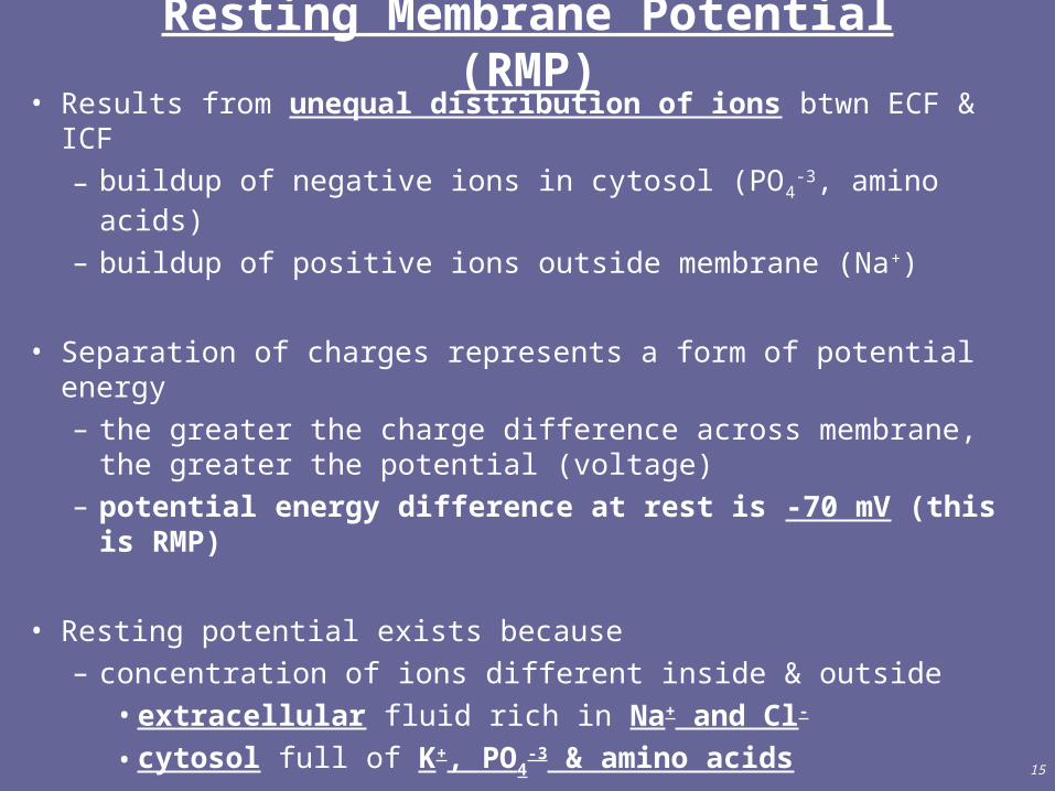
Resting Membrane Potential (RMP)• Results from unequal distribution of ions btwn ECF & ICF
– buildup of negative ions in cytosol (PO4-3, amino acids)
– buildup of positive ions outside membrane (Na+)
• Separation of charges represents a form of potential energy– the greater the charge difference across membrane, the
greater the potential (voltage) – potential energy difference at rest is -70 mV (this is
RMP)
• Resting potential exists because– concentration of ions different inside & outside
• extracellular fluid rich in Na+ and Cl-
• cytosol full of K+, PO4-3 & amino acids

16
Resting Membrane Potential (RMP)
• Resting potential exists because– membrane permeability differs for Na+ and K+
• 50-100x greater permeability for K+
• inward flow of Na+ can’t keep up with outward flow of K+
• ***Na+/K+ ATPase pump maintains R. M. P.***– w/o this pump, ion concentrations would reach
equilibrium and the membrane potential (excitability) would be destroyed
– K+ has a natural tendency to leak out of cell and Na+ tends to flow into the cell (down their respective gradients)
– pump returns 3 Na+ to ECF and 2 K+ to cytosol
17
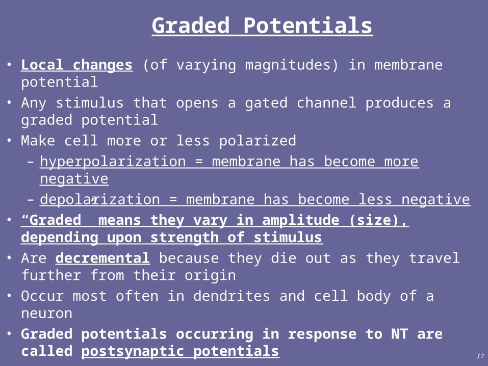
Graded Potentials
• Local changes (of varying magnitudes) in membrane potential• Any stimulus that opens a gated channel produces a graded
potential• Make cell more or less polarized
– hyperpolarization = membrane has become more negative– depolarization = membrane has become less negative
• “Graded” means they vary in amplitude (size), depending upon strength of stimulus
• Are decremental because they die out as they travel further from their origin
• Occur most often in dendrites and cell body of a neuron• Graded potentials occurring in response to NT are called
postsynaptic potentials

18
How do Graded Potentials Arise?
• Source of stimuli– mechanical stimulation of membranes with mechanical
gated ion channels (pressure)– chemical stimulation of membranes with ligand gated ion
channels (neurotransmitter)
• Graded/postsynaptic/receptor or generator potential– ions flow through ion channels and change membrane
potential locally– amount of change varies with strength of stimuli
• Flow of current (ions) is local change only
19
Generation of an Action Potential• Action potential = sequence of rapidly occurring events that
briefly reverse membrane potential due to rapid changes in membrane permeability– depolarization = membrane becomes less negative inside– repolarization = restoration of RMP (-70 mV)– threshold potential = -55 mV
• potential at which an action potential is generated• all-or-none principle: if stimulus causes depolarization
to threshold, action potential is generated – no “large” or “small” a.p. – stronger stimulus will not cause a larger impulse
• Action potentials can travel over long distances w/o dying out

20
Depolarizing Phase of Action Potential
• In resting membrane, Na+ inactivation (inner) gate open & activation (outer) gate is closed (Na+ cannot get in)
• Depolarizing graded potential or some stimulus initiates movemt of Na+ into cell (↓ potential)
• This further depolarization activates Na+-gated channels which open & allow rapid influx of Na+ until threshold reached
• @ threshold (-55mV), both Na+ gates open & Na+ enters & membrane becomes several hundred times more permeable to Na+
• more channels open in adjacent regions of membrane (positive FB)
• influx of Na+ makes inside less negative (up to +30 mV)• @ +30 mV, Na+ inner (inactivation) gates close

21
Repolarizing Phase of Action Potential
• As Na+ gates close (at +30 mV), K+ gates are activated & membrane permeability to K+ is increased
• K+ flows out of cell (down its gradient) until RMP is reached
• If the cell “overshoots” K+ efflux, hyperpolarization results– -90 mV cell further from threshold no a.p. can occur
• K+ channels close and the membrane potential returns to the resting potential of -70mV via action of Na+/ K+ ATPase pump

22
Refractory Period of Action Potential
• Period of time during which neuron cannot generate another action potential
• Absolute refractory period– even very strong stimulus will not produce another a.p.– inactivated Na+ channels must return to the resting state
before they can be reopened– Na+ inner gates closed & cannot reopen
• Relative refractory period– 2nd a.p. can be generated by very strong stimulus– Na+ channels have been restored to resting state, but
K+ channels are still open• Allows unidirectional transmission of impulses• Axons w/ large diameter have greater membrane surf. area
& shorter abs. refract. periods than small-diameter axons
23
Propagation of Nerve Impulses
• Continuous conduction (local current flow)– starts @ axon hillock where membrane is most sensitive
to changes in potential– step-by-step depolarization of adjacent segments of
membrane– membrane polarity is reversed (out becomes (-) & in
becomes (+)– inactive area of membrane (downstream) has resting
polarity opposite charges attract (+) “pulls” (–)– this opens voltage-gated channels in adjacent regions of
membrane & a.p. moves along axon– occurs in muscle fibers & unmyelinated axons

24
Propagation of Nerve Impulses• Saltatory conduction
– in myelinated axons only– depolarization occurs in similar way @ nodes of Ranvier
where voltage-gated channels are concentrated– current flows thru aqueous cytosol & ECF of Schwann cells– nerve impulses appear to jump from node to node– much quicker/more energy efficient
• open fewer voltage channels• less use of Na+/K+ pump less ATP used
• Axon diameter– large fibers are all myelinated fastest– medium fibers myelinated, but slower (b/c less surf. area)– small fibers unmyelinated & slowest (longest abs. refr. per.)

25
Encoding of Stimulus Intensity
• How do we differentiate a light touch from a firmer touch?
• Perception of intensity results from frequency of impulses (not the magnitude of an impulse)
– frequency of impulses• firm pressure generates impulses at a higher
frequency
– number of sensory neurons activated• firm pressure stimulates more neurons than does a
light touch

26
SIGNAL TRANSMISSION AT SYNAPSES
• Presynaptic neuron = neuron sending the signal
• Postsynaptic neurono = neuron receiving chem/elec signal
• Electrical synapses– ionic current spreads to next cell through gap junctions– advantages
• faster transmission of impulses a. p. jumps directly from pre-synaptic to post-synaptic neuron
• capable of synchronizing groups of neurons as in the contraction of cardiac & visceral smooth muscles

27
SIGNAL TRANSMISSION AT SYNAPSES
• Chemical synapses– Synaptic cleft separates pre/post-syn neurons chem
signals can’t “jump” from one neuron to next
– Presynaptic neuron releases NT into cleft; NT binds receptor on post-synaptic neuron
– Binding of NT produces graded (postsynaptic) potential• Repeated binding eventually produces a.p.
– Synaptic delay = time required for events to occur @ chemical synapse

28
Mechanism of Chemical Synapse• Action potential reaches end bulb and voltage-gated Ca+2
channels open
• Ca+2 flows inward & triggers release of neurotransmitter
• NT crosses synaptic cleft & binds to ligand-gated receptors – ligand-gated channels activated & ions flow across
membrane• ion flow can change postsyn. potential • If Na+ in depolarization• If Cl- in or K+ out hyperpolariz
• If depolarizing potentials reach threshold, a.p. is triggered

29
Excitatory & Inhibitory Potentials
• If NT causes depolarization excitatory postsynaptic potential (EPSP) is generated– excitatory = a.p. generated if sum of EPSPs exceeds -55mV– usually results from cation channels opening– partial depolarization makes cell more excitable
• If NT causes hyperpolarization inhibitory PSP (IPSP)– inhibitory because membrane is further from threshold– usually result of K+ or Cl- channels opening

30
Removal of Neurotransmitter
• Diffusion: NT diffuses away from cleft & is no longer effective
• Enzymatic degradation– EX: acetylcholinesterase breakdown of ACh
• Cellular uptake– Uptake by nearby neuroglia– Re-uptake by secreting axon– Clinical application: some drugs block uptake process
• EX: Prozac = SSRI blocks serotonin reuptake serotonin’s effects are prolonged

31
Summation of PSPs• Summation = integration of synaptic inputs• Spatial summation results when several presynaptic
neurons secrete NT that affects single postsynaptic neuron • Temporal summation results from repeated release of NT
from single presynaptic neuron• One postsynaptic neuron can receive numerous
excitatory/inhibitory inputs• Sum of inputs determines postsynaptic response
– EPSP: excitatory input > inhibitory input• above threshold a.p. generated• below threshold cell more sensitive b/c partial
depolarized– IPSP: inhibitory input > excitatory input
• membrane is hyperpolarized & no a.p. occurs

32
Summation of PSPs
• Clinical relevance: strychnine poisoning
– Under normal conditions: inhibitory neurons in spinal cord release glycine (a NT) which inhibits XS contractions of skeletal muscle
– Strychnine binds & inactivates glycine receptors• inhibitory effects of glycine are removed• uncontrolled muscle contraction results
– diaphragm remains fully contracted death ensues via suffocation

33
Small-Molecule Neurotransmitters
• Acetylcholine (ACh)– excitatory effect @ NMJ via direct ligand-channel binding– inhibitory @ some parasympathetic synapses
• indirect activation of receptors via G-protein• slows heart rate
– inactivated by acetylcholinesterase• Amino Acids
– excitatory: glutamate & aspartate– inhibitory: GABA & glycine
• generate IPSP via opening of Cl- channels• Valium enhances GABA effects
– prolongs effects of GABA – acts as anti-anxiety drug

34
• Biogenic Amines– catecholamines
• norepinephrine (NE) & epinephrine (Epi)– also act as hormones when released from adrenal
gland• dopamine: responsible for emotions, addictive
behaviors– Regulates skeletal muscle tone– Parkinson’s disease result of degeneration of
dopamine-secreting neurons• serotonin responsible for mood control, appetite, sleep
induction– SSRIs prevent reuptake– Zoloft, Prozac for treatment of depression
Small-Molecule Neurotransmitters
35
• Nitric oxide (NO)– potent vasodilator: increases blood flow in regions where
it is released– unique because is formed on demand & acts immediately– first recognized as vasodilator that helped lower blood
pressure– extremely toxic in high quantities– metabolic pathway = target of Viagra
Small-Molecule Neurotransmitters

36
Neuropeptides
• 3-40 amino acids linked peptide bonds• Can be excitatory or inhibitory• Brain has receptors for binding opiate drugs
– Enkephalins have potent analgesic effect (200x morphine)– Opiod peptides = body’s natural painkillers
• Dynorphins• Endorphins: responsible for “runner’s high” experienced
after exercise• Substance P transmits pain-related input from PNS to CNS
– Enhances perception of pain– Suppressed by enkephalins & endorphins

37
Modifying Effects of NTs• NT synthesis can be stimulated or inhibited
– Parkinson’s patients benefit from L-dopa b/c it boosts dopamine production for limited time
• NT release can be enhanced or blocked– Amphetamines promote release of dopamine & NE– Botulinum toxin inhibits release of Ach paralysis
• NT receptors can be activated or blocked– Agonists activate: Isoproterenol activates NE & Epi
receptors dilate airways during asthma attack– Antagonists block: Zyprexa blocks dopamine/serotonin
receptors treatment of schizophrenia• NT removal can be stimulated or inhibited
– Cocaine blocks dopamine reuptake euphoric feeling
Related Documents