1. X-RAY PRODUCTION....................................................................................................................1 2. X-RAY INTERACTIONS.................................................................................................................7 3. PROJECTION RADIOGRAPHY I...................................................................................................14 4. PROJECTION RADIOGRAPHY II.................................................................................................20 5. COMPUTED TOMOGRAPHY........................................................................................................25 6. IMAGE QUALITY.........................................................................................................................31 7. RADIOBIOLOGY/ PATIENT DOSIMETRY....................................................................................37 8. RADIATION PROTECTION..........................................................................................................44 9. NUCLEAR MEDICINE..................................................................................................................50 10. ULTRASOUND..........................................................................................................................56 11. MAGNETIC RESONANCE..........................................................................................................62 PRACTICE EXAMINATION A:.........................................................................................................68 PRACTICE EXAMINATION B:.........................................................................................................90 1. X-RAY PRODUCTION 1.1 Which of the following is not considered a force? a. Electrostatic b. Weak c. Strong d. Gravity e. Electricity 1.1e. Electricity is the flow of charge, and is measured in amps (C/s). 1.2 Which of the following is not a unit of energy? a. Erg b. Joule c. Watt d. Calorie e. eV 1.2c. The watt is a unit of power, measured in J/s.

Welcome message from author
This document is posted to help you gain knowledge. Please leave a comment to let me know what you think about it! Share it to your friends and learn new things together.
Transcript
1. X-RAY PRODUCTION.........................................................................................................................1
2. X-RAY INTERACTIONS......................................................................................................................7
3. PROJECTION RADIOGRAPHY I.....................................................................................................14
4. PROJECTION RADIOGRAPHY II....................................................................................................20
5. COMPUTED TOMOGRAPHY...........................................................................................................25
6. IMAGE QUALITY..............................................................................................................................31
7. RADIOBIOLOGY/ PATIENT DOSIMETRY......................................................................................37
8. RADIATION PROTECTION..............................................................................................................44
9. NUCLEAR MEDICINE.....................................................................................................................50
10. ULTRASOUND................................................................................................................................56
11. MAGNETIC RESONANCE..............................................................................................................62
PRACTICE EXAMINATION A:....................................................................................................................68
PRACTICE EXAMINATION B:....................................................................................................................90
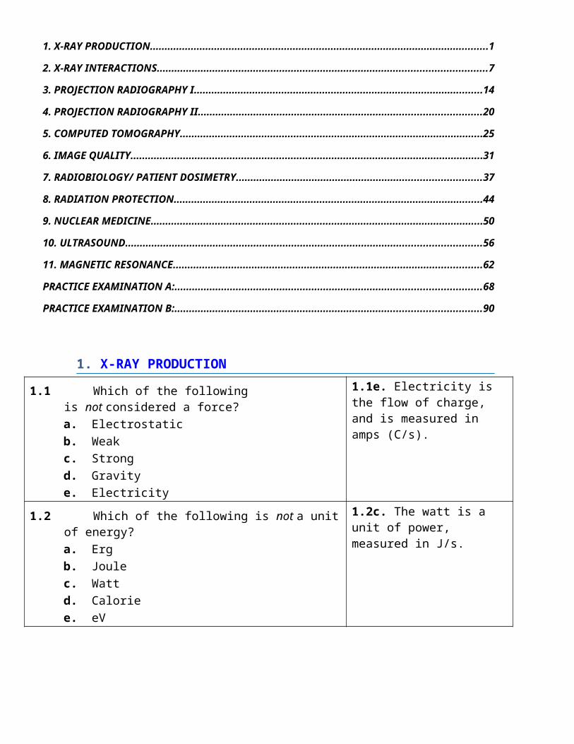
1. X-RAY PRODUCTION
1.1 Which of the following is not considered a force?a. Electrostaticb. Weakc. Strongd. Gravitye. Electricity
1.1e. Electricity is the flow of charge, and is measured in amps (C/s).
1.2 Which of the following is not a unit of energy?a. Ergb. Joulec. Wattd. Caloriee. eV
1.2c. The watt is a unit of power, measured in J/s.
1.3 Which of the following would most likely be attracted to an anode?a. Protonb. Neutronc. Electrond. Positrone. Alpha particle
1.3c. Electron, since it has a negative charge that is attracted to the positive anode.
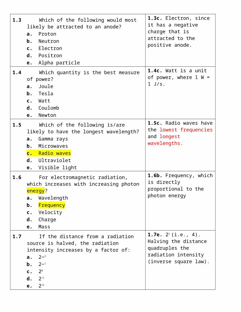
1.4 Which quantity is the best measure of power?a. Jouleb. Teslac. Wattd. Coulombe. Newton
1.4c. Watt is a unit of power, where 1 W = 1 J/s.
1.5 Which of the following is/are likely to have the longest wavelength?a. Gamma raysb. Microwavesc. Radio wavesd. Ultraviolete. Visible light
1.5c. Radio waves have the lowest frequencies and longest wavelengths.
1.6 For electromagnetic radiation, which increases with increasing photon energy?a. Wavelengthb. Frequencyc. Velocityd. Chargee. Mass
1.6b. Frequency, which is directly proportional to the photon energy
1.7 If the distance from a radiation source is halved, the radiation intensity increases by a factor of:a. 2−2
b. 2−1
c. 20
d. 2+1
e. 2+2
1.7e. 22 (i.e., 4). Halving the distance quadruples the radiation intensity (inverse square law).
1.8 X-ray generators have a power level (kW) of approximately:a. 0.1b. 1c. 10d. 100e. 1,000
1.8d. 100 kW is typical of the power of x-ray generators in radiography and CT.
1.9 Which of the following is not a type of x-ray generator?a. Single phaseb. Double phasec. Six pulsed. Twelve pulsee. High frequency
1.9b. There are no double-phase generators.

1.10 The purpose of x-ray transformers is most likely to change the:a. magnetic fieldb. electrical voltagec. power leveld. waveform frequencye. current intensity
1.10b. Transformers change (increase or decrease) voltages.
1.11 When a secondary coil has 500 more turns than a primary coil, the ratio of the secondary voltage to the primary voltage is most likely:a. 500b. 5000.5
c. 1/500d. 1/5000.5
e. Depends on AC frequency
1.11a. 500. The increase in voltage is directly proportional to the increase in the number of turns.
1.12 Which of the following generators is likely to have the largest waveform ripple?a. Constant potentialb. High frequencyc. Single phased. Six pulsee. Twelve pulse
1.12c. The ripple on a single-phase generator is 100%.
1.13 Electrons passing through matter lose energy primarily by producing:a. bremsstrahlungb. characteristic x-raysc. atomic ionizationsd. Compton electronse. photoelectrons
1.13c. Electrons lose most of their kinetic energy by knocking out (or exciting) outer shell electrons.
1.14 Tungsten is most likely used as an x-ray target because it has a high:a. physical densityb. electron densityc. electrical resistanced. melting pointe. ionization potential
1.14d. Tungsten can tolerate very high temperatures, which makes it an attractive target material in x-ray tubes.
1.15 The maximum photon energy in x-ray beams is determined by the x-ray tube:a. currentb. exposure timec. target materiald. anode–cathode voltagee. total filtration
1.15d. The voltage across the x-ray tube determines the kinetic energy imparted to the electrons that are accelerated from the cathode (filament) to the anode (target), and thereby the maximum x-ray photon energy.

1.16 The most likely characteristic x-ray energy (keV) from x-ray tubes used in chest radiography is:a. 19b. 33c. 65d. 75e. 140
1.16c. The chest x-ray unit will use a W target; the characteristic x-ray energy is therefore ~65 keV.
1.17 At 65 kV and with a tungsten target, the percentage (%) of K-shell x-rays in the x-ray beam is most likely:a. 0b. 1c. 10d. 50e. 99
1.17a. There will be no characteristic x-rays, as the electron kinetic energy (65 keV) is insufficient to eject W K-shell electrons that have a binding energy of 70 keV.
1.18 The average photon energy of an x-ray beam is least likely to be affected by changes in the:a. tube currentb. tube voltagec. voltage waveformd. target compositione. beam filtration
1.18a. The tube current does not affect the average (or maximum) photon energy in x-ray beams.
Doubling the current will double the x-ray tube output but have no effect on the x-ray spectrum shape.)
1.19 The number of electrons accelerated across an x-ray tube is most strongly influenced by:a. anode speedb. focus sizec. filament currentd. tube filtratione. tube voltage
1.19c. The filament current affects the temperature of the filament and thereby how many electrons the filament “bubbles off’’.
1.20 The most likely x-ray tube filament current (mA) is:a. 0.4b. 4c. 40d. 400e. 4,000
1.20e. X-ray tube filaments are about 4 A, or 4,000 mA.V = 10 VP = 4 x 10 = 40 w
1.21 Changing x-ray tube current (mA) most likely changes the x-ray:a. field of viewb. maximum energyc. average energyd. anode anglee. beam intensity
1.21e. Tube current controls the x-ray beam intensity, or the total number of x-ray photons produced.

1.22 The large focus dimension is most likely larger (%) than that of the small focus by:a. 10b. 25c. 50d. 75e. 100
1.22e. The large focal spot is typically 1.2 mm, and the small focal spot is 0.6 mm (i.e., 100% larger).
1.23 The anode angle (degrees) in an x-ray tube used for chest radiography is most likely:a. 15b. 30c. 45d. 60e. 75
1.23a. 15 degrees is a typical anode angle.
1.24 X-ray tube output would likely increase the most when increasing the x-ray tube:a. voltageb. anode anglec. target Zd. currente. exposure time
1.24a. The x-ray tube output is (approximately) proportional to the square of the x-ray tube voltage.Output ~ mAs x kVP2
1.25 A chest x-ray examination on a dedicated chest unit would be least likely to use:a. 60-kV voltageb. 800-mA tube currentc. 10-ms exposure timed. 1-mm focuse. 5-mm Al filtration
1.25a. Chest x-rays are performed at high voltage (120 kV).
1.26 For specification of anode heat capacities, one heat unit corresponds to energy (J) of:a. 0.9b. 0.8c. 0.7d. 0.5e. 0.3
1.26c. The heat unit is 0.7 joule, and is an anachronism in modern radiology.
1.27 At the same peak voltage, which generator likely deposits most energy into an anode?a. Constant potentialb. High frequencyc. Three phase (12 pulse)d. Three phase (6 pulse)e. Single phase
1.27a. Constant potential, since it has negligible ripple and the voltage across the x-ray tube is always the maximum possible value.

1.28 Heat stored in x-ray tube anodes is most likely dissipated by:a. convectionb. conductionc. radiationd. air coolinge. oil cooling
1.28c. Anodes get to be white hot and lose energy by radiation (light) to the tube housing.
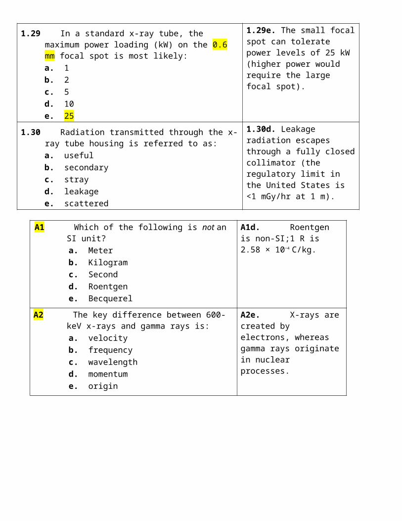
1.29 In a standard x-ray tube, the maximum power loading (kW) on the 0.6 mm focal spot is most likely:a. 1b. 2c. 5d. 10e. 25
1.29e. The small focal spot can tolerate power levels of 25 kW (higher power would require the large focal spot).
1.30 Radiation transmitted through the x-ray tube housing is referred to as:a. usefulb. secondaryc. strayd. leakagee. scattered
1.30d. Leakage radiation escapes through a fully closed collimator (the regulatory limit in the United States is <1 mGy/hr at 1 m).
A1 Which of the following is not an SI unit? a. Meter b. Kilogram c. Second d. Roentgen e. Becquerel
A1d. Roentgen is non-SI;1 R is 2.58 × 10⁻⁴ C/kg.
A2 The key difference between 600-keV x-rays and gamma rays is:
a. velocity b. frequency c. wavelength d. momentum e. origin
A2e. X-rays are created by electrons, whereas gamma rays originate in nuclear processes.
A3 What is the air kerma (mGy) 10 m from a radiation source when the air kerma at 1 m is 100 mGy?
a. 1 b. 10 c. 100 d. 1,000 e. 10,000
A3a. One mGy, since increasing the distance from the source tenfold reduces the radiation intensity a hundredfold.
= 100 x (1/100)

A4 For a fixed kVp, which generator likely results in the shortest exposure time?
a. Constant potential b. High frequency c. Three phase (12 pulse) d. Three phase (6 pulse) e. Single phase
A4a. A constant potential generator has no ripple and therefore produces the most x-rays per unit time.
A5 The maximum photon energy in an x-ray beam is determined by the x-ray tube:
a. current (mA) b. voltage (kV) c. exposure time (s) d. ripple (%) e. filtration (mm Al)
A5b. Voltage (kV) determines the maximum x-ray photon energy produced in x-ray tubes.
A6 Characteristic x-rays are characteristic of the material in the:
a. target b. anode c. filter d. window e. filament
A6a. A tungsten target produces characteristic x-rays whose energy is ~65 keV, just below the K-shell binding energy (70 keV).
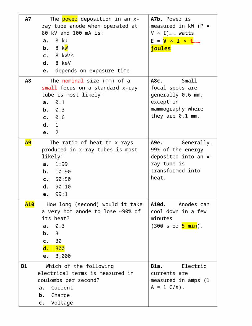
A7 The power deposition in an x-ray tube anode when operated at 80 kV and 100 mA is:
a. 8 kJ b. 8 kW c. 8 kW/s d. 8 keV e. depends on exposure time
A7b. Power is measured in kW (P = V × I)…… wattsE = V × I × t…… joules
A8 The nominal size (mm) of a small focus on a standard x-ray tube is most likely:
a. 0.1 b. 0.3 c. 0.6 d. 1 e. 2
A8c. Small focal spots are generally 0.6 mm, except in mammography where they are 0.1 mm.
A9 The ratio of heat to x-rays produced in x-ray tubes is most likely:
a. 1:99 b. 10:90 c. 50:50 d. 90:10 e. 99:1
A9e. Generally, 99% of the energy deposited into an x-ray tube is transformed into heat.
A10 How long (second) would it take a very hot anode to lose ~90% of its heat?
a. 0.3 b. 3 c. 30 d. 300 e. 3,000
A10d. Anodes can cool down in a few minutes(300 s or 5 min).
B1 Which of the following electrical terms is measured in coulombs per second?
a. Current b. Charge c. Voltage d. Resistance e. Power
B1a. Electric currents are measured in amps (1 A = 1 C/s).
B2 Power (W) for a constant-potential generator at 100 kV and 1,000 mA is most likely:
a. 10 b. 100 c. 1,000 d. 10,000 e. 100,000
B2e. Power is 100,000 W or 100 kW (P = V x I x T).
B3 A rectification circuit is most likely to contain: a. resistors b. transistors c. diodes d. inductances e. capacitors
B3c. Diodes are used in rectification circuits, and they permit electrical currents to flow in only one direction.
B4 The most likely x-ray tube target material is: a. iron b. copper c. zinc d. tungsten e. lead
B4d. Most x-ray tubes use tungsten targets.
B5 What is the maximum photon energy (keV) of an x-ray produced at an x-ray tube voltage of 120 kV?
a. 12 b. 25 c. 40 d. 60 e. 120
B5e. The maximum energy is 120 keV, since the maximum electron kinetic energy is 120 keV and all of this kinetic energy can be transformed into an x-ray photon.
B6 100-keV electrons most likely produce x-ray photons with average energy (keV) of:
a. 20
B6c. The average photon energy is between one half and one third of the maximum photon energy.

b. 30 c. 45 d. 55 e. 70
B7 The power (W) dissipated in the x-ray tube filament is most likely:
a. 0.4 b. 4 c. 40 d. 400 e. 4,000
B7c. X-ray tube filaments are operated at 4 A and 10 V and deposit 40 W of power, just like the filament in a light bulb.
B8 X-ray tube output is unlikely to be increased by increasing:
a. tube voltage (kV) b. anode capacity (MJ) c. target atomic number (Z) d. tube current (mA) e. exposure time (s)
B8b. Anode capacity has no direct relevance to the x-ray tube output.
B9 The energy deposited in an anode with a constant-potential generator operated at voltage kV, tube current mA, and exposure time t is:
a. kV mA t b. kV2 mA t c. kV mA2 t d. (kV mA)2 t e. (kV mA)/t
B9a. Energy deposited in the anode in joules is kV mA t (at constant voltage).
B10 Which of the following is the most likely x-ray tube power level (kW) during routine abdominal fluoroscopy?
a. 0.3 b. 1 c. 3 d. 10 e. 30
B10a. In fluoroscopy, the tube voltage is ~100 kV and tube current is ~3 mA, so the power is ~300 W (i.e., 0.3 kW).
2. X-RAY INTERACTIONS
2.1 Which of the following refers to the number of nucleons in a nucleus?a. Mass numberb. Atomic numberc. Avogadro’s numberd. Atomic mass unite. Nuclear density
2.1a. The mass number is the total number of protons and neutrons (i.e., nucleons) in the atom’s nucleus.
2.2 Which element has an atomic number of 56 and a K-shell binding energy of 37 keV?a. Calciumb. Seleniumc. Molybdenumd. Bariume. Tungsten
2.2d. Barium has 56 protons and a K-shell binding energy that is slightly higher than that of Iodine (33 keV).TABLE 2.2
2.3 The outer shell electrons most likely have binding energies (keV) of approximately:a. 0.001b. 0.005 keVc. 0.025d. 0.1e. 0.5
2.3b. The outer shell binding energy is ~5 eV (i.e., 0.005 keV).
2.4 Ionizing radiations are least likely to include:a. x-ray photonsb. energetic electronsc. infrared radiationd. alpha particlese. fast neutrons
2.4c. Infrared radiation is nonionizing, as the photon energy is less than 1 eV.
2.5 The total number of atomic ionizations produced following absorption of a 30 keV photon is most likely:a. 10b. 100c. 1,000d. 10,000e. 100,000
2.5c. 1,000 ionization events are likely since it takes 30 eV or so to produce one ionization and there is 30,000 eV available.
30 eV one ionization
2.6 The most likely percentage (%) of coherent scatter photons in an x-ray beam emerging from a patient having a chest x-ray is:a. <5b. 5c. 10d. 20e. >20
2.6a. Coherent scatter never accounts for more than 5% of all interactions in diagnostic radiology.

2.7 The energy (E) dependence of photoelectric absorption above the K edge varies as:a. 1/E3
b. 1/E2
c. 1/Ed. E2
e. E3
2.7a. Above the K edge, the photoelectric effect is proportional to Z3/E3.
↑ KVp ↓ PE more than ↓ Compton Compton predominates
2.8 After an x-ray undergoes photoelectric absorption by a K-shell electron, which emission is least likely?a. Photoelectronb. Scattered photonc. K-shell x-rayd. L-shell x-raye. Auger electron
2.8b. There are no scattered photons in photoelectric absorption; scatter occurs with Compton interactions.
2.9 The likelihood of Compton interactions is best quantified using:a. physical density……… PEb. electron densityc. atomic numberd. K-shell energye. outer shell energy
2.9b. The likelihood of Compton interactions is approximately proportional to the electron density.
= number of free outer shell electrons
2.10 For a given absorber, if the Compton attenuation coefficient at 50 keV is 0.1 cm–1, its value at 100 keV (cm–1) is most likely:a. 0.01b. 0.025c. 0.05 (1/E)d. 0.1e. 0.2
2.10c. 0.05 cm–1, since Compton interactions vary as 1/E, so doubling the photon energy will halve the probability of this interaction. 1/E ½
2.11 In bone, at what photon energy are photoelectric and Compton effects approximately equal?a. 4.0b. 25c. 40d. 70e. 88
2.11c. Compton and photoelectric interactions are equally probable at 40 keV in bone.
2.12 If the linear attenuation coefficient is 0.1 cm–1,( 10% are lost per 1 cm) how many x-ray photons (%) are lost in 1 mm?a. 0.1b. 1 1%=(0.01)c. 10d. e–1
e. e–0.01
2.12b. 10% are lost in 1 cm, so 1% must be lost in each mm.

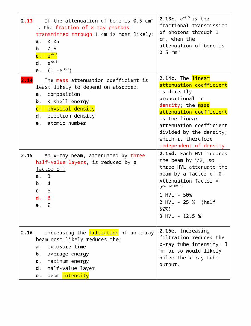
2.13 If the attenuation of bone is 0.5 cm–1, the fraction of x-ray photons transmitted through 1 cm is most likely:a. 0.05b. 0.5c. e–0.5
d. e+0.5
e. (1 –e–0.5)
2.13c. e–0.5 is the fractional transmission of photons through 1 cm, when the attenuation of bone is 0.5 cm–1
2.14 The mass attenuation coefficient is least likely to depend on absorber:a. compositionb. K-shell energyc. physical densityd. electron densitye. atomic number
2.14c. The linear attenuation coefficient is directly proportional to density; the mass attenuation coefficient is the linear attenuation coefficient divided by the density, which is therefore independent of density.
2.15 An x-ray beam, attenuated by three half-value layers, is reduced by a factor of:a. 3b. 4c. 6d. 8e. 9
2.15d. Each HVL reduces the beam by 1/2, so three HVL attenuate the beam by a factor of 8.Attenuation factor = 2no. of HVL’s
1 HVL – 50%2 HVL – 25 % (half 50%)3 HVL – 12.5 %
2.16 Increasing the filtration of an x-ray beam most likely reduces the:a. exposure timeb. average energyc. maximum energyd. half-value layere. beam intensity
2.16e. Increasing filtration reduces the x-ray tube intensity; 3 mm or so would likely halve the x-ray tube output.
2.17 The x-ray tube output most likely increases when reducing the:a. mAb. exposure timec. kVd. filtratione. focal spot
2.17d. A reduction in filtration means that more x-rays can get out.
Outpu = Gy = joule/kgJoule = kvp x mA
2.18 Adding Aluminum filters to an x-ray beam is most likely to increase x-ray:a. intensity (air kerma)b. air kerma–area productc. maximum energyd. beam hardeninge. leakage radiation
2.18d. Increasing (Al) filtration always increases beam hardening in radiology.

2.19 The adequacy of the filtration of an x-ray tube is best determined by measuring the:a. tube voltageb. air kermac. field sized. half-value layere. leakage radiation
2.19d. The HVL is a good measure of the adequacy of filtration and at 80 kV should be greater than ~2.5 mm Al.
2.20 The heel effect most likely increases when reducing the:a. tube voltageb. tube currentc. anode angled. filtratione. field size
2.20c. Reducing the anode angle will increase the heel effect.
2.21 In abdominal imaging, the scatter to primary ratio of photons leaving the patient is most likely:a. 0.2b. 0.5c. 1d. 2e. 5
2.21e. There are about five scattered photons exiting the patient for every one primary photon.
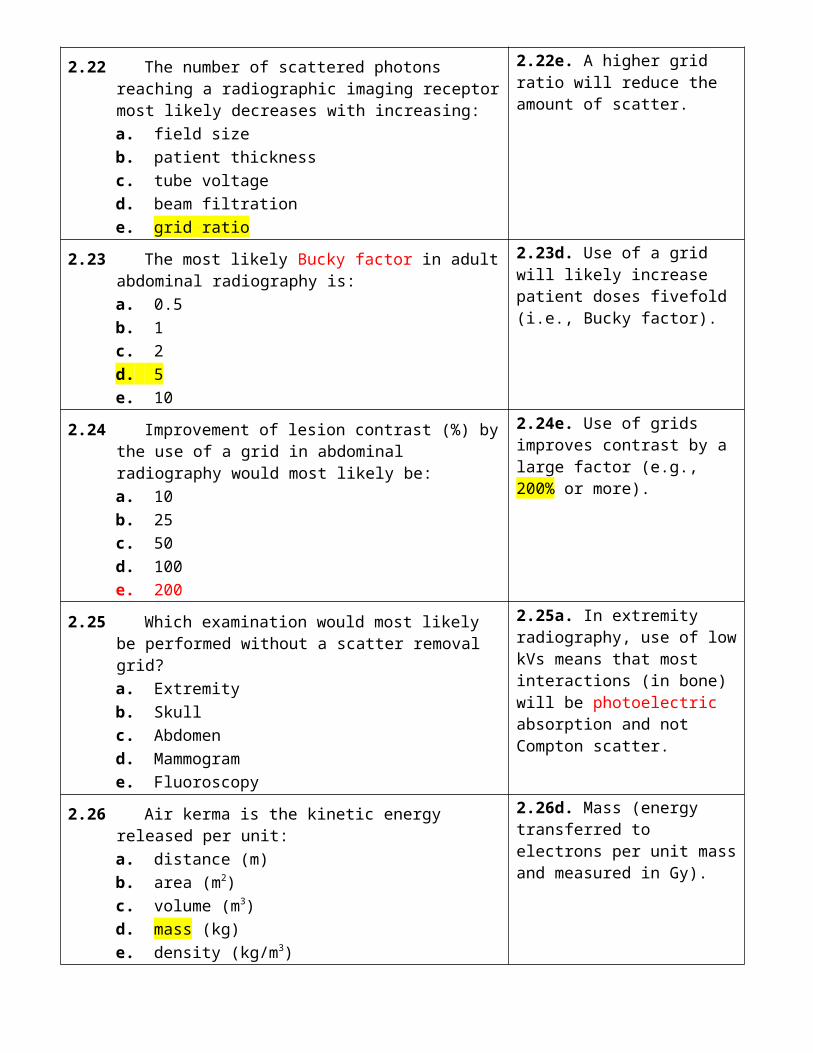
2.22 The number of scattered photons reaching a radiographic imaging receptor most likely decreases with increasing:a. field sizeb. patient thicknessc. tube voltaged. beam filtratione. grid ratio
2.22e. A higher grid ratio will reduce the amount of scatter.
2.23 The most likely Bucky factor in adult abdominal radiography is:a. 0.5b. 1c. 2d. 5e. 10
2.23d. Use of a grid will likely increase patient doses fivefold (i.e., Bucky factor).
2.24 Improvement of lesion contrast (%) by the use of a grid in abdominal radiography would most likely be:a. 10b. 25c. 50d. 100e. 200
2.24e. Use of grids improves contrast by a large factor (e.g., 200% or more).

2.25 Which examination would most likely be performed without a scatter removal grid?a. Extremityb. Skullc. Abdomend. Mammograme. Fluoroscopy
2.25a. In extremity radiography, use of low kVs means that most interactions (in bone) will be photoelectric absorption and not Compton scatter.
2.26 Air kerma is the kinetic energy released per unit:a. distance (m)b. area (m2)c. volume (m3)d. mass (kg)e. density (kg/m3)
2.26d. Mass (energy transferred to electrons per unit mass and measured in Gy).
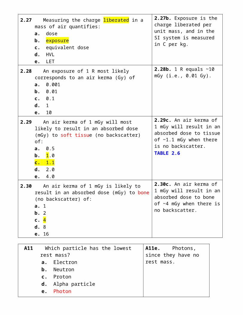
2.27 Measuring the charge liberated in a mass of air quantifies:a. doseb. exposurec. equivalent dosed. HVLe. LET
2.27b. Exposure is the charge liberated per unit mass, and in the SI system is measured in C per kg.
2.28 An exposure of 1 R most likely corresponds to an air kerma (Gy) ofa. 0.001b. 0.01c. 0.1d. 1e. 10
2.28b. 1 R equals ~10 mGy (i.e., 0.01 Gy).
2.29 An air kerma of 1 mGy will most likely to result in an absorbed dose (mGy) to soft tissue (no backscatter) of:a. 0.5b. 1.0c. 1.1d. 2.0e. 4.0
2.29c. An air kerma of 1 mGy will result in an absorbed dose to tissue of ~1.1 mGy when there is no backscatter.TABLE 2.6
2.30 An air kerma of 1 mGy is likely to result in an absorbed dose (mGy) to bone (no backscatter) of:a. 1b. 2c. 4d. 8e. 16
2.30c. An air kerma of 1 mGy will result in an absorbed dose to bone of ~4 mGy when there is no backscatter.

A11 Which particle has the lowest rest mass? a. Electron b. Neutron c. Proton d. Alpha particle e. Photon
A11e. Photons, since they have no rest mass.
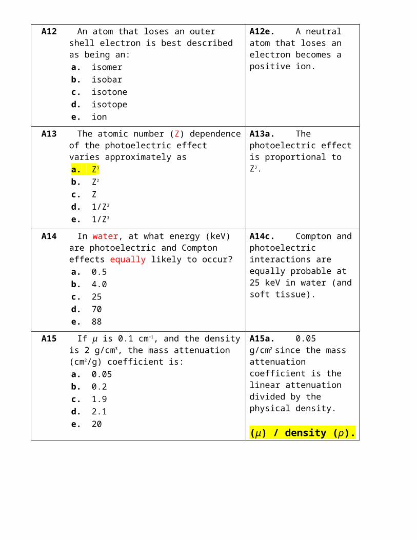
A12 An atom that loses an outer shell electron is best described as being an:
a. isomer b. isobar c. isotone d. isotope e. ion
A12e. A neutral atom that loses an electron becomes a positive ion.
A13 The atomic number (Z) dependence of the photoelectric effect varies approximately as
a. Z3
b. Z2
c. Z d. 1/Z2
e. 1/Z3
A13a. The photoelectric effect is proportional to Z3.
A14 In water, at what energy (keV) are photoelectric and Compton effects equally likely to occur?
a. 0.5 b. 4.0 c. 25 d. 70 e. 88
A14c. Compton and photoelectric interactions are equally probable at 25 keV in water (and soft tissue).
A15 If μ is 0.1 cm−1, and the density is 2 g/cm3, the mass attenuation (cm2/g) coefficient is:
a. 0.05 b. 0.2 c. 1.9 d. 2.1 e. 20
A15a. 0.05 g/cm2 since the mass attenuation coefficient is the linear attenuation divided by the physical density.
(μ) / density (ρ).
A16 The total attenuation by 10 half-value layers is most likely:
a. 64 b. 128 c. 256 d. 512 e. 1,024
A16e. (1/2)10 is 1/1,024, or ~0.1%.
Attenuation factor = 2n
n = no. of HVL
Transmision %=1002(n )
Attenuation%=100−1002 (n)
A17 The x-ray beam HVL is least likely to be affected by the x-ray tube:
a. output (mGy) b. voltage (kV) c. voltage ripple (%) d. filtration (mm Al) e. target material (Z)
A17a. X-ray beam air kerma has no effect on the x-ray beam HVL.
A18 What grid characteristic is most likely to determine the scatter removal performance?
a. Grid ratio b. Focus distance c. Gap distance d. Strip height e. Line density
A18a. The grid ratio is the most important parameter that determines the scatter removal performance of a grid.
A19 The most likely reason that grids are seldom used for portable chest radiography is that:
a. portable x-ray output is low b. lower kV won’t penetrate grid c. grid alignment is difficult d. scatter is very low e. air gap minimizes scatter
A19c. Grid alignment is very difficult in bedside radiography.
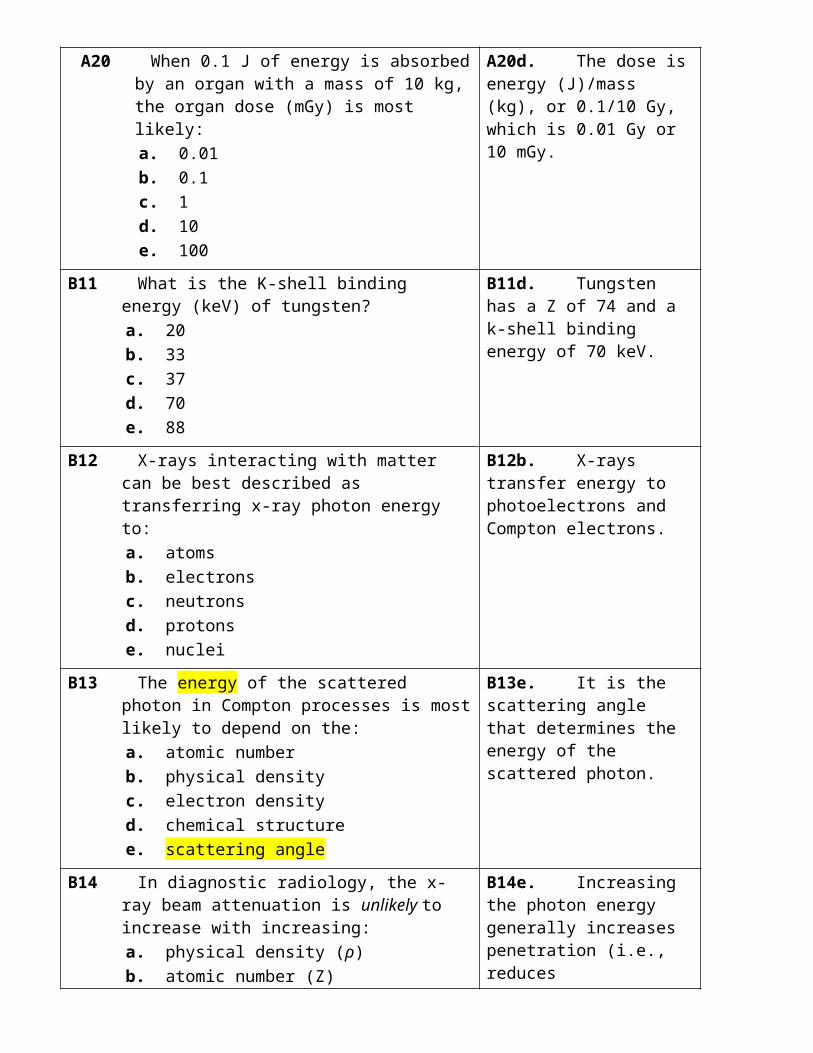
A20 When 0.1 J of energy is absorbed by an organ with a mass of 10 kg, the organ dose (mGy) is most likely:
a. 0.01 b. 0.1 c. 1 d. 10 e. 100
A20d. The dose is energy (J)/mass (kg), or 0.1/10 Gy, which is 0.01 Gy or 10 mGy.
B11 What is the K-shell binding energy (keV) of tungsten?
a. 20 b. 33 c. 37
B11d. Tungsten has a Z of 74 and a k-shell binding energy of 70 keV.

d. 70 e. 88
B12 X-rays interacting with matter can be best described as transferring x-ray photon energy to:
a. atoms b. electrons c. neutrons d. protons e. nuclei
B12b. X-rays transfer energy to photoelectrons and Compton electrons.
B13 The energy of the scattered photon in Compton processes is most likely to depend on the:
a. atomic number b. physical density c. electron density d. chemical structure e. scattering angle
B13e. It is the scattering angle that determines the energy of the scattered photon.
B14 In diagnostic radiology, the x-ray beam attenuation is unlikely to increase with increasing:
a. physical density (ρ) b. atomic number (Z) c. electron density (e/cm3) d. attenuator thickness (cm) e. photon energy (keV)
B14e. Increasing the photon energy generally increases penetration (i.e., reduces attenuation).
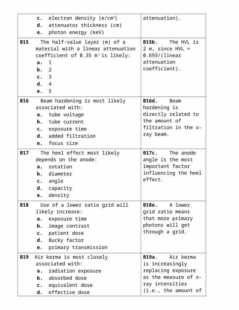
B15 The half-value layer (m) of a material with a linear attenuation coefficient of 0.35 m−1 is likely:
a. 1 b. 2 c. 3 d. 4 e. 5
B15b. The HVL is 2 m, since HVL = 0.693/(linear attenuation coefficient).
B16 Beam hardening is most likely associated with: a. tube voltage b. tube current c. exposure time d. added filtration e. focus size
B16d. Beam hardening is directly related to the amount of filtration in the x-ray beam.
B17 The heel effect most likely depends on the anode: a. rotation b. diameter c. angle d. capacity e. density
B17c. The anode angle is the most important factor influencing the heel effect.
B18 Use of a lower ratio grid will likely increase: B18e. A lower grid ratio
a. exposure time b. image contrast c. patient dose d. Bucky factor e. primary transmission
means that more primary photons will get through a grid.
B19 Air kerma is most closely associated with: a. radiation exposure b. absorbed dose c. equivalent dose d. effective dose e. dose area product
B19a. Air kerma is increasingly replacing exposure as the measure of x-ray intensities (i.e., the amount of radiation in an x-ray beam).
B20 The non-SI unit of absorbed dose is the: a. mR b. mrad c. mrem d. mCi e. mBq
B20b. Rads are non-SI units of absorbed dose (1 rad is equal to 10 mGy).
3. PROJECTION RADIOGRAPHY I
3.1 X-ray film emulsion contains crystals of:a. calcium tungstateb. silver bromidec. lanthanum oxybromided. silver nitratee. cesium iodide
3.1b. Silver bromide grains are the light-sensitive grains found in film emulsions.
silver halide (iodobromide)
3.2 In film processing, the developer most likely:a. modifies developer pHb. removes unexposed grainsc. attaches silver to the based. removes brominee. reduces silver halide
3.2e. Film development is the reduction (i.e., addition of electrons) of silver halide grains to “a clump of silver atoms.’’
3.3 If Io is the light incident on a film, and It is the light transmitted, optical density is:a. Io +It
b. Io –It
c. Io/It
d. log(Io/It)e. log(Io–It)
3.3d. Film density is log(Io/It).
3.4 The fraction of light transmitted by a film with an optical density of 2 is:a. 0.5b. 0.1c. 0.05d. 0.01e. 0.005
3.4d. A film with density of 2 transmits 1% (0.01) of the incident light.
3.5 The maximum slope of the characteristic curve is called the film:a. densityb. gammac. transmittanced. opacitye. lambda
3.5b. Film gamma is the maximum slope.
3.6 Films used for chest x-ray examinations are likely to have increased:a. gradientb. gammac. speedd. latitudee. density
3.6d. Chest films require wide-latitude films to capture intensities transmitted through the lungs and mediastinum.
↑ latitude ↓ gradient ↓ contrast
3.7 The percentage of x-ray photons (%) absorbed by a radiographic film alone (no screen) is most likely:a. 0.001b. 0.01c. 0.1d. 1e. 10
3.7d. A film alone absorbs ~1% of the incident x-ray photons.
Absorption = 1% exposure (no IS) = 50% exposure (with IS)Conversion into light = 10% Absorption2eV 1 photon30 eV 1 ionization
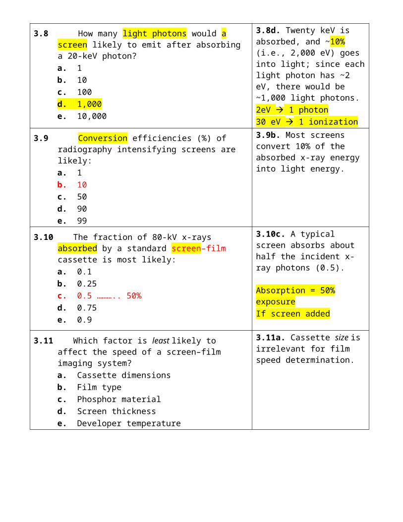
3.8 How many light photons would a screen likely to emit after absorbing a 20-keV photon?a. 1b. 10c. 100d. 1,000e. 10,000
3.8d. Twenty keV is absorbed, and ~10% (i.e., 2,000 eV) goes into light; since each light photon has ~2 eV, there would be ~1,000 light photons.2eV 1 photon30 eV 1 ionization
3.9 Conversion efficiencies (%) of radiography intensifying screens are likely:a. 1b. 10c. 50d. 90e. 99
3.9b. Most screens convert 10% of the absorbed x-ray energy into light energy.
3.10 The fraction of 80-kV x-rays absorbed by a standard screen–film cassette is most likely:a. 0.1b. 0.25c. 0.5 ……….. 50%d. 0.75e. 0.9
3.10c. A typical screen absorbs about half the incident x-ray photons (0.5).
Absorption = 50% exposureIf screen added
3.11 Which factor is least likely to affect the speed of a screen–film imaging system?a. Cassette dimensionsb. Film typec. Phosphor materiald. Screen thicknesse. Developer temperature
3.11a. Cassette size is irrelevant for film speed determination.
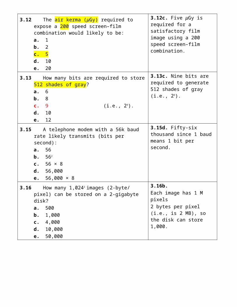
3.12 The air kerma (μGy) required to expose a 200 speed screen–film combination would likely to be:a. 1b. 2c. 5d. 10e. 20
3.12c. Five μGy is required for a satisfactory film image using a 200 speed screen–film combination.
3.13 How many bits are required to store 512 shades of gray?a. 6b. 8c. 9 (i.e., 29).d. 10e. 12
3.13c. Nine bits are required to generate 512 shades of gray (i.e., 29).
3.15 A telephone modem with a 56k baud rate likely transmits (bits per second):a. 56b. 562
c. 56 × 8d. 56,000e. 56,000 × 8
3.15d. Fifty-six thousand since 1 baud means 1 bit per second.
3.16 How many 1,0242 images (2-byte/ pixel) can be stored on a 2-gigabyte disk?a. 500b. 1,000c. 4,000d. 10,000e. 50,000
3.16b.Each image has 1 M pixels2 bytes per pixel (i.e., is 2 MB), so the disk can store 1,000.

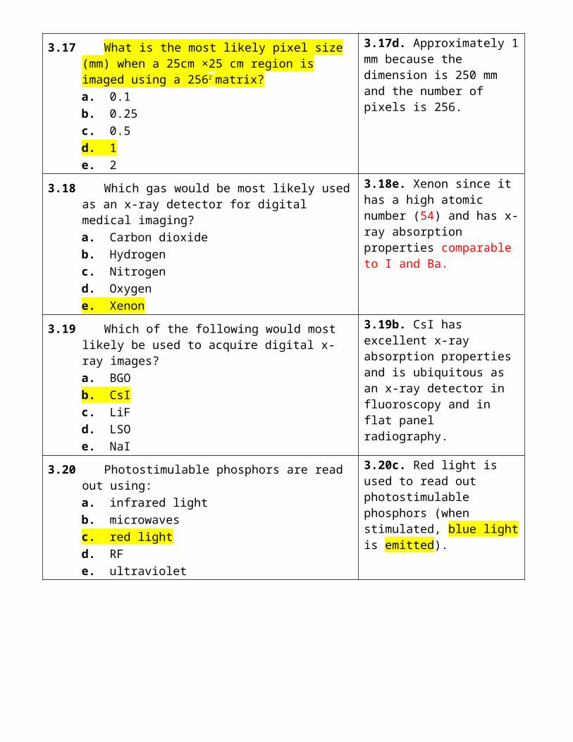
3.17 What is the most likely pixel size (mm) when a 25cm ×25 cm region is imaged using a 2562 matrix?a. 0.1b. 0.25c. 0.5d. 1e. 2
3.17d. Approximately 1 mm because the dimension is 250 mm and the number of pixels is 256.
3.18 Which gas would be most likely used as an x-ray detector for digital medical imaging?a. Carbon dioxideb. Hydrogenc. Nitrogend. Oxygene. Xenon
3.18e. Xenon since it has a high atomic number (54) and has x-ray absorption properties comparable to I and Ba.
3.19 Which of the following would most likely be used to acquire digital x-ray images?a. BGOb. CsIc. LiFd. LSOe. NaI
3.19b. CsI has excellent x-ray absorption properties and is ubiquitous as an x-ray detector in fluoroscopy and in flat panel radiography.
3.20 Photostimulable phosphors are read out using:a. infrared lightb. microwavesc. red lightd. RFe. ultraviolet
3.20c. Red light is used to read out photostimulable phosphors (when stimulated, blue light is emitted).
3.21 The dynamic range of a photostimulable phosphor is most likely:a. 10:1b. 102:1c. 103:1d. 104:1e. 105:1
3.21d. The typical dynamic range of a photostimulable phosphor is 104:1.
3.22 The x-ray absorber material most likely used in indirect flat panel x-ray detectors is:a. BaFBrb. CsIc. NaId. PbIe. Se
3.22b. Indirect x-ray detectors mainly use CsI as the x-ray absorbing phosphor material.
3.23 Which of the following materials is most likely used as a photoconductor in direct flat panel x-ray detectors?a. Xeb. Brc. Sed. Bae. Cs
3.23c. Se is the most common photoconductor in use in medical imaging today (2008).
3.24 A typical pixel size (μm) in a digital diagnostic chest x-ray image is most likely:a. 50b. 100c. 175d. 300e. 500
3.24c. The typical matrix size is 175 microns (i.e., 350-mm width of a chest x-ray film divided by 2,000 pixels, or 430-mm height divided by 2,500).35 x 43 cm /2000 x 2500
3.25 Typical maximum brightness (cd/m2) of a digital image display is most likely:a. 3b. 10c. 30d. 100e. 300
3.25e. Diagnostic workstations have a maximum image brightness of 300 cd/m2.
3.26 The display capacity (megapixel, MP) of a radiology diagnostic workstation is most likely:a. 0.25b. 0.5c. 1d. 2e. 3
3.26e. Diagnostic workstations typically use 3 MP displays (5 MP are used in mammography).
3.27 Processing a digital x-ray image by unsharp mask enhancement likely increases the:a. limiting resolutionb. visibility of edgesc. patient dosed. matrix sizee. image magnification
3.27b. Visibility of edges improves in images processed by unsharp mask enhancement.
3.28 Reducing image noise by smoothing the acquired data is most likely to reduce:a. data contentb. lesion contrastc. matrix sized. patient dosee. spatial resolution
3.28e. Average pixel values to reduce random fluctuations (mottle) will blur the image and reduce spatial resolution.
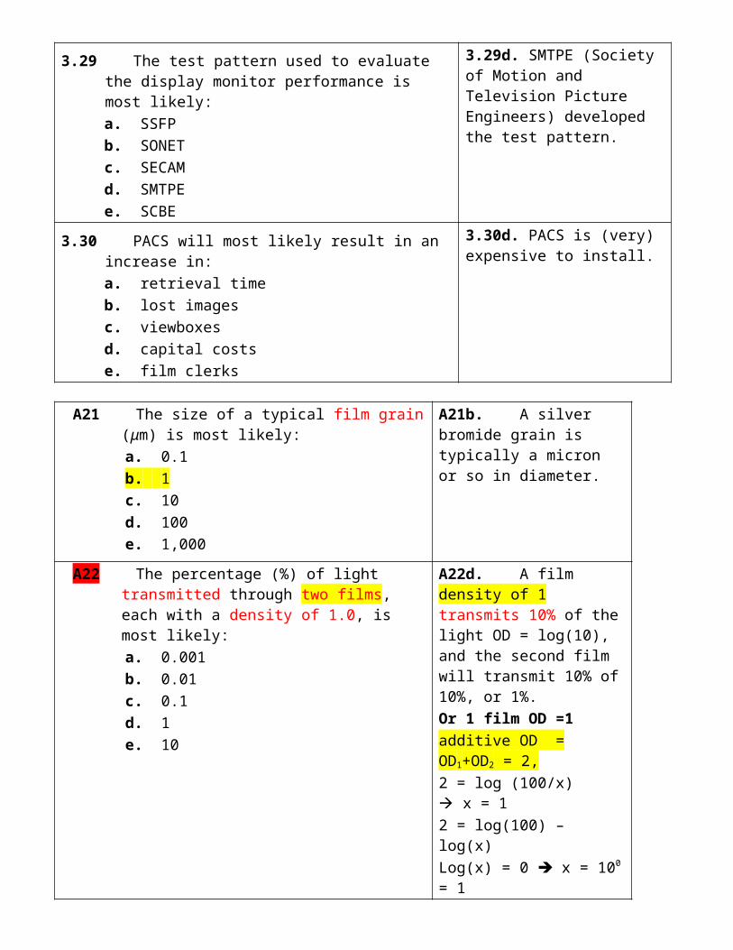
3.29 The test pattern used to evaluate the display monitor performance is most likely:a. SSFPb. SONETc. SECAMd. SMTPEe. SCBE
3.29d. SMTPE (Society of Motion and Television Picture Engineers) developed the test pattern.
3.30 PACS will most likely result in an increase in:a. retrieval timeb. lost imagesc. viewboxesd. capital costse. film clerks
3.30d. PACS is (very) expensive to install.
A21 The size of a typical film grain (μm) is most likely: a. 0.1 b. 1 c. 10 d. 100 e. 1,000
A21b. A silver bromide grain is typically a micron or so in diameter.
A22 The percentage (%) of light transmitted through two films, each with a density of 1.0, is most likely:
a. 0.001 b. 0.01 c. 0.1 d. 1 e. 10
A22d. A film density of 1 transmits 10% of the light OD = log(10), and the second film will transmit 10% of 10%, or 1%.Or 1 film OD =1additive OD = OD1+OD2 = 2,2 = log (100/x) x = 12 = log(100) – log(x)Log(x) = 0 x = 100 = 1
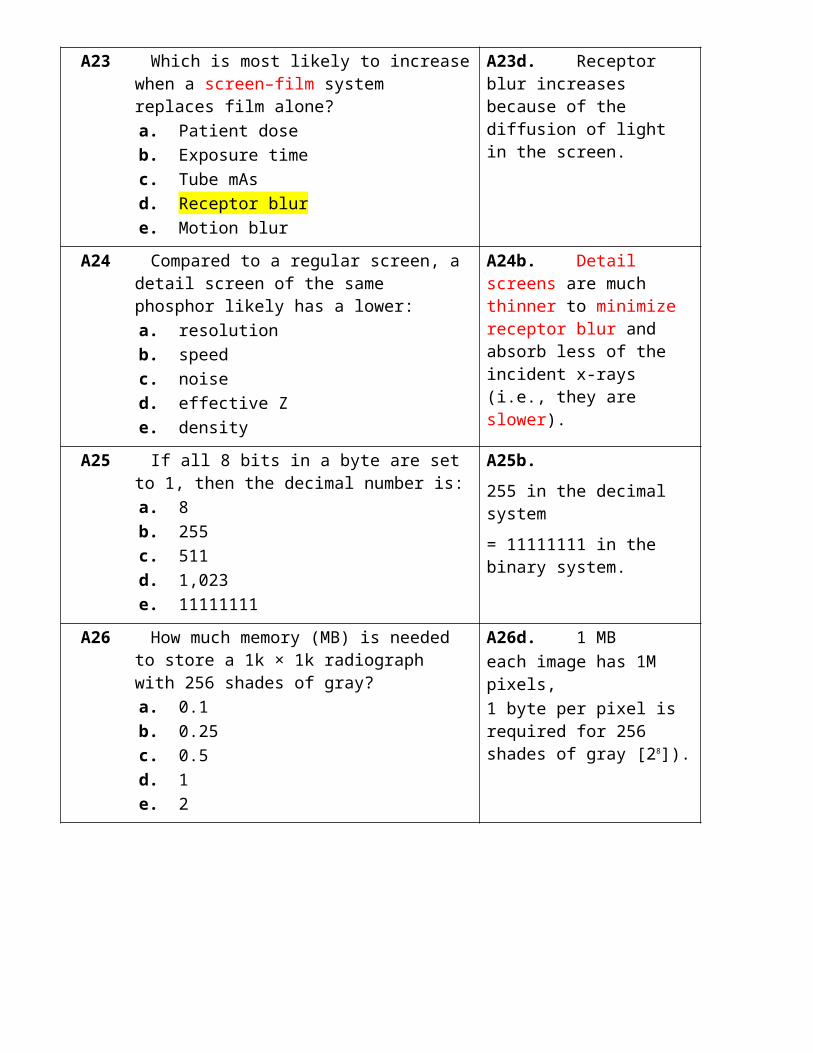
A23 Which is most likely to increase when a screen–film system replaces film alone?
a. Patient dose b. Exposure time c. Tube mAs d. Receptor blur e. Motion blur
A23d. Receptor blur increases because of the diffusion of light in the screen.
A24 Compared to a regular screen, a detail screen of the same phosphor likely has a lower:
a. resolution b. speed c. noise d. effective Z e. density
A24b. Detail screens are much thinner to minimize receptor blur and absorb less of the incident x-rays (i.e., they are slower).

A25 If all 8 bits in a byte are set to 1, then the decimal number is:
a. 8 b. 255 c. 511 d. 1,023 e. 11111111
A25b.255 in the decimal system
= 11111111 in the binary system.
A26 How much memory (MB) is needed to store a 1k × 1k radiograph with 256 shades of gray?
a. 0.1 b. 0.25 c. 0.5 d. 1 e. 2
A26d. 1 MBeach image has 1M pixels,1 byte per pixel is required for 256 shades of gray [28]).
A27 Which of the following materials is most likely a photostimulable phosphor?
a. BaFBr b. CsI c. NaI d. PbI e. Se
A27a. BaFBr is a photostimulable phosphor.
A28 Photoconductors convert x-ray energy directly into: a. light b. charge c. heat d. voltage e. radio waves
A28b. Photoconductors absorb x-rays which is converted into charge.
A29 Replacing analog chest imaging with digital technology is least likely to improve image:
a. resolution b. processing c. retrieval d. storage e. transmission
A29a. Resolution in digital imaging is generally lower than that of analog imaging.
A30 The minimum number of images required to perform energy subtraction is:
a. 1 b. 2 c. 3 d. 4 e. >4
A30b. Two images are required—one at a low kV and one at a high kV.
B21 In film processing, the fixer: a. modifies developer pH
B21b. Fixers remove unexposed grains of silver

b. removes unexposed grains c. fixes silver to the emulsion d. removes bromine e. reduces silver halide
bromide.
B22 Reducing the film processor temperature will most likely decrease the:
a. image contrast b. quantum mottle c. focal blur d. screen blur e. patient dose
B22a. Lower film temperatures will reduce the average film gradient, and therefore image contrast.
B23 Matching the screen K-edge with incident x-ray energy will most likely increase screen–film:
a. conversion efficiency b. fog level c. image blur d. average gradient e. relative speed
B23e. Relative speed increases since the matching exercise means that more x-rays will be absorbed by the screen.
B25 What is the data transfer speed (Mbit/s) of Gigabit Ethernet?
a. 1 b. 10 c. 100 d. 1,000 e. 10,000
B25d. Giga Ethernet transfers data at 1,000 Mbit/s.
B26 Which of the following is least likely to be used for the detection of diagnostic x-rays?
a. Photoconductor b. Scintillator c. Charged couple device d. Photostimulable phosphor e. Intensifying screen
B26c. A charged couple device detects light, not x-rays.
B27 Which of the following x-ray detector materials most likely emits light?
a. Xe b. CsI c. Se d. PbI e. HgI
B27b. CsI converts 10% of the absorbed energy into light energy.
B28 How many pixels (million) are most likely generated by a film digitizer processing a diagnostic chest radiograph?
a. 0.5 b. 1
B28d. Digitized chest x-rays are likely to have amatrix size of 2,000 × 2,5000, or 5 million pixels.
Image size 10 Mb
c. 2 d. 5 e. 10
B29 Which of the following is least related to image processing?
a. Histogram equalization b. Low-pass filtering c. Background subtraction d. Bow tie filtering e. Energy subtraction
B29d. Bow tie filtering refers to the use of a bow tie–shaped filter in CT imaging.
B30 Which of the following does not relate to computer networks?
a. Token ring b. Ethernet c. Backbone d. JPEG e. Bridge
B30d. JPEG is a file format that includes standards for compression.
4. PROJECTION RADIOGRAPHY II
4.1 The ideal photon energy (not tube voltage) (keV) for performing mammography is most likely:a. 10b. 15c. 19d. 25e. 33
4.1c. Photons of 19 keV have sufficient energy to penetrate the breast but are low enough to offer high image contrast.
4.2 The standard focal spot size (mm) in mammography is:a. 0.1b. 0.2c. 0.3d. 0.6e. 1.2
4.2c. The standard focal spot size in mammography is 0.3 mm.
4.3 Molybdenum filters in mammography most likely have a thickness (μm) of:a. 1b. 3c. 10d. 30e. 100
4.3d. A typical molybdenum filter thickness is 30 μm.
4.4 The most likely grid ratio in contact mammography is:a. no grid usedb. 2:1c. 5:1d. 8:1e. 12:1
4.4c. Mammography imaging systems usually use grids of ~5:1 because there is less scatter at the low energies that are used (e.g., 25 kV).
4.5 The average gradient of a mammography film is most likely:a. 1b. 2c. 3d. 5e. 10
4.5c. Mammography films have gradients of ~3.
4.6 The optimum film density in mammography is:a. 0.8b. 1.4c. 1.8d. 2.2e. 2.6
4.6c. The optimum for mammography is ~1.8, as this maximizes image contrast.
4.7 Calcifications in mammograms are visible because of their:a. atomic numberb. physical densityc. electron densityd. cross-sectional areae. linear thickness
4.7a. The high atomic number of calcium (Z = 20) strongly absorbs the low-energy x-rays used in mammography.
4.8 Compression in mammography increases:a. tube loadingb. breast thicknessc. x-ray penetrationd. average glandular dosee. image magnification
4.8c. X-ray penetration will increase with compression.
4.9 Power (kW) supplied to a mammography x-ray tube is most likely:a. 1b. 3c. 10d. 30e. 100
4.9b. Mammography x-ray tubes require about 3 kW of electrical power.
4.10 The most likely x-ray tube voltage (kV) in a screening film mammogram is:a. 17b. 20c. 25d. 30e. 35
4.10c. Mammography techniques typically use 25 kV and 100 mAs; note that the average energy in mammography is likely to be ~19 keV, and it is primarily influenced by the Mo characteristic x-rays.
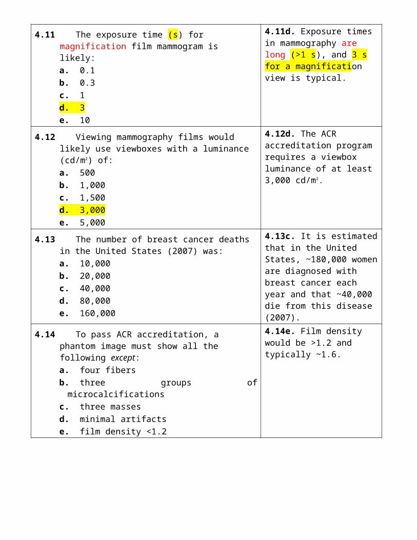
4.11 The exposure time (s) for magnification film mammogram is likely:a. 0.1b. 0.3c. 1d. 3e. 10
4.11d. Exposure times in mammography are long (>1 s), and 3 s for a magnification view is typical.
4.12 Viewing mammography films would likely use viewboxes with a luminance (cd/m2) of:a. 500b. 1,000c. 1,500d. 3,000e. 5,000
4.12d. The ACR accreditation program requires a viewbox luminance of at least 3,000 cd/m2.
4.13 The number of breast cancer deaths in the United States (2007) was:a. 10,000b. 20,000c. 40,000d. 80,000e. 160,000
4.13c. It is estimated that in the United States, ~180,000 women are diagnosed with breast cancer each year and that ~40,000 die from this disease (2007).
4.14 To pass ACR accreditation, a phantom image must show all the following except:a. four fibersb. three groups of microcalcificationsc. three massesd. minimal artifactse. film density <1.2
4.14e. Film density would be >1.2 and typically ~1.6.
4.15 Which repeat rate (%) is most likely to occur in screen–film mammography?a. 0.1b. 0.3c. 1d. 3e. 10
4.15d. A repeat rate of 3% is typical for a screen–film mammography facility.

4.16 The II input phosphor is most likely made of:a. NaIb. PbIc. LiFd. CsIe. Se
4.16d. The input to an image intensifier is normally made of CsI, which has excellent x-ray absorption properties.Scintillator
4.17 The II flux gain is most likely:a. 2b. 5c. 10d. 25e. 50
4.17e. A typical II flux gain is ~50.
4.18 The brightness gain of a 250-mmdiameter II is most likely:a. 3b. 10c. 30d. 100e. >100
4.18e. II brightness gains are ~5,000 (flux gain of ~50 and minification gain of ~100).
4.19 Image intensifier output brightness during fluoroscopy is least influenced by:a. tube voltageb. tube currentc. exposure timed. II diametere. phosphor thickness
4.19c. Exposure time does not affect image brightness during fluoroscopy.
4.20 Falloff in brightness at the periphery of a fluoroscopic image is called:a. vignettingb. pincushion distortionc. barrel distortiond. S-wave distortione. edge packing
4.20a. Fall off in brightness at the periphery of a fluoroscopic image is called vignetting.
4.21 The aspect ratio of high-definition TV is:a. 4:3b. 5:4c. 7:5d. 12:7e. 16:9
4.21e. HTDV uses a 16:9 aspect ratio, whereas traditional (analog) TV uses a 4:3 aspect ratio.

4.22 Replacing a TV camera with a CCD would likely improve (%) fluoroscopy signal to noise ratio by:a. 0b. 25c. 50d. 100e. >100
4.22a. Fluoroscopy is quantum noise limited imaging, which means that the TV/CCD will not be an additional source of significant noise.
4.23 The most likely tube current (mA) in fluoroscopy is:a. 3b. 10c. 30d. 100e. 300
4.23a. Low tube currents (~3 mA) are the norm in fluoroscopy.
4.24 Pulsed fluoroscopy would likely acquire images at a rate (frames per second) of:a. 15b. 30c. 45d. 60e. >60
4.24a. Since pulsed fluoroscopy uses <30 frames per second, it would acquire 15 frames per second.
4.25 Automatic brightness control (ABC) in fluoroscopy attempts to maintain a constant:a. tube voltageb. tube currentc. exposure timed. patient dosee. II brightness
4.25e. Automatic brightness control (ABC) is used to maintain a constant brightness at the output of an II.
4.26 For constant techniques (kV/mA), switching an II from 250 mm to 125 mm input diameter likely increases skin doses (%) by:a. 25b. 50c. 100d. 200e. 400
4.26e. Halving the II input diameter will reduce the exposed CsI phosphor area to a quarter and require a fourfold increase (400%) in the radiation intensity if the II output brightness intensity is to be kept constant.
4.27 Tube currents (mA) in photospot imaging are most likely:a. 0.3b. 3c. 30d. 300e. 3,000
4.27d. A typical tube current used to generate a digital photospot image is 300 mA.
4.28 What is the most likely matrix size of digital photospot image?a. 2562
b. 5122
c. 1,0242
d. 2,0482
e. 4,0962
4.28c. The most common matrix size in digital photospot imaging is 1,0242.
4.29 Use of temporal filtering in digital fluoroscopy would likely increase:a. noiseb. scatterc. dosed. lage. contrast
4.29d. Filtering requires frame averaging, which must increase image lag.
4.30 Increasing the DSA matrix size would likely decrease:a. pixel sizeb. digitization ratec. image contrastd. data storagee. processing time
4.30a. Pixel size always decreases with increasing matrix size.
A31 The target material in a mammography x-ray tube is most likely:
a. Be (Z = 4) b. Al (Z = 13) c. Mo (Z = 42) d. Ag (Z = 47) e. Ba (Z = 56)
A31c. Mo is the most common target material in mammography x-ray tubes.
A32 In a linear grid for mammography, a fiber interspaced grid is preferred over aluminum because it likely reduces: a. dose b. scatter c. mottle d. receptor blur e. focal blur
A32a. Fiber has a lower atomic number and density than Al, and will therefore transmit more primary photons (i.e., it reduces the patient dose).
A33 High image contrast is least likely achieved in mammography by the use of:
a. low photon energies b. high film gradients c. short exposures (<0.1 s) d. breast compression e. scatter removal grids
A33c. Exposure time has no direct impact on image contrast.
A34 The optimal grid ratio in magnification mammography is most likely:
a. no grids used b. 2:1 c. 4:1 d. 8:1 e. 16:1
A34a. Grids are not used in magnification mammography, since the air gap will minimize scatter at the image receptor.
A35 The Mammography Quality Standards Act does not require:
a. reject analysis b. processor sensitometry c. physics testing d. ACR accreditation e. FDA certification
A35d. The MQSA requires accreditation from an approved body, but it does not have to be the ACR.
A36 The purpose of photocathodes in image intensifiers is to convert light into:
a. x-rays b. heat c. voltages d. electrons e. ultraviolet
A36d. Photocathodes absorb light photons, and emit low-energy electrons.
A37 A typical II conversion factor (cd/m2 per μGy/s), is most likely:
a. 2 b. 20 c. 200 d. 2,000 e. 20,000
A37b. A typical II conversion factor is ~20 cd/m2 per μGy/s.
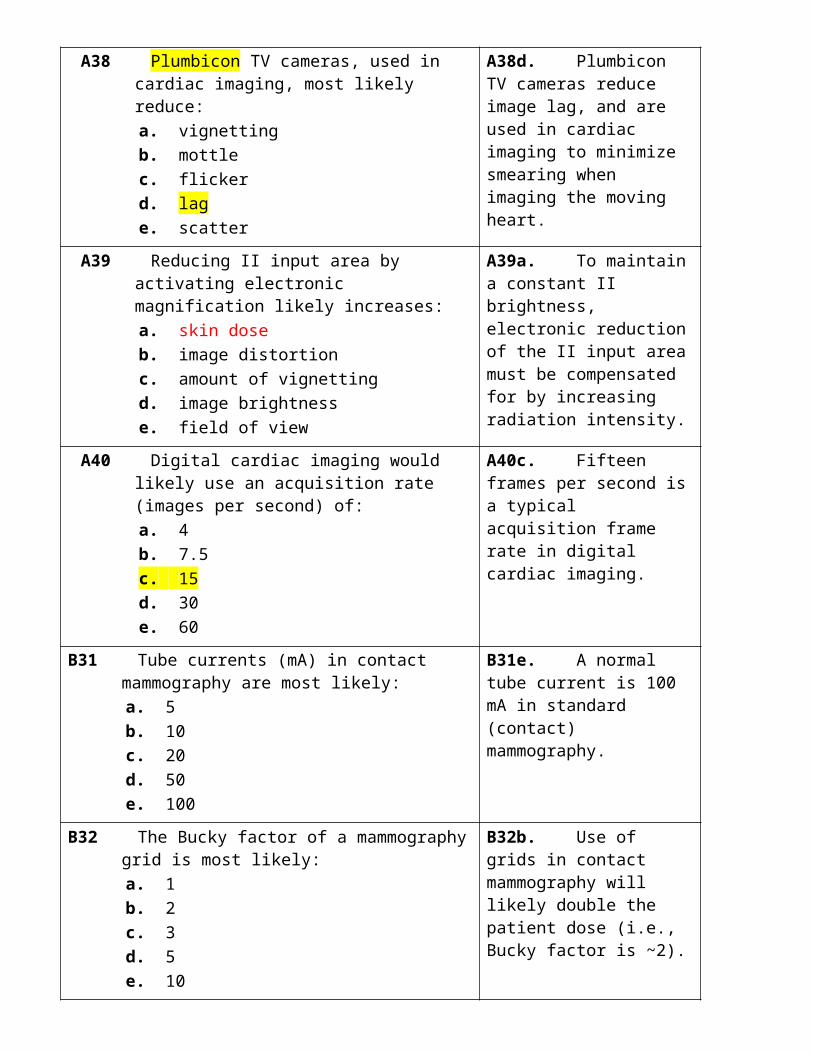
A38 Plumbicon TV cameras, used in cardiac imaging, most likely reduce:
a. vignetting b. mottle c. flicker d. lag e. scatter
A38d. Plumbicon TV cameras reduce image lag, and are used in cardiac imaging to minimize smearing when imaging the moving heart.
A39 Reducing II input area by activating electronic magnification likely increases:
a. skin dose b. image distortion c. amount of vignetting d. image brightness e. field of view
A39a. To maintain a constant II brightness, electronic reduction of the II input area must be compensated for by increasing radiation intensity.

A40 Digital cardiac imaging would likely use an acquisition rate (images per second) of:
a. 4 b. 7.5 c. 15 d. 30 e. 60
A40c. Fifteen frames per second is a typical acquisition frame rate in digital cardiac imaging.
B31 Tube currents (mA) in contact mammography are most likely:
a. 5 b. 10 c. 20 d. 50 e. 100
B31e. A normal tube current is 100 mA in standard (contact) mammography.
B32 The Bucky factor of a mammography grid is most likely:
a. 1 b. 2 c. 3 d. 5 e. 10
B32b. Use of grids in contact mammography will likely double the patient dose (i.e., Bucky factor is ~2).
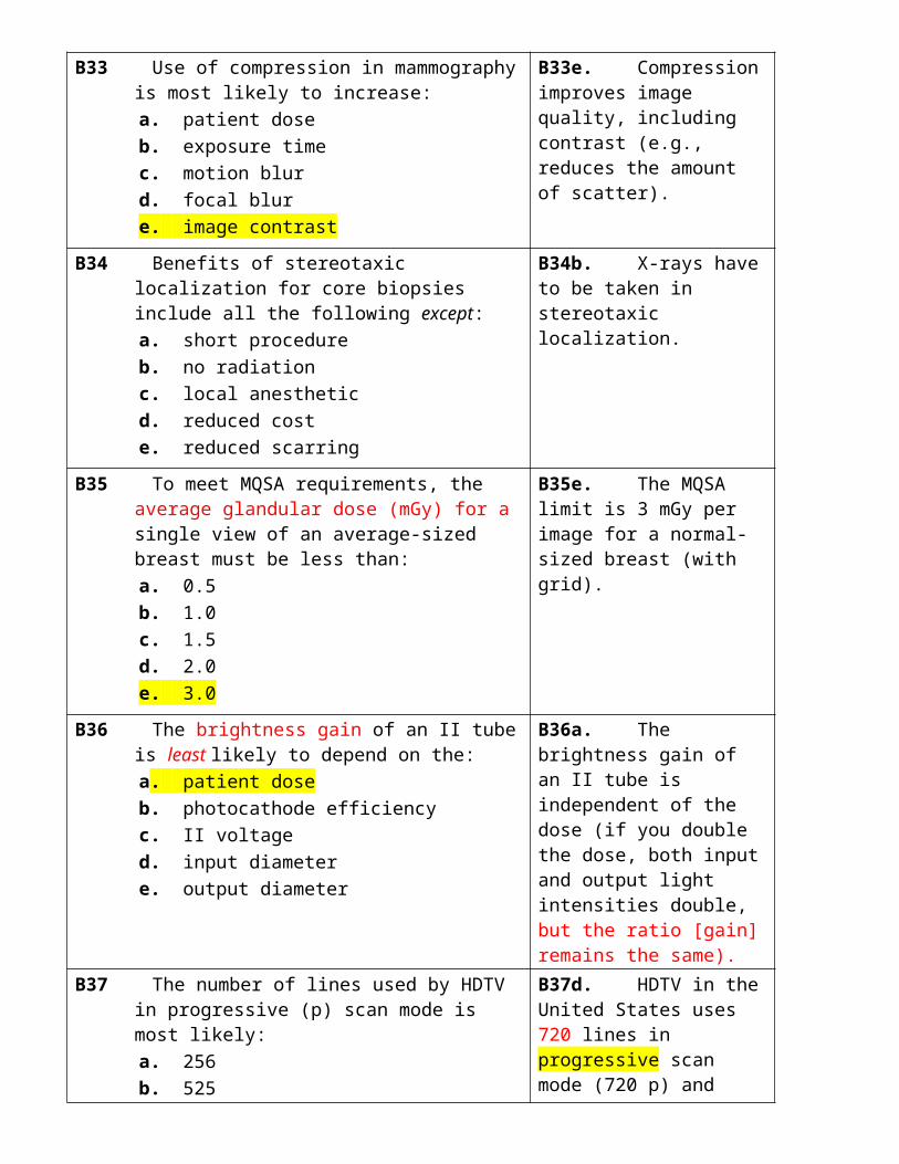
B33 Use of compression in mammography is most likely to increase:
a. patient dose b. exposure time c. motion blur d. focal blur e. image contrast
B33e. Compression improves image quality, including contrast (e.g., reduces the amount of scatter).
B34 Benefits of stereotaxic localization for core biopsies include all the following except:
a. short procedure b. no radiation c. local anesthetic d. reduced cost e. reduced scarring
B34b. X-rays have to be taken in stereotaxic localization.
B35 To meet MQSA requirements, the average glandular dose (mGy) for a single view of an average-sized breast must be less than:
a. 0.5 b. 1.0 c. 1.5 d. 2.0 e. 3.0
B35e. The MQSA limit is 3 mGy per image for a normal-sized breast (with grid).
B36 The brightness gain of an II tube is least likely to B36a. The brightness gain of

depend on the: a. patient dose b. photocathode efficiency c. II voltage d. input diameter e. output diameter
an II tube is independent of the dose (if you double the dose, both input and output light intensities double, but the ratio [gain] remains the same).
B37 The number of lines used by HDTV in progressive (p) scan mode is most likely:
a. 256 b. 525 c. 625 d. 720 e. 1,080
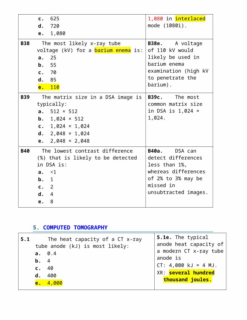
B37d. HDTV in the United States uses 720 lines in progressive scan mode (720 p) and 1,080 in interlaced mode (1080i).
B38 The most likely x-ray tube voltage (kV) for a barium enema is:
a. 25 b. 55 c. 70 d. 85 e. 110
B38e. A voltage of 110 kV would likely be used in barium enema examination (high kV to penetrate the barium).
B39 The matrix size in a DSA image is typically: a. 512 × 512 b. 1,024 × 512 c. 1,024 × 1,024 d. 2,048 × 1,024 e. 2,048 × 2,048
B39c. The most common matrix size in DSA is 1,024 × 1,024.
B40 The lowest contrast difference (%) that is likely to be detected in DSA is:
a. <1 b. 1 c. 2 d. 4 e. 8
B40a. DSA can detect differences less than 1%, whereas differences of 2% to 3% may be missed in unsubtracted images.
5. COMPUTED TOMOGRAPHY
5.1 The heat capacity of a CT x-ray tube anode (kJ) is most likely:a. 0.4b. 4c. 40d. 400e. 4,000
5.1e. The typical anode heat capacity of a modern CT x-ray tube anode isCT: 4,000 kJ = 4 MJ.XR: several hundred thousand
joules.

5.2 The power (kW) applied to a modern CT x-ray tube is most likely:a. 1b. 3c. 10d. 30e. 100
5.2e. The most common power level in CT today (2008) is 100 kW.
120 mA x 750 kv = 90000 W = 90 kW
5.3 A CT beam shaping filter (bow tie) is most likely made out of:a. aluminumb. copperc. molybdenumd. Teflone. tin
5.3d. Teflon (i.e., tissue like) is used as the CT beam shaping filter material to minimize beam hardening artifacts.
a low Z material
5.4 CT collimation is most likely used to change the x-ray beam:a. widthb. intensityc. HVLd. FOVe. isocenter
5.4a. CT collimation changes the x-ray beam width.
5.5 The most likely x-ray beam width (mm) on a 64-row CT scanner is:a. 0.5b. 5c. 10d. 20e. 40
5.5e. The most likely x-ray beam width is 40 mm because the detector thickness is comparable to the in-plane pixel size of ~0.6 mm.
64 x 0.625 = 40
5.6 The total number of individual detector elements on a 64-row CT scanner is most likely:a. 64 × 100b. 64 × 200c. 64 × 400d. 64 × 800e. 64 × 1,600
5.6d. 64 × 800 Since each slice would make use of ~800 individual detectors.
5.7 The percentage (%) of incident radiation likely captured by a CT x-ray detector is:a. 30b. 45c. 60d. 75e. >75
5.7e. CT x-ray detectors are very efficient and capture well over 75% of the incident radiation (e.g., 90%).

5.8 The number of projections obtained per 360-degree rotation of the x-ray tube in a single-slice CT scanner is most likely:a. 500b. 1,000c. 2,000d. 4,000e. 8,000
5.8b. In CT, ~1,000 projections are obtained for a single rotation of the x-ray tube.
5.9 Use of a bone filter, as opposed to a soft tissue filter, to reconstruct CT images would likely improve:a. subject contrastb. image contrastc. scatter rejectiond. spatial resolutione. data storage
5.9d. Bone filters improve spatial resolution but also result in higher mottle (noise).
5.10 The CT number (HU) is directly proportional to the pixel:a. mass attenuationb. linear attenuationc. physical densityd. electron densitye. atomic number
5.10b. CT numbers are directly proportional to the pixel linear attenuation.
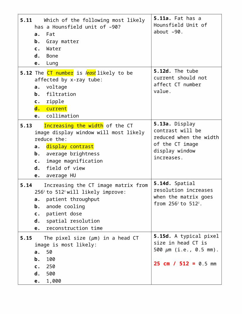
5.11 Which of the following most likely has a Hounsfield unit of –90?a. Fatb. Gray matterc. Waterd. Bonee. Lung
5.11a. Fat has a Hounsfield Unit of about –90.
5.12 The CT number is least likely to be affected by x-ray tube:a. voltageb. filtrationc. rippled. currente. collimation
5.12d. The tube current should not affect CT number value.
5.13 Increasing the width of the CT image display window will most likely reduce the:a. display contrastb. average brightnessc. image magnificationd. field of viewe. average HU
5.13a. Display contrast will be reduced when the width of the CT image display window increases.

5.14 Increasing the CT image matrix from 2562 to 5122 will likely improve:a. patient throughputb. anode coolingc. patient dosed. spatial resolutione. reconstruction time
5.14d. Spatial resolution increases when the matrix goes from 2562 to 5122.
5.15 The pixel size (μm) in a head CT image is most likely:a. 50b. 100c. 250d. 500e. 1,000
5.15d. A typical pixel size in head CT is 500 μm (i.e., 0.5 mm).
25 cm / 512 = 0.5 mm
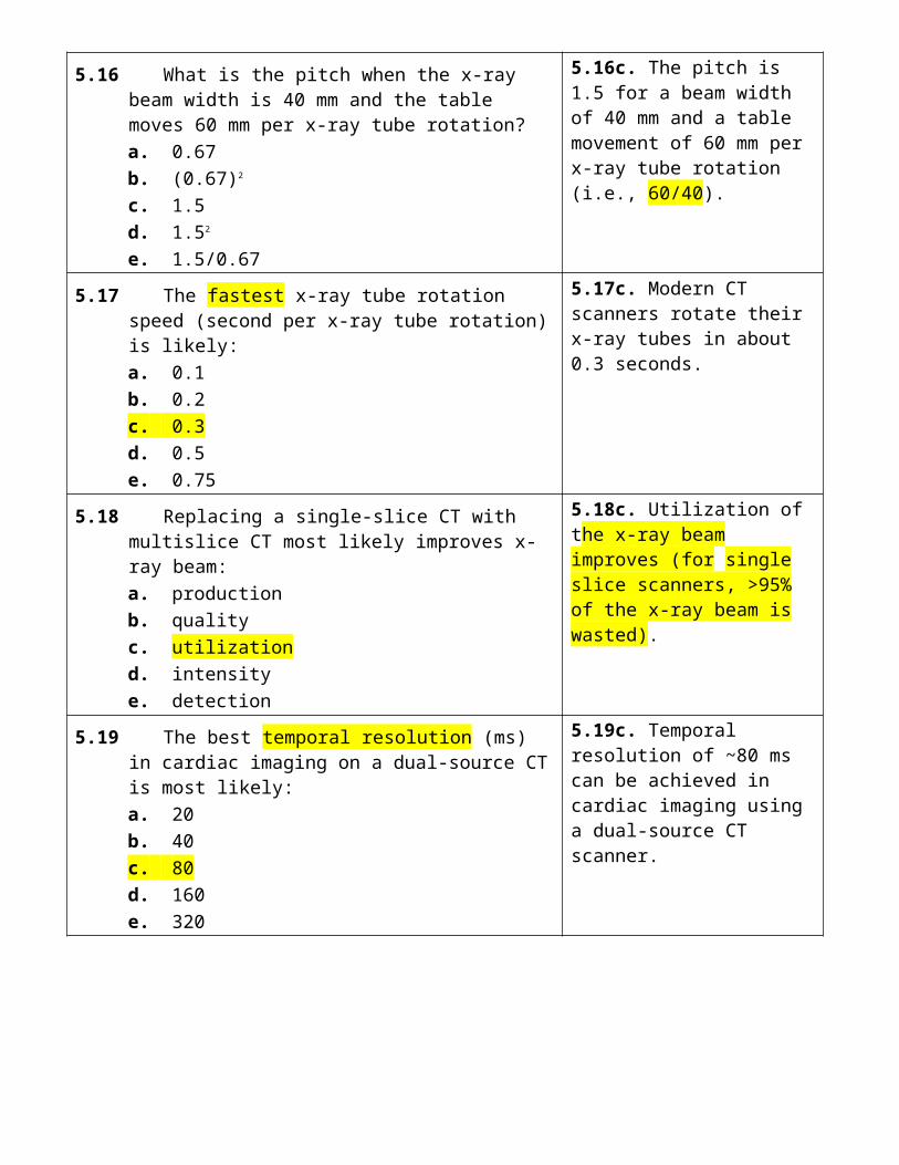
5.16 What is the pitch when the x-ray beam width is 40 mm and the table moves 60 mm per x-ray tube rotation?a. 0.67b. (0.67)2
c. 1.5d. 1.52
e. 1.5/0.67
5.16c. The pitch is 1.5 for a beam width of 40 mm and a table movement of 60 mm per x-ray tube rotation (i.e., 60/40).
5.17 The fastest x-ray tube rotation speed (second per x-ray tube rotation) is likely:a. 0.1b. 0.2c. 0.3d. 0.5e. 0.75
5.17c. Modern CT scanners rotate their x-ray tubes in about 0.3 seconds.
5.18 Replacing a single-slice CT with multislice CT most likely improves x-ray beam:a. productionb. qualityc. utilizationd. intensitye. detection
5.18c. Utilization of the x-ray beam improves (for single slice scanners, >95% of the x-ray beam is wasted).
5.19 The best temporal resolution (ms) in cardiac imaging on a dual-source CT is most likely:a. 20b. 40c. 80d. 160e. 320
5.19c. Temporal resolution of ~80 ms can be achieved in cardiac imaging using a dual-source CT scanner.
5.20 The number of x-ray tube rotations required to measure the CTDIc in a head phantom is:a. 1b. 2c. 3d. 7e. 10
5.20a. One, as each individual computed tomography dose index (CTDI) measurement is obtained for a single x-ray tube rotation.
5.21 The ratio of the peripheral CTDI to the central CTDI in a body phantom is most likely:a. 0.25b. 0.5c. 1d. 2e. 4
5.21d. Two, since the peripheral dose in a 32-cm acrylic cylinder is generally double that of the central dose.
5.22 If the peripheral CTDI is 12 mGy and the central CTDI is 6 mGy, the weighted CTDIw (mGy) is:a. 7b. 8c. 9d. 10e. 11
5.22d. Ten mGy (CTDIw is one-third the central CTDI plus two thirds the peripheral CTDI).
2/3 CTDIp + 1/3 CTDIc
5.23 What is the dose length product DLP (mGycm) for a CTDIw of 20 mGy, pitch of 2, and scan length of 100 cm?a. 200b. 400c. 500d. 800e. 1,000
5.23e. One thousand mGy obtained by multiplying the scan length by CTDIvol, which is the CTDIw divided by the pitch (i.e., 100 cm × 20 mGy/2).
CTDIw / pitch x scan length
5.24 The reference dose (CTDIvol mGy) recommended by the ACR (2008) for an adult head CT is:a. 25b. 50c. 75d. 100e. 125
5.24c. The CTDIvol reference dose (mGy) currently recommended by the ACR for an adult head CT (2008) is 75 mGy.
5.25 If an adult head CT scan uses 100%, the most likely technique (%) for a 1year-old is:a. 15b. 30c. 45d. 60e. 85
5.25e. Head techniques in a 1-year-old are reduced by 15%, so 85% would be used.
85% of adult

5.26 CT fluoroscopy best minimizes radiation doses by reducing:a. beam filtrationb. focus sizec. tube currentd. slice thicknesse. matrix size
5.26c. Tube currents are generally reduced in CT fluoroscopy.
5.27 The optimal x-ray tube voltage (kV) for performing CT angiography is most likely:a. 80b. 100c. 120d. 140e. >140
5.27a. A voltage of 80 kV maximizes iodine contrast by bringing the average x-ray energy closer to the iodine K-shell energy (33 keV).
5.28 The most likely voltages (kV) used in dual-energy CT are:a. 80 and 100b. 80 and 120c. 80 and 140d. 100 and 140e. 120 and 140
5.28c. Voltages of 80 kV and 140 kV would likely be used.
5.29 Ring artifacts in CT are most likely caused by:a. beam hardeningb. metallic implantsc. faulty detectorsd. patient motione. scattered x-rays
5.29c. Faulty detectors can result in ring artifacts.
5.30 CT beam hardening artifacts are minimized by increasing the:a. tube voltageb. tube currentc. scan timed. matrix sizee. helical pitch
5.30a. Increasing tube voltage in CT minimizes beam hardening.
A41 The typical anode cooling rate (kW) of a standard CT x-ray tube is most likely:
a. 1 b. 3 c. 10 d. 30 e. 100
A41c. Ten kW is a common anode cooling rate (kW) in CT with standard x-ray tubes; the Straton x-ray tube is an exception that has a cooling rate of ~60 kW.

A42 A beam-shaping filter is most likely used in CT scanners to reduce:
a. detector dynamic range b. beam hardening c. detector cross-talk d. off-focus radiation e. scatter radiation
A42a. Beam-shaping filters reduce the CT detector dynamic range.
A43 The detected x-ray pattern transmitted through the patient at a single x-ray tube angle is best described as a:
a. ray b. projection c. back projection d. convolution e. tomographic slice
A43b. The detected x-ray pattern at a single x-ray tube angle is called a projection.
A44 Use of a soft tissue filter, as opposed to a bone filter, to reconstruct CT images would most likely reduce:
a. mottle b. scatter c. dose d. artifacts e. scan times
A44a. Soft tissue filters reduce mottle at the cost of inferior spatial resolution performance.
A45 A window width of 100 and window level of 50 likely results in a pixel value of 10 appearing as:
a. black b. almost black c. gray d. almost white e. white
A45b. A HU of 10 will look almost black with a window width of 100 and window level of 50 (50 looks gray, and 0 looks black).
Center = gray
A46 The advantage of helical over axial CT is most likely a reduction in:
a. radiation doses b. scan times c. scatter radiation d. reconstruction times e. slice sensitivity profiles
A46b. Scan times are markedly reduced when axial scanning is replaced with helical CT.
A47 Total exam time (s) for a single-phase adult abdomen on a 64-slice MDCT is likely:
a. 0.3 b. 1 c. 3 d. 10 e. 30
A47c. Three seconds should be possible (patient coverage per rotation of 4 cm; tube rotation time 0.3 s; ten rotations).Slice = 4cm.Rotations = 40/4 = 10Time = 0.3 x 10 = 3

A48 When the weighted CTDIw is 10 mGy, and the pitch is 0.25, the volume CTDIvol (mGy) is most likely:
a. 2.5 b. 5 c. 10 d. 20 e. 40 10 mGy/0.25
A48e. Forty mGy since CTDIvol = CTDIw / pitch(i.e., 10 mGy/0.25).
10 mGy/0.25
A49 Compared to an adult (mAs = 100%), the most likely mAs (%) for a body CT scan of a 1-year-old would be:
a. 10 b. 30 c. 50 d. 70 e. >70
A49c. Body techniques (mAs) in a 1-year-old can be reduced to ~50% with no loss of diagnostic information.
Head: 25% reduction
Body: 50% reduction
TABLE 5.8
A50 Partial-volume artifacts in CT are best minimized by reducing:
a. section thickness b. scan time c. matrix size d. focal blur e. scan length
A50a. Reducing the section thickness will minimize partial volume artifacts.
B41 The most likely filtration (mm Al) used in CT x-ray tubes is:
a. 1 b. 2 c. 3 d. 6 e. 12
B41d. Filtration is frequently 6 mm in CT x-ray tubes (excluding the bow tie filter).
B42 Which material is least likely to be used to construct an array of CT detectors?
a. Bismuth germanate b. Cadmium tungstate c. Xenon (high pressure) d. Sodium iodide e. Lithium fluoride
B42e. Lithium fluoride is a thermoluminescent dosimeter, which would be useless in CT imaging.
B43 Which CT image reconstruction algorithm is most likely used in clinical practice?
a. 2D Fourier transform b. 3D Fourier transform c. Back projection d. Filtered back projection e. Iterative reconstruction
B43d. CT images are generally reconstructed using filtered back projection.
CT: filtered back projection.MRI: 2d fourier transform
B44 The difference (%) in x-ray attenuation between 40 B44b. A difference of 10

HU and 50 HU is: a. 0.1 b. 1 c. 10 d. 25 e. 45
HU corresponds to a difference in x-ray attenuation of
1% difference = 10 HU
B45 The data acquisition geometry (i.e., generation) of a 64-slice CT scanner is most likely:
a. first b. second c. third d. fourth e. fifth
B45c. MSCT uses third-generation acquisition geometry (x-ray tube and detector array both rotate).
B46 The temporal resolution (ms) of a dual-source CT scanner (gantry rotation speed of 0.30 s) in cardiac imaging is most likely:
a. 37.5 b. 75 c. 150 d. 30 e. 600
B46b. The resolution is 75 ms; for a single-source CT scanner, the temporal resolution is approximately half the rotation time, and a dual-source CT scanner is half the value of a single-source system.
B47 The ratio of the peripheral to central CTDI in a head phantom is most likely:
a. 0.25:1 b. 0.5:1 c. 1:1 d. 2:1 e. 4:1
B47c. The ratio is 1:1, as doses in head CT are pretty uniform in 16-cm-diameter phantoms.
B48 The reference dose (CTDIvol mGy) recommended by the ACR (2008) for an adult abdominal CT is most likely:
a. 25 b. 50 c. 75 d. 100 e. 125
B48a. The CTDIvol reference dose currently (2008) recommended by the ACR for an adult abdomen CT is 25 mGy.Abdomen : 25Head: 75TABLE 5.6
B49 The most likely pitch in retrospective gating cardiac imaging is:
a. 0.25 b. 0.5 c. 1.0 d. 1.5 e. 2.0
B49a. A typical pitch is 0.25 in retrospective gating cardiac imaging.

B50 Which of the following artifacts is least likely in CT? a. Motion b. Zipper c. Streak d. Ring e. Beam-hardening
B50b. Zipper artifacts are observed in MR, not CT.
6. IMAGE QUALITY
6.1 Subject contrast is most likely to be affected by the:a. exposure timeb. tube current (mA)c. tube voltage (kV)d. focus sizee. display window
6.1c. The x-ray tube voltage (average photon energy) is the most important factor that affects subject contrast.
6.2 The most important factor for maximizing film contrast is most likely the film:a. optical densityb. base thicknessc. exposure timed. processing timee. fog level
6.2a. Film optical density is critical (light and dark films have very little image contrast).
6.3 Film contrast is inversely related to film:a. fogb. noisec. latituded. speede. resolution
6.3c. Film contrast (i.e., gradient) is inversely related to film latitude.
6.4 A characteristic curve with a high gamma likely results in images with a high:a. patient doseb. film densityc. quantum mottled. image contraste. fog level
6.4d. Gamma is the maximum film gradient; high gamma produces a high image contrast.
6.5 Screen–film mammography contrast is likely improved by increasing:a. tube voltageb. target atomic numberc. screen thicknessd. film latitudee. film gradient
6.5e. A high film gradient in mammography (~3 or more) results in high image contrast.
6.6 Increasing the kV alone in CT scanning would most likely reduce:a. anode loadingb. image mottlec. patient dosed. reconstruction timee. scan time
6.6b. Mottle will be reduced at higher kV because more x-rays are produced, and the x-ray beam is more penetrating, which will increase the number of detected photons.↑ penetration, increase the number of detected photons.
6.7 Increasing the amount of scatter in a radiograph reduces:a. image contrastb. focal blurc. screen blurd. image mottlee. patient dose
6.7a. Image contrast is reduced when scatter increases.
6.8 Which of the following is least likely a measure of spatial resolution?a. ROCb. PSFc. LSFd. FWHMe. MTF
6.8a. ROC is receiver operating characteristic, which measures diagnostic performance (PSF is the point spread function; LSF is the line spread function; FWHM is the full width half maximum; and MTF is the modulation transfer function).
6.9 Spatial resolution is important when detecting lesions that are characterized as being:a. small sizeb. low contrastc. high contrastd. less attenuatinge. more attenuating
6.9a. Spatial resolution is important for detecting, differentiating, and characterizing lesions that have a small size.
6.10 The most likely limitation of geometric magnification is an increase in:a. focal blurb. screen blurc. scattered photonsd. quantum mottlee. detector exposure
6.10a. Focal blur is extremely important in magnification imaging.
6.11 Radiographic spatial resolution performance can be best improved by reducing:a. beam filtrationb. detector exposurec. detector thicknessd. grid ratioe. tube voltage
6.11c. Detector thickness (e.g., screen thickness) is important for determining the spatial resolution performance.
6.12 Increasing the detector thickness to absorb more x-rays will most likely increase image:a. contrastb. magnificationc. blurd. mottlee. brightness
6.12c. Image blur will increase with a thicker x-ray detector.
6.13 When the full width half maximum FWHM of an imaged slit is 0.1 mm, the limiting resolution (line pairs per mm) is most likely:a. 1b. 2c. 3d. 5e. 10
6.13d. Five line pairs per mm, since the achievable number of lp/mm is normally taken to be ~1/(2 × FWHM).
6.14 The MTF value (%) at the lowest spatial frequencies is most likely:a. 100b. 75c. 50d. 25e. 0
6.14a. The MTF value is 100% for all imaging systems; this effectively means that huge objects can very easily be seen.
6.15 The limiting spatial resolution (lp/mm) of a (dedicated) chest screen– film unit is likely:a. 0.5b. 1c. 2.5d. 5e. 10
6.15d. Screen–film (200 speed) should achieve 5 lp/mm.
6.16 Actual vertical resolution (line pairs) achieved with a 525-line TV monitor is:a. 180 (525/2v= 262 pairs ….. actual = 70% of 262)b. 262c. 370d. 425e. 525
6.16a. A TV with 525 lines can display 262.5 line pairs, but TV achieves only 70% of this value, so actual vertical TV resolution is 180 line pairs.
6.17 The horizontal resolution of a TV system is primarily determined by the:a. image lagb. bandwidthc. TV linesd. frame ratee. camera size
6.17b. The TV bandwidth determines the horizontal TV resolution (usually the same as the vertical resolution).
6.18 Digital fluoroscopy spatial resolution would most likely be improved by increasing the:a. grid ratiob. II input diameterc. air kermad. tube voltagee. image matrix
6.18e. Increasing the image matrix size will normally improve spatial resolution.
6.19 The maximum number of line pairs that can be observed using a 1k matrix size is:a. 50b. 100c. 250d. 500 (1 line pair needs two pixels, one that is white and one
that is black).e. 1,000
6.19d. A 1k matrix can display 500 line pairs (1 line pair needs two pixels, one that is white and one that is black).
6.20 The best achievable head CT limiting resolution (line pairs/mm) using a 5122 matrix and 25 cm field-of-view is most likely:a. 0.25b. 0.5c. 1.0 (1/2 X sampling frequency = ½ 512/250)d. 2.0e. 4.0
6.20c. One line pair per mm, since the pixel size is 0.5 mm.
6.21 If an average of 10,000 photons are detected per mm2, the chance (%) of detecting between 9,700 and 10,300 counts in any exposed mm2 is:a. 67b. 90c. 95d. 99e. 99.9
6.21d. Ninety-nine percent, since the standard deviation is 100, and the limits correspond to three standard deviations.
6.22 X-ray quantum mottle is best characterized by quantifying:a. x-ray beam filtrationb. detector air kermac. average photon energyd. scintillator conversion efficiencye. image receptor thickness
6.22b. The detector air kerma determines the number of x-ray photons used to make an image.

6.23 For comparable image mottle in an abdominal radiograph, which image receptor would likely result in the highest patient dose?a. Screen-filmb. Photostimulable phosphorc. Direct flat panel detectord. Indirect flat panel detectore. Digital photospot
6.23b. Photostimulable phosphor requires more radiation as it must be thin to minimize light scatter during the readout process.
6.24 The dominant source of image mottle in a radiographic flat panel detector is most likely:a. detector granularityb. electronic noisec. digitization noised. quantum mottlee. monitor structure
6.24d. Quantum mottle, as virtually all radiographic and CT imaging is quantum noise limited.
6.25 The detector air kerma (μGy) producing a digital photospot image in a Ba enema examination is most likely:a. 1b. 5c. 25d. 100e. 500
6.25a. The detector air kerma in digital photospot imaging is 1 μGy.
6.26 The II air kerma (μGy) needed to produce a single digital fluoroscopy image (frame) is most likely:a. 0.01b. 0.03c. 0.1d. 0.3e. 1
6.26a. The air kerma that produces a single digital fluoroscopy frame is 0.01 μGy.
6.27 CT image mottle is least likely to be affected by changing the:a. section thicknessb. reconstruction algorithmc. patient sized. x-ray intensitye. window width
6.27e. The display window width will not affect the image data, only the way it appears on the monitor.
6.28 Sensitivity is given by the:a. true-negative fractionb. true-positive fractionc. (1 – true-positive fraction)d. (1 + true-negative fraction)e. true positives + true negatives
6.28b. The true-positive fraction is the sensitivity.

6.29 Specificity is given by the:a. true-negative fractionb. true-positive fractionc. (1 – true-positive fraction)d. (1 + true-negative fraction)e. true positives + true negatives
6.29a. The true-negative fraction is the specificity.
6.30 Relaxing the threshold criterion in a ROC study increases false-positive fraction as well as the test:a. performanceb. specificityc. ROC aread. sensitivitye. accuracy
6.30d. Sensitivity will increase as the threshold criterion increases and one moves up the ROC curve from lower left to upper right.
A51 The highest subject contrast of an iodinated blood vessel likely occurs at a photon energy (keV) of:
a. 30 b. 40 closest larger than 33 c. 50 d. 70 e. 100
A51b. 40 keV will achieve the maximum absorption by iodine (K-shell binding energy of 33 keV) and maximize subject contrast.
A52 Lowering the kV in screen–film mammography most likely reduces:
a. contrast b. dose c. mAs d. scatter e. exposure time
A52d. Scatter is lower at lower energies since photoelectric absorption will be more important than Compton scatter.
A53 What x-ray tube voltage (kV) would likely maximize the visibility of iodinated contrast in the carotid arteries?
a. 30 b. 50 c. 70 d. 90 e. 110
A53c. Seventy kV will have an average energy (1/2 to 1/3 of the 70 keV maximum) that is close to the iodine K-edge of 33 keV.
A54 Which of the following factors is least likely to affect image sharpness?
a. Detector composition b. Focal spot size c. Exposure time d. Detector thickness e. Image magnification
A54a. Detector composition has negligible impact on spatial resolution performance.
A55 When the MTF from focal and receptor blur are both equal to 0.1 (at 2 lp/ mm), the imaging system MTF at this spatial frequency is most likely:
a. 0.2 b. 0.1 c. 0.05 d. 0.02 e. 0.01
A55e. Most likely 0.01 since the system MTF is the product of the component MTF values at each spatial frequency.
A56 Spatial resolution of a standard fluoroscopy unit is most likely limited by the:
a. focal spot b. input phosphor c. output phosphor d. optical system e. TV system
A56e. The TV system is the weak link in the fluoroscopy imaging chain.
A57 CT scanner spatial resolution performance would most likely improve when increasing the:
a. focal spot b. detector width c. tube current d. scan time e. image matrix
A57e. An increase in the image matrix size could improve spatial resolution.
A58 Visibility of low-contrast lesions in a digital radiograph would most likely be improved when increasing:
a. focus size b. image magnification c. air kerma d. beam filtration e. display luminance
A58c. Increasing the receptor air kerma would reduce mottle and improve visibility of low contrast lesions.
A59 The detector air kerma (μGy) that produces one frame in DSA imaging is most likely:
a. 5 x 5 b. 15 c. 50 d. 150 e. 500
A59a. The image receptor air kerma in DSA is 5 μGy, or five times higher than in digital photospot imaging (1 μGy).
A60 A receiver operator characteristic curve likely measures:
a. diagnostic performance b. error rate c. test specificity d. test sensitivity e. cost-effectiveness
A60a. A receiver operator characteristic curve measures diagnostic performance.
B51 In screen–film radiography, raising the tube voltage (kV) likely reduces:
a. half-value layer b. scatter radiation c. patient transmission d. subject contrast e. grid penetration
B51d. Subject contrast is reduced when kV increases.
B52 Increasing kV in digital mammography most likely increases:
a. image contrast b. quantum mottle c. breast penetration d. focal blur e. exposure time
B52c. Breast penetration increases at higher kV.
B53 Spatial resolution performance is least likely to be assessed using a:
a. line pair phantom b. line spread function c. full width half maximum d. modulation transfer function e. pixel standard deviation
B53e. The pixel standard deviation is a measure of mottle (noise), not spatial resolution.
B54 Minimizing which factor would most likely improve spatial resolution?
a. Exposure time b. Tube voltage c. Tube current d. Beam filtration e. Window width
B54a. Minimizing exposure time reduces motion blur.
B55 Measured limiting spatial resolution (lp/mm) of screen–film mammography is likely:
a. 1 b. 2 c. 4 d. 8 e. 16
B55e. Screen–film mammography normally achieves ~16 lp/mm (the ACR limit is ~12 lp/mm).
B56 Going from a 2562 to a 5122 matrix size is most likely to double the:
B56a. Doubling the matrix size could double the spatial

a. spatial resolution b. number of pixels c. gray levels d. transmission time e. storage requirements
resolution.
B57 When the average number of x-ray photons detected by a pixel is 100, the standard deviation is most likely:
a. 1 b. 3 c. 10 d. 30 e. 100
B57c. Ten, as the standard deviation is the square root of the mean number of counts.
B58 The detector air kerma (μGy) in digital mammography is most likely:
a. 1 b. 3 c. 10 d. 30 e. 100
B58e. is the detector air kerma in mammography is ~100 μGy.
B59 Visibility of large low-contrast CT lesions likely improves with increasing:
a. beam filtration b. tube current c. field of view d. matrix size e. window width
B59b. Higher tube current will reduce mottle and improve the visibility of (large) low-contrast lesions.
B60 A diagnostic test is of no value when the area under the ROC curve (%) has a value of:
a. 0 b. 25 c. 50 d. 75 e. 100
B60c. Random guessing corresponds to an ROC area of 50%.

7. RADIOBIOLOGY/ PATIENT DOSIMETRY
7.1 What fraction of cell damage most likely results from direct action of x-ray radiation?a. 1/6b. 1/3c. 1/2d. 2/3e. 5/6
7.1b. One third is direct, and the remaining 2/3 is indirect damage.
7.2 How many cells exposed to an LD50 dose are most likely to be killed (%)?a. 5b. 25c. 50d. 75e. 95
7.2c. LD50 kills 50% of the exposed cells (by definition).
7.3 Which cells are likely to be the most resistant to ionizing radiation?a. Marrow cellsb. Neuronal cellsc. Lymphoid tissuesd. Spermatidse. Skin cells
7.3b. Neuronal cells are resistant to radiation because they are highly differentiated and nondividing.
7.4 The energy lost per unit length along the track of charged particles is most likely a measure of:a. ionizationb. scintillationc. linear attenuation coefficientd. mass energy absorptione. linear energy transfer
7.4e. The energy lost (keV) per micron is the linear energy transfer.
7.5 The radiation weighting factor (wR)is used to convert absorbed dose into:a. exposureb. air kermac. equivalent dosed. effective dosee. collective dose
7.5c. The radiation weighting factor (wR) converts absorbed dose into equivalent dose.

7.6 If the absorbed dose to lungs from radon daughters (i.e., alpha emitters) is 10 mGy, the lung equivalent dose (mSv) is:a. 10b. 20c. 50d. 100e. 200
7.6e. An alpha dose of 10 mGy corresponds to an equivalent dose of 200 mSv, since theweighting factoralpha : 20gamma/xray: 1.
7.7 After an acute whole-body dose of 1 Gy, which effect is most likely to be observed?a. Reduced lymphocytesb. Skin erythemac. Patient diarrhead. Eye cataractse. Hair epilation
7.7a. A whole body dose of 1 Gy will reduce the number of lymphocytes.
7.8 The threshold dose (Gy) for the induction of deterministic effects in interventional radiology is likely to be taken as:a. 0.5b. 1c. 2d. 3e. 5
7.8c. Two Gy is the practical skin dose below which deterministic effects will not occur.
7.9 The threshold dose (Gy) for temporary epilation is most likely:a. 1b. 3c. 5d. 7e. 10
7.9b. Three Gy is the threshold dose for temporary epilation.
7.10 The time (days) before radiation-induced skin necrosis will manifest is most likely:a. 0.5b. 1c. 2d. 5e. 10
7.10e. Serious burns become visible ~10 days after the exposure occurs.
7.11 The acute threshold dose (Gy) for cataract induction is most likely:a. 1b. 2c. 5d. 10e. >10
7.11b. Two Gy is the acute threshold dose for inducing eye cataracts.
Acute: 2Chronic: 5

7.12 The fractionated dose (Gy) that can induce permanent sterility in males is most likely:a. 0.5b. 1c. 3d. 5e. 10
7.12c. Three Gy (fractionated) could produce male sterility.
Single: 6Fractionated: 3Prepubertal female: 12Adult female: 2
7.13 Stochastic effects of radiation include:a. epilationb. sterilityc. carcinogenesisd. cataractse. erythema
7.13c. Carcinogenesis is one of the two important stochastic radiation risks (hereditary effects is the other one).
7.14 Which of the following is least likely to show evidence of radiation-induced cancers?a. Radiation therapy patientb. Lung fluoroscopy patientc. Radium dial painterd. Nuclear medicine (NM) imaging patiente. A-bomb survivor
7.14d. NM imaging patients have not been shown to have elevated risks of any type of cancer.
7.15 Which of the following is most sensitive to radiation-induced carcinogenesis?a. Heart muscleb. Brain tissuec. Gall bladderd. Adrenal glande. Thyroid gland
7.15e. The (young) thyroid is relatively sensitive to radiation. (The cancer fatality rate is relatively low [~5%], which explains why the thyroid weighting factor w is only 0.04.)
7.16 Which radiation-induced cancers have the shortest latency period in exposed adults?a. Breastb. Colonc. Leukemiad. Lunge. Stomach
7.16c. Leukemia has a much shorter latency period than all solid tumors.
7.17 The dose and dose-rate effectiveness factor (DDREF) is most likely:a. 0.5b. 2c. 3d. 5e. 10
7.17b. Two is the DDREF factor used by the ICRP.

7.18 The relative cancer radiosensitivity of a child compared to 70-year-olds is most likely:a. 1:1b. 3:1c. 10:1d. 30:1e. 100:1
7.18c. Newborns are taken to be ~10 times more sensitive than 70-year-olds.
7.19 Which group of irradiated individuals have demonstrated hereditary effects of radiation?a. A-bomb survivorsb. Radiotherapy patientsc. Uranium minersd. 131I therapy patientse. No human data
7.19e. There are no human data for the induction of hereditary effects by ionizing radiation.
7.20 The average number of hereditary effects in the first two generations following an exposure of 1 mGy to the gonads is most likely:a. 1 in 50b. 1 in 500c. 1 in 5,000d. 1 in 50,000e. 1 in 500,000
7.20e. One in 500,000 (i.e., 0.2% per Gy).
7.21 Gross malformation is most likely to occur:a. preimplantationb. early organogenesisc. late organogenesisd. early fetal periode. late fetal period
7.21b. Gross malformation occurs in early organogenesis.
7.22 The conceptus dose (mGy) that triggers consideration of medical intervention is likely:a. 1b. 3c. 10d. 30e. 100
7.22e. The ICRP states that below 100 mGy, no medical intervention would be warranted.
7.23 A fetal dose of 10 mGy likely increases the incidence (%) of childhood cancer by about:a. 0 (no risk)b. 0.5c. 2.5d. 10e. 40
7.23e. Forty percent, although the absolute risk is much lower at only ~0.06% (i.e., natural background incidence of childhood cancer is very low).
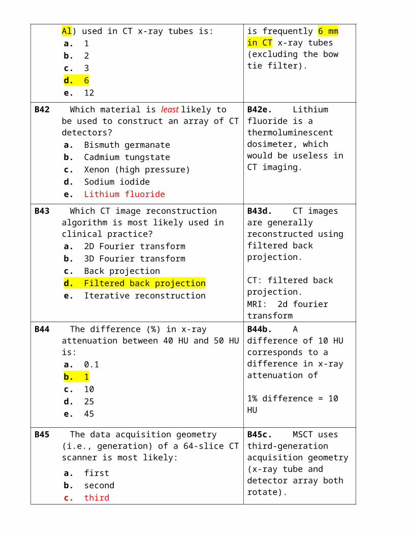
7.24 The backscatter factor in diagnostic radiology is most likely:a. 1.1b. 1.4c. 1.8d. 2.5e. >2.5
7.24b. A common backscatter value used in radiology is 1.4.
7.25 An air kerma of 1 mGy will most likely result in a skin dose (mGy) of:a. 0.5b. 1.5c. 3d. 10e. 20
7.25b. The skin dose is 1.5 mGy after accounting for differences in dose to air/tissue (10%) and backscatter (40%).
7.26 If the entrance air kerma in an adult PA chest x-ray is 0.1 mGy, the air kerma– area product (Gy-cm2) is most likely:a. 0.1b. 1c. 10d. 100e. 1,000
7.26a. A KAP of 0.1 Gy-cm2, since the exposed area at the patient entrance is ~1,000 cm2.
TABLE 7.3
7.27 If the skin dose in a lateral abdominal examination is 100%, the embryo dose (%) is most likely:a. 50b. 20c. 5d. 1e. 0.2
7.27c. The fetal dose is ~5% of the skin dose for a lateral projection in abdominal radiography.AP: 1/3PA: 1/6Lateral: 1/20
7.28 Which of the following is least likely to affect the fetal dose in a radiographic examination?a. Beam areab. Beam HVLc. Focal spotd. Projectione. Skin dose
7.28c. The size of the focal spot has negligible effect on any patient (or operator) dose.
7.29 All of the following organs have a tissue weighting factor (w) of 0.04 except:a. bladderb. esophagusc. braind. livere. thyroid
7.29c. The brain tissue weighting factor is 0.01.TABLE 7.4
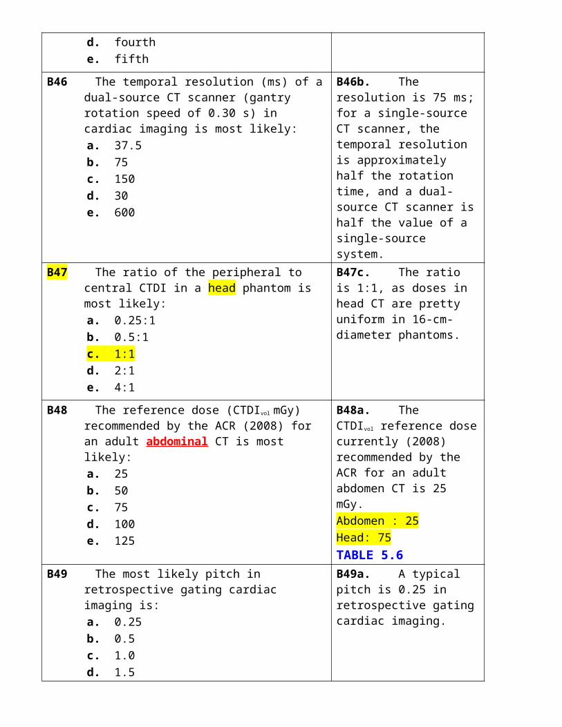
7.30 For radiation protection purposes, an effective dose of 1 mSv corresponds to an average patient detriment (%) of about:a. 50b. 5c. 0.5d. 0.05e. 0.005
7.30e. A determinant of 0.005%, or 5 × 10⁻⁵, is the most likely risk of detriment (harm) from 1 mSv (i.e., ~5% per Sv).
A61 The most radiosensitive part of the cell is most likely the:
a. cell membrane b. chloroplast c. nucleus d. mitochondrion e. ribosome
A61c. The nucleus, which contains DNA, is the most sensitive part of the cell.
A62 Which particle is likely to have the highest linear energy transfer (LET)?
a. Electron b. Positron c. Proton d. Neutron e. Alpha particle
A62e. Alpha particle LET is ~100 keV/ μm,
whereas x-rays are ~1 keV/μm.
A63 The uniform whole-body dose (Gy) that would kill half the exposed population is most likely:
a. 1 b. 2 c. 4 d. 8 e. 16
A63c. A 4-Gy uniform whole-body dose would kill half the exposed population.
A64 The chronic threshold dose (Gy) for cataract induction is most likely:
a. 1 b. 2 c. 5 d. 10 e. >10
A64c. 5 Gy is the chronic threshold dose for inducing eye cataracts.
A65 The threshold equivalent dose (mSv) for the induction of stochastic effects is likely:
a. 0 (no threshold) b. 0.1 c. 1 d. 10 e. 100
A65a. Zero (no threshold) is assumed for exposure to ionizing radiations.
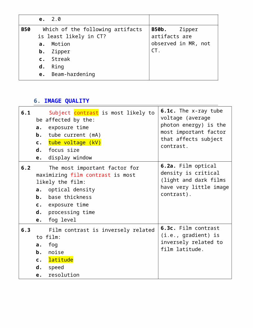
A66 Which of the following does not concern itself with radiation risk estimates?
a. ICRP b. UNSCEAR c. BEIR d. ICRU e. NCRP
A66d. ICRU is the International Commission on Radiological Units and Measurements, which addresses dose quantities such as exposure and air kerma but does not deal with any radiation risk estimates.
A67 The organ weighting factor for gonad exposure recommended by the ICRP (Publication 103) in 2007 is:
a. 0.01 b. 0.04 c. 0.08 d. 0.20 e. 0.30
A67c. The gonad weighting factor recommended by the ICRP in 2007 is 0.08, which replaced the value of 0.20 recommended in 1990.
A68 The most sensitive period for the induction of severe mental retardation in pregnant patients is most likely:
a. up to 10 days b. 2 to 7 weeks c. 7 to 15 weeks d. 15 to 25 weeks e. >25 weeks
A68c. Severe mental retardation in pregnant patients likely occurs at 7 to 15 weeks.
A69 Which is likely the best indicator of the risk of a radiation-induced skin reaction?
a. backscatter fraction b. entrance air kerma c. energy imparted d. air kerma–area product e. skin dose
A69e. The skin dose (mGy) is the best indicator of the possible harm done to the skin from x-ray exposure.
A70 The total energy (J) deposited in a patient undergoing a head CT scan is most likely:
a. 0.1 b. 1 c. 10 d. 100 e. 1,000
A70a. The energy absorbed by the patient is 0.1 J; this may be contrasted to the 600 J deposited each second when a chicken is heated in a 600-W microwave.
B61 The most likely oxygen enhancement ratio for x-rays is:
a. 1.5 b. 2.5 c. 5 d. 10 e. 20
B61b. A typical OER for low LET x-rays is 2.5.
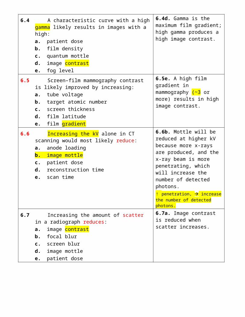
B62 The radiation weighting factor (wR) for 80-kV x-rays is likely:
a. 0.3 b. 0.5 c. 1.0 d. 2.0 e. 3.0
B62c. The radiation weighting factor is 1.0 for radiations used in diagnostic radiology.
B63 The threshold dose (Gy) for permanent epilation is most likely:
a. 1 b. 3 c. 5 d. 7 e. 10
B63d. Permanent epilation occurs at ~7 Gy.
B64 The chance (%) of a radiation-induced cataract from ten head CT examinations is most likely:
a. 0 b. 0.1 c. 0.25 d. 0.5 e. 1
B64a. Zero since ten head CTs would result in an eye lens dose of ~600 mGy, well below the threshold dose for chronic exposure (i.e., 5 Gy).
B65 Which of the following tissues is likely the least sensitive to radiation-induced carcinogenesis:
a. Breast b. Colon c. Kidney d. Lung e. Stomach
B65c. The kidney is least sensitive to radiation-induced cancers of those listed.
B66 For young adults, a uniform whole-body dose of 1 Sv will result in a cancer incidence risk (%) that is most likely:
a. 0.1 b. 0.3 c. 1 d. 3 e. 10
B66e. Approximately 10% of 30-year-olds might suffer a radiation-induced cancer following exposure to an effective dose of 1 Sv (1,000 mSv).
B67 The gonad dose (Gy) that would most likely double the spontaneous mutation incidence is most likely:
a. 2 b. 5 c. 10 d. 20 e. 50
B67a. The commonly accepted value of the doubling dose for hereditary effects is 2 Gy.

B68 The dose to the fetus (mGy) after 5 minutes of pelvic fluoroscopy (PA) is most likely:
a. 0.3 b. 3 c. 30 d. 300 e. 3,000
B68c. The dose is 30 mGy, which is 30% of the patient skin dose of 100 mGy (i.e., 5 minutes at 20 mGy/min for average-sized patients).
TABLE 7.3
B69 If the entrance air kerma in an adult PA chest x-ray is 0.1 mGy, the air kerma– area product (Gy-cm2) is most likely:
a. 0.1 b. 1 c. 10 d. 100 e. 1,000
B69a. The KAP is 0.1 Gy-cm2, since the exposed are at the patient entrance is ~1,000 cm2.
B70 If a newborn patient (3.5 kg) absorbs the same total energy as an adult (70 kg), the newborn’s dose will likely be higher by a factor of:
a. 2 b. 4 c. 10 d. 20 e. 50
B70d. A factor of 20 since the newborn has a mass that is twenty times lower, and dose = energy/mass.
8. RADIATION PROTECTION
8.1 Which is least likely to be categorized as an x-ray detector?a. Ionization chamberb. Scintillation detectorc. Geiger-Muller counterd. Photostimulable phosphore. Photomultiplier tube
8.1e. Photomultiplier tubes detect light, not x-rays.
8.2 Absorbed x-ray doses may be quantified by heating thermoluminescent dosimeters and measuring the emitted:a. radio wavesb. microwavesc. infraredd. visible lighte. ultraviolet
8.2d. TLDs emit visible light when heated.

8.3 When ionization chambers absorb x-rays, they most likely measure the resultant:a. chargeb. heatc. lightd. photonse. voltage
8.3a. Ionization chambers collect and measure charge (or charge per second in fluoroscopy).
8.4 Which of the following works on the principle of air ionization?a. Intensifying screenb. Thermoluminescent dosimeterc. Photostimulable phosphord. Radiographic filme. Geiger counter
8.4e. Geiger counters are essentially (air) ionization chambers, but operated at a high voltage.
8.5 Which dosimeter would likely be used when a parent holds a child for an x-ray examination?a. Ionization chamberb. Geiger counterc. TLDd. Film badgee. Pocket dosimeter
8.5e. Pocket ionization chamber because it is so easy to read (just point at the window, and read off the exposure that was received).
8.6 Who coordinates the radiation control programs in all 50 states in the United States?a. CRCPDb. ICRPc. BEIRd. NCRPe. NRC
8.6a. Conference of Radiation Control Program Directors (CRCPD).
8.7 The regulatory (2008) effective dose limit (mSv/year) for U.S. x-ray technologists is:a. 1b. 5c. 10d. 20e. 50
8.7e. Fifty mSv per year is the legal limit in the United States for radiation workers.
8.8 The regulatory (2008) effective dose limit (mSv) for a patient chest CT scan is:a. 1b. 5c. 20d. 50e. no limit
8.8e. There are no dose limits in radiology (except for fluoroscopy and mammography).
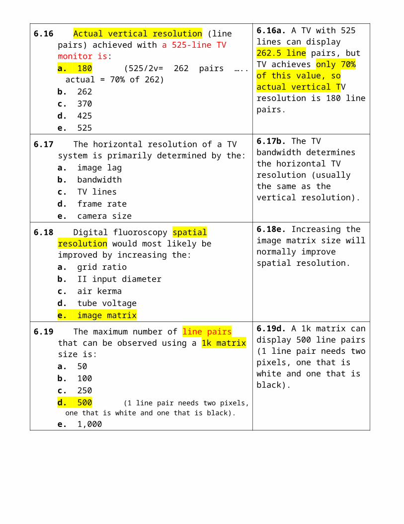
8.9 Regulatory dose limits for the public include only doses received from:a. dental radiographsb. airplane flightc. terrestrial radioactivityd. screening radiographse. radiology cafeterias
8.9e. Imaging center cafeteria since the dose received comes from the (shielded) CT facility and must not exceed 1 mSv.
8.10 The regulatory (2008) dose limit (mSv/year) to a member of the public is:a. 0.25b. 0.5c. 1d. 2e. 5
8.10c. One mSv is the current regulatory dose limit to members of the public.
8.11 Scattered radiation intensities at 1 m in diagnostic examination, expressed as a percentage (%) of the patient skin dose, is most likely:a. 0.01b. 0.03c. 0.1d. 0.3e. 1
8.11c. The scattered intensity is likely 0.1% of what the patient gets; at 1 m from a fluoroscopy patient, the radiologist is exposed to 0.02 mGy/minute since the patient receives 20 mGy/minute.
8.12 Leakage radiation (mGy per hour) at 1 m from an x-ray tube must not exceed:a. 0.01b. 0.1c. 1d. 10e. 100
8.12c. One mGy per hour is the current regulatory limit in the United States (100 mR/hour).
8.13 The transmission of x-rays (%) by a 0.5-mm Pb apron in diagnostic radiology is most likely:a. 5b. 15c. 25d. 35e. 45
8.13a. Five percent is likely to get through a lead apron, and the remaining 95% absorbed.5-10 %
8.14 Which is least likely to be required in designing the shielding for an x-ray room?a. Beam filtrationb. Occupancy factorc. Room dimensionsd. Use factore. Workload
8.14a. Beam filtration is not used in shielding calculations.

8.15 The annual effective dose (mSv) received by a nuclear medicine technologist is most likely:a. 0.3b. 1c. 3d. 10e. 30
8.15c. Three mSv per year is a typical NM operator dose.
8.16 If a radiologist were to increase the distance to a fluoroscopy patient from 1 to 2 m, his or her radiation dose would likely be:a. 1/2b. 1/3c. 1/4d. 1/5e. 1/8
8.16c. One fourth by the inverse square law.
8.17 Entrance skin dose (mGy) for an AP abdominal x-ray examination is most likely:a. 0.1b. 0.5c. 2d. 10e. >10
8.17c. Two mGy is a typical skin dose in abdominal radiography for an AP projection.
8.18 The patient exposure rate during fluoroscopy is least likely to be affected by the:a. exposure timeb. grid ratioc. patient thicknessd. tube currente. tube voltage
8.18a. Exposure time is relevant only for the total exposure, not the exposure rate.
8.19 The average glandular dose per film (mGy) in screening mammography is most likely:a. 0.5b. 1.5c. 3d. 5e. 10
8.19b. For an average-sized breast, 1.5 mGy per image is typical (3 mGy is the ACR/MQSA limit).
8.20 The chance (%) that a patient undergoing an IR procedure in a radiology department will suffer a serious deterministic injury is likely:a. 10b. 1c. 0.1d. 0.01e. 0.001
8.20d. The chance is 0.01% or 1 in 10,000 patients for a serious injury.
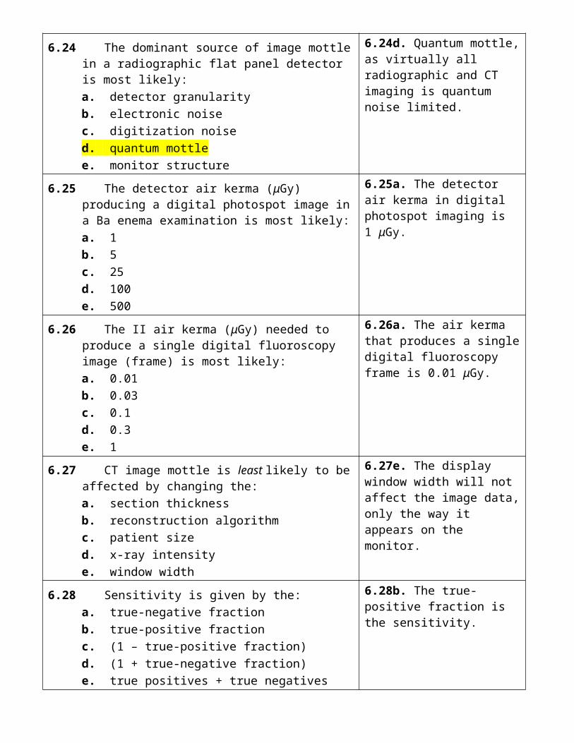
8.21 Fetal doses in CT would likely be reduced with increasing:a. patient sizeb. tube voltagec. tube currentd. scan timee. scan length
8.21a. Larger patient size will reduce fetal dose because of additional attenuation (dilution) of the x-ray beam.
8.22 The most likely patient effective dose (mSv) from a four-phase abdominal CT examination is most likely:a. 5b. 10c. 25d. 75e. 150
8.22c. The most likely dose would be 25 mSv (6 mSv/scan would be typical).
8.23 The benefit–risk ratio of screening mammography is most likely:a. 2:1b. 4:1c. 8:1d. 16:1e. >16:1
8.23e. A ratio of >16:1 (a value of 30:1 was obtained in this book).
8.24 The average effective dose (mSv/year) from background radiation in the United States, excluding radon, is likely:a. 0.1b. 0.3c. 1d. 3e. 10
8.24c. One mSv per year in the United States, with an additional 2 mSv from radon.
8.25 Which are the most damaging emissions from the decay of 222Rn (radon) and its daughters?a. Alphab. Beta plusc. Beta minusd. Neutrinoe. Gamma ray
8.25a. Alpha particles are emitted by radon and its daughters.
8.26 The largest exposure to the U.S. population from man-made radiation is the result of:a. A-bomb falloutb. diagnostic x-raysc. industrial radiographyd. nuclear power plantse. nuclear waste sites
8.26b. Diagnostic x-rays are the dominant contributor to man-made radiation exposures in the United States.
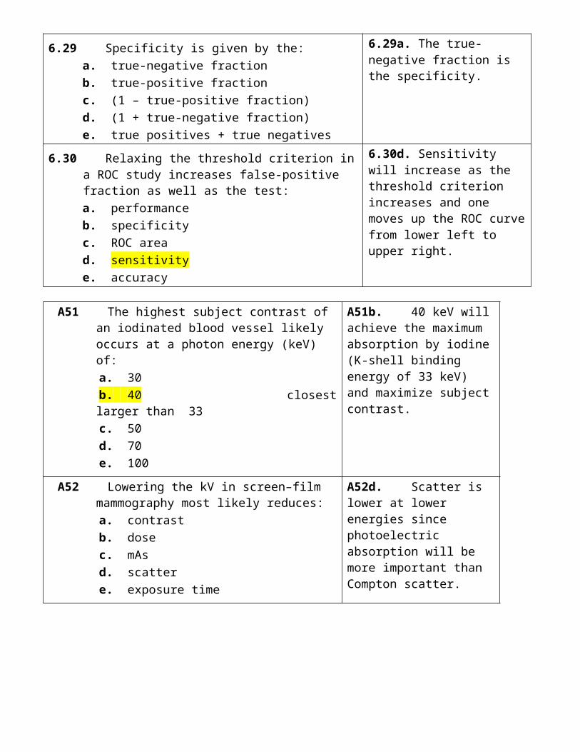
8.27 The average effective dose to the U.S. population from diagnostic imaging in 2006 was most likely:a. 0.5b. 1c. 2d. 3e. 5
8.27d. Three mSv is the average dose to the U.S. population from diagnostic imaging.
8.28 The average patient effective dose (mSv) in NM imaging (2006) was most likely:a. 0.5b. 1c. 2.5d. 5e. 10
8.28e. Ten mSv (NM is the imaging modality with the highest average patient doses).
8.29 The number of diagnostic x-ray examinations (millions) performed in the United States in 2006 was most likely:a. 100b. 250c. 500d. 1,000e. 2,000
8.29c. About 500 million examinations were performed in the United States in 2006, including dental x-rays.
8.30 The contribution (%) of medical imaging to the U.S. population dose from all man-made radiation exposure is most likely:a. 50b. 75c. 90d. 95e. >95
8.30e. More than 95% of man-made exposures are from diagnostic imaging.
A71 For the same air kerma, blackening of film by 30 keV photons, compared to the blackening by 300 keV photons, is most likely:
a. much less b. slightly less c. similar d. slightly more e. much more!!!
A71e. Much more because of the k-edge energy of silver is 25 keV, which will result in many more 30-keV than 300-keV photons being absorbed.
A72 A Geiger-Muller detector would likely be used to measure:
a. low-level contamination b. x-ray tube outputs c. patient exposures d. operator exposures e. x-ray leakage
A72a. Low-level contamination is normally detected using Geiger-Muller detectors.
A73 The effective dose (mSv per year) to an interventional radiology fellow is most likely:
a. 1 b. 5 c. 10 d. 20 e. 50
A73b. 5 mSv is the annual effective dose received by the most highly exposed radiation workers (e.g., IR fellows).Maximum 50 mSv
A74 The current (2008) regulatory dose limit (mSv per year) to the eye lens of a radiologist is:
a. 50 b. 100 c. 150 d. 300 e. 500
A74c. The limit is 150 mSv per year, which should prevent the induction of an eye cataract (deterministic effect) with a threshold dose of ~5 Sv.
A75 The amount of lead shielding (mm) in the wall of a CT facility is most likely:
a. 0.5 b. 1 c. 1.5 d. 3 e. 5
A75c. Virtually all diagnostic x-ray rooms have 1.5 mm of lead shielding.
Apron : 0.25 mm - 0.5 mm
A76 The effective dose (mSv) from a chest x-ray examination is most likely:
a. 0.05 b. 0.2 c. 1 d. 3 e. 10
A76a. A chest radiographic examination (PA plus lateral) would result in an effective dose of ~0.05 mSv. = 0.02 + 0.04Skin dose = 0.5 mGy
A77 The effective dose (mSv) for an upper barium examination is most likely:
a. 0.2 b. 1 c. 5 d. 25 e. >25
A77c. A typical value is 5 mSv for upper barium studies (higher for barium enemas).X100 CXR!!!!
A78 The risk of a breast cancer from a screening mammogram in a 50-yearold woman is most likely three per:
a. 100 b. 1,000 c. 10,000 d. 100,000 e. 1,000,000
A78d. Three per 100,000 breast cancers is the mammogram radiation risk, with a quarter being fatal.
A79 The average dose (mSv per year) from radon (+ daughters) in the United States is most likely:
a. 1 b. 2 c. 3 d. 4 e. 5
A79b. The average U.S. dose from radon is 2 mSv, but there are large variations depending on the type of dwelling and geographic location.
A80 Which imaging modality contributed least to the U.S. population medical dose (2006)?
a. Interventional radiology b. Radiography/fluoroscopy c. CT d. Nuclear medicine e. Mammography
A80e. The contribution of mammography to US medical doses (2006) is negligible.
B71 Which of the following materials is most likely to be used as a TLD for occupational dosimetry?
a. BaFBr b. LiF c. NaCl d. PbI e. Se
B71b. LiF is the most popular TLD material in medical radiation dosimetry.
B72 Agreement states are most likely to regulate the operations in:
a. Radiography b. Mammography c. Computed tomography d. Nuclear medicine e. Interventional radiology
B72d. Nuclear medicine (radioactivity); nonagreement states are regulated by the NRC.
B73 The fetus of an x-ray technologist has an equivalent dose limit (mSv/month) that is most likely:
a. 0.5 b. 1 c. 2 d. 5 e. 10
B73a. A dose limit of 0.5 mSv per month (to the fetus); 50 mSv per year to the mother.
B74 The current (2008) regulatory dose limit (mSv per B74e. A limit of 500 mSv
year) to the hands of a radiopharmacist is: a. 50??? b. 100 c. 150 d. 300 e. 500
per year is designed to prevent the induction of chronic deterministic effects.
B75 If both occupancy factor and work load double, personnel doses are likely to increase by (%):
a. 50 b. 100 c. 200 d. 400 e. 800
B75d. Doses increase by 400%, since each of these factors alone would likely double operator doses.
B76 The maximum patient entrance air kerma (mGy per minute) in fluoroscopy is most likely:
a. 5 b. 10 c. 25 d. 50 e. >50
B76e. The maximum is >50 mGy; 100 mGy/min is the limit in normal fluoroscopy, and 200 mGy/min is permitted in high-dose mode with alarms to indicate the high-dose mode.
B77 The breast dose (mGy) from a single chest CT scan is most likely:
a. 0.02 b. 0.2 c. 2 d. 20 e. 200
B77d. The dose is 20 mGy, which is much higher than in mammography (~4 mGy for a two-view exam).
B78 Dose reductions (%) for follow-up scoliosis digital radiographs are most likely:
a. 10 b. 25 c. 50 d. 80 e. >80
B78e. More than 80% as follow-up digital radiographs using a tenth of the initial dose are adequate for assessing the spine curvature.
B79 Which imaging modality produced the highest collective medical dose in 2006?
a. Computed tomography b. Dental radiography c. Interventional radiology d. Mammography e. Radiography
B79a. Computed tomography accounts for nearly half of the total population medical dose.
B80 The contribution (%) of CT to the total U.S. population dose from medical imaging is likely:
a. 2.5
B80e. CT contributes 50% of the U.S. medical radiation

b. 7.5 c. 15 d. 30 e. 50
dose.
9. NUCLEAR MEDICINE
9.1 15O and 16O are examples of:a. isotopesb. isotonesc. isomersd. isobarse. metastable states
9.1a. Isotopes since both 15O and 16O have 8 protons.
9.2 37 MBq is equal to (mCi):a. 0.1b. 1c. 10d. 100e. 1,000
9.2b. One mCi is 37 MBq.
9.3 Which of the following decay modes most likely changes the mass number (A) of an unstable nucleus?a. Beta minus decayb. Beta plus decayc. Alpha decayd. Isomeric transitione. Electron capture
9.3c. Alpha decay will reduce the radionuclide atomic number by two and mass number by four.
9.4 60Co (Z = 27)decaying to 60Ni (Z = 28) is an example of:a. β+ decayb. β− decayc. electron captured. alpha decaye. isomeric transition
9.4b. β− decay since the daughter radionuclide atomic number has increased by one by emitting a β− particle.
9.5 Electron capture nuclei are most likely to produce:a. antineutrinosb. internal conversion electronsc. characteristic x-raysd. β+ particlese. β− particles
9.5c. Electron capture always results in characteristic x-rays, since the inner shell vacancy will be filled by an outer shell electron resulting in characteristic x-ray emission.
9.6 A radionuclide produced in a nuclear reactor is most likely to decay by:a. beta minus decayb. beta plus decayc. alpha decayd. isomeric transitione. electron capture
9.6a. Radionuclides produced in a nuclear reactor are neutron rich and therefore decay via beta minus emission.
9.7 After ten half-lives, the fraction of activity remaining is:a. depends on the initial activityb. 1/10c. (1/10)2
d. (1/2)10
e. (1/10)10
9.7d. (1/2)10, or 1/1,024 which is ~0.1%.
9.8 After one day, the remaining activity (%) of a 123I source will most likely to be about:a. 50b. 25c. 12.5d. 6.3e. 3.2
9.8b. the half-life of 123I is 13 hours, so after a day (~2 half-lives), about 25% will remain.
9.9 The time (hour) when a daughter radionuclide (T1/2 = 1 hour) reaches approximate equilibrium with its long-lived parent is most likely:a. 1b. 2c. 4d. 8e. 16
9.9c. Four hours, since equilibrium is always established after four daughter half-lives.
For Tc = 4x6 = 24 hrs
9.10 Which type of collimator will likely result in the highest resolution for imaging the thyroid?a. High sensitivityb. Divergingc. High energyd. All purposee. Pinhole
9.10e. Pinhole collimators offer the highest resolution for thyroid imaging.
9.11 Scintillation camera detectors are most likely made of:a. CsIb. NaIc. Nad. Cse. NaCl
9.11b. NaI is the scintillator material in most current scintillation cameras.
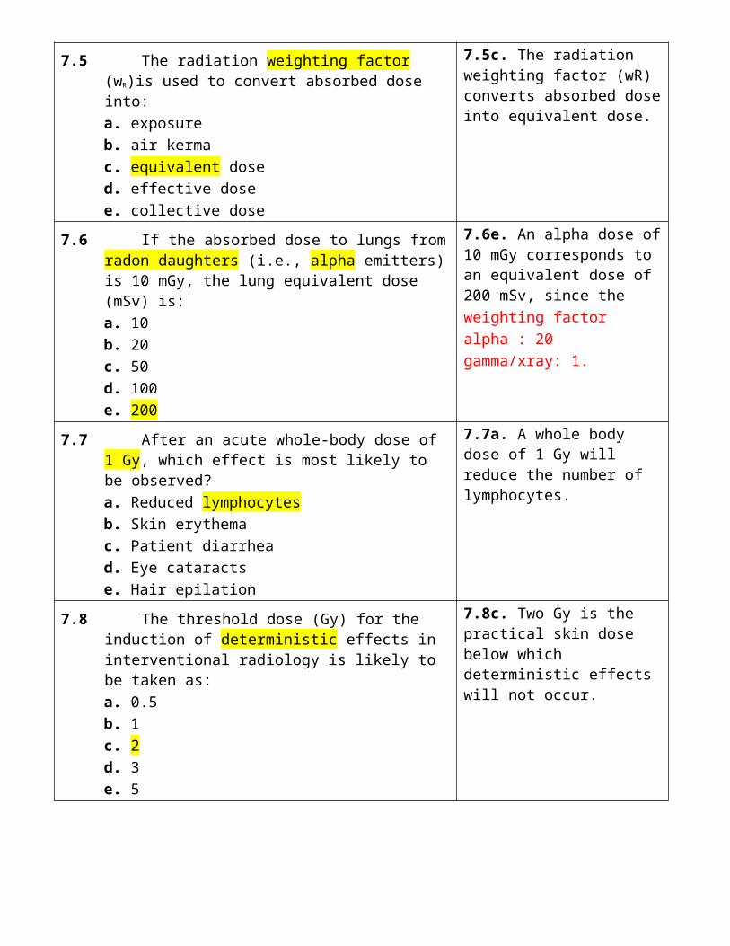
9.12 The percentage (%) of 140-keV photons absorbed in a scintillating camera crystal is most likely:a. 20b. 40c. 60d. 80e. >80
9.12e. A scintillation camera crystal will absorb most (i.e., >80%) of incident 140-keV gamma rays.TABLE 9.5
9.13 A pulse height analyzer window width of 20% used with 99mTc would likely reject energies (keV) that are less than:a. 140b. 136c. 126d. 120e. 112
9.13c. A PHA window width of 20% selected for 99mTc gamma rays would reject energies that are less than 126 keV and more than 156 keV.
20% window (±10%) 126-156 keV
9.14 The most likely number of counts (k) in a scintillation camera image is most likely:a. 5b. 50c. 500d. 5,000e. 50,000
9.14c. A scintillation camera image is most likely to have ~500k counts.
9.15 The most likely image matrix size for cardiac SPECT imaging is:a. 32 × 32b. 64 × 64c. 128 × 128d. 256 × 256e. 512 × 512
9.15b. The normal cardiac SPECT image matrix size is 64 × 64 (most SPECT studies are cardiac examinations).
Typical: 128 × 128SPECT: 64 × 64whole-body: 1,024 × 1,024
9.16 Pixel values in SPECT images represent:a. physical densitiesb. gamma ray absorptionc. clearance ratesd. radioisotope concentrationse. effective half-lives
9.16d. Pixel values in SPECT images represent radioisotope concentrations.
9.17 Which is least likely to be used as a detector material in PET imaging?a. BGOb. GSOc. LSOd. HCOe. YSO
9.17d. HCO, which consists of hydrogen (H), carbon (C), and oxygen (O), and which would therefore be useless for detecting 511-keV photons because of their low atomic numbers.
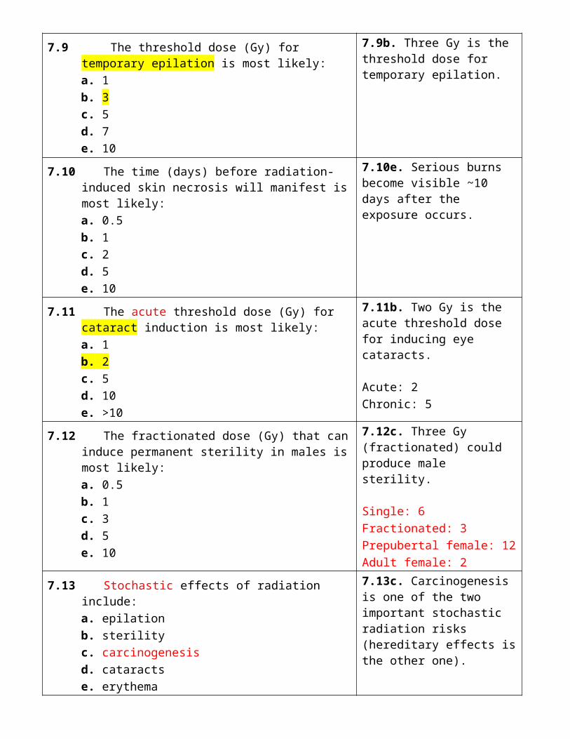
9.18 The amount of 18F (mCi)most likely to be administered in a PET/CT scan is:a. 1b. 4c. 16d. 64e. 256
9.18c. A typical administered activity for a PET/CT scan is 16 mCi of 18F.
9.19 The standard limit for 99Mo breakthrough (μCi 99Mo per mCi 99mTc) is most likely:a. 0.15b. 0.3c. 0.5d. 1.0e. 1.5
9.19a. The limit for 99Mo breakthrough is 0.15 μCi 99Mo per mCi 99mTc.
5.5 kBq of 99Mo per 37 Bq of 99mTc.
9.20 The most likely radionuclide for performing a scintillation camera flood uniformity test is:a. 57Cob. 60Coc. 137Csd. 131
I
e. 226Ra
9.20a. 57Co would be used for a flood uniformity test because its photon energy (122 keV) is close to that of 99mTc (140 keV).
9.21 The most likely radionuclide used to check the constancy of a dose calibrator is:a. 14C
b. 51Crc. 137Csd. 131Ie. 32P
9.21c. 137Cs, which is a long-lived (30 year half-life) gamma emitter (661 keV).
9.22 Radionuclides with a higher photon energy would likely increase scintillation camera:a. detection efficiencyb. septal penetrationc. radionuclide sensitivityd. spatial resolutione. image magnification
9.22b. Higher photon energies increase collimator septal penetration.
9.23 The full width half maximum width (mm) of a line source that is achieved by scintillation cameras is most likely:a. 1b. 2c. 4d. 8e. 16
9.23d. Eight mm is a typical full width half maximum width of an image of a line source obtained using a scintillation camera using a low-energy high-resolution (LEHR) collimator.

9.24 The variance (i.e., σ2) of a NM image pixel with an average of 100 counts would likely be:a. 10b. 20c. 30d. 50e. 100
9.24e. The variance (σ2) is 100 when a NM image pixel has an average of 100 counts (i.e., σ is 10).
9.25 A single circular cold spot artifact in a scintillation camera image would likely be the result of a:a. cracked crystalb. damaged collimatorc. defective PMTd. faulty PHAe. high count rate
9.25c. A defective PMT would likely result in a single circular cold spot in a scintillation camera image.
9.26 For 99mTc, which of the following is least likely to contribute to the patient dose?a. Auger electronsb. Beta particlesc. Internal conversion electronsd. Gamma rayse. Characteristic x-rays
9.26b. Beta particles, since these are not emitted by 99mTc.
9.27 If both physical and biologic half lives are 2 hours, the effective half life (hour) is:a. 0.5b. 1c. 2d. 3e. 4
9.27b. One, since the effective half-life is given by the relationship 1/Teffective = 1/Tphysical + 1/Tbiologic.
9.28 Cumulative activity in an organ is least likely to depend on the:a. administered activityb. organ uptakec. organ massd. physical half-lifee. biologic clearance
9.28c. Organ mass has no direct relationship to any cumulative activity.
9.29 Adult effective doses (mSv) for a 99mTc labeled radiopharmaceutical are most likely:a. <1b. 1c. 2.5d. 5e. 10
9.29d. Five mSv is a representative adult effective dose for a 99mTc labeled radiopharmaceutical.NM: 5 mSvPET: 10 mSvPET/CT: 15 mSv

9.30 Adult effective doses (mSv) in PET imaging are most likely:a. 0.3b. 1c. 3d. 10e. 30
9.30d. Ten mSv is a representative adult effective dose for a PET study.
A81 An activity of 1 mCi equals (Bq): a. 37,000 b. 370,000 c. 3,700,000 d. 37,000,000 e. 370,000,000
A81d. One mCi is 37 MBq (i.e., 37,000,000 Bq)
A82 Which of the following emits positrons? a. 3H b. 32P c. 18F d. 99mTc e. 226Ra
A82c. 18F is a positron emitter that is used in most PET imaging studies.
A83 A radionuclide produced in a cyclotron is most likely to decay by:
a. Beta minus decay b. Beta plus decay c. Alpha decay d. Isomeric transition e. Neutron emission
A83b. Radionuclides produced in cyclotrons are proton rich, and can decay via beta plus emission.
A84 Which of the following is not a radiopharmaceutical localization mechanism?
a. Diffusion b. Phagocytosis c. Capillary blockage d. Elution e. Cell sequestration
A84d. Elution is not a radiopharmaceutical localization mechanism, but rather, extracting a substance that is adsorbed to another by washing with a solvent.
A85 Which nuclide would be most likely to make use multiple PHA windows?
a. 67Ga b. 123I c. 131I d. 99mTc e. 133Xe
A85a. 67Ga, which has three photon energies (93 keV, 185 keV, and 300 keV).
A86 Which radionuclide is least likely to be used for PET imaging?
a. 18F
A86b. 67Ga is a gamma ray emitter, whereas all the others emit positrons and produce annihilation radiation (511 keV).
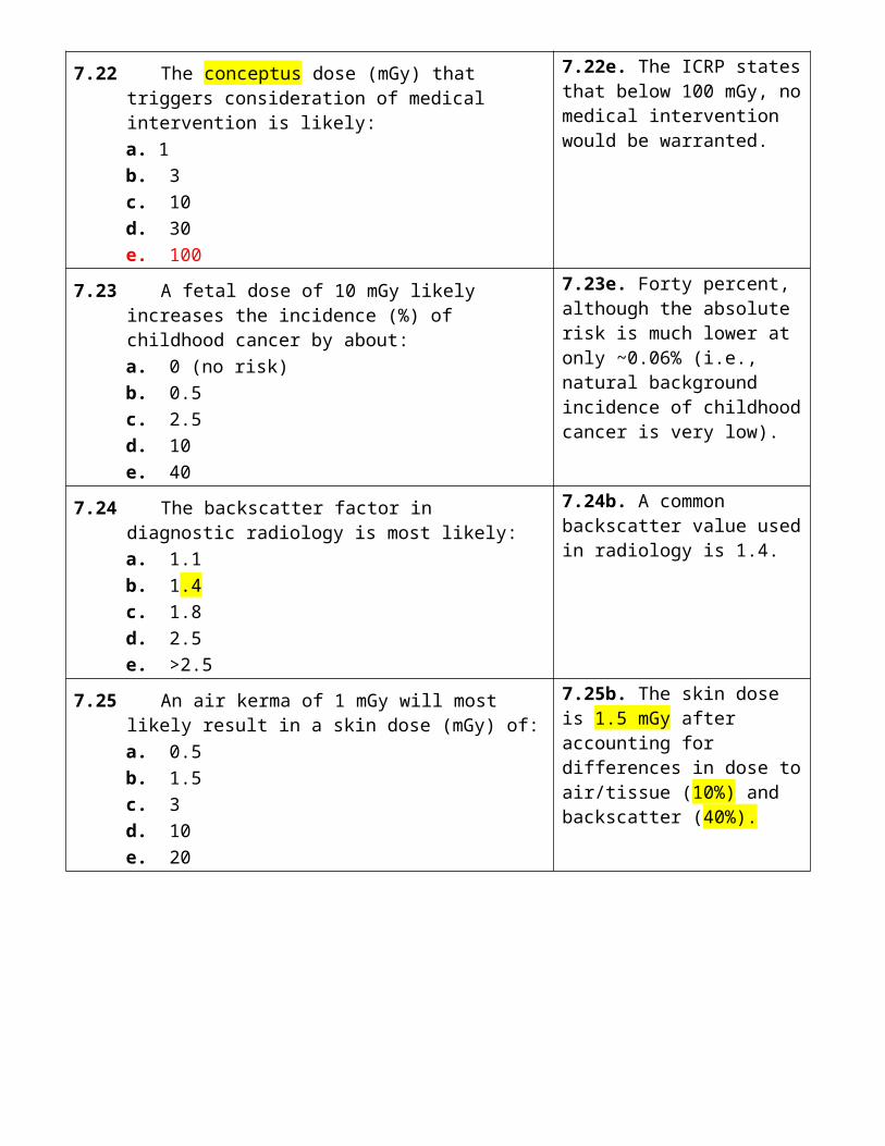
b. 67Ga c. 68Ga d. 15O e. 82Rb
A87 Which of the following tests is least likely to be performed on a radiopharmaceutical?
a. Flood uniformity b. Pyrogenicity c. Radiochemical purity d. Radionuclide purity e. Sterility
A87a. Flood uniformity is a scintillation camera test, not a radiopharmaceutical QC test.
A88 The intrinsic (RI) and collimator (RC) resolution are related to the system resolution (R) as:
a. RI + RC
b. (RI − RC)1/2
c. 1/(RI + RC)1/2
d. (RI2 + RC
2) e. (RI
2 + RC2)1/2
A88e. System resolution R is equal to (R2
I + R2C)1/2, where RI
and RC are the intrinsic and collimator resolutions, respectively.
A89 Which is least related to artifacts in scintillation camera imaging?
a. Chemical shift b. Cracked crystal c. Edge packing d. Nonuniformity e. Off-peak imaging
A89a. Chemical shift artifacts occur in MR, not nuclear medicine.
A90 Which organ likely receives the highest dose for an uptake of 1 μCi activity (no biologic clearance)?
a. Adult thyroid b. Fetal thyroid c. Spleen d. Liver e. Kidneys
A90b. Fetal thyroid because it has the smallest mass.
B81 Which is least likely to be emitted during radioactive decay?
a. Electrons b. Protons c. Positrons d. Gamma rays e. Neutrinos
B81b. There are no known radionuclides that emit protons.
B82 Electron capture is most likely to compete with: a. positron decay b. beta minus decay c. alpha decay
B82a. Electron capture competes with positron decay.
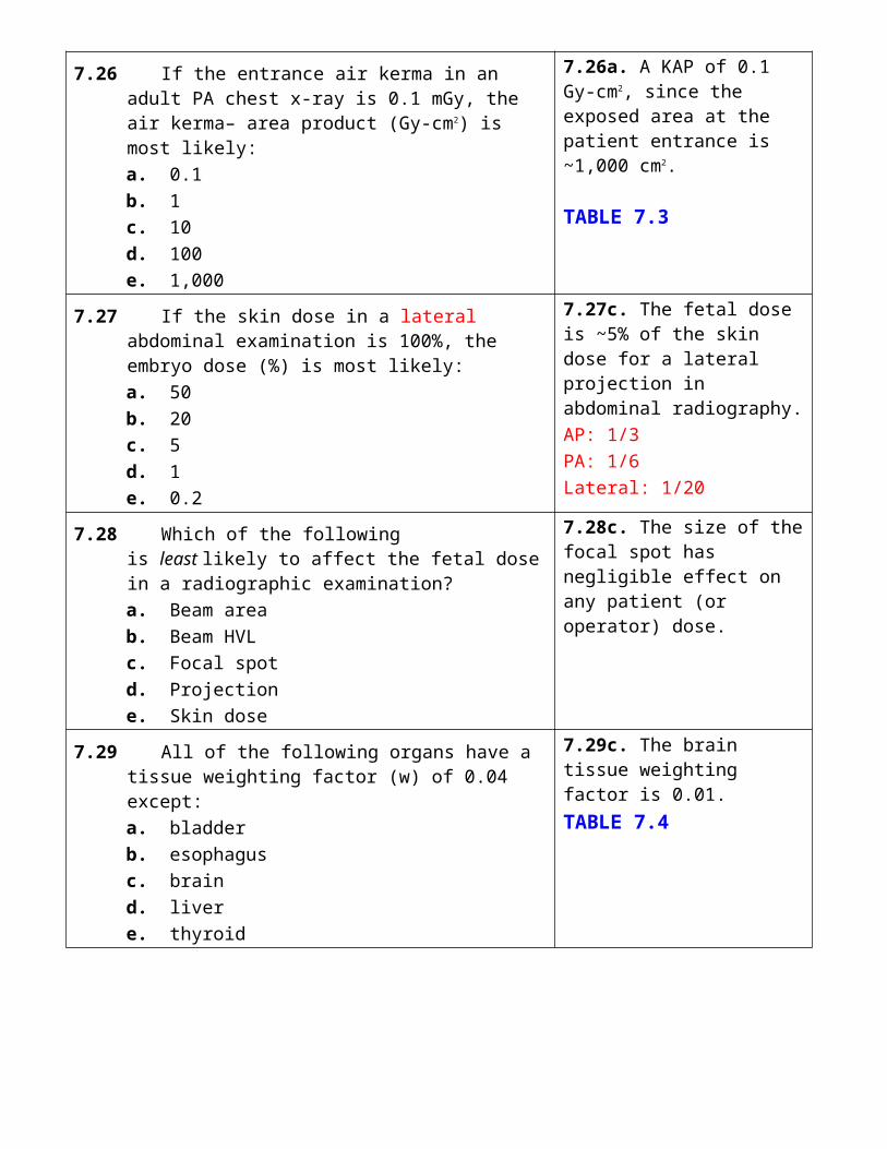
d. isomeric transition e. nuclear fission
B83 Which radionuclide has a primary photopeak energy of 365 keV?
a. Oxygen-15 b. Technetium-99m c. Iodine-131 d. Thallium-201 e. Indium-111
B83c. Iodine-131 emits 365 keV gamma ray photons.
B84 The sensitivity of a low-energy (highresolution) collimator is most likely to be:
a. 10−1
b. 10−2
c. 10−3
d. 10−4
e. 10−5
B84d. 10−4, as only about 1 in 10,000 gamma rays incident on a collimator are expected to get through and contribute to the image.
B85 The angular rotation (degrees) of a dual-camera SPECT system when imaging the liver would likely be:
a. 90 b. 135 c. 180 d. 270 e. 360
B85c. The rotation is 180 degrees, which permits the acquisition of projections through 360 degrees.
B86 A PET scanner obtains spatial information by detecting: a. positrons and electrons in coincidence b. positrons and electrons in anticoincidence c. photons and positrons in coincidence d. annihilation photons in coincidence e. annihilation photons in anticoincidence
B86d. PET scanners obtain spatial information by detecting annihilation photons in coincidence.
B87 Which of the following is not a quality control test performed on a scintillation camera?
a. Field uniformity b. 99Mo breakthrough c. Extrinsic flood d. Spatial resolution e. Linearity
B87b. 99Mo breakthrough is used to test the eluted solution from a 99mTc/99Mo generator.
B88 The full width half maximum width (mm) of a line source obtained using a PET scanner is most likely:
a. 1 b. 2.5 c. 5 d. 10 e. 20
B88c. A typical full width half maximum width of an image of a line source obtained with a PET imaging system is 5 mm.
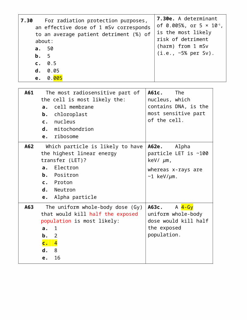
B89 A radionuclide with a shorter half-life likely results in lower:
a. count rates b. patient doses c. biologic clearance d. scattered photons e. photopeak energy
B89b. A shorter half-life means a lower cumulative activity (fewer nuclear transformations) and therefore a lower patient dose.
B90 The dose rate near a 131I therapy patient is least likely to depend on the:
a. administered activity b. physical half-life c. biological half-life d. patient weight e. distance to patient
B90d. Patient weight will have negligible impact on the dose rate in the vicinity of a 131I therapy patient (365 keV gamma rays).
10. ULTRASOUND
10.1 In a sound wave, the physical quantity varying with time is:a. voltageb. magnetic fieldc. pressured. chargee. current
10.1c. A sound wave is a variation of pressure with time.
10.2 The wavelength of a 3-MHz sound beam is shortest in:a. airb. fatc. muscled. bonee. PZT
10.2a. Air, since wavelength is velocity/ frequency and air has the lowest velocity.wavelength = velocity/ frequency
λ = c / fwavelength is proportional to speed
10.3 A signal attenuated to 1% (= 99% attenuation) of its original intensity corresponds to attenuation (−dB) of:a. 1b. 5c. 10d. 20e. 100
10.3d. Attenuation of −20 dB is 1% (−10 dB is 10%, and −30 dB is 0.1%).
dB = 10 × log10(I/I0)= 10 × log10(1/100) = 10 x -2
10.4 Which of the following has the highest acoustic impedance?a. Boneb. Fatc. Aird. Watere. Eye lens
10.4a. Bone, since acoustic impedance is density times sound velocity, which are both highest in bone.
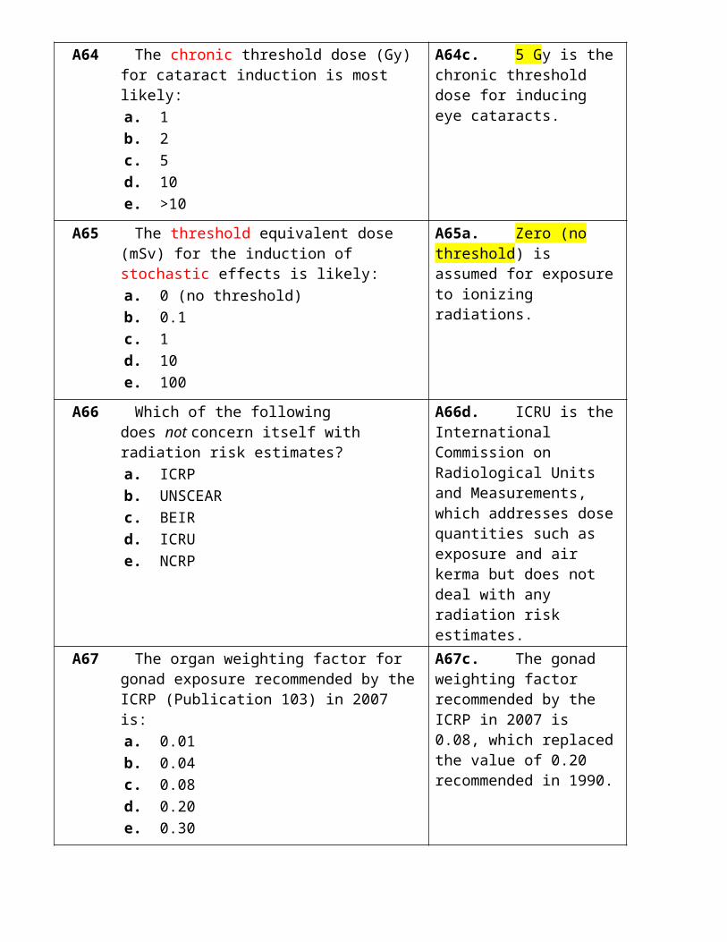
10.5 An ultrasound beam traveling through tissue is least likely to be:a. absorbedb. amplifiedc. scatteredd. reflectede. refracted
10.5b. Amplified, since ultrasound beams are normally strongly attenuated at a rate of about −0.5 dB/cm/MHz.
10.6 Reflections are least likely to occur from:a. smooth surfacesb. kidney interiorc. fat–kidney interfacesd. bladder walle. bladder contents
10.6e. Bladder contents are normally fluids, which are most unlikely to give rise to any kind of specular or nonspecular reflections.
10.7 The largest ultrasound reflections most likely occur between soft tissue and:a. waterb. fatc. boned. bloode. air
10.7e. Air–soft tissue interfaces will give rise to the largest echoes because of the large mismatch in the acoustic properties of air (very low) and soft tissue (average).
10.8 Snell’s law describes the relation between the:a. angle of incidence and transmissionb. Fraunhofer angle and wavelengthc. near field and frequencyd. angle of incidence and reflectione. focus and transducer curvature
10.8a. Snell’s law describes the relation between the angle of incidence and transmission.
10.9 Depth gain compensation accounts for tissue attenuation by increasing the:a. transducer outputb. echo amplificationc. focal lengthd. ultrasound velocitye. pulse repetition frequency
10.9b. Depth gain compensation accounts for tissue attenuation by increasing echo amplification for later echoes.
10.10 Attenuation of ultrasound in soft tissue at 2 MHz is most likely (dB/cm):a. 0.25b. 0.5c. 1d. 2e. 4
10.10c. One dB, since the value used in clinical ultrasound is 0.5 dB/cm per MHz.

10.11 Increasing the transducer thickness is most likely to increase the sound:a. frequencyb. velocityc. wavelengthd. intensitye. attenuation
10.11c. Wavelength, since the crystal thickness is one half of the ultrasound wavelength.
crystal thickness = ½ wavelength
10.12 The damping material behind the crystal transducer reduces the:a. tissue attenuationb. pulse lengthc. operating frequencyd. lateral resolutione. penetration depth
10.12b. The pulse length is reduced by the introduction of a damping material behind a transducer.Damping = tungsten/rubber in an epoxy resinreduce vibration (ring-down time) and shorten pulses.
10.13 An ultrasound near field is made longer when increasing the transducer:a. operating frequencyb. time gain compensationc. pulse repetition frequencyd. physical densitye. acoustic impedance
10.13a. An increase in operating frequency reduces the wavelength, and the near field is inversely proportional to the wavelength.
10.14 A 4-kHz PRF corresponds to a listening time (μs) of:a. 60b. 125c. 250d. 500e. 1,000
10.14c. The pulse repetition period (listening time) is 250 μs, which is given by 1/PRF (i.e., 1/4,000 seconds).
PRP (μs) = 1/PRF (Hz)
10.15 Ultrasound signals are converted to a video monitor display using:a. log amplifiersb. array processorsc. scan convertersd. pulse height analyzerse. analog-to-digital converters
10.15c. Ultrasound signals are converted from digital data to a video monitor display using scan converters.
10.16 An echo received 26 μs after the signal is sent is likely from a interface depth (cm) of:a. 1b. 2c. 3d. 4e. 5
10.16b. Two cm, since it takes 13 μs to get an echo from an interface from a depth of 1 cm.
13 JanuaryD (m) = 1/2 x t (s) x c (m/s)

10.17 The matrix size of a digitized ultrasound frame sent to a PACS is most likely:a. 64 × 64b. 128 × 128c. 256 × 256d. 512 × 512e. 1,024 × 1,024
10.17d. The typical matrix of the displayed US image on a typical monitor is 512 × 512 (note that the acquired image in the scan converter may have a different matrix size).
10.18 The most likely frame rate (frames per second) in real-time ultrasound imaging is:a. 1b. 5c. 20d. 100e. 500
10.18c. Twenty frames per second is the only plausible value (5 is too low, and 100 is too high).
10.19 Which is least likely an ultrasound display mode?a. Ab. Bc. Td. T-Me. M
10.19c. There is no T (time) display mode in ultrasound.
10.20 The Doppler shift from a moving object is least likely to depend on the:a. ultrasound velocityb. ultrasound frequencyc. beam directiond. object depthe. object speed
10.20d. Object depth has no direct relationship to the Doppler shift.
10.21 The maximum Doppler frequency shift likely occurs when the angle (degrees) between the moving reflector and ultrasound beam is:a. 0b. 23c. 45d. 68e. 90
10.21a.Zero degrees largest Doppler shift, Cos 0 = 1
90 degrees no Doppler shift, , Cos 90 = 1
10.22 What is the minimum PRF (kHz) required to accurately measure a 1-kHz Doppler frequency shift?a. 0.25b. 0.5c. 1d. 2e. 4
10.22d. Two kHz, since one has to sample at twice the maximum frequency shift one is trying to detect (1 kHz in this example).

10.23 In color Doppler, a red intensity most likely signifies that the blood flow is:a. toward the transducerb. away from the transducerc. perpendicular to the transducerd. generating aliased datae. turbulent
10.23a. In color Doppler, a red intensity most likely signifies that the blood flow is toward the transducer.
10.24 Choice of frequency in ultrasound is most likely a trade-off between patient penetration and:a. image contrastb. axial resolutionc. lateral resolutiond. speckle noisee. image artifacts
10.24b. The choice of frequency in ultrasound is a trade-off between patient penetration and axial resolution.
10.25 Ultrasound with a short pulse length is most likely to result in improved:a. axial resolutionb. lateral resolutionc. echo intensityd. tissue penetratione. frame rate
10.25a. Axial resolution is approximately half the spatial pulse length.
10.26 Lateral resolution in ultrasound imaging would most likely be improved by increasing the:a. transducer thicknessb. pulse repetition frequencyc. lines per framed. frame ratee. pulse length
10.26c. Lateral resolution improves by increasing the number of lines in each frame.
improve lateral resolution by: Focus the beam (width) Increase number of scan lines
10.27 Shadowing artifacts would be least likely to occur behind:a. lungb. bonec. air cavitiesd. bladdere. clips
10.27d. Bladder contents have negligible attenuation and are more likely to show enhancement (not shadowing).
10.28 Which of the following is least likely to be an ultrasound artifact?a. Mirror imageb. Reverberationc. Edge packingd. Speed displacemente. Refraction
10.28c. Edge packing is a nuclear medicine artifact.

10.29 B-mode ultrasound beam intensities (W/cm2) are most likely:a. 0.001b. 0.01c. 0.1d. 10e. 100
10.29b. B-mode US intensities are most likely 0.01 W/cm2, which is 10 mW/cm2.
Range:1-10 mW/cm2
= 0.001 -0.01 W/cm2
10.30 The thermal index (TI) value indicates the possible increase in tissue:a. cavitationb. cell deathc. densityd. shearinge. temperature
10.30e. Thermal index (TI) values indicate an increase in tissue temperature.
A91 A 2-MHz transducer has an wavelength (mm) in tissue of approximately:
a. 0.01 b. 0.03 c. 0.1 d. 0.3 e. 1
A91e. The approximate wavelength is 1 mmv = f ×λ (m/s). 1,540 m/s = 2 × 10⁶ Hz x ff = 0.77 mm
A92 When an ultrasound beam is attenuated −30 dB, the percentage (%) of the initial intensity that remains is:
a. 70 b. 30 c. 10 d. 1 e. 0.1
A92e. The remaining intensity is
0.1% at −30 dB
1% at −20 dB,
10% at −10 dB).
A93 What fraction of ultrasound is reflected from an interface where Z1 = 1 and Z2 = 2?
a. 1/2 b. 1/3 c. 1/5 d. 1/8 e. 1/9
A93e. The fraction is 1/9 since reflection is
= [(Z2 − Z1)/(Z2 + Z1)]2
= [(2 − 1)/ (2 + 1)]2.
A94 The resonant frequency of an ultrasound transducer is determined primarily by:
a. crystal thickness b. Snell’s law c. sound bandwidth d. applied voltage e. pulse repetition frequency
A94a. The crystal thickness determines the ultrasound frequency and wavelength (thicker crystals have longer wavelengths and lower frequencies).
Thickness = ½ wavelength

A95 The ultrasound PRF (kHz) is most likely to be: a. 0.04 b. 0.4 c. 4 d. 40 e. 400
A95c. A typical ultrasound PRF value is 4 kHz (4,000 pulses per second).
PRF = 4 kHz = 4,000 Hz
A96 How long (μs) will it take to receive the ultrasound echo from an object 10 cm away?
a. 0.13 b. 1.3 c. 13 d. 130 e. 1,300
A96d. It will take 130 μs, since it takes 13 μs to get an echo from an interface from a depth of 1 cm.Velocity = 1540 m/s distance 1/1540 s/m = 0.00064 s/mEcho = 2 * 0.00064 = 0.0013 s/m= 13 μs/cm
A97 Which is the likely frequency (MHz) detected and analyzed for making a harmonic image with a 3-MHz transducer?
a. 1.5 b. 3 c. 6 d. 9 e. 12
A97c. The frequency is 6 MHz, because harmonic images are obtained at double the fundamental frequency (i.e., 2 × 3 MHz).
A98 In spectral analysis, the detected frequency shift is plotted as a function of:
a. angle b. distance c. echo intensity d. frequency e. time
A98e. In spectral analysis, the detected frequency shift is plotted as a function of time.
A99 If the ultrasound pulse length is 1 mm, the axial resolution (mm) is likely:
a. 0.25 b. 0.5 c. 1 d. 2 e. 4
A99b. The axial resolution is 0.5 mm, since it is approximately half the spatial pulse length.
A100 Below a structure, a very faint image of the structure is probably due to:
a. reverberation artifact b. side lobes c. specular reflection d. nonspecular reflection e. incorrect TCG
A100a. Reverberation artifacts are faint image of the structure below the structure.

B91 Which material has the highest ultrasound propagation velocity?
a. Air b. Fat c. Soft tissue d. Bone e. PZT
B91e. PZT; the less compressible a material, the higher the velocity and vice versa (compressible air has the lowest velocity).
B92 Which of the following has the lowest acoustic impedance?
a. Bone b. Fat c. Air d. Water e. Eye lens
B92c. Air since acoustic impedance is density times sound velocity, which are both lowest in air.
B93 Depth gain compensation most likely corrects for: a. specular scatter b. nonspecular scatter c. tissue attenuation d. transducer damping e. shadowing losses
B93c. Depth gain compensation (TGC) mainly corrects for tissue attenuation.
B94 Increasing the transducer bandwidth will most likely reduce the ultrasound pulse:
a. velocity b. duration c. reflection d. attenuation e. frequency
B94b. Duration and bandwidth are inversely related (e.g., pure sounds clearly have a narrow bandwidth and thus last a long time).
B95 The pulse repetition frequency (PRF) is least likely to affect the:
a. listening time b. frame rate c. line density d. penetration depth e. operating frequency
B95e. The operating frequency has no direct relationship to the PRF.
B96 The number of bytes used to code for one pixel in a B-mode image is most likely:
a. 0.5 b. 1 c. 2 d. 4 e. 8
B96b. Each pixel is coded using 1 byte (8 bits), and can display 256 shades of gray.
B98 The main advantage of power Doppler for detecting blood flow is likely its:
a. sensitivity to slow flow
B98a. Power Doppler is very sensitive to slow flow.
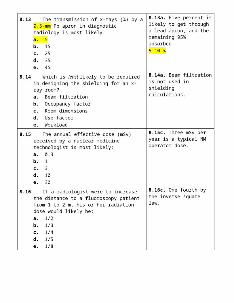
b. increased intensity c. increased echo strength d. quantitative flow data e. ability to provide directional data
B99 Lateral resolution is most influenced by the ultrasound beam:
a. velocity b. frequency c. intensity d. width e. duration
B99d. lateral resolution is determined by the ultrasound beam width.
B100 The mechanical index (MI) value indicates the possible increase in tissue:
a. cavitation b. cell death c. density d. shearing e. temperature
B100a. Mechanical Index (MI) values indicate the possibilities of tissue cavitation.
11. MAGNETIC RESONANCE
11.1 Which would be least useful for magnetic resonance imaging?a. 1Hb. 13Cc. 16Od. 23Nae. 31P
11.1c. 16O, which has an even number of protons (8) and an even number of neutrons (8).
11.2 The ratio of the electron magnetic moment to that of a proton is most likely:a. 4:1b. 16:1c. 50:1d. 200:1e. 800:1
11.2e. The ratio of the electron to proton magnetic moments is 800:1.
11.3 The resonance frequency (kHz) for protons in a 1-T magnetic field is likely:a. 4.2b. 42c. 420d. 4,200e. 42,000
11.3e. The resonance frequency is 42,000 kHz, which is 42 MHz.

11.4 If a 90-degree RF pulse has a duration of t seconds, a 180-degree pulse is likely to have a duration of:a. t/4b. t/2c. td. 2te. 4t
11.4d. A duration of 2t, since doubling the RF pulse duration doubles the angular rotation of the longitudinal magnetization, assuming RF intensity stays constant.
11.5 The maximum free induction decay signal is obtained when using a flip angle of (degrees):a. 0b. 45c. 90d. 135e. 180
11.5c. Pulses of 90 degrees rotate the longitudinal magnetization through ninety degrees (i.e., into the transverse plane) that maximizes the FID signal.
11.6 Which of the following is likely to have the shortest T1 times?a. Fatb. Liverc. Kidneyd. White mattere. CSF
11.6a. Fat T1 times are shorter than those of most tissues and all fluids.
11.7 Which of the following is likely to have the longest T1 times?a. Fatb. Liverc. Kidneyd. White mattere. CSF
11.7e. CSF, since all fluids have long T1 relaxation times compared to fat and tissues.
11.8 After 90-degree RF pulses, spins lose phase coherence in a time comparable to:a. T1b. T2c. TE/2d. TEe. TR
11.8b. T2, which is a measure of spin dephasing in the transverse plane due to spin–spin interactions.
11.9 Which of the following likely has the longest T2 relaxation time?a. Fatb. Liverc. Kidneyd. White mattere. CSF
11.9e. CSF and all fluids have long T2 times compared to fat and soft tissues.
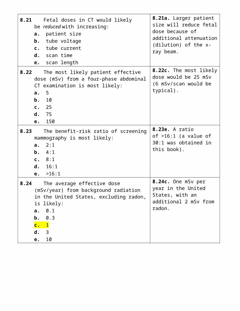
11.10 In MR, cortical bone most likely appears black because bone has:a. short T2b. long T2c. short T1d. long T1e. little hydrogen
11.10a. Short T2, which is characteristic of all solids (μs) and which causes transverse magnetization to disappear extremely quickly.
11.11 A 1-T magnetic field is greater than the earth’s magnetic field by a factor of:a. 5,000b. 10,000c. 20,000d. 40,000e. 80,000
11.11c. A factor of 20,000, since the earth’s magnetic field is only 0.05 mT (0.5 Gauss).
11.12 Increasing the main magnetic field will most likely reduce:a. T1 relaxation (slower)b. T2 relaxationc. signal-to-noise ratiod. equipment costse. resonance frequency
11.12a. T1 relaxation is reduced, which causes T1 times to increase (increased relaxation reduces T1).Faster moving protons are less efficient at transferring energy to the slower moving lattice
11.13 Superconducting MR magnets are most likely kept cold by using liquid:a. airb. carbon dioxidec. heliumd. nitrogene. oxygen
11.13c. Liquid helium is used to cool superconducting magnets.
11.14 Coils used to adjust main magnetic field uniformity are called:a. shimb. Helmholtzc. saddled. surfacee. RF
11.14a. Shim coils are used to correct for inhomogeneities in the main magnetic field.
11.15 These coils are used to localize the MR signal:a. shimb. gradientc. phased arrayd. surfacee. RF
11.15b. Magnetic gradient coils are used to localize the MR signal.

11.16 The best material for Faraday cages that shield against RF interference is most likely:a. aluminumb. copperc. rhodiumd. leade. tin
11.16b. Copper is generally used for RF shielding in Faraday cages.
11.17 The number of phase-encode gradient steps applied when generating M × M SE images is likely:a. Mb. M2
c. M3
d. 2 × Me. 3 × M
11.17a. M phase-encode steps are required to generate an MR image with a matrix size of M × M.
11.18 The reconstruction algorithm used in clinical MRI is most likely:a. 2D Fourier transformb. Iterativec. algebraic reconstructiond. back projectione. filtered back projection
11.18a. 2D Fourier transform is the reconstruction algorithm used in clinical MRI.
11.19 Acquisition time(s) for a 192 × 128 SE image (TE = 100 ms; TR = 1,000 ms) is likely:a. 12.8b. 19.2c. 128d. 192e. 128 + 192
11.19c. Acquisition time is 128 seconds, since there are at least 128 repetitions, each with a different phase-encoding gradient, and each repetition takes 1 s (TR time).TR x M x N
11.20 Following a 90-degree pulse in SE imaging, the echo signal would likely be measured at:a. immediately (t = 0)b. TEc. 4 × T1d. T2e. TR
11.20b. TE is the time when the echo (signal) is produced and measured.
11.21 A decreased MR signal is most likely when there is an increase in:a. T2b. T2*
c. blood flowd. spin densitye. spin dephasing
11.21e. Spin dephasing must reduce the MR signal.
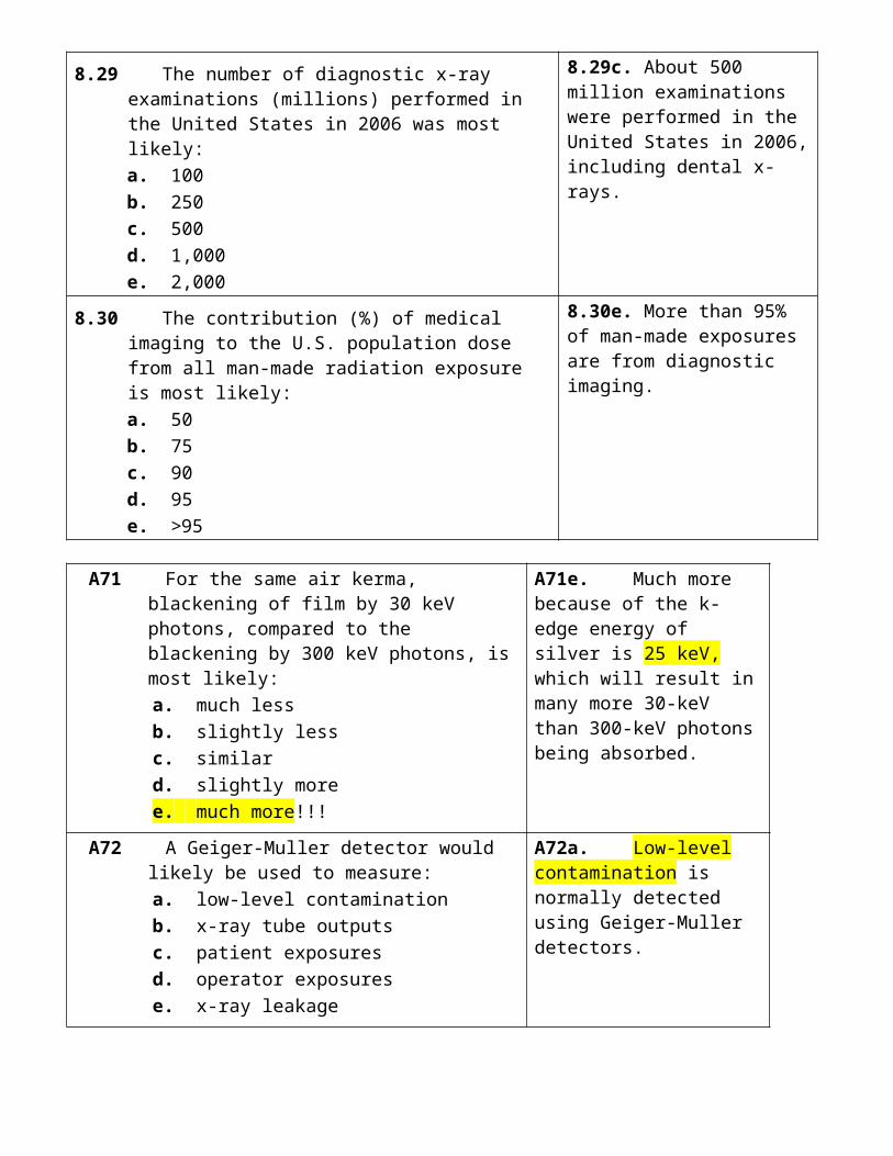
11.22 Which of the following is most likely to suppress signals from fat?a. FLASHb. Fast SEc. STIRd. FLAIRe. BOLD
11.22c. STIR, which stands for short tau inversion recovery, will suppress signals from fat.
11.23 The limiting spatial resolution (line pairs per mm) in current clinical MR is most likely:a. 0.1b. 0.3c. 1d. 2e. 5
11.23b. A typical resolution in clinical MR is 0.3 lp/mm, and about half the resolution in clinical CT.
11.24 When N MR image acquisitions are averaged, the resultant signal-to-noise ratio likely improves by:a. N0.5
b. Nc. 2 × Nd. N1.5
e. N2
11.24a. N0.5, which means that SNR doubles when the number of image acquisitions quadruples.
11.25 In MR, motion results in ghost images that appear in which direction?a. Read encodeb. Phase encodec. Slice selection axisd. PAe. Lateral
11.25b. Phase encode is the direction in which ghost artifacts from motion appear.
11.26 Chemical shift artifacts are most likely caused by fat and water differences in:a. T1b. T2c. T2*
d. Larmor frequencye. spin density
11.26d. Differences in Larmor frequency give rise to fat/water chemical shift artifacts.
11.27 The FDA guideline for limiting RF absorption (W per kg) in any gram of extremity of tissue is:a. 1b. 2c. 4d. 8e. 16
11.27d. The FDA guideline for limiting RF absorption in any gram of tissue is 8 W per kg in the extremities.TABLE 11.6
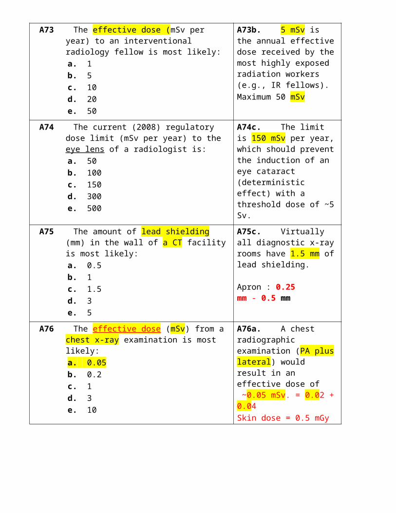
11.28 The largest susceptibility artifacts are likely to be seen between tissue and:a. airb. bloodc. fatd. marrowe. bone
11.28a. Air and soft tissue interfaces will likely generate the highest susceptibility artifacts.
11.29 Proton relaxation by Gd-DTPA is most likely due to the gadolinium:a. nuclear fieldb. chelate (DTPA)c. unpaired electronsd. K-edge energye. electron density
11.29c. Unpaired electrons in Gd will increase spin-lattice relaxation and thereby shorten T1 times.
11.30 The physical size (μm) of a superparamagnetic particle of SPIO or USPIO is most likely:a. 0.004b. 0.04c. 0.4d. 4e. 40
11.30b. The size (single domain) of a superparamagnetic particle such as SPIO is 0.04 μm.
A101 Which magnetic nucleus is most likely to result in the largest MR signal intensity?
a. 1H b. 2H c. 13C d. 23Na e. 31P
A101a. 1H protons have the highest Larmor frequency at a fixed magnetic field and produce the largest MR signal intensities.
A102 If the transverse magnetization is Mxy the free induction decay signal is proportional to:
a. (Mxy)−1
b. (Mxy)−0.5
c. (Mxy)0.5
d. (Mxy)1
e. independent of Mxy
A102d. (Mxy)1, which simply means that the FID signal is proportional to the transverse magnetization value.
A103 To maximize T1 weighting, the most likely TR time (ms) at 1.5 T would be:
a. 300 b. 600 c. 900 d. 1,500 e. 3,000
A103a. A time of 300 ms since T1 weighting is achieved by the use of TR times that comparable to fat and tissue T1 times.
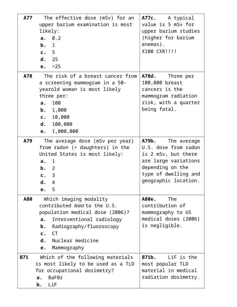
A104 The magnitude (%) of magnetic inhomogeneities responsible for T2∗ dephasing is likely:
a. 1 b. 0.1 c. 0.01 d. 0.001 e. <<0.001
A104e. A magnitude of <<0.001% since typical magnetic field inhomogeneities are only a few parts per million.
A105 Electric currents (A) in superconducting MR magnets are most likely:
a. 5 b. 50 c. 500 d. 5,000 e. 50,000
A105c. Approximately 500 amp would be the typical electrical current in a superconducting magnet.
A106 Thinner slices in MR imaging are most likely obtained by increasing the:
a. magnetic gradient b. RF frequency c. RF bandwidth d. TR time e. TE time
A106a. Increasing the magnetic gradient will generally result in thinner MR slices.
A107 Which is least likely to affect the total scan time in MR? a. Frequency-encode matrix size b. Number of phase-encoding steps c. Pulse sequences in study d. Number of acquisitions e. TR (repetition time)
A107a. The frequency-encode matrix size has no effect on the total MR scan time.
A108 Typical tissue differences (%) in spin density are most likely:
a. 1 b. 3 c. 10 d. 30 e. 100
A108c. A typical spin density difference is 10% between any two different types of soft tissue.
A109 Which is least likely to be an MR artifact? a. Chemical shift b. Bounce point c. Zipper d. Susceptibility e. Vignetting
A109e. Vignetting is the loss of image intensity in the periphery of an image intensifier used in fluoroscopy.
A110 A patient’s foot undergoing an MR scan is unlikely to exceed a temperature (◦C) of:
a. 38 b. 39 c. 40 d. 41 e. 42
A110c. The FDA recommends 40oC as the maximum temperature to an extremity during MRI.
TABLE 11.7

B101 At 1.5 T, the excess number (%) of proton spins in the low-energy state over the high-energy state is most likely:
a. 10 b. 1 c. 0.1 d. 0.01 e. <<0.01
B101e. Much less than 0.01 (<<0.01), as the excess number of protons is only a few per million.
B102 Following a 90-degree pulse, the longitudinal magnetization will most likely recover in a time that is four times:
a. T1 b. T2 c. T2* d. TE e. TR
B102a. T1 since the magnetization recovers in 4 × T1.
B103 If T2 for gray matter is 100 ms at 1.5 T, its value at 3Tis most likely:
a. 50 b. 70 c. 100 d. 140 e. 200
B103c. Its value is still 100 since T2 times show little dependence on field strength in clinical MR.
B104 Soft tissue T2∗ times (μs) are most likely: a. 5 b. 50 c. 500 d. 5,000 e. 50,000
B104d. Times are 5,000 μs, which is 5 ms and a typical tissue T2* value
B105 Gradient magnetic fields in MR are used most commonly to:
a. increase T2 b. shorten T1 c. localize signal d. amplify signal e. minimize stray fields
B105c. Magnetic field gradients are used to localize the MR signal. LG
B106 The number of 1D Fourier transforms in reconstructing a M × N image is most likely:
a. M b. N c. M + N d. M − N e. M × N
B106c. Each row must undergo a FT (M) followed by each column (N), which results in a total of M + N.
B107 Which is the most likely TR value (ms) for a FLASH B107b. The most likely TR
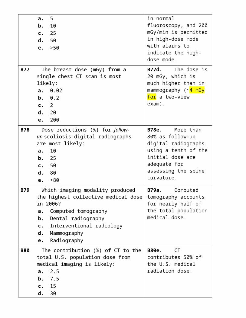
gradient recalled echo pulse sequence? a. 1 b. 10 c. 100 d. 1,000 e. 10,000
time for a fast low-angle shot pulse sequence is 10 ms.
B108 MR signal-to-noise ratio (SNR) is likely reduced when there is an increase in:
a. coil diameter b. number of acquisitions c. magnetic field strength d. section thickness e. pixel dimension
B108a. Larger coil diameter will generally reduce the SNR in MR.
B109 Which line is an exclusion zone (mT) for persons with pacemakers?
a. 0.5 b. 1 c. 2 d. 5 e. 10
B109a. The magnetic field exclusion zone is 0.5 mT (5 gauss).
B110 Which of the following is least likely to exhibit paramagnetism?
a. Ba b. Cr c. Fe d. Gd e. Mn
B110a. Ba is an x-ray contrast material that does not exhibit paramagnetism
Related Documents