Serum Protein Electrophoresis Dr. Nitin Inamdar Department of Biochemistry Tata Memorial Center, Parel, Mumbai.

Welcome message from author
This document is posted to help you gain knowledge. Please leave a comment to let me know what you think about it! Share it to your friends and learn new things together.
Transcript
Serum Protein Electrophoresis
Dr. Nitin Inamdar Department of BiochemistryTata Memorial Center, Parel,Mumbai.

Phoresis: Separation or migration or movement
Electro: Under influence of electric field
Electrophoresis

Serum
Electrophoresis
Mixture of proteins
Amino acids:◦ Amino group (NH2)◦ Carboxyl group (COOH)
Two amino acids join each other with polypeptide bond to form polypeptide chain & many polypeptide chains make a protein.
Ph 8.6/0.075M Barbitone buffer
1%Agarose
Constant current 5mAM/Slide

Electrophoresis separates proteins based ontheir physical properties.
Serum is placed on a specific medium, and a charge is applied. The net charge (positive or negative) and
the size and shape of the subsets of these proteins are used in interpreting the results.

Serum ElectrophoresisPaper electrophoresis
Starch gel electrophoresisAgarosre gel electrophoresis
Cellulose acetate gel electrophoresisCapillary gel electrophoresis
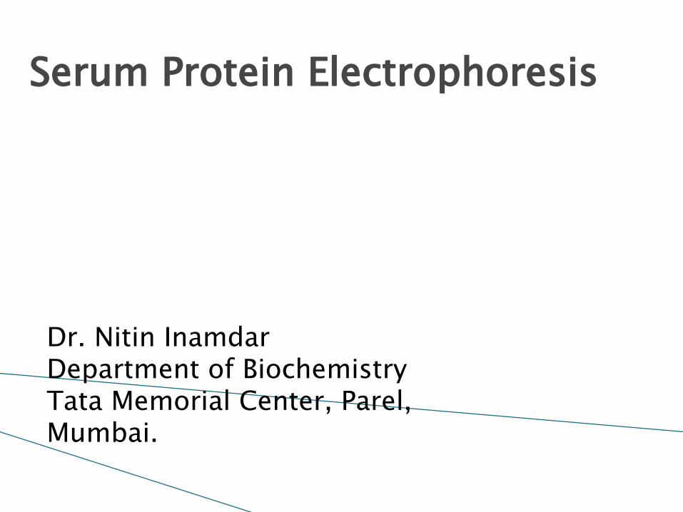
Serum protein electrophoresis on agarose gel is a type of horizontal gel electrophoresis

Equipment used for the gel electrophoresis
power supply (direct current)
electrophoresis chamber
containers for staining and destaining gel
applicator
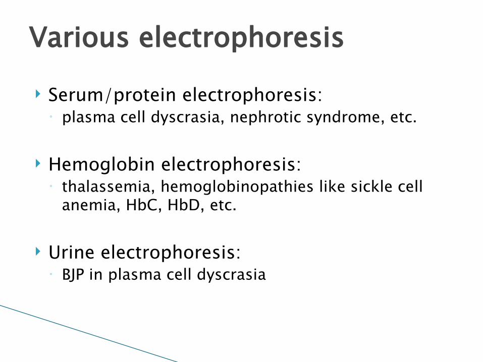
Serum/protein electrophoresis: ◦ plasma cell dyscrasia, nephrotic syndrome, etc.
Hemoglobin electrophoresis: ◦ thalassemia, hemoglobinopathies like sickle cell
anemia, HbC, HbD, etc.
Urine electrophoresis: ◦ BJP in plasma cell dyscrasia
Various electrophoresis

Diagnostic: ◦ Diagnosis of plasma cell dyscrasia◦ Diagnosis of Waldenstrom’ macroglbulinemia
Monitoring of disease: ◦ Monitoring of plasma cell dyscrasia
Indication of serum electrophoresis
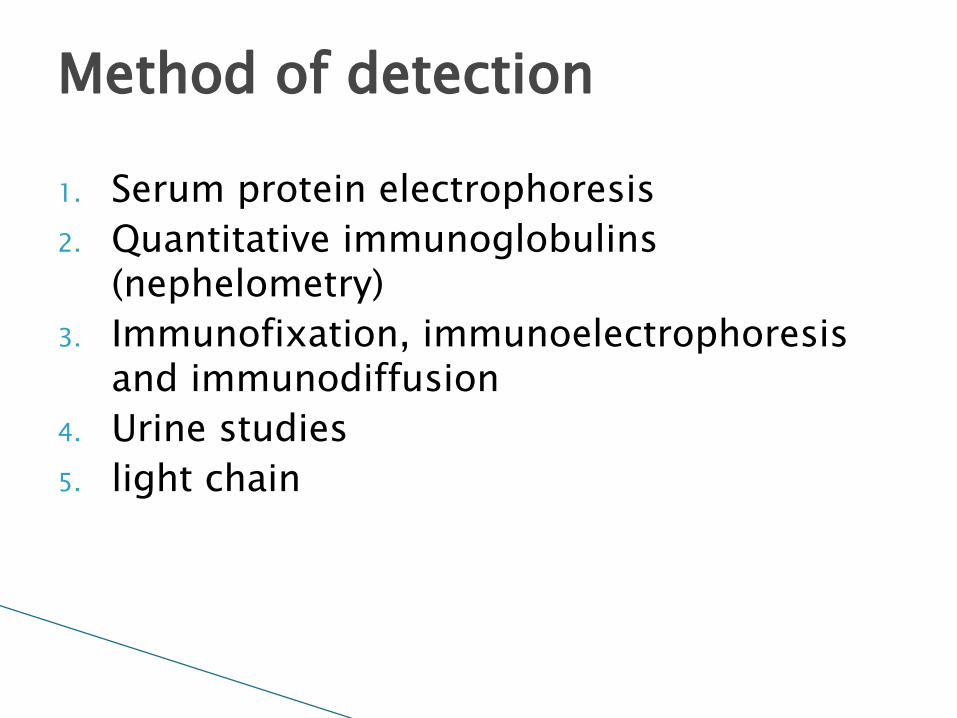
1. Serum protein electrophoresis2. Quantitative immunoglobulins
(nephelometry)3. Immunofixation, immunoelectrophoresis
and immunodiffusion4. Urine studies5. light chain
Method of detection
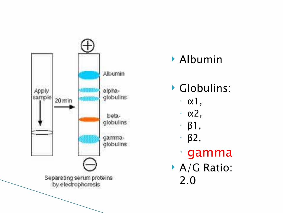
Albumin
Globulins:◦ α1, ◦ α2, ◦ β1, ◦ β2, ◦ gamma
A/G Ratio: 2.0

Normal serum electrophoresispH=8.6, 1% agarose gel, sodium barbitone buffer
Anode +ve Cathode -ve

Abnormal serum electrophoresis showing M band
Anode +ve Cathode -ve
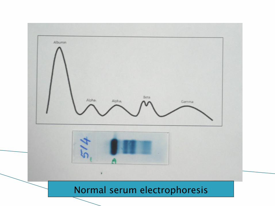
Normal serum electrophoresis
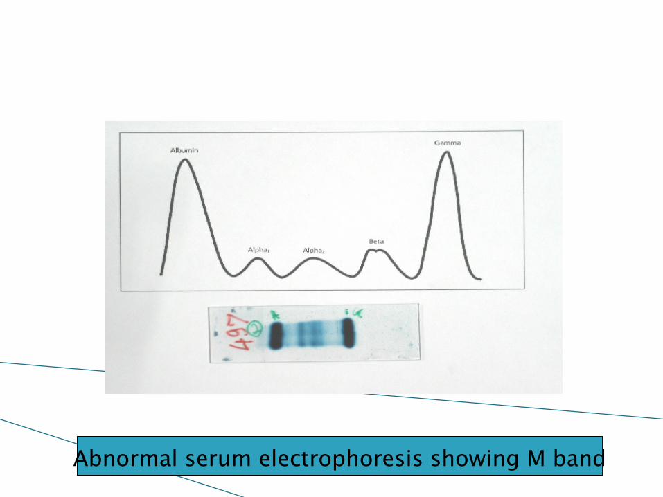
Abnormal serum electrophoresis showing M band
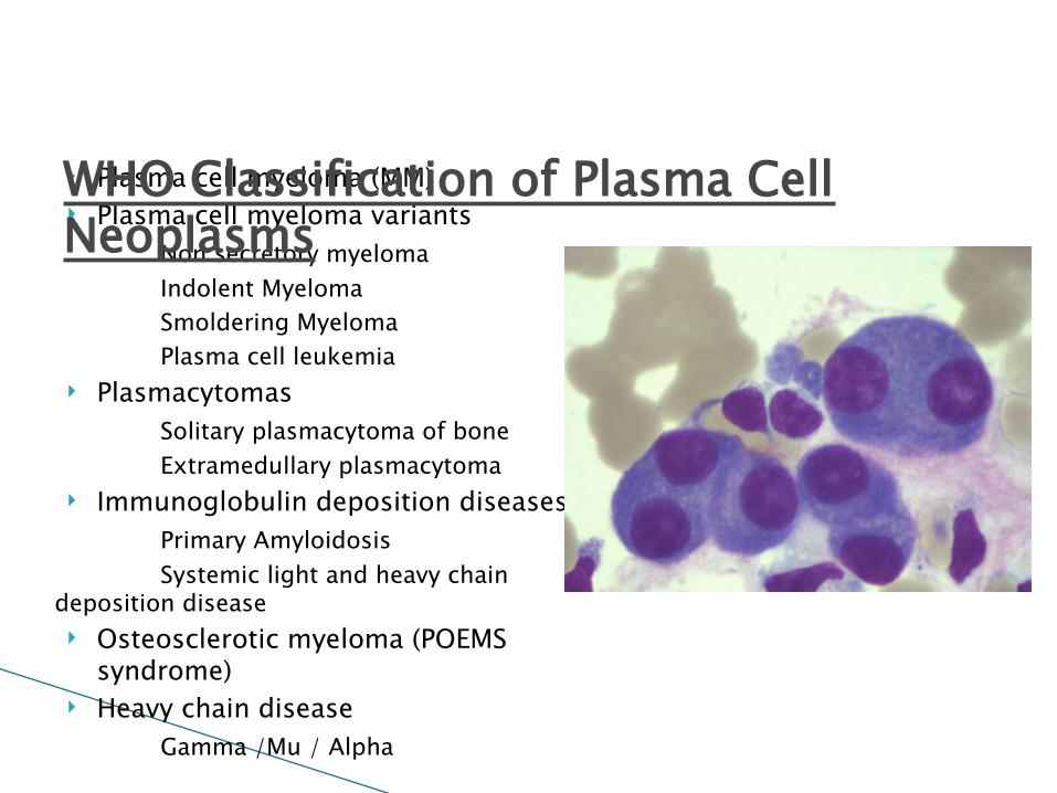
Plasma cell myeloma (MM) Plasma cell myeloma variants
Non secretory myelomaIndolent MyelomaSmoldering MyelomaPlasma cell leukemia
PlasmacytomasSolitary plasmacytoma of boneExtramedullary plasmacytoma
Immunoglobulin deposition diseasesPrimary AmyloidosisSystemic light and heavy chain
deposition disease Osteosclerotic myeloma (POEMS
syndrome) Heavy chain disease
Gamma /Mu / Alpha
WHO Classification of Plasma Cell Neoplasms
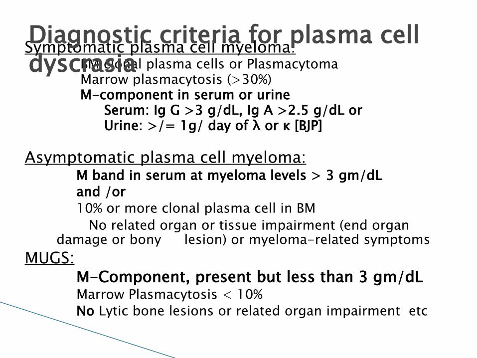
Symptomatic plasma cell myeloma:BM clonal plasma cells or PlasmacytomaMarrow plasmacytosis (>30%)M-component in serum or urine
Serum: Ig G >3 g/dL, Ig A >2.5 g/dL or Urine: >/= 1g/ day of λ or κ [BJP]
Asymptomatic plasma cell myeloma:M band in serum at myeloma levels > 3 gm/dL and /or 10% or more clonal plasma cell in BM No related organ or tissue impairment (end organ
damage or bony lesion) or myeloma-related symptoms MUGS:
M-Component, present but less than 3 gm/dLMarrow Plasmacytosis < 10%No Lytic bone lesions or related organ impairment etc
Diagnostic criteria for plasma cell dyscrasia
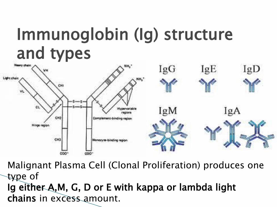
Immunoglobin (Ig) structure and types
Malignant Plasma Cell (Clonal Proliferation) produces one type of Ig either A,M, G, D or E with kappa or lambda light chains in excess amount.

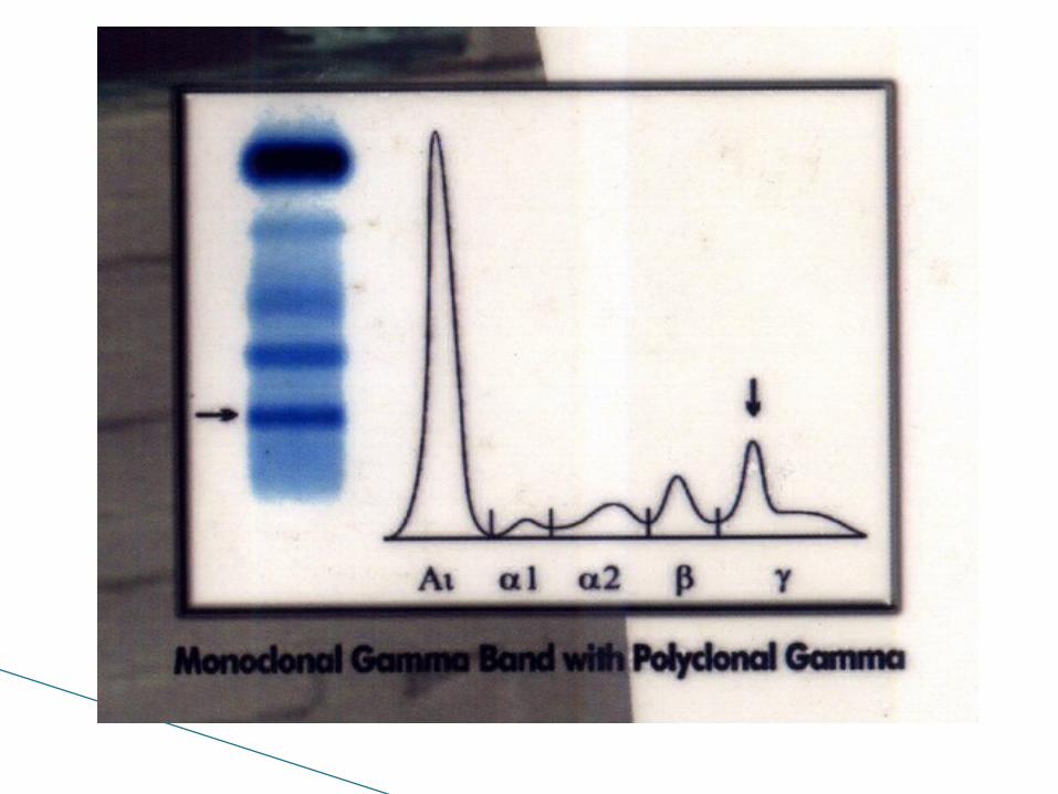
Various patterns


Analbuminemia


Haptoglobin phenotype
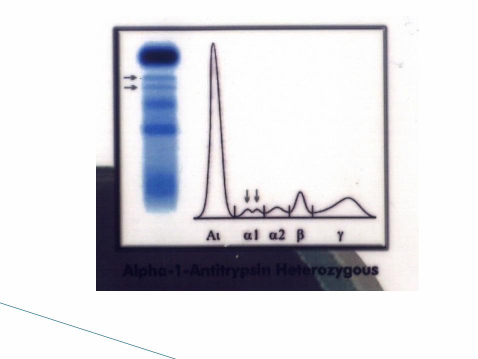
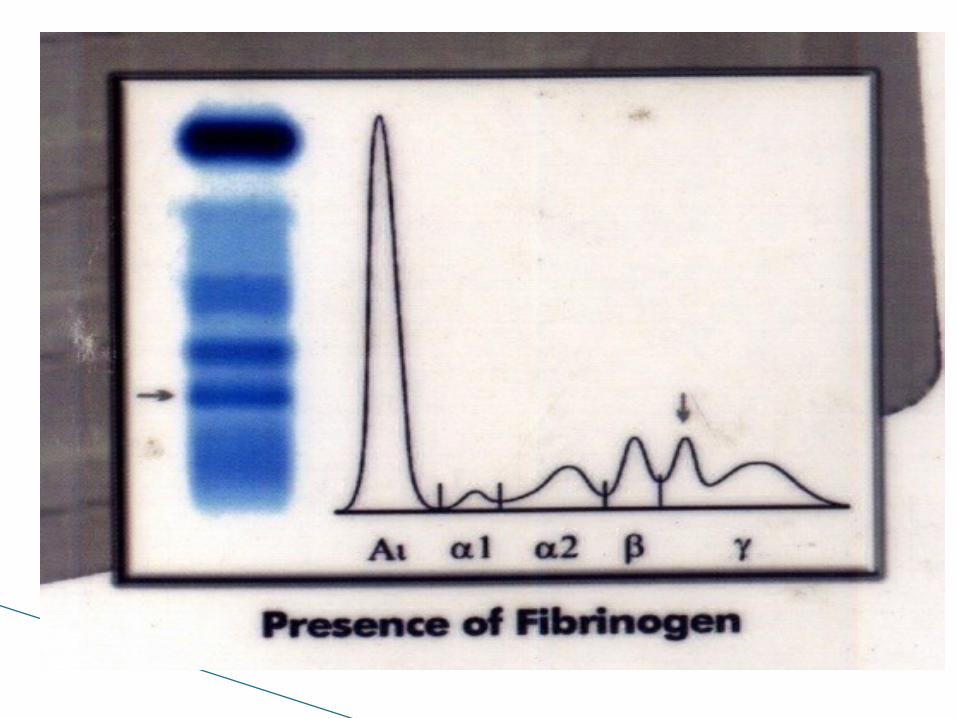


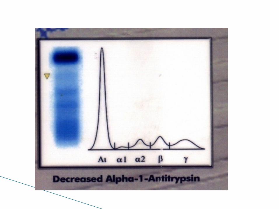

Bis-albuminemia


Nephrotic syndrome

Hypergammaglobulinemia

M band in β region

M band in γ region


Nephelometry Antigen-antibody complexes, when formed at a
high rate, will precipitate out of a solution resulting in a turbid or cloudy appearance.
• Nephelometry: is indirect measurement of light scattered by the antigen-antibody complexes.

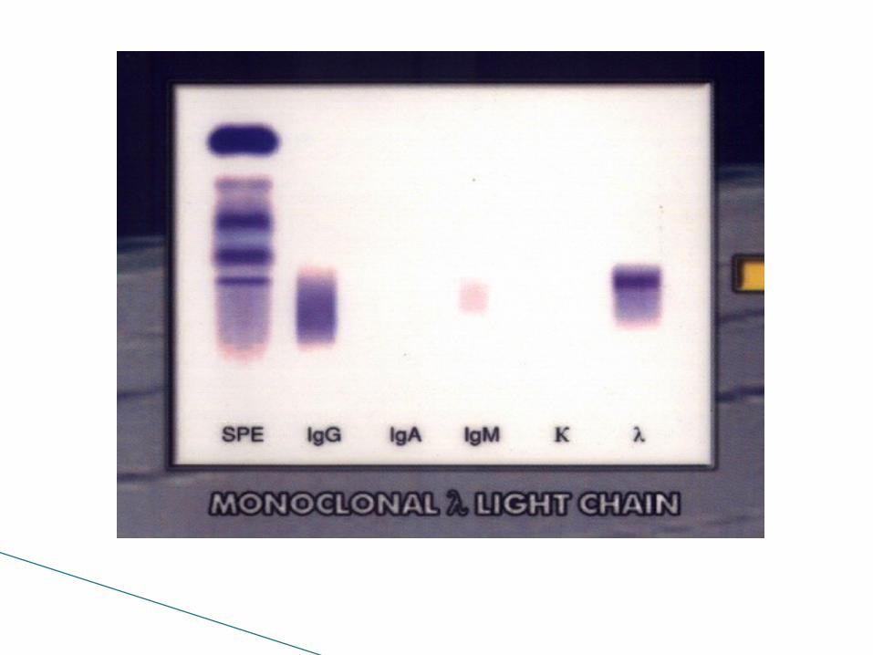
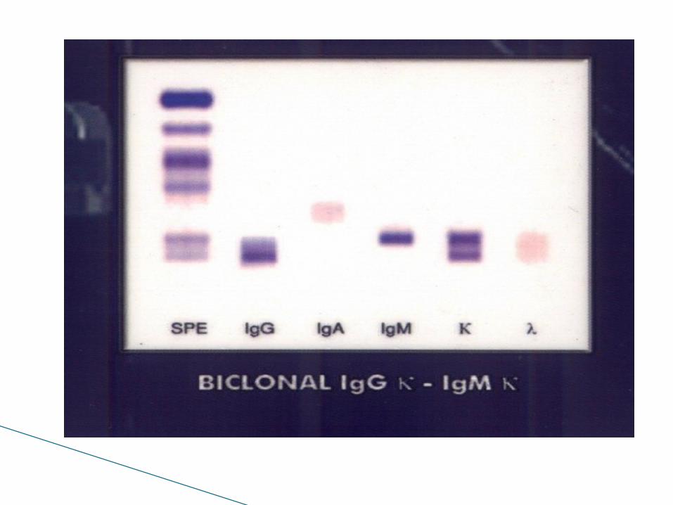
After separation of serum proteins by electrophoresis separated serum in different lanes is incubated with monospecific anti sera against IgA, M G and κ λ
Immunofixation electrophoresis


Intravascular hemolysis
A type of multiple myeloma in which plasma cell tumors produce only monoclonal light chain proteins. Persons with light chain disease may develop lytic bone lesions, hypercalcemia, impaired kidney function, and amyloidosis.
light chain myeloma
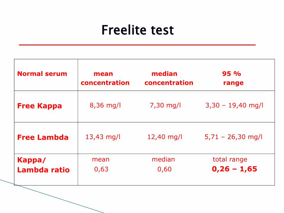
Normal serum mean median 95 %concentration concentration range
Free Kappa 8,36 mg/l 7,30 mg/l 3,30 – 19,40 mg/l
Free Lambda 13,43 mg/l 12,40 mg/l 5,71 – 26,30 mg/l
Kappa/
Lambda ratio
mean median total range
0,63 0,60 0,26 – 1,65
Freelite test

THANK YOU
Related Documents